Adjuvant chemoradiotherapy versus radiotherapy alone for ...

University of Groningen
Response evaluation of chemoradiotherapy in esophageal cancerSmit, Justin Kendrick
IMPORTANT NOTE: You are advised to consult the publisher's version (publisher's PDF) if you wish to cite fromit. Please check the document version below.
Document VersionPublisher's PDF, also known as Version of record
Publication date:2013
Link to publication in University of Groningen/UMCG research database
Citation for published version (APA):Smit, J. K. (2013). Response evaluation of chemoradiotherapy in esophageal cancer Groningen: s.n.
CopyrightOther than for strictly personal use, it is not permitted to download or to forward/distribute the text or part of it without the consent of theauthor(s) and/or copyright holder(s), unless the work is under an open content license (like Creative Commons).
Take-down policyIf you believe that this document breaches copyright please contact us providing details, and we will remove access to the work immediatelyand investigate your claim.
Downloaded from the University of Groningen/UMCG research database (Pure): http://www.rug.nl/research/portal. For technical reasons thenumber of authors shown on this cover page is limited to 10 maximum.
Download date: 03-12-2018


Response evaluation of chemoradiotherapy in esophageal cancer
J.K. Smit 2013

The studies in this thesis were financially supported by: Post-graduate School of Medicine GUIDE, Jan Kornelis de Cock Stichting, Stichting Scoren Tegen Kanker, Departments of Surgery, Radiotherapy and Pathology of the UMCG.
The printing of this thesis was financially supported by: Stichting Noordelijk Chirurgisch Oncologisch Fonds, ERBE Nederland B.V., Harlan Laboratories B.V., Roche Nederland B.V., Chipsoft B.V., University of Groningen and UMCG.
Smit, J.K.Response evaluation of chemoradiotherapy in esophageal cancer
PhD Dissertation, University of Groningen, The Netherlands
ISBN: 978-90-367-6109-3ISBN: 978-90-367-6108-6 (electronic version)
© Copyright 2013 Justin K. Smit, The NetherlandsAll rights reserved. No part of this book may be reproduced, stored in a retrieval system or transmitted in any form or by any means, without prior permission of the author.
Lay out & cover design: Saeideh JabaniPrinted by: Ipskamp Drukkers, Enschede

RIJKSUNIVERSITEIT GRONINGEN
RESPONSE EVALUATION OF CHEMORADIOTHERAPY IN ESOPHAGEAL CANCER
Proefschrift
ter verkrijging van het doctoraat in de Medische Wetenschappen
aan de Rijksuniversiteit Groningenop gezag van de
Rector Magnificus, dr. E. Sterken,in het openbaar te verdedigen op
woensdag 24 april 2013om 16.15 uur
door
Justin Kendrick Smitgeboren op 13 februari 1985
te Curaçao, Nederlandse Antillen

Prof.dr. J.Th.M. PlukkerProf.dr. R.P. CoppesProf.dr. H. Hollema
Promotores:
Beoordelingscommissie: Prof.dr. E. HeinemanProf.dr. M.A. CuestaProf.dr. J.A. Langendijk

Joost KamstraMichael Dickinson
Paranimfen:


Contents
Chapter 1
Chapter 2
Chapter 3
Chapter 4
Chapter 5
Chapter6
Chapter 7
Chapter 8
Chapter 9
Chapter 10
General introduction, rationale and outline of thesis
Prognostic factors and patterns of recurrence in esophageal cancer assert arguments for extended two-field transthoracic esophagectomy
Different recurrence pattern after neoadjuvant chemoradiotherapy compared to surgery alone in esophageal cancer catients
Survival after definitive (chemo)radiotherapy in esophageal cancer patients: a population-based study in the north-east netherlands
Demarcation of radiotherapy target volumes to improve pathologic evaluation of neo-adjuvant chemoradiation for esophageal cancer
Pathologic evaluation of radiotherapy target volumes as quality control after neo-adjuvant chemoradiation for esophageal cancer
Prediction of response to radiotherapy in the treat-ment of esophageal cancer using stem cell markers.
Neoadjuvant therapy reduces the incidence of nodal micrometastases in esophageal adenocarcinoma
Hedgehog Signaling Pathway and Prediction of Recurrence after Neoadjuvant Chemoradiotherapy in Esophageal Cancer
Summarizing discussion and future perspectives
Nederlandse samenvatting
Dankwoord
List of publications and presentations
Curriculum Vitae
11
23
43
61
79
97
117
149
167
187
195
203
209
213


General introduction, rationale and outline of thesis
Chapter 1
J.K. Smit
12
Introduction
Esophageal cancer (EC) is the 7th leading cause of cancer related death with an estimated worldwide prevalence of nearly 500.000 patients, accounting for 4% to 5% of the total cancer burden 1-3. Currently it has the most rapidly rising incidence of the solid malignancies in Western countries, which is mainly due to the steadily increased rate of esophageal reflux with Barrett esophagus and obesity especially in the early age category 2, 4-6. Usually the tumor is located at the distal one-third of the esophagus and most patients are ≥ 65 years of age at the time of diagnosis. Recently the 7th TNM edition of the American Joint Committee on Cancer (AJCC) and Union for International Cancer Control (UICC) Cancer Staging Manual for the esophagus and esophagogastric junction tumors has been employed in the standard workup of esophageal cancer patients (table 1) 7, 8. As in most solid tumors, depth of invasion and locoregional nodal involvement are the key prognostic factors for survival after surgical resection (figure 1) 8. Although EC commonly presents in a relatively advanced stage of the disease, surgical resection is still the cornerstone of curative treatment in these patients 9. During the past decade the use of neoadjuvant chemoradiotherapy (CRT) has been increasingly propagated to complement the surgical resection10-13. Using this trimodality treatment, 5-year survival rates of 45% have been achieved in contrast to rates of 20% and 35% by surgical resection alone in non-expert and expert centers, respectively 13, 14. In the last decade patient selection also has improved considerably using sophisticated imaging techniques, including hybrid 18-F-Fluorodeoxyglucose-positron emission and computed tomography (FDG-PET-CT) preferably with a 64 multidetector (MD)CT and the additional use of endoscopic ultrasonographic guided fine needle aspiration (EUS-FNA) of suspected nodes. Advancements have been made in radiotherapy techniques and more precise imaging of the extent and the location of the primary tumor and suspected nodes. Moreover, prognosis and survival have changed steadily due to proper surgical approaches with adequate nodal dissection and effective intensive care treatment. Nonetheless, the strongest modulator on the improving patient outcome has been the centralization of treatment with adequate functioning collaborative multidisciplinary tumor boards and implementation of approved neoadjuvant chemoradiotherapy regimens as standard of care.

13
1
Notes :* Includes all noninvasive neoplastic epithelium that was previously called carcinoma in situ Cancers stated to be noninvasive or in situ are classifi ed as Tis** Number must be recorded for total number of regional nodes sampled and total number of reported nodes with metastases*** Location (primary cancer site) is defined by position of upper (proximal) edge of tumor in esophagus
Table 1. TNM 7th edition of the AJCC Cancer Staging Manual: esophagus and esoph-agogastric junction tumors 7, 8.
Primary Tumor (T)TX Primary tumor cannot be assessedT0 No evidence of primary tumorTis High-grade dysplasia*T1 Tumor invades lamina propria, muscularismucosae, or submucosaT1a Tumor invades lamina propria or muscularismucosaeT1b Tumor invades submucosaT2 Tumor invades muscularis propriaT3 Tumor invades adventitiaT4 Tumor invades adjacent structuresT4a Resectable tumor invading pleura, pericardium,or diaphragmT4b Unresectable tumor invading other adjacentstructures, such as aorta, vertebral body,trachea, etc.Regional Lymph Nodes (N)**NX Regional lymph nodes cannot be assessedN0 No regional lymph node metastasisN1 Regional lymph node metastases involving 1 to 2NodesN2 Regional lymph node metastases involving 3 to 6NodesN3 Regional lymph node metastases involving 7 ormore nodesDistant Metastasis (M)MX Distant metastasis cannot be assessedM0 No distant metastasisM1 Distant metastasisHistopathologic Cell TypeSquamous cell carcinomaAdenocarcinomaHistologic Grade (G)GX Grade cannot be assessed – stage grouping as G1G1 Well differentiatedG2 Moderately differentiatedG3 Poorly differentiatedG4 Undifferentiated – stage grouping as G3Squamous cell carcinomaTumor Location***Upper or middle—cancers above lower border of inferior pulmonaryVeinLower—below inferior pulmonary vein

14
Figure 1. Schematic representation of the information in table 1 (From Rice WR: Diagnosis and staging of esophageal carcinoma. In Pearson FG, Copper JD, Deslau-riers J, et al [eds]: Esophageal syrgery, ed 2, New York, 2002, Churchill Livingstone, p 687).
RationaleEC remains an aggressive disease in which the results of treatment generally
depend on the stage at diagnosis and involved biological factors. One of the strongest prognostic factors which inform us about the different outcomes are the presence of locoregional nodal involvement, especially the total number of nodal metastases and ratio between involved and examined lymphnodes (L/N ratio), the obtained pathological radicality (R0 resection) and lymphangioinva-sion. Besides improvement in radicality due to advancement of surgical resection techniques with adequate nodal dissection through a transthoracic approach, great progress has been made by adding neoadjuvant chemoradiotherapy (CRT) in the treatment of esophageal cancer patients. Although several meta-analyses already had noted better outcome with combined trimodality treatment, convincing data on the effect of neoadjuvant CRT in the treatment of EC was only recently

15
1
obtained by the publication of a large multicenter Dutch randomized controlled study, the CROSS trial 13. In the CROSS trial, standard surgical resection alone was compared with surgical resection in combination with pre-operative chemoradiotherapy. The radiotherapy schedule consisted of a total dose of 41.4 Gy in daily fractions of 1.8 Gy, given five times per week (23 fractions). Patients received concurrent chemotherapy consisting of 5 weekly courses of paclitaxel (50 mg/m2) and carboplatin (area under the curve, AUC= 2). The CROSS-schedule followed by surgical resection with curative intent (R0 resection) achieved a median survival gain from 24 to 49.4 months 13. The publication of the CROSS data can be considered as a great step forward in the treatment of EC during the last decade.
However, further improvements can certainly be made in the treatment of EC. For instance, previous research has shown a pathological complete response (absence of viable tumor cells), which strongly correlates with survival, in only 25% to 30% of the patients who received neoadjuvant CRT 13, 15-17. But does response to CRT really predict outcome in EC patients? Unfortunately however, even patients with a clinical complete response on currently used sophisticated imaging methods, but with microscopic residual vital tumor cells after neoadjuvant CRT, have a significant reduction in survival 15, 17. So, it is still not possible to indentify all these non-responding patients beforehand. In order to treat patients more adequately, biological tumormarkers predicting efficacy and treatment benefit have to be defined. This thesis focuses on exploring some of the prognostic and predictive factors in the treatment with neoadjuvant CRT. This research may help to improve selection of suitable candidates for CRT or surgery after CRT and to get better insight into the effect of these factors on outcome of neoadjuvant and definitive CRT in the treatment of esophageal cancer patients.
In summary, as neoadjuvant CRT is an established part in the curative treatment of esophageal cancer patients, the major issues are the prediction and increase of response to CRT. Understanding treatment efficacy is important in facilitating shared decision-making, as patients may need a different surgical and/or CRT approach to prevent progression of disease and prevent devastating unresectabillity or early recurrences. This translates into the concrete research question: Which patients are at risk and which patients may benefit most from additional surgical or CRT treatment?
16
Outline of chapters
Chapter 2The arguments for a transthoracic esophagectomy with two-field
lymphadenectomy are described. Currently, the optimal curative treatment of esophageal cancer consists of neoadjuvant chemoradiotherapy, usually a combination of carboplatin and paclitaxel and 41.4 Gy according to the CROSS study, followed by a radical surgical resection. Chapter 2 is important, as surgery still is the mainstay of treatment and not all patients can or will be treated by a trimodality approach. The quintessence of the surgical treatment is to obtain adequate locoregional control. Local recurrence should be considered as the ultimate failure to treatment. Therefore we analyzed the most important prognostic factors in developing locoregional recurrences and especially with respect to nodal involvement.
Chapter 3Even though the percentage of successful surgical resections is high
with a standard transthoracic approach as propagated and reported in chapter 2, the recurrence rate is still unacceptably high. Therefore, neoadjuvant chemoradiotherapy (CRT), as described in the CROSS trial, has been added in addition to the standard surgical treatment of esophageal cancer patients. It is postulated that neoadjuvant CRT facilitates the possibility for curative resection (R0) and will improve local control rate and disease free survival, which may be considered as clinical readouts to treatment response. It is believed that this can be achieved by tumor downsizing and downstaging through elimination of micrometastatic disease, both at distant and in locoregional lymph nodes. In this chapter we compare the recurrence pattern of neoadjuvantly treated patients with those treated by surgery alone, with a particular focus on differences in local recurrence pattern and distant disease between both groups. Furthermore, the outcome stratification based on response to neoadjuvant CRT is discussed.

17
1
Chapter 4Unfortunately not all patients with esophageal cancer can undergo a
surgical resection, usually due to severe pre-existing co-morbidities and/or technical inability to achieve a complete resection (T4b, see figure 1). Definitive (chemo)radiotherapy (dCRT) with a curative intent, commonly given in a chemorediothrapy scheme of ≥ 50Gy or definitive radiotherapy (dRT) of ≥ 60Gy in fractions of 2Gy ± intraluminal radiation, is the first choice of treatment for these patients. In this chapter we described the results in a population-based cohort study of 287 patients. Different prognostic factors for survival and recurrent disease in these patients are discussed. The data from this chapter could help to improve treatment selection of suitable candidates for definitive (chemo)radiotherapy.
Chapters 5 & 6The gold standard in assessing treatment success (response assessment)
after neoadjuvant CRT is the pathologic evaluation of the resected specimen by an experienced pathologist. Complete pathologic response (pCR) to neoadjuvant CRT is of more clinical relevance than complete clinical response (CCR), which is based on the absence of any lesion at the site of the initially tumor on both imaging and gross examination. Current standard pathological protocols were designed and explored in the era when patients were treated by surgery alone. However, optimal pathological evaluation after neoadjuvant CRT with esophagectomy is hampered by CCR at the primary tumor site. Histological changes, including different proportions of necrosis and fibrosis in the resected specimens due to the additional CRT are categorized in the so-called Mandard classification system 18. Previously developed and currently still used pathological protocols do not meet the current demands for adequate pathologic evaluation of response and are difficult to evaluate without a good judgment of the radiation target volumes. In these chapters we propose our new standardized method for a better evaluation of the pathologic response and adequate analysis of the given radiotherapy, with the surgeon in a key position by in vivo marking of radiological reference points used in the radiation planning. Furthermore, the findings with our new method are tested in a prospective dataset.
18
Chapter 7Unfortunately not all esophageal cancer patients respond adequately
to neoadjuvant chemoradiotherapy (CRT). Moreover, when evaluating the pathologic specimen approximately 25 to 30% of the patients have a pathological complete response (pCR), while the majority of patients still have a considerable amount of vital cancer cells. The presence of vital tumor cells has a great negative impact on short- and longterm survival. Patients who do not respond adequately to neoadjuvant CRT do not benefit from this treatment in terms of overall and disease free survival. Moreover, these patients only experience the disadvantages, including treatment related toxicity and even mortality. At present it is not possible to predict response to neoadjuvant CRT properly. In this chapter pre-clinical studies are performed to indentify biological predictive factors for the response to neoadjuvant CRT in esophageal cancer and in particular with respect to radioresistant cells. The data are also translated to a patient series to confirm our pre-clinical results.
Chapter 8Locoregional lymph node disease has a strong negative impact on survival
(chapter 2). As is given in the TNM staging, the prognosis of esophageal cancer patients worsens with increasing numbers of involved nodes. In chapter 8 this robust prognostic factor on early failure is elaborated further on with respect to the presence of nodal micrometastases (NMM). One of the arguments to introduce neoadjuvant CRT is the elimination or reduction of micrometastatic disease at distant and regional sites. There is limited knowledge of the reduction of NMM disease and the impact on survival or recurrences after CRT. The present chapter defines some of these aspects and the possible clinical relevance with therapeutic purposes.
Chapter 9Previous research has shown a pathological complete response in only
25-30% of the patients who received neoadjuvant CRT which greatly improves both, the disease free and overall survival. On the other hand microscopic presence of residual vital tumor cells, implicating lack of response, in the resected specimen after neoadjuvant CRT is known to reduce the survival. Therefore, it

19
1
is of utmost importance to find additional therapeutic ways by which response rates can be improved for patients with EC treated with neoadjuvant CRT. Therapeutic failure resulting in early recurrence i.e. short disease-free survival time can be considered as clinical substitute for lack of response to neoadjuvant CRT. Clinicopathologic prognostic factors for short disease-free survival (DFS) after neoadjuvant CRT, in patients with microscopic residual disease (mRD) in the resected specimen could be of help in selecting patients improving current treatment outcome. This may lead in optimizing the radiation fields, or extending the time interval between completion of CRT and resection or even omit surgical intervention. Determining enhanced expression of candidate biological markers in the resected tissues with mRD, may provide better insight into additional therapeutic options. This could lead in reducing or even eliminating mRD in the resected specimen (increasing response rates) after neadjuvant CRT and therefore greatly improving patient survivals.
In this chapter clinicopathological factors and biomarkers (like the pre-clinical results obtained from chapter 7) which could be implicated in the presence of mRD are investigated in our patient cohort, which was treated with neoadjuvant CRT (CROSS schedule).
Chapter 10General discussion on the research performed in chapters 2 to 9. Also
possible future research directed are elaborated on.multidisdciplinary discussion in close collaboration with surgeon,
medical oncologist, gastro-enterologist and radiotherapeutic oncologist. As reported previously by our group, treatment consisted of best supportive care, radiotherapy alone or combined with chemotherapy, chemotherapy alone or stenting17. Different combinations of these treatment modalities were also given. Curatively intended radiotherapy was usually given in doses of 50-60 Gy and/or in combination with 5-fluoracil and cisplatin.

20
RefeRences 1. Ferlay J, Shin HR, Bray F, et al. Estimates of worldwide burden of cancer in 2008: GLOBOCAN 2008. Int J Cancer 2010;127:2893-2917.2. Netherlands cancer registry. [Dutch cancer figures web site]. 2013. Available at: http://www.cijfersoverkanker.nl/?language=en. 3. Parkin DM, Bray F, Ferlay J, et al. Global cancer statistics, 2002. CA Cancer J Clin 2005;55:74-108.4. Crane LM, Schaapveld M, Visser O, et al. Oesophageal cancer in The Netherlands: increasing incidence and mortality but improving survival. Eur J Cancer 2007;43:1445-51.5. Brown LM, Devesa SS, Chow WH. Incidence of adenocarcinoma of the esophagus among white Americans by sex, stage, and age. J Natl Cancer Inst 2008;100:1184-1187.6. Pohl H, Welch HG. The role of overdiagnosis and reclassification in the marked increase of esophageal adenocarcinoma incidence. J Natl Cancer Inst 2005;97:142-146.7. Edge SB, Byrd DR, Compton CC, et al. AJCC Cancer Staging Manual (7th ed). New York, NY: Springer; 2010.8. Rice TW, Blackstone EH, Rusch VW. 7th edition of the AJCC Cancer Staging Manual: esophagus and esophagogastric junction. Ann Surg Oncol 2010;17:1721-1724.9. Mariette C, Piessen G, Triboulet JP. Therapeutic strategies in oesophageal carcinoma: role of surgery and other modalities. Lancet Oncol 2007;8:545-53.10. Gebski V, Burmeister B, Smithers BM, et al. Survival benefits from neoadjuvant chemoradiotherapy or chemotherapy in oesophageal carcinoma: a meta-analysis. Lancet Oncol 2007;8:226-234.11. Sjoquist KM, Burmeister BH, Smithers BM, et al. Survival after neoadjuvant chemotherapy or chemoradiotherapy for resectable oesophageal carcinoma: an updated meta-analysis. Lancet Oncol 2011;12:681-692.12. Kaklamanos IG, Walker GR, Ferry K, et al. Neoadjuvant treatment for resectable cancer of the esophagus and the gastroesophageal junction: a meta-analysis of randomized clinical trials. Ann Surg Oncol 2003;10:754-61.13. van Hagen P, Hulshof MC, van Lanschot JJ, et al. Preoperative chemoradio-therapy for esophageal or junctional cancer. N Engl J Med 2012;366:2074-2084.14. Smit JK, Pultrum BB, van Dullemen HM, et al. Prognostic factors and patterns of recurrence in esophageal cancer assert arguments for extended two-field transthoracic esophagectomy. Am J Surg 2010;200:446-453.15. Berger AC, Farma J, Scott WJ, et al. Complete response to neoadjuvant chem-oradiotherapy in esophageal carcinoma is associated with significantly improved survival. J Clin Oncol 2005;23:4330-4337.16. Scheer RV, Fakiris AJ, Johnstone PA. Quantifying the benefit of a pathologic complete response after neoadjuvant chemoradiotherapy in the treatment of esophageal cancer. Int J Radiat Oncol Biol Phys 2011;80:996-1001.17. Ajani JA, Correa AM, Hofstetter WL, et al. Clinical parameters model for

21
1
predicting pathologic complete response following preoperative chemoradiation in patients with esophageal cancer. Ann Oncol 2012;23:2638-2642.18. Mandard AM, Dalibard F, Mandard JC, et al. Pathologic assessment of tumor regression after preoperative chemoradiotherapy of esophageal carcinoma. Clin-icopathologic correlations. Cancer 1994;73:2680-2686.


Prognostic factors and patterns of recurrence in esophageal cancer assert arguments for extended two-field transthoracic esophagec-tomy
Chapter 2
J.K. Smit, B.B. Pultrum, H.M. van Dullemen, G.M. van Dam, H. Groen and J.Th.M. Plukker
Am J Surg 2010 Oct;200(4):446-453.

24
Abstract
Background: High recurrence-rates (RR) determine the dismal outcome in esophageal cancer. We reviewed our experiences and defined prognostic factors and patterns of recurrences after curatively intended transthoracic-esophagectomy (TTE).
Methods: Between January 1991 and December 2005, 212 consecutive patients underwent a radical TTE with extended two-field lymphadenectomy. RR, survival and prognostic factors were analyzed (minimal follow-up: 2 years).
Results: Radicality was obtained in 85.6%. Median follow-up was 26.6 months. Overall RR at one, three and five years were: 28%, 44% and 64% and locoregional RR: 17%, 27% and 43%, respectively. Overall survival-rates, including postoperative deaths, were 45% and 34% at 3 and 5 years. pT-stage and lymph node (LN) ratio >0.20 were independent prognostic factors for survival and recurrences. Radicality was most prognostic for survival and N+> 4 positive LN for recurrences.
Conclusions: Radicality and LN-ratio are strong prognostic factors. High radicality and adequate nodal assessment are guaranteed by extended transthoracic approach.

25
2
Introduction
Annually more than 1500 new patients are diagnosed with esophageal cancer in The Netherlands and 460,000 new patients worldwide, with an increasing incidence1,2. These tumors are difficult to treat as reflected by a relatively low yearly rate of 40% in curatively intended treated patients. Over the years different treatment modalities have been proposed but surgical resection remains the mainstay of treatment3,4. Even with significant advances in the surgical techniques and peri-operative treatment, the 5-year survival rate after curative intended surgery is rarely above 25%5. One of the important reasons is a relatively high recurrence rate of more than 50% in these patients, leading to an ongoing debate about the optimal surgical procedure, with a neoadjuvant combined treatment modality, regarding better local tumor control, prognosis and survival6-8.
Though the extended two-field transthoracic esophagectomy has been associated with lower locoregional recurrences, it has not yet translated into significantly better survival rates compared with the less extensive transhiatal blunt dissection9,10. However, a recently performed randomized Dutch study of Hulscher et. al.9,11 and the updated results demonstrated a trend towards a better survival for the transthoracic approach, even in the distal region. The rationale for the extended transthoracic method, which is the recommended procedure in our centre, is to diminish local recurrences by providing an optimal local radicality, eradicating regional (micro)metastases, which occur frequently in esophageal cancer. Therefore, we investigated the impact of radicality of surgery on survival, patterns of recurrences and different prognostic factors in a relatively large equally staged and treated group of patients, who underwent a curatively intended esophageal resection with a standard two-field lymphadenectomy in our hospital during a 15-year period.
We compared our data with the results of several large series in the literature about the quality of surgery regarding radicality in order to get better insight in the prognostic factors for recurrence and survival in these patients.
26
Patients and methods
PatientsBetween January 1991 and December 2005, a total of 220 consecutive
patients with histologically proven cancer of the esophagus and gastroesophageal junction underwent a curative intended radical transthoracic resection with an extended 2-field lymph node dissection (2-FLND).
The database of these patients included demographic information, tumor characteristics such as tumor size, grade, histology, stage, therapeutic information and survival data collected prospectively during follow-up. Informed consent was obtained in all patients with approval from the institutional ethics board. In this study we excluded patients (n=8) with a high-grade dysplasia (carcinoma in situ) from the analyses.
Except from the overall survival calculations, we also excluded those with macroscopic irradicality (n=1), the so-called R2 resections according to the International Union Against Cancer Classification (UICC)12 and those, who died within 30 days or in-hospital (n=9; 4.1%).
Consequently we analyzed 212 patients in the survival calculations, most (85%) were adenocarcinomas. Eight of the 10 excluded patients (from recurrences analyses) had stage III tumors, while the other two had stage II tumors. Microscopic radical resection (R0) was achieved in 87% (186/212). Average number of resected nodes was 11 (standard deviation (SD) 8.1; range, 3-61, median 10). The median follow-up was 26.6 months (SD, 41.1; range, 0.13-197).
In the recurrence analyses (n=202), sixteen patients (7.9%) received neo-adjuvant chemoradiotherapy. Male to female ratio was 4.8 to 1 with a median age of 63.5 years. In this group 174 patients (86.1%) had an adenocarcinomas and most tumors were located in the distal part of the esophagus (55.9%, n=113, Table 1). Generally, the tumors (n=132; 65.3%) were locally advanced T3 or resectable T4 tumors and more than half of the patients (56.9%: n=115) had regional node metastases. Of these patients, 13 (11.3%) had distant nodal, M1a metastasis. The most frequent performed approach was through a left-thoracolaparotomy with an intrathoracic anastomosis. R0 resection was achieved in 181 patients (181/202; 89.6%).

27
2 Gender Male / FemaleAge (yrs)Median Localization (%)Mid/upper Distal GEJHistology (%)Adenocarcinoma / SCCType of resection (%)Left TT / Right TTAnastomosis site (%)Intrathoracic / CervicalPathologic T-stage (%)T1T2T3T4Pathologic N-stage (%)N0 / N1Pathologic M-stage (%) M0 / M1aTumor stage (%)IIIaIIbIIIIVaRadicality (%)R0 / R1 > 4 positive nodes (%) Yes / No> 0.20 ratio positive nodes (%)Yes / NoPerineural invasion (%)Yes / NoLymphangio invasion (%)Yes / NoAdjuvant Therapy (%)Yes / No
99 / 20 (83.2)
62.0 / (28.8-80.9)
9 (7.6)66 (55.5)44 (37.0)
104 / 15 (87.4)
63 / 56 (52.9)
75 / 44 (63.0)
3 (2.5)17 (14.3)89 (74.8)10 (8.4)
33 / 86 (27.7)
108 / 11 (90.8)
3 (2.5)28 (23.5)9 (7.6)69 (58.0)10 (8.4)
101 / 18 (84.9)
33 / 86 (27.7)
61 / 58 (51.3)
37 / 82 (31.1)
43 / 76 (36.1)
12 / 107 (10.1)
68 / 15 (81.9)
66.7 / (41.1-81.8)
8 (9.6)47 (56.6)28 (33.7)
70 / 13 (84.3)
44 / 39 (53.0)
50 / 33 (60.2)
24 (28.9)26 (31.3)29 (34.9)4 (4.8)
54 / 29 (65.1)
81 / 2 (97.6)
22 (26.5)29 (34.9)12 (14.5)18 (21.7)2 (2.4)
80 / 3 (96.4)
3 / 80 (3.6)
13 / 70 (15.7)
8 / 75 (9.6)
13 / 70 (15.7)
4 / 79 (4.8)
0.816
0.038
0.540
0.537
0.992
0.689
<0.001
<0.001
0.052
<0.001
0.009
<0.001
<0.001
<0.001
<0.001
0.174
Table 1. Clinicopathological characteristics of patients divided in the recurrent and non-recurrent group. GEJ = Gastroesophageal junction, SCC = Squamouscell carcinoma , TT = Transthoracic.
Characteristic No-recurrence N=83
P valueRecurrence N=119

28
Pre-operative staging procedureThe preoperative work-up consisted of an endoscopic ultrasonography
(EUS) eventually with a fine needle aspiration (FNA) of the pathological nodes that would change the preoperative staging (N0 vs N+ and M0 vs M1a), 16-64 multidetector Computed Tomography (CT) scan of the neck, chest and abdomen and ultrasonography (US) of the cervical region to rule out tumors with local irresectability or distant metastases (M1b). Since the introduction of 18F-Fluoro-2-Deoxy-d-Glucose Positron Emission Tomography (FDG-PET) scan in our hospital (1996), patients with a T3 or resectable T4 and or N1 tumor had an additional FDG-PET13. After the clinical work-up all patients were discussed in a multidisciplinary panel.
Surgical ApproachAll patients underwent an extended transthoracic resection by the same
surgical group. The surgical procedure started with a laparotomy exploring the peritoneal cavity to exclude distant metastatic disease (M1b) or local irresectability (T4). Resection was performed through a left thoraco-laparotomy with intrathoracic anastomosis in case of lower third esophageal and gastroesophageal junction (GEJ) tumors, as categorized by Siewert14 or through a right thoracolaparotomy with cervical anastomosis in squamous cell tumors and the more proximal adenocarcinomas, including all Barrett tumors.
Routinely we performed an en-bloc esophagectomy with a 2-FLND of the mediastinal and abdominal nodes, including the nodes at the celiac trunk, along the common hepatic artery and upper border of the pancreas and the para-aortic regional nodes. Reconstruction usually consisted of a gastric tube, vascularized on the right gastroepiploic vessels or a colonic interponate in case of gastric surgery in the past.
Pathological AssessmentThe resected specimens were examined according to the standard
pathological procedures. Depth of tumor invasion (pathological or pT-stage), nodal involvement, distal and proximal resection margins were examined routinely and we reported the presence of lymph/angioinvasion and perineural invasion. The 6th UICC/TNM classification was the basis for pathologic staging
29
2
in these patients15. Based on the prognostic significance in the literature we also incorporated the number of resected nodes, the presence of more than 4 positive lymph nodes and the ratio of positive nodes to the total number of resected lymph nodes in the pathologic staging reports16.
Follow-Up and Survival Patients were followed every 3 months for the first postoperative year,
every 6 months for the next year and then annually for ten years. Last follow-up was in January 2008 ensuring a minimum of 2 years. All data was collected prospectively in a patient research database.
Relevant information regarding follow-up was collected from our research database, medical records, general practitioners and data from the Comprehensive Cancer Center North Netherlands. Follow-up was calculated from the time of resection until death from any cause or last follow-up, the overall survival (OS). Disease-free survival was calculated from time of operation until recurrence, last follow-up or death from any cause.
Recurrence DefinitionAny cytologic or histologic proof, unequivocal or strong radiologic (CT,
MRI, PET, Bone-scan and Ultrasonography) suspicious lesions or obvious clinical evidence of tumor was regarded as recurrent disease. Recurrences were classified in three categories, local, regional and distant disease. Depending on the location of the primary tumor, local recurrence at the anastomotic site was defined as cancer recurrence at the anastomosis or at the whole upper mediastinum for upper and mid-esophageal tumors and for distal and GEJ tumors as recurrence at the anastomosis or at the distal mediastinum and hiatal region. Regional recurrence was defined as non-local recurrences within the two-field area. Distant recurrence was categorized according to the involved organ in; hepatic, pulmonary, skeletal, cerebral, skin or soft tissue and peritoneal metastases. Any additional recurrence found within 6 weeks of the first recurrence was considered as occurred simultaneously.

30
Treatment of recurrenceDepending on the presenting complaints, site and type of recurrences,
treatment was considered to be palliative or with curative intent. In case of a localized or locoregional recurrence, treatment with curative intention was offered to the patient whenever possible. The decision to treat was taken in a multidisdciplinary discussion in close collaboration with surgeon, medical oncologist, gastro-enterologist and radiotherapeutic oncologist. As reported previously by our group, treatment consisted of best supportive care, radiotherapy alone or combined with chemotherapy, chemotherapy alone or stenting17. Different combinations of these treatment modalities were also given. Curatively intended radiotherapy was usually given in doses of 50-60 Gy and/or in combination with 5-fluoracil and cisplatin.
Statistical AnalysisContinuous variables were compared with the T-test and categorical
variables were compared with the Chi-square test. Survival and recurrence rates were calculated according to the Kaplan-Meier method and if applicable compared using the log-rank test. Univariate and multivariate Cox-regression analyses were performed to identify prognostic factors for survival and recurrent disease. Factors with a P-value <0.1 in the univariate analysis were included in the multivariate Cox-regression analysis. A P-value <0.05 (CI 95%) was considered as significant. The statistical analyses were performed by using the Statistical Package for Social Sciences (SPSS) version 14.0 software.
Results
RecurrencesDuring the follow-up recurrent disease was observed in 119 patients
(58.9%; table 1). The diagnosis of recurrence was mainly (92%) based on radiological evidence of disease (CT, MRI, bone-scan, FDG-PET or US) or confirmed by histological or cytological examination during endoscopy. In 10 patients the diagnosis of recurrent disease was solely based on clinical evidence of disease without further diagnostic examinations.

31
2
As shown in table 1, the 202 patients were divided in two groups; the recurrence group (n=119) and the non-recurrence group (n=83). Gender, histology, localization, type of resection, anastomotic site, M-stage and adjuvant therapy did not differ significantly between the groups.
Patients with recurrent disease were generally younger than those without recurrent disease, 62.0 versus 66.7 years (P=0.038), respectively. The tumors in the recurrence group had a more advanced tumor invasion (pT-stage), and more often involvement of > 4 locoregional lymph nodes. In addition, an LN ratio of more than 0.20 was significantly more prevalent in patients with recurrent tumors. Furthermore, perineural and lymphangio-invasion were encountered more often and at pathological examination a microscopically involved surgical resection margin (R1) was found more often.
The overall recurrence rates (ORR) at one, three and five years after resection were 28%, 44% and 64%, while locoregional recurrence rates (LRR) occurred in 17%, 27% and 43%, respectively.
Table 2 shows the LRR site classified according to the primary tumor localization. Distant recurrent disease (table 3) occurred frequently in the liver (33%) and the skin or soft tissue (40.3%). One of the soft tissue recurrences was located in the orbital region. Cerebral recurrences were diagnosed relatively often (5.6%).
Survival The patients (N=212), including those who died postoperatively (n=9),
in this study had a crude overall survival (OS) of 74%, 45% and 34% after one, three and five years, respectively (figure 1). The ten-year OS survival rate was 27%. When we include only those who had a successful resection (n=202), the crude overall survival was 78%, 47% and 36% after one, three and five years, respectively.
Patients without recurrences had a significantly higher five-year survival than those who developed recurrent disease; 73% and 8%, respectively (figure 2; P=<0.001).

32
N (%)
3 (17.6)
30 (16.2)24 (13.0)
4 (23.5)
N (%)
9
Mid/Upper (N=17)AnastomoticMediastinalDistal/GEJ (N=185)AnastomoticMediastinal/hiatalRegional recurrences
Primary localization of tumor
Table 2. Locoregional recurrence; GEJ = Gastroesophageal junction.
Table 3. Hematogenous recurrence site (N=72).
Hematogenous recurrence N (%)
LiverLungBoneCerebralSkin or soft tissuePeritoneal
24 (33.3)10 (13.8)18 (25.0)4 (5.6)29 (40.3)18 (25.0)
Prognostic factors for survival and recurrent diseasePrognostic factors for survival out of the univariate analysis were: pT-stage
(pT2 Hazard ratio (HR)=4.7, pT3 HR=11.4 and pT4 HR=21.7), pN-stage (HR=3.1), pM-stage (HR=2.3), outcome (HR=2.4), more than 4 positive lymph nodes (HR=2.3), positive lymph node ratio greater than 0.20 (HR=3), perineural invasion (HR=1.8) and lymphangio invasion (HR=1.7). Independent prognostic factors for survival and recurrent disease are displayed in table 4. Factors that were not significant for both survival and recurrent disease were pN-stage, pM-stage, outcome, perineural invasion and lymphangio-invasion.
Prognostic factors for recurrent disease from the univariate analysis were: pT-stage (pT2 HR=5.2, pT3 HR=13.8 and pT4 HR=20.4), pN-stage (HR=3.5), pM-stage (HR=3.1), outcome (HR=2.5), more than 4 positive lymph nodes (HR=4.9), positive lymph node ratio greater than 0.20 (HR=3.8), perineural invasion (HR=2.3) and lymphangio invasion (HR=2.1).
Of the dependent factors that are displayed in table 5, the pT3/T4, radicality (R0 vs R1), lymph node ratio greater than 0.20 and perineural invasion were independent prognostic factors for LRR (table 6). In both the univariate

33
2
and multivariate pT1 versus pT2 was not significant.Year of surgery was not prognostic for survival (P=0.632) or recurrence
(P=0.926) in the univariate analyses.
Discussion
The results of this study show that a transthoracic esophagectomy with 2-FLND provides good disease control in patients with esophageal cancer. Usually, better results can be achieved in high volume centers with experienced surgeons generally implementing a uniform treatment policy. The reported 5-year survival rate in the literature rarely exceeded 25%5. In this study the 5-year overall survival with and without the postoperative deaths was 34% and 36% respectively, which is in concordance with reported results in other experienced centers9-11,18. Our results confirm that a transthoracic extended procedure remains an important curative option in the surgical treatment of these patients.
The reported early OS rate at one and three-years as well as the late 5- and 10 year overall survival rates in this study are relatively high with 74% / 45% and 34% / 27%, respectively. Taken into consideration that most patients (65.3%) had a T3 tumor or more, the high-grade dysplasia or in situ cancers were excluded, and the inclusion of locoregional M1a tumors, one should agree that these figures are in line with those of expert centers. The study of Portale et. al.19
reported a higher survival rate of 50%, but the patient population consisted of a large group of stage I tumors ( 37%), compared with 13.4% in our study.
The rate of microscopic radicality, expressed as a R0 resection was 89.6%, resulted in a rate of LRR of 21% (N=41) in the resected tumors, which is relatively low, particularly in the light of the low number of neoadjuvant treated patients in this group (7.9%). Usually the reported microscopic radicality (R0 resection) rate is between 57% and 72%9,20. The relatively high rate of R0 resections in our study (84.9%) can be explained by the standard transthoracic surgical procedure with a 2-FLND. Surgeons who routinely performed a transthoracic esophagectomy have been associated with better survival outcome21. In a previous reported comparative study in the Northern part of the Netherlands we demonstrated improved treatment result in the University hospital in relation to other teaching

34
Figure 2. Kaplan-Meier analysis for 202 patients: Survival for the recurrence and non-recurrence group.
Figure 1. Kaplan-Meier analysis: overall survival (OS) in 212 patients.

35
2 11.69124.12043.347
2.616
3.180
0.012<0.001<0.001
0.024
<0.001
P value
14.70831.41662.395
3.952
3.159
0.021<0.0010.001
0.001
0.003
3.9888.51817.280
1.706
2.550
pT-stage (compared with T1) pT2 pT3 pT4
Outcome
Lymph node ratio >0.20 (yes vs no)
Survival
Table 4. Multivariate Cox regression analysis: Independent prognostic factors for survival (N=212) and recurrent disease (N=202) after extended esophagectomy for carcinoma of the esophagus.
1.3613.1704.447
1.071
1.593
Hazard Ratio 95% Confidence intervalLower Upper
4.287 9.77516.625
2.361
2.004
pT-stage (compared with T1) pT2 pT3 pT4
> 4 positive lymph nodes (yes vs no)
Lymph node ratio >0.20 (yes vs no)
Recurrence
1.2503.0424.430
1.411
1.271
48.091102.067
6.889
10.304
9.217
14.967
9.727
5.645
5.719
0.001 0.002
<0.001
0.014
<0.001
<0.001
<0.001
0.002
<0.001
P value
11.53116.596
3.716
3.655
4.832
8.1
5.417
2.907
3.184
pT-stage (compared with T1) pT3 pT4
pN-stage (negative vs positive)
pM-stage (negative vs positive)
Outcome (R0 vs R1)
> 4 positive lymph nodes (yes vs no)
Lymph node ratio >0.20 (yes vs no)
Perineural invasion (yes vs no)
Lymphangio invasion (yes vs no)
Factor
Table 5. Univariate Cox regression analysis: prognostic factors for locoregional recurrent disease after extended esophagectomy for carcinoma of the esophagus (N=202).
2.7652.698
2.005
1.297
2.533
4.351
3.017
1.497
1.773
Hazard Ratio 95% Confidence intervalLower Upper

36
and non-teaching hospitals in the region22. Moreover, as established in the present study, the radicality of the surgical procedure was an independent prognostic factor for locoregional recurrences. This is expected from what is known in literature on the effect R0 resections8,12. Despite a high R0 resection rate the overall 5–year recurrence rate is disappointing in this and other studies, providing additional arguments for the use of neoadjuvant treatment modalities. As was strongly suggested in the meta-analyses of Gebski et al23, to increase locoregional control by achieving a higher number of R0 resections. The importance in a literature overview on table 7 argumented the importance of radicality (R0) obtained by extended surgical resection. Currently, neoadjuvant chemoradiation contributes considerably in these efforts, preventing the occurrence of LRR.
Otherwise, radiotherapy eventually combined with chemotherapy was considered as the treatment of choice in recurrent disease, which was used in 48% of our patients with recurrent disease. Studies have shown that aggressive radiotherapy treatment could be beneficial for survival and local control reducing dysphagia24,25. This approach may contribute to the relatively high overall survival rate in our total study population.The recurrence group consisted of younger patients (p=0.038). An explanation for this observation may be the presentation of more advanced disease and a delayed diagnosis26.
The outcome of surgery in patients with a positive LNR of more than 0.20 is a strong prognostic factor for a worse survival. In a review article Lagarde et al.27 found the LNR and number of positive lymph nodes to be of strong prognostic value for the survival. Dependent prognostic factors for recurrent disease were pT-stage, outcome of surgical margin, more than 4 positive lymph nodes, positive LNR greater than 0.20, perineural invasion and lymphangio invasion. Independent prognostic factors for recurrent disease were pT-stage, more than 4 positive lymph nodes and positive LNR more than 0.20.
Our findings give single institute data for the surgical treatment of esophageal cancer with good insights into the prognostic factors for recurrent disease.
A possible weakness of this study is that the follow up was primarily based on clinical symptoms followed by further investigation when necessary and not on routinely based radiological examinations. Determination of the moment of recurrent disease as accurately as possible (lead time bias) is important for

37
2
calculating the disease-free survival used in the regression analysis for recurrent disease. As we did not implement routinely radiological examinations during follow up our lead-time could be confounding. However it could be reduced to a minimum by including patients in a thorough follow up scheme.
By incorporation more than 4 positive lymph nodes and more than 0.20 positive lymph node ratio, into the staging procedure one can predict the prognoses more accurately and adjust the treatment accordingly. This is not a new idea as recently published studies also advocate determining these factors, routinely16. We think that this study adds important information to this concept.
Figure 1 clearly demonstrated the impact of recurrence on survival
27.17349.918
5.901
6.717
4.047
0.0150.034
0.002
<0.001
0.050
P value
6.2217.627
3.627
3.627
2.010
pT-stage (compared with T1) pT3 pT4
Outcome (R0 vs R1)
Lymph node ratio >0.20 (yes vs no)
Perineural invasion (yes vs no)
Factor
1.4241.165
1.516
1.958
0.999
Hazard Ratio 95% Confidence intervalLower Upper
Table 6. Multivariate Cox regression analysis: Independent prognostic factors for locoregional recurrent disease after extended esophagectomy for carcinoma of the esophagus (N=202).
Mariette (2003)
Altorki (2001)Omloo (2007) THETTENakagawa (2004)Dresner (2000)
Present Study (2009)
Study Survival R0-rateHistologyMortality (in-hospital and 30-day)
No. patients439
111
95110171176
212
4.5% (in-hospital)2.4% (30-day)5.4%
2%7%1.7% (30-day)4% (in-hospital)2% (30-day)4.1%
AC 17.5%
AC 73%Only AC
Only SCCAC 64%
AC 85%
Only R0
97.3%
72%72%96%Only RO
87%
3y 54%5y 41%5y 40% 5y 34%5y 36%5y 55.6%1y 83%%5y 31%1y 74% (78%)*3y 45% (47%)5y 34% (36%)
Table 7. Literature overview; AC=Adenoca., SCC=Squamouscellca., THE=Transhiatal resection, TTE=Transthoracic resection. ( )* excluding postoperative mortality

38
(p<0.001). It is therefore important to understand what factors predict recurrence. This study described these factors and therefore clinicians can predict which patients get recurrence more accurately than based solely on the TNM.
ConclusionExtended radical resections through a transthoracic approach provide
relatively good local control with high early and late survival. Nodal involvement, including more than 4 positive lymph nodes and a LNR > 0.20 are strong prognostic factors for recurrent disease, particularly locoregional recurrences. This study also demonstrated that the quality of surgery is an independent significant factor effecting both recurrences and survival.

39
2
RefeRences
1. Crane LM, Schaapveld M, Visser O, Louwman MW, Plukker JT, van Dam GM. Oesophageal cancer in The Netherlands: increasing incidence and mortality but improving survival. Eur J Cancer 2007 June;43(9):1445-51.2. Parkin DM, Bray F, Ferlay J, Pisani P. Global cancer statistics, 2002. CA Cancer J Clin 2005 March;55(2):74-108.3. Mariette C, Piessen G, Triboulet JP. Therapeutic strategies in oesophageal carcinoma: role of surgery and other modalities. Lancet Oncol 2007 June;8(6):545-53.4. Wu PC, Posner MC. The role of surgery in the management of oesophageal cancer. Lancet Oncol 2003 August;4(8):481-8.5. Enzinger PC, Mayer RJ. Esophageal cancer. N Engl J Med 2003 December 4;349(23):2241-52.6. Kato H, Tachimori Y, Watanabe H, Igaki H, Nakanishi Y, Ochiai A. Recurrent esophageal carcinoma after esophagectomy with three-field lymph node dissection. J Surg Oncol 1996 April;61(4):267-72.7. Dresner SM, Griffin SM. Pattern of recurrence following radical oesophagectomy with two-field lymphadenectomy. Br J Surg 2000 October;87(10):1426-33.8. Hulscher JB, van Sandick JW, Tijssen JG, Obertop H, van Lanschot JJ. The recurrence pattern of esophageal carcinoma after transhiatal resection. J Am Coll Surg 2000 August;191(2):143-8.9. Hulscher JB, van Sandick JW, de Boer AG, Wijnhoven BP, Tijssen JG, Fockens P, Stalmeier PF, Ten Kate FJ, van DH, Obertop H, Tilanus HW, van Lanschot JJ. Extended transthoracic resection compared with limited transhiatal resection for adenocarcinoma of the esophagus. N Engl J Med 2002 November 21;347(21):1662-9.10.Altorki N, Skinner D. Should en bloc esophagectomy be the standard of care for esophageal carcinoma? Ann Surg 2001 November;234(5):581-7.11. Omloo JM, Lagarde SM, Hulscher JB, Reitsma JB, Fockens P, van DH, Ten Kate FJ, Obertop H, Tilanus HW, van Lanschot JJ. Extended transthoracic resection compared with limited transhiatal resection for adenocarcinoma of the mid/distal esophagus: five-year survival of a randomized clinical trial. Ann Surg 2007 December;246(6):992-1000.12. Wittekind C, Compton CC, Greene FL, Sobin LH. TNM residual tumor classification revisited. Cancer 2002 May 1;94(9):2511-6.13. Kole AC, Nieweg OE, Pruim J, Paans AM, Plukker JT, Hoekstra HJ, Schraffordt Koops H, Vaalburg W. Standardized uptake value and quantification of metabolism for breast cancer imaging with FDG and L-[1-11C]tyrosine PET. J Nucl Med 1997 May;38(5):692-6.14. Rudiger Siewert J, Feith M, Werner M, Stein HJ. Adenocarcinoma of the esophagogastric junction: results of surgical therapy based on anatomical/topographic classification in 1,002 consecutive patients. Ann Surg 2000 September;232(3):353-61.15. Sobin LH. TNM, sixth edition: new developments in general concepts and

40
rules. Semin Surg Oncol 2003;21(1):19-22.16. Mariette C, Piessen G, Briez N, Triboulet JP. The number of metastatic lymph nodes and the ratio between metastatic and examined lymph nodes are independent prognostic factors in esophageal cancer regardless of neoadjuvant chemoradiation or lymphadenectomy extent. Ann Surg 2008 February;247(2):365-71.17.Pultrum BB, van Westreenen HL, Mulder NH, van Dullemen HM, Plukker JT. Outcome of palliative care regimens in patients with advanced oesophageal cancer detected during explorative surgery. Anticancer Res 2006 May;26(3B):2289-93.18. Mariette C, Balon JM, Piessen G, Fabre S, Van S, I, Triboulet JP. Pattern of recurrence following complete resection of esophageal carcinoma and factors predictive of recurrent disease. Cancer 2003 April 1;97(7):1616-23.19. Portale G, Hagen JA, Peters JH, Chan LS, DeMeester SR, Gandamihardja TA, DeMeester TR. Modern 5-year survival of resectable esophageal adenocarcinoma: single institution experience with 263 patients. J Am Coll Surg 2006 April;202(4):588-96.20. Surgical resection with or without preoperative chemotherapy in oesophageal cancer: a randomised controlled trial. Lancet 2002 May 18;359(9319):1727-33.21. Sutton DN, Wayman J, Griffin SM. Learning curve for oesophageal cancer surgery. Br J Surg 1998 October;85(10):1399-402.22. Verhoef C, van de WR, Schaapveld M, Bastiaannet E, Plukker JT. Better survival in patients with esophageal cancer after surgical treatment in university hospitals: a plea for performance by surgical oncologists. Ann Surg Oncol 2007 May;14(5):1678-87.23. Gebski V, Burmeister B, Smithers BM, Foo K, Zalcberg J, Simes J. Survival benefits from neoadjuvant chemoradiotherapy or chemotherapy in oesophageal carcinoma: a meta-analysis. Lancet Oncol 2007 March;8(3):226-34.24. Homs MY, Steyerberg EW, Eijkenboom WM, Tilanus HW, Stalpers LJ, Bartelsman JF, van Lanschot JJ, Wijrdeman HK, Mulder CJ, Reinders JG, Boot H, Aleman BM, Kuipers EJ, Siersema PD. Single-dose brachytherapy versus metal stent placement for the palliation of dysphagia from oesophageal cancer: multicentre randomised trial. Lancet 2004 October 23;364(9444):1497-504.25. Shioyama Y, Nakamura K, Ohga S, Nomoto S, Sasaki T, Yamaguchi T, Toba T, Yoshitake T, Terashima H, Honda H. Radiation therapy for recurrent esophageal cancer after surgery: clinical results and prognostic factors. Jpn J Clin Oncol 2007 December;37(12):918-23.26. Portale G, Peters JH, Hsieh CC, Tamhankar AP, Almogy G, Hagen JA, DeMeester SR, Bremner CG, DeMeester TR. Esophageal adenocarcinoma in patients < or = 50 years old: delayed diagnosis and advanced disease at presentation. Am Surg 2004 November;70(11):954-8.27. Lagarde SM, Ten Kate FJ, Reitsma JB, Busch OR, van Lanschot JJ. Prognostic factors in adenocarcinoma of the esophagus or gastroesophageal junction. J Clin Oncol 2006 September 10;24(26):4347-55.


Different recurrence pattern after neoad-juvant chemoradiotherapy compared to surgery alone in esophageal cancer catients
Chapter 3
J.K. Smit, S. Güler, J.C. Beukema, V.E. Mul, J.G.M. Burgerhof, G.A.P. Hospers, J.Th.M. Plukker
Submitted for publication

44
Abstract
Purpose: To evaluate the rate and pattern of recurrences after neoadjuvant chemoradiotherapy (CRT) in esophageal cancer (EC) patients.
Methods: We described survival and differences in recurrences from a single center between neoadjuvant CRT (carboplatin/paclitaxel and 41.4 Gy) and surgery alone (period 2000-2011). To reduce bias we performed a propensity score matched analysis.
Results: 204 patients were analyzed, 75 were treated with neoadjuvant CRT and 129 with surgery alone. The pathologic response to neoadjuvant CRT was 69% with a complete response rate of 25%. After matching, baseline characteristics between the groups (both n=75) were equally distributed. The 3 and 5 years disease-free survival was 53% and 42% in the neoadjuvant CRT group compared with 24% and 18% in the surgery alone group (P=0.011). After 3 and 5 years CRT patients had an estimated locoregional recurrence free survival of 83% and 73% compared with 52% and 49% in the surgery alone group (P=0.015). The distant recurrence free survival was comparable in both groups. Locoregional recurrences were located less in the para-esophageal lymph nodes in the CRT group than in the surgery alone group, 9% versus 21%, respectively (P=0.041). With respect to differences in distant recurrences we observed more skeletal recurrences in the surgery alone group compared to CRT, 12% versus 1% (P=0.009).
Conclusions: The employed neoadjuvant CRT regimen offers a significant improvement in outcome, with a different recurrence pattern compared with surgery alone. This effect is probably due to both the pathologic complete response and eradication of micrometastases in CRT group.

45
3
Introduction
In the Netherlands, as well as in many western countries, the incidence of esophageal cancer (EC) has increased steadily from 1700 to 2500 newly diagnosed patients annually in the last decade 1-3. Based on positive results in different meta-analyses, neoadjuvant chemoradiotherapy (CRT) has been increasingly employed in esophageal cancer patients during the past decade 4-6. After the recent publication of the results of a Dutch randomized controlled (CROSS) trial enough evidence-based data was provided to justify the standard use of neoadjuvant CRT in the treatment of esophageal cancer 7. Generally, the 5 year survival gain after neoadjuvant CRT is between 10-15% 4-7. This is achieved by downstaging / sizing of the primary tumor and a possible reduction of micrometastases in adequately responding patients 7, 8. Several studies have shown that good response to CRT strongly correlates with better survival outcome9, 10. However, recurrences still occur in an unacceptably high amount of the cases. In the absence of effective treatment, recurrent disease even after neoadjuvant CRT is the most important factor associated with death in esophageal cancer. Efforts have been intensified to increase the pathological complete response rate, which is currently around 25-30%, by adding targeted treatment to the CRT regimen7, 9-12. Therefore we need more insight in the rate and changes in patterns and time of recurrences in patients treated with neoadjuvant CRT. Because all patients in our institute underwent an uniform surgical procedure, consisting of a transthoracic esophagectomy with extended 2-field lymphadenectomy, we were able to explore and evaluate the rate and changing pattern of recurrences in patients treated with neoadjuvant CRT according to the CROSS regimen. Therefore, we could compare all surgically treated EC patients after CRT in our hospital with a statistically matched (propensity score matched) cohort of only surgically treated patients. Furthermore, we included survival analysis according to the response rate in this study.

46
Patients and methods
Study population and matching procedureIn the present study, we included EC patients who underwent a potentially
curative surgical resection in our center during the period January 2000 and August 2011. From 2006 onwards patients were treated with neoadjuvant carboplatin/paclitaxel and 41.4 Gy, first within the previous mentioned CROSS trial and from 2009 on as standard treatment. Data retrieval from our research database identified 272 patients in the period of 2000-2011, who met the inclusion criteria. Of these 68 patients were not eligible for neoadjuvant CRT (only given in the period 2006-2011), due to pre-existing comorbidity according to the criteria described in the CROSS trial 7. This led to an inclusion of 204 patients in the final analysis, of which 75 received neoadjuvant CRT and 129 only a surgical resection. Based on their history and tumor characteristics these last 129 patients would have received neoadjuvant CRT, if it were given standard at that time.
Because of the non-randomized nature of this study we created statistically comparable groups, using the propensity score matching 13, 14. The 75 patients in the neoadjuvant CRT group were matched with 75 patients from the in total 129 patients treated with surgery alone. Patients were matched on age, sex, histology (adenocarcinoma [AC] and squamous cell carcinoma [SCC]), cT-stage, cN-stage, localization (mid/upper, distal or gastroesophageal junction [GEJ] tumors), tumor length measured endoscopically, type of resection and post-operative mortality.
The present study was performed according to national guidelines and the rules approved by the local ethics committee (www.ccmo.nl).
Staging procedureAfter diagnosis of esophageal cancer, all patients had an endoscopic
ultrasonography (EUS) with fine needle aspiration of suspected locoregional lymph nodes. Furthermore, a 64 multi-slice CT-scan of the neck, chest and abdomen was performed to exclude distant metastases and to determine local resectability with curative intent. All patients with a T2-T4a locally advanced tumor or involved regional lymph nodes (N+) had an 18-F-fluorodeoxyglucose positron emission tomography (FDG-PET) to optimize staging by excluding

47
3
distant disease (M1). Staging occurred according to the Union for International Cancer Control TNM 7th edition and when the TNM 6th edition was initially used, the staging information was converted to the 7th edition15, 16. After the diagnostic and staging information were complete, the patients were discussed in a weekly multidisciplinary tumor board.
Neoadjuvant chemoradiotherapyDetails of the neoadjuvant regimen are described elsewhere 7, 17. In short,
the treatment regimen consisted of radiotherapy in a total dose of 41.4 Gy in daily fractions of 1.8 Gy, five times per week (23 fractions). Patients received concurrent chemotherapy consisted of 5 weekly courses of paclitaxel (50 mg/m2) and carboplatin (area under the curve, AUC= 2). Patients underwent a surgical resection with curative intent within 6 to 8 weeks after completing neoadjuvant treatment.
SurgeryAll patients were operated by the same experienced surgical team at our
tertiary center during the study period, through a transthoracic resection with a two-field nodal dissection in mediastinum and abdomen, as described previously18. Our surgical approach remained unchanged during the study period.
Histopathological examinationThe resection specimen was examined according to a standard protocol
consisting of radicality of resection margins, including the circumferential margin (CRM), histological subtype, depth of invasion (pT or ypT; y denoting after neoadjuvant treatment), lymph node involvement and tumor regression after neoadjuvant CRT. Tumor regression was classified according to the Mandard criteria into three subcategories: complete response ([CR], Mandard 1), partial response ([PR], Mandard 2-3) and hardly any response or non-response ([NR], Mandard 4-5) 19.
Follow-up and recurrence identification Patients were seen for regular follow-up according to national guidelines
at 4 to 8 weeks after completion of initial treatment. Then every 3 months in the

48
first year, every 4 months in the second and every 6 months in the third year. From then on annually for up to 5 years. Radiological investigations, usually with CT or PET-CT were performed based on clinical suspicion of recurrent disease or on endoscopic findings. Recurrence site was defined as local (esophageal bed), regional (lymph nodes) or distant metastases. All recurrences were proven by cyto/histological and/or radiological examinations. Complete follow-up was recorded at the end of August 2012.
StatisticsOverall survival (OS) was defined as the time interval between the start
of neoadjuvant chemoradiotherapy or surgery and the date of death or last follow-up. Disease free survival (DFS), locoregional recurrence-free survival (LRRFS) and distant recurrence-free survival (DRFS) were determined from the start of treatment to documented date of first recurrence, last follow-up or death of any cause.
Survival was calculated according to the Kaplan-Meier method and compared using the log-rank test. Cox-regression analysis was performed to correct for baseline characteristics on the OS. Groups were compared using a t-test for continuous response variables and a Chi-square test for categorical response variables. A P-value <0.05 (corresponding to a 95% confidence interval [CI]) was considered as significant. The statistical analyses were performed by using the International Business Machines Statistical Package for Social Sciences (IBM SPSS, Armonk, New York, USA) version 20.0.
Results
Patient and clinicopathologic characteristics Table 1 summarizes the clinicopathologic characteristics in the unmatched
and matched patients. Before matching, patients in the surgery alone group had more cT1 (12%) and less cT3 tumors (63%) compared to the neoadjuvant CRT group, with 0% and 77%, respectively (table 1, P=0.02). Also before matching, the patients in the surgery alone group had less cN1 tumors (64%) compared to the neoadjuvant CRT group, with 77% (table1, P=0.05). There was a trend

49
3
(P=0.09) towards older patients in the surgery alone group compared to the neoadjuvant group, with 64.9 years versus 62.9 years, respectively. The other baseline characteristics as displayed in table 1 were equally distributed between both groups before matching. After matching, no differences were observed in age, cT-stage and cN1-stage between both groups.
Disease-free survival and overall survivalIn the unmatched situation the higher DFS in the neoadjuvant CRT
group did not reach statistical significance (figure 1a, P=0.050). After matching the higher DFS for the neoadjuvant group did reach statistical significance, with a DFS of 53% and 42%, estimated at 3 and 5 years in the neoadjuvant CRT group compared to 24% and 18%, respectively in the surgery alone group (figure 1b, P=0.011). The OS difference also did not reach statistical significance in the
Figure 1. Comparison of the Kaplan-Meier curves of the disease-free survival (DFS) between neoadjuvant chemoradiotherapy (CRT) and surgery (S) alone groups, in an unmatched (fig 1a) and matched situation (fig1b). Comparison of the overall survival (OS) between neoad-juvant CRT and S alone groups, in unmatched (fig 1c) and matched situations (fig1d).

50
Tabl
e 1.
Pat
ient
s and
clin
icop
atho
logi
c cha
ract
erist
ics d
ivid
ed in
to tw
o gro
ups -
neo
adju
vant
chem
othe
rapy
and
surg
ery
alon
e - a
ccor
ding
to b
oth
unm
atch
ed a
nd m
atch
ed si
tuat
ions
. AC
=ade
noca
rcin
oma,
SC
C=s
quam
ousc
ellc
arci
nom
a,
GEJ
=gas
troes
opha
geal
junc
tion,
EU
S=en
dosc
opic
ultr
asou
nd.
Age
in y
ears
(mea
n)
Sex
mal
e F
emal
e
Hist
olog
y A
C
SC
C
cT-s
tage
cT
1 c
T2
cT
3 c
T4
cN1
Loca
lizat
ion
M
id
Dist
al
GEJ
EUS-
tum
or le
ngth
(cm
)
Mor
talit
y
62.9
(38-
83)
75%
(N=5
6)25
% (N
=19)
79%
(N=5
9)21
% (N
=16)
0%
(N=0
)19
% (N
=14)
77%
(N=5
8)4%
(N
=3)
77%
(N=5
8)
11%
(N
=8)
72%
(N=5
4)17
% (N
=13)
5.3
(1-1
1)
2.7%
(N=2
)
64.9
(44-
81)
81%
(N=1
05)
19%
(N=2
4)
86%
(N=1
11)
14%
(N=1
8)
12%
(N=1
5)21
% (N
=26)
63%
(N=8
2)4%
(N
=6)
64%
(N=8
3)
9% (N
=12)
72%
(N=9
3)19
% (N
=24)
5.3
(1-1
5)
4.7%
(N=6
)
0.09
0.26
0.17
0.02
0.05
0.94
0.87
0.71
62.9
(38-
83)
75%
(N=5
6)25
% (N
=19)
79%
(N=5
9)21
% (N
=16)
0%
(N=0
)19
% (N
=14)
77%
(N=5
8)4%
(N
=3)
77%
(N=5
8)
11%
(N
=8)
72%
(N=5
4)17
% (N
=13)
5.3
(1-1
1)
2.7%
(N=2
)
63.9
(49-
78)
77%
(N=5
8)23
% (N
=17)
80%
(N=6
0)20
% (N
=15)
1%
(N=1
)15
% (N
=11)
79%
(N=5
9)5%
(N
=4)
75%
(N=5
6)
11%
(N
=8)
69%
(N=5
2)20
% (N
=15)
5.4
(1-1
4)
1.3%
(N=1
)
0.45
0.70
0.84
0.68
0.70
0.91
0.84
1.00
PS
(N=7
5)C
RT
(N=7
5)P
S (N
=129
)C
RT
(N=7
5)
Unm
atch
edM
atch
ed
51
3
unmatched situation, but was higher in the neoadjuvant CRT group (figure 1c, P=0.98). Importantly after matching, the higher OS for the neoadjuvant group did reach significance, estimated after 3 and 5 years to be 53% and 48% in the neoadjuvant CRT group and 35% and 28% in the surgery alone group, respectively (figure 1d, P=0.021).
Difference in recurrence patterns between neoadjuvant CRT and surgery alone groupsIn the matched (figure 2a) situation LRRFS was higher in the neoadjuvant
CRT group compared to surgery alone (P=0.015). Moreover, the estimated LRRFS rates after 3 and 5 years were 83% and 73% in the neoadjuvant CRT group, whereas they were 52% and 49% in the surgery alone group (figure 2a).
The DRFS did not differ statistically between the neoadjuvant CRT group compared to surgery alone (P=0.248). Moreover, in the matched situation the estimated DRFS was 65% after 3 years and 57% after 5 years in the neoadjuvant CRT group compared to 45% and 34% in the surgery alone group, respectively (figure 2b).
Site-specific recurrence analysis occurring within 2 years of follow-up and after correction for different follow-up times, are detailed in table 2. As expected the total numbers of locoregional recurrences was markedly decreased in the CRT group, 12% versus 28% (P=0.014). This was primarily due to a significant reduction in nodal recurrences at the para-esophageal region (P=0.041) (table 2). As expected there were no significant differences in distant recurrences between the CRT and surgery alone groups (P=0.212; table 2). However, sub-analysis showed that there was a significant difference in skeletal recurrences between the two groups with 12% in the surgery alone group and 1% in the CRT group (P=0.009).
Pathologic response to neoadjuvant chemoradiotherapyThe majority of patients (69%, 52/75) showed a response to neoadjuvant
CRT. The pathologic CR rate was 25% (n=19), the PR rate was 44% (n=33) and the NR rate was 31% (n=23). Complete TNM 7th edition pathology evaluation after neoadjuvant CRT is displayed in supplementary table 1. Importantly 48% of patients were found to have ypT0-ypT1a-b tumors and 65% had no pathologic

52
detectable lymph nodes (ypN0) after surgery.Patients that achieved a CR had an estimated 3 and 5 year DFS of both
79% compared to 26% and 0% for patients with a NR (figure 2c, P=0.002). The OS for patients that achieved a CR was estimated at 79% after both 3 and 5 years, compared to 37% and 28% for NR patients, respectively (figure 3b, P=0.035).
The R0 resection rate was higher in the neoadjuvant group compared to the surgery alone group, with 99% (74/75) versus 88% (66/75), respectively (P=0.018).
Multivariate Cox-regression analysis for the OS on baseline characteristicsWe performed a multivariate Cox-regression analysis to determine whether
the strong positive effect for neoadjuvant CRT on the OS was still present after correction for baseline characteristics. Indeed neoadjuvant CRT had a strong independent positive effect on the OS, with a 42% reduction of risk of death
Figure 2. Comparison of the Kaplan-Meier curves of the locoregional recurrence free survival (LRRFS, fig 2a) and distant recurrence free survival (DRFS, fig 2b) between neoadjuvant chemoradiotherapy (CRT) and surgery (S) alone groups, in a matched situation. The disease-free survival (DFS, fig 2c) and overall survival (OS, fig 2d) is according to the pathologic response. Divided in CR = complete response, PR = partial response and NR = non response.

53
3
Table 2. Site specific recurrence analysis of recurrences occurring within 2 years after reat-ment.
12% (N=9)12% (N=9)4% (N=3)
25% (N=19)11% (N=8)12% (N=9)1% (N=1)3% (N=2)4% (N=3)1% (N=1)5% (N=5)7% (N=5)
28% (N=21)21% (N=16)12% (N=9)
35% (N=26)12% (N=9)9% (N=7)12% (N=9)1% (N=1)8% (N=6)1% (N=1)5% (N=4)3% (N=2)
PS (N=75)CRT (N=75)
0.010.040.07
0.210.790.59<0.010.560.301.000.730.24
Locoregional recurrencesPara-esophageal lymph nodesAnastomotic
Distant recurrences LiverLungSkeletalCerebralSkin or soft tissuePeritonealDistant lymph nodesOther solid organs
3.312
2.252
2.019
1.328
1.167
1.051
0.940
1.003
0.051
0.536
0.57
0.396
0.309
0.071
0.027
0.082
P value
1.818
1.215
1.058
0.805
1.054
0.560
0.584
0.974
cT-stage
cN-stage (negative vs positive)
Histology (AC vs SCC)
Localization
EUS-tumor length
Sex (Female vs Male)
Neoadjuvant CRT (yes vs no)
Age
Table 3. Multivariate Cox-regression analysis for the overall survival on the baseline charac-teristics. AC= adenocarcinoma, SCC=squamouscellcarcinoma, EUS=endoscopic ultrasound, CRT= chemoradiatherapy.
0.997
0.656
0.555
0.489
0.952
0.299
0.363
0.945
Hazard Ratio 95% Confidence interval Lower Upper

54
(Hazard ratio [HR]: 0.58 95% CI 0.36-0.94, P=0.027; table 3). The other characteristics did not significantly influence the OS in this multivariate model.
Discussion
The present study adds to a better insight into outcome and recurrence patterns after neoadjuvant CRT consisting of a combined treatment of carboplatin/paclitaxel and 41.4Gy external beam radiotherapy compared to surgery alone from a single center. In line with the recently published CROSS data, the treatment in this study provided good oncologic outcomes in favor of the neoadjuvant CRT group. We showed that this was primarily based on less locoregional recurrence rates, which was achieved by a pathologic response rate of 69% accompanied with considerable downstaging and increased R0 resections. In the site-specific recurrence analysis, it became evident that locoregional recurrence was particularly reduced at the para-esophageal area in the CRT group, indicating a beneficial effect of radiotherapy on regional lymph nodes. Furthermore as the DFS improved considerably in the neoadjuvant CRT group, these patients usually had a longer period without recurrent obstructive symptoms and therefore possibly an improved quality of life. From previous studies it is well known that recurrent disease negatively influences the quality of life in cancer patients 20-23. Particular, locoregional recurrences in esophageal cancer patients may be difficult to palliate without drastic measures, reflecting a considerable negative impact on patient’s quality of life 24-27.
With a high observed DFS and OS rates in patients within the neoadjuvant CRT group with a pCR, only a minority had developed recurrent disease. Therefore, future studies have to focus more on identifying predictive factors for pCR response to neoadjuvant CRT, as these patients will benefit the most 28. Moreover we should intensify the research on new agents to improve the overall response rate. Recently EGFR inhibitors has gained some attention as a possible usable target agent 11, 29. However, a cautionary note should be made because recent results from the REAL 3 phase III study, in which 553 locally advanced or metastatic esophagogastric adenocarcinoma patients were randomized for either epirubicin, oxaliplatin and capecitabine (EOC) or EOC with addition

55
3
of panitumumab, showed a worsened outcome in the panitumumab group 30. A possible more promising path could be HER-2 receptor inhibitors, which has recently gained more attention 12, 31. Of particular interest is the currently open National Cancer Institute sponsored RTOG 1010 study, assigning patients randomly with HER-2 positive esophageal adenocarcinoma tumors for neoadjuvant CRT± trastuzumab, including in an adjuvant setting 32.
Another interesting finding from the present study is that the DRFS did not differ between the neoadjuvant CRT and surgery alone groups. Reducing DRFS, by eliminating more micrometastases, is another possibility to improve current oncologic outcome in neoadjuvant CRT treated patients. Potential possibilities are improving response and/or reducing micrometastases by enlarging the time interval between completion of neoadjuvant treatment and surgical resection as was reported in a recent study in rectal cancer or sustaining response by adding adjuvant target agents as suggested in RTOG 1010 study32, 33.
A possible limitation of this study is its non-randomized nature. We reduced this bias by creating two matched groups based on the propensity score. Propensity score matching was specifically designed for this purpose 13, 14. In the analyses therefore the baseline characteristics of both groups were consequently equally distributed (table 1).
In figures 2c and 2d the survival curves cross each other, and therefore a note of caution has to be taken into consideration when interpreting the P-value of these log-rank tests.
In conclusion, patients treated with neoadjuvant CRT showed an improved oncological outcome compared to surgery alone with a different recurrence pattern. A considerable downstaging effect (pathological response rate of 69% and pCR of 25%) accompanied an increased R0 resection rate. This treatment yielded a marked difference in local recurrences with a changed pattern in distant recurrences, providing the rationale for eradication of micrometastases.
AcknowledgementsSupported by: GUIDE post-graduate school of Medicine, University of GroningenThe authors would like to thank Peter Nagle for correcting English grammar.

56
RefeRences
1. Brown LM, Devesa SS, Chow WH. Incidence of adenocarcinoma of the esophagus among white Americans by sex, stage, and age. J Natl Cancer Inst 2008;100:1184-7.2. Crane LM, Schaapveld M, Visser O, et al. Oesophageal cancer in The Netherlands: increasing incidence and mortality but improving survival. Eur J Cancer 2007;43:1445-51.3. Netherlands cancer registry. [Dutch cancer figures web site]. 2013. Available at: http://www.cijfersoverkanker.nl/?language=en. 4. Sjoquist KM, Burmeister BH, Smithers BM, et al. Survival after neoadjuvant chemotherapy or chemoradiotherapy for resectable oesophageal carcinoma: an updated meta-analysis. Lancet Oncol 2011;12:681-692.5. Gebski V, Burmeister B, Smithers BM, et al. Survival benefits from neoadjuvant chemoradiotherapy or chemotherapy in oesophageal carcinoma: a meta-analysis. Lancet Oncol 2007;8:226-34.6. Kaklamanos IG, Walker GR, Ferry K, et al. Neoadjuvant treatment for resectable cancer of the esophagus and the gastroesophageal junction: a meta-analysis of randomized clinical trials. Ann Surg Oncol 2003;10:754-61.7. van Hagen P, Hulshof MC, van Lanschot JJ, et al. Preoperative chemoradio-therapy for esophageal or junctional cancer. N Engl J Med 2012;366:2074-2084.8. Berger AC, Farma J, Scott WJ, et al. Complete response to neoadjuvant chem-oradiotherapy in esophageal carcinoma is associated with significantly improved survival. J Clin Oncol 2005;23:4330-4337.9. Berger AC, Farma J, Scott WJ, et al. Complete response to neoadjuvant chem-oradiotherapy in esophageal carcinoma is associated with significantly improved survival. J Clin Oncol 2005;23:4330-4337.10. Scheer RV, Fakiris AJ, Johnstone PA. Quantifying the benefit of a pathologic complete response after neoadjuvant chemoradiotherapy in the treatment of esophageal cancer. Int J Radiat Oncol Biol Phys 2011;80:996-1001.11. Ruhstaller T, Pless M, Dietrich D, et al. Cetuximab in combination with chemoradiotherapy before surgery in patients with resectable, locally advanced esophageal carcinoma: a prospective, multicenter phase IB/II Trial (SAKK 75/06). J Clin Oncol 2011;29:626-631.12. Bang YJ, Van Cutsem E, Feyereislova A, et al. Trastuzumab in combination with chemotherapy versus chemotherapy alone for treatment of HER2-positive advanced gastric or gastro-oesophageal junction cancer (ToGA): a phase 3, open-label, randomised controlled trial. Lancet 2010;376:687-697.13. Weitzen S, Lapane KL, Toledano AY, et al. Principles for modeling propensity scores in medical research: a systematic literature review. Pharmacoepidemiol Drug Saf 2004;13:841-853.14. D’Agostino RB,Jr. Propensity score methods for bias reduction in the comparison of a treatment to a non-randomized control group. Stat Med 1998;17:2265-2281.

57
3
15. Rice TW, Blackstone EH, Rusch VW. 7th edition of the AJCC Cancer Staging Manual: esophagus and esophagogastric junction. Ann Surg Oncol 2010;17:1721-1724.16. Sobin LH. TNM, sixth edition: new developments in general concepts and rules. Semin Surg Oncol 2003;21:19-22.17. van Meerten E, Muller K, Tilanus HW, et al. Neoadjuvant concurrent chemoradiation with weekly paclitaxel and carboplatin for patients with oesophageal cancer: a phase II study. Br J Cancer 2006;94:1389-1394.18. Smit JK, Pultrum BB, van Dullemen HM, et al. Prognostic factors and patterns of recurrence in esophageal cancer assert arguments for extended two-field transthoracic esophagectomy. Am J Surg 2010;200:446-453.19. Mandard AM, Dalibard F, Mandard JC, et al. Pathologic assessment of tumor regression after preoperative chemoradiotherapy of esophageal carcinoma. Clin-icopathologic correlations. Cancer 1994;73:2680-2686.20. Bull AA, Meyerowitz BE, Hart S, et al. Quality of life in women with recurrent breast cancer. Breast Cancer Res Treat 1999;54:47-57.21. Camilleri-Brennan J, Steele RJ. The impact of recurrent rectal cancer on quality of life. Eur J Surg Oncol 2001;27:349-353.22. Northouse LL, Mood D, Kershaw T, et al. Quality of life of women with recurrent breast cancer and their family members. J Clin Oncol 2002;20:4050-4064.23. Oh S, Heflin L, Meyerowitz BE, et al. Quality of life of breast cancer survivors after a recurrence: a follow-up study. Breast Cancer Res Treat 2004;87:45-57.24. Knyrim K, Wagner HJ, Bethge N, et al. A controlled trial of an expansile metal stent for palliation of esophageal obstruction due to inoperable cancer. N Engl J Med 1993;329:1302-1307.25. Cwikiel W, Tranberg KG, Cwikiel M, et al. Malignant dysphagia: palliation with esophageal stents--long-term results in 100 patients. Radiology 1998;207:513-518.26. Fan Y, Song HY, Kim JH, et al. Evaluation of the incidence of esophageal complications associated with balloon dilation and their management in patients with malignant esophageal strictures. AJR Am J Roentgenol 2012;198:213-218.27. Caspers RJ, Welvaart K, Verkes RJ, et al. The effect of radiotherapy on dysphagia and survival in patients with esophageal cancer. Radiother Oncol 1988;12:15-23.28. Lambin P, van Stiphout RG, Starmans MH, et al. Predicting outcomes in radiation oncology-multifactorial decision support systems. Nat Rev Clin Oncol 2012;10:27-40.29. Trial register: NCT01107639. [ClinicalTrials.gov web site]. 2013. Available at: http://www.clinicaltrials.gov/ct2/show/NCT01107639?term=ruhstaller&rank=1. 30. Waddell T, Chau I, Barbachano Y, et al. A randomized multicenter trial of epirubicin, oxaliplatin, and capecitabine (EOC) plus panitumumab in advanced esophagogastric cancer (REAL3). J Clin Oncol 2012;30, suppl; abstr LBA4000.31. Yoon HH, Shi Q, Sukov WR, et al. Adverse prognostic impact of

58
intratumor heterogeneous HER2 gene amplification in patients with esophageal adenocarcinoma. J Clin Oncol 2012;30:3932-3938.32. NCI RTOG 1010 trial register: NCT01196390. [ClinicalTrials.gov web site]. Accessed Jan 31 2013, 2012. Available at: http://clinicaltrials.gov/show/NCT01196390. 33. Wolthuis AM, Penninckx F, Haustermans K, et al. Impact of interval between neoadjuvant chemoradiotherapy and TME for locally advanced rectal cancer on pathologic response and oncologic outcome. Ann Surg Oncol 2012;19:2833-2841.

59
3
Supplementary Information
Supplementary table 1 : Pathologic evaluation after neodjuvant chemoradiotherapy
(N=75) according to the TNM 7th edition.
Response rate to CRT
Complete response Partial response Non-response
ypT-stage ypT0 ypT1a ypT1b ypT2 ypT3
ypN-stage ypN0 ypN1 ypN2 ypN3
ypM-stage ypM1
25% (N=19)44% (N=33)31% (N=23)
31% (N=23)2% (N=2)15% (N=11)15% (N=11)37% (N=28)
65% (N=49)23% (N=17)9% (N=7)2% (N=2)
0% (N=0)


Survival after definitive (chemo)radiotherapy in esophageal cancer patients: a population-based study in the north-east netherlands
Chapter 4
J.K. Smit, C.T. Muijs, J.G.M. Burgerhof, G. Paardekooper, P.R. Timmer, K. Muller, D. Woutersen, V.E. Mul, J.C. Beukema, G.A.P. Hospers, B.A.C. van Dijk , J.A. Langendijk, J.Th.M. Plukker
Ann Surg Oncol. 2012 Dec 29. [Epub ahead of print].

62
Abstract
Background: Definitive (chemo) radiotherapy is employed in esophageal cancer patients, as an alternative for patients considered medically unfit for surgery or having unresectable tumors. We evaluated a population-based cohort to improve the selection for intensified non-surgical strategies and to identify prognostic factors.
Methods: Patients that had squamous cell carcinoma (SCC) or adenocarcinoma (AC) were treated in four referral centers in the North-East Netherlands with definitive chemoradiotherapy (dCRT) or radiotherapy (dRT) between 1996 and 2008.
Results: Of the 287 included patients, 110 were treated with dCRT and 177 with dRT. Median overall survival (OS) was 11 months (95% CI: 10 to 12) with an OS of 22% and 8% and disease free survival (DFS) of 16% and 5%, at 2 and 5 years, respectively. DFS at 2 and 5 years was 24% and 9% for SCC versus 10% and 2% for AC patients (P=0.006). OS after 2 and 5 years was 29% and 14% for SCC patients versus 17% and 3% for AC patients (P=0.044). In a multivariate Cox-regression SCC was an independent prognostic factor for DFS (P=0.020, HR=0.71) and OS (P=0.047, HR=0.76). In a matched cohort analysis, DFS was higher in the dCRT group compared to dRT patients (P=0.016). Locoregional failure-rate was lower in the dCRT group and in SCC patients (P=0.001 and P=0.046).
Conclusions: Long-term results and local control rate in SCC patients were better after definitive (chemo) radiotherapy compared to AC patients. SCC was an independent prognostic factor for survival. Definitive chemoradiotherapy leads to improved local control rate and DFS.

63
4
Introduction
As in many western countries, the annual incidence of esophageal carcinoma in the Netherlands markedly increased from 1100 to 2000 newly diagnosed patients in the last decade 1-3. Unfortunately, around half of these patients were considered to be incurable due to the extent of the primary tumor and/or the presence of distant metastases at the time of presentation 4. Radical resection remains the mainstay of treatment. However, many patients have unresectable tumors or are medically unfit for surgery due to severe co-morbidity and/or poor general condition, often related to the relatively high aged population 4, 5. Only 30% to 40% of patients with non-metastatic esophageal cancer are considered eligible for radical resection with curative intent and generally the 5-year survival in these patients is between 35-38% 6, 7. At present, pre-operative chemoradiotherapy (CRT) is given as standard care in this patient category. This is based on the results of a number of randomized controlled trials showing an improvement of 10-15% in the 5-year survival as compared to surgery alone8-10. For patients with non-metastatic localized unresectable tumors or those not amenable for surgery, definitive CRT (dCRT) or definitive radiotherapy (dRT) are viable options for curatively intended strategies 4, 11-14. Currently, the most frequently used concurrent CRT regimens in the neo-adjuvant setting are also on occasion applied for dCRT and include radiation dosages of 40-50 Gy/ in 4-6 weeks given concurrently with platinum based chemotherapy combined with either paclitaxel or 5-fluorouracil (5-FU) 8-12, 15. Although the long-term results of these regimens are encouraging with 3-year survival rates up to 25%, especially among patients treated with dCRT, these treatment regimens are generally associated with significant acute and late treatment-related morbidity11, 13, 16-18. Identification of prognostic factors is warranted to optimize future treatment approaches and/or to identify patient categories that may not benefit from these treatments.
The primary objectives of this population- based study were to evaluate the results after primary (chemo)radiation and to identify prognostic factors for tumor control and survival.

64
Patients and methods
Patients This historical cohort study initially composed of 312 patients treated
between 1996 and 2008 in four radiotherapy referral centers in the Northeastern part of The Netherlands for non-metastatic esophageal cancer with curative intent by dRT (≥50Gy) or dCRT (≥50Gy combined with platinum based chemotherapy, see below). Thirteen patients were excluded because of missing data, such as incomplete staging information, discontinuation of treatment and inadequate follow-up. Twelve other patients were excluded because of histology other than adenocarcinoma (AC) or squamous cell carcinoma (SCC). Therefore after exclusion, 287 patients were analyzed including 110 (38.3%) patients treated with dCRT and 177 (61.7%) patients treated with dRT alone. The indications for employing definitive CRT or RT were locoregionally unresectable tumors, medically unfit or inoperable patients based on either severe co-morbidity or high age based frailty and/or patient’s own choice.
Staging procedureThe pre-treatment workup generally consisted of endoscopic
ultrasonography (EUS) with fine needle aspiration (FNA) of suspected lymph nodes, 16-64 multidetector Computed Tomography (md-CT) scans of the neck, chest and abdomen and cervical echographic examination. EUS information could be obtained in 90% of the patients. When available, 18-F-fluorodeoxy-glucose positron emission tomography (FDG-PET) was also applied. FDG-PET was standard procedure from 2002 onwards. Bronchoscopy was required when the tumor was tethered to the trachea or main stem bronchus. Patients were staged according to the Union for International Cancer Control TNM 6th edition, which was the most current TNM classification at time of staging 19.
Treatment regimen The radiotherapy planning was carried out after direct simulation, based
on diagnostic CT images, or by the use of treatment planning CT images. During direct simulation patients had to swallow barium contrast to facilitate identification and localization of the primary tumor. For the treatment planning

65
4
CT scan, patients received oral contrast.The gross tumor volume (GTV), defined as the macroscopic primary
tumor and regional lymph node metastases, was reconstructed using all available information derived from endoscopy, EUS, CT and from FDG-PET if available.
At direct simulation, margins from GTV to field margin were 5 cm in caudal/cranial direction and 2 cm margin in transversal plane. If the treatment planning was based on a planning CT, the clinical target volume (CTV) was obtained by adding a 3 cm margin in cranial-caudal direction and 1 cm margin in transversal plane. A 1 cm margin was used around the pathological lymph nodes. The planning target volume (PTV) was generated by expanding the CTV with a margin of 1 cm in all directions to account for setup uncertainties and organ motion.
The dRT regime consisted of external beam radiotherapy (EBRT), which was combined with intraluminal brachytherapy (ILBT) in 93% of the patients. A total dose of 50-70 Gy (median dose 60Gy) was given in fractions of 2 Gy, 5 times per week and was generally delivered by 3D-conformal radiotherapy (3D-CRT) with at least 6 MV photons. The ILBT was given in 2 fractions of 6 Gy or a single fraction of 10 Gy. The treated length was equal to the tumor length estimated at endoscopy plus 1-2 cm margins in cranial and caudal directions. The required dose was specified at 1 cm from the radiation source or 0.5 cm under the mucosa surface.
Due to positive results in favor of dCRT in prospective randomized studies comparing dCRT to dRT for example the RTOG 85-01 study 11, dCRT was gradually introduced from 2005 onwards as standard treatment in the four radiotherapy referral centers.
Patients who underwent dCRT received a total radiation dose of 50-54 Gy (median dose 50.4Gy) in daily fractions of 1.8-2 Gy.
One chemotherapy regime consisted of cisplatin/5-fluorouracil (FU) at week 1 and 5 during RT, with two additional courses at week 8 and 11 (RTOG 85-01 scheme) employed in 64 patients (64/110 patients) 11.
The other employed chemotherapy scheme consisted of carboplatin/paclitaxel at week 1, 8, 15, 22, 29 and 35 during RT employed in 46 patients (46/110 patients) 20.
In the dCRT group, ILBT was employed in 36% of the patients.

66
Data acquisitionData was obtained using the medical records of the four radiotherapy
referral centers. Additionally, information from comprehensive cancer centers was also acquired. From these two data sets a database was constructed.
Data was collected anonymously and was only known to the principle investigators (JKS, CTM & JTP). According to national guidelines and ethics codes no institutional board review was required for this study.
Follow-upIn general, patients were seen for regular follow up according to national
guidelines at 4 to 8 weeks after completion of treatment, every 3 months in the first year, every 4-6 months in the second and third year and annually up to 5 years or until death. Only one center routinely performed CT-scans during follow-up, the other centers performed radiological examinations solely based on clinical grounds. A recurrence site was defined as either a local (esophageal bed), regional (lymph nodes) or distant metastases.
StatisticsOverall survival (OS) was defined as the time interval between the
starting date of the (chemo) radiotherapy treatment and documentation of the date of death or last follow-up. Disease-free survival (DFS) and locoregional recurrence-free survival (LRFS) were determined from the starting date of treatment to documented date of first recurrence, last follow-up or death of any cause. OS, DFS and LRFS rates were calculated according to the Kaplan-Meier method and compared using the log-rank test. Cox-regression analyses were performed to identify independent prognostic factors. Groups were compared with t-tests or Mann-Whitney tests in the case of a continuous response variable, and with Fisher’s exact test in the case of a categorical outcome. Because of the non-randomized nature of this study, we also compared the dCRT and dRT groups matched on possible confounding variables (TNM-stage and age). Matched cohorts were created according to the propensity score, which is a balancing score implemented in our statistical package. 21, 22 The matching procedure was performed with the help of an experienced biostatistician (JGMB). First, we randomly selected 75 dCRT patients from the 110 in the total dCRT group.

67
4
Then a matched group of 75 patients was formed from the 177 in the total dRT group based on age, T, N and M-stage. Matching with a larger population instead of 75 patients corrected less based on the propensity score. Importantly, the characteristics listed in tables 1 and 2 between the 75 random selected patients and the initial 110 were not different.
A p-value <0.05 (95% confidence interval [CI]) was considered significant. The statistical analyses were performed by using the Statistical Package for Social Sciences (SPSS, Chicago IL, USA); software version 18.0.
Characteristics
Gender Male / FemaleAge (years)Mean (range)Localization primary tumor Mid / upper Distal / GEJT-stageT1T2T3T4 N-stageN1M-stage M1aTreatment dCRT dRTDose MedianILBTYes
90-day mortality
Table 1. Clinicopathological characteristics between Adenocarcinoma (AC) and Squamous-cell carcinoma (SCC) patients. GEJ = Gastroesophageal junction, SCC = Squamous-cell carcinoma, AC = Adenocarcinoma, NS= not significant, ILBT= Intra-luminal brachy therapy.
ACN=164
136 / 28
72 (37-88)
11%88%
7%16%59%18%
65%
10%
34%66%
54Gy
74%
3.0%
SCCN=123
88 / 35
65 (32-87)
49%51%
5%14%63%18%
65%
4%
44%56%
54Gy
65%
4.9%
P value
0.030
<0.001
<0.001
0.838 (NS)
0.682 (NS)
0.107 (NS)
0.111 (NS)
0.877 (NS)
0.091 (NS)
0.538 (NS)

68
Tabl
e 2.
Clin
icop
atho
logi
cal c
hara
cter
istic
s bet
wee
n pa
tient
s tre
ated
with
dC
RT a
nd d
RT, b
efor
e an
d af
ter m
atch
ing.
GEJ
= G
astro
esop
hage
al ju
nctio
n,
SCC
= S
quam
ous-
cell
carc
inom
a, A
C =
Ade
noca
rcin
oma,
NS=
not
sign
ifica
nt, I
LBT
= In
tra-
lum
inal
bra
chy
ther
apy.
Gen
der
Mal
e /
Fem
ale
Age
(yea
rs)
M
ean
(ran
ge)
Loca
lizat
ion
M
id /
uppe
r
Dist
al /
GEJ
Hist
olog
y
AC
/ SC
C
T-sta
ge
T1
T
2
T3
T
4
N-s
tage
N
1
M-s
tage
M
1a
ILBT
Y
es 90
-day
mor
talit
y
88 /
22
61.7
(32-
82)
35%
65%
51%
/ 49
%
1% 7% 59%
33%
84%
15%
36%
3.6%
136
/ 41
71.1
(36-
88)
25%
75%
61%
/ 39
%
9% 20%
61%
10%
52%
3% 93%
4.0%
dCR
TN
=75
P-v
alue
dRT
N=1
77dC
RT
N
=110
Bef
ore
mat
chin
gA
fter
mat
chin
g
0.56
0 (N
S)
<0.0
01
0.07
2 (N
S)
0.11
1 (N
S)
<0.0
01
<0.0
01
<0.0
01
<0.0
01 1.
000
(NS)
59 /
16
61.5
(42-
82)
34%
66%
55%
/ 45
%
1% 5% 66%
28%
87%
16%
32%
2.6%
57 /
18
65.7
(32-
82)
29%
71%
52%
/ 48
%
0% 13%
68%
19%
81%
7% 91%
5.3%
0.84
6 (N
S)
0.00
5
0.59
1 (N
S)
0.87
0 (N
S)
0.17
1 (N
S)
0.50
5 (N
S)
0.12
0 (N
S)
<0.0
01 0.
681
(NS)
dR
T N
=75
P-v
alue

69
4
Results
Comparison of clinical characteristicsCharacteristics between AC en SCC patients were compared in table 1.
Male to female ratio (136/28 versus 88/35, P=0.030), age (72 years versus 65 years, P<0.001,) and localization differed (more distal/gastro-esphogeal junction [GEJ] tumors 88% compared to 51%, P<0.001) between AC and SCC patients. Stage and treatment related characteristics did not differ between AC and SCC patients. Co-morbidities and reasons not to perform primary resection were not routinely recorded in the data administration of the Comprehensive Cancer Centre The Netherlands, but sufficient information could be obtained from patient records in referral centers (84%, n=241). A trend (P=0.06) was observed in the distribution difference between the dCRT and dRT groups regarding these pre-treatment co-morbidities (table 3). To adequately evaluate the results between the dCRT and dRT groups and to reduce selection bias, we created matched groups with similar baseline characteristics (see methods and table 2).
The characteristics of the matched patients treated with dCRT and dRT are displayed in table 2. Patients treated with dCRT were relatively younger (61.5 versus 65.7 years) compared to dRT patients. The T, N, M -stages were comparable in both groups.
The post-treatment 90-day mortality did not differ between both groups (P=1.000 and P=0.681, table 2).
Pulmonary
Cardiovascular
Unresectable
Age
6%
16%
57%
21%
9%
30%
45%
16%
Table 3. Clinicopathological characteristics of patients divided in the recurrent and non-recurrent group. GEJ = Gastroesophageal junction, SCC = Squamouscell carcinoma , TT = Transthoracic.
Factor dRTN=152
dCRTN=89

70
Disease-free and overall survivalThe median DFS and OS of the whole group (n=287) were respectively
8 months (95% CI: 7 to 9) and 11 months (95% CI: 10 to 12). The 1-, 2- and 5-year survival rates were 37%, 16% and 5% for DFS (Figure 1a) and 49%, 22% and 8% for OS (Figure 1b).
The DFS for patients with SCC was significantly better compared to patients with AC (1- , 2- and 5-years: 40%, 24% and 9% versus 34%, 10% and 2% (P=0.006, log-rank test) (Figure 1c).
Similarly, the OS for SCC patients was significantly better than in AC patients, with 47%, 29% and 14% at 1-, 2- and 5-years for SCC versus 50%, 17% and 3% for AC patients (P=0.044, log-rank test) (Figure 1d).
In the matched analysis, patients in the dCRT group showed a better DFS compared to the dRT group (P=0.016, log-rank test). The difference in OS did
Figure 1. Kaplan-Meier survival curves of the disease-free (DFS: fig 1a) and overall survival (OS; fig 1b) in the total population (n=287). Survival curves of the DFS (fig 1c) and OS (fig 1d) between squamous cell carcinoma (SCC) versus adenocarcinoma (AC) patients. Survival curve of the locoregional recurrence-free survival (LRFS) between SCC and AC patients (fig1e).

71
4
not reach significance, although there was a trend towards a better survival in favor of the dCRT group (P=0.071, log-rank test) (figure 2b).
In a multivariate Cox-regression analysis on the whole group (n=287), tumor histology was an independent prognostic factor for the DFS with a Hazard ratio (HR) of 0.72 (95% CI: 0.54-0.95) and for the OS (HR of 0.76 95% CI: 0.58-0.99). The following factors were included in this analysis; T-stage, N-stage, M-stage, histology (AC versus SCC), type of treatment (dCRT versus dRT), gender and age. No other independent prognostic factors for OS and DFS could be identified.
Locoregional free survivalThe median LRFS in the whole group (n=287) was 13 months (95%
CI: 10 to 16 months) with a 1-, 2- and 5-year LRFS of 55%, 31% and 12% respectively. The LRFS in SCC patients was significantly higher compared to AC patients, with 17% at 5-years for SCC patients versus 7% for AC patients (P=0.046, log-rank test) (Figure 1e).
In the matched group analysis the dCRT group showed a better LRFS compared to the dRT group with 27% versus 9% at 5 years (P=0.001, log-rank test) (Figure 2c).
Figure 2. Kaplan-Meier survival of the disease-free survival (DFS; fig 2a) and overall survival (DFS; fig 2b) of the dCRT (n=75) and dRT (n=75) groups, according to matched cohorts. Survival curves of the locoregional recurrence-free survival (LRFS) between definitive chemoradiotherapy (dCRT) and radiotherapy (dRT) groups (fig 2c).

72
Potential staging effect during the study periodTo determine whether there was a different staging effect due to more
appropriated staging procedures, patients were divided in to two groups, namely group 1 treated before 2002 (n=92) and group 2 treated from 2002 (n=195). This time point coincided with the halfway point of the study period and the introduction of FDG-PET in the staging procedure (see methods section). Both groups were well balanced with regard to T-stage and N-stage (P=0.21 and P=0.78). Patients in group 2 had relatively higher rates of M1a stage (according to the 6th TNM) patients compared to the group treated before 2002 (10% versus 1%, P=0.005). The OS and DFS between these two groups did not differ statistically.
Discussion
The present study of a relatively large population-based cohort of patients treated with dCRT or dRT provides important prognostic information with possible consequences in current clinical practice.
The long term DFS and OS at 5-years of the whole group were grim. Other centers have reported higher 5-year survival rates. One could speculate that the differences in survival are due to stricter selection criteria 11, 12, 14, 23. Therefore a direct comparison with these survival rates is difficult to make, since we included all curatively treatable stage categories, including a high percentage of T4 tumors and previously M1a-staged patients compromising the survival rates.
In the present study we found a difference in the DFS and OS rates between the SCC and AC group. At 5 years, the OS rate in the AC group was 3% whereas in the SCC group this was 14%. Furthermore in this study SCC tumors showed an improved locoregional control compared to AC tumors. To date, little is known about the outcome of RT or CRT between SCC and AC tumors. Cooper et. al 11 has shown, in a prospective study, an improved OS at 2 and 5 years for the SCC compared to AC (38% and 21% versus 22% and 13%) after dCRT treatment. However, this difference was not statistically significant, arguably due to the small study population. Burmeister et. al 24 has also shown in a histology sub-group analysis of a RCT, that SCC is associated with a better progression free survival
73
4
compared to non-SCC (HR 0.47 versus HR 1.02 in risk of death). However, these patients underwent pre-operative CRT instead of dCRT or dRT. Finally, a recent Dutch RCT, comparing surgery versus pre-operative CRT has also shown a better outcome for SCC-patients compared to AC-patients (HR; 0.34 and 0.82 in risk of death) 25. The improved outcome of preoperative CRT on SCC tumors in these studies generally increases the positive results of preoperative CRT in the whole group. However, this improvement in survival in the pre-operative CRT arm for the SCC histology subtype could not be reproduced by a recent updated meta-analysis 10. In contrast, Fiorica et. al 26 has described, in a meta-analysis, a benefit of pre-operative CRT for AC compared to SCC. The extensiveness of surgery may be an important confounding factor for the comparison of the results of AC and SCC after pre-operative CRT. SCC tumors are usually localized in the mid or upper esophagus and treated using a transthoracic approach, while AC tumors are located in the distal esophagus or gastro-esophageal junction (GEJ), for which many surgeons will opt for a transhiatal approach. However, there are indications that patients who were operated through a transthoracic approach have a better outcome compared to the transhiatal approach 6, 27. Large trials in the past have showed an advantage for pre-operative chemotherapy compared to surgery alone for distal or GEJ esophageal AC tumors 28, 29. Therefore some centers still opt for the exclusion of radiotherapy in de pre-operative regimen in case of distal or GEJ, AC tumors. This study adds further evidence that AC tumors have less survival benefit when RT is part of the treatment regimen. The choice to add RT to the treatment regimen, whether in definitive or pre-operative setting should be considered carefully, as the addition of RT may induce significant morbidity, such as life threatening ulceration with perforation, fistula or stricture forming 13, 16-18.
In the matched analysis between dCRT and dRT in this study, the DFS was better in the dCRT compared to the dRT group. We only observed a trend of significance in OS (P=0.07) towards a better survival for the dCRT group, which could be based on pre-treatment existing differences in co-morbidities between the two groups. Also, local control (LRFS), as a surrogate for response in the dCRT group was better. A possible explanation is the radiosensitizing effect of concomitant chemotherapy. This is supported by previously published studies describing a better outcome for dCRT patients compared to dRT 11, 12, 14. The

74
radiation dose is lower in the dCRT group since the chemotherapy will act as a radiosensitizer. Therefore, the additional use of ILBT is mostly applied in the dRT group. Furthermore, the effect of dose escalation is still questionable. This was shown in the RTOG 94-05 study in which a higher radiation dose did not translate into improved locoregional tumor control or in overall survival 30.
An important note is that with increasing age and the presence of concomitant co-morbidity elderly patients usually receive dRT, while younger patients are generally treated with a combined modality of dCRT. Indeed in our group the dCRT patients were significantly younger with a mean age of 61.7 years versus 71.2 years in the dRT group. This age difference was reduced after matching to 61.5 versus 65.7 years. Moreover, there was no difference in the presence of pre-treatment co-morbidity and reasons to abandon surgery as we here show in a subgroup analysis of 241 patients.
This study is subjected to some limitations. A possible drawback is that the information was limited to the data recorded in the patient and cancer registry files. Despite the large time interval in the current study no staging dependent effect was observed during the treatment period.
It was noted that patients with SCC were significantly younger than AC patients. This could be confounding when interpreting the survival data, but as shown in our multivariate analysis patient age did not impact the DFS or OS.
ConclusionsIn this study we show that SCC of the esophagus has better long-term
results and local control rate than AC tumors after definitive (chemo) radiation. This histological subtype demonstrated to be an independent prognostic factor for both OS and DFS in patients treated with (chemo) radiation. Although the OS difference between dRT and dCRT is small, the matched analysis showed that dCRT seems to offer a better LRFS and DFS compared to dRT.
AcknowledgmentsSupported by: GUIDE post-graduate school of Medicine, University of GroningenThe authors would like to thank Michael G. Dickinson MD for editing our manuscript.

75
4
RefeRences
1. Brown LM, Devesa SS, Chow WH. Incidence of adenocarcinoma of the esophagus among white Americans by sex, stage, and age. J Natl Cancer Inst 2008;100:1184-7.2. Crane LM, Schaapveld M, Visser O, et al. Oesophageal cancer in The Netherlands: increasing incidence and mortality but improving survival. Eur J Cancer 2007;43:1445-51.3. Netherlands cancer registry. [Dutch cancer figures web site]. 2013. Available at: http://www.cijfersoverkanker.nl/?language=en. 4. Smith GL, Smith BD, Buchholz TA, et al. Patterns of care and locoregional treatment outcomes in older esophageal cancer patients: The SEER-Medicare Cohort. Int J Radiat Oncol Biol Phys 2009;74:482-489.5. Mariette C, Piessen G, Triboulet JP. Therapeutic strategies in oesophageal carcinoma: role of surgery and other modalities. Lancet Oncol 2007;8:545-53.6. Omloo JM, Lagarde SM, Hulscher JB, et al. Extended transthoracic resection compared with limited transhiatal resection for adenocarcinoma of the mid/distal esophagus: five-year survival of a randomized clinical trial. Ann Surg 2007;246:992-1000; discussion 1000-1.7. Smit JK, Pultrum BB, van Dullemen HM, et al. Prognostic factors and patterns of recurrence in esophageal cancer assert arguments for extended two-field transthoracic esophagectomy. Am J Surg ;200:446-53.8. Gebski V, Burmeister B, Smithers BM, et al. Survival benefits from neoadjuvant chemoradiotherapy or chemotherapy in oesophageal carcinoma: a meta-analysis. Lancet Oncol 2007;8:226-34.9. Kaklamanos IG, Walker GR, Ferry K, et al. Neoadjuvant treatment for resectable cancer of the esophagus and the gastroesophageal junction: a meta-analysis of randomized clinical trials. Ann Surg Oncol 2003;10:754-61.10. Sjoquist KM, Burmeister BH, Smithers BM, et al. Survival after neoadjuvant chemotherapy or chemoradiotherapy for resectable oesophageal carcinoma: an updated meta-analysis. Lancet Oncol 2011;12:681-692.11. Cooper JS, Guo MD, Herskovic A, et al. Chemoradiotherapy of locally advanced esophageal cancer: long-term follow-up of a prospective randomized trial (RTOG 85-01). Radiation Therapy Oncology Group. JAMA 1999;281:1623-1627.12. Herskovic A, Martz K, al-Sarraf M, et al. Combined chemotherapy and radiotherapy compared with radiotherapy alone in patients with cancer of the esophagus. N Engl J Med 1992;326:1593-1598.13. Muijs CT, Beukema JC, Mul VE, et al. External beam radiotherapy combined with intraluminal brachytherapy in esophageal carcinoma. Radiother Oncol 2011.14. Smith TJ, Ryan LM, Douglass HO,Jr, et al. Combined chemoradiotherapy vs. radiotherapy alone for early stage squamous cell carcinoma of the esophagus: a study of the Eastern Cooperative Oncology Group. Int J Radiat Oncol Biol Phys

76
1998;42:269-276.15. Geh JI, Bond SJ, Bentzen SM, et al. Systematic overview of preoperative (neoadjuvant) chemoradiotherapy trials in oesophageal cancer: evidence of a radiation and chemotherapy dose response. Radiother Oncol 2006;78:236-244.16. Stahl M, Stuschke M, Lehmann N, et al. Chemoradiation with and without surgery in patients with locally advanced squamous cell carcinoma of the esophagus. J Clin Oncol 2005;23:2310-2317.17. Bedenne L, Michel P, Bouche O, et al. Chemoradiation followed by surgery compared with chemoradiation alone in squamous cancer of the esophagus: FFCD 9102. J Clin Oncol 2007;25:1160-1168.18. Gaspar LE, Winter K, Kocha WI, et al. A phase I/II study of external beam radiation, brachytherapy, and concurrent chemotherapy for patients with localized carcinoma of the esophagus (Radiation Therapy Oncology Group Study 9207): final report. Cancer 2000;88:988-995.19. Sobin LH. TNM, sixth edition: new developments in general concepts and rules. Semin Surg Oncol 2003;21:19-22.20. Wang H, Ryu J, Gandara D, et al. A phase II study of paclitaxel, carboplatin, and radiation with or without surgery for esophageal cancer. J Thorac Oncol 2007;2:153-157.21. D’Agostino RB,Jr. Propensity score methods for bias reduction in the comparison of a treatment to a non-randomized control group. Stat Med 1998;17:2265-2281.22. Weitzen S, Lapane KL, Toledano AY, et al. Principles for modeling propensity scores in medical research: a systematic literature review. Pharmacoepidemiol Drug Saf 2004;13:841-853.23. Sykes AJ, Burt PA, Slevin NJ, et al. Radical radiotherapy for carcinoma of the oesophagus: an effective alternative to surgery. Radiother Oncol 1998;48:15-21.24. Burmeister BH, Smithers BM, Gebski V, et al. Surgery alone versus chem-oradiotherapy followed by surgery for resectable cancer of the oesophagus: a randomised controlled phase III trial. Lancet Oncol 2005;6:659-668.25. Gaast AV, van Hagen P, Hulshof M, et al. Effect of preoperative concurrent chemoradiotherapy on survival of patients with resectable esophageal or esophagogastric junction cancer: Results from a multicenter randomized phase III study. J Clin Oncol 2010 (suppl; abstr 4004);28:15s.26. Fiorica F, Di Bona D, Schepis F, et al. Preoperative chemoradiotherapy for oesophageal cancer: a systematic review and meta-analysis. Gut 2004;53:925-930.27. Lee PC, Mirza FM, Port JL, et al. Predictors of recurrence and disease-free survival in patients with completely resected esophageal carcinoma. J Thorac Cardiovasc Surg 2011;141:1196-1206.28. Cunningham D, Allum WH, Stenning SP, et al. Perioperative chemotherapy versus surgery alone for resectable gastroesophageal cancer. N Engl J Med 2006;355:11-20.29. Ychou M, Boige V, Pignon JP, et al. Perioperative chemotherapy compared with surgery alone for resectable gastroesophageal adenocarcinoma: an FNCLCC

77
4
and FFCD multicenter phase III trial. J Clin Oncol 2011;29:1715-1721.30. Minsky BD, Pajak TF, Ginsberg RJ, et al. INT 0123 (Radiation Therapy Oncology Group 94-05) phase III trial of combined-modality therapy for esophageal cancer: high-dose versus standard-dose radiation therapy. J Clin Oncol 2002;20:1167-1174.


Demarcation of radiotherapy target volumes to improve pathologic evaluation of neo-adjuvant chemoradiation for esophageal cancer
Chapter 5
C.T. Muijs, J.K. Smit, J.C. Beukema, V.E. Mul, A. Karrenbeld, G.M. van Dam, G.A.P. Hospers, P.M. Kluin, J.A. Langendijk, J.Th.M. Plukker
In preparation for publication

80
Abstract
Background: After neo-adjuvant chemoradiation (neo-CHRT) for esophageal cancer, pathologic evaluation may be hampered by tumor response. The aim of this study was to evaluate a new method to reconstruct radiotherapy target volumes intra-operatively on the esophageal resection specimen to facilitate optimal pathologic examination.
Methods: Thirty-six esophageal cancer patients underwent neo-CHRT, followed by a transthoracic esophagectomy with two-field lymphadenectomy. The gross and clinical target volumes (GTV and CTV, respectively) borders were delineated on the planning computed tomography (CT) and marked in vivo on the esophagus. Macroscopic and microscopic tumor responses were evaluated according to the Mandard classification.
Results: Twenty-two patients (61%) showed complete macroscopic tumor response. A complete (Mandard 1), partial (Mandard 2-3) or none (Mandard 4-5) microscopic tumor response was seen in seven (19%), 25 (69%) and four (11%) patients. The response classification could be based on the GTV alone in 29 patients (81%) and was correct for all patients when based on the GTV plus CTV. Positive lymph nodes were seen in 15 patients (42%) and remained within the GTV and CTV. The median number of harvested nodes per patient was 18 (range 7-30). Significant differences in histomorphologic stromal and vascular changes between the GTV, CTV and the regions outside the CTV, supported the accuracy of the demarcation.
Conclusions: Macroscopically recognizable residual tumor was absent in a significant proportion of patients. Our proposed method for intraoperative demarcation of the GTV and CTV may improve detection and response classification of the original tumor area and adjacent lymph nodes.

81
5
Introduction
Neo-adjuvant chemoradiotherapy (neo-CHRT) is increasingly applied to patients with locally advanced esophageal carcinoma in an effort to downstage the tumor and consequently increase the rate of radical resections (R0) 1-3.
Adequate histopathological tumor response to neo-CHRT predicts both locoregional tumor control and survival 4,5. Furthermore, tumor response is significantly associated with the pattern of failure. Patients with a complete pathological response had a significantly longer disease free survival and a lower incidence of observed distant metastases, as compared to partial or non-responders4.
At presence, there is no validated clinical imaging modality for accurate assessment of tumor response. Several studies have shown a significant association between decreased metabolic activity between pre- and post-treatment 18F-fluorodeoxyglucose positron emission tomography (FDG-PET) scans and histopathological tumor response 6,7 but this could not be confirmed by others 8. Furthermore, FDG-PET (-CT) cannot distinguish effectively between complete, partial and non-responders. Therefore, evaluation of the tumor response by the pathologist remains the ‘gold standard’.
Macroscopic identification of the original tumor location is essential for an accurate pathologic evaluation. However, this is often hampered by tumor response induced by chemoradiation. Furthermore, the microscopic distribution of the tumor and lymph node metastases may skip reactive lesions, such as ulceration or fibrosis 9. As a consequence, random sampling in ulcerated or scarred area is unreliable and thus evaluation of the complete specimen is required. However, this procedure is time consuming. A proper demarcation of the radiotherapy target volumes on the esophageal specimen in its original mediastinal bed and length in vivo might be a simple yet effective method to decrease the difficulty to identify the original tumor area.
The aim of this study was to evaluate a method to reconstruct the gross tumor volume (GTV) and clinical target volume (CTV) as delineated by the radiation oncologist on the planning CT-scan during surgery on the esophageal resection specimen to facilitate an accurate pathologic examination of the original tumor area.

82
Materials and methods
The study population of this prospective study was composed of 36 patients with clinical stage T1N1-3M0 and T2-4N0-3M0 esophageal carcinoma (adenocarcinoma or squamous cell (AC or SCC), who were eligible for curatively intended treatment consisting of neo-CHRT, followed by surgical resection.
All patients were staged according to the 7th TNM-system 10, based on physical examination, endoscopic ultrasonography (EUS), and CT of neck, thorax and abdomen. For patients with tumors invading the adventitia or nodal involvement, a whole body FDG-PET was performed as well. Other additional investigations were carried out when indicated. All patients were discussed in a multidisciplinary tumor board.
The study was performed according to the rules approved by the local ethics committee.
The 36 patients had a median age of 65 years (range: 41-75 years). Patients and tumor characteristics are listed in Table 1.
Target volume delineationThe GTV was delineated by experienced radiation oncologists on the
planning CT-scan, using all available diagnostic information. The GTV contained the primary tumor and pathologic lymph nodes. The CTV was obtained by adding a 1 cm margin in the transversal plane and a 3.5 cm margin in cranial and caudal directions (2.5 cm margin if the tumor expanded into the stomach) to the primary tumor and 1 cm margin around pathological lymph nodes. The planning target volume (PTV) was generated by expanding the CTV with 5 mm margins.
TreatmentAll patients were treated in our tertiary referral center from August 2009 to
April 2011. Neo-adjuvant radiotherapy consisted of 41.4 Gy in daily fractions of 1.8 Gy, five times per week. Patients received weekly concurrent chemotherapy, which consisted of paclitaxel (50 mg/m2) and carboplatin (AUC= 2). All patients underwent a transthoracic esophagectomy with two-field lymphadenectomy approximately six weeks after completion of neo-CHRT. All surgical procedures were performed by two experienced surgical teams.

83
5
Demarcation We developed a practical method for demarcation of the radiation target
volumes on the esophagus. For the orientation of the GTV and CTV, we defined five anatomical reference points. These reference points were determined in such a way that they could be easily identified on the CT images by the radiation oncologist as well as by the surgeon during the esophagectomy. Reference points were based on individual anatomical landmarks including: the caudal border of the arcus aortae, the tracheal bifurcation, the vena azygos, the origo of the left gastric artery and the celiac trunk.
Distances for the GTV and CTV borders to the aforementioned reference points were measured in longitudinal direction on the CT images (Figure 1). During the surgical procedure, the measured distances from the reference points to the GTV and CTV borders were projected on the esophagus in situ, before the
SexMale Female Age (years)MedianRange HistologyACSC LocalizationHighMidDistal/GEJ Clinical stageT2N0M0T2N1M0T3N0M0T3N1M0T3N2M0T3N3M0T4N1M0
Table 1. Patient and tumor characteristics.
29 (81)7 (19) 6541-75 30 (83)6 (17) 0036 (100) 5 (14)3 (8)5 (14)9 (25)12 (33)1 (3)1 (3)
Characteristics n=36 (%)

84
intersection. During operation, marking stitches were placed on the esophagus to demarcate the GTV and CTV borders. This was performed in vivo to avoid the influence of shrinkage of the esophagus after resection.
Before intersection, we determined in situ the longitudinal esophageal resection length, from the upper border of the diaphragm at the hiatus to the upper resection line. After resection the removed specimen was pinned on a flat board along the intra-operatively taken lace and stretched carefully to the original measured length in vivo as described by Mamede et al. 11.
Pathologic evaluationThe specimen was fixed in 10% phosphate-buffered formalin for
approximately 48 hours. The surgical specimens were divided in five areas (Figure 1 and 2): 2 cm proximal of the CTV border (I), the proximal CTV (II), GTV (III), distal CTV (IV) and 2 cm below the distal CTV border (V). To identify the different areas microscopically, the outer surfaces were inked in different colors before the specimen was cut into transverse slices at a thickness of approximately 3 mm. All slices were ordered by area of origin and evaluated macroscopically
Figure 1. A: Esophageal specimen with 5 inked areas; I: above CTV; II: proximal CTV; III: GTV; IV: distal CTV; V: below CTV. B: Systematic overview. C: CT images with the GTV in green, CTV in blue and PTV in red. Distances from GTV and CTV borders to the anatomical reference points (1=arcus aortae, 2= tracheal bifurcation, 3= truncus coeliacus) are measured in longitudinal direction on the CT images
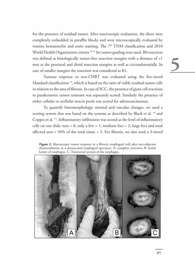
85
5
for the presence of residual tumor. After macroscopic evaluation, the slices were completely embedded in paraffin blocks and were microscopically evaluated by routine hematoxylin and eosin staining. The 7th TNM classification and 2010 World Health Organization criteria 10,12 for tumor grading were used. R0 resection was defined as histologically tumor-free resection margins with a distance of >1 mm at the proximal and distal resection margins as well as circumferentially. In case of smaller margins the resection was considered as R1.
Tumour response to neo-CHRT was evaluated using the five-tiered Mandard classification 13, which is based on the ratio of viable residual tumor cells in relation to the area of fibrosis. In case of SCC, the presence of giant cell reactions to parakeratotic tumor remnant was separately scored. Similarly the presence of either cellular or acellular mucin pools was scored for adenocarcinomas.
To quantify histomorphologic stromal and vascular changes, we used a scoring system that was based on the systems as described by Black et al. 14 and Coppes et al. 15. Inflammatory infiltration was scored as the level of inflammatory cells on one slide: non = 0; only a few = 1; medium foci = 2; large foci and total affected area > 50% of the total tissue = 3. For fibrosis, we also used a 3-tiered
Figure 2. Macroscopic tumor response in a fibrotic esophageal wall after neo-adjuvant chemoradiation in a demarcated esophageal specimen. A: complete overview. B: Inside lumen of esophagus. C: Transversal section of the esophagus.

86
scoring system: non = 0; little fibrosis, thickening submucosa and hyalinized, broadened collagen fibers = 1; moderate fibrosis, submucosa 3-4 times thickened by abnormal collagen, increased and histologically abnormal periesophageal collagen = 2; severe fibrosis, massive mural fibrosis with replacement of muscularis by abnormal collagen extending into periesophageal tissues = 3. Vascular damage, described as vascular hypertrophy, was scored as 0-2: non = 0; slight thickening and hyalinization of vessel walls in the submucosa = 1; heavily affected, double thickness of vessel walls with marked hyalinization and possible reduction in lumen diameter = 2 (Figure 3 & Table 2).
The analysis and classification was performed by an experienced gastrointes-tinal pathologist (A.K.).
Statistical analyses
Figure 3. Scoring system for morphologic changes in normal tissue and tumor areas after neo-adjuvant chemoradiation of the esophagus. A: inflammatory cells and fibrosis grade II; B: inflammatory cells and fibrosis grade II; C: inflamma-tory cells and fibrosis grade III; D: vascular hypertrophy grade I; E: vascular hypertrophy grade II; F: giant cell reaction in former tumor area

87
5
Tabl
e 2.
Sco
re o
verv
iew
for t
he d
iffer
ent a
reas
(I-V
) con
cern
ing
tum
or re
spon
se, g
iant
cel
l rea
ctio
n, m
ucin
ous p
ools,
and
hist
omor
-ph
olog
ic st
rom
al a
nd v
ascu
lar c
hang
es.
I (p
roxi
mal
CT
V)
Man
dard
cla
ssifi
cati
on
Gia
nt c
ell r
eact
ion
to
kera
tin
in sq
uam
ous c
ell
carc
inom
a
Muc
inou
s poo
ls in
ad
enoc
arci
nom
a Va
scul
ar c
hang
es/d
amag
e
Infla
mm
atio
n
Fibr
osis
Tota
l sco
resc
ore
1 (%
)sc
ore
2 (%
)sc
ore
3 (%
)sc
ore
4 (%
)
N (%
)
N (%
)
Tota
l sco
resc
ore
1 (%
)sc
ore
2 (%
)To
tal s
core
scor
e 1
(%)
scor
e 2
(%)
scor
e 3
(%)
Tota
l sco
resc
ore
1 (%
)sc
ore
2 (%
)sc
ore
3 (%
)
0 0 0 0 0 0 (0
)
0 (0
)
1 3 0 16 44 0 0 11 31 0 0
25 3 17 6 3 0,0
1 (3
) 18 42 3 44 61 31 0 40 56 28 0
70 31 39 25 3 3 (5
0)
5 (1
7)
47 53 39 70 14 64 17 78 8 50 36
45 28 17 8 8 3 (5
0)
2 (7
) 26 28 22 42 58 25 3 39 36 19 11
16 3 8 3 3 0,0
0 (0
)
6 17 0 13 36 0 0 12 28 3 0
II (C
TV-
GT
V)
III
(GT
V)
IV (G
TV-
CT
V)
V (d
ista
l CT
V)

88
The scores for morphology changes in the normal tissue in the different areas were compared using a Mann Whitney U test. P-values <0·05 were considered statistically significant.
Results
All patients underwent neo-CHRT without severe (≥ grade 3) toxicities. However, six patients (17%) did not receive the final cycle of chemotherapy because of hematologic toxicities (n=5) and an allergic reaction (n=1). Another patient only received 2 cycles of chemotherapy because of hearing loss during the chemotherapy.
The transthoracic esophagectomy was performed after a median time of 42 days (range: 26-62 days) after completion of neo-CHRT. In 32 patients (94%), a complete resection (R0) was obtained. In 2 patients, the resection margin was ≤ 1 mm (R1).
Demarcations of the GTV and CTV were performed correctly in 33 patients. In 3 patients the demarcations were incomplete. However, in these patients the missing demarcations did not influence the analysis (Table 3).
Tumor responseIn 22 patients (62%) no macroscopic tumor was recognized after
neo-CHRT. At microscopic evaluation, 9 patients (25%) showed a complete response of the primary tumor (ypT0). However, 2 patients had residual tumor
CTV demarcations
lower GTV and CTV border
upper CTV and GTV border
Table 3. Influence of missing demarcations.
Complete microscopic response; no influence
Reconstruction performed, seemed correct; no influenceTumor foci were only located proximal
Missing demarcation Influence of analysis
1
2
3

89
5
cells in their lymph nodes (ypT0N1). Overall, a complete (Mandard 1), partial (Mandard 2-3) and almost none
(Mandard 4-5) response was found in 7 (19%), 25 (69%) and 4 (11%) patients, respectively (Table 1).
The Mandard classification could be based on the GTV (area III) alone in 29 patients (81%), meaning that in these patients the Mandard classification based on area III corresponded well with the Mandard classification based on all the areas (I-V). For the other patients (19%), the CTV had to be incorporated (area II-IV) for a correct evaluation of the tumor response. Incorporation of area V and I did not result in a different Mandard classification. The microscopic tumor extended to and beyond the borders of the CTV in three patients (8%). In all of these patients, the closest margin was located in the CTV or at the border.
A granulomatous reaction to parakeratotic cells was only found in the GTV and/or CTV in 4 patients (67%) with SCC. Mucin pools as remnants of AC were seen in 5 patients (14%) with AC and remained also restricted to the GTV and/or CTV. According to the current TNM classification, these lesions were considered as tumor negative, but may indicate previous localizations of tumor 10,16.
The median number of resected lymph nodes per patient was 18 (range 7-30). Tumor positive lymph nodes were found in 15 patients (42%; range 1-8 positive lymph nodes). For the lymph node positive patients, the median lymph node ratio was 0.11 (range 0.04-0.44).
All positive lymph nodes were located in area II-IV (GTV and CTV). However, in 2 patients the location of the positive lymph nodes was not documented.
Verification of intra-operatively marked areas by analyzing stromal changes To verify the accuracy of the intra-operative demarcation procedures for
the GTV and CTV, we separately analyzed stromal reactions to the radiotherapy. Inflammatory cells, fibrosis and vascular hypertrophy were observed in all regions (Table 2). However, the level of inflammation and fibrosis was significantly (P<0.001) higher in area III (GTV), as compared to area II and IV (both CTV’s). Moreover, these levels were also significantly higher than in regions I and V, proximal and distal of the two CTV’s (P<0.001). As expected, the level of inflammation and fibrosis was not significantly different between the area II

90
and IV (CTV) and between area I and V. Similar effects were seen for vascular damage.
Discussion
Accurate identification of the original tumor bed is essential for proper pathologic evaluation. However, this can be hampered by a complete macroscopic response resulting from neo-CHRT. In the present study, 22 patients (61%) showed a complete macroscopic tumor response. In these patients random sampling in ulcerated or scarred area is unlikely to assure representative microscopic evaluation. Most importantly, microscopic tumor spread and skip lesions, which are often seen in esophageal cancer, will be missed at macroscopic palpation and inspection.
Previous studies, in which random sampling was performed in patients with a macroscopic response, showed no significant survival difference between patient groups with 0% or 1-10% residual tumor cells 17. This suggests that viable tumor cells could be missed in a considerable percentage using this method.
An alternative is an in toto examination of the esophageal specimen. However, this method is time consuming and possibly less effective as the sensitivity of the histological examination may decrease with an increased volume of slides (usually >100) to be assessed.
The original tumor site and areas at risk for microscopic spread can be identified by exact demarcation of the GTV and CTV. This starts with an accurate localization and delineation by the radiation oncologist. However, despite thorough preparations, the intra- and interobserver variability may still be an important issue 18,19. Furthermore, microscopic spread can easily be missed. A CTV margin of at least 30 mm is required to cover the extent of microscopic spread within the esophagus in about 94% of cases 14, except for adenocarcinoma along the GEJ and stomach cardia, which might require a larger margin. In the current study, a CTV margin of 35 mm was taken. To be certain that we included all microscopic tumor, area V and I were also incorporated in the study design.
PTV margins are set to compensate for set up uncertainties to assure adequate dose coverage of the CTV. The PTV itself will receive a radiation dose

91
5
that will be less than 95% of the prescribed dose depending on the size and type (random or systematic) of the set up deviations 4. Therefore, and to reduce the number of demarcations, we only included the GTV and CTV borders, and did not demarcate the PTV borders as well.
Measurement uncertainties, which might occur while measuring the distances from GTV and CTV borders to the anatomical reference points, compromise the accuracy of the demarcation. To minimize these uncertainties, we used at least 2 reference points to relate the distances to. Differences in patient position at the time of measurements may also affect the accuracy of the demarcation. An immobilization device is used for the arms to obtain the appropriate radiotherapy treatment position, which is absent during surgical resection. Furthermore, during surgical resection muscle relaxation might also influence the position of the esophagus in relation to the anatomical reference points.
However, the accuracy of our procedure was supported by the presence of giant cell reactions to keratin remnants and mucin pools as remnants of AC, which were only present in the GTV and/or CTV. Furthermore, significant morphologic differences were found between histomorphologic stromal and vascular changes in the GTV, CTV and outside the CTV. The level of fibrosis, inflammatory cells and vascular hypertrophy in the GTV was also significantly different from the CTV, even though these areas received a similar radiation dose. These differences suggest that in the different areas, different mechanisms are responsible for these morphologic changes in the normal tissue. For fibrosis, it can be difficult to differentiate between radiation induced effects or pre-existing tumor desmoplasia. Indirect radiation induced effects, the so-called out of field effect, may also play a role 15.
The Mandard system relates the residual tumor to the amount of fibrosis to estimate the tumor response to the neo-adjuvant treatment. However, this principle should only be applied to previous tumor locations, since fibrosis does not only exist as a result of tumor response to neo-CHRT, but also as a response of normal tissue to irradiation or as a result of peri-tumor inflammation. Identification of the original tumor bed based on the absence of presence of fibrosis will therefore be inaccurate and might compromise the quality of the Mandard classification. Therefore, we incorporated other radiation-induced

92
changes as well to improve the accuracy of identifying the original tumor bed.Detection and analysis of the lymph nodes is another important part of
the tumor response evaluation. Several studies emphasized both the number of lymph node metastasis and the lymph node ratio as prognostic factors 21,22. Furthermore, Greenstein et al 22 showed a significantly higher cancer-specific survival with an increasing number of negative lymph nodes. They suggest that at least 18 lymph nodes should be removed for an adequate staging. However, the patients in that study were treated with surgery alone. Mariette et al 21 did a similar study after neo-CHRT. They concluded that the number of lymph node metastases could be used in adequately staged patients (≥15 examined lymph nodes), whereas the lymph node ratio should be used for inadequately staged patients (<15 examined lymph nodes).
The use of neo-CHRT may lead to nodal regression and atrophy of lymph nodes in the irradiated area to such an extent that they may become invisible and impalpable at macroscopic examination. Moreover, the number of lymph nodes found following neo-CHRT is significantly lower, as compared to patients treated with surgery alone 22. In the current study, the mean number of collected glands was 18.6, which is relatively high after neoadjuvant treatment since lymph nodes may regress and become too small to detect at gross examination. This suggests that our method increases the number of harvested lymph nodes, which increases the prognostic value of the nodal stage 21,22.
The demarcation procedure and the detailed report of the pathologist on the individual zones I – V also results in a comprehensive feedback to the radiation oncologist and surgeon. The surgeon will be even more aware of the areas at risk for microscopic spread (CTV). This implies that the CTV-borders should always be included in the surgical specimen, which may contribute to further reduction of tumor positive resection margins. Furthermore, the additional information concerning the exact tumor extension in relation to the GTV and CTV can be used for quality assurance after radiotherapy, and may lead to improved GTV and CTV definition and delineation.
ConclusionMacroscopically recognizable residual tumor, as a clinical guidance for
pathologic examination, was absent in 22 patients (61%). The proposed method
93
5
for intra-operative demarcation of the radiotherapy target volumes facilitates the detection and histological examination of the original tumor bed and adjacent lymph nodes.
Conflict of Interest Statement
The authors state that the research presented in this manuscript is free of conflicts of interest.

94
RefeRences
1. Sujendran V, Wheeler J, Baron R, et al. Effect of neoadjuvant chemotherapy on circumferential margin positivity and its impact on prognosis in patients with resectable esophageal cancer. Br J Surg 2008 Feb;95(2):191-194. 2. Gebski V, Burmeister B, Smithers BM, et al. Survival benefits from neoadjuvant chemoradiotherapy or chemotherapy in esophageal carcinoma: a meta-analysis. Lancet Oncol 2007 03;8(3):226-234. 3. van Hagen P, Hulshof MC, van Lanschot JJ, et al. Preoperative chemo-radiotherapy for esophageal or junctional cancer. N Engl J Med 2012 May 31;366(22):2074-2084. 4. Rohatgi PR, Swisher SG, Correa AM, et al. Failure patterns correlate with the proportion of residual carcinoma after preoperative chemoradiotherapy for carcinoma of the esophagus. Cancer 2005 Oct 1;104(7):1349-1355. 5. Brucher BL, Becker K, Lordick F, et al. The clinical impact of histopatho-logic response assessment by residual tumour cell quantification in esophageal squamous cell carcinomas. Cancer 2006 May 15;106(10):2119-2127. 6. Brucher BL, Weber W, Bauer M, et al. Neoadjuvant therapy of esophageal squamous cell carcinoma: response evaluation by positron emission tomography. Ann Surg 2001 Mar;233(3):300-309. 7. Wieder HA, Brucher BL, Zimmermann F, et al. Time course of tumour metabolic activity during chemoradiotherapy of esophageal squamous cell carcinoma and response to treatment. J Clin Oncol 2004 Mar 1;22(5):900-908. 8. Vallbohmer D, Holscher AH, Dietlein M, et al. 18F]-Fluorodeoxyglucose-positron emission tomography for the assessment of histopathologic response and prognosis after completion of neoadjuvant chemoradiation in esophageal cancer. Ann Surg 2009 Dec;250(6):888-894. 9. Hosch SB, Stoecklein NH, Pichlmeier U, et al. Esophageal cancer: the mode of lymphatic tumour cell spread and its prognostic significance. J Clin Oncol 2001 Apr 1;19(7):1970-1975. 10. Sobin LH. TNM classification of malignant tumours. 7th ed. 2009. 11. Mamede M, El Fakhri G, Abreu-e-Lima, et al. Pre-operative estimation of esophageal tumour metabolic length in FDG-PET images with surgical pathology confirmation 14. Ann Nucl Med 2007 12;21(10):553-562. 12. Hamilton SR AL. WHO classification of tumours, Pathology and Genetics, Tumours of the digestive system. 2000. 13. Mandard AM, Dalibard F, Mandard JC, et al. Pathologic assessment of tumour regression after preoperative chemoradiotherapy of esophageal carcinoma. Clin-icopathologic correlations. Cancer 1994 Jun 1;73(11):2680-2686. 14. Black QC, Grills IS, Kestin LL, et al. Defining a radiotherapy target with positron emission tomography. Int J Radiat Oncol Biol Phys 2004 11/15;60(4):1272-1282. 15. Coppes RP, Muijs CT, Faber H, et al. Volume-Dependent Expression of In-Field and Out-of-Field Effects in the Proton-Irradiated Rat Lung. Int J Radiat Oncol Biol Phys 2011 Sep 1;81(1):262-269.

95
5
16. Baldus SE, Monig SP, Schroder W, et al. Regression of esophageal carcinomas after neoadjuvant radiochemotherapy: criteria of the histopathological evaluation. Pathologe 2004 Nov;25(6):421-427. 17. Chirieac LR, Swisher SG, Ajani JA, et al. Posttherapy pathologic stage predicts survival in patients with esophageal carcinoma receiving preoperative chemora-diation. Cancer 2005 Apr 1;103(7):1347-1355. 18. Tai P, Van Dyk J, Yu E, et al. Variability of target volume delineation in cervical esophageal cancer. Int J Radiat Oncol Biol Phys 1998 Sep 1;42(2):277-288. 19. Schreurs LM, Busz DM, Paardekooper GM, et al. Impact of 18-fluorode-oxyglucose positron emission tomography on computed tomography defined target volumes in radiation treatment planning of esophageal cancer: reduction in geographic misses with equal inter-observer variability: PET/CT improves esophageal target definition. Dis Esophagus 2010 Aug;23(6):493-501. 20. Gao XS, Qiao X, Wu F, et al. Pathological analysis of clinical target volume margin for radiotherapy in patients with esophageal and gastresophageal junction carcinoma. Int J Radiat Oncol Biol Phys 2007 Feb 1;67(2):389-396. 21. Mariette C, Piessen G, Briez N, et al. The number of metastatic lymph nodes and the ratio between metastatic and examined lymph nodes are independent prognostic factors in esophageal cancer regardless of neoadjuvant chemoradiation or lymphadenectomy extent. Ann Surg 2008 Feb;247(2):365-371. 22. Greenstein AJ, Litle VR, Swanson SJ, et al. Effect of the number of lymph nodes sampled on postoperative survival of lymph node-negative esophageal cancer. Cancer 2008 Mar 15;112(6):1239-1246. 23. Pultrum BB, Honing J, Smit JK, et al. A critical appraisal of circumferential resection margins in esophageal carcinoma. Ann Surg Oncol 2010 Mar;17(3):812-820.


Pathologic evaluation of radiotherapy target volumes as quality control after neo-adju-vant chemoradiation for esophageal cancer
Chapter 6
C.T. Muijs, J.K. Smit, A. Karrenbeld, J.C. Beukema, V.E. Mul, G.M. van Dam, G.A.P. Hospers, P.M. Kluin, J.A. Langendijk, J.Th.M. Plukker
Submitted for publication

98
Abstract
Purpose: The aim of this study was to analyze the accuracy of gross tumor volume (GTV) delineation and clinical target volume (CTV) margins for neo-adjuvant chemoradiotherapy (neo-CHRT) in esophageal carcinoma by detailed pathologic analysis and to study the impact on survival.
Methods: The study population consisted of 63 esophageal cancer patients treated with neo-CHRT. GTV and CTV borders were demarcated in situ during surgery on the esophagus, using anatomical reference points, to provide information regarding tumor location at pathologic evaluation. To identify prognostic factors for disease free survival (DFS) and overall survival (OS), a Cox-regression analysis was performed.
Results: After resection, macroscopic residual tumor was found outside the GTV in 7 patients (11%). Microscopic residual tumor was located outside the CTV in 9 patients (14%). The median follow up was 15.6 months. With multivariate analysis, only microscopic tumor outside the CTV (HR 4.96, 95%CI 1.03-15.36) and perineural growth (HR 5.77, 95%CI 1.27-26.13) were identified as independent factors for OS. The one year OS was 20% vs. 86%, for patients with or without tumor outside the CTV (P<0.01). For DFS, microscopic tumor outside the CTV (HR 5.92, 95%CI 1.89-18.54) and ypN+ (HR3.36, 95%CI 1.33-8.48) were identified as independent adverse prognostic factors. The one year DFS was 23% vs. 77%, for patients with or without tumor outside the CTV (P<0.01).
Conclusion: The presence of microscopic tumor outside the CTV, associated with a markedly poorer survival after neo-CHRT warrants improvement of GTV delineation and CTV margins in the radiotherapy planning.

99
6
Introduction
Neo-adjuvant chemoradiotherapy (neo-CHRT) followed by surgical resection increases the rate of microscopic radical resections and significantly improves disease free survival (DFS) and overall survival (OS), as compared to surgical resection alone 1,2. Therefore, neo-CHRT has become standard of care for operable patients with curatively resectable non-metastatic esophageal carcinoma.
The histopathological tumor response to neo-CHRT is an important predictor for both locoregional tumor control and survival 3,4. To gain optimal tumor response, accurate radiotherapy, and therefore accurate identification and delineation of the target volumes, is essential. However, delineation of the gross tumor volume (GTV) in esophageal cancer remains difficult. Currently, computer tomography (CT) is the reference imaging modality for delineation of the GTV for radiotherapy of esophageal cancer, integrated with information derived from other diagnostic modalities. However, the discriminative value of CT is generally poor and not sufficient to detect all aspects of tumor extension, such as submucosal spread. Therefore, the current standard is to use relatively large margins from GTV to clinical target volume (CTV) to account for possible microscopic tumor spread, in particular in the cranial-caudal directions.
FDG-positron emission tomography (PET) scans may improve tumor delineation by incorporation of metabolic information 5-7. However, the use of FDG-PET/CT for tumor delineation in esophageal cancer may result in an increase or decrease of the GTV, and pathologic validation is still limited 8,9. So far, only four studies, comparing the delineated target volume to pathological specimen in esophageal cancer patients, have been published 5,10-12. However, these studies only evaluated the correspondence in tumor length between imaging techniques and pathology but did not take into account the accuracy of the localization of the GTV.
Anatomical reference points can be used to reconstruct and to demarcate the radiotherapy target volumes borders in vivo on the esophageal specimen. With the use of this method, information regarding the exact localization of residual tumor in relation to the GTV and CTV, as delineated on the currently used planning CT scans, can be obtained at pathologic examination.
The aim of this study was to analyze the accuracy of GTV delineation and

100
CTV margins for neo-CHRT in esophageal carcinoma by detailed pathologic analysis and to study the impact on DFS and OS.
Materials and methods
PatientsThe study population was composed of 63 patients with clinical stage
T1N1-3M0 and T2-4aN0-3M0 esophageal cancer (adeno- or squamous cell carcinoma (AC or SCC), who were eligible for curative treatment consisting of neo-CHRT, followed by surgical resection.
All patients were staged according to the 7th TNM-system of the Union International Contre le Cancer (UICC) 13 based on the following examinations: physical examination, endoscopic ultrasonography (EUS) and CT. For patients with tumors invading the adventitia and/or positive locoregional lymph nodes, an FDG-PET was performed as well. Other additional investigations were carried out when indicated. All patients were discussed in a multidisciplinary tumor board.
The study was performed according to the rules approved by the local ethics committee.
The 63 patients had a median age of 65 (range: 41-83) years. Patients and tumor characteristics are listed in Table 1.
Target volume delineationThe GTV was delineated by experienced radiation oncologists on planning
CT scan, using all available diagnostic information. The GTV contained the primary tumor and pathologic lymph nodes. The CTV was obtained by adding a 1 cm margin in the transversal plane and a 3.5 cm margin in cranial and caudal directions (2.5 cm margin if the tumor expanded into the stomach) to the primary tumor and 1 cm margin around pathologic lymph nodes. In addition, the CTV was adjusted to anatomical structures. The planning target volume (PTV) was generated by expanding the CTV with 5 mm margins to account for setup uncertainties to assure adequate dose coverage of the CTV.

101
6
TreatmentAll patients were treated with neo-CHRT followed by a surgical resection
at the University Medical Center Groningen from August 2009 to June 2012. Radiotherapy consisted of 41.4 Gy in daily fractions of 1.8 Gy, five times
a week. Patients received weekly concurrent chemotherapy, which consisted of paclitaxel (50 mg/m2) and carboplatin (AUC= 2). All patients underwent a transthoracic esophagectomy with 2-field lymphadenectomy 4 to12 weeks after completion of neo-CHRT. All surgical procedures were performed by two experienced surgical teams.
Demarcation of the GTV and CTV bordersFor the orientation of the GTV and CTV, we defined five anatomical
reference points; the caudal border of the arcus aortae, the tracheal bifurcation,
SexMale Female Age (years)MedianRange HistologyACSC LocalizationHighMidDistal/GEJ Clinical stageT2N0M0T2N1M0T2N2M0T3N0M0T3N1M0T3N2M0T3N3M0T4N1M0TxN1
Table 1. Patient characteristics
48 (76)15 (24) 6541-83 52 (83)11 (17) 01 (2)62 (98) 6 (9)5 (8)1 (2)7 (11)15 (24)23 (36)4 (6)1 (2)1 (2)
Characteristics n=63 (%)
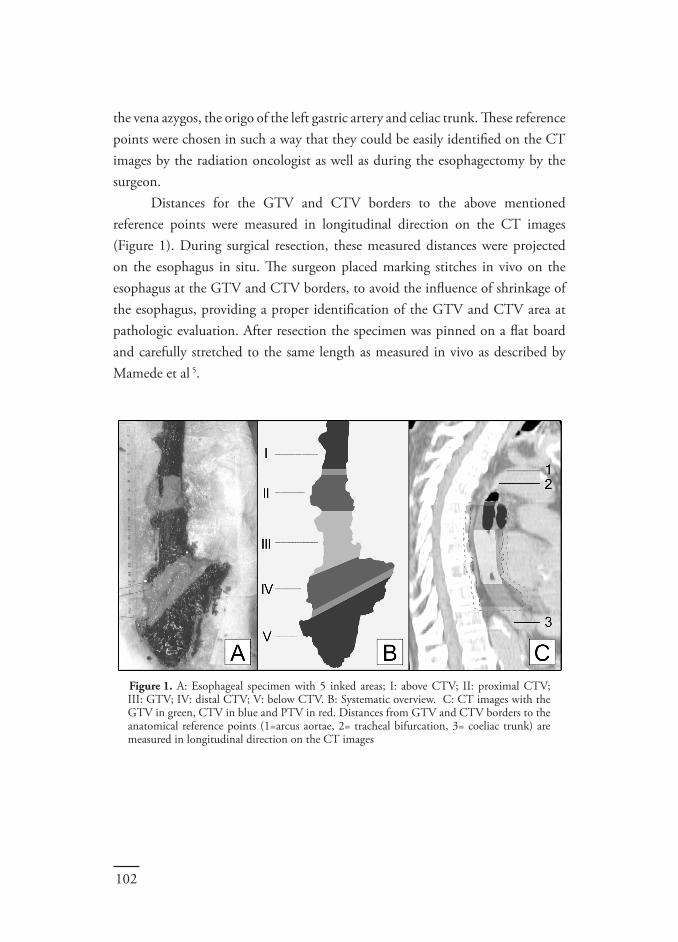
102
the vena azygos, the origo of the left gastric artery and celiac trunk. These reference points were chosen in such a way that they could be easily identified on the CT images by the radiation oncologist as well as during the esophagectomy by the surgeon.
Distances for the GTV and CTV borders to the above mentioned reference points were measured in longitudinal direction on the CT images (Figure 1). During surgical resection, these measured distances were projected on the esophagus in situ. The surgeon placed marking stitches in vivo on the esophagus at the GTV and CTV borders, to avoid the influence of shrinkage of the esophagus, providing a proper identification of the GTV and CTV area at pathologic evaluation. After resection the specimen was pinned on a flat board and carefully stretched to the same length as measured in vivo as described by Mamede et al 5.
Figure 1. A: Esophageal specimen with 5 inked areas; I: above CTV; II: proximal CTV; III: GTV; IV: distal CTV; V: below CTV. B: Systematic overview. C: CT images with the GTV in green, CTV in blue and PTV in red. Distances from GTV and CTV borders to the anatomical reference points (1=arcus aortae, 2= tracheal bifurcation, 3= coeliac trunk) are measured in longitudinal direction on the CT images

103
6
Pathologic evaluationThe specimen was fixed in 10% phosphate-buffered formalin for
approximately 48 hours. The surgical specimen was divided in five areas: an area of 2 cm proximal of the CTV border (I), the proximal CTV (II), GTV (III), distal CTV (IV) and an area of 2 cm below the distal CTV border (V). To identify the different areas microscopically, the outer surface of these five areas was inked in different colors before the specimen was cut into transverse slices at a thickness of approximately 3 mm. All slices were ordered by area of origin and evaluated macroscopically for the presence of residual tumor or other abnormalities. After macroscopic evaluation, the slices were completely embedded in paraffin blocks and were microscopically evaluated by routine hematoxylin and eosin staining. All five areas were separately scored by the pathologist for tumor involvement, lymph node involvement and signs of regression. The 7thTNM classification and 2010 World Health Organization criteria for tumor grading were used 14,15. R0 resection was defined as histologically tumor-free resection margins with a distance of >1 mm between tumor and all resection margins, including the circumferential margin (CRM) according to the RCP 16,17. In case of microscopic tumor at a distance of ≤ 1mm, the resection was considered as R1.
Tumor response to neo-CHRT was evaluated using the five-tiered Mandard classification 18, which is based on the ratio of viable residual tumor cells in relation to the area of fibrosis. Furthermore, we evaluated the localization of the original tumor bulk based on the presence of residual tumor and/or regressive changes. The analysis and classification was performed by an experienced gastrointestinal pathologist (A.K.).
Follow upAfter resection, routine follow up was performed every 3 months in the
first year and 4 months in the second years, followed by a yearly control. For 43 patients, who participated in a trial, a CT-scan of the thorax and abdomen was part of this follow up for the first 2 years. In the other 20 patients, further radiological investigations were performed based on clinical suspicion of recurrent disease.

104
StatisticsOS time was calculated from the first day of neo-CHRT, according to
the Kaplan-Meier method and compared using the Log-rank test. To identify prognostic factors for DFS and OS, univariate Cox proportional hazards analyses were performed. Multivariate analyses were performed using the Cox proportional hazards model entering the parameters of influence on outcome according to univariate analysis (defined as those with P<0.1) using backwards selection. A P-value <0.05 was considered significant. The statistical analyses were performed by using the Statistical Package for Social Sciences (SPSS, Chicago IL, USA) version 18.0 software.
ResultsAll patients underwent neo-CHRT, without severe (≥ grade 3) toxicities.
However, 11 patients (18%) did not receive the final cycle of chemotherapy course because of hematologic toxicities (n=10) and general physical condition (n=1). Another patient only received 2 cycles because of hearing loss during the chemotherapy.
The transthoracic esophagectomy with lymphadenectomy was performed after a median time of 48 days (range: 26-89) after neo-CHRT. Fifty-four patients (86%) received a complete resection with CRM > 1mm. In 9 patients, the CRM was ≤ 1 mm (R1, according to RCP). Pathologic characteristics are listed in table 2. Demarcations of the GTV and CTV were performed correctly in 58 patients. In 5 patients the demarcations were incomplete. However, in these patients the missing demarcations did not influence the analysis (Table 3).
Accuracy of the gross tumor delineation (GTV)Macroscopically evident residual tumor was found in 19 patients (30%),
which was located in the GTV in 12 patients and outside the GTV in 7 patients (11%). All macroscopic residual tumor cells remained within the CTV. Using the Mandard classification 4 of these 7 patients were partial responders (Mandard 2-3) and 3 were minimal or no responders (Mandard 4-5).
The bulk of the tumor, based on residual tumor and evident regressive changes indicative for preexistent carcinoma, was located in the GTV in 47 patients (75%), in both GTV and CTV in 7 patients (11%) and in the CTV

105
6 Pathology characteristics
Table 2. Pathologic characteristics
n= 63 (%)
Tumor outsideCTV n=9 (%)
Tumor withinCTV n=54 (%)
Histopathological T-stage ypT0 15 (24) 1 (11) 14 (27)ypT1 12 (19) 0 (0) 12 (22)ypT2 9 (14) 2 (22) 7 (13)ypT3 27 (43) 6 (67) 21 (38)ypT4 0 (0) 0 (0) 0 (0) Histopathological N-stage ypN0 38 (60) 1 (11) 37 (69)ypN1 17 (27) 4 (45) 13 (24)ypN2 3 (5) 2 (22) 1 (2)ypN3 5 (8) 2 (22) 3 (5) Macroscopic tumor extension within GTV 19 (30) 6 (67) 38 (70)outside GTV 7 (11) 3 (33) 16 (30) Microscopic tumor extension within CTV 47 (75) outside CTV 9 (14) Completeness of resection R0 53 (84) 6 (67) 47 (87)R1 (according to RCP) 10 (16) 3 (33) 7 (13) Lymphangio invasion Negative 48 (76) 4 (44) 44 (81)Positive 15 (24) 5 (56) 10 (19) Perineural growth Negative 56 (89) 6 (67) 50 (93)Positive 7 (11) 3 (33) 4 (7) Tumor regression grade – overall I 12 (19) 0 (0) 12 (22)II 29 (46) 6 (67) 23 (44)III 13 (20) 0 (0) 13 (24)IV 9 (14) 2 (22) 7 (13)V 1 (1) 1 (11) 0 (0)

106
margin in 8 patients (13%). In one patient, the tumor affected the GTV and CTV as well as the area cranial to the CTV, and no clear tumor bulk could be identified.
Accuracy of the CTV margin for microscopic tumor spread A complete microscopic response of the primary tumor (ypT0) was seen in
15 patients (24%). However, 4 of them had residual tumor cells in their lymph nodes (ypT0N1).
Overall, a complete (Mandard 1), partial (Mandard 2-3) and almost no (Mandard 4-5) response were found in 12 (19%), 41 (65%) and 10 (16%) patients, respectively.
The microscopic residual tumor remained restricted to the CTV in 86% of the patients. However, in 9 patients (14%) microscopic tumor was also found outside the CTV. In 6 of these patients, tumor was found caudal from the distal CTV margin. In four of them, a CTV margin of 25 mm was used because of expansion into the stomach. In one patient microscopic tumor was found beyond both CTV borders, while another patient showed extension only cranial to the proximal CTV margin. The final patient showed exclusively a positive lymph node cranial to the proximal CTV margin. Importantly, in none of these patients the CTV demarcations were incomplete.
R1-resections were found in 3 out of these 8 patients (excluding the ypN+ patient), including 2 with circumferential invasion and only one with invasion of the cranial and caudal borders.
Tumor characteristics of these 8 patients are listed in table 4. Interestingly,
Table 3. Influence of missing demarcations
Complete microscopic response; no influenceReconstruction performed, seemed correct; no influenceTumor foci were only located proximal
Reconstruction performed, no proximal residual tumor; no influenceNo macroscopic residual tumor; no influence
Influence of analysis
1
2
3
4
5
CTV demarcations
lower GTV and CTV border
upper CTV and GTV border
upper GTV border
GTV demarcations
Missing demarcationPatient

107
6
these patients also showed different distributions of their tumor bulk; it was located within the GTV in only 3 patients, in the caudal CTV in 3 patients and within the GTV/CTV area in 2 patients. In one patient no clear tumor bulk could be identified.
According to the Mandard classification, 6 of these 9 patients were partial responders, while 3 patients showed almost no response to neo-CHRT.
The median number of resected lymph nodes was 17 (range 7-33). Positive lymph nodes were seen in 25 patients (40%). For lymph node positive patients, the median positive to negative ratio was 0.17 (range 0.04-0.69).
Most positive lymph nodes (72%) were located in the GTV and CTV, except for 4 patients (16%) with positive lymph nodes in area I or V (outside the CTV). In 3 patients the location of the positive lymph nodes was not documented.
Influence of pathological findings after neo-CHRT on DFS and OS At the time of analysis, 19 out of 63 (30.2%) patients had died within 4 to
29 months after start of treatment. The median follow up time was 16.6 months (95% CI 14.0-19.2).
The one year OS and DFS was 79% and 71%, respectively. The mean OS and DFS were 24.9 (95% CI 21-28) and 24.2 (95% CI 20-28) months.
In univariate analysis, microscopic tumor extension outside the CTV, ypN+, lymphangio-invasion, perineural growth, lymph node ratio >0.10 and >5 positive lymph nodes were associated with worse DFS and OS (Table 5). In the multivariate analysis, only perineural growth (HR 5.77, 95%CI 1.27-26.13) and microscopic tumor extension outside the CTV (HR 4.96, 95%CI 1.03-15.36) were significantly associated with OS. The one year OS was 20% if tumor was
MMMFFMMM
Sex
6263694669645666
T3N1T3N0T3N3TxN1T2N0T3N1T3N3T3N2
R1R0R0R0R0R1R1R0
ACACACACACACACAC
33302333
yesnonononoyesyesno
yesnoyesyesyesnoyesno
60220870
20110330
42224252
AgeClinical stage
Histo-logy
Resection margin
ypT-stage
ypN-stage
Positive LN’s
Lymph-angio invasion
Perineural growth
Tumor regression grade
Table 4. Tumor characteristics of patients with microscopic tumor outside the CTV

108
2,73
4,74 11,4398,60
5,7310,990,00
6,89
2,53
3,98
2,495,075,45
7,29
79,80
10,02
7,88
10,489,3920,24
8,5518,59
44,33
9,23
21,84
7,35
5,84
4,77
7,25
3,43
man 48female 15≥65 30<65 33cT2 12cT3 49cT4a 1cN0 13cN1 22cN2 24cN3 4no 13yes 50no 37yes 26AC 52PCC 11ypT1 15ypT2 12ypT3 9ypT4 27ypN0 38ypN+ 25no 57yes 6no 51yes 12R0 53R1 10Mandard 1 12Mandard 2 29Mandard 3 13Mandard 4 8Mandard 5 1CR 11PR 43NR 9no 56yes 7no 48yes 15no 54
yes 9no 56
yes 7no 59yes 3no 33yes 29no 51yes 12no 36yes 27
Table 5. Univariate analysis for overall survival
lowerSex
Age
cT
cN
cN+
Tumor length >5
PA type
ypT
ypN+
LN positive >5
LN ratio>0.1
R-margin
Tumor response Tumor response Perineural growth Lymphangio invasion Microscopic residual tumor outside CTV
Macroscopic residual tumor outside GTV
Upstaging T
Downstaging T
Upstaging N
Downstaging N
1,00 0,90 0,301,00 1,85 0,721,00 2,570 0,588,490 0,731,00 1,430 0,362,970 0,80,000 0,001,00 2,00 0,581,00 1,02 0,411,00 1,31 0,431,00 ,280 0,03,930 0,171,750 0,561,00 2,86 1,131,00 6,12 1,891,00 3,91 1,521,00 2,15 0,591,00 2,280 0,501,710 0,313,260 0,53,000 1,00 1,930 0,442,990 0,481,00 11,27 2,871,00 3,47 1,311,00
6,80 2,121,00
2,41 0,791,00 0,76 0,101,00 1,82 0,701,00 2,80 1,081,00 1,39 0,56
0,85 0,20 0,22 0,32 0,28 0,97 0,63 0,27 0,03* 0,00* 0,01* 0,25 0,77 0,50 0,00* 0,01* 0,00*
0,12
0,80 0,22 0,03* 0,48
11,0411,6612,446,9611,433,1012,0411,2111,1710,261,012,041,0003,3901,0006,86014,9512,5612,371,933,84012,013,4519,7113,321
5,871
210,9212,2213,0511,64
0,38
0,67
0,560,62
0,360,850
0,59
0,5
0,39
0,030,190,67
1,350
2,390
1,97
0,82
0,520,350,69
0,450,62
2,68
1,33
1,94
0,67
0,12
0,87
1,24
0,68
2,9
4,12
10,6678,89
5,7311,280
6,99
2,92
3,51
2,295,546,28
8,510
19,670
12,44
8,03
10,8310,5721,42
8,8519,34
35,3
8,29
17,75
6,03
6,93
5,66
7,51
3,97
0,94
0,27
0,27
0,26
0,26
0,68
0,78
0,15
0,01*
0,00*
0,00*
0,11
0,65
0,36
0,00*
0,01*
0,00*
0,21
0,93
0,10*
0,02*
0,27
lowerupper upper
95% CI95% CIDFSOS
pvalueHRHRNunivariaatCOX regression
pvalue

109
6
found outside the CTV vs. 86% for patients without tumor outside the CTV (P-value: <0.01). The one year OS was 0% vs. 85% for patients with or without perineural growth (P-value <0.01). For DFS, microscopic tumor extension outside the CTV (HR 5.92, 95%CI 1.89-18.54) and ypN+ (HR3.36, 95%CI 1.33-8.48) were identified as independent adverse prognostic factors. The one year DFS was 23% vs. 77% for patients with or without tumor outside the CTV (P-value <0.01). For patients with ypN+ the one year OS was 58% vs. 80% for patients with ypN0 (P-value <0.01).
Discussion
The present study demonstrated that macroscopic tumor was located outside the GTV in 35% of the patients with macroscopic residual tumor. Furthermore, microscopic tumor was found outside the CTV in 14% of the patients, after neo-CHRT.
The mismatch of the GTV and macroscopic tumor suggests inaccurate delineation. Accurate delineation of the GTV is a prerequisite for successful preoperative treatment of esophageal cancer with neo-CHRT. However, it is well known that the intra- and interobserver variability for the tumor delineation of esophageal cancer can be rather large 19,20. Furthermore, the GTV delineation can be hampered by the poor discriminative value of the currently used CT, and the inability to relate endoscopic (ultrasound) information to CT-images. Several authors speculated about the incorporation of FDG-PET data to improve the accuracy of the tumor delineation by identifying a metabolic tumor volume 5,10. However, the results of a recently published review showed that there is currently insufficient evidence regarding pathologic and clinical validation, to support the use of FDG-PET/CT in the tumor delineation process for radiotherapy 9.
Microscopic tumor spread can easily be missed on CT, but also on FDG-PET. Moreover, FDG-PET is not able to identify T1-tumors and failed to identify microscopic residual tumor in 18% of cases in a study by Swisher et al 21.
Literature concerning the extent of submucosal spread in esophageal cancer is limited. Most surgical studies examined the resection margins of surgical specimens and did not report on the minimal margin that is required to

110
encompass the microscopic tumor 22,23.The only study, explicitly performed to define the optimal CTV margin,
stated that a CTV margin of at least 30 mm (proximal and distal) is required to cover the extent of microscopic spread within the esophagus in 94% of patients with SCC 24. For AC located at the GEJ, a 30 mm proximal CTV margin would cover microscopic tumor in up to 100% of cases. However, for tumors along the GEJ and gastric cardia a 50 mm distal CTV margin is required to cover microscopic tumor in 94% of cases 24.
In the current study, a CTV margin of 35 mm was taken in cranial and caudal direction. However, if the tumor expanded into the gastric cardia only a 25 mm margin was taken. In line with the findings of Gao et al 24, this 25 mm margin seemed insufficient, since most cases of microscopic tumor outside the CTV were located caudally of the distal CTV margin into the gastric cardia.
Another explanation for macroscopic or microscopic residual tumor outside the delineated GTV or CTV is persistent tumor growth. In the current study, half of the patients with microscopic extension beyond the CTV border were classified as non-responders to neo-CHRT. For these relatively radiotherapy-resistant tumors, neo-CHRT delays further treatment, i.e. the surgical resection, which gives the tumor the opportunity to grow, resulting in tumor extension beyond the radiotherapy target volumes. Several studies described newly detected post-neoadjuvant metastases, also suggesting tumor growth during or after the neo-adjuvant treatment. In these studies, the incidence of post-neoadjuvant metastases varied from 8 to 17% 25-28. Moreover, tumor progression can even be observed before the start of neo-CHRT. Muijs et al 29 found tumor progression in terms of increased tumor length and/or more advanced TNM-stage, in 31% and 27% of the patients, within a median time interval of 22 days between diagnostic imaging and imaging for radiotherapy planning.
Despite some limitations of the demarcation method, we demonstrated that the presence of microscopic tumor spread beyond CTV borders was associated with a significant worse OS. Furthermore, the accuracy of our procedure was supported by the presence of giant cell reactions to keratin remnants and mucin pools as remnants of AC, which were only present in the GTV and/or CTV.
The presence of microscopic tumor spread beyond the CTV borders might be a clinical sign of a biologically more aggressive tumor behavior, suggesting that

111
6
these tumors might be more progressive and tend to metastasize in an earlier stage. Perineural growth was also an independent prognostic factor for OS. However, the multivariate analysis in the current study, including all pre-treatment tumor characteristics and pathological tumor characteristics after treatment, showed no other factors that were prognostic.
This might be explained by the relatively small study population with, consequently a rather low incidence of events, which is a limitation of this study, and might lead to an under- or overestimation of the effect.
Conclusion Macroscopic tumor outside the GTV and microscopic tumor outside the
CTV were found in a substantial proportion of patients, suggesting incorrect delineation, inadequate CTV-margins, or tumor growth before, during or after the neo-CHRT. Moreover, the presence of microscopic tumor spread beyond the CTV borders had a significant adverse impact on DFS and OS. These findings emphasize the importance of an accurate delineation of the GTV and indicate that the currently used length of the distal CTV, in particular in caudal direction, is not sufficient.
Conflict of Interest StatementThe authors state that the research presented in this manuscript is free of conflicts of interest.

112
RefeRences
1. Sujendran V, Wheeler J, Baron R, Warren BF, Maynard N. Effect of neoadjuvant chemotherapy on circumferential margin positivity and its impact on prognosis in patients with resectable oesophageal cancer. Br J Surg 2008;95:191-194. 2. van Hagen P, Hulshof MC, van Lanschot JJ et al. Preoperative chemoradio-therapy for esophageal or junctional cancer. N Engl J Med 2012;366:2074-2084. 3. Rohatgi PR, Swisher SG, Correa AM et al. Failure patterns correlate with the proportion of residual carcinoma after preoperative chemoradiotherapy for carcinoma of the esophagus. Cancer 2005;104:1349-1355. 4. Brucher BL, Becker K, Lordick F et al. The clinical impact of histopathologic response assessment by residual tumor cell quantification in esophageal squamous cell carcinomas. Cancer 2006;106:2119-2127. 5. Mamede M, El Fakhri G, Abreu-e-Lima, Gandler W, Nose V, Gerbaudo VH. Pre-operative estimation of esophageal tumor metabolic length in FDG-PET images with surgical pathology confirmation 14. Ann Nucl Med 2007;21:553-562. 6. Leong T, Everitt C, Yuen K et al. A prospective study to evaluate the impact of FDG-PET on CT-based radiotherapy treatment planning for oesophageal cancer. Radiother Oncol 2006;78:254-261. 7. Lambrecht M, Haustermans K. Clinical evidence on PET-CT for radiation therapy planning in gastro-intestinal tumors. Radiother Oncol 2010;96:339-346. 8. Muijs CT, Schreurs LM, Busz DM et al. Consequences of additional use of PET information for target volume delineation and radiotherapy dose distribution for esophageal cancer. Radiother Oncol 2009;93:447-453. 9. Muijs CT, Beukema JC, Pruim J et al. A systematic review on the role of FDG-PET/CT in tumour delineation and radiotherapy planning in patients with esophageal cancer. Radiother Oncol 2010. 10. Zhong X, Yu J, Zhang B et al. Using (18)F-Fluorodeoxyglucose Positron Emission Tomography to Estimate the Length of Gross Tumor in Patients with Squamous Cell Carcinoma of the Esophagus. Int J Radiat Oncol Biol Phys 2008. 11. Han D, Yu J, Yu Y et al. Comparison of (18)F-fluorothymidine and (18)F-fluorodeoxyglucose PET/CT in delineating gross tumor volume by optimal threshold in patients with squamous cell carcinoma of thoracic esophagus. Int J Radiat Oncol Biol Phys 2010;76:1235-1241. 12. Jeganathan R, McGuigan J, Campbell F, Lynch T. Does pre-operative estimation of oesophageal tumour metabolic length using 18F-fluorodeoxyglu-cose PET/CT images compare with surgical pathology length? Eur J Nucl Med Mol Imaging 2011;38:656-662. 13. Edge SB, Byrd DR, Compton CC et al. AJCC cancer staging manual, 7th edition. New York: Springer. 2009. 14. Sobin LH. TNM classification of malignant tumours. 7th ed. 2009. 15. Hamilton SR AL. WHO classification of tumours, Pathology and Genetics,

113
6
Tumours of the digestive system. 2000. 16. The Royal College of Pathologists. Dataset for the histopathological reporting of oesophageal carcinoma, 2nd edition. Document G006. 2007. 17. Griffiths EA, Brummell Z, Gorthi G, Pritchard SA, Welch IM. The prognostic value of circumferential resection margin involvement in oesophageal malignancy. Eur J Surg Oncol 2006;32:413-419. 18. Mandard AM, Dalibard F, Mandard JC et al. Pathologic assessment of tumor regression after preoperative chemoradiotherapy of esophageal carcinoma. Clin-icopathologic correlations. Cancer 1994;73:2680-2686. 19. Schreurs LM, Busz DM, Paardekooper GM et al. Impact of 18-fluorode-oxyglucose positron emission tomography on computed tomography defined target volumes in radiation treatment planning of esophageal cancer: reduction in geographic misses with equal inter-observer variability*. Dis Esophagus 2010. 20. Tai P, Van Dyk J, Yu E, Battista J, Stitt L, Coad T. Variability of target volume delineation in cervical esophageal cancer. Int J Radiat Oncol Biol Phys 1998;42:277-288. 21. Swisher SG, Erasmus J, Maish M et al. 2-Fluoro-2-deoxy-D-glucose positron emission tomography imaging is predictive of pathologic response and survival after preoperative chemoradiation in patients with esophageal carcinoma. Cancer 2004;101:1776-1785. 22. Mariette C, Castel B, Balon JM, Van Seuningen I, Triboulet JP. Extent of oesophageal resection for adenocarcinoma of the oesophagogastric junction. Eur J Surg Oncol 2003;29:588-593. 23. Barbour AP, Rizk NP, Gonen M et al. Adenocarcinoma of the gastroesopha-geal junction: influence of esophageal resection margin and operative approach on outcome. Ann Surg 2007;246:1-8. 24. Gao XS, Qiao X, Wu F et al. Pathological analysis of clinical target volume margin for radiotherapy in patients with esophageal and gastroesophageal junction carcinoma. Int J Radiat Oncol Biol Phys 2007;67:389-396. 25. Bruzzi JF, Swisher SG, Truong MT et al. Detection of interval distant metastases: clinical utility of integrated CT-PET imaging in patients with esophageal carcinoma after neoadjuvant therapy. Cancer 2007;109:125-134. 26. Cerfolio RJ, Bryant AS, Ohja B, Bartolucci AA, Eloubeidi MA. The accuracy of endoscopic ultrasonography with fine-needle aspiration, integrated positron emission tomography with computed tomography, and computed tomography in restaging patients with esophageal cancer after neoadjuvant chemoradiotherapy. J Thorac Cardiovasc Surg 2005;129:1232-1241. 27. Flamen P, Van Cutsem E, Lerut A et al. Positron emission tomography for assessment of the response to induction radiochemotherapy in locally advanced oesophageal cancer. Ann Oncol 2002;13:361-368. 28. Blom RL, Schreurs WM, Belgers HJ, Oostenbrug LE, Vliegen RF, Sosef MN. The value of post-neoadjuvant therapy PET-CT in the detection of interval metastases in esophageal carcinoma. Eur J Surg Oncol 2011;37:774-778. 29. Muijs CT, Pruim J, Beukema JC, Berveling MJ, Plukker JT, Langendijk JA. Oesophageal tumour progression between the diagnostic (18)F-FDG-PET and

114
the (18)F-FDG-PET for radiotherapy treatment planning. Radiother Oncol 2012.


Prediction of response to radiotherapy in the treatment of esophageal cancer using stem cell markers.
Chapter 7
J.K. Smit, H. Faber, M. Niemantsverdriet, M. Baanstra, J. Bussink, H. Hollema, R.P. van Os, J.Th.M. Plukker, R.P. Coppes
Under revision Radiotherapy and Oncology 2013

118
Abstract
Background: and Purpose: In this study, we investigated whether cancer stem cell marker expressing cells can be identified, that predict for the response of esophageal cancer (EC) to chemoradiotherapy (CRT).
Materials: and Methods: EC cell-lines OE-33 and OE-21 were used to assess in vitro, stem cell activity, proliferative capacity and radiation response. Xenograft tumors were generated using NOD/SCID mice to assess in vivo proliferative capacity and tumor hypoxia. Archival and fresh EC biopsy tissue was used to confirm our in vitro and in vivo results.
Results: We showed that the CD44+/CD24- subpopulation of EC cells exerts a higher proliferation rate and sphere forming potential and is more radioresistant in vitro, when compared to unselected or CD44+/CD24+ cells. Moreover, CD44+/CD24- cells formed xenograft tumors faster and were often located in hypoxic tumor areas.
In a study of archival pre-neoadjuvant CRT biopsy material from EC adenocarcinoma patients (n=27), this population could only be identified in 50% (9/18) of reduced-responders to neoadjuvant CRT, but never (0/9) in the complete responders (P=0.009).
Conclusions: These results warrant further investigation into the possible clinical benefit of CD44+/CD24- as a predictive marker in EC patients for the response to CRT.

119
7
Introduction
Esophageal cancer (EC) is an aggressive disease with increasing incidence and a low curability rate 1-4. In specialized centers, the 5-year survival after surgery for all stage groups together is only 20-40%5, 6. Multimodality treatments with preoperative (neo-adjuvant) irradiation in combination with chemotherapy (chemo-radiation) have recently become common practice7, 8, 8. These multimodality treatments achieve a gain in 5-year survival of only 10-15%7, 8. However, a significant proportion of 60-70% does not respond well to these treatments and are thus unnecessarily experiencing severe side-effects 7-9.
Factors predicting the response to neoadjuvant chemoradiation may identify the group of non-responders before treatment is given 10. This may help to reduce the number of unnecessarily treated patients and lead to investigations on new and more effective therapies for this patient group.
Recently, evidence has accumulated that many solid tumors are driven and managed by a small population of cancer stem cells (CSCs), tumor-initiating cells or cancer stem cell like cells11-15, which may be more resistant to treatment16,17. It is postulated that in most cases, these CSCs are, in part, responsible for the inadequate treatment response of certain tumors16-19. It is therefore of great importance for the radiation oncology field to intensify research into CSCs, which could be complimentary to yet established or to be investigated predictive factors for the response to radiotherapy 20-23.
More research is necessary into what extent CSCs are present in EC and what would be their response to radiotherapy 24, 25. In several models for cancers, cell surface markers have been used to identify CSCs such as CD133, CD44, CD24, CD90, CD326 (Epcam) and combinations hereof 11, 13, 14, 16, 26-28. These proteins often activate tumor-specific, downstream pathways and may therefore be possible targets for further therapy 12, 14, 26, 29, 30.
In this study, we hypothesized that a subpopulation of cells may exist in EC that could predict for treatment resistance. Hereto, we tested CSC marker expression, in vitro growth of spheroids, radiation sensitivity, and in vivo growth of several EC derived cell lines derived sub-populations. In EC, a putative CSC-like population was identified with superior in vitro and in vivo growth as well as increased radiation resistance on basis of CD44 and CD24 expression.

120
In patient material the same markers could be detected which suggests that a population may exist that can predict treatment response in a clinical setting.
Methods
For details see supplementary methods section.
Cell culture The OE-33 cell-line derived from a poorly differentiated Barrets
adenocarcinoma of the lower esophagus and the OE-21 cell-line derived from a squamous cell carcinoma of the upper esophagus, were cultured under standard conditions with RPMI 1640 growth medium supplemented with 10% Fetal Calf Serum, 1% Penicilline/Streptomycine in a humidified atmosphere and 5% CO2 at 37°C 31. Cells were passaged at 50-80% confluence. Both cell-lines were independently DNA authenticated by the Leibniz Institute DSMZ-German Collection of Microorganisms and Cell Cultures (Braunschweig, Germany). Serum-free and low adherent growing conditions (ultra-low adherent plates, Corning Inc., Corning, New York, USA) were employed to grow the cells as spheroids using Neural Basal A medium containing N2, bFGF and FGF-2 as previously described by Vermeulen et al. 14
Flow cytometrySingle cell suspensions obtained from tumor tissue or cell-lines were
resuspended in PBS with 0.2% bovine serum albumin (BSA). Primary flow cytometric antibodies were: CD44-PE (BD Biosciences, Franklin Lakes, New Jersey, USA), CD24-FITC (BD Biosciences, Franklin Lakes, New Jersey, USA), EPCAM-Alexa fluor® 647 (eBioscience, San Diego, California, USA), CD133/1-APC (Bergisch Gladbach, Germany), CD29-PE (BD Biosciences, Franklin Lakes, New Jersey, USA), CD-90-FITC (eBioscience, San Diego, California, USA). Flow cytometric analysis was performed on the FACS-Calibur (BD Biosciences, Franklin Lakes, New Jersey, USA) or LSR-II (BD Biosciences, Franklin Lakes, New Jersey, USA). Flow cytometric data were analyzed using Flojo version 7.6 software (Treestar Inc., Ashland, Oregon, USA). To isolate cells with

121
7
a putative stem cell phenotype cell sorting was performed using a MoFlo-XDP or MoFLoAstrios cell sorter (Beckman Coulter previously DakoCytomation, Glostrup, Denmark).
In vitro radiation experiments, clonogenic assaysSorted single cell suspensions of the different subpopulations obtained
after flow-cytometric sorting were counted and plated immediately in standard growth medium (RPMI, see cell culture section). Cells were allowed to attach overnight and (sham) irradiated (Cesium 137, IBL) with 0, 2, 4 and 6 Gy at a dose rate of 0.65 Gy/min. After irradiation cells were trypsinized, replated and concentrations were adjusted according to the expected survival. Colonies were allowed to grow for 10-14 days, fixated and stained (coomassie brillant blue). Surviving fractions were determined by dividing the average number of colonies at different doses by the average number of colonies in the non-irradiated control.
Animal experimentsFemale NOD/SCID mice were purchased from Harlan laboratories
(NOD.CB17-Prkdcscid/NCrHsd). Mice were subcutaneously injected with tumor cells in a 1:1 suspension with matrigel (BD Biosciences, Franklin Lakes, New Jersey, USA) under general anesthesia (isoflurane 2,5%). A total of 1.5x10⁵
OE-21 cells or 6.0x10⁵ OE-33 cells were injected for tumor generation. Single cells were obtained by using a modified method from previously published study on salivary glands 32. In selected cases, mice were intraperitoneally injected with pimonidazole HCI 60mg/kg (Hydroxyprobe™-1, NPI, Inc., Burlington, Massachusetts, USA) as a marker for hypoxia. It is important to note that between 1st and 2nd generation and 2nd and 3rd generation tumors, the cells were not resorted for either CD44+/CD24- or CD44/CD24+ subpopulations.
All animal experiments were performed according to our institutional animal ethics guidelines and were reviewed by an animal ethics committee.
Human tissue samples experimentsHuman tissue biopsies were obtained from patients with confirmed
histological diagnosis of esophageal cancer during routine staging with gastrointes-tinal endo-echography (EUS) or from rest material after surgical resection of

122
the tumor with informed consent. Tissue samples were immediately placed in phosphate buffer with antibiotics and antimycotics. In the lab, the tissue was washed and incubated for at least 4 hours in RPMI with antibiotics and antimycotics and subsequently dissociated into single cells, as described above. The single cells obtained after this process were used for direct FACS analysis (Figure S3 for gating strategy).
All human tissue collection experiments were reviewed by the institutional human ethics commission (Institutional board review). The ethics guidelines comply with the Helsinki Declaration on experiments with humans.
Immunohistochemistry and immunofluoresence imagingImmunohistochemical staining was performed on 5 μm tissue sections
from archival patient material or tumor xenografts using primary antibodies against CD44 (Biolegend, San Diego, California, USA), CD24 (Santa Cruz Biotechnologies Inc., Santa Cruz, California, USA) and pimonidazole (J.A. Raleigh, Department of Radiation Oncology and Toxicology, University of North Carolina, Chapel Hill, North Carolina, USA ) in paraffin 33 or frozen sections 34.
Quantification of CD44+/CD24- population in patient biopsy samples was performed as following: first, the serially stained sections with CD44 and CD24, were scored blindly by an experienced pathologist (H.H.). When there were cases in which there was both CD44 and CD24 positivity, the pathologist was asked if there were tumor areas that were CD44+/CD24-, yes or no. Furthermore, in case there was only single positivity of CD44, these cases were considered as CD44+/CD24- cases. In cases of single positivity of CD24, these were considered as not having CD44+/CD24-. Both CD44 and CD24 results were based exclusively on membrane staining.
Statistical analysisExperiments are representative of at least 3 experiments unless otherwise
stated. All data are presented as mean and ± SD/SEM. Groups were compared with the student’s t test. Correlations were determined with the Pearson’s bi-variant comparison.
123
7
Results
To identify progenitor cell markers on subpopulations in EC cell lines (OE-33 and OE-21), it was analyzed whether these markers were expressed in various culture conditions. The markers CD90, CD29 and CD133 were not present at the surface of OE-33 or OE-21 cells, whereas CD326 (EpCam) was expressed ubiquitously (Supplementary Table 1 and Figure S2). Interestingly, a subpopulation of CD44+/CD24+ and CD44+/CD24- of different sizes could be identified in both the OE-33 and the OE-21 cell line (Supplementary Table 1 and Figure S2). Since cancer cells growing in non-adherent conditions are less likely to differentiate than cells growing in adherent conditions with serum 11, 13, 16,35, we investigated whether the expression of CD44 and CD24 was dependent on culture conditions. Approximately 40% (44.5 ± 7.9%) of OE-33 cells growing sparsely at 15-30% confluency were CD44+ but CD24-, whereas in cells growing at 90-95% confluence only 3.6 ± 2.3% were CD44+/CD24- (P=0.010, Figure 1A and S1). Moreover, in non-adherent serum free growing conditions OE-33 cells formed spheroid structures (Figure S1) that expressed CD44+/CD24- in 59.3 ± 2.1 % of the cells (Figure 1A and S1). These results indicate that in OE-33, the CD44+/CD24- expression is dependent on the culture conditions. Next, it was assessed whether this shift from a high percentage of cells expressing CD44+/CD24- to lower percentages was observed after prolonged spheroid culture conditions. Indeed, a gradual shift from a predominantly CD44+/CD24- phenotype towards the CD44+/CD24+ phenotype was found in time concomitant with increasing sphere size (P= 0.019 and P=0.020, Figure 1B). To test for differences in proliferative potential, FACS sorted cell populations were allowed to grow into colonies and CD44+/CD24- cells formed larger adherent colonies after 13 days in culture compared to unsorted cells or CD44+/CD24+ cells (P<0.001 and P=0.020, Figure 1C and S1). In spheroid 3D cultures, FACS sorted CD44+/CD24- cells showed a 2.2-fold higher sphere forming capacity compared with the CD44+/CD24+ cells (P=0.014, Figure 1D). Taken together, these results suggest that in OE-33 CD24- may develop into CD24+ cells in prolonged in vitro sphere culture. Under adherent conditions, cells that lack CD24 expression (within CD44+ cells) may represent a more progenitor like population with higher proliferative capacity.

124
It has been postulated that cancer cell subpopulations that are enriched with cancer progenitor cells form xenograft tumors grow more easily and more aggressively 11, 13, 17, 26, 36. Therefore, the NOD/SCID mouse model was used to assess the in vivo tumor forming ability of both the OE-33 and OE-21 cell-lines. Animals were subcutaneously injected with unsorted cells or FACS sorted cell suspensions (CD44+/CD24- or CD44+/CD24+). Tumor growth was accurately monitored. After 9 weeks the tumor volumes formed by CD44+/CD24- OE-33 cells were much larger (433 ± 127 mm3) when compared to tumors formed by CD44+/CD24+ (131 ± 59 mm3, P=0.020) and unsorted (187 ± 104 mm3, P=0.062) cells (Figure 2A). Similarly, tumors derived from OE-21 CD44+/CD24- cells grew faster and were larger after 6 weeks (733 ± 81 mm3) when compared with tumors grown from CD44+CD24+ (301 ± 151 mm3, P=0.023) and unsorted cells (383 ± 174 mm3, P=0.050) (Figure 2B). The tumor-take rates were 3 out 3 for OE-33 in all 3 cell-compartments. In OE-21 the take rates were 3 out of 3 for CD44+/CD24- and 2 out of 3 for both CD44+/CD24+ and unsorted cells.
To further assess which cell fraction determines tumor growth rate, FACS analysis was performed on single cell suspensions obtained from first, second, and third generation tumors. Since the OE-33 tumors had slower in vivo growth rates compared to OE-21 tumors, serial transplant experiments were performed only with OE-21. In the 2nd and 3rd tumors, the size of the CD44+/CD24- or CD44+/CD24+ populations were analyzed and correlated with growth rate. The proportion of CD44+/CD24- cells correlated strongly with in vivo growth rate (R2=-0.66, P=0.025) (Figure 2C), whereas CD44+/CD24+ showed an inverse correlation with the in vivo growth speeds (R2=0.38, P=0.238) (Figure S4). The average latency for CD44+/CD24- derived tumors was 32 ± 12 days and 44.5 ± 21 days for CD44+/CD24+ derived tumors (P=0.265). The average doubling time between the volumes 100 mm3 and 400 mm3 was 7.9±2.4 days for CD44+/CD24- and 9.3±5.9 days for CD44+/CD24+ derived tumors (P=0.079). Importantly, the OE-21 cells grew into tumors, which were morphologically very similar to primary human esophageal tumors in all three generations, as determined by an experienced pathologist (H.H.) (Figure 2D). Moreover, these tumors where of the squamous-cell carcinoma subtype classified according to the Union for International Cancer Control TNM 7th edition guidelines37. Overall,
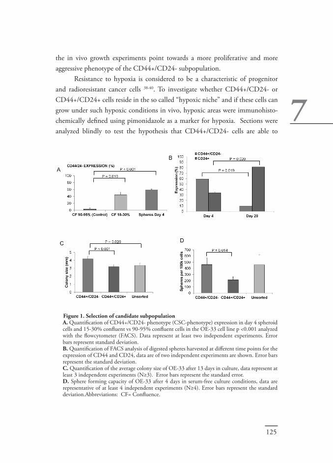
125
7
Figure 1. Selection of candidate subpopulation A. Quantification of CD44+/CD24- phenotype (CSC-phenotype) expression in day 4 spheroid cells and 15-30% confluent vs 90-95% confluent cells in the OE-33 cell line p <0.001 analyzed with the flowcytometer (FACS). Data represent at least two independent experiments. Error bars represent standard deviation.B. Quantification of FACS analysis of digested spheres harvested at different time points for the expression of CD44 and CD24, data are of two independent experiments are shown. Error bars represent the standard deviation.C. Quantification of the average colony size of OE-33 after 13 days in culture, data represent at least 3 independent experiments (N≥3). Error bars represent the standard error.D. Sphere forming capacity of OE-33 after 4 days in serum-free culture conditions, data are representative of at least 4 independent experiments (N≥4). Error bars represent the standard deviation.Abbreviations: CF= Confluence.
the in vivo growth experiments point towards a more proliferative and more aggressive phenotype of the CD44+/CD24- subpopulation.
Resistance to hypoxia is considered to be a characteristic of progenitor and radioresistant cancer cells 38-40. To investigate whether CD44+/CD24- or CD44+/CD24+ cells reside in the so called “hypoxic niche” and if these cells can grow under such hypoxic conditions in vivo, hypoxic areas were immunohisto-chemically defined using pimonidazole as a marker for hypoxia. Sections were analyzed blindly to test the hypothesis that CD44+/CD24- cells are able to

126
Figure 2. CD44+/CD24- phenotype displays aggressive tumor growth in vivoA. Growth curves of xenograft tumors generated in NOD/SCID mice of OE-33 cell-line after sorting for CD44+/CD24- and CD44+/CD24+ subpopulations for first generation tumors, data represent at least 3 independent experiments. Error bars represent the standard error.B. Growth curves of xenograft tumors generated in NOD/SCID mice of OE-21 cell-line after sorting for CD44+/CD24- and CD44+/CD24+ subpopulations for first generation tumors. One CD44+/CD24+ and one of the unsorted mice did not produce a tumor, therefore N=2 were compared with N=3 of the CD44+/CD24- tumors. Error bars represent the standard error.C. Quantification of FACS analysis of single cells obtained from the OE-21 xenograft tumors in all generations (1st-3rd). The CD44+/CD24- expression was analyzed for correlations with growth speed using the pearson correlation coefficient. D. Squamous cell carcinoma OE-21 cell-line generated by injecting the CD44+/CD24- subpopulation in NOD/SCID mice (left). With increasing generation number (from left to right); Second generation tumor generated by injecting single cells from a first generation tumor. A third generation tumor generated by injecting single from a second generation tumor.

127
7
reside in hypoxic areas within the tumor. A proportion of CD44+/CD24- cells were located near or in hypoxic areas, whereas CD44+/CD24+ cells were never observed in hypoxic areas in (Figure 3). These data might indicate that CD44+/CD24- cells, growing in vivo, are more resistant to hypoxia compared to CD44+/CD24+ cells.
To test whether the CD44+CD24- population may be used to predict the response to radiation, their sensitivity for radiation was compared to CD44+CD24+ and unsorted cells. To this end, an in vitro clonogenic assay was used. After a dose of 6Gy the OE-33 cell line showed a significantly higher survival for the CD44+/CD24- subpopulation compared to the CD44+/
Figure 3. CD44+/CD24- cells are present in hypoxic areas in vivoA. Pseudo-colored, composite image after multiple immunofloresent staining and scanning at 10x of an OE-21 tumor. Red = CD44 (left)/CD24 (right), Green = pimonidazole, Blue = Dap. Arrow denotes the CD44+/CD24- areas which are located in hypoxic regions (CD44+/CD24-/Pimonidazole+).B. Pseudo-colored, composite image after multiple immunofloresent staining and scanning at 10x of another OE-21 tumor. Red = CD44 (left)/CD24 (right), Green = pimonidazole, Blue = Dap. Arrow denotes the CD44+/CD24- areas, which are located in hypoxic regions (CD44+/CD24-/Pimonidazole+).

128
CD24+ (P=0.036) fraction and unsorted fraction (P=0.033) (Figure 4A and B). In the OE-21 cell line, also a significantly higher survival for the CD44+/CD24- subpopulation was observed when compared with the CD44+/CD24+ subpopulation (P=0.017) and unsorted fractions at 2Gy (Figure 4C) and 6Gy (P=0.020) (Figure 4C and D). These results indicate that cells with the CD44+/CD24- phenotype are more radioresistant than cells with the CD44+/CD24+ phenotype and unselected OE-21 / OE-33 cells.
The in vitro and in vivo experiments indicate that the CD44+/CD24-
Figure 4. CD44+/CD24- is more resistant to A. Clonogenic survival assay with corresponding doses-response curves of the OE-33 cell line sorted for the CD44+/CD24- and CD44+/CD24+ phenotypes, data represent 4 independent experiments (N≥4).B. Bar chart of clonogenic survival assay of figure 5A at 6Gy. Error bars represent the standard deviation.C. Clonogenic survival assay with corresponding doses-response curves of the OE-21 cell line sorted for the CD44+/CD24- and CD44+/CD24+ phenotypes, data represent 3 independent experiments (N≥3).D. Bar chart of clonogenic survival assay of figure 5C at 6Gy. Error bars represent the standard deviation. radiotherapy

129
7
phenotype may be an interesting marker combination for the prediction of response to radiotherapy. One important determinant for clinical use is that the phenotype can be identified in fresh tumor tissue 13, 28. Therefore, we prospectively analyzed freshly obtained tumor material from 8 different patients, suffering from carcinoma of the distal esophagus, for CD44 and CD24 expression (Figure 5A and S3). Although the profiles for CD44 and CD24 were very variable, in most cases a distinct CD44+/CD24- and CD44+/CD24+ subpopulation could be identified.
To test for a correlation between the CD44+/CD24- marker combination and the response to neoadjuvant chemoradiation in a clinical setting, pre-treatment biopsy specimens were investigated for CD44 and CD24 expression using immunohistochemistry. From a database in our institution 27 patients who
Figure 5.The CD44+/CD24- subpopulation can be prospectively identified in human material and is a predictor of response to neoadjuvant chemoradiation A. FACS plots of prospectively isolated patient material analyzed for CD44 and CD24 marker expression. B. Light microscope image of an esophageal adenocarcinoma pre-treatment biopsy serial section stained for CD24 and CD44, at 20x magnification. The arrows point to the cancer cells which are CD44+/CD24-. The patient had no response to the neoadjuvant chemoradiation.

130
received neoadjuvant chemoradiation were randomly selected. All 27 patients had non-diffuse intestinal type adenocarcinoma of the esophagus. Eighteen patients (n=18) were determined to have vital tumor (morphologically intact tumor cells) after pathological assessment of the surgical specimen, and 9 patients had no vital tumor tissue after the neoadjuvant chemoradiation. From the patients with vital tumor tissue, 9 out of 18 patients (50%) showed the presence of CD44+/CD24- cells in the pre-treatment biopsy specimen, whereas CD44+/CD24- cells were never found in patients without vital tumor (0/9: 0%) (Table 1, P=0.009). The 9 patients with CD44+/CD24- cells in their biopsy samples could not be further distinguished from the other 9 patients with vital tumor tissue, using regression grades. In figure 5B one of the CD44+/CD24- areas is visible in a pre-treatment biopsy specimen from a non-responder patient. In table 1, the results of the CD44 and CD24 staining and pathological characteristics of all 27 patients are shown. Thus, in this preliminary study, the presence of CD44+/CD24- cells in EC pre-treatment biopsy tissue indicates a lack of response to chemoradiation.
P-value
0.0530.0490.0090.6870.1361.0000.317
CD44+CD24+CD44+/CD24-cT-stage@ (T1 /T2 /T3 /T4)cN-stage& (N0 / N1)HistologyHistological grade (1/2/3/4)$
Table 1.Immunohistochemistry results for staining of esophageal cancer pre-treatment biopsy tissue in 27 patients.
72% (N=13)50% (N=9)50% (N=9)0%/ 6% / 88% / 6%39% / 61%Intestinal type AC 100%6% / 61% / 33% / 0%
Marker / clinicopathologic factor
After neoadjuvant chemoradiation
No vital tumor* (N=9)Vital tumor* (N=18)
33% (N=3)89% (N=8)0%0% / 0% / 88% / 12%12% / 88%Intestinal type AC 100%0% / 88% / 12% / 0%
*: Morphologically intact tumor cellsAC: adenocarcinoma.@: pre-treatment T-stage&: pre-treatment N-stage$: Pre-treatment histological grade according to the 4-tier system.

131
7
Discussion
This study proposes that cells with a CD44+/CD24- phenotype are more proliferation prone, grow more aggressively, reside in the radioresistant hypoxic niche and are a radioresistant subset in EC cell lines. In patient samples, a similar population was found that identified a group of patients with a lack of response to CRT. Therefore, the presence of CD44+/CD24- cells may predict for the (lack of response to (chemo) radiotherapy in esophageal carcinoma patients and can have a negative impact on the survival using current therapies. Identification of these new markers for esophageal cancer is essential, because these factors make it possible to identify patients that do not benefit from current therapies.
Previous clinical data shows a clear survival disadvantage for patients that had hardly any response to the neoadjuvant chemoradiotherapy, after pathologic evaluation of the resection specimen (according to standard pathologic guidelines)41, 42. This may be caused by a treatment resistant sub-population of tumor cells.
It has been suggested that each tumor contains cells with stem cell-like characteristics (cancer stem cells (CSC)), which are resistant to therapy and drive tumor regrowth 16-18. But the exact CSC or normal stem cell in EC and the esophagus remains elusive 25, 43. Research into the CD44 status has been performed previously by Takaishi et al. 18 on gastric cancer tissue, which has a close tumor-biological relation to esophageal cancer. In this study, it was shown that the CD44 positive subfraction selects for more chemoradiation resistant cells. Another study by Winder et al. 44 also demonstrated that CD44 positivity correlates with a reduced survival in gastric cancer patients. Furthermore, a recent study in laryngeal cancer patients showed that CD44 predicted for a higher chance on local recurrences (a clinical surrogate for response assessment) 45. Interestingly in line with our results, also in esophageal squamous cell carcinoma it was shown that, CD44 correlates with increased therapy resistance and aggressive tumor growth 46. Our study revealed that in EC cell lines and tumor biopsies the CD44 population can be subdivided into at least 2 subpopulations with different characteristics. The CD44+/CD24- sub-fraction of two EC cell lines displayed CSC-like characteristics and were found to be highly proliferative, formed more and bigger spheres, were less abundantly present in culture conditions that
132
induce differentiation and were more resistant to radiation when compared to CD44+/CD24+ or unselected cells. Indeed, the CD44+/CD24- subpopulation has previously been shown to select for CSCs and chemoradiation resistant cells, in breast cancer cell-lines and primary breast tumor tissue 11, 16. To translate our results into a clinical setting it was determined whether the CD44+/CD24- subpopulation was present in primary EC material. Although marker expression was rather heterogeneous, both the CD44+/CD24- and CD44+/CD24+ subpopulations could be identified in primary human EC. About half of the patients had a CD44+/CD24- expression above 40%, which does not indicate a rare primitive or stem cell like population. This suggests that additional markers may be required to have a more accurate estimation of CSCs or therapy resistant cells. One possible candidate for this is EPCAM, which from studies in colon cancer was shown to select for CSCs in combination with CD44 28. The biopsies in our study contained a CD44+/CD24-/EPCAM+ subpopulation of a much smaller size, which would be more in line with the amount of CSCs in other malignancies (Figure S5) 17, 26.
In a pilot study performed on a historical sample of esophageal cancer patients, the presence of CD44+/CD24- immunohistochemically determined was predictive for the response to chemoradiation. The expression of CD44 in our cell lines did not correlate with detected expression in our primary biopsy archival material. This can possibly be explained by the high selectiveness for aggressive cells within the investigated cell-lines. However, the presence in CD44+CD24- cells in patient samples, either or not in combination with EPCAM, could be a signature for CSC cells in EC. But more studies are needed to validate these results, like for instance limiting dilution tumor-initiating experiments, and validation studies on larger and/or prospective datasets. It seems obvious that this is an inherent limitation of investigations with cancer cell-lines and warrants caution for over-interpreting these results. This can be overcome by performing tumorigenicity experiments with primary human esophageal cancer tissue. Unfortunately, we were not able to grow tumors from isolated human cells. Others have also reported about the same problem25.
Detection of CSC markers in tumor biopsies could distinguish patients that will not respond to the current therapies. For these patients toxic treatments could be avoided providing a better quality of life, and warrant investigations into

133
7
alternative treatment options. In conclusion, the CD44+/CD24- subpopulation is present in primary EC
material and possibly predicts a reduced response to chemoradiation. CD44+/CD24- EC cells are more resistant to radiation in vitro. Furthermore, CD44+/CD24- cells exhibit some CSC-like characteristics such as increased growth in vivo and in vitro. These results warrant further investigation into the possible clinical benefit of CD44+/CD24- in EC patients as a predictive marker for the response to chemoradiation.
AcknowledgmentsNone
Conflict of InterestNone
Funding SourceUniversity of Groningen post-graduate School of Medicine GUIDE. The authors declare that the funding source was in no manner involved in this study.

134
RefeRences
1. Mariette C, Piessen G, Triboulet JP. Therapeutic strategies in oesophageal carcinoma: role of surgery and other modalities. Lancet Oncol 2007;8:545-53.2. Brown LM, Devesa SS, Chow WH. Incidence of adenocarcinoma of the esophagus among white Americans by sex, stage, and age. J Natl Cancer Inst 2008;100:1184-1187.3. Crane LM, Schaapveld M, Visser O, et al. Oesophageal cancer in The Netherlands: increasing incidence and mortality but improving survival. Eur J Cancer 2007;43:1445-51.4. Parkin DM, Bray F, Ferlay J, et al. Global cancer statistics, 2002. CA Cancer J Clin 2005;55:74-108.5. Omloo JM, Lagarde SM, Hulscher JB, et al. Extended transthoracic resection compared with limited transhiatal resection for adenocarcinoma of the mid/distal esophagus: five-year survival of a randomized clinical trial. Ann Surg 2007;246:992-1000; discussion 1000-1.6. Smit JK, Pultrum BB, van Dullemen HM, et al. Prognostic factors and patterns of recurrence in esophageal cancer assert arguments for extended two-field transthoracic esophagectomy. Am J Surg 2010;200:446-453.7. Gebski V, Burmeister B, Smithers BM, et al. Survival benefits from neoadjuvant chemoradiotherapy or chemotherapy in oesophageal carcinoma: a meta-analysis. Lancet Oncol 2007;8:226-234.8. van Hagen P, Hulshof MC, van Lanschot JJ, et al. Preoperative chemoradio-therapy for esophageal or junctional cancer. N Engl J Med 2012;366:2074-2084.9. Nguyen NP, Krafft SP, Vinh-Hung V, et al. Feasibility of tomotherapy to reduce normal lung and cardiac toxicity for distal esophageal cancer compared to three-dimensional radiotherapy. Radiother Oncol 2011;101:438-442.10. Lambin P, van Stiphout RG, Starmans MH, et al. Predicting outcomes in radiation oncology-multifactorial decision support systems. Nat Rev Clin Oncol 2012;10:27-40.11. Al-Hajj M, Wicha MS, Benito-Hernandez A, et al. Prospective identification of tumorigenic breast cancer cells. Proc Natl Acad Sci U S A 2003;100:3983-8.12. Zhu L, Gibson P, Currle DS, et al. Prominin 1 marks intestinal stem cells that are susceptible to neoplastic transformation. Nature 2009;457:603-7.13. O’Brien CA, Pollett A, Gallinger S, et al. A human colon cancer cell capable of initiating tumour growth in immunodeficient mice. Nature 2007;445:106-10.14. Vermeulen L, De Sousa E Melo F, van der Heijden M, et al. Wnt activity defines colon cancer stem cells and is regulated by the microenvironment. Nat Cell Biol 2010;12:468-476.15. Oonishi K, Cui X, Hirakawa H, et al. Different effects of carbon ion beams and X-rays on clonogenic survival and DNA repair in human pancreatic cancer stem-like cells. Radiother Oncol 2012;105:258-265.16. Phillips TM, McBride WH, Pajonk F. The response of CD24(-/low)/CD44+ breast cancer-initiating cells to radiation. J Natl Cancer Inst 2006;98:1777-85.

135
7
17. Todaro M, Alea MP, Di Stefano AB, et al. Colon cancer stem cells dictate tumor growth and resist cell death by production of interleukin-4. Cell Stem Cell 2007;1:389-402.18. Takaishi S, Okumura T, Tu S, et al. Identification of gastric cancer stem cells using the cell surface marker CD44. Stem Cells 2009;27:1006-20.19. Baumann M, Krause M, Hill R. Exploring the role of cancer stem cells in radioresistance. Nat Rev Cancer 2008;8:545-554.20. Begg AC. Predicting recurrence after radiotherapy in head and neck cancer. Semin Radiat Oncol 2012;22:108-118.21. de Jong MC, Pramana J, Knegjens JL, et al. HPV and high-risk gene expression profiles predict response to chemoradiotherapy in head and neck cancer, independent of clinical factors. Radiother Oncol 2010;95:365-370.22. Gurtner K, Hessel F, Eicheler W, et al. Combined treatment of the immu-noconjugate bivatuzumab mertansine and fractionated irradiation improves local tumour control in vivo. Radiother Oncol 2012;102:444-449.23. Gurtner K, Deuse Y, Butof R, et al. Diverse effects of combined radiotherapy and EGFR inhibition with antibodies or TK inhibitors on local tumour control and correlation with EGFR gene expression. Radiother Oncol 2011;99:323-330.24. Huang D, Gao Q, Guo L, et al. Isolation and identification of cancer stem-like cells in esophageal carcinoma cell lines. Stem Cells Dev 2009;18:465-73.25. Grotenhuis BA, Dinjens WN, Wijnhoven BP, et al. Barrett’s oesophageal adenocarcinoma encompasses tumour-initiating cells that do not express common cancer stem cell markers. J Pathol ;221:379-89.26. Ricci-Vitiani L, Lombardi DG, Pilozzi E, et al. Identification and expansion of human colon-cancer-initiating cells. Nature 2007;445:111-5.27. Yang ZF, Ho DW, Ng MN, et al. Significance of CD90+ cancer stem cells in human liver cancer. Cancer Cell 2008;13:153-66.28. Dalerba P, Dylla SJ, Park IK, et al. Phenotypic characterization of human colorectal cancer stem cells. Proc Natl Acad Sci U S A 2007;104:10158-63.29. Dontu G, Jackson KW, McNicholas E, et al. Role of Notch signaling in cell-fate determination of human mammary stem/progenitor cells. Breast Cancer Res 2004;6:R605-15.30. Liu S, Dontu G, Mantle ID, et al. Hedgehog signaling and Bmi-1 regulate self-renewal of normal and malignant human mammary stem cells. Cancer Res 2006;66:6063-71.31. Rockett JC, Larkin K, Darnton SJ, et al. Five newly established oesophageal carcinoma cell lines: phenotypic and immunological characterization. Br J Cancer 1997;75:258-263.32. Lombaert IM, Brunsting JF, Wierenga PK, et al. Rescue of salivary gland function after stem cell transplantation in irradiated glands. PLoS One 2008;3:e2063.33. Khramtsov AI, Khramtsova GF, Tretiakova M, et al. Wnt/beta-catenin pathway activation is enriched in basal-like breast cancers and predicts poor outcome. Am J Pathol ;176:2911-20.34. Rademakers SE, Lok J, van der Kogel AJ, et al. Metabolic markers in relation

136
to hypoxia; staining patterns and colocalization of pimonidazole, HIF-1alpha, CAIX, LDH-5, GLUT-1, MCT1 and MCT4. BMC Cancer 2011;11:167.35. Yeung TM, Gandhi SC, Wilding JL, et al. Cancer stem cells from colorectal cancer-derived cell lines. Proc Natl Acad Sci U S A ;107:3722-7.36. Deng S, Yang X, Lassus H, et al. Distinct expression levels and patterns of stem cell marker, aldehyde dehydrogenase isoform 1 (ALDH1), in human epithelial cancers. PLoS One 2010;5:e10277.37. Rice TW, Blackstone EH, Rusch VW. 7th edition of the AJCC Cancer Staging Manual: esophagus and esophagogastric junction. Ann Surg Oncol 2010;17:1721-1724.38. Schwartz DL, Bankson J, Bidaut L, et al. HIF-1-dependent stromal adaptation to ischemia mediates in vivo tumor radiation resistance. Mol Cancer Res 2011;9:259-270.39. McGee MC, Hamner JB, Williams RF, et al. Improved intratumoral oxygenation through vascular normalization increases glioma sensitivity to ionizing radiation. Int J Radiat Oncol Biol Phys 2010;76:1537-1545.40. Conley SJ, Gheordunescu E, Kakarala P, et al. Antiangiogenic agents increase breast cancer stem cells via the generation of tumor hypoxia. Proc Natl Acad Sci U S A 2012;109:2784-2789.41. Scheer RV, Fakiris AJ, Johnstone PA. Quantifying the benefit of a pathologic complete response after neoadjuvant chemoradiotherapy in the treatment of esophageal cancer. Int J Radiat Oncol Biol Phys 2011;80:996-1001.42. Berger AC, Farma J, Scott WJ, et al. Complete response to neoadjuvant chem-oradiotherapy in esophageal carcinoma is associated with significantly improved survival. J Clin Oncol 2005;23:4330-4337.43. Doupe DP, Alcolea MP, Roshan A, et al. A single progenitor population switches behavior to maintain and repair esophageal epithelium. Science 2012;337:1091-1093.44. Winder T, Ning Y, Yang D, et al. Germline polymorphisms in genes involved in the CD44 signaling pathway are associated with clinical outcome in localized gastric adenocarcinoma. Int J Cancer 2011;129:1096-1104.45. de Jong MC, Pramana J, van der Wal JE, et al. CD44 expression predicts local recurrence after radiotherapy in larynx cancer. Clin Cancer Res 2010;16:5329-5338.46. Zhao JS, Li WJ, Ge D, et al. Tumor initiating cells in esophageal squamous cell carcinomas express high levels of CD44. PLoS One 2011;6:e21419.

137
7
Supplementary information
Supplementary Methods
Cell culture The OE-33 cell-line derived from a poorly differentiated Barrets
adenocarcinoma of the lower esophagus and the OE-21 cell-line derived from a squamous cell carcinoma of the upper esophagus (both kind gifts from Dr. F.A. Kruyt, Department of Medical Oncology, University Medical Center Groningen) were cultured under standard conditions with RPMI 1640 growth medium supplemented with 10% Fetal Calf Serum, 1% Penicilline/Streptomycine in a humidified atmosphere and 5% CO2 at 37°C. Cells were passaged at 50-80% confluence1. Both cell-lines were independently DNA authenticated by the Leibniz Institute DSMZ-German Collection of Microorganisms and Cell Cultures (Braunschweig, Germany). The cells were not used for more than 12 passages after which they were discarded. Serum-free and low adherent growing conditions (ultra-low adherent plates, Corning Inc., Corning, New York, USA) were employed to grow the cells as spheroids using Neural Basal A medium containing N2, bFGF and FGF-2 as previously described by Vermeulen et al. 2. Depending on the experiment, the spheres were harvested and used for enzymatic digestion with Accutase (Sigma-Aldrich, St. Louis, Missouri, USA) to obtain a single cell suspension for use in cell biological experiments. In the sphere quantification experiments, cells were counted and plated in low-adherent plates (Corning Inc, Corning, New York, USA) directly after sorting for the different subpopulations. The concentration was 75.000-90.000 cells/mL. After 4-5 days the amount of spheres formed was evaluated by aspirating 3 samples of 100μL medium from each well and counting the amount of spheres. Each experiment was performed at least 4 times. To analyze spheres at different time points, OE-33 cells were grown adherently until they reached a confluence of 80-95% then the cells were trypsinized and replated in spheroid growing conditions described above. The medium was refreshed every 3-4 days. In some experiments, the spheres were harvested at various time points and analyzed with FACS for CD44 and CD24 marker expression.

138
Flow cytometrySingle cell suspensions obtained from tumor tissue or cell-lines were
resuspended in PBS with 0.2% bovine serum albumin (BSA). Concentrations of cells were adjusted if necessary. The cells were incubated for 30 minutes at 4°C. Flow cytometric analysis was performed on the FACS-Calibur (BD Biosciences, Franklin Lakes, New Jersey, USA) or LSR-II (BD Biosciences, Franklin Lakes, New Jersey, USA). Flow cytometric data were analyzed using Flojo version 7.6 software (Treestar Inc., Ashland, Oregon, USA). To isolate cells with a putative stem cell phenotype cell sorting was performed using a MoFlo-XDP or MoFLoAstrios cell sorter (Beckman Coulter previously DakoCytomation, Glostrup, Denmark). Each population was gated according to its isotype control. The 5-15% most extreme of the subpopulations were used.
In vitro radiation experiments, clonogenic assaysSorted single cell suspensions of the different subpopulations obtained
after flow-cytometric sorting were counted and plated immediately in standard growth medium (RPMI, see cell culture section). Cells were allowed to attach overnight and (sham) irradiated (Cesium 137, IBL) with 0, 2, 4 and 6 Gy at a dose rate of 0.65 Gy/min. After irradiation cells were trypsinized, replated and concentrations were adjusted according to the expected survival. Colonies were allowed to grow for 10-14 days, fixated and stained (coomassie brillant blue). Surviving fractions were determined by dividing the average number of colonies at different doses by the average number of colonies in the non-irradiated control.
Animal experimentsFemale NOD/SCID mice were purchased from Harlan laboratories
(NOD.CB17-Prkdcscid/NCrHsd). Animals were fed ad libitum, kept under sterile conditions in individually ventilated cages. To achieve in vivo tumor propagation 8-12 week old mice were subcutaneously injected with tumor cells in a 1:1 suspension with matrigel (BD Biosciences, Franklin Lakes, New Jersey, USA) under general anesthesia (isoflurane 2,5%). A total of 1.5x10⁵ OE-21 cells or 6.0x10⁵ OE-33 cells were injected for tumor generation. The injected volume never exceeded 100μL. Tumor growth was monitored twice weekly and measured with calipers. Tumor volume was determined with the formula

139
7
(π)/6 x (large diameter) x (small diameter) 2. When tumors reached a volume of about 900 mm3 or when ulcers appeared, mice were euthanized. Tumors were extracted under a-septic conditions and divided in different pieces depending on the experiments. A part of the tumor tissue was fixed with 5% paraformaldehyde for use in immunohistochemical experiments. Another part was digested into a single cell suspension for use in FACS experiments, in vitro sphere growth and/or serial transplantations. Single cells were obtained by using a modified from previously published method on salivary glands. Briefly, the tissue was washed and then minced into single cells with standard scissors, incubated with an enzyme mixture of hyaluronidinase (20 ug/mL) and collagenase type II (1.5 mg/mL) for 80 minutes. After washing the cells, DNASE (1 mg/mL) was added to reduce aggregates of cell clumps. The procedure for digestion of xenograft tumors was identical as for patient material. In selected cases, mice were injected with pimonidazole HCI 60mg/kg (Hydroxyprobe™-1, NPI, Inc., Burlington, Massachusetts, USA) as a marker forhypoxia. This was performed by intraperitoneal injection of 1000μL of the hypoxia marker 30 minutes before the animals were euthanized to allow perfusion in the tumor. Afterwards the tumor was extracted and frozen in liquid nitrogen and stored at -150 degrees Celsius. Xenograft tumors generated from transplanting CD44+/CD24- and CD44+/CD24+ subpopulations were disassociated into single cells and directly injected subcutaneously into the flanks of NOD/SCID mice (one injection per mice). The amount of cells in serial transplantation was similar as in the primary transplantation (1.5x10⁵ for the OE-21 xenograft tumors). It is important to note that between 1st and 2nd generation and 2nd and 3rd generation tumors, the cells were not resorted for either CD44+/CD24- or CD44/CD24+ subpopulations. At least three independent experiments were done.
All animal experiments were performed according to our institutional animal ethics guidelines and were reviewed by an animal ethics committee.
Human tissue samples experimentsHuman tissue biopsies were obtained from patients with confirmed
histological diagnosis of esophageal cancer during routine staging with gastrointes-tinal endo-echography (EUS) or from rest material after surgical resection of the tumor with informed consent. Tissue samples were immediately placed

140
in phosphate buffer with antibiotics and antimycotics. In the lab, the tissue was washed and incubated for at least 4 hours in RPMI with antibiotics and antimycotics and subsequently dissociated into single cells, as described above. The single cells obtained after this process were used for direct FACS analysis (Figure S3 for gating strategy).
All human tissue collection experiments were reviewed by the institutional human ethics commission (Institutional board review). The ethics guidelines comply with the Helsinki Declaration on experiments with humans.
Immunohistochemistry and immunofluoresence imagingImmunohistochemical staining was performed on 5 μm tissue sections
from archival patient material or tumor xenografts using primary antibodies against CD44 (Biolegend, San Diego, California, USA) and CD24 (Santa Cruz Biotechnologies Inc., Santa Cruz, California, USA). The procedure has been previously described 4. In short, the tissue sections were deparaffinized and subsequently immersed in PBS 2% hydrogen peroxidase to block endogenous peroxidase activity. Antigen-retrieval was performed and the sections were incubated overnight at 4 °C with the primary antibodies, both at 1:100 dilution. Tissue sections were then incubated with biotinylated secondary antibodies at 1:300 dilutions. The ABC complex was formed using a Vectashield ABC kit. This complex was visualized with SIGMA FAST 3,3’-Diaminobenzidine tablets (Sigma-Aldrich, St. Louis, Missouri, USA). In the final step, sections were counterstained with haematoxylin. For detection of pimonidazole (hypoxic cells) expression in frozen sections after fixation, sections were incubated with rabbit-anti-pimo antibody (J.A. Raleigh, Department of Radiation Oncology and Toxicology, University of North Carolina, Chapel Hill, North Carolina, USA) diluted 1:1000 in primary antibody diluent (PAD, Abcam, Cambridge, UK) for 30 minutes at 37°C. The second incubation step was with donkey-anti-rabbit Alexa488 (Molecular Probes, Leiden, The Netherlands) diluted 1:600 in PBS. Analyses of the stained sections were performed with standard light microscopy (Leica Microsystems GmbH, Wetzlar, Germany) or fluorescence microscopy (Axioskop, Zeiss, Gottingen, Germany) and a computer-controlled motorized stepping stage using IPlab software (Scanalytics Inc., Fairfax, Virginia,

141
7
USA) as described previously 5. Leica Acquiring Software (Leica Microsystems GmbH, Wetzlar, Germany) version 4.2 was used for image processing of the light microscopy images.
Statistical analysisExperiments are representative of at least 3 experiments unless otherwise
stated. All data are presented as mean and ± SD/SEM. Groups were compared with the student’s t test. Correlations were determined with the Pearson’s bi-variant comparison. Statistical analysis was performed in SPSS version 18 (IBM Inc, Armonk, New York, USA.).
94.8 ± 2.5 3.6 ± 2.3 99.8 ± 0.1
Supplementary table 1 (STable 1): Table displaying the expression of different CSC markers analyzed with the flowcytometer on esophageal cancer cell lines, which were grown adherently (2D-culture) to 90-95% confluence. The CD44+/CD24- and CD44+/CD24+ phenotypes are present in these cell lines. Mean and ± standard deviation of 3 independent experiments are shown
65.2 ± 1.3 35.1 ± 1.399.2 ± 0.2
Marker expression (%)
CD44+/CD24+ CD44+/CD24- EPCAM
OE-21 cell-line OE-33 cell-line

142
Supplementary figure 1 (S1): A. Representative FACS-plots from left to right of OE-33 cells after culture in standard adherent conditions with serum at a confluence of 90-95%, at a confluence of 15-30% and OE-33 spheres after 4 days in serum-free culture. Note: Gating was consistently performed with isotype controls explaining the difference in gate sizes.B. Light microscopy image of OE-33 spheres after 4-5 days in culture at 5X and 40X.C. Images of representative 6-well plates of colony sizes of OE-33 after sorting for CD44+/CD24- and CD44+/CD24+ 13 days in culture, representative example of data of 4 independent experiments.

143
7
Supplementary Figure 2 (S2): Representative FACS plots of marker expression shown in the table from figure 1. The OE-33 and OE-21 cell-lines were stained with CD44-PE, CD24-FITC, EPCAM-AlexaFluor-647, CD90-FITC, CD133-PE and CD29-PE.

144
Supplementary figure 3 (S3): Gating strategy for single cell suspensions obtained from xenograft tumor and patient material. In this example the gating strategy for patient 1 is shown. The cells were stained with propidium iodide (PI), CD44-PE (BD), CD24-FITC (BD) and isotype controls. A: The live cell populations are gated according to forward scatter and PI positive cells. B: Next PE and FITC positive gates were determined first using with the negative stained sample.C: Then the PE and FITC positive gates were confirmed first using the isotype controls. D: The stained sample with CD44-PE and CD24-FITC positivity according to gates deter-mined with the controls.

145
7
Suplementary figure 4 (S4): Quantification of FACS analysis of single cells obtained from the OE-21 xenograft tumors in all generations (1st-3rd). The CD44+/CD24+ expression was analyzed for correlations with growth speed using the pearson correlation coefficient.
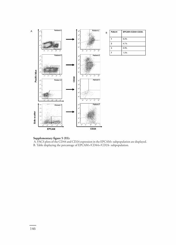
146
Supplementary figure 5 (S5): A. FACS plots of the CD44 and CD24 expression in the EPCAM+ subpopulation are displayed.B. Table displaying the percantage of EPCAM+/CD44+/CD24- subpopulation.


Neoadjuvant therapy reduces the incidence of nodal micrometastases in esophageal adenocarcinoma
Chapter 8
D. Wang, * J.K. Smit, * E. Zwaan, C.T. Muijs, H. Groen, H. Hollema , J.Th.M. Plukker
*Both authors contributed equally to this work
Under revision American Journal of Surgery 2013
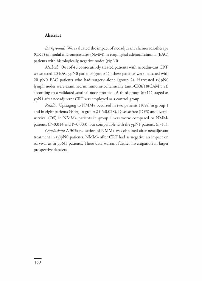
150
Abstract
Background: We evaluated the impact of neoadjuvant chemoradiotherapy (CRT) on nodal micrometastases (NMM) in esophageal adenocarcinoma (EAC) patients with histologically negative nodes (y)pN0.
Methods: Out of 48 consecutively treated patients with neoadjuvant CRT, we selected 20 EAC ypN0 patients (group 1). These patients were matched with 20 pN0 EAC patients who had surgery alone (group 2). Harvested (y)pN0 lymph nodes were examined immunohistochemically (anti-CK8/18(CAM 5.2)) according to a validated sentinel node protocol. A third group (n=11) staged as ypN1 after neoadjuvant CRT was employed as a control group.
Results: Upstaging to NMM+ occurred in two patients (10%) in group 1 and in eight patients (40%) in group 2 (P=0.028). Disease free (DFS) and overall survival (OS) in NMM+ patients in group 1 was worse compared to NMM- patients (P=0.014 and P=0.003), but comparable with the ypN1 patients (n=11).
Conclusions: A 30% reduction of NMM+ was obtained after neoadjuvant treatment in (y)pN0 patients. NMM+ after CRT had as negative an impact on survival as in ypN1 patients. These data warrant further investigation in larger prospective datasets.

151
8
Introduction
Despite recent advances in cancer treatment, patients with esophageal cancer (EC) still have a relatively poor prognosis. More than 80% of the EC patients present with a locally advanced tumor and nodal involvement or metastatic disease at the time of diagnosis. It is known that histologically proven nodal involvement (pN1), total number of resected, number of tumor positive nodes and lymph node ratio (number of involved/number of examined nodes) are independent prognostic factors for overall (OS) and disease free survival (DFS) 1-4. The importance of an adequate nodal resection is subjected to the strong prognostic value of the number of resected nodes on the outcome 3. As demonstrated in some studies an extended two-field nodal resection should be recommended, usually through a transthoracic (TT) route 5, 6. However, even patients with histological node negative (pN0) status will develop (early) locoregional recurrences which can be explained by the presence of nodal micrometastases (NMM) 7, 8. These NMM are not detected by routine hematoxylin and eosin (H&E) methods, but usually immunohistochemically with antibodies against cytokeratins specific for epithelial tissue 8-10. Patients with pN0 tumors, but with NMM, have a significantly worse survival rate than those without NMM. These NMM can be divided into isolated tumor cells (ITC) and micrometastases (MM) 10. MM have a worse effect on survival than ITC 7, 8, 10, 11.
Neoadjuvant treatment is currently the standard of care in the experienced centers 12-16. The rationale behind this is that tumor downstaging/sizing and elimination of NMM leads to improved resectability and curability rates 12,13,17. The improvement on survival rates after chemoradiotherapy (CRT) in a neoadjuvant setting is considerable, between 10-15% 12, 13. Previous research has shown that response to neoadjuvant CRT reduces NMM in esophageal cancer 17. But these studies are scarce and little has been published regarding the rate and the effect of neoadjuvant CRT on NMM in pN0 esophageal cancer patients. Therefore we evaluated the effect of CRT on NMM in ypN0 esophageal cancer patients after neoadjuvant treatment compared to pN0 in the surgery alone group.

152
Patients and methods
PatientsA total of 380 patients with histological proven esophageal cancer were
identified in the prospective database of our tertiary referral medical center. All patients had a surgical resection with a curative intent. From 2005 onwards, patients were treated with neoadjuvant treatment in a RCT trial or as standard procedure. The preoperaitive diagnostic workup, the surgical procedure and/or surgical team and follow-up did not change during the study period. In total 332 patients received surgery alone and 48 received neoadjuvant CRT followed by surgical resection. According to national guidelines no ethics board review was required for the present study (www.ccmo.nl). Archival tissue was handled according to the Dutch Code for proper use of Human Tissue (www.federa.org).
Matching and construction of surgery alone and ypN1 groupsFrom our prospective database one group of 20 consecutive adenocarcinoma
(AC) patients who received neoadjuvant CRT and staged histologically as ypN0 were selected (group 1). This group was matched on cT-stage or best-case lower cT-stage match with 20 AC patients with pN0 who were treated with surgery alone (group 2). Age was not a matching criterion. Consequently patients from group 1 were only treated in the period after 2005 and patients from group 2 during both periods. Additionally a third group which consisted of all 11 AC patients treated with neoadjuvant CRT but classified as ypN1 after routine pathologic evaluation, was used as a control group for the NMM+ patients in group 1 in the analyses. For the analyses we could therefore include 51 patients in this study.
Staging procedureThe diagnostic staging procedure consisted of endoscopic ultrasonography
(EUS) with fine needle aspiration (FNA) of suspected lymph nodes, 16-64 multidetector Computed Tomography (md-CT) scans of the neck, chest and abdomen, and cervical echographic examination. In case of T2-T4a tumors, or involved regional lymph nodes (N+), 18-F-fluorodeoxyglucose positron emission tomography (18-F-FDG-PET) was performed to exclude distant disease 18.

153
8
After staging in accordance with the Union for International Cancer Control TNM 7th edition, all patients were discussed by a multidisciplinary tumor board for an adequate treatment planning 19, 20.
Pre-operative treatmentThe neoadjuvant chemoradiotherapy regimen consisted of radiotherapy
with a total dose of 41.4-45 Gy in daily fractions of 1.8 Gy, five times per week (n=30). Patients received concurrent chemotherapy, which consisted of 5 weekly courses of paclitaxel (50 mg/m2) and carboplatin (area under the curve= 2). One patient received a neo-adjuvant chemotherapy scheme consisting of 3 courses of epirubicin, cisplatin, capecitabine (ECC).
Surgical procedureThe patients were operated by two experienced surgeons at our center. All
patients underwent a standard radical resection through a transthoracic approach en-bloc with an extended 2-field nodal dissection, as described in detail in a previous study of our group 21. These nodes were located in the mediastinum and the abdomen, including the nodes at the celiac trunk, along the common hepatic artery and a.lienalis at the upper border of the pancreas and the proximal para-aortic regional nodes.
Lymph node examinationAll identified lymph nodes, which were obtained from the surgical
specimen by the standard pathology procedures, were embedded in paraffin blocks and evaluated microscopically by routine H&E staining. For the purpose of this study, all lymph nodes in group 1 and group 2 were reconfirmed as pN0 by an experienced pathologist (HH).
Reassessment of lymph nodesReassessment of lymph nodes was performed according to a sentinel
lymph node sectioning protocol 22. Each lymph node was sectioned at four different levels at a distance of 100μm. After H&E staining was performed and showed to be negative, immunohistochemical (IHC) staining was carried out using anti-CK8/18 (CAM 5.2) to detect NMM. CAM 5.2 is a monoclonal

154
IgG2 antibody reacting against keratins 8/18 present in most adenocarcinomas23. NMM with deposits ≤ 0.2 mm and > 0.2 mm to ≤ 2 mm were considered as ITC and MM, respectively. The slides were blindly evaluated independently by two researchers. In case of disagreement a third judgment by an experienced pathologist was decisive.
Pathologic response assessmentPathologic response was classified according to the 5-tier, so called
Mandard criteria and divided into three subcategories: complete response ([CR], Mandard 1), partial response ([PR], Mandard 2-3) and hardly any response or non-response ([NR], Mandard 4-5) 24.
Follow-upPatients were seen for regular follow up according to national guidelines
at 4 to 8 weeks after completion of treatment, every 3 months in the first year, every 4-6 months in the second and third year and annually up to 5 years or until death. This follow-up regimen remained unchanged during the study period. Further radiological investigations were performed based on clinical suspicion of recurrent disease. A recurrence site was defined as local (esophageal bed), regional (lymph nodes) or distant metastases.
StatisticsOS was defined as the time interval between the starting date of the
neoadjuvant chemoradiotherapy or surgery and documentation of the day of death or last follow-up. DFS, locoregional recurrence-free survival (LRFS) and distant recurrence-free survival (DRFS) were determined from the starting date of treatment to documented date of first recurrence, last follow-up or death of any cause
Categorical data were assessed using Pearson’s Chi Square test. Continuous data Mann-Whitney U test. The DFS and OS were calculated according to the Kaplan-Meier method and compared with the Log-Rank test. P-values <0.05 were considered as statistically significant. All data were collected and analyzed using Statistical Package for Social Sciences (SPSS) version 18.0 (Chicago, IL, USA).
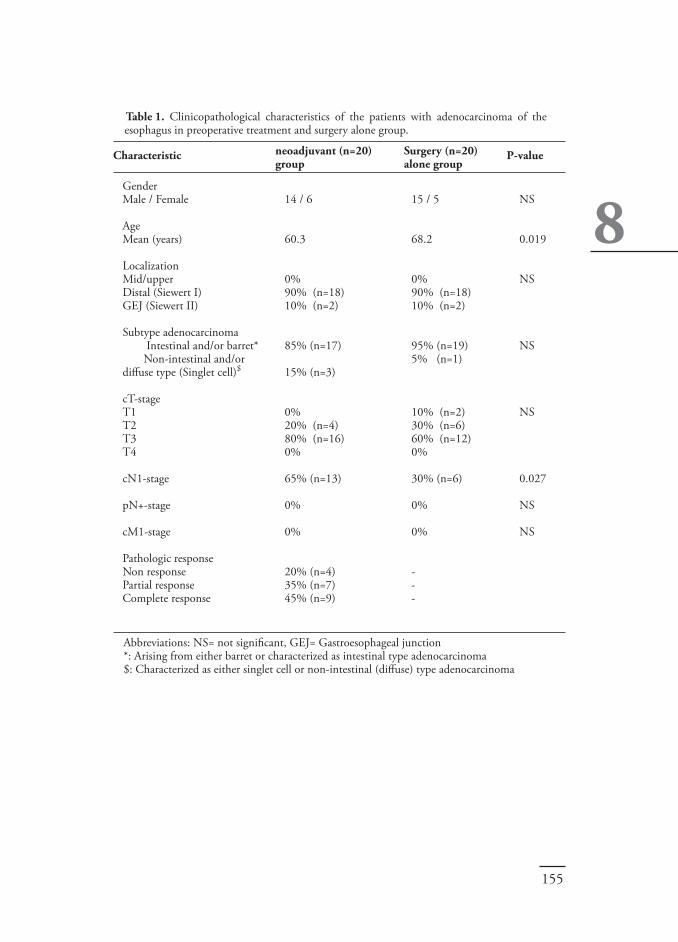
155
8 P-value
NS
0.019
NS
NS
NS
0.027
NS
NS
Gender Male / Female
Age Mean (years)
Localization Mid/upper Distal (Siewert I)GEJ (Siewert II)
Subtype adenocarcinoma Intestinal and/or barret* Non-intestinal and/or diffuse type (Singlet cell)$
cT-stageT1T2T3T4
cN1-stage
pN+-stage
cM1-stage
Pathologic response Non responsePartial responseComplete response
Table 1. Clinicopathological characteristics of the patients with adenocarcinoma of the esophagus in preoperative treatment and surgery alone group.
14 / 6
60.3
0%90% (n=18)10% (n=2)
85% (n=17)
15% (n=3)
0%20% (n=4)80% (n=16)0%
65% (n=13)
0%
0%
20% (n=4)35% (n=7)45% (n=9)
Characteristic Surgery (n=20)alone group
neoadjuvant (n=20)group
15 / 5
68.2
0%90% (n=18)10% (n=2)
95% (n=19)5% (n=1)
10% (n=2)30% (n=6)60% (n=12)0%
30% (n=6)
0%
0%
---
Abbreviations: NS= not significant, GEJ= Gastroesophageal junction*: Arising from either barret or characterized as intestinal type adenocarcinoma$: Characterized as either singlet cell or non-intestinal (diffuse) type adenocarcinoma

156
Results
Patient characteristics between the two groupsPatient characteristics were equally distributed, except for age for which
the groups were not matched (table 1). Patients in the neoadjuvant group (group 1) were significantly younger than those treated with surgery alone (group 2: P=0.019). The tumors were mainly located in the distal part of the esophagus (90% in both groups 1 and 2) and mainly staged as cT3 (80% and 60%, respectively) at time of surgery. None of the patients had distant metastases at the start of their treatment and microscopic radicality (R0) was achieved in all patients. The clinicopathological characteristics of the ypN1 group are displayed in table 2. The post-operative mortality was 0% in all three groups (group 1, group 2 and ypN1).
Gender Male / Female
Age Mean (years)
Localization Mid/upper Distal GEJ
cT-stageT1T2T3T4
cN1-stage
Pathologic response Non responsePartial responseComplete response
Table 2. Clinicopathological characteristics of patients in the control group of ypN1 patients. Abbreviations: GEJ= Gastroesophageal junction
10 / 1
63 (38-74)
0%82% (n=9)18% (n=2)
0%9% (n=1)73% (n=8)18% (n=2)
64% (n=7)
46% (n=5)54% (n=6)0%
Characteristic ypN1 (n=11)

157
8
Response rateIn the neoadjuvant group 9 patients (9/20; 45%) had a CR, 35% (n=7) a PR and 20% (n=4) a NR.
Reduction of nodal micrometastases after neoadjuvant treatmentAll resected lymph nodes were histologically evaluated. The total number
of evaluated lymph nodes was 533; 251 in group 2 with surgery alone and 282 in group 1 with neo-adjuvant treatment. The median number of resected nodes in group 2 was 13 (5-20) versus 15 (4-20) (P=0.527) in group 1. The median resected lymph nodes in the ypN1 group (n=11) was 12 (5-30), which was comparable with groups 1 and 2 (P=0.632)
Consequently, more patients in the surgery alone group were upstaged due to positive NMM as compared to the neoadjuvant group; 40% (8 out of 20 patients) versus 10% (2 out of 20 patients), respectively (P=0.028, figure 1).
Figure 1. Lower incidence of nodal micrometastasis (NMM) in (y)pN0 patients after neoadjuvant treatment (CRT group; n=20) compared to pN0 in surgery alone (n=20) group.The number of patients with NMM was significantly lower in the CRT group (n=2) compared to the surgery alone group (n=8). P-value = 0.028.

158
There was a trend (P=0.050) towards less NMM+ positive nodes in the neoadjuvant group (3 out of 282; 1%) compared with the surgery alone group (9 out of 251; 3.6%).
Interestingly, when the data were analyzed more carefully (table 1), the reduction of NMM+ in the neoadjuvant group is even greater since this group contains more cN1 positive (pre-treatment nodal staging) tumors compared to the surgery alone group (65% versus 30%, P=0.027)
Localization of positive NMM in the neoadjuvant groupAll three NMM positive nodes in the two upstaged patients were located in
Figure 2. Survival in NMM+ patients (n=2) compared to NMM- patients (n=18) in the neoadjuvant group. Survival of the NMM+ patients compare to ypN1 patients (n=11). a. Disease-free survival (DFS) was significantly worse in NMM+ patients compared to
NMM- patients (P=0.013).b. Overall survival (OS) was significantly more poor in NMM+ patients compared to
NMM- patients (P=0.001).c. Disease-free survival (DFS) was significantly worse in NMM+ patients and ypN1
compared to NMM negative patients (P=0.014).d. Overall survival (OS) was significantly worse in NMM+ patients and ypN1 compared
to NMM negative patients (P=0.003).
159
8
the radiation planning-field (para-esophageal region). Interestingly, both patients responded well to neoadjuvant treatment with a classification of pathologic CR (Mandard 1) and pathologic PR respectively.
Effect of nodal micrometastases on prognosisIn the neoadjuvant group, the 2 NMM positive patients clearly showed
a worse median DFS (figure 2a) compared to the 18 NMM negative patients (P=0.013), with 8 months in NMM+ patients and not yet reached in NMM- patients.
The median OS was also significantly lower in NMM+ patients compared to NMM- patients (P=0.001), with 14 months in NMM+ patients and not yet reached in NMM- patients (figure 2b). Furthermore, we analyzed the survival in a group of ypN1 patients (n=11) as a control because the relatively small number of NMM+ patients (n=2). As shown in figure 2c and figure 2d, the median DFS and OS of the ypN1 patients were comparable with the NMM+ patients, with 13 months and 24 months respectively (P=0.014 and P=0.003). The DFS and OS did not differ between the neodjuvant and surgery group (P=0.269 and P=0.388). NMM positivity did not have an effect on the OS (P=0.812) in the surgery alone group, but did show a difference towards worse DFS for NMM+, which did also not reach statistical significance (P=0.160).
Discussion
Currently, neoadjuvant chemoradiotherapy followed by surgical resection is considered as standard of care for resectable tumors of the esophagus in stage II-III patients 12-16. This is strengthened by the favorable results for the CRT-arm in a recent Dutch RCT (CROSS-study) 25. It is postulated that the favorable results for CRT in the neoadjuvant setting, are achieved by tumor downstaging and downsizing, resulting in higher microscopic radical resection rates (R0 resections) 25.
Another postulated effect of pre-operative treatment is a reduction of micrometastatic disease explaining the smaller burden of disease observed during follow up of these patients 13-16. Even though there is evidence showing reduction
160
in micrometastatic disease after pre-operative CRT in patients who achieved a complete response to the CRT, no data have been published about the exact reduction of micrometastatic disease in esophageal cancer patients 17. Therefore, the present study adds important information to the effect of neoadjuvant treatment on the prevalence and clinical relevance of NMM. Moreover, in the neoadjuvant group we observed a reduction in upstaging (from ypN0 to ypNMM+) of 30% compared to the surgery alone group. In absolute numbers we also observed a trend towards a reduction in NMM+ lymph nodes in the neoadjuvant group compared to the surgery alone group. This reduction was even more apparent when considering the fact that in the pre-treatment phase patients in the neoadjuvant group had significantly more cN1 tumors (65%) compared to surgery alone patients. There are two aspects, which could potentially have an impact on clinical decision-making. One is that even after responses to neoadjuvant treatment an extended nodal dissection should not be omitted, based on the presence of NMM even in these patients. Moreover the two patients (n=2) in the neoadjuvant group with NMM+ were both responders to the pre-operative CRT. Indeed one of these patients had a complete response (Mandard 1). As the NMM positive lymph nodes were located in the para-esophageal region and therefore within the irradiation field. Secondly the information of this study may have an impact on future adjuvant trials for a more appropriate stratification based on NMM disease. Even after routine pathology evaluations, we should be aware of the presence of NMM and the potential impact on outcome. Excluding NMM at least immunohistochemically may increase the rigor of determining cases either true node positive or node negative. Our results should also encourage us and other study groups to validate future data in prospective analyses regarding its true clinical relevance.
Although the Kaplan-Meier survival estimation of the analyzed neoadjuvant group is relatively small (n=20), the two NMM+ patients had a significantly worse DFS and OS compared to the NMM negative (n=18) patients. Furthermore, we performed an analysis on a control group of 11 ypN1 patients, and found that the DFS and OS were comparable to NMM+ patients. This is in line with previous studies that showed a worse prognosis when NMMs were present 7-10. Additionally, even though the surgery alone group also represents a relatively small sample size of 20 pN0 patients, the upstaging expressed by the

161
8
rate of 40% in NMM positive patients was in line with a previous study at our institute by Heeren et al. 8 consisting of 60 pN0 patients, where the upstaging rate was 30%. Heeren et al. used exactly the same antibody (anti- CAM 5.2, DAKO, Carpinteria, CA, USA) in their study as described in the present study.
A limitation of the current study is the relatively small size of 51 patients, which may induce a form of sample bias, which possibly reduces the impact of the present study. To reduce this type of bias, we carefully matched the neoadjuvant group (n=20) with a control group of 20 patients (surgery alone). All patients had adenocarcinoma of the distal/GEJ esophagus and were (y)pN0 after routine pathologic evaluation. We also used the cT status in the matching procedure as it is known that there is a strong correlation between increasing depth of primary tumor invasion and the presence of nodal disease, even in submucosal disease 2,
6, 21, 26, 27. Furthermore we included a control group of 11 ypN1 patients in the survival analyses in the neoadjuvant group according to NMM status.
Response to neoadjuvant treatment, specifically pCR, strongly predicts an increased survival and is one of the key criteria to evaluate the success of the given treatment 28-30. In the present study, the given pre-operative treatment regimens provided good responses, with 35% (n=7) PR rate and 45% (n=8) CR rate.
In conclusion, a 30% reduction of NMM positivity was obtained after neoadjuvant treatment in ypN0 patients. NMM+ after CRT had an equal negative impact on DFS and OS as in ypN1 patients. Based on the presence of NMM (10%) after neoadjuvant CRT, within the irradiation field, we still advocate a standard nodal dissection even in patients with good responses. Furthermore the data from this study warrants caution when considering patients ypN0 after routine pathological examination with H&E staining and the data should also be reconfirmed preferably in a larger prospective datasets.

162
Conflict of interest statementNone
AcknowledgmentsSupported by: GUIDE post-graduate school of Medicine, University of Groningen. The funding source had no role in this study. We would like to thank Peter W.K. Nagle MSc for correcting English grammar.

163
8
RefeRences
1. Hsu WH, Hsu PK, Hsieh CC, et al. The metastatic lymph node number and ratio are independent prognostic factors in esophageal cancer. J Gastrointest Surg 2009;13:1913-1920.2. Mariette C, Piessen G, Briez N, et al. The number of metastatic lymph nodes and the ratio between metastatic and examined lymph nodes are independent prognostic factors in esophageal cancer regardless of neoadjuvant chemoradiation or lymphadenectomy extent. Ann Surg 2008;247:365-371.3. Altorki NK, Zhou XK, Stiles B, et al. Total number of resected lymph nodes predicts survival in esophageal cancer. Ann Surg 2008;248:221-226.4. Smit JK, Pultrum BB, van Dullemen HM, et al. Prognostic factors and patterns of recurrence in esophageal cancer assert arguments for extended two-field transthoracic esophagectomy. Am J Surg 2010;200:446-453.5. Hagen JA, DeMeester SR, Peters JH, et al. Curative resection for esophageal adenocarcinoma: analysis of 100 en bloc esophagectomies. Ann Surg 2001;234:520-30; discussion 530-1.6. Lerut T, Nafteux P, Moons J, et al. Three-field lymphadenectomy for carcinoma of the esophagus and gastroesophageal junction in 174 R0 resections: impact on staging, disease-free survival, and outcome: a plea for adaptation of TNM classification in upper-half esophageal carcinoma. Ann Surg 2004;240:962-72; discussion 972-4.7. Zingg U, Montani M, Busch M, et al. Prognostic influence of immunohis-tochemically detected lymph node micrometastasis and histological subtype in pN0 oesophageal cancer. Eur J Surg Oncol 2009;35:593-599.8. Heeren PA, Kelder W, Blondeel I, et al. Prognostic value of nodal micrometastases in patients with cancer of the gastro-oesophageal junction. Eur J Surg Oncol 2005;31:270-276.9. Prenzel KL, Holscher AH, Drebber U, et al. Prognostic impact of nodal micrometastasis in early esophageal cancer. Eur J Surg Oncol 2012;38:314-318.10. Thompson SK, Ruszkiewicz AR, Jamieson GG, et al. Isolated tumor cells in esophageal cancer: implications for the surgeon and the pathologist. Ann Surg 2010;252:299-306.11. de Boer M, van Deurzen CH, van Dijck JA, et al. Micrometastases or isolated tumor cells and the outcome of breast cancer. N Engl J Med 2009;361:653-663.12. Gebski V, Burmeister B, Smithers BM, et al. Survival benefits from neoadjuvant chemoradiotherapy or chemotherapy in oesophageal carcinoma: a meta-analysis. Lancet Oncol 2007;8:226-34.13. Sjoquist KM, Burmeister BH, Smithers BM, et al. Survival after neoadjuvant chemotherapy or chemoradiotherapy for resectable oesophageal carcinoma: an updated meta-analysis. Lancet Oncol 2011;12:681-692.14. Allum WH, Stenning SP, Bancewicz J, et al. Long-term results of a randomized trial of surgery with or without preoperative chemotherapy in esophageal cancer. J Clin Oncol 2009;27:5062-5067.

164
15. Burmeister BH, Smithers BM, Gebski V, et al. Surgery alone versus chem-oradiotherapy followed by surgery for resectable cancer of the oesophagus: a randomised controlled phase III trial. Lancet Oncol 2005;6:659-668.16. Cunningham D, Allum WH, Stenning SP, et al. Perioperative chemotherapy versus surgery alone for resectable gastroesophageal cancer. N Engl J Med 2006;355:11-20.17. Prenzel KL, Konig A, Schneider PM, et al. Reduced incidence of nodal micrometastasis after major response to neoadjuvant chemoradiation in locally advanced esophageal cancer. Ann Surg Oncol 2007;14:954-959.18. Kole AC, Plukker JT, Nieweg OE, et al. Positron emission tomography for staging of oesophageal and gastroesophageal malignancy. Br J Cancer 1998;78:521-527.19. Rice TW, Blackstone EH, Rusch VW. 7th edition of the AJCC Cancer Staging Manual: esophagus and esophagogastric junction. Ann Surg Oncol 2010;17:1721-1724.20. Sobin LH. TNM, sixth edition: new developments in general concepts and rules. Semin Surg Oncol 2003;21:19-22.21. Smit JK, Pultrum BB, van Dullemen HM, et al. Prognostic factors and patterns of recurrence in esophageal cancer assert arguments for extended two-field transthoracic esophagectomy. Am J Surg ;200:446-53.22. Weaver DL. Pathology evaluation of sentinel lymph nodes in breast cancer: protocol recommendations and rationale. Mod Pathol 2010;23 Suppl 2:S26-32.23. Makin CA, Bobrow LG, Bodmer WF. Monoclonal antibody to cytokeratin for use in routine histopathology. J Clin Pathol 1984;37:975-983.24. Mandard AM, Dalibard F, Mandard JC, et al. Pathologic assessment of tumor regression after preoperative chemoradiotherapy of esophageal carcinoma. Clin-icopathologic correlations. Cancer 1994;73:2680-2686.25. van Hagen P, Hulshof MC, van Lanschot JJ, et al. Preoperative chemoradio-therapy for esophageal or junctional cancer. N Engl J Med 2012;366:2074-2084.26. Ancona E, Rampado S, Cassaro M, et al. Prediction of lymph node status in superficial esophageal carcinoma. Ann Surg Oncol 2008;15:3278-3288.27. Holscher AH, Bollschweiler E, Schroder W, et al. Prognostic impact of upper, middle, and lower third mucosal or submucosal infiltration in early esophageal cancer. Ann Surg 2011;254:802-7; discussion 807-8.28. Berger AC, Farma J, Scott WJ, et al. Complete response to neoadjuvant chem-oradiotherapy in esophageal carcinoma is associated with significantly improved survival. J Clin Oncol 2005;23:4330-4337.29. Donahue JM, Nichols FC, Li Z, et al. Complete pathologic response after neoadjuvant chemoradiotherapy for esophageal cancer is associated with enhanced survival. Ann Thorac Surg 2009;87:392-8; discussion 398-9.30. Schneider PM, Baldus SE, Metzger R, et al. Histomorphologic tumor regression and lymph node metastases determine prognosis following neoadjuvant radiochemotherapy for esophageal cancer: implications for response classification. Ann Surg 2005;242:684-692.


Hedgehog Signaling Pathway and Predic-tion of Recurrence after Neoadjuvant Chem-oradiotherapy in Esophageal Cancer
Chapter 9
J.K. Smit, J. Honing, D. Wang, B. van Etten, G.M. van Dam, F. Kruyt, H. Hollema, R.P. Coppes, J.Th.M. Plukker
Submitted for publication

168
Abstract
Purpose: We aimed to identify candidate chemoradiotherapy (CRT) response markers and prognostic factors for disease-free survival (DFS) after neoadjuvant CRT in esophageal cancer.
Methods: Immune-histochemical (IHC) expression of markers expected to be associated with poor response were investigated: Sonic Hedgehog (SHH), CD44, CD24, ERCC1, OCT4 and PARP1 in resected mRD (microscopic residual disease) specimens were compared with matched controls of surgery only specimens. For identification of prognostic factors Cox-regression analyses were performed.
Results: Between 2006 and 2011, a total of 83 consecutive patients were included. Average DFS was 39 months for mRD patients (n=61) and 76 months for pathologic complete response (pCR) patients (n=22). The estimated 3- and 5-year OS was both 75% for pCR patients and 42% and 27% for mRD patients, respectively.
Compared to the matched controls, a 64% enhanced IHC expression of SHH (P=0.003) and a 55% enhanced expression of CD44 (P=0.031) was observed in mRD tissue. SHH expression showed a positive correlation coefficient (CC) of 0.551 (P=0.027) with CD44 expression in mRD tissue. SHH expression was also positively correlated with ERCC1 (CC 0.512; P=0.043). Pathological lymph node (ypN) status was the only independent prognostic factor for DFS in mRD patients (n=61) : ypN1 (hazard ratio [HR] 4.0 95% CI 1.5-10.4), ypN2 (HR 6.4 95% CI 2.1-19.2) and ypN3 (HR 9.6 95% CI 2.3-39.6).
Conclusions: This study showed that SHH, CD44, and ERCC1 are associated with poor response to neoadjuvant. Furthermore it underlines the adverse impact of ypN+ status on DFS.
169
9
Introduction
Esophageal cancer is the 7th leading cause of cancer death with a worldwide incidence of nearly 500.000 patients 1-3. Currently it is one of the most rapidly increasing solid malignancies in Western countries 2,4-6. During the past decade, the use of neoadjuvant chemoradiotherapy (CRT) has been increasingly propagated to complement surgical resection 7-10. With a trimodality treatment, 5-year survival rates of 45% have been achieved in contrast with obtained rates of 35% after surgery alone in expert centers 10,11. Previous research has shown a pathological complete response (pCR i.e absence of vital tumor cells in the resection specimen) in only 25-30% of the patients who received neoadjuvant CRT, which greatly improves the survival 10,12-14. The microscopic presence of residual vital tumor cells, implicating lack of response, in the resected specimen after neoadjuvant CRT, is known to reduce patient survival.
Therefore, it is of utmost importance to find additional therapeutic ways by which response rates can be improved for patients with EC treated with neoadjuvant CRT.
Therapeutic failure resulting in early recurrence i.e. short disease-free survival time can be considered as a clinical substitute for lack of response to neoadjuvant CRT. Clinicopathologic prognostic factors for short disease-free survival (DFS) after neoadjuvant CRT in patients with microscopic residual disease (mRD) in the resected specimen could be of help in selecting patients for improving current treatment outcome. For instance, improved identification and selection of patients with short DFS could lead to extending the surgical resection, optimizing the radiation planning (including 2-field locoregional lympnodes) or adding selective post-operative radiotherapy therapy in order to obtain better treatment outcomes. Identification of novel biological markers in the resected mRD tissue could provide better means for patient selection and additional therapeutic options. This could lead to reducing or even eliminating mRD in the resected specimen (increasing response rates) after neoadjuvant CRT and therefore greatly improving patient survivals.
The Hedgehog (Hh) pathway is found to be inactive in most normal adult tissues, but reactivation of Hh has been found in the pathogenesis of several cancers 15,16. Studies have demonstrated an over-activation of the Hedgehog

170
signaling pathway in cancer cells of several gastro-intestinal tumors, including esophageal (EC) and gastric cancer 15,16. This is as a result of Sonic and Indian hedgehog ligand overexpression. Sonic hedgehog (SHH) ligands bind to the cell surface receptor Patched (PTCH), which leads to diminished inhibitory activity of PTCH on Smoothend (SMO). SMO is a transmembrane ligand, which in turn allows a further downstream intracellular activation of transcriptional factors, contributing to tumor growth 15,16. Other markers which have been implicated in relation to tumor response to irradiation are, CD44, CD24 & OTC4 (stem cell markers), ERCC1 (platinum resistance and and DNA repair enzyme) and PARP1 (DNA damage repair gene) 17-28. The aims of this study are therefore to analyse the expression of candidate CRT response markers, in the resected mRD tissue and to indentify prognostic factors for DFS in mRD patients.
Patients and methods
Primary study cohortIn the present study, we included EC patients who underwent a surgical
resection in our center during the period between 2006 and 2011. Only patients with histologically proven adenocarcinoma (AC) or squamous cell carcinoma (SCC) of the esophagus or gastro-esophageal junction (GEJ) after potentially curative resections were included. Furthermore all patients had to be treated with neoadjuvant CRT according to the CROSS regimen 10,29. Data was retrieved from our prospective research database and identified 83 patients who met these inclusion criteria. The follow-up was complete until December 2012.
The study was conducted according to the guidelines of our ethics hospital board (www.ccmo.nl). Archival tissue was handled according to the Dutch Code for proper use of Human Tissue (www.federa.org).
Staging procedureAfter the diagnosis of esophageal cancer, all patients underwent an
endoscopic ultrasonography with fine needle aspiration (EUS-FNA) of suspected locoregional lymph nodes. A 64 multi-slice CT-scan of the neck, chest and abdomen was performed to exclude distant metastases and to determine

171
9
resectability with curative intent. Furthermore, 18-F-fluorodeoxyglucose positron emission tomography (FDG-PET) was performed in all T2 -T4a tumors or in case of involved regional lymph nodes (N+) to optimize staging by excluding distant disease (M1-status). The Union for International Cancer Control TNM 7th edition was employed in all patients and in case the TNM 6th edition was initially used the staging information was converted to the 7th edition 30,31.
Chemoradiotherapy regimenDetails of the neoadjuvant CROSS regimen are described elsewhere 10,29.
After CT has excluded interval distant disease patients underwent a surgical resection with curative intent within 6 to 8- weeks after completing neoadjuvant treatment .
SurgeryThe same experienced surgical team at our center operated the patients
during the study period (J.T.M.P., G.M.v.D and B.v E.). After excluding distant metastases or local irresectability at laparotomy a curative intended transthoracic resection en-bloc with a 2-field nodal dissection in mediastinum and abdomen, including the celiac trunk nodes was performed.
Histopathological examinationThe resected specimen was examined according to a standard protocol.
Microscopic residual disease (mRD) was considered as the presence of vital invasive tumor (at least ypT1a) cells after neoadjuvant CRT in the primary tumor bed or locoregional lymph nodes (at least ypN1). Consequently ypT0N0 tumors were classified as pathologic complete response (pCR, Mandard regression grades1)32.
Immunohistochemical secondary study cohortThe immune-histochemical (IHC) staining cohort was constructed as
following: First we selected all patients in the mRD group which were classified as Mandard regression grades 4-5 (non-responders) and who had adequate tissue available (N=16) 32. These 16 patients were matched (1:2) with 32 untreated with neoadjuvant CRT controls. Matching was based on histology and depth of tumor

172
invasion (T-stage). The 32 matched patients were selected from a prospective research database of 450 patients with esophageal cancer treated by only surgical resection. Tissue microarrays (TMAs) were constructed as previously described33. After matching and construction of TMAs one patient from the untreated matched control group had no evaluable tissue and could not be included in the analysis.
Immunohistochemical procedure secondary study cohortImmunohistochemical staining was performed on 5 μm tissue sections
from archival patient material using primary antibodies against CD44 (1:100, Biolegend, San Diego, California, USA), CD24 (1:100, Santa Cruz Biotechnologies Inc., Santa Cruz, California, USA), SHH (1:100, ABCAM, Cambridge, United Kingdom), PARP1 (1:100, ABCAM, Cambridge, United Kingdom), ERCC1 (1:100, ABCAM, Cambridge, United Kingdom), OCT4 (1:250, ABCAM, Cambridge, United Kingdom). The tissue sections were de-paraffinized and subsequently immersed in PBS 2% hydrogen peroxidase to block endogenous peroxidase activity. Antigen-retrieval was performed and the sections were incubated overnight at 4 °C (SHH, OCT4, CD44, CD24 and ERCC1) or for 2 hours (PARP1) at room temperature with the primary antibodies. Tissue sections were then incubated with biotinylated secondary antibodies at 1:300 dilutions. The ABC complex was formed using a Vectashield ABC kit (Burlingame, California, USA). This complex was visualized with SIGMA FAST 3,3’-Diaminobenzidine tablets (Sigma-Aldrich, St. Louis, Missouri, USA). In the final step, sections were counterstained with haematoxylin.
Evaluation of immunohistochemistryImmunohistochemical staining was scored semi-quantitatively as: no
staining (0), weakly positive (1+), positive (2+) or intense (3+) by two independent observers (J.K.S and D.W.) without prior knowledge of the clinical outcome. Discordant cases were reviewed, and scores were reassigned on consensus of opinion. The cellular localization of the staining pattern (membranous, nuclear or cytoplasmatic) was only considered positive based on the expected performance and manufacturers information of the respective antibodies.

173
9
StatisticsSurvival was calculated according to the Kaplan-Meier method and
compared using the log-rank test. IHC results were compared with the Mann-Whitney U test. Correlations between IHC markers were determined with the Spearman’s rank correlation coefficient.
Univariate Cox-regression analysis was performed to indentify dependent prognostic factors. Factors with a P-value <0.1 were included in the stepwise (backwards conditional method) multivariate Cox-regression analysis for determination of independent prognostic factors for DFS.
A P-value <0.05 (corresponding to a 95% confidence interval [CI]) was considered as significant. The statistical analyses were performed by using the International Business Machines Statistical Package for Social Sciences (IBM SPSS, Armonk, New York, USA) version 20.0.
Figure 1. Comparison of the disease-free survival (fig 1a) and overall survival (fig 1b) between pathologic complete response (pCR, N=22) and microscopic residual disease (mRD, N=61) groups.

174
Results
Patient and clinicopathologic characteristics Table 1 describes the clinicopathologic characteristics of the study cohort
(N=83). In the IHC study cohort, the matched characteristics were as expected not
different between mRD and matched controls. AC/SCC distribution was 75% (N=12)/25% (N=4) versus 74% (N=23) / 26% (N=8) and T2/T3 distribution was 12% (N=2) /88% (N=14) versus 10% (N=3) /90% (N=28), in the mRD and matched controls groups (both P=1.000).
Disease-free and overall survival rates between pCR and mRD groupsTo assess whether the presence of mRD in the resected specimens would
lead to early recurrence (shorter disease-free survival [DFS]) in our study cohort, Kaplan-Meier survival estimations were calculated. As expected the DFS was significantly shorter in mRD patients compared to pCR patients (Log-rank P=0.004). Moreover, the estimated average DFS rate was 39 months (95% CI 28 – 50) for mRD patients and 76 months (95% CI 65 – 87) for pCR patients (figure 1a). The shorter DFS for mRD patients also translated into a significantly shorter overall survival (OS) rate for mRD patients compared with pCR patients (Log-rank P=0.041). Moreover, the estimated 3- and 5-year OS was both 75% for pCR patients and 42% and 27% for mRD patients, respectively (Figure 1b).
Relative enhanced IHC expression and correlations of biomarkers in mRD tissue To determine if the Hh pathway is implicated in mRD tissue, and therefore
possibly a lack of response to neoadjuvant CRT, the IHC expression of Sonic Hedgehog (SHH) in mRD tissue was compared to a matched control group of surgery only treated patients (figure 2a). Indeed, a 64% relative enhanced expression of SHH was determined in mRD tissue compared to matched controls (P=0.003, figure 2a). In figure 2b, representative samples of weakly positive (1+) and intense (3+) staining of SHH are displayed.

175
9 Sex
Age
Histology
Localization
Endoscopic tumor length
cT-stage
cN-stage
ypT-stage
ypN-stage
Circumferential resectionmargin involved (<1 mm)
Outcome resection
Resected locoregionalLymphnodes
Lymphangioinvasion
Perineuralgrowth
Pathologic complete response
Post-operative mortality
Table 1. Clinicopathologic characteristics of study cohort (N=83) treated with neoadjuvant CRT (41.4Gy and carboplatin/paclitaxel)
MaleFemale
Median (range)
AdenocarcinomaSquamous cell carcinoma
Mid/upperDistal/GEJ
Median (range)
cT1cT2cT3cT4
cN0cN1
ypT0ypT1aypT1bypT2ypT3ypN0ypN1ypN2ypN3
Yes
R0R1Median (range)
yes
yes
yes
Characteristic75% (N=62)25% (N=21)
64 years (38 – 83)
78% (N=65)22% (N=18)
11% (N=9)89% (N=74)
5 cm (1 – 11)
0% (N=0)17% (N=4)78% (N=65)5% (N=4)
23% (N=19)73% (N=64)
29% (N=24)2% (N=2)13% (N=11)17% (N=14)39% (N=32)64% (N=53)24% (N=20)8% (N=7)4% (N=3)
12% (N=10)
99% (N=82)1% (N=1)14 (2 – 31))
17% (N=14)
15% (N=12)
27% (N=22)
3.6% (N=3)R0: proximal and distal resection margins free of microscopic diseaseR1: proximal and distal resection margins free of macroscopic disease but microscopically involvedypT: pathologic T-stage after chemoradiotherapy according to the TNM 7th edition guidelinesypN: pathologic N-stage after chemoradiatherapy according to the TNM 7th edition guidelinescT: Radiological pre-operative T-stagecN: Radiological pre-operative N-stage

176
The additional candidate markers, which are expected also to be implicated in mRD tissue (lack of response to CRT) in our study setup, were also compared between the two groups. The additional candidate markers were CD44, CD24, ERCC1, PARP1 and OCT4. Of these markers only CD44 showed a significant relative enhanced IHC expression of 55% in the mRD tissue compared to the matched controls (P=0.031, figure 2a). In figure 2c representative samples of weakly positive (1+) and intense (3+) staining of CD44 are displayed.
We further determined if there were correlations between the IHC expression of SHH and CD44 and the other markers. Indeed, SHH expression showed a strong positive correlation coefficient (CC) of 0.551 (P=0.027) with CD44 expression in mRD tissue (table 2). Interestingly, SHH expression was also positively correlated with the platinum resistance marker and DNA repair enzyme ERCC1 (CC 0.512 P=0.043) (table 2). Furthermore, SHH expression showed a negative correlation of -0.596 (P=0.015) with stem cell marker, OCT4 (table 2). CD44 did not show any correlations with the additional candidate markers in mRD tissue, except of course with SHH (table 2).
Cox-regression analysis to indentify prognostic factors for disease-free survivalTo identify prognostic factors for early recurrence (short DFS) in mRD
patients, Cox-regression analysis were performed. First, dependent prognostic factors were identified using the univariate Cox-regression analysis in the mRD group (N=61). From this it was determined that, ypN1 (hazard ratio [HR] 4.0
CD44
CD24
SHH
ERCC1
PARP1
OCT4
Table 2. Clinicopathological characteristics of patients in the control group of ypN1 patients. Abbreviations: GEJ= Gastroesophageal junction
0.551
-0.297
1.000
0.512
-0.287
-0.596
IHC MarkerP-value
0.027
0.264
-
0.043
0.281
0.015
1.000
-0.299
0.551
0.194
0.299
-0.380
-
0.260
0.027
0.472
0.260
0.146
Correlation coeff. Correlation coeff. P-valueSHH CD44

177
9
95% CI 1.5-10.4), ypN2 (HR 6.4 95% CI 2.1-19.2), ypN3 (HR 9.6 95% CI 2.3-39.6), Circumferential resection margin (CRM) free (HR 0.3 95% CI 0.1-0.7) and lymph-angio invasion (HR 3.0 95% CI 1.1-7.7) were dependent prognostic factors for short DFS (Table 3). The factors that had a P-value lower than 0.1 were included in the multivariate Cox-regression analysis to identify independent prognostic factors for short DFS (table 3). The only independent prognostic factor for shortened DFS in the mRD group was, pathological lymph node status stratification: ypN1 (HR 4.0 95% CI 1.5-10.4), ypN2 (HR 6.4 95% CI 2.1-19.2) and ypN3 (HR 9.6 95% CI 2.3-39.6) (Table 3).
Figure 2.a. Comparison of immunohistochemical expression between microscopic residual disease (mRD) after neoadjuvant CRT resection specimens (N=16) and untreated with neoadjuvant CRT matched controls resection specimens (N=31). b. Representative sample of weakly positive (1+) Sonic Hedgehog (SHH) expression (left) and intense (3+) SHH expression (right). c. Representative sample of weakly positive (1+) CD44 expression (left) and intense (3+) CD44 expression (right). mRD: Microscopic residual disease.**: significant on level of < 0.01 (Mann-Whitney U test)*: significant on level of < 0.05 (Mann-Whitney U test)

178
Sex (female vs male)Age (years)Histology (SCC vs AC)Localization Distal (compared to medial) GEJ (compared to medial)Endoscopic tumor length (cm)ypT-stage ypT1a (compared to ypT0) ypT1b (compared to ypT0) ypT2 (compared to ypT0) ypT3 (compared to ypT0)ypN-stage ypN1 (compared to ypN0) ypN2 (compared to ypN0) ypN3 (compared to ypN0)CRM free (yes vs no)Lymph-angio invasionPerineural growthOutcome resection (R1 vs R0)
Disease-free survival in the mRD group (N=61)
Univariate
2.2501.0081.939
3.2913.8611.313
0.0008.68220.08724.296
10.47519.28139.6370.7667.7464.66643.394
Upper0.8500.9590.583
0.9700.9121.103
0.0000.7392.2563.085
4.0116.4069.6550.3323.0481.9635.496
HR 95% CI
0.7440.0970.379
0.9600.9010.269
0.9800.8100.4660.285
0.0050.0010.0020.0100.0190.1270.106
P-value
0.3210.9130.175
0.2860.2160.927
0.0000.0630.2530.392
1.5352.1282.3520.1441.1990.8260.696
Lower
Multivariate
10.47519.28139.637
Upper•¶•
•••
••••
4.0116.4069.655¶¶••
HR 95% CI
0.0050.0010.002
P-value
1.5352.1282.352
Lower
Sex (female vs male)Age (years)mRD (yes vs no)Histology (SCC vs AC)Localization Distal (compared to medial) GEJ (compared to medial)Endoscopic tumor length (cm)ypT-stage ypT1a (compared to ypT0) ypT1b (compared to ypT0) ypT2 (compared to ypT0) ypT3 (compared to ypT0)ypN-stage ypN1 (compared to ypN0) ypN2 (compared to ypN0) ypN3 (compared to ypN0)CRM free (yes vs no)Lymph-angio invasionPerineural growthOutcome resection (R1 vs R0)
Disease-free survival in the whole group (N=83)
Univariate
1.2731.02126.7631.280
2.0602.9691.261
0.00018.88544.17947.871
13.72527.28551.9090.5589.3886.36159.208
Upper0.4840.9756.3390.384
0.6820.7661.080
0.0002.5748.82810.846
5.5149.40013.0460.2453.7842.7077.500
HR 95% CI
0.1410.2830.0120.119
0.4980.7000.329
0.9860.3520.0080.002
<0.001<0.001<0.0010.0010.0040.0220.056
P-value
0.1840.9321.5020.115
0.2260.1980.926
0.0000.3511.7642.457
2.2163.2393.2790.1081.5251.1520.950
Lower
Multivariate
0.00011.99732.32124.593
10.86516.34534.211
Upper••¶•••••
0.0001.6236.4105.098
4.2494.8327.735¶¶¶¶
HR 95% CI
0.9840.6350.0240.042
0.0030.0110.007
P-value
0.0000.2201.2711.057
1.6611.4291.749
Lower
Table 3. Univariate and multivariate Cox-regression analysis for disease-free survival in the microscopic residual disease patients (N=61) and the whole study cohort (N=83).
•: Not included in de multivariate analysis, ¶: Not included in the final step of the multivariate anal-ysis, mRD: Microscopic residual disease after neoadjuvant CRT, ypT: pathologic T-stage after CRT according to the TNM 7th edition guidelines, ypN: pathologic N-stage after CRT according to the TNM 7th edition guidelines, AC: adenocarcinoma, SCC: squamous cell carcinoma, CRM: circumferential resection margin free denoted as > 1 mm, R0: margins free of microscopic disease, R1: margins free of macroscopic disease but microscopically involved, GEJ: Gastro-esophageal junction.

179
9
The same procedure was followed to identify dependent and independent prognostic factors for shortened DFS in the whole group (including non-mRD patients [pCR]) (table 3).
Discussion
The present study shows that expression of the Hh signaling pathway (in form of SHH expression) and CD44 are enhanced in the resected mRD tissue after neoadjuvant CRT in EC. Interestingly, Hh signaling displayed a positive correlation with ERCC1 expression in resected mRD tissue. ERCC1 has been previously shown to be a robust factor related to platinum resistance, including in EC 24,26,27. This indicates that platinum chemotherapeutic resistance may be related to Hh signaling and could be modulated via common pathways. The enhanced CD44 expression in resected mRD tissue is in line with recent evidence that supports CD44 as a robust cancer stem cell (CSC) marker and also possibly of (chemo) radioresistance 17-20,22,23,28. Therefore the above mentioned data from the present study point towards Hh signaling together with ERCC1 and CD44 as markers of a common (chemo)radiotherapy -resistant population EC cells, which could be related to the lack of response to neoadjuvant CRT in EC. The association of Hh signaling with esophageal cancer growth and a lack of response after CRT has previously been proven using different approaches in pre-clinical and translational studies of other research groups 15,16,34,35. The current study further underlines that inhibition of Hh signaling could provide us a future therapeutic approach by which response rates to neoadjuvant CRT could be increased.
Recently, an Hh pathway inhibitor, GDC-0449 also known as Vismodegib (SMO inhibitor) has been approved by the Food and Drug Administration (FDA). This inhibitor, has shown to have impressive tumor response rates in basal cell carcinoma patients 36. Prospective trials are currently recruiting patients in particular for pancreatic cancer (NCT01537107 and NCT01195415 www.clinicaltrials.gov). Based on the results from this study and others, future trials should investigate the additional effect of Hh inhibition in a selected group of EC patients in combination with standard neoadjuvant CRT37. Also it would
180
be interesting to investigate if Hh signaling, in form of SHH expression could predict the expected response to CRT. This could possibly help future clinicians to make choices on neoadjuvant CRT treatment based on SHH expression in combination with other clinical and biological factors. Recently Ajani et al. (14)
have published a prediction nomogram for response to CRT, solely based on clinicolpathological factors. This nomogram has to be validated in other cohorts and can be complimentary to other biological factors, like the ones proposed in the current study.
As stated a secondary aim of this study was to identify prognostic factors for shortened DFS in patients who received neoadjuvant CRT. From this it becomes apparent, that lymph node status is the strongest prognostic factor for a shortened DFS. This is in line with a previous paper from our research group that identified prognostic factors for recurrence in patients treated with surgery alone (without neoadjuvant CRT) 11. There are two primary reasons why this finding is important. First, it provides important arguments for an adequate nodal dissection and may contribute to the ongoing debate, whether less extended nodal dissection is a viable option 38-40. To add to this debate is another option, minimal invasive surgery, which has gained a renewed interest due to recently published prospective randomized controlled trial 41. Secondly, in the setting of adjuvant chemotherapy, pathologic locoregional lymph node status could be one criterion by which patients could be stratified. For instance, only adding adjuvant therapy to patients with at least ypN1.
The negative correlation between SHH and OCT4 expression in mRD tissue is contradictory to a previous report, in which OCT4 was implicated in radioresistance in EC cell-lines using in vitro pre-clinical studies 42. Including the finding that OCT4 knockdown shifts embryonic stem cells from a more pluripotent towards a more differentiated state and can be downregulated by inhibition of Hedgehog pathway in pancreatic cancer 43,44. An explanation for this contradictory finding is that Hh signaling could interact with OCT4 expression regulators in a yet unidentified manner in EC.
In conclusion, this study correlates Hh, CD44 and ERCC1 in lack of response to neoadjuvant CRT in EC. Furthermore it underscores that locoregional lymph node status has a strong adverse impact on DFS after neoadjuvant CRT.

181
9
Possibly patients with ypN+ may need a different approach regarding future adjuvant treatments or follow-up schemes.
Financial supportThis study was financially supported by research grants from J.K. de Cock Stichting and Stichting Scoren Tegen Kanker.
AcknowledgementsNone

182
RefeRences
1. Ferlay J, Shin HR, Bray F, Forman D, Mathers C, Parkin DM. Estimates of worldwide burden of cancer in 2008: GLOBOCAN 2008. Int J Cancer 2010 Dec 15;127(12):2893-2917.2. Netherlands cancer registry. 2013; Available at: http://www.cijfersoverkanker.nl/?language=en. Accessed 1/31, 2013.3. Parkin DM, Bray F, Ferlay J, Pisani P. Global cancer statistics, 2002. CA Cancer J Clin 2005 Mar-Apr;55(2):74-108.4. Crane LM, Schaapveld M, Visser O, Louwman MW, Plukker JT, van Dam GM. Oesophageal cancer in The Netherlands: increasing incidence and mortality but improving survival. Eur J Cancer 2007 Jun;43(9):1445-51.5. Brown LM, Devesa SS, Chow WH. Incidence of adenocarcinoma of the esophagus among white Americans by sex, stage, and age. J Natl Cancer Inst 2008 Aug;100(16):1184-1187.6. Pohl H, Welch HG. The role of overdiagnosis and reclassification in the marked increase of esophageal adenocarcinoma incidence. J Natl Cancer Inst 2005 Jan 19;97(2):142-146.7. Gebski V, Burmeister B, Smithers BM, Foo K, Zalcberg J, Simes J, et al. Survival benefits from neoadjuvant chemoradiotherapy or chemotherapy in oesophageal carcinoma: a meta-analysis. Lancet Oncol 2007 Mar;8(3):226-234.8. Sjoquist KM, Burmeister BH, Smithers BM, Zalcberg JR, Simes RJ, Barbour A, et al. Survival after neoadjuvant chemotherapy or chemoradiotherapy for resectable oesophageal carcinoma: an updated meta-analysis. Lancet Oncol 2011 Jul;12(7):681-692.9. Kaklamanos IG, Walker GR, Ferry K, Franceschi D, Livingstone AS. Neoadjuvant treatment for resectable cancer of the esophagus and the gastro-esophageal junction: a meta-analysis of randomized clinical trials. Ann Surg Oncol 2003 Aug;10(7):754-61.10. van Hagen P, Hulshof MC, van Lanschot JJ, Steyerberg EW, van Berge Henegouwen MI, Wijnhoven BP, et al. Preoperative chemoradiotherapy for esophageal or junctional cancer. N Engl J Med 2012 May 31;366(22):2074-2084.11. Smit JK, Pultrum BB, van Dullemen HM, Van Dam GM, Groen H, Plukker JT. Prognostic factors and patterns of recurrence in esophageal cancer assert arguments for extended two-field transthoracic esophagectomy. Am J Surg 2010 Oct;200(4):446-453.12. Berger AC, Farma J, Scott WJ, Freedman G, Weiner L, Cheng JD, et al. Complete response to neoadjuvant chemoradiotherapy in esophageal carcinoma is associated with significantly improved survival. J Clin Oncol 2005 Jul;23(19):4330-4337.13. Scheer RV, Fakiris AJ, Johnstone PA. Quantifying the benefit of a pathologic complete response after neoadjuvant chemoradiotherapy in the treatment of esophageal cancer. Int J Radiat Oncol Biol Phys 2011 Jul 15;80(4):996-1001.14. Ajani JA, Correa AM, Hofstetter WL, Rice DC, Blum MA, Suzuki A, et al.

183
9
Clinical parameters model for predicting pathologic complete response following preoperative chemoradiation in patients with esophageal cancer. Ann Oncol 2012 Oct;23(10):2638-2642.15. Berman DM, Karhadkar SS, Maitra A, Montes De Oca R, Gerstenblith MR, Briggs K, et al. Widespread requirement for Hedgehog ligand stimulation in growth of digestive tract tumours. Nature 2003 Oct 23;425(6960):846-851.16. Yauch RL, Gould SE, Scales SJ, Tang T, Tian H, Ahn CP, et al. A paracrine requirement for hedgehog signalling in cancer. Nature 2008 Sep 18;455(7211):406-410.17. de Jong MC, Pramana J, van der Wal JE, Lacko M, Peutz-Kootstra CJ, de Jong JM, et al. CD44 expression predicts local recurrence after radiotherapy in larynx cancer. Clin Cancer Res 2010 Nov 1;16(21):5329-5338.18. Takaishi S, Okumura T, Tu S, Wang SS, Shibata W, Vigneshwaran R, et al. Identification of gastric cancer stem cells using the cell surface marker CD44. Stem Cells 2009 May;27(5):1006-20.19. Liu CG, Lu Y, Wang BB, Zhang YJ, Zhang RS, Lu Y, et al. Clinical implications of stem cell gene Oct-4 expression in breast cancer. Ann Surg 2011 Jun;253(6):1165-1171.20. Phillips TM, McBride WH, Pajonk F. The response of CD24(-/low)/CD44+ breast cancer-initiating cells to radiation. J Natl Cancer Inst 2006 Dec 20;98(24):1777-85.21. Al-Hajj M, Wicha MS, Benito-Hernandez A, Morrison SJ, Clarke MF. Prospective identification of tumorigenic breast cancer cells. Proc Natl Acad Sci U S A 2003 Apr 1;100(7):3983-8.22. Winder T, Ning Y, Yang D, Zhang W, Power DG, Bohanes P, et al. Germline polymorphisms in genes involved in the CD44 signaling pathway are associated with clinical outcome in localized gastric adenocarcinoma. Int J Cancer 2011 Sep 1;129(5):1096-1104.23. Ryu HS, Park do J, Kim HH, Kim WH, Lee HS. Combination of epithelial-mesenchymal transition and cancer stem cell-like phenotypes has independent prognostic value in gastric cancer. Hum Pathol 2012 Apr;43(4):520-528.24 Leichman LP, Goldman BH, Bohanes PO, Lenz HJ, Thomas CR, Billingsley KG, et al. S0356: a phase II clinical and prospective molecular trial with oxaliplatin, fluorouracil, and external-beam radiation therapy before surgery for patients with esophageal adenocarcinoma. J Clin Oncol 2011 Dec 1;29(34):4555-4560.25. Li C, Yan Y, Ji W, Bao L, Qian H, Chen L, et al. OCT4 Positively Regulates Survivin Expression to Promote Cancer Cell Proliferation and Leads to Poor Prognosis in Esophageal Squamous Cell Carcinoma. PLoS One 2012;7(11):e49693.26. Gao R, Price DK, Sissung T, Reed E, Figg WD. Ethnic disparities in Americans of European descent versus Americans of African descent related to polymorphic ERCC1, ERCC2, XRCC1, and PARP1. Mol Cancer Ther 2008 May;7(5):1246-1250.27. Alexander BM, Wang XZ, Niemierko A, Weaver DT, Mak RH, Roof KS, et al. DNA repair biomarkers predict response to neoadjuvant chemoradiotherapy

184
in esophageal cancer. Int J Radiat Oncol Biol Phys 2012 May 1;83(1):164-171.28. Zhao JS, Li WJ, Ge D, Zhang PJ, Li JJ, Lu CL, et al. Tumor initiating cells in esophageal squamous cell carcinomas express high levels of CD44. PLoS One 2011;6(6):e21419.29. van Meerten E, Muller K, Tilanus HW, Siersema PD, Eijkenboom WM, van Dekken H, et al. Neoadjuvant concurrent chemoradiation with weekly paclitaxel and carboplatin for patients with oesophageal cancer: a phase II study. Br J Cancer 2006 May 22;94(10):1389-1394.30. Rice TW, Blackstone EH, Rusch VW. 7th edition of the AJCC Cancer Staging Manual: esophagus and esophagogastric junction. Ann Surg Oncol 2010 Jul;17(7):1721-1724.31. Sobin LH. TNM, sixth edition: new developments in general concepts and rules. Semin Surg Oncol 2003;21(1):19-22.32. Mandard AM, Dalibard F, Mandard JC, Marnay J, Henry-Amar M, Petiot JF, et al. Pathologic assessment of tumor regression after preoperative chemoradio-therapy of esophageal carcinoma. Clinicopathologic correlations. Cancer 1994 Jun 1;73(11):2680-2686.33. Noordhuis MG, Eijsink JJ, Ten Hoor KA, Roossink F, Hollema H, Arts HJ, et al. Expression of epidermal growth factor receptor (EGFR) and activated EGFR predict poor response to (chemo)radiation and survival in cervical cancer. Clin Cancer Res 2009 Dec 1;15(23):7389-7397.34. Yoshikawa R, Nakano Y, Tao L, Koishi K, Matsumoto T, Sasako M, et al. Hedgehog signal activation in oesophageal cancer patients undergoing neoadjuvant chemoradiotherapy. Br J Cancer 2008 May 20;98(10):1670-1674.35. Sims-Mourtada J, Izzo JG, Apisarnthanarax S, Wu TT, Malhotra U, Luthra R, et al. Hedgehog: an attribute to tumor regrowth after chemoradiotherapy and a target to improve radiation response. Clin Cancer Res 2006 Nov 1;12(21):6565-6572.36. Sekulic A, Migden MR, Oro AE, Dirix L, Lewis KD, Hainsworth JD, et al. Efficacy and safety of vismodegib in advanced basal-cell carcinoma. N Engl J Med 2012 Jun 7;366(23):2171-2179.37. Wang Y, Ding Q, Yen CJ, Xia W, Izzo JG, Lang JY, et al. The crosstalk of mTOR/S6K1 and Hedgehog pathways. Cancer Cell 2012 Mar 20;21(3):374-387.38. Omloo JM, Lagarde SM, Hulscher JB, Reitsma JB, Fockens P, van Dekken H, et al. Extended transthoracic resection compared with limited transhiatal resection for adenocarcinoma of the mid/distal esophagus: five-year survival of a randomized clinical trial. Ann Surg 2007 Dec;246(6):992-1000; discussion 1000-1.39. Hulscher JB, van Sandick JW, de Boer AG, Wijnhoven BP, Tijssen JG, Fockens P, et al. Extended transthoracic resection compared with limited transhiatal resection for adenocarcinoma of the esophagus. N Engl J Med 2002 Nov;347(21):1662-1669.40. Orringer MB, Marshall B, Chang AC, Lee J, Pickens A, Lau CL. Two thousand transhiatal esophagectomies: changing trends, lessons learned. Ann

185
9
Surg 2007 Sep;246(3):363-72; discussion 372-4.41. Biere SS, van Berge Henegouwen MI, Maas KW, Bonavina L, Rosman C, Garcia JR, et al. Minimally invasive versus open oesophagectomy for patients with oesophageal cancer: a multicentre, open-label, randomised controlled trial. Lancet 2012 May 19;379(9829):1887-1892.42. Zhang X, Komaki R, Wang L, Fang B, Chang JY. Treatment of radioresistant stem-like esophageal cancer cells by an apoptotic gene-armed, telomerase-specific oncolytic adenovirus. Clin Cancer Res 2008 May 1;14(9):2813-23.43. Rodova M, Fu J, Watkins DN, Srivastava RK, Shankar S. Sonic hedgehog signaling inhibition provides opportunities for targeted therapy by sulforaphane in regulating pancreatic cancer stem cell self-renewal. PLoS One 2012;7(9):e46083.44. Babaie Y, Herwig R, Greber B, Brink TC, Wruck W, Groth D, et al. Analysis of Oct4-dependent transcriptional networks regulating self-renewal and pluripotency in human embryonic stem cells. Stem Cells 2007 Feb;25(2):500-510.


Summarizing discussion and future perspectives
Chapter 10
J.K. Smit

188
Summarizing discussion and future perspectives
The studies described in this thesis provide insight into a better assessment of recurrences and in the prediction of response following primary (chemo)radiotherapy or neoadjuvant chemoradiotherapy in esophageal cancer patients. It is well recognized that recurrence rate and time interval to recurrence are mainly related to tumor stage and several histological risk factors, including tumor type and grade. However, there is an urgent need for additional information of biological molecular factors that may be associated with a high or low risk for recurrence or therapeutic failure (response assessment). With respect to the accuracy of assessing recurrences with the traditional Tumor-Node-Metastasis (TNM)-classification, there is a particular interest in the additional information of locoregional nodal metastasis stratification in estimating recurrence free survival, as also is described in this thesis 1. Other authors have also argued for improvement of the TNM-classification 6th edition, by re-incorporating locoregional nodal stratification to this staging system 2-5. Although this time locoregional nodal stratification had to be based on more data-driven findings, including the number of positive locoregional lymph nodes 1-5. This prompted the collaboration between the Union for International Cancer Control (UICC) and the American Joint Committee on Cancer (AJCC) to incorporate the locoregional nodal stratification into the 7th edition of the TNM classification for cancers in the esophagus and esophagogastric junction 6-8 6, 7. An unavoidable limitation of this new data-driven TNM 7th edition is that it was based on primarily data from patients treated by surgery alone. As neoadjuvant chemoradiotherapy is increasingly employed lately and is currently standard of treatment in many Western countries, this patient category was not or underrepresented in the TNM 7th edition datasets. Therefore data on prediction of recurrence free survival in patients treated with neoadjuvant chemoradiotherapy and stratified/staged according to the TNM 7th edition, as performed in this thesis is in great need by the scientific community. From this data it becomes apparent that locoregional lymph node status stratification also contains important information on the prediction of recurrence in this patient category. Interestingly the impact on (recurrence free)- survival of locoregional lymph node status (N-status in the TNM-classification) stratification is even greater than the depth of primary tumor

189
10
invasion, the T-status in the TNM-classification. This observation is in contrast to patients treated with surgery alone, in which both T and N-status have an about equal impact on the prognosis 1. It could be therefore postulated that, the relatively lower amount of local recurrences and relatively more distant recurrences in neoadjuvant chemoradiotherapy compared to surgery alone treated patients, as was seen in some of the data from this thesis, is another clinical implication of this discrepancy. A possible explanation for these observed findings is that nodal micrometastatic disease, which is considered to be the culprit for the huge negative impact locoregional nodal disease has on (disease free)- survival, is a prerequisite to hematological spread of tumor cells in neoadjuvant treated patients 8. Indeed, in this thesis we provide intriguing data to this theory, in which neoadjuvant treated patients, who showed (nearly) complete responses, nodal micrometastatic disease had a huge negative impact on (disease free)- survival.
All the above-mentioned problems and arguments regarding “locoregional nodal disease” in this thesis, underline the fact that an extended nodal dissection via the transthoracic-route should be considered as a standard procedure. This is in stark contrast to the less invasive blunt nodal dissection via the transhiatal-route, as propagated by many surgeons 9. Using the surgical techniques as underlined in this thesis, we can confidently state that currently the maximum surgical benefit has been reached by operating on sound oncological principles. Recently another interesting surgical approach has been introduced, using a minimal invasive laparoscopic assisted transthoracic resection with nodal dissection, without a thoracotomy 10. This surgical procedure tries to combine the best of both worlds, i.e. the oncological benefits of open techniques and with regard to the post-operative morbidity, the benefits of minimal invasive techniques. However, the interpretation of the results from studies may be limited by a potential lack of uniformity in evaluating “resectability” and “quality“ of surgical resection with respect to casemix of co-morbidity and pathologic evaluation of histological risk factors, including involvement and definition of circumferential resection margin (CRM) and extent of nodal involvement using the lymph node ratio with total node numbers. The main pragmatic and important question is how to improve preoperative strategy? From a surgical point of view future research should therefore focus on a better selection of patients who may benefit from surgery in general and in particular the surgical procedure (open transthoracic and minimal

190
invasive techniques), hereby taken into account long-term outcome data to truly justify possible new directions. From a non-surgical standpoint (systemic and radiotherapeutic standpoint) the “locoregional nodal disease problems and arguments”, with as a clinical consequence relatively more distant recurrences, as described in this thesis, should also be the focus of future research. Moreover, new chemotherapeutic agents and in specific smart drugs targeting metastatic disease should be added to the current standard chemoradiotherapy schedules. With regard to this aspect interesting pre-clinical and translational data has been generated on the implication of the hedgehog (Hh) signaling pathway activation in esophageal cancer and other gastro-intestinal solid malignancies11-14. Interestingly Hh pathway activity was shown to be essential for gastrointestinal tumor growth and hence to the occurrence of metastasis, not by mutations, but by ligand overexpression. Hh pathway activity can be measured by antibodies directed to Sonic Hedgehog ligands (SHH) 11, 14. SHH signals diminish the inhibitory activity of patched (PTC) on Smoothend (SMO), a transmembrane ligand, which in turn allows the further downstream intracellular activation of transcriptional factors essential for tumor growth 11, 14. Inhibition of Hh pathway using a SMO inhibitor has recently been shown to have impressive tumor response rates in basal cell carcinoma 15. Prospective randomized controlled trials are currently recruiting patients in particular for pancreatic cancer (NCT01537107 and NCT01195415 www.clinicaltrials.gov). In this thesis we also show that Hh activity, measured as SHH expression is increased in residual tumor tissue after neoadjuvant chemoradiotherapy. This might emphasize the possibility that hedgehog inhibition could not only be effective in reducing distant recurrences but also improving primary tumor response rates. Moreover, previous research has shown that between 25-30% of esophageal tumors show hardly any response to neoadjuvant chemoradiotherapy 16-19. Further adding weight to the argument that Hh inhibition in specifically esophageal cancer should be the focus of future phase II and III studies. Primary tumor response rates can also be improved by specifically targeting radioresistant, cancer stem cell like, cells or their (hypoxic) microenvironment 20-24. The candidate (chemo)radioresistant CSC subpopulation or signature proposed in this thesis could also be used in future trials as a predictive factor for the primary tumor response. In the future biological information from pretreatment biopsy may be useful to stratify treatments based
191
10
on their predicted response rates. At first hand screening and identification of the (chemo)radioresistant signature as described in this thesis was done using in vitro radioresistance assays. Therefore future research should focus on a different approach for validating and or indentifying new radioresistant signatures using in vivo xenograft tumor radioresistance models. For instance, by generating (chemo)radioresistant xenograft tumors and then screening these tumors for new candidate signatures. Radioresistant signature identification could also be performed by obtaining blood samples from responding and non-responding patients, and extracting genomic DNA data for genome wide association studies (GWAS) to identify new germline polymorphisms, in form of single nucleotide polymorphisms (SNPs), associated with CRT response 25. This approach has obvious advantages like the ease of use, safety, experience and possible quick results when performing analysis on blood samples. The information gained from these approaches would translate more easily into a clinical setting. Recently a clinicopathological factors -based prediction nomogram for response after neoadjuvant chemoradiotherapy in esophageal cancer was proposed 19. This nomogram has to be validated in other cohorts and could then be complimentary to other biological prediction signatures.
In conclusion, prediction of response/recurrence after (chemo)radiotherapy is an exciting area of research. With the pre-clinical, translation and clinical data obtained from this thesis more insight is gained and enough future research questions are generated on this subject. Future studies, which tackle these, are certainly eagerly awaited.

192
RefeRences
1. Smit JK, Pultrum BB, van Dullemen HM, et al. Prognostic factors and patterns of recurrence in esophageal cancer assert arguments for extended two-field transthoracic esophagectomy. Am J Surg 2010;200:446-453.2. Hsu WH, Hsu PK, Hsieh CC, et al. The metastatic lymph node number and ratio are independent prognostic factors in esophageal cancer. J Gastrointest Surg 2009;13:1913-1920.3. Hu Y, Hu C, Zhang H, et al. How does the number of resected lymph nodes influence TNM staging and prognosis for esophageal carcinoma? Ann Surg Oncol 2010;17:784-790.4. Thompson SK, Ruszkiewicz AR, Jamieson GG, et al. Improving the accuracy of TNM staging in esophageal cancer: a pathological review of resected specimens. Ann Surg Oncol 2008;15:3447-3458.5. Mariette C, Piessen G, Briez N, et al. The number of metastatic lymph nodes and the ratio between metastatic and examined lymph nodes are independent prognostic factors in esophageal cancer regardless of neoadjuvant chemoradiation or lymphadenectomy extent. Ann Surg 2008;247:365-371.6. Rice TW, Blackstone EH, Rusch VW. 7th edition of the AJCC Cancer Staging Manual: esophagus and esophagogastric junction. Ann Surg Oncol 2010;17:1721-1724.7. Edge SB, Byrd DR, Compton CC, et al. AJCC Cancer Staging Manual (7th ed). New York, NY: Springer; 2010.8. Heeren PA, Kelder W, Blondeel I, et al. Prognostic value of nodal micrometas-tases in patients with cancer of the gastro-oesophageal junction. Eur J Surg Oncol 2005;31:270-276.9. Orringer MB, Marshall B, Chang AC, et al. Two thousand transhiatal esophagectomies: changing trends, lessons learned. Ann Surg 2007;246:363-72; discussion 372-4.10. Biere SS, van Berge Henegouwen MI, Maas KW, et al. Minimally invasive versus open oesophagectomy for patients with oesophageal cancer: a multicentre, open-label, randomised controlled trial. Lancet 2012;379:1887-1892.11. Berman DM, Karhadkar SS, Maitra A, et al. Widespread requirement for Hedgehog ligand stimulation in growth of digestive tract tumours. Nature 2003;425:846-851.12. Yoshikawa R, Nakano Y, Tao L, et al. Hedgehog signal activation in oesophageal cancer patients undergoing neoadjuvant chemoradiotherapy. Br J Cancer 2008;98:1670-1674.13. Sims-Mourtada J, Izzo JG, Apisarnthanarax S, et al. Hedgehog: an attribute to tumor regrowth after chemoradiotherapy and a target to improve radiation response. Clin Cancer Res 2006;12:6565-6572.14. Yauch RL, Gould SE, Scales SJ, et al. A paracrine requirement for hedgehog signalling in cancer. Nature 2008;455:406-410.15. Sekulic A, Migden MR, Oro AE, et al. Efficacy and safety of vismodegib in advanced basal-cell carcinoma. N Engl J Med 2012;366:2171-2179.

193
10
16. Berger AC, Farma J, Scott WJ, et al. Complete response to neoadjuvant chem-oradiotherapy in esophageal carcinoma is associated with significantly improved survival. J Clin Oncol 2005;23:4330-4337.17. van Hagen P, Hulshof MC, van Lanschot JJ, et al. Preoperative chemoradio-therapy for esophageal or junctional cancer. N Engl J Med 2012;366:2074-2084.18. Scheer RV, Fakiris AJ, Johnstone PA. Quantifying the benefit of a pathologic complete response after neoadjuvant chemoradiotherapy in the treatment of esophageal cancer. Int J Radiat Oncol Biol Phys 2011;80:996-1001.19. Ajani JA, Correa AM, Hofstetter WL, et al. Clinical parameters model for predicting pathologic complete response following preoperative chemoradiation in patients with esophageal cancer. Ann Oncol 2012;23:2638-2642.20. Phillips TM, McBride WH, Pajonk F. The response of CD24(-/low)/CD44+ breast cancer-initiating cells to radiation. J Natl Cancer Inst 2006;98:1777-85.21. Todaro M, Alea MP, Di Stefano AB, et al. Colon cancer stem cells dictate tumor growth and resist cell death by production of interleukin-4. Cell Stem Cell 2007;1:389-402.22. Vermeulen L, De Sousa E Melo F, van der Heijden M, et al. Wnt activity defines colon cancer stem cells and is regulated by the microenvironment. Nat Cell Biol 2010;12:468-476.23. Al-Hajj M, Wicha MS, Benito-Hernandez A, et al. Prospective identification of tumorigenic breast cancer cells. Proc Natl Acad Sci U S A 2003;100:3983-8.24. Takaishi S, Okumura T, Tu S, et al. Identification of gastric cancer stem cells using the cell surface marker CD44. Stem Cells 2009;27:1006-20.25. Chen PC, Chen YC, Lai LC, et al. Use of germline polymorphisms in predicting concurrent chemoradiotherapy response in esophageal cancer. Int J Radiat Oncol Biol Phys 2012;82:1996-2003.


Nederlandse samenvatting

196
Hoofdstuk 1 - inleiding en argumentaties
Slokdarmkanker is de zevende belangrijkste oorzaak van kanker gerelateerde sterfgevallen, met een geschatte wereldwijde prevalentie van bijna 500.000 patiënten, goed voor 4% tot 5% van de totale kankerlast. Momenteel heeft slokdarmkanker de snelst stijgende incidentie van de solide tumoren in westerse landen. Slokdarmkanker blijft een agressieve ziekte waarbij de resultaten van de behandeling meestal afhankelijk zijn van het stadium bij diagnose en betrokken biologische factoren. Een van de sterkste prognostische factoren die ons informeren over de verschillende uitkomsten zijn de aanwezigheid van locoregionale lymfkliermetastasen, in het bijzonder het totale aantal lymfekliermetastasen en de verhouding tussen de betrokken en aantal onderzochte lymfeklieren, de verkregen pathologische radicaliteit (R0 resectie) en vaat- en lymfebaan ingroei. Door verbeteringen in chirurgische technieken verbeterde het aantal oncologisch succesvolle resecties. Ook is er grote vooruitgang geboekt door toevoeging van neoadjuvante (preoperatieve) chemoradiotherapie bij de behandeling van slokdarmkankerpatiënten. Tegenwoordig is neoadjuvante chemoradiotherapie een vast onderdeel in de curatieve behandeling van slokdarmkankerpatiënten. De belangrijkste onbeantwoorde vragen zijn, de voorspelling en de toename van de respons op chemoradiotherapie.
Inzicht en informatie over effectiviteit van de behandeling is belangrijk voor het faciliteren van gezamenlijke besluitvorming, omdat patiënten eventueel een andere chirurgische en/of chemoradiotherapie behandeling behoeven. Hiermee kan progressie van de ziekte voorkomen worden met als gevolg verwoestende niet-reseceerbaarheid of vroege recidieven. Dit laat zich vertalen in de concrete onderzoeksvraag: Welke patiënten lopen het risico en welke patiënten kunnen het meest profiteren van additionele / andere chirurgische of chemoradiotherapie behandeling?
Hoofdstuk 2
In dit hoofdstuk worden de argumenten voor een transthoracale slokdarmresectie met twee-veld lymfeklierdissectie beschreven. Momenteel bestaat de optimale curatieve behandeling van slokdarmkanker uit neoadjuvante

197
chemoradiotherapie, meestal in combinatie met carboplatine en paclitaxel + 41.4 Gy volgens de CROSS-studie, gevolgd door een radicale chirurgische resectie. Hoofdstuk 2 is van belang, omdat de chirurgie nog steeds de belangrijkste pijler van de behandeling is en niet alle patiënten kunnen of zullen worden behandeld door een multimodale aanpak. De kwintessens van de chirurgische behandeling is het verkrijgen van een adequate locoregionale controle. Lokale recidieven worden beschouwd als het ultieme oncologisch falen van de behandeling. Daarom hebben we de belangrijkste prognostische factoren in ontwikkeling van locoregionale recidieven beschreven, vooral met betrekking tot lymfkliermetastasen. Met een radicaliteit van ongeveer 86% in ons centrum in het tijdperk van chirurgie alleen, wordt dit als hoog beschouwd. De belangrijkste prognostische factoren voor een lange-termijn overleving van ≥ 5 jaar (36% in de onderzochte groep) toonde aan dat dit histologisch bewezen radicaliteit zijn (R0 resectie) en nodale (N) betrokkenheid waren. De prognose van patiënten was beduidend slechter in de groep met positieve / ≥ 4 lymfeklieren en lymfeklier ratio (positieve klieren ÷ totaal gereseceerde ) van 0.20.
Hoofdstuk 3
Hoewel het percentage succesvolle chirurgische resecties hoog is met een standaard transthoracale benadering, zoals gepropageerd en gerapporteerd in hoofdstuk 2, is de kans op recidieven nog steeds onaanvaardbaar hoog. Daarom is neoadjuvante chemoradiotherapie, zoals beschreven in de CROSS resultaten en in dit hoofdstuk, toegevoegd aan de standaard chirurgische behandeling van slokdarmkankerpatiënten. In dit hoofdstuk vergelijken we het recidiefpatroon van neoadjuvant behandelde patiënten met patiënten behandeld met chirurgie alleen, met de nadruk op de verschillen in lokaal recidief patroon en afstandsrecidieven tussen beide groepen. Neoadjuvante chemoradiotherapie (carboplatine / paclitaxel en 41.4 Gy radiotherapie) had een beter resultaat in vergelijking met chirurgie alleen. De respons op de chemoradiotherapie van 69% in dit onderzoek ging niet alleen gepaard met een aanzienlijk ‘downstaging’ effect en verhoogde microscopisch radicale resectie, maar ook met een veranderend patroon van afstandsrecidieven in combinatie met een vermindering van de locoregionale recidieven.

198
Hoofdstuk 4
Helaas kunnen niet alle patiënten met slokdarmkanker een chirurgische resectie ondergaan, meestal als gevolg van ernstige reeds bestaande co-morbiditeit en/of technische onmogelijkheid om tot een succesvolle resectie te komen. Een in opzet curatieve definitieve (chemo)radiotherapie, meestal gegeven in een chemoradiotherapie schema van ≥ 50Gy of definitieve radiotherapie schema van ≥ 60Gy in fracties van 2Gy ± intra-luminale bestraling, is de eerste keuze van behandeling voor deze patiënten. In dit hoofdstuk beschrijven we de resultaten van een ‘population-based’ cohortstudie van 287 patiënten. Hoewel definitieve chemoradiotherapie lokale controle en ziektevrije overleving verbetert, werd geen significante invloed waargenomen op de totale overleving. Echter, in de aanvullende analyses van recidiefpatronen tussen verschillende subgroepen van patiënten vonden we een betere ziektevrije overleving en algemene overleving voor patiënten met een plaveiselcelcarcinoom in vergelijking met patiënten met een adenocarcinoom. Bovendien toonde een analyse van gematchte groepen een beter lokaal recidiefvrije overleving in de definitieve chemoradiotherapie groep vergeleken met de definitieve radiotherapie groep. De gegevens uit dit hoofdstuk zouden kunnen helpen om de behandeling en selectie van geschikte kandidaten voor definitieve (chemo)radiotherapie te verbeteren.
De hoofdstukken 5 en 6
De gouden standaard voor de beoordeling van behandelingssucces (respons evaluatie) na neoadjuvante chemoradiotherapie is de pathologische evaluatie van de resectiepreparaat door een ervaren patholoog. Volledige pathologische respons op neoadjuvante chemoradiotherapie is meer van klinisch belang dan volledige klinische respons, die gebaseerd is op het ontbreken van een laesie op de plaats van de tumor met behulp van beeldvormende technieken. De huidige standaard pathologische protocollen werden ontworpen en onderzocht in het tijdperk waarin patiënten werden behandeld met alleen chirurgie. Echter, optimale pathologische evaluatie na neoadjuvante chemoradiotherapie, gevolgd door slokdarmresectie, wordt gehinderd door een volledige/gedeeltelijke klinische respons van de tumor. Histologische veranderingen, waaronder verschillende verhoudingen

199
van necrose en fibrose in de resectiepreparaten vanwege chemoradiotherapie, worden geclassificeerd in het zogenaamde Mandard-classificatiesysteem. Eerder ontwikkelde en op dit moment nog steeds gebruikte pathologische protocollen voldoen niet aan de huidige eisen voor een adequate pathologische evaluatie van de respons, zijn moeilijk te interpreteren zonder een goede beoordeling van de bestralings ‘target volumes’.
In deze hoofdstukken beschreven we een nieuwe gestandaardiseerde methode, welke een betere evaluatie van de pathologische respons en een adequate analyse van de gegeven radiotherapie (in eerste instantie in 36 patiënten). In deze nieuwe methode heeft de chirurg een sleutelpositie door in-vivo markering (intra-operatief ) van radiotherapeutische referentiepunten, die bij de stralingsplanning gebruikt zijn. Hierna testen wij onze nieuwe methode in een grotere prospectieve dataset (n=63). Hiermee werd vastgesteld dat indien macroscopische tumor buiten de ‘gross target volume (GTV)’ en microscopische tumor buiten de ‘clinical target volume (CTV)’ gevonden werden in een aanzienlijk deel van patiënten, hetgeen verkeerde radiotherapeutische planning, onvoldoende klinische doelvolume marges of tumorgroei vóór, tijdens of na de neoadjuvante chemoradiotherapie suggereert. Bovendien had de aanwezigheid van microscopische tumor uitbreiding buiten de CTV grenzen een zeer negatieve impact op de ziektevrije overleving en algemene overleving. Deze bevindingen benadrukken het belang van een accurate inplanning van het GTV en geven aan dat de momenteel gebruikte CTV, met name in caudale richting, onvoldoende is.
Hoofdstuk 7
In dit hoofdstuk worden preklinische studies uitgevoerd om biologische predictieve factoren te identificeren voor de respons op neoadjuvante chemoradiotherapie bij slokdarmkanker en in het bijzonder met betrekking tot de radioresistente cellen. Uit deze studies werd vastgesteld dat CD44+ /CD24- slokdarmkankercellen resistent zijn tegen straling in vitro. Verder vertonen CD44+/CD24- cellen kankerstamcel-achtige kenmerken zoals verhoogde groei in vivo en in vitro. De radioresistente CD44+/ CD24- subpopulatie is ook aanwezig in primair slokdarmkanker materiaal en voorspelt eventueel een verminderde respons op chemoradiotherapie. Deze resultaten rechtvaardigen verder onderzoek

200
naar de mogelijke klinische implementatie van CD44+/CD24- als een predictieve marker voor de respons op chemoradiotherapie bij slokdarmkanker patiënten
Hoofdstuk 8Positieve locoregionale lymfeklieren hebben een sterk negatief effect op
de overleving (hoofdstuk 2). Zoals aangegeven in de TNM is de prognose van slokdarmkankerpatiënten slechter naarmate er een toenemende aantal positieve klieren aanwezig zijn. In hoofdstuk 8 wordt deze robuuste prognostische factor op vroegtijdig recidiveren nader geanalyseerd met betrekking tot de aanwezigheid van lymfeklier micrometastasen (NMM). Een van de argumenten om neoadjuvante chemoradiotherapie te introduceren is het wegnemen of beperken van micro-metastatische ziekte op in de lymfeklieren. Een 30% reductie van NMM positiviteit werd verkregen na neoadjuvante behandeling bij ypN0 patiënten. NMM+ na chemoradiotherapie had een gelijke negatieve invloed op de ziektevrije overleving en algemene overleving als ypN1 patiënten. Gebaseerd op de aanwezigheid van NMM (10%) na neoadjuvante chemoradiotherapie binnen het bestralingsveld, pleiten we voor een standaard lymfeklierdissectie, zelfs bij patiënten met een goede respons. Verder moeten de gegevens ook bevestigd worden in een grotere prospectieve datasets.
Hoofdstuk 9
Identificatie van nieuwe biologische markers in het gereseceerde microscopische residuele ziekte (mRD) weefsel na neoadjuvante chemoradiotherapie, kan zorgen voor betere aanvullende therapeutische middelen en voor selectie van geschikte patiënten. Dit kan leiden tot het verminderen of zelfs elimineren van mRD in het gereseceerde preparaat (toenemende respons) na neoadjuvante chemoradiotherapie en dus patiënt overleving. Therapeutisch falen met vroegtijdige recidieven en kortdurende ziektevrije overleving, kan worden beschouwd als een substituut voor gebrek aan klinische respons op neoadjuvante chemoradiotherapie. Clinicopathologische prognostische factoren voor korte ziektevrije overleving na neoadjuvante chemoradiotherapie bij patiënten met een mRD in het gereseceerde preparaat zou kunnen helpen bij het selecteren van geschikte patiënten. De doelstellingen van dit onderzoek zijn dan ook het

201
identificeren van kandidaat-chemoradiotherapie respons markers in de verwijderde mRD weefsel en prognostische factoren voor de ziektevrije overleving in mRD. In dit hoofdstuk wordt de hedgehog signaleringsroute, CD44 (stamcel marker) en ERCC1 (platina weerstand en DNA reparatie-enzym) gecorreleerd met een gebrek aan respons op neoadjuvante chemoradiotherapie bij slokdarmkanker. Verder onderstreept dit hoofdstuk dat locoregionale lymfeklierstatus een sterke negatieve invloed op de ziektevrije overleving na neoadjuvante chemoradiotherapie heeft. Patiënten met ypN+ hebben mogelijk een andere aanpak met betrekking tot toekomstige adjuvante behandelingen of follow-up schema’s nodig.
Tot slot is voorspellen van de respons / recidief na (chemo) radiotherapie een interessant onderzoeksgebied. Met de preklinische en klinische gegevens verkregen uit dit proefschrift wordt meer inzicht verkregen en voldoende toekomstige onderzoeksvragen gegenereerd over dit onderwerp. Toekomstige studies, die dit zullen aanpakken, zijn van essentieel belang.


Dankwoord

204
Dankwoord
Met erg veel trots presenteer ik u dit proefschrift. Sinds 2008 heb ik hier met plezier aan gewerkt. Graag zou ik een aantal personen willen bedanken.
Mijn eerste promotor, prof.dr. J.Th.M. Plukker. Beste John, wat een genoegen om jou promovendus te mogen zijn. Vanaf mijn 3de/4de jaar als geneeskundestudent heb je mij enthousiast gemaakt voor het doen van onderzoek. Ik durf met alle zekerheid te stellen dat er maar weinigen zijn die zo’n bevlogenheid hebben zoals jij. Het is zeer motiverend om onderzoek met jou te doen. Je hebt mij altijd veel ruimte en vertrouwen gegeven om mijn kwaliteiten te ontwikkelen, maar je weet ook te zorgen dat alles tot een goed eind komt. Het heeft mij altijd verbaasd hoe jij ten alle tijden zorgt dat je alles zorgvuldig nakijkt. Zeker gezien het feit dat er geen week voorbij ging waarin jij hemel en aarde moest bewegen om met mij zaken te bespreken, vanwege je drukke schema. Ook heb jij mij heel belangrijke carrière- en levensadviezen gegeven en altijd de spijker op zijn kop geslagen . Hier ging het ook erom hoe mij mijn warme Caribische / tropische bloed (dat wij delen) te leren koelen in minder leuke situaties of tegenslagen. Ik hoop in de toekomst nog vele mooie projecten en samenwerkingsverbanden met jou op te kunnen zetten.
Mijn tweede promotor, prof.dr. R.P. Coppes. Beste Rob, als bioloog en met name met jou nadruk op de zuivere wetenschappelijke kern (waar wij als artsen af en toe nogal van weten af te wijken), bracht de nodige extra dimensie aan dit proefschrift. Toch weet jij op een zeer positieve manier de vraagstukken uit het lab naar de kliniek te vertalen. Ik denk dat je grootste talent daarin schuilt. Reeds in een vroeg stadium raakte je betrokken bij dit project en heb je mij de weg gewezen in het doen van preklinische onderzoek. Je eist veel van je promovendi, maar daar krijg je als promovendus ook wat voor terug. Verder waardeer ik ook alle discussies over alles en nog wat, welke wij op jou kamer gevoerd hebben. Ondanks je drukke schema stond je deur altijd open. Ik hoop in de toekomst nog volop gebruik te kunnen maken van je adviezen en expertise.

205
Mijn derde promotor, prof.dr. H. Hollema. Beste Harry, jou pathofysiologische kennis van de histologie van weefsels en kanker heeft altijd een diepe indruk op mij gemaakt. De overleggen ware altijd zeer nuttig, dat waardeer ik enorm. Je hebt ondanks de schijnbare chaos die er heerst op je kamer een ontzettende relaxte uitstraling en weet altijd alles rustig aan mij uit te leggen. Je wist mij ook altijd op de juiste momenten te remmen in mijn soms over-enthousiasme. Ook zullen de leermomenten (niet alleen medisch inhoudelijk) achter de microscoop mij altijd bijblijven. Ik weet zeker dat jou deur in de toekomst altijd voor mij openstaat voor leuke ideeën of projecten.
De leden van de beoordelingscommissie, prof.dr. E. Heineman, prof.dr. M.A. Cuesta en prof.dr. J.A. Langendijk, dank dat jullie mijn proefschrift wilden beoordelen.
De Junior Scientific Masterclass (JSM). Dank voor de mogelijkheid om mijn coschappen te mogen combineren met het doen van promotieonderzoek in dit zware maar erg nuttige MD/PhD programma.
Veel dank ben ik verschuldigd aan alle patiënten en familieleden die meegewerkt hebben aan de verschillende onderzoeken.
Alle medeauteurs van de verschillende artikelen. Hartelijk dank voor de samenwerking.
Secretaresses en datamanagers uit het RISO, Deventer Ziekenhuis, Medische Spectrum Twente en Isala Klinieken, bedankt voor het helpen van verzamelen van de gegevens namens de CURRAD-studie.
Collega onderzoekers uit het veld van de afdeling chirurgie in het UMCG dank voor de samenwerking, discussies en natuurlijk prettige niet-wetenschappelijke momenten.
In het bijzonder, kamergenoten Dirk Bosch, Maarten Niebling en onze eigen abonnee Kevin Wevers. De tijd was eigenlijk veel te kort waarbij wij allen tegelijk daar waren.

206
Onderzoekers van de Plukker slokdarmgroep: Bastiaan Pultrum, Liesbeth Schreurs, Dirk Bosch, Judith Honing, Daisy Wang en Jan-Binne Hulshof. Natuurlijk ook alle studenten die de afgelopen jaren aan allerlei projecten hadden mee gewerkt . Bedankt voor de goede sfeer en wacht (die van jou heb ik al Bastiaan) jullie proefschriften rustig af.
People from the Coppes group: Hette Faber, Marianne van der Zwaag, Mirjam Baanstra, Wytse Hogewerf, Yamini Nanduri, Sarah Pringle, Maarten Niemantsverdriet, Marti Maimets, Nynke Hosper, Peter Nagle, Sonja van der Veen, Ghazale Gobadi and Roland Chiu, thank you guys for helping, this sometimes dumb physician, in my lab adventures. You guys helped me in so many ways and made my time in the lab way more fun!
Off course I want to thank all the other colleges for everything (too many to mention) from 5th floor.
Stemcell guys, thank you for everything. Specially Ronald van Os: Ronald, erg bedankt voor de goede begeleiding op het gebied van stamcel onderzoek. Je inzet heb ik altijd enorm gewaardeerd. Je bent kritisch op de juiste momenten en positief wanneer het moet. Ik heb jou leren kennen als een zeer bekwame stamcelbioloog. Ik verwacht dat we in de toekomst nog vele keren met elkaar zullen samenwerken.
Collegae uit het Klinisch Trainingscentrum van het UMCG. Ik heb het als een genoegen ervaren om samen met jullie les te geven. Ik heb ontzettend veel van jullie geleerd!
Dr. F.C. den Boer en Dr. J.W.R. Mulder, dank voor het vertrouwen dat jullie mij gegeven hebben om aan de slag te gaan als ANIOS in het Zaans Medisch Centrum. Ik heb er zin in!
Mijn paranimfen: Joost Kamstra en Michael Dickinson. Met grote trots sta ik naast jullie dit proefschrift te verdedigen. Dank voor alle steun in de laatste fase van mijn proefschrift.

207
Lieve (schoon)familie en vrienden. Dank voor alle momenten van ontspanning buiten mijn onderzoeksactiviteiten.
Mijn ouders, broertje en zusje. Dit proefschrift draag ik aan jullie op. Jullie steun heeft er altijd voor gezorgd dat ik mij kon concentreren op mijn onderzoek. Hoewel jullie niet altijd begrepen waarmee ik bezig was, waren jullie altijd bereid om een luisterend oor te bieden. Nu weten jullie waar ik al die jaren in het verre Nederland mee bezig was.
Crystal en Jerald. Jullie deur staat altijd open voor mij en ik kom altijd met een groot plezier langs. Ik zie het altijd als zeer positief dat wanneer ik bij jullie langs geweest ben, ik meer heimwee naar mijn eiland krijg. Mijn eigen Aruba in Nederland, door jullie wordt de afstand kleiner.
Lieve Co. Ik zou nog duizend proefschriften kunnen schrijven met alleen maar dankwoorden aan jou, maar één zin is genoeg. Het leven is een stuk saaier zonder jou, ik hou van jou!


List of publications and presentations

210
List of publications
1. Prognostic factors and patterns of recurrence after surgery in esophageal cancer assert arguments for extended two-field transthoracic esophagectomy. J.K. Smit, B.B. Pultrum, H. Groen, H.M. van Dullemen, G.M. Van Dam, J.Th.M. Plukker. Am J Surg. 2010 Oct; 200(4):446-53
2. A critical appraisal of the prognostic value of circumferential resection margins in esophageal carcinoma. B.B. Pultrum, J. Honing, J.K. Smit, H.M. van Dullemen, H.Groen, G.M. van Dam, H. Hollema, J.Th.M. Plukker. Ann Surg Oncol. 2010 Mar;17(3):812-20.
3. Definitive (chemo)radiotherapy in esophageal cancer patients: a population-based study in north-east netherlands. J.K. Smit, C.T. Muijs, J.G.M. Burgerhof, G. Paardekoper, P.R. Timmer, J.C. Beukema, V.E.M. Mul, J.C. Beukema, G.A.P. Hospers, B.A.C. van Dijk, J.A. Langendijk, J.Th.M. Plukker. Ann Surg Oncol. 2012 Dec 29. [Epub ahead of print].
4. Prediction of response to radiotherapy in the treatment of esophageal cancer using stem cell markers. J. K. Smit, M. Niemantsverdriet, R. P. van Os, H. Hollema, J. T. Plukker, R. P. Coppes. Under revision Radiotherapy & Oncology 2013.
5. Different recurrence pattern after neoadjuvant chemoradiotherapy compared to surgery alone in esophageal cancer patients. J.K. Smit, S. Güler, J.C. Beukema, V.E.M. Mul, J.G.M. Burgerhof, G.A.P. Hospers, J.Th.M. Plukker. Submitted for publication.
6. Hedgehog signaling pathway and prediction of recurrence after neoadjuvant chemoradiotherapy in esophageal cancer. J.K. Smit, J. Honing, D. Wang, B. van Etten, G.M. van Dam, F. Kruyt, H. Hollema, R.P. Coppes, J.Th.M. Plukker. Submitted for publication.

211
7. Neoadjuvant therapy reduces the incidence of nodal micrometastases in esophageal adenocarcinoma. D. Wang*, J.K. Smit*, E. Zwaan, C.T. Muijs, H. Groen, H. Hollema, J. Th.M. Plukker. Under revision American Journal of Surgery 2013. *both authors contributed equally
8. Pathologic evaluation of radiotherapy target volumes as quality control after neo-adjuvant chemoradiation for esophageal cancer. C.T. Muijs, J.K. Smit, A.Karrenbeld, J.C. Beukema, V.E.M. Mul, G.M. van Dam, G.A.P. Hospers, P.M. Kluin, J.A. Langendijk, J.Th.M. Plukker. Submitted for publication.
9. Demarcation of radiotherapy target volumes to improve pathologic evaluation of neo-adjuvant chemoradiation for esophageal cancer. C.T. Muijs, J.K. Smit, J.C. Beukema, V.E.M. Mul, A. Karrenbeld, G.M. van Dam, G.A.P. Hospers, P.M. Kluin, J.Th.M. Plukker. In preparation for publication.
10. A rationale for choosing carboplatin with paclitaxel above 5-fluoroucil with cisplatinum in definitive chemoradiation in esophageal cancer patients. J. Honing*, J.K. Smit*, C.T. Muijs, J.W. de Groot, G. Paardekooper, K. Muller, D. Woutersen, M.J.C. Legdeur, A. Slot, W.E. Fiets, J.A. Langendijk, J.Th.M. Plukker, G.A.P. Hospers. In preparation for publication. *both authors contributed equally.
11. The efficacy of computed tomography in determining progression of esophageal cancer after neoadjuvant chemoradiotherapy prior to surgery. J.B.Hulshoff, J.K. Smit, E.J. van der Jagt, J.Th.M. Plukker. In preparation for publication

212
Presentations
Definitive (Chemo)Radiotherapy in Esophageal Cancer Patients: a Population-based Study in North-East Netherlands. 1) Oral presentation SSO 65th Annual Cancer Symposium, Orlando, USA March 23 of 2012
Evaluation of Radioresistant Esophageal Cancer Stem Cells.1) Poster presentation ASCO GI 2011 San Francisco, USA & SSO 64th Annual Cancer Symposium, San Antonio, USA. 2) Oral Presentation in the Excellent abstract session SEOHS 2010, 12 November 2010, Rotterdam.
Patterns and predictors of recurrence in esophageal cancer after extended two-field transthoracic esophagectomy: a single center experience. 1) Poster presentation, ASCO GI 2009, January 15-17, 2009, San Francisco, USA. 2) Poster presentation, ISCOMS 2009, 2-5 June 2009, Groningen. 3) Poster presentation, Chirurgendagen, 14 en 15 mei 2009, Veldhoven.
Circumferential resection margins in esophageal carcinoma. 1) Oral Presentation, ISCOMS 2009, 2-5 June 2009, Groningen.Curriculum Vitea

Curriculum Vitae

214
Curriculum Vitea
Justin Kendrick Smit werd geboren op 13 februari 1985 te Curaçao. Na het doorlopen van zijn Voorbereidend Wetenschappelijk Onderwijs op het Colegio Arubano te Oranjestad, Aruba, werd het tijd voor de bovengenoemde om ‘de eilanden’ te verlaten.
In 2003 begon hij met zijn studie geneeskunde aan de Rijksuniversiteit Groningen. Al snel werd hem duidelijk dat zijn interesses in de snijdende vakken lagen. Ook had hij wetenschappelijke ambities. Dit uitte zich als eerste in 2008 waar hij een onderzoeksproject begon op de afdeling Chirurgie onder leiding van prof.dr. J.Th.M. Plukker. Het combineren van wetenschappelijk onderzoek en zijn studie geneeskunde beviel goed en leidde uiteindelijk tot zijn eerste publicatie en het toetreden van het MD/PhD programma van het Junior Scientific Masterclass (JSM) programma van de RUG onder leiding van prof.dr. J.Th.M. Plukker, prof.dr. R.P. Coppes en prof.dr. H. Hollema. Dit programma biedt excellente geneeskunde studenten de optie om naast hun coschappen een promotietraject te volgen. Dit traject leidde uiteindelijk tot het onderzoek zoals beschreven in dit proefschrift. Zijn coschappen doorliep hij achtereenvolgens in het UMCG, Isala Klinieken en Martini Ziekenhuis.
Naast het drukke bestaan als onderzoeker heeft Justin zijn werk gecombineerd met het lesgeven van Bachelor geneeskunde studenten eerst als tutor en daarna in de Masters fase aan coassistenten als arts-docent in het Klinisch Trainingscentrum van het UMCG. Momenteel is Justin werkzaam als ANIOS chirurgie in het Zaans Medisch Centrum te Zaandam.
JJG KamstraMG Dickinson