University of Groningen Intraoperative Fluid Restriction ...
15
University of Groningen Intraoperative Fluid Restriction is Associated with Functional Delayed Graft Function in Living Donor Kidney Transplantation Nieuwenhuijs-Moeke, Gertrude J.; Huijink, Tobias M.; Pol, Robert A.; El Moumni, Mostafa; Burgerhof, Johannes G. M.; Struys, Michel M. R. F.; Berger, Stefan P. Published in: Journal of Clinical Medicine DOI: 10.3390/jcm8101587 IMPORTANT NOTE: You are advised to consult the publisher's version (publisher's PDF) if you wish to cite from it. Please check the document version below. Document Version Publisher's PDF, also known as Version of record Publication date: 2019 Link to publication in University of Groningen/UMCG research database Citation for published version (APA): Nieuwenhuijs-Moeke, G. J., Huijink, T. M., Pol, R. A., El Moumni, M., Burgerhof, J. G. M., Struys, M. M. R. F., & Berger, S. P. (2019). Intraoperative Fluid Restriction is Associated with Functional Delayed Graft Function in Living Donor Kidney Transplantation: A Retrospective Cohort Analysis. Journal of Clinical Medicine, 8(10), [1587]. https://doi.org/10.3390/jcm8101587 Copyright Other than for strictly personal use, it is not permitted to download or to forward/distribute the text or part of it without the consent of the author(s) and/or copyright holder(s), unless the work is under an open content license (like Creative Commons). The publication may also be distributed here under the terms of Article 25fa of the Dutch Copyright Act, indicated by the “Taverne” license. More information can be found on the University of Groningen website: https://www.rug.nl/library/open-access/self-archiving-pure/taverne- amendment. Take-down policy If you believe that this document breaches copyright please contact us providing details, and we will remove access to the work immediately and investigate your claim. Downloaded from the University of Groningen/UMCG research database (Pure): http://www.rug.nl/research/portal. For technical reasons the number of authors shown on this cover page is limited to 10 maximum.
Transcript of University of Groningen Intraoperative Fluid Restriction ...

University of Groningen
Intraoperative Fluid Restriction is Associated with Functional Delayed Graft Function in LivingDonor Kidney TransplantationNieuwenhuijs-Moeke, Gertrude J.; Huijink, Tobias M.; Pol, Robert A.; El Moumni, Mostafa;Burgerhof, Johannes G. M.; Struys, Michel M. R. F.; Berger, Stefan P.Published in:Journal of Clinical Medicine
DOI:10.3390/jcm8101587
IMPORTANT NOTE: You are advised to consult the publisher's version (publisher's PDF) if you wish to cite fromit. Please check the document version below.
Document VersionPublisher's PDF, also known as Version of record
Publication date:2019
Link to publication in University of Groningen/UMCG research database
Citation for published version (APA):Nieuwenhuijs-Moeke, G. J., Huijink, T. M., Pol, R. A., El Moumni, M., Burgerhof, J. G. M., Struys, M. M. R.F., & Berger, S. P. (2019). Intraoperative Fluid Restriction is Associated with Functional Delayed GraftFunction in Living Donor Kidney Transplantation: A Retrospective Cohort Analysis. Journal of ClinicalMedicine, 8(10), [1587]. https://doi.org/10.3390/jcm8101587
CopyrightOther than for strictly personal use, it is not permitted to download or to forward/distribute the text or part of it without the consent of theauthor(s) and/or copyright holder(s), unless the work is under an open content license (like Creative Commons).
The publication may also be distributed here under the terms of Article 25fa of the Dutch Copyright Act, indicated by the “Taverne” license.More information can be found on the University of Groningen website: https://www.rug.nl/library/open-access/self-archiving-pure/taverne-amendment.
Take-down policyIf you believe that this document breaches copyright please contact us providing details, and we will remove access to the work immediatelyand investigate your claim.
Downloaded from the University of Groningen/UMCG research database (Pure): http://www.rug.nl/research/portal. For technical reasons thenumber of authors shown on this cover page is limited to 10 maximum.

Journal of
Clinical Medicine
Article
Intraoperative Fluid Restriction is Associated withFunctional Delayed Graft Function in Living DonorKidney Transplantation: A RetrospectiveCohort Analysis
Gertrude J Nieuwenhuijs-Moeke 1,* , Tobias M Huijink 2, Robert A Pol 2 ,Mostafa El Moumni 2 , Johannes GM Burgerhof 3, Michel MRF Struys 1,4 and Stefan P Berger 5
1 Department of Anesthesiology, University of Groningen, University Medical Centre Groningen,Hanzeplein 1, 9713 GZ, Groningen, The Netherlands; [email protected]
2 Department of Surgery, University of Groningen, University Medical Centre Groningen, Hanzeplein 1,9713 GZ, Groningen, The Netherlands; [email protected] (T.M.H.); [email protected] (R.A.P.);[email protected] (M.E.M.)
3 Department of Epidemiology, University of Groningen, University Medical Centre Groningen, Hanzeplein 1,9713 GZ, Groningen, The Netherlands; [email protected]
4 Department of Anesthesiology and Peri-operative Medicine, Ghent University, Corneel Heymanslaan 10,9000 Gent, Belgium
5 Department of Nephrology, University of Groningen, University Medical Centre Groningen,Groningen, The Netherlands; [email protected]
* Correspondence: [email protected]; Tel.: +31-631623075
Received: 8 August 2019; Accepted: 23 September 2019; Published: 2 October 2019�����������������
Abstract: Background: In 2016 we observed a marked increase in functional delayed graft function(fDGF) in our living donor kidney transplantation (LDKT) recipients from 8.5% in 2014 and 8.8% in2015 to 23.0% in 2016. This increase coincided with the introduction of a goal-directed fluid therapy(GDFT) protocol in our kidney transplant recipients. Hereupon, we changed our intraoperative fluidregimen to a fixed amount of 50 mL/kg body weight (BW) and questioned whether the intraoperativefluid regimen was related to this increase in fDGF. Methods: a retrospective cohort analysis of alldonors and recipients in our LDKT program between January 2014–February 2017 (n = 275 pairs).Results: Univariate analysis detected various risk factors for fDGF. Dialysis dependent recipientswere more likely to develop fDGF compared to pre-emptively transplanted patients (p < 0.001).Recipients developing fDGF received less intraoperative fluid (36 (25.9–50.0) mL/kg BW vs. 47(37.3–55.6) mL/kg BW (p = 0.007)). The GDFT protocol resulted in a reduction of intraoperativefluid administration on average by 850 mL in total volume and 21% in mL/kg BW compared toour old protocol (p < 0.001). In the unadjusted analysis, a higher intraoperative fluid volume inmL/kg BW was associated with a lower risk for the developing fDGF (OR 0.967, CI (0.941–0.993)).After adjustment for the confounders, prior dialysis and the use of intraoperative noradrenaline,the relationship of fDGF with fluid volume was still apparent (OR 0.970, CI (0.943–0.998)). Conclusion:Implementation of a GDFT protocol led to reduced intraoperative fluid administration in the LDKTrecipients. This intraoperative fluid restriction was associated with the development of fDGF.
Keywords: fluid management; kidney transplantation; delayed graft function; goal-directed fluid therapy
1. Introduction
During the procedure of organ donation and transplantation a number of potentially harmfulprocesses will inevitably occur, affecting the viability of the kidney graft. Both donor and recipient are
J. Clin. Med. 2019, 8, 1587; doi:10.3390/jcm8101587 www.mdpi.com/journal/jcm

J. Clin. Med. 2019, 8, 1587 2 of 14
subjected to anesthesia and surgery, which will produce a sequence of systemic and local changes,including a significant proinflammatory and procoagulatory response [1]. The donor organ isby definition, exposed to a number of phases of injury from the moment the donor suffers fromcerebral injury (in case of brain death) until the kidney is reconnected to the circulation in therecipient. These phases include a profound systemic and local proinflammatory and procoagulatoryresponse during donor management and retrieval, associated with hypoxia and ischemia of the kidney.In addition, prolonged warm ischemia in the deceased circulatory death (DCD) donor will affect theviability of the donor kidney. These combined effects on the graft-to-be result in a cascade of renaldamage that will reveal itself at the time of transplantation, when the donor kidney is reperfused inthe recipient and has been named an ischemia-reperfusion injury (IRI) [2]. Typically, IRI will clinicallymanifest as immediate nonfunction of the transplant with the need for dialysis treatment until thegraft recovers from the insult and starts eventually to function. This ‘secondary’ recovery is calleddelayed graft function.
DGF, a form of acute kidney injury post-transplantation, is an uncommon complication after livingdonor kidney transplantation (LDKT), most likely due to very short ischemia times and healthy livingdonors. Incidences reported vary between 1%–8% [3,4]. In transplantation with kidneys from deceasedbrain death (DBD) donors, however, the incidence of DGF increases to 15%–25% and may rise up to72% in transplantation with kidneys from deceased DCD donors [5,6]. DGF is a risk factor for acuterejection (AR) and the combination of DGF and AR reduces graft and patient survival [7–9]. Also in theabsence of AR, DGF has been shown to be an independent risk factor for long term graft loss. Reportedrisk factors for DGF are: deceased donor, longer ischemia times, donor and recipient older age, femaledonor, male recipient, history of dialysis, higher body mass index (BMI), hypertension in the donor,diabetes in the recipient, retransplantation, higher panel-reactive antibody levels, and higher humanleukocyte antigens (HLA) mismatch [3,5,7,10]. This variety of risk factors underscores the complexpathological mechanisms underlying DGF.
Regarding the intraoperative period, several studies suggest that an adequate/supranormalfluid state is associated with a reduced risk of DGF [5,7,11–14]. These studies, however, are mainlyretrospective and often comprise a variety of donor types with variable incidences of DGF hamperingan adequate analysis. Central venous pressure (CVP)-guided fluid therapy has been suggesteduntil recently [11,12], but CVP does not correlate well with intravascular fluid state and its use toguide fluid therapy is currently discouraged [15]. Blood pressure and heart rate are also affected byseveral variables, unrelated to the circulatory state of the patient, like pain, temperature, anesthetics,and analgesics, making them less suitable as an indicator of the intravascular volume [16,17].
Recently, goal directed fluid therapy (GDFT) has been shown to improve patient outcomes aftermajor (abdominal) surgery [18–20]. During 2015, our department implemented a GDFT approach inkidney transplant recipients to replace our standard intraoperative fluid regimen of four to five liters(L) of balanced crystalloids. In the first half year of 2016 a marked increase in DGF and functional(f)DGF in our LDKT population was noticed. During 2014 and 2015, respectively, 8.5% and 8.8% ofthe patients experienced fDGF. From January to June 2016 the incidence of functional delayed graftfunction (fDGF) rose to 23.0%, which was a significant increase compared to 2014 and 2015 (p = 0.039and p = 0.021, respectively). Since the incidence of fDGF in this population has been stable over thepast two decades and no protocol changes were implemented with the exception of the GDFT protocol,we questioned whether this increase in fDGF was due to the altered fluid regimen. To our surprise,a retrospective analysis revealed that the implementation of GDFT protocol had resulted in a reducedintraoperative fluid administration which seemed associated with the increase in fDGF. Based on theseresults, we promptly changed the intraoperative fluid protocol in September 2016 to a fixed amount of50 mL/kg BW with a lower limit of 2500 mL and upper limit of 6000 mL (50 kg–120 kg), unless patientscomorbidity determined otherwise. After six months the incidence of fDGF was back to baselineat 8.2%.
J. Clin. Med. 2019, 8, 1587 3 of 14
Since we were interested in whether the amount of fluid administered intraoperatively was indeedan independent factor predicting fDGF in this LDKT population, we performed a retrospective cohortanalysis of all donors and recipients in our living donor program between January 2014–February 2017.
2. Materials and Methods
2.1. Study Design and Population
This retrospective cohort analysis comprised all consecutive donor and recipient pairs of theLDKT program of the University Medical Centre of Groningen (UMCG) between January 2014 andFebruary 2017. The Institutional Review Board approved the study (METc 201600968), which wasconducted in adherence to the Declaration of Helsinki. Due to the observational and retrospectivecharacter of the analysis, the requirement for informed consent was waived.
2.2. Definition of DGF
Twenty-two definitions of DGF were identified in literature based on dialysis, serum creatininelevels, urine output or a combination of these 3 [21], Most commonly used was dialysis requirementthe first week after transplantation (also used in this analysis for DGF). This dialysis-based definition,however, is criticized for its subjectivity since there are center- or physician-specific thresholds forthe use of dialysis after transplantation [22]. Furthermore, since approximately half of our LDKTpopulation was transplanted preemptively, this dialysis-based definition was unsuitable for thisanalysis. Another definition, referred to as functional (f)DGF, is failure of serum creatinine level todecrease spontaneously by at least 10% daily on 3 consecutive days during the first postoperativeweek, discounting creatinine decreases due to dialysis. Moore and colleagues showed that fDGF isindependently associated with reduced death-censored graft survival in contrast to DGF based on thedialysis definition and suggested a superiority of this definition over the dialysis-based definition [23].To prevent misclassification in patients with excellent early graft function, failure of creatinine todecrease on postoperative day three was not classified as fDGF if optimal graft function had alreadybeen achieved by day 2. In this analysis, we compared patients undergoing LDKT with fDGF andwithout fDGF (nofDGF).
2.3. Intra- and Postoperative Management and Surgical Procedure
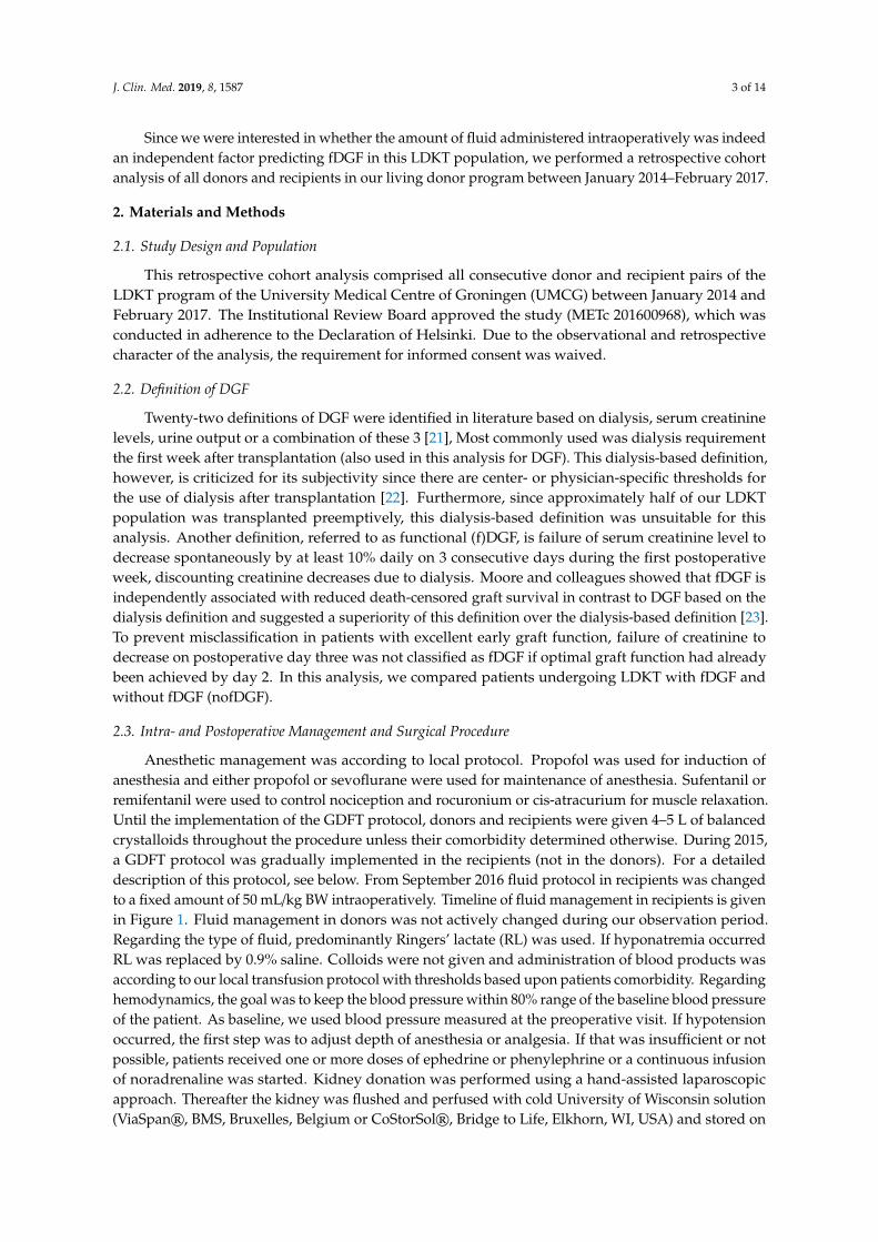
Anesthetic management was according to local protocol. Propofol was used for induction ofanesthesia and either propofol or sevoflurane were used for maintenance of anesthesia. Sufentanil orremifentanil were used to control nociception and rocuronium or cis-atracurium for muscle relaxation.Until the implementation of the GDFT protocol, donors and recipients were given 4–5 L of balancedcrystalloids throughout the procedure unless their comorbidity determined otherwise. During 2015,a GDFT protocol was gradually implemented in the recipients (not in the donors). For a detaileddescription of this protocol, see below. From September 2016 fluid protocol in recipients was changedto a fixed amount of 50 mL/kg BW intraoperatively. Timeline of fluid management in recipients is givenin Figure 1. Fluid management in donors was not actively changed during our observation period.Regarding the type of fluid, predominantly Ringers’ lactate (RL) was used. If hyponatremia occurredRL was replaced by 0.9% saline. Colloids were not given and administration of blood products wasaccording to our local transfusion protocol with thresholds based upon patients comorbidity. Regardinghemodynamics, the goal was to keep the blood pressure within 80% range of the baseline blood pressureof the patient. As baseline, we used blood pressure measured at the preoperative visit. If hypotensionoccurred, the first step was to adjust depth of anesthesia or analgesia. If that was insufficient or notpossible, patients received one or more doses of ephedrine or phenylephrine or a continuous infusionof noradrenaline was started. Kidney donation was performed using a hand-assisted laparoscopicapproach. Thereafter the kidney was flushed and perfused with cold University of Wisconsin solution(ViaSpan®, BMS, Bruxelles, Belgium or CoStorSol®, Bridge to Life, Elkhorn, WI, USA) and stored on
J. Clin. Med. 2019, 8, 1587 4 of 14
ice. Transplantation was performed according to local, standardized protocol. Postoperative fluidmanagement comprised 1 L NaCl 0.45%-Glucose 2.5% per 24 h, complemented with the volume ofdiuresis in the former hour.
Figure 1. Timeline of various intraoperative fluid protocols in recipients. L: liters; RL: Ringers’ lactate;GDFT: goal directed fluid therapy, BW: body weight.
2.4. Goal-Directed Fluid Therapy Protocol.
GDFT was performed with the use of the FloTrac®in combination with the EV1000®monitor(Edwards Lifesciences Corporation, Irvine, CA, USA). The system was used according to manufacturer’sinstructions. A standard institutional GDFT protocol was used with adjustment of the goal. Insteadof a stroke volume variation (SVV) < 12%, commonly used in abdominal surgery, we aimed fora SVV < 10% throughout the procedure. When the SVV was >10% additional fluid was given until SVVwas <10%. If SVV < 10%, fluid administration was left to the discretion of the attending anesthesiologist,however, when cardiac index (CI) was below age-adjusted normal values, a noradrenaline infusion wasstarted. If measurement of the SVV was not possible (e.g., due to cardiac arrhythmias) a protocol basedon stroke volume (SV) was used. In this case, if a fluid bolus of 250 mL resulted in an increase of the SVof 10%, additional fluid was given, if not, the trend of the SV was monitored and fluid administrationwas left to the discretion of the attending anesthesiologist. When SV decreased >10%, additional fluidwas given. The FloTrac®was used with the EV1000 monitor, which does not communicate with ourdigital PDMS. Therefore SV, SVV, and CI values could not be retrieved for this analysis.
2.5. Patient Data
Demographic and postoperative data were obtained from digital patient medical records. The followingvariables were taken into account: age, gender, BMI, smoking, hypertension, use of antihypertensive drugs,measured glomerular filtration rate (mGFR) with use of iodine 125-iothalamate in the donor, blood pressure(measured the day of hospital admission), difference in blood pressure between donor and recipientmeasured by systolic/diastolic/mean of the recipient minus systolic/diastolic/mean of the donor, underlyingkidney disease, number of HLA mismatches, history of dialysis, related or unrelated donor transplantation.For all recipients, the age-adjusted Charlson comorbidity index (CCI) [24] and length of hospital stay wascalculated. Intraoperative data were retrieved from our digital patient data monitoring system (PDMS,CS-EZIS, Chipsoft B.V., Amsterdam, the Netherlands) and consisted of duration of surgery, intraoperativevolume and type of fluid, cumulative hypotensive periods defined as a systolic blood pressure < 80 mmHgand MAP < 60 mmHg, intraoperative use of vasoactive substances, ischemia times, left/right kidney,side of implantation, number of arteries, sacrifice of an accessory artery, and urinary output the first 2h postoperatively. Regarding the use of vasoactive substances, patients were scored on receiving one ormore boluses of ephedrine and/or phenylephrine and whether or not noradrenaline was administered asa continuous infusion. Additionally, the maximum noradrenaline infusion rate during the procedure wasnoted. This was grouped into 3 categories: low infusion rate (0.02–0.10 mg/h), intermediate (0.10–0.20 mg/h),and high (>0.20 mg/h) infusion rate.
2.6. Statistics
For the statistical analysis SPSS version 23 (IBM Corp, Armonk, NY, USA) and GraphPad Prismversion 7.02 (GraphPad Software Inc, La Jolla, CA, USA) were used. We performed univariate analyses

J. Clin. Med. 2019, 8, 1587 5 of 14
to identify factors associated with fDGF. Categorical data were analyzed by chi-square or Fisher’sexact tests. Continuous data were analyzed with an unpaired t-test in the case of normally distributedvalues. If variables were not normally distributed Mann–Whitney test was applied. Multivariateanalysis was performed by means of binary logistic regression. We adjusted the amount of fluidadministered intraoperatively in recipients for potentially relevant confounders with high significancein the univariate analysis. Additionally, we were interested in the impact of implementation of ourGDFT protocol on the incidence of fDGF and on the amount of fluid administered intraoperatively.We therefore analyzed these data between the different time periods 1–3 (described above) with theuse of Fisher’s exact test and Kruskal–Wallis test. Post-hoc analysis with Mann–Whitney was used.Values are given as number (%), mean ± standard deviation (SD) or median with interquartile range(IQR). All reported p-values are two-sided. A p-value of 0.05 or less was considered significant.
3. Results
3.1. Univariate Analysis
3.1.1. Patient Characteristics
Between January 2014 and February 2017, 275 living donor kidney transplant procedures wereperformed in our center. Of the 275 recipients, 31 patients experienced fDGF and 244 recipients didnot (nofDGF). Donor and recipients characteristics of fDGF and nofDGF kidneys are listed in Table 1.There were no statistically significant differences in baseline characteristics and kidney function (mGFR)in donors of kidneys with our without fDGF. Recipients developing fDGF were more likely to bedialysis-dependent at the time of transplantation (25 (81%) vs. 105 (43%), p < 0.001). The compositionof the group of dialysis dependent patients did not differ between nofDGF and fDGF recipients. In thenofDGF group 76 (72%) patients were on hemodialysis at the time of transplantation and 29 (28%)on peritoneal dialysis. In the fDGF group, this was the case for 19 (76%) and six (24%), respectively.All patients on hemodialysis were dialyzed the day before transplantation to 1 kg above dry weight.
Table 1. Donor and recipient demographics. Data given as number (%), mean (SD), or median (IQR).
nofDGF fDGF p
Donor N = 244 N = 31
Age year 54 (11.6) 51 (12.4) 0.104
Gender male 117 (48%) 20 (65%) 0.089
BMI 26.1 (3.0) 25.1 (2.7) 0.075
Smoking 67 (27%) 13 (42%) 0.140
Blood pressureS-RR mmHg 136 (15.3) 136 (11.8) 0.848D-RR mmHg 79 (73–84) 81 (73–86) 0.548MAP mmHg 98 (9.4) 98 (6.7) 0.897
Hypertension 38 (16%) 2 (6%) 0.277Anti-hypertensive drugs
Diuretics 11 1 >0.999B-blocker 13 1 >0.999
Ca antagonist 10 0 0.610ACE-I 4 0 >0.999
AT-II-ant. 16 1 0.703
mGFRNon-stimulated mL/min 109 (97–23) 107 (95–128) 0.846
Stimulated mL/min 116 (103–133) 118 (100–140) 0.764∆GFR 7 (2–12) 7 (−1–12) 0.810

J. Clin. Med. 2019, 8, 1587 6 of 14
Table 1. Cont.
nofDGF fDGF p
Recipient N = 244 N = 31
Age year 54 (41−61) 55 (43−62) 0.991
Gender male 138 (57%) 21 (68%) 0.254
BMI 25.6 (22.6–28.4) 25.8 (24.0–29.8) 0.267
Smoking 45 (18%) 7 (23%) 0.626
Blood pressureS-RR mmHg 143 (20.4) 138 (23.7) 0.196D-RR mmHg 79 (73–84) 81 (73–86) 0.548MAP mmHg 97 (9.4) 98 (6.6) 0.897
∆ blood pressure with donor∆ S-RR mmHg 7.1 (22.8) 2.5 (29.1) 0.308∆ D-RR mmHg 3.1 (13.9) 1.0 (15.0) 0.336∆ MAP mmHg 4 (−6–14) 8 (−10–12) 0.756
Hypertension 175 (72%) 21 (68%) 0.675Antihypertensive drugs
Diuretics 84 (34%) 8 (25%) 0.421B-blocker 124 (51%) 10 (32%) 0.058
Ca antagonist 131 (54%) 15 (48%) 0.703ACE-I. 46 (19%) 5 (16%) 0.811
AT-II-ant 55 (23%) 7 (23%) >0.999
CCI 3 (2–4) 3 (2–6) 0.157
Underlying kidney diseaseDM 15 (6%) 5 (16%) 0.358PKD 57 (23%) 5 (16%) 0.495
Systemic autoimmunediseases 25 (10%) 3 (10%) >0.999
Glomerulonephritis 47 (19%) 4 (13%) 0.4713Other 100 (41%) 14 (45%) 0.701
HLA mm < 3 55 (23%) 8 (25%) 0.655
Dialysis dependent 105 (43%) 25 (81%) <0.001 *
LURD 164 (67%) 19 (61%) 0.547
fDGF: functional delayed graft function; BMI: body mass index; S-RR: systolic blood pressure; D-RR: diastolic bloodpressure; MAP: mean arterial pressure; ACE-I: angiotensin-converting enzyme inhibitor; AT-II-ant: angiotensinII receptor antagonist; CCI: Charlson comorbidity index; mGFR: measured glomerular filtration rate measuredwith use of iodine 125-iothalamate; DM: diabetes mellitus; PKD: polycystic kidney disease; HLA: human leucocyteantigen; LURD: living unrelated donation; *: statistically significant.
3.1.2. Intra- and Postoperative Data
Intraoperative data of donors of fDGF and nofDGF kidneys showed no differences with exceptionof the total amount of fluid, in which donors of fDGF kidneys received less fluid intraoperatively,which was the case for total volume (3545 mL (778.2) vs. 3845 mL (799.1), p = 0.050) and mL/kg BW(45 mL/kg BW (10.3) vs. 49 mL/kg BW (11.4), p = 0.053).
Recipients who developed fDGF received significantly less intraoperative fluid, which was thecase for the total amount of fluid (3000 mL (2250–3680) vs. 3500 mL (2900–4075), p = 0.023) and mLkg-1BW (36 mL/kg BW (25.9–50.0) vs. 47 mL/kg BW (37.3–55.6), p = 0.007). Predominantly RL wasgiven, but in case of hyponatremia RL was partially replaced by saline. This was the case in 48 (20%)of the recipients without fDGF and in 8 (26%) of the patients with fDGF (p = 0.477). Median volumereplaced by saline was 1000 mL (500–2000) in the nofDGF group and 800 mL (500–1075) in the fDGFgroup (p = 0.865). Blood loss was comparable between groups and transfusion of red blood cells was

J. Clin. Med. 2019, 8, 1587 7 of 14
applied in 10 (4.1%) of the patients in the noFDGF group and two (6.4%) of the fDGF group. Patientsshowed no difference in hypotensive periods, but recipients experiencing fDGF were treated morefrequently with noradrenaline continuous infusion (p = 0.034), which was only the case for low doseinfusion with a maximum of 0.1 mg/h. For noradrenaline administered at higher dosage (>0.1 mg/h),there was no difference between the two groups. fDGF was associated with a lower urine outputduring the first two hours after transplantation (p = 0.005 for the first hour and p = 0.002 for the secondhour). Ten patients in the fDGF group were dialyzed after transplantation versus zero patients inthe nofDGF group (p < 0.001). Eight of these kidneys gained function after a mean of 10.3 (3.1) days.Two kidneys suffered primary nonfunction due to a combination of ATN and mild antibody-mediatedrejection (patient 114, transplanted June 2015) and non-HLA-mediated hyperacute rejection (patient273, transplanted November 2016). Recipients experiencing fDGF showed a longer hospital stay(14 (10–20) vs. 9 (7–13) days p < 0.001) (Table 2).
Table 2. Intra- and postoperative donor and recipient data. Data given as number (%), mean (SD),or median (IQR).
nofDGF fDGF p
Donor n = 244 n = 31
Duration min 227 (38.2) 216 (36.8) 0.134
FluidTotal mL 3845 (799.1) 3545 (778.2) 0.050*
mL/kg BW 49 (11.4) 45 (10.3) 0.053
Intraoperative blood pressureS-RR ≤ 80 mmHG 137 (56%) 21 (68%) 0.251
Cumulative duration (min) 10 (5–15) 10 (5–15) 0.772
Vasoactive substancesEphedrine 178 (73%) 25 (71%) 0.515
Phenylephrine 22 (9%) 4 (13%) 0.512Noradrenaline 61 (25%) 11 (35%) 0.277
Recipient n = 244 n = 31
Duration min 212 (189–239) 224 (190–260) 0.390
Fluid
Total mL 3500(2900–4075)
3000(2250–3680) 0.023*
mL/kg BW 47 (37.3–55.6) 36 (25.9–50.0) 0.007*
Intraoperative blood pressureS-RR < 80 mmHg 49 (20%) 6 (19%) >0.999
Cumulative duration min 5 (5–10) 7.5 (4.5–11.2) 0.679MAP < 60 mmHg 93 (38%) 11 (35%) 0.846
Cumulative duration min 10 (5–10) 5 (5–20) 0.759
Vasoactive substancesEphedrine 93 (38%) 16 (52%) 0.174
Phenylephrine 26 (11%) 3 (10%) >0999Noradrenaline 129 (53%) 23 (74%) 0.034*
0.02–0.10 mg h−1 37 (15%) 10 (32%) 0.024*0.10–0.20 mg h−1 42 (17%) 7 (23%) 0.459
>0.20 mg h−1 49 (20%) 6 (19%) >0.999
Ischemia times (min)WIT 3 (3–4) 3 (3–4) 0.724CIT 154 (140–173) 158 (141–178) 0.646
WIT2 39 (33–45) 38 (33–45) 0.982

J. Clin. Med. 2019, 8, 1587 8 of 14
Table 2. Cont.
nofDGF fDGF p
Kidney left 177 (73%) 19 (61%) 0.209Right fossa 203 (83%) 26 (84%) >0.999>1 artery 49 (20%) 8 (26%) 0.482
Artery sacrificed 11 (5%) 4 (13%) 0.074
Blood loss (mL) 250 (150–400) 250 (162.5–500) 0.499
Urineproduction n = 230 n = 301st h (mL) 405 (250–675) 255 (75–512) 0.005*2nd h (mL) 350 (250–550) 183 (64–462) 0.002*
n = 244 n = 31Dialysis after transplantation 0 (0%) 10 (32%) <0.001*Length of hospital stay days 9 (7–13) 14 (10–20) <0.001*
Min: minutes; BW: bodyweight; S-RR: systolic blood pressure; MAP: mean arterial pressure; WIT: warm ischemiatime; CIT: cold ischemia time: WIT2: warm ischemia time 2; *: statistically significant
3.2. Multivariate Logistic Regression Analysis
In the unadjusted analysis, a higher intraoperative administered fluid volume was associatedwith 3% lower odds for the development of fDGF per mL/kg BW (OR 0.967, CI (0.941–0.993), model 1).We adjusted for potentially relevant confounders with high significance in the univariate analysis,i.e., a history of dialysis and the use of intraoperative noradrenaline, after which the relationship wasstill apparent (OR 0.970, CI (0.943–0.998), model 2). Since the intraoperative amount of fluid in thedonors approached significance in the univariate analysis with lower volumes given in the fDGF group,we also adjusted for amount of fluid in the donor, after which the relationship was still apparent (OR0.969, CI (0.941–0.997), model 3) (Table 3).
Table 3. Multivariate logistic regression on risk factors of functional delayed graft function (fDGF).
Model Odds ratio(95% CI) p
1. Unadjusted analysis, model 1
• Amount of fluid administered intraoperatively,recipient, mL/kg BW
0.967(0.941–0.993) 0.015
2. Adjusted analysis, model 2
• Amount of fluid administered intraoperatively,recipient, mL/kg BW
0.970(0.943–0.998) 0.036
• No dialysis dependence at timeof transplantation
0.186(0.073–0.475) <0.001
• Use of noradrenaline continuous infusionyes/no
2.018(0.834–4.878) 0.119
3. Adjusted analysis, model 3
• Amount of fluid administered intraoperatively,recipient, mL/kg BW
0.969(0.941–0.997) 0.029
• No dialysis dependence at timeof transplantation
0.181(0.071–0.464) <0.001
• Amount of fluid administered intraoperatively,donor, mL/kg BW
0.978(0.942–1.014) 0.231

J. Clin. Med. 2019, 8, 1587 9 of 14
3.3. Influence of the GDFT Protocol on the Intraoperative Fluid Volume.
Additionally, we were interested in the impact of implementation of our GDFT protocol on theincidence of fDGF and on the amount of fluid administered intraoperatively. The GDFT protocol wasgradually implemented during 2015 and in 2016 (up to September) all recipients were treated followingthis protocol (Figure 1). Data of the EV1000 monitor were not recorded in our PDMS, therefore wewere unable to see which patients in 2015 were treated according the GDFT protocol and disregardedthis period (March 2015–December 2015) in this specific analysis. We compared patients transplantedbetween January 2014–February 2015 (period 1, n = 84, old protocol) to patients transplanted betweenJanuary 2016–June 2016 (period 2, n = 52, GDFT protocol) and patients transplanted between September2016–February 2017 (period 3, n = 61, new protocol).
Incidence of fDGF during the different periods are shown in Figure 2. Implementation of GDFTwas accompanied by an increase in fDGF from 8.3% in period 1 to 23% in period 2. The implementationof the new protocol in period 3 resulted in a reduction of the incidence of fDGF back to baseline (8.2%,p = 0.029).
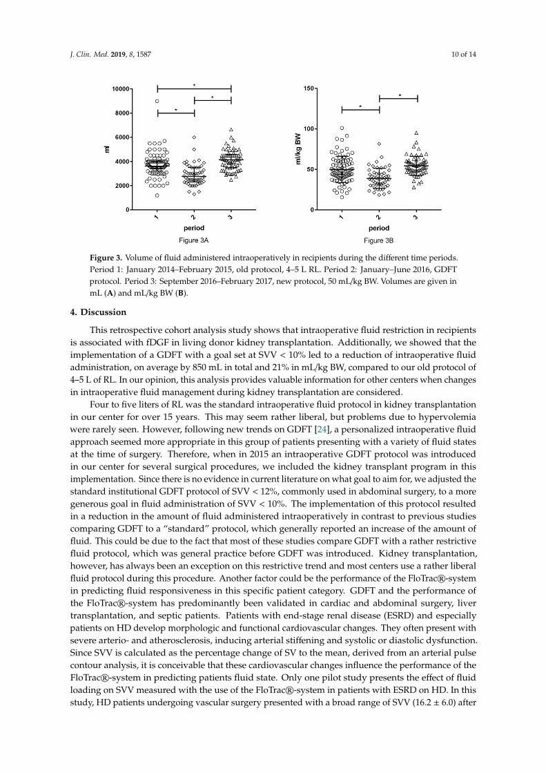
Total amount of intraoperative administered fluid and mL/kg BW in recipients in the different timeperiods are shown in Figure 3A,B, respectively. Total amount of fluid and mL/kg BW were significantlydifferent between the three time periods (p < 0.001, p < 0.001). Implementation of the GDFT (period 2)resulted in a decrease of intraoperative fluid administration compared to our old protocol (period 1),which was the case for total volume (2775 mL (2313–3500) vs. 3625 mL (3213–4000), p < 0.001) andmL/kg BW (38 mL/kg BW (30.3–45.3) vs. 48 mL/kg BW (40–60), p < 0.001). The implementation ofthe new protocol (period 3) resulted in an increase in intraoperative fluid administration to 4150 mL(3475–4575) mL and 54 mL/kg BW (47.4–60.1) compared to the old (total volume p = 0.037, mL/kg BWp = 0.053) and GDFT (total volume p < 0.001, mL/kg BW p < 0.001).
Figure 2. Incidence of fDGF in recipients during the different time periods. Period 1: January2014–February 2015, old protocol, 4–5 L RL. Period 2: January–June 2016, GDFT protocol. Period 3:September 2016–February 2017, new protocol, 50 mL/kg BW. p = 0.029.
J. Clin. Med. 2019, 8, 1587 10 of 14
Figure 3. Volume of fluid administered intraoperatively in recipients during the different time periods.Period 1: January 2014–February 2015, old protocol, 4–5 L RL. Period 2: January–June 2016, GDFTprotocol. Period 3: September 2016–February 2017, new protocol, 50 mL/kg BW. Volumes are given inmL (A) and mL/kg BW (B).
4. Discussion
This retrospective cohort analysis study shows that intraoperative fluid restriction in recipientsis associated with fDGF in living donor kidney transplantation. Additionally, we showed that theimplementation of a GDFT with a goal set at SVV < 10% led to a reduction of intraoperative fluidadministration, on average by 850 mL in total and 21% in mL/kg BW, compared to our old protocol of4–5 L of RL. In our opinion, this analysis provides valuable information for other centers when changesin intraoperative fluid management during kidney transplantation are considered.
Four to five liters of RL was the standard intraoperative fluid protocol in kidney transplantationin our center for over 15 years. This may seem rather liberal, but problems due to hypervolemiawere rarely seen. However, following new trends on GDFT [24], a personalized intraoperative fluidapproach seemed more appropriate in this group of patients presenting with a variety of fluid statesat the time of surgery. Therefore, when in 2015 an intraoperative GDFT protocol was introducedin our center for several surgical procedures, we included the kidney transplant program in thisimplementation. Since there is no evidence in current literature on what goal to aim for, we adjusted thestandard institutional GDFT protocol of SVV < 12%, commonly used in abdominal surgery, to a moregenerous goal in fluid administration of SVV < 10%. The implementation of this protocol resultedin a reduction in the amount of fluid administered intraoperatively in contrast to previous studiescomparing GDFT to a “standard” protocol, which generally reported an increase of the amount offluid. This could be due to the fact that most of these studies compare GDFT with a rather restrictivefluid protocol, which was general practice before GDFT was introduced. Kidney transplantation,however, has always been an exception on this restrictive trend and most centers use a rather liberalfluid protocol during this procedure. Another factor could be the performance of the FloTrac®-systemin predicting fluid responsiveness in this specific patient category. GDFT and the performance ofthe FloTrac®-system has predominantly been validated in cardiac and abdominal surgery, livertransplantation, and septic patients. Patients with end-stage renal disease (ESRD) and especiallypatients on HD develop morphologic and functional cardiovascular changes. They often present withsevere arterio- and atherosclerosis, inducing arterial stiffening and systolic or diastolic dysfunction.Since SVV is calculated as the percentage change of SV to the mean, derived from an arterial pulsecontour analysis, it is conceivable that these cardiovascular changes influence the performance of theFloTrac®-system in predicting patients fluid state. Only one pilot study presents the effect of fluidloading on SVV measured with the use of the FloTrac®-system in patients with ESRD on HD. In thisstudy, HD patients undergoing vascular surgery presented with a broad range of SVV (16.2 ± 6.0) after

J. Clin. Med. 2019, 8, 1587 11 of 14
induction of anesthesia. After a fluid bolus of only 500 mL of a colloid solution almost all patientsshowed a SVV < 10% (6.2 ± 2.8), the threshold in our protocol [25].
The debate on perioperative fluid management is still ongoing. Controversy exists regardingassessment of the intravascular volume state, which goals to aim for, how to measure these goals,and what type of fluid should be used. Hypovolemia leads to a decreased oxygen supply to organs andtissues and may cause hypoxia, which can lead to organ dysfunction. Hypervolemia, on the other hand,can damage the endothelial glycocalyx resulting in a fluid shift from the intravascular compartment tothe interstitial space and tissue edema [26]. Shin and colleagues report in their large cohort analysis of92.094 patients undergoing noncardiac surgery that both too little and too much intraoperative fluidis associated with increased morbidity, mortality, costs, and length of hospital stay [27]. Myles andcolleagues randomLy assigned 3000 patients undergoing a major abdominal procedure to a restrictiveor liberal fluid regimen. In their study, a restrictive regimen was associated with increased risk ofacute kidney injury with a hazard ratio of 1.71 (95% CI 1.29–2.27) [28]. These studies, however, do nottake kidney transplant recipients into account. In the normal kidney, blood flow is regulated byan autoregulatory mechanism, ensuring adequate perfusion in a broad blood pressure range by afferentand efferent arterioles. In the transplanted, denervated kidney, this haemodynamic autoregulationis impaired making the renal blood flow linearly dependent on the systemic blood flow [29–31].Furthermore, reperfusion of the ischemic kidney can be followed by vasoconstriction in the afferentarterioles. This may result in a reduced GFR due to a decrease in glomerular transcapillary hydraulicpressure difference [7,32,33]. Ensuring an adequate volume state in this specific patient category,therefore, is essential to obtain an adequate circulation both on macro- and microcirculatory level.Recently, Cavalari and colleagues reported the results of their prospective observational study, in whichthey compared a prospectively observed cohort of 33 deceased donor kidney transplant recipientstreated with a GDFT protocol to a historical cohort of 33 kidney transplant recipients treated with theirconventional fluid therapy [34]. They observed a significant reduction of cardiovascular complications,DGF. and surgical complications in the GDFT group. Surprisingly, in this study both groups receivedthe same amount of fluid throughout the transplant procedure. Studies including deceased donorkidneys, however, comprise a variety of donor types with variable incidences of DGF hamperingan adequate analysis and conclusions.
The most important predictor of fDGF in our analysis was dialysis dependency at the time oftransplantation. A history of dialysis and especially hemodialysis prior to transplantation is a knownrisk factor of DGF [5,7,35,36]. Hypovolemia at the time of transplantation is one of the proposedunderlying mechanisms [37]. Our hypothesis before implementation of the GDFT protocol was thatthese hypovolemic dialysis patients would present with higher SVV at time of surgery, demanding morefluid intraoperatively, compared to the relatively normovolemic or slightly hypervolemic preemptivelytransplanted patients. Surprisingly, comparable amounts of fluids were given to the two groups.
In our GDFT protocol, noradrenaline was used when CI was below an age-adjusted value.Therefore, an increased use of noradrenaline was seen in period 2 compared to period 1 (71% vs. 41%p = 0.001) due to the implementation of the GDFT. In period 3, the use of noradrenaline decreased to 50%of the patients. In the univariate analysis, the use of noradrenaline was correlated with developmentof fDGF, but after multivariate logistic regression this was no longer the case. However, Morita andcoworkers showed that in a rat model, transplanted kidneys responded to sympaticomimetics witha reduction in renal blood flow (RBF) in contrast to the increase in RBF seen in native rat kidneys [38].
There are some limitations of this analysis that have to be addressed: A major limitation isthat we were unable to evaluate outcome directly according to the fluid protocol (4–5L RL vs.GDFT) and are unable to present information or draw any conclusions regarding actual SV, SVV,CO or CI values and their relation to the observed increase of fDGF. Other limitations are those ofa retrospective observational trial. There is the potential of confounding by unmeasured factors.Regarding postoperative fluid volume, the exact amount of fluid given could not be retrieved ina reliable way from our PDMS and is therefore not implemented in this analysis. Postoperative fluid

J. Clin. Med. 2019, 8, 1587 12 of 14
management was according to a standardized protocol and comprised of 1 L NaCl 0.45%-Glucose2.5% per 24 h, complemented with the volume of diuresis in the former hour. This means that whenthe kidney produces less urine the patient will be given less fluid postoperatively. Since fDGF wasassociated with a lower urinary output the first two hours, it is very likely that patients experiencingfDGF received less fluid postoperatively. Whether this contributed to development of fDGF or is moreof a symptom remains unknown. Backpressure from congested tubules obstructed with cellular debrismay contribute to a reduction in GFR [39,40]. A higher volume of urine in the first hours may have ledto washout of this debris.
Finally, due to the fact that there are only 31 events there is always the possibility of overestimatingthe strength of associations using a multivariate analysis. A strong argument, however, is that nopolicy changes were implemented during the study period with the exception of the intraoperativefluid regimen. Furthermore the incidence of fDGF in our LDKT population has been stable over manyyears and after changing the fluid regimen back to a more liberal fixed amount of 50 mL/kg BW theincidence of fDGF instantly returned to baseline.
DGF after transplantation is a clinically relevant problem. It is associated with an increase inmorbidity, patient anxiety, increased risk of acute rejection, and additional diagnostic procedures andcosts. In our population the median hospital stay in patients experiencing fDGF was prolonged byfive days. Furthermore, this study shows that strict protocols for perioperative fluid management areneeded when studies in kidney transplantation are designed. Fluid restriction can be an important riskfactor for DGF, a frequently used primary end point, even in the setting of LDKT.
5. Conclusions
Implementation of a goal-directed approach to fluid administration with a goal set at a SVV < 10%throughout the procedure led to reduced intraoperative fluid administration in the LDKT recipients inour center. This intraoperative fluid restriction was associated with the development of more fDGF.A thorough validation of GDFT protocols in patients with renal insufficiency is warranted before theseare implemented in this population.
Author Contributions: G.J.N.-M.: participated in research design, data collection, data analysis, writing of thepaper. T.M.H.: participated in data collection, writing of the paper; R.A.P.: participated in writing of the paper;M.E.M.: participated in data analysis, writing of the paper; J.G.B.: participated in data analysis, writing of thepaper; M.M.S.: participated in research design, data analysis, writing of the paper; S.P.B.: participated in researchdesign, data analysis, writing of the paper.
Funding: This research received no external funding.
Acknowledgments: We would like to thank Cordelia Hempel and Tamar van den Berg for data retrieval andprofessor Anthony Absalom for his critical comments and help with language aspects.
Conflicts of Interest: The authors declare no conflict of interest
References
1. Nijboer, W.N.; Schuurs, T.A.; van der Hoeven, J.A.; Leuvenink, H.G.; van der Heide, J.J.; van Goor, H.; Ploeg, R.J.Effects of brain death on stress and inflammatory response in the human donor kidney. Transplant. Proc.2005, 37, 367–369. [CrossRef] [PubMed]
2. Salvadori, M.; Rosso, G.; Bertoni, E. Update on ischemia-reperfusion injury in kidney transplantation:Pathogenesis and treatment. World J. Transplant. 2015, 5, 52–67. [CrossRef] [PubMed]
3. Simpkins, C.E.; Montgomery, R.A.; Hawxby, A.M.; Locke, J.E.; Gentry, S.E.; Warren, D.S.; Segev, D.L. Coldischemia time and allograft outcomes in live donor renal transplantation: Is live donor organ transportfeasible? Am. J. Transplant. 2007, 7, 99–107. [CrossRef] [PubMed]
4. Redfield, R.R.; Scalea, J.R.; Zens, T.J.; Muth, B.; Kaufman, D.B.; Djamali, A.; Astor, B.C.; Mohamed, M.Predictors and outcomes of delayed graft function after living-donor kidney transplantation. Transpl. Int.2016, 29, 81–87. [CrossRef] [PubMed]
J. Clin. Med. 2019, 8, 1587 13 of 14
5. Siedlecki, A.; Irish, W.; Brennan, D.C. Delayed graft function in the kidney transplant. Am. J. Transplant.2011, 11, 2279–2296. [CrossRef]
6. Snoeijs, M.G.; Winkens, B.; Heemskerk, M.B.; Hoitsma, A.J.; Christiaans, M.H.; Buurman, W.A.; vanHeurn, L.E. Kidney transplantation from donors after cardiac death: A 25-year experience. Transplantation2010, 90, 1106–1112. [CrossRef]
7. Perico, N.; Cattaneo, D.; Sayegh, M.H.; Remuzzi, G. Delayed graft function in kidney transplantation. Lancet2004, 364, 1814–1827. [CrossRef]
8. Wu, W.K.; Famure, O.; Li, Y.; Kim, S.J. Delayed graft function and the risk of acute rejection in the modernera of kidney transplantation. Kidney Int. 2015, 88, 851–858. [CrossRef]
9. Fonseca, I.; Teixeira, L.; Malheiro, J.; Martins, L.S.; Dias, L.; Castro Henriques, A.; Mendonça, D. The effect ofdelayed graft function on graft and patient survival in kidney transplantation: An approach using competingevents analysis. Transpl. Int. 2015, 28, 738–750. [CrossRef]
10. Sharif, A.; Borrows, R. Delayed graft function after kidney transplantation: The clinical perspective.Am. J. Kidney Dis. 2013, 62, 150–158. [CrossRef]
11. Othman, M.M.; Ismael, A.Z.; Hammouda, G.E. The impact of timing of maximal crystalloid hydrationon early graft function during kidney transplantation. Anesth. Analg. 2010, 110, 1440–1446. [CrossRef][PubMed]
12. Bacchi, G.; Buscaroli, A.; Fusari, M.; Neri, L.; Cappuccilli, M.L.; Carretta, E.; Stefoni, S. The influence ofintraoperative central venous pressure on delayed graft function in renal transplantation: A single-centreexperience. Transplant. Proc. 2010, 42, 3387–3391. [CrossRef] [PubMed]
13. Snoeijs, M.G.; Wiermans, B.; Christiaans, M.H.; van Hooff, J.P.; Timmerman, B.E.; Schurink, G.W.H.;Buurman, W.A.; Van Heurn, L.W.E. Recipient hemodynamics during non-heart-beating donor kidneytransplantation are major predictors of primary nonfunction. Am. J. Transplant. 2007, 7, 1158–1166.[CrossRef] [PubMed]
14. Aulakh, N.K.; Garg, K.; Bose, A.; Aulakh, B.S.; Chahal, H.S.; Aulakh, G.S. Influence of hemodynamics andintraoperative hydration on biochemical outcome of renal transplant recipients. J. Anaesthesiol. Clin. Pharmacol.2015, 31, 174–179. [CrossRef] [PubMed]
15. Marik, P.E.; Cavallazzi, R. Does the central venous pressure predict fluid responsiveness? An updatedmeta-analysis and a plea for some common sense. Crit. Care Med. 2013, 41, 1774–1781. [CrossRef] [PubMed]
16. Ferris, R.L.; Kittur, D.S.; Wilasrusmee, C.; Shah, G.; Krause, E.; Ratner, L. Early hemodynamic changes afterrenal transplantation: Determinants of low central venous pressure in the recipients and correlation withacute renal dysfunction. Med. Sci. Monit. 2003, 9, 61–66.
17. Le Manach, Y.; Hofer, C.K.; Lehot, J.J.; Vallet, B.; Goarin, J.P.; Tavernier, B.; Cannesson, M. Can changes inarterial pressure be used to detect changes in Cardiac output during volume expansion in the perioperativeperiod? Anesthesiology 2012, 117, 1165–1174. [CrossRef] [PubMed]
18. Benes, J.; Giglio, M.; Brienza, N.; Michard, F. The effects of goal-directed fluid therapy based on dynamicparameters on post-surgical outcome: A meta-analysis of randomized controlled trials. Crit. Care 2014, 18,584. [CrossRef] [PubMed]
19. Ripollés-Melchor, J.; Espinosa, Á.; Martínez-Hurtado, E.; Abad-Gurumeta, A.; Casans-Frances, R.;Fernandez-Perez, C.; Lopez-Timoneda, F.; Calvo-Vecino, J.M. Perioperative goal-directed hemodynamictherapy in noncardiac surgery: A systematic review and meta-analysis. J. Clin. Anesth. 2016, 28, 105–115.[CrossRef] [PubMed]
20. Sun, Y.; Chai, F.; Pan, C.; Romeiser, J.L.; Gan, T.J. Effect of perioperative goal-directed hemodynamic therapyon postoperative recovery following major abdominal surgery-a systematic review and meta-analysis ofrandomized controlled trials. Crit. Care 2017, 21, 141. [CrossRef]
21. Decruyenaere, P.; Decruyenaere, A.; Peeters, P.; Vermassen, F. A Single-Centre Comparison of 22 CompetingDefinitions of Delayed Graft Function After Kidney Transplantation. Ann. Transplant. 2016, 21, 152–159.[CrossRef] [PubMed]
22. Akkina, S.K.; Connaire, J.J.; Israni, A.K.; Snyder, J.J.; Matas, A.J.; Kasiske, B.L. Similar outcomes with differentrates of delayed graft function may reflect centre practice, not centre performance. Am. J. Transplant. 2009, 9,1460–1466. [CrossRef] [PubMed]

J. Clin. Med. 2019, 8, 1587 14 of 14
23. Moore, J.; Shabir, S.; Chand, S.; Bentall, A.; McClean, A.; Chan, W.; Jham, S.; Benavente, D.; Sharif, A.; Ball, S.;et al. Assessing and comparing rival definitions of delayed renal allograft function for predicting subsequentgraft failure. Transplantation 2010, 90, 1113–1116. [CrossRef] [PubMed]
24. Pearse, R.M.; Harrison, D.A.; McDonald, N. Effect of a perioperative, cardiac output guided hemodynamictherapy algorithm on outcomes following major gastrointestinal surgery: A randomized clinical trial andupdated systematic review. JAMA 2014, 311, 2181–2190. [CrossRef] [PubMed]
25. Kanda, H.; Hirasaki, Y.; Iida, T.; Kanao-Kanda, M.; Toyama, Y.; Kunisawa, T.; Iwasaki, H. Effect of fluidloading on left ventricular volume and stroke volume variability in patients with end-stage renal disease:A pilot study. Ther. Clin. Risk Manag. 2015, 11, 1619–1625. [CrossRef] [PubMed]
26. Chappell, D.; Jacob, M.; Hofmann-Kiefer, K.; Conzen, P.; Rehm, M. A rational approach to perioperativefluid management. Anesthesiology 2008, 109, 723–740. [CrossRef] [PubMed]
27. Shin, C.H.; Long, D.R.; McLean, D.; Grabitz, S.D.; Ladha, K.; Timm, F.P.; Thevathasan, T.; Pieretti, A.;Ferrone, C.; Hoeft, A.; et al. Effects of intraoperative Fluid Management on Postoperative Outcomes:A Hospital Registry Study. Ann. Surg. 2018, 267, 1084–1092. [CrossRef]
28. Myles, P.S.; Bellomo, R.; Corcoran, T.; Forbes, A.; Peyton, P.; Story, D.; Christophi, C.; Leslie, K.; McGuinness, S.;Parke, R.; et al. Restrictive versus Liberal Fluid Therapy for Major Abdominal Surgery. N. Engl. J. Med. 2018,378, 2263–2274. [CrossRef] [PubMed]
29. Adams, P.L.; Adams, F.F.; Bell, P.D.; Navar, L.G. Impaired renal blood flow autoregulation in ischemic acuterenal failure. Kidney Int. 1980, 18, 68–76. [CrossRef]
30. Shannon, J.L.; Headland, R.; MacIver, A.G.; Ferryman, S.R.; Barber, P.C.; Howie, A.J. Studies on theinnervation of human renal allografts. J. Pathol. 1998, 186, 109–115. [CrossRef]
31. Thomas, M.C.; Mathew, T.H.; Russ, G.R.; Rao, M.M.; Moran, J. Perioperative blood pressure control, delayedgraft function and acute rejection after renal transplantation. Transplantation 2003, 75, 1989–1995. [CrossRef][PubMed]
32. Alejandro, V.; Scandling, J.D., Jr.; Sibley, R.K.; Dafoe, D.; Alfrey, E.; Deen, W.; Myers, B.D. Mechanisms offiltration failure during postischemic injury of the human kidney. A study of the reperfused renal allograft.J. Clin. Investig. 1995, 95, 820–831. [PubMed]
33. Wilhelm, S.M.; Simonson, M.S.; Robinson, A.V.; Stowe, N.T.; Schulak, J.A. Endothelin up-regulation andlocalization following renal ischemia and reperfusion. Kidney Int. 1999, 55, 1011–1018. [CrossRef] [PubMed]
34. Cavaleri, M.; Veroux, M.; Palermo, F.; Vasile, F.; Mineri, M.; Palumbo, J.; Salemi, L.; Astuto, M.; Murabito, P.Perioperative Goal-Directed Therapy during Kidney Transplantation: An Impact Evaluation on the MajorPostoperative Complications. J. Clin. Med. 2019, 8, 80. [CrossRef]
35. Van Loo, A.A.; Vanholder, R.C.; Bernaert, P.R.; Vermassen, F.E.; van der Vennet, M.; Lameire, N.H.Pretransplantation hemodialysis strategy influences early renal graft function. J. Am. Soc. Nephrol. 1998, 9,473–481.
36. Snyder, J.J.; Kasiske, B.L.; Gilbertson, D.T.; Collins, A.J. A comparison of transplant outcomes in peritonealand hemodialysis patients. Kidney Int. 2002, 62, 1423–1430. [CrossRef]
37. Bogaard, H.J.; de Vries, J.P.; de Vries, P.M. Assessment of refill and hypovolaemia by continuous surveillanceof blood volume and extracellular fluid volume. Nephrol. Dial. Transplant. 1994, 9, 1283–1287.
38. Morita, K.; Seki, T.; Nonomura, K.; Koyanagi, T.; Yoshioka, M.; Saito, H. Changes in renal blood flow inresponse to sympathomimetics in the rat transplanted and denervated kidney. Int. J. Urol. 1999, 6, 24–32.[CrossRef]
39. Alejandro, V.S.; Nelson, W.J.; Huie, P.; Sibley, R.K.; Dafoe, D.; Kuo, P.; Scandling, J.D., Jr.; Myers, B.D.Postischemic injury, delayed function and Na+/K+-ATPase distribution in the transplanted kidney. Kidney Int.1995, 48, 1308–1315. [CrossRef]
40. Tanner, G.A.; Sloan, K.L.; Sophasan, S. Effects of renal artery occlusion on kidney function in the rat. Kidney Int.1973, 4, 377–389. [CrossRef]
© 2019 by the authors. Licensee MDPI, Basel, Switzerland. This article is an open accessarticle distributed under the terms and conditions of the Creative Commons Attribution(CC BY) license (http://creativecommons.org/licenses/by/4.0/).