University of Groningen Cerebral and splanchnic ...
15
University of Groningen Cerebral and splanchnic oxygenation and necrotizing enterocolitis in preterm infants Schat, Trijntje Eelkje DOI: 10.1016/j.earlhumdev.2014.04.008 IMPORTANT NOTE: You are advised to consult the publisher's version (publisher's PDF) if you wish to cite from it. Please check the document version below. Document Version Publisher's PDF, also known as Version of record Publication date: 2015 Link to publication in University of Groningen/UMCG research database Citation for published version (APA): Schat, T. E. (2015). Cerebral and splanchnic oxygenation and necrotizing enterocolitis in preterm infants. University of Groningen. https://doi.org/10.1016/j.earlhumdev.2014.04.008 Copyright Other than for strictly personal use, it is not permitted to download or to forward/distribute the text or part of it without the consent of the author(s) and/or copyright holder(s), unless the work is under an open content license (like Creative Commons). The publication may also be distributed here under the terms of Article 25fa of the Dutch Copyright Act, indicated by the “Taverne” license. More information can be found on the University of Groningen website: https://www.rug.nl/library/open-access/self-archiving-pure/taverne- amendment. Take-down policy If you believe that this document breaches copyright please contact us providing details, and we will remove access to the work immediately and investigate your claim. Downloaded from the University of Groningen/UMCG research database (Pure): http://www.rug.nl/research/portal. For technical reasons the number of authors shown on this cover page is limited to 10 maximum. Download date: 25-02-2022
Transcript of University of Groningen Cerebral and splanchnic ...

University of Groningen
Cerebral and splanchnic oxygenation and necrotizing enterocolitis in preterm infantsSchat, Trijntje Eelkje
DOI:10.1016/j.earlhumdev.2014.04.008
IMPORTANT NOTE: You are advised to consult the publisher's version (publisher's PDF) if you wish to cite fromit. Please check the document version below.
Document VersionPublisher's PDF, also known as Version of record
Publication date:2015
Link to publication in University of Groningen/UMCG research database
Citation for published version (APA):Schat, T. E. (2015). Cerebral and splanchnic oxygenation and necrotizing enterocolitis in preterm infants.University of Groningen. https://doi.org/10.1016/j.earlhumdev.2014.04.008
CopyrightOther than for strictly personal use, it is not permitted to download or to forward/distribute the text or part of it without the consent of theauthor(s) and/or copyright holder(s), unless the work is under an open content license (like Creative Commons).
The publication may also be distributed here under the terms of Article 25fa of the Dutch Copyright Act, indicated by the “Taverne” license.More information can be found on the University of Groningen website: https://www.rug.nl/library/open-access/self-archiving-pure/taverne-amendment.
Take-down policyIf you believe that this document breaches copyright please contact us providing details, and we will remove access to the work immediatelyand investigate your claim.
Downloaded from the University of Groningen/UMCG research database (Pure): http://www.rug.nl/research/portal. For technical reasons thenumber of authors shown on this cover page is limited to 10 maximum.
Download date: 25-02-2022

Trijntje E. Schat*, Fardou H. Heida*, Maarten Schurink, Michelle E. van der Laan, Christian V. Hulzebos,
Arend F. Bos, Elisabeth M.W. Kooi, Jan B.F. Hulscher
* Both authors contributed equally
Submitted
THE ASSOCIATION OF TISSUE OXYGENATION AND INTESTINAL
FATTY ACID-BINDING PROTEIN IN PLASMA DURING THE DEVELOPMENT
OF NECROTIZING ENTEROCOLITIS
CHAPTER 3

36 The association of tissue oxygenation and intestinal fatty acid-binding protein in plasma during the development of necrotizing enterocolitis
ABSTRACT
Background: The underlying pathophysiology of necrotizing enterocolitis (NEC) remains incompletely understood, particularly the role of intestinal perfusion.Objectives: The main aim of the study was to determine the relation between cerebral and splanchnic fractional tissue oxygen extraction (FTOE) with intestinal fatty acid-binding protein in plasma (I-FABPp), a marker for intestinal damage, in infants with NEC. Furthermore, we investigated the courses of cerebral and splanchnic FTOE values and I-FABPp levels in uncomplicated (conservative treatment) and complicated NEC (surgery, or death). Methods: We included 19 preterm infants with NEC (9 uncomplicated, 10 complicated). Using near-infrared spectroscopy, we measured regional cerebral and splanchnic tissue oxygen saturations continuously for 48 hours after NEC onset. We measured I-FABPp levels simultaneously. We used Spearman correlation tests to calculate correlation coefficients between FTOE values and I-FABPp levels in uncomplicated and complicated NEC. Results: Median (range) gestational age was 28 (25-36) weeks and median (range) birth weight was 1290 (740-2400) grams. Cerebral and splanchnic FTOE values correlated strongly with I-FABPp levels (rho between .745 and .900; P < .001 - .037) during the first 16 hours after NEC onset. Thereafter, in uncomplicated NEC, splanchnic FTOE values increased while I-FABPp levels decreased concomitantly. In complicated NEC both splanchnic FTOE values and I-FABPp levels decreased.Conclusion: Combining cerebral and splanchnic FTOE values with I-FABPp levels may enable us to discriminate between the progression of and recovery from intestinal damage in NEC.
3
The association of tissue oxygenation and intestinal fatty acid-binding protein in plasma during the development of necrotizing enterocolitis 37
INTRODUCTION
In preterm infants, necrotizing enterocolitis (NEC) is the leading cause of death from gastrointestinal diseases.1 The underlying pathophysiology of NEC remains incompletely understood, particularly the role of intestinal perfusion.2,3
The suggested role of impaired intestinal perfusion in NEC can be investigated by using near-infrared spectroscopy (NIRS). NIRS is a non-invasive method used increasingly in preterm infants to assess cerebral and intestinal perfusion.4-9 NIRS measures regional tissue oxygen saturation (rSO2) of underlying tissue continuously.10 When transcutaneous arterial oxygen saturation (SpO2) is measured simultaneously, fractional tissue oxygen extraction (FTOE) can be calculated.11 FTOE is thought to reflect the balance between tissue oxygen supply and consumption and may, therefore, be an early indicator of impaired tissue perfusion.11 A possible way of gaining more insight into the role of cerebral and intestinal perfusion during the development of NEC is to combine FTOE values with intestinal fatty acid-binding protein (I-FABP), a marker for intestinal damage.12,13 I-FABP is an intracellular protein located specifically in the epithelium of the small bowel. Following enterocyte damage, it is rapidly released into the circulation due, for example, to hypoxia.12,13 In preterm infants with NEC, I-FABP levels in plasma (I-FABPp) are elevated and can serve as a predictor of the severity and extent of intestinal damage.14-19
Our first aim was to investigate whether FTOE values reflect intestinal damage due to NEC. We did so by relating cerebral and splanchnic FTOE values with I-FABPp levels in preterm infants with NEC. Secondly, we investigated whether, during the first 48 hours after NEC onset, the courses of cerebral and splanchnic FTOE values in relation to the course of I-FABPp levels differed in infants with uncomplicated and complicated NEC.
METHODS
Ethical approval
This study was part of a larger, prospective, observational cohort study registered with the Dutch Trial Registry under number NTR3239. The study was approved by the ethical review board of University Medical Center Groningen. Written informed parental consent was obtained in all cases.
Patient population
We included preterm infants admitted to the neonatal intensive care unit of University Medical Center Groningen between October 2010 and October 2012, who were suspected of or had recently been diagnosed with NEC. Suspected NEC was defined as the presence of non-specific NEC symptoms, such as bloody stools or distended abdomen. NEC was definitely confirmed if pneumatosis intestinalis, portal venous gas, or both were present according to the modified Bell’s staging criteria.20 As soon as a suspicion of NEC arose, the neonatologist put the patients on a nil per os and gastric decompression regime, and treated

38 The association of tissue oxygenation and intestinal fatty acid-binding protein in plasma during the development of necrotizing enterocolitis
them with broad-spectrum antibiotics until radiographic signs of NEC resolved and clinical signs stabilized.The study design included measuring I-FABPp levels repeatedly, and measuring rSO2 of cerebral and splanchnic tissue continuously, for 48 hours after NEC onset or until surgery, whichever came first. We defined NEC onset as the time of the first radiographic abdominal examination after clinical suspicion of NEC had arisen, including abdominal X-rays that had been taken in referring hospitals. After completing the study, a team of four consultants, blinded as to the results of the NIRS and I-FABP measurements, determined the modified Bell’s staging criteria at NEC onset.20 We also determined the end-stage Bell’s stage. Agreement was reached in all cases. In this paper we present data of the subgroup of preterm infants with definite NEC only, i.e., Bell’s stage ≥ 2. We excluded those infants in whom NEC was not confirmed. We assigned the infants with NEC to one of two groups: those with uncomplicated NEC (conservative treatment sufficed) and those with complicated NEC (surgery was required, or the infant died). Indications for surgery, i.e. laparotomy, were bowel perforation (Bell’s stage 3B) or lack of improvement despite optimal conservative therapy.
I-FABPp
With every routine blood analysis after definite NEC onset, an extra sample of 100 µL was obtained in an EDTA tube for study purposes. Ideally, blood samples were collected every 8 hours during the first 24 hours after definite NEC onset, and every 12 hours during the following 24 hours. Blood samples were fractioned by centrifuging for 10 minutes at approximately 2000 x g. Plasma was then collected in a 5 mL Sarstedt tube and stored at -80°C. Next, a laboratory technician, who was blinded as to the clinical data, performed the I-FABPp measurements. We used the commercially available ELISAs for determining I-FABPp levels (Human FABP2 kit from R&D systems, Minneapolis, USA).
NIRS
We used the INVOS 5100C near-infrared spectrometer (Covidien, Mansfield, USA) in combination with the neonatal SomaSensors (Covidien) to measure cerebral and splanchnic oxygen saturation continuously for 48 hours after NEC onset. To measure cerebral tissue oxygen saturation (rcSO2), we placed the neonatal SomaSensor on the right or left frontoparietal side of the infant’s head. We measured splanchnic oxygen saturation at two locations: just below the right costal arch to measure liver oxygen saturation (rlivSO2), and infraumbilically on the central abdomen to measure intestinal oxygen saturation (rintSO2). The SomaSensors were held in place by elastic bandaging or Mepitel (Mölnlycke, Sweden). During routine nursing care, clinical assessments, and radiographic examinations the sensor was temporarily removed. Afterwards, it was replaced in the same location. The data, collected prospectively, were stored off-line for future analysis.
3
The association of tissue oxygenation and intestinal fatty acid-binding protein in plasma during the development of necrotizing enterocolitis 39
Additionally, we collected SpO2 (Nellcor, Covidien), and calculated FTOE using the formula: FTOE = (SpO2 - rSO2) / SpO2.
11 FTOE reflects the balance between tissue oxygen delivery and tissue oxygen consumption.11 Changes in FTOE reflect changes in tissue perfusion. In this way tissue hypoxia can be detected.
Demographic and clinical variables
We collected the following patient characteristics from patient reports: gestational age, birth weight, postnatal age at NEC onset, gender, whether surgery was required, and mortality. Furthermore, we documented the first concentrations during the study period of hemoglobin, thrombocytes, pH, C-reactive protein, and lactate. Additionally, we documented the need for mechanical ventilation, treatment of circulatory failure (volume expansion, vasoactive drugs), and the presence of a (hemodynamically significant) patent ductus arteriosus during the study period. It was defined as a diastolic forward flow in the branches of the pulmonary artery, a diastolic backflow in the descending aorta, and a left ventricular end diastolic diameter > p 95.
Data and statistical analysis
For the first aim of this study, we correlated cerebral and splanchnic FTOE values with I-FABPp levels in preterm infants with Bell’s stage ≥ 2. Subsequently, we analyzed the courses of cerebral and splanchnic FTOE values in association with I-FABPp levels in preterm infants in whom NEC developed without complications and in preterm infants with complicated NEC. Cerebral and splanchnic regional tissue oxygen saturations were collected once every 6 seconds, whilst SpO2 values were collected once every 5 minutes. We matched each SpO2 value with the corresponding single rSO2 value, leaving one coupled measurement every 5 minutes. Next, we calculated FTOE values using these combined SpO2 and rSO2 values for the cerebral, liver, and intestinal regions separately. Since I-FABPp levels were collected once every 8 hours in the first 24 hours after NEC onset and once every 12 hours between 24 and 48 hours after NEC onset, we calculated 8-hour mean FTOE values in the first 24 hours after NEC onset and subsequently 12-hour mean FTOE values for the remaining study period. Next, we calculated correlation coefficients between cerebral and splanchnic FTOE values and I-FABPp levels during the first 48 hours after NEC onset using the Spearman rank correlation test. For our second aim, we constructed courses of cerebral and splanchnic FTOE values together with I-FABPp levels for infants with uncomplicated NEC and complicated NEC after logarithmic transformation of the I-FABPp levels.Categorical data were tested by the chi-square test or Fischer’s exact test and continuous data by the Mann-Whitney test. We used SPSS 22.0 software for Windows (IBM SPSS Statistics 22, IBM Corp., Armonk, New York, USA) for the statistical analyses. We considered a P value < .05 statistically significant.

40 The association of tissue oxygenation and intestinal fatty acid-binding protein in plasma during the development of necrotizing enterocolitis
RESULTS
Patient characteristics
We enrolled 19 preterm infants with NEC Bell’s stage ≥ 2 in whom we were able to measure cerebral and splanchnic tissue oxygen saturation simultaneously and collect plasma for analyzing I-FABP levels (Figure 1). I-FABPp levels could not be assessed in cases in which the condition of the infant was too critical to obtain routine blood analysis. The median (range) time between NEC onset and the beginning of NIRS monitoring was 7 (2-31) hours. NIRS data could not be assessed in cases of incorrect sensor placement, or for logistic reasons when NIRS devices were not available. In nine infants the course of NEC was uncomplicated. One of these infants required surgery as a result of post-NEC stricture 74 days after NEC onset. Of the ten infants with complicated NEC, two infants who were diagnosed with Bell’s stage 3A, died as a consequence 5 and 35 days after NEC onset. Eight infants were found to have Bell’s stage 3B, with bowel perforation. Eight infants with complicated NEC required surgery; seven of them were operated on during the study period, with a median of 33 hours (range, 9-165) between the onset of NEC symptoms and surgery. We present the patient characteristics in Table 1.
Figure 1. Flow diagram of the study.
The correlation between cerebral and splanchnic FTOE values and I-FABPp levels
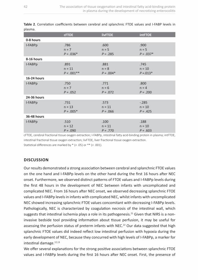
In Table 2 we present the correlation coefficients between cerebral and splanchnic FTOE values and I-FABPp levels. We found strong correlation coefficients between these variables during the first 16 hours after NEC onset (Table 2). Additionally, cerebral FTOE values correlated significantly with I-FABPp levels between 24 and 36 hours after NEC onset (Table 2).

3
The association of tissue oxygenation and intestinal fatty acid-binding protein in plasma during the development of necrotizing enterocolitis 41
Table 1. Patient characteristics. Uncomplicated NEC (n = 9)
Complicated NEC (n = 10)
P value
Gestational age, weeks 31.6 (25.7-35.9) 26.7 (25.0-34.0) .025*Birth weight, grams 1520 (740-2400) 980 (790-2280) .050Male:Female 6:3 8:2 .628PNA at NEC diagnosis, days 8 (3-29) 9 (7-22) .589Hemoglobin, mmol/L 8.7 (7.0-12.4) 8.2 (6.0-10.3) .567Thrombocytes, 109/L 235 (131-491) 202 (42-405) .142pH 7.34 (7.19-7.39) 7.24 (7.09-7.42) .130C-reactive protein, mg/L 33 (0-166) 30 (0-95) .838Lactate, mmol/L 2.7 (1.2-4.5) (n = 4) 2.0 (1.0-11.9) (n = 8) .799Mechanical ventilation (%) 3 (33) 7 (70) .179PDA (%) 1 (11) 3 (30) .582Hemodynamically significant (%) - 2 (20) .474RBC transfusion (%) 3 (33) 4 (40) .999Fluid resuscitation (%) 4 (44) 8 (80) .170Inotropes (%) - 6 (60) .011*Surgery (%) 1 (11) 8 (80) .005*Mortality (%) - 6 (60) .011*
Data are expressed as median (range) or as numbers unless specified otherwise.
Abbreviations: NEC, necrotizing enterocolitis; PDA, patent ductus arteriosus; PNA, postnatal age; RBC, red blood cell.
Statistical differences between the two groups are marked by * (< .05)
The courses of cerebral and splanchnic FTOE values and I-FABPp levels in infants with complicated and uncomplicated NEC
Figure 2 shows the courses of cerebral, liver, and intestinal FTOE values and I-FABPp levels in preterm infants with uncomplicated NEC and infants with complicated NEC. During the first 16 hours after NEC onset we found little difference in the courses of cerebral and splanchnic FTOE values and I-FABPp levels between infants with uncomplicated and complicated NEC. From 16 hours after NEC onset, however, we found that both splanchnic FTOE values and I-FABPp levels decreased in preterm infants with complicated NEC. In preterm infants with uncomplicated NEC we observed the opposite: splanchnic FTOE values gradually increased whilst I-FABPp levels decreased. We observed this increase and decrease of splanchnic FTOE values particularly in the intestinal region, as compared to the liver region. Please note that we constructed these courses using all the data available for the included infants, even though the FTOE values and/or I-FABPp levels were not available at each time period for every infant.
42 The association of tissue oxygenation and intestinal fatty acid-binding protein in plasma during the development of necrotizing enterocolitis
Table 2. Correlation coefficients between cerebral and splanchnic FTOE values and I-FABP levels in plasma.
cFTOE livFTOE intFTOE0-8 hoursI-FABPp .786
n = 7P = .036*
.600n = 5P = .285
.900n = 5P = .037*
8-16 hoursI-FABPp .891
n = 11P < .001**
.881n = 8P = .004*
.745n = 10P =.013*
16-24 hoursI-FABPp .750
n = 7P = .052
.771n = 6P = .072
.800n = 4P = .200
24-36 hoursI-FABPp .731
n = 13P = .005*
.573n = 11P = .066
-.285n = 10P = .425
36-48 hoursI-FABPp .510
n = 12P = .090
.100n = 11P = .770
.188n = 10P = .603
cFTOE, cerebral fractional tissue oxygen extraction; I-FABPp, intestinal fatty acid-binding protein in plasma; intFTOE,
intestinal fractional tissue oxygen extraction; livFTOE, liver fractional tissue oxygen extraction.
Statistical differences are marked by * (< .05) or ** (< .001).
DISCUSSION
Our results demonstrated a strong association between cerebral and splanchnic FTOE values on the one hand and I-FABPp levels on the other hand during the first 16 hours after NEC onset. Furthermore, we observed distinct patterns of FTOE values and I-FABPp levels during the first 48 hours in the development of NEC between infants with uncomplicated and complicated NEC. From 16 hours after NEC onset, we observed decreasing splanchnic FTOE values and I-FABPp levels in infants with complicated NEC, whilst infants with uncomplicated NEC showed increasing splanchnic FTOE values concomitant with decreasing I-FABPp levels. Pathologically, NEC is characterized by coagulation necrosis of the intestinal wall, which suggests that intestinal ischemia plays a role in its pathogenesis.21 Given that NIRS is a non-invasive bedside tool providing information about tissue perfusion, it may be useful for assessing the perfusion status of preterm infants with NEC.22 Our data suggested that high splanchnic FTOE values did indeed reflect low intestinal perfusion with hypoxia during the early development of NEC, because they concurred with high levels of I-FABPp, a marker for intestinal damage.12,13
We offer several explanations for the strong positive associations between splanchnic FTOE values and I-FABPp levels during the first 16 hours after NEC onset. First, the presence of

3
The association of tissue oxygenation and intestinal fatty acid-binding protein in plasma during the development of necrotizing enterocolitis 43
ischemia may cause intestinal epithelial cell damage. Second, intestinal ischemia and hypoxia may develop as a consequence of circulatory insufficiency in the presence of intestinal epithelial cell damage. Finally, intestinal circulation may be affected locally as a result of intestinal injury. Whatever the case may be, our data suggested that splanchnic FTOE values can be used to gain information about the degree of intestinal damage during NEC. Interestingly, we not only found strong correlations between splanchnic FTOE values and I-FABPp levels, but also between cerebral FTOE values and I-FABPp levels. This finding may be a reflection of the fact that by the time NEC becomes clinically evident, it has already had systemic effects on hemodynamics. In this case, systemic circulatory insufficiency, e.g., the need for volume expansion and/or inotropes, may have occurred earlier on, compromising cerebral perfusion as well.23 Another explanation is that cerebrovascular autoregulation in these preterm infants might have been compromised.24,25
Figure 2. Graphs showing the median (dots and squares) and interquartile range (lines) of cerebral and splanchnic FTOE values and I-FABPp levels of infants with uncomplicated and complicated NEC.

44 The association of tissue oxygenation and intestinal fatty acid-binding protein in plasma during the development of necrotizing enterocolitis
In all infants with NEC, irrespective of whether the disease developed with or without complications, we found decreasing I-FABPp levels 16 hours after NEC onset. This may be the result of one of two mechanisms: either expansion of damage or recovery of intestinal tissue. In case of complicated NEC, it could be caused by intestinal necrosis leaving no villi to secrete I-FABP or the absence of blood flow through a demarcated necrotic bowel segment .13,19 Conversely, when the infant’s condition is ameliorating and the intestinal tissue is not injured any further, secretion of I-FABP into the circulation will diminish.19 On the basis of concentrations of I-FABPp levels alone we cannot differentiate between these two hypothesized mechanisms. With simultaneous knowledge of FTOE values, however, we were able to differentiate between the aforementioned supposed mechanisms. We identified two distinct patterns during the 48 hours during which the disease was developing, one predominant in infants with uncomplicated NEC and the other in infants with complicated NEC. In infants with uncomplicated NEC, we observed low splanchnic FTOE values during the first 16 hours after NEC onset that increased during the remainder of the study period. We hypothesize that hyperemia is present during the first hours after NEC onset due to an inflammatory response. As time progresses, hyperemia gradually disappears, which explains the increasing FTOE values. We hypothesize that this course of FTOE values in combination with decreasing I-FABPp levels represents recovery of intestinal tissue. We observed the opposite in infants with complicated NEC, i.e., relatively high initial splanchnic FTOE values, and gradually decreasing intestinal FTOE as the disease developed. We speculate that the high splanchnic FTOE values during the first 16 hours after NEC onset were the result of compromised intestinal perfusion. From 16 hours after NEC onset, however, the splanchnic FTOE values decreased, which suggests decreasing or absent intestinal metabolism due to the presence of necrotic bowel. In these infants, therefore, decreasing I-FABPp levels could possibly have been the result of increasing intestinal injury. In the intestinal region the increase and decrease of FTOE values was more distinct than in the region of the liver. This could be explained by the liver’s unique blood supply - in addition of receiving partially deoxygenated blood from the intestinal region through the portal vein, it also receives oxygenated blood from the hepatic artery.26 The strength of this study was that we obtained plasma samples at regular intervals and that we measured the cerebral and splanchnic rSO2 values with NIRS simultaneously. Furthermore, we measured cerebral and splanchnic oxygenation for 48 consecutive hours. Finally, this was the first study to correlate FTOE values with a marker for intestinal damage. A limitation of this study was the relatively small sample size. Nevertheless, we found very strong associations between cerebral and splanchnic FTOE and I-FABPp levels. Moreover, we included all available data, including data of infants for whom FTOE values and/or I-FABPp levels were not available for each time period. This might have led to a selection bias. Finally, we did not include a control group. However, the aim of this study was to investigate whether intestinal perfusion plays a role in the course of NEC, and, together with I-FABPp

3
The association of tissue oxygenation and intestinal fatty acid-binding protein in plasma during the development of necrotizing enterocolitis 45
measurements differentiated between a complicated or uncomplicated course. Future research should also investigate differences in intestinal perfusion measures between infants with NEC and relatively healthy and stable preterm infants. Our findings may have clinical implications. They suggested that impaired intestinal perfusion played a pivotal role in the development of complications during the early stages of the disease. Combined measurements of splanchnic FTOE values and I-FABPp levels in the initial phase of NEC might indicate individual infants who are at high risk of developing intestinal perforations. Early detection of impaired intestinal perfusion and hypoxia would be most helpful, because the assessment of intestinal necrosis and the timing of surgery for NEC, especially in the absence of perforation, remain difficult. It may also lead to new interventions, other than surgical ones, aimed at counteracting the progression of NEC into complicated disease. Further research is warranted to confirm this hypothesis.
CONCLUSION
We found strong associations between FTOE values of cerebral and splanchnic tissue on one hand and I-FABPp levels on the other during the first 16 hours after NEC onset, suggesting that FTOE values can be used to gain information about the degree of intestinal damage. Additionally, during the first 48 hours after NEC onset, we identified distinct splanchnic FTOE and I-FABPp courses in preterm infants with uncomplicated NEC and complicated NEC. This finding suggests that impaired intestinal perfusion and hypoxia play an important role early on in the development of complicated NEC.
ACKNOWLEDGMENTS
This study was part of the research program of the Graduate School of Medical Sciences, the Groningen University Institute for Drug Exploration, and the postgraduate school for Behavioural and Cognitive Neurosciences, University of Groningen. T.E. Schat, F.H. Heida, and M.E. van der Laan were financially supported by a grant from the Junior Scientific Master Class of University of Groningen. Finally, we greatly acknowledge the help of Dr. T. van Wulfften Palthe for correcting the English manuscript.

46 The association of tissue oxygenation and intestinal fatty acid-binding protein in plasma during the development of necrotizing enterocolitis
REFERENCES
1. Neu J, Walker WA. Necrotizing enterocolitis. N Engl J Med. 2011;364(3):255-264. 2. Nowicki PT. Ischemia and necrotizing enterocolitis: Where, when, and how. Semin Pediatr Surg.
2005;14(3):152-158. 3. Watkins DJ, Besner GE. The role of the intestinal microcirculation in necrotizing enterocolitis.
Semin Pediatr Surg. 2013;22(2):83-87. 4. Cortez J, Gupta M, Amaram A, Pizzino J, Sawhney M, Sood BG. Noninvasive evaluation of
splanchnic tissue oxygenation using near-infrared spectroscopy in preterm neonates. J Matern Fetal Neonatal Med. 2011;24(4):574-582.
5. Gay AN, Lazar DA, Stoll B, et al. Near-infrared spectroscopy measurement of abdominal tissue oxygenation is a useful indicator of intestinal blood flow and necrotizing enterocolitis in premature piglets. J Pediatr Surg. 2011;46(6):1034-1040.
6. McNeill S, Gatenby JC, McElroy S, Engelhardt B. Normal cerebral, renal and abdominal regional oxygen saturations using near-infrared spectroscopy in preterm infants. J Perinatol. 2011;31(1):51-57.
7. Gillam-Krakauer M, Cochran CM, Slaughter JC, et al. Correlation of abdominal rSO2 with superior mesenteric artery velocities in preterm infants. J Perinatol. 2013;33(8):609-612.
8. Pellicer A, Greisen G, Benders M, et al. The SafeBoosC phase II randomised clinical trial: A treatment guideline for targeted near-infrared-derived cerebral tissue oxygenation versus standard treatment in extremely preterm infants. Neonatology. 2013;104(3):171-178.
9. Patel AK, Lazar DA, Burrin DG, et al. Abdominal near-infrared spectroscopy measurements are lower in preterm infants at risk for necrotizing enterocolitis. Pediatr Crit Care Med. 2014;15(8):735-741.
10. Wyatt JS, Cope M, Delpy DT, Wray S, Reynolds EO. Quantification of cerebral oxygenation and haemodynamics in sick newborn infants by near infrared spectrophotometry. Lancet. 1986;2(8515):1063-1066.
11. Naulaers G, Meyns B, Miserez M, et al. Use of tissue oxygenation index and fractional tissue oxygen extraction as non-invasive parameters for cerebral oxygenation. A validation study in piglets. Neonatology. 2007;92(2):120-126.
12. Gollin G, Marks C, Marks WH. Intestinal fatty acid binding protein in serum and urine reflects early ischemic injury to the small bowel. Surgery. 1993;113(5):545-551.
13. Lieberman JM, Sacchettini J, Marks C, Marks WH. Human intestinal fatty acid binding protein: Report of an assay with studies in normal volunteers and intestinal ischemia. Surgery. 1997;121(3):335-342.
14. Edelson MB, Sonnino RE, Bagwell CE, Lieberman JM, Marks WH, Rozycki HJ. Plasma intestinal fatty acid binding protein in neonates with necrotizing enterocolitis: A pilot study. J Pediatr Surg. 1999;34(10):1453-1457.
15. Evennett N, Alexander N, Petrov M, Pierro A, Eaton S. A systematic review of serologic tests in the diagnosis of necrotizing enterocolitis. J Pediatr Surg. 2009;44(11):2192-2201.
16. Aydemir C, Dilli D, Oguz SS, et al. Serum intestinal fatty acid binding protein level for early diagnosis and prediction of severity of necrotizing enterocolitis. Early Hum Dev. 2011;87(10):659-661.
17. Schurink M, Scholten IG, Kooi EM, et al. Intestinal fatty acid-binding protein in neonates with imminent necrotizing enterocolitis. Neonatology. 2014;106(1):49-54.
18. Heida FH, Hulscher JB, Schurink M, et al. Intestinal fatty acid-binding protein levels in necrotizing enterocolitis correlate with extent of necrotic bowel: Results from a multicenter study. J Pediatr Surg. 2014.

3
The association of tissue oxygenation and intestinal fatty acid-binding protein in plasma during the development of necrotizing enterocolitis 47
19. Schurink M, Kooi EM, Hulzebos CV, et al. Intestinal fatty acid-binding protein as a diagnostic marker for complicated and uncomplicated necrotizing enterocolitis: A prospective cohort study. PLoS One. 2015;10(3):e0121336.
20. Walsh MC, Kliegman RM. Necrotizing enterocolitis: Treatment based on staging criteria. Pediatr Clin North Am. 1986;33(1):179-201.
21. Ballance WA, Dahms BB, Shenker N, Kliegman RM. Pathology of neonatal necrotizing enterocolitis: A ten-year experience. J Pediatr. 1990;117(1 Pt 2):S6-13.
22. Moore JE. Newer monitoring techniques to determine the risk of necrotizing enterocolitis. Clin Perinatol. 2013;40(1):125-134.
23. Hanson SJ, Berens RJ, Havens PL, Kim MK, Hoffman GM. Effect of volume resuscitation on regional perfusion in dehydrated pediatric patients as measured by two-site near-infrared spectroscopy. Pediatr Emerg Care. 2009;25(3):150-153.
24. Greisen G. Autoregulation of cerebral blood flow in newborn babies. Early Hum Dev. 2005;81(5):423-428.
25. Verhagen EA, Hummel LA, Bos AF, Kooi EM. Near-infrared spectroscopy to detect absence of cerebrovascular autoregulation in preterm infants. Clin Neurophysiol. 2014;125(1):47-52.
26. Teller J, Wolf M, Keel M, Bucher HU, Fanconi S, Baenziger O. Can near infrared spectroscopy of the liver monitor tissue oxygenation? Eur J Pediatr. 2000;159(7):549.