University of Groningen Airway epithelium in obliterative ... · PDF filemodels have also been...
21
University of Groningen Airway epithelium in obliterative airway disease Qu, Ning IMPORTANT NOTE: You are advised to consult the publisher's version (publisher's PDF) if you wish to cite from it. Please check the document version below. Document Version Publisher's PDF, also known as Version of record Publication date: 2005 Link to publication in University of Groningen/UMCG research database Citation for published version (APA): Qu, N. (2005). Airway epithelium in obliterative airway disease s.n. Copyright Other than for strictly personal use, it is not permitted to download or to forward/distribute the text or part of it without the consent of the author(s) and/or copyright holder(s), unless the work is under an open content license (like Creative Commons). Take-down policy If you believe that this document breaches copyright please contact us providing details, and we will remove access to the work immediately and investigate your claim. Downloaded from the University of Groningen/UMCG research database (Pure): http://www.rug.nl/research/portal. For technical reasons the number of authors shown on this cover page is limited to 10 maximum. Download date: 10-05-2018
Transcript of University of Groningen Airway epithelium in obliterative ... · PDF filemodels have also been...

University of Groningen
Airway epithelium in obliterative airway diseaseQu, Ning
IMPORTANT NOTE: You are advised to consult the publisher's version (publisher's PDF) if you wish to cite fromit. Please check the document version below.
Document VersionPublisher's PDF, also known as Version of record
Publication date:2005
Link to publication in University of Groningen/UMCG research database
Citation for published version (APA):Qu, N. (2005). Airway epithelium in obliterative airway disease s.n.
CopyrightOther than for strictly personal use, it is not permitted to download or to forward/distribute the text or part of it without the consent of theauthor(s) and/or copyright holder(s), unless the work is under an open content license (like Creative Commons).
Take-down policyIf you believe that this document breaches copyright please contact us providing details, and we will remove access to the work immediatelyand investigate your claim.
Downloaded from the University of Groningen/UMCG research database (Pure): http://www.rug.nl/research/portal. For technical reasons thenumber of authors shown on this cover page is limited to 10 maximum.
Download date: 10-05-2018

Chapter 1
General Introduction

CHAPTER 1
2

General introduction
3
1. Obliterative bronchiolitis and obliterative airway disease
1.1. Obliterative Bronchiolitis
Lung transplantation is currently the only available treatment for end-stage lung disease patients. Despite the success of improved modern lung transplantation with the introduction of new surgical techniques, improved immunosuppressive agents and innovations in managing of acute rejection and infection, the survival rate of recipients is 75% at 1 year and less than 50% at 5 years. (1) One of the most severe complications after lung transplantation is obliterative bronchiolitis (OB) which affect over 40% of the recipients within 5 years during the post-transplantation period.(1).
OB is a chronic disease that develops from months and mostly years after lung transplantation (2-4). It is characterized by progressive bronchial inflammation, epithelial injury and luminal fibrosis.(5-7). There is no treatment for human OB and insight into the understandings of its mechanism is still lacking. Presently, all clinical efforts are directed at slowing down the process. These efforts include pre-transplant treatment to the donor lung to reduce inflammatory inducing factors, employment of aggressive peri-transplant administration with antibiotics (3;8-11) and improved immunosuppressive regimens. Only re-transplantation appears to be a curable solution, but is mostly not possible due to the limited availability of donor organs. Clinical investigation of OB in humans is restricted by the limited amount of patient material available for research. This makes the development of new treatments a difficult and time consuming task (12). Thus, a simple animal model that resembles the development of OB in human is a desirable goal.
1.2. Animal transplant models of obliterative airway disease (OAD) for human OB As pointed out above an animal model is needed for studying the pathophysiology and possible treatment of OB. A rat lung transplantation model was developed in Groningen and described by Jochum Prop and colleagues in 1984 (13). Using this technique they

CHAPTER 1
4
were the first to show that OB was secondary to acute pulmonary rejection (14). Also, OB-like airway damage was seen during chronic rejection and viral infections in this rat lung transplantation model. (15) Hertz and his colleagues published a much simpler model in 1993, where they transplanted murine heterotopic tracheas subcutaneously (16), and found that allogeneic tracheas were rejected with massive cellular infiltration and epithelium loss as opposed to isografts which did not show these changes. This led by day 21 to obliteration of the transplanted trachea lumen by the fibroproliferation. This pathological process is called the obliterative airway disease (OAD) and is supposed to reflect the changes seen in OB. Large animal OAD models have also been studied in the past decades. Porcine, canine or even primate tracheas (17) and lungs (18-21) were transplanted and shown to mimic the human OB, since similar histological findings such as graft infiltration, epithelium loss and fibrotic luminal occlusion were observed. Although large animals may be close to humans in size and body structure, the limitations of these large animal models are also obvious: the lack of inbred strains, the cost of purchasing large animals and the peri-transplantation management. The biggest obstacle, however, is the unstable results (22). In summary, OAD in small animal models share histopathological similarities to human OB and is presently the most useful tool for human OB study.
1.3. The histopathology and immunology of OB/OAD
Clinical OB is a chronic inflammatory disease of the airways, involving the bronchial epithelium and leading to the gradual obliteration of small and large airways by inflammatory infiltrates, proliferating fibroblasts, mature collagen and extracellular matrix (3). Although the mechanisms underlying the development of OB are not clear yet, the syndrome may be divided into two distinct phases; an acute alloimmune phase with lymphocytic infiltration of the bronchiolar structures followed by a chronic fibroproliferative phase leading to partial or total occlusion of the airway lumen (3;4;10). These pathogenic phases appear to mimic a tissue 'injury-repair' type of pattern in which episodes of potentially reversible acute rejection (injury) lead to the irreversible chronic state of rejection (insufficient repair leading to scar tissue formation).
General introduction
5
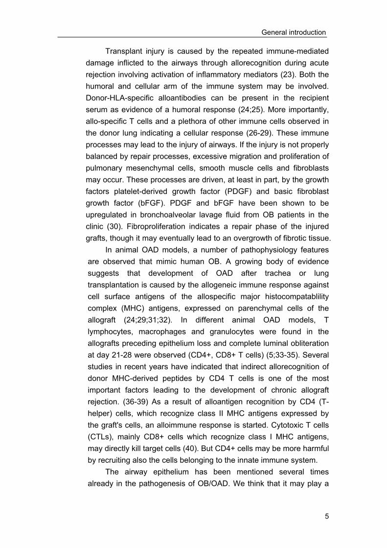
Transplant injury is caused by the repeated immune-mediated damage inflicted to the airways through allorecognition during acute rejection involving activation of inflammatory mediators (23). Both the humoral and cellular arm of the immune system may be involved. Donor-HLA-specific alloantibodies can be present in the recipient serum as evidence of a humoral response (24;25). More importantly, allo-specific T cells and a plethora of other immune cells observed in the donor lung indicating a cellular response (26-29). These immune processes may lead to the injury of airways. If the injury is not properly balanced by repair processes, excessive migration and proliferation of pulmonary mesenchymal cells, smooth muscle cells and fibroblasts may occur. These processes are driven, at least in part, by the growth factors platelet-derived growth factor (PDGF) and basic fibroblast growth factor (bFGF). PDGF and bFGF have been shown to be upregulated in bronchoalveolar lavage fluid from OB patients in the clinic (30). Fibroproliferation indicates a repair phase of the injured grafts, though it may eventually lead to an overgrowth of fibrotic tissue.
In animal OAD models, a number of pathophysiology features are observed that mimic human OB. A growing body of evidence suggests that development of OAD after trachea or lung transplantation is caused by the allogeneic immune response against cell surface antigens of the allospecific major histocompatablility complex (MHC) antigens, expressed on parenchymal cells of the allograft (24;29;31;32). In different animal OAD models, T lymphocytes, macrophages and granulocytes were found in the allografts preceding epithelium loss and complete luminal obliteration at day 21-28 were observed (CD4+, CD8+ T cells) (5;33-35). Several studies in recent years have indicated that indirect allorecognition of donor MHC-derived peptides by CD4 T cells is one of the most important factors leading to the development of chronic allograft rejection. (36-39) As a result of alloantigen recognition by CD4 (T-helper) cells, which recognize class II MHC antigens expressed by the graft's cells, an alloimmune response is started. Cytotoxic T cells (CTLs), mainly CD8+ cells which recognize class I MHC antigens, may directly kill target cells (40). But CD4+ cells may be more harmful by recruiting also the cells belonging to the innate immune system.
The airway epithelium has been mentioned several times already in the pathogenesis of OB/OAD. We think that it may play a
CHAPTER 1
6
role both in the induction of the injury and in the repair process. In the following part we will focus on this aspect.
2. Airway epithelium in transplantation
2.1. Airway epithelium structure and function
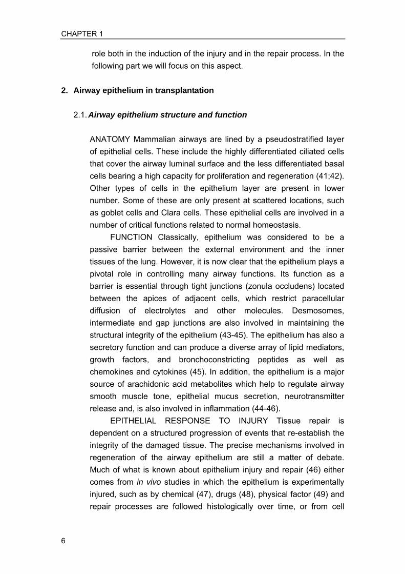
ANATOMY Mammalian airways are lined by a pseudostratified layer of epithelial cells. These include the highly differentiated ciliated cells that cover the airway luminal surface and the less differentiated basal cells bearing a high capacity for proliferation and regeneration (41;42). Other types of cells in the epithelium layer are present in lower number. Some of these are only present at scattered locations, such as goblet cells and Clara cells. These epithelial cells are involved in a number of critical functions related to normal homeostasis.
FUNCTION Classically, epithelium was considered to be a passive barrier between the external environment and the inner tissues of the lung. However, it is now clear that the epithelium plays a pivotal role in controlling many airway functions. Its function as a barrier is essential through tight junctions (zonula occludens) located between the apices of adjacent cells, which restrict paracellular diffusion of electrolytes and other molecules. Desmosomes, intermediate and gap junctions are also involved in maintaining the structural integrity of the epithelium (43-45). The epithelium has also a secretory function and can produce a diverse array of lipid mediators, growth factors, and bronchoconstricting peptides as well as chemokines and cytokines (45). In addition, the epithelium is a major source of arachidonic acid metabolites which help to regulate airway smooth muscle tone, epithelial mucus secretion, neurotransmitter release and, is also involved in inflammation (44-46).
EPITHELIAL RESPONSE TO INJURY Tissue repair is dependent on a structured progression of events that re-establish the integrity of the damaged tissue. The precise mechanisms involved in regeneration of the airway epithelium are still a matter of debate. Much of what is known about epithelium injury and repair (46) either comes from in vivo studies in which the epithelium is experimentally injured, such as by chemical (47), drugs (48), physical factor (49) and repair processes are followed histologically over time, or from cell

General introduction
7
culture models (50). It is clear that the airway epithelium has a tremendous capacity to repair itself after injury. In a recent in vivo study, Erjefalt et al. showed that in as little as 15 min after an 800-µm wide wound was made in the tracheal epithelium of guinea pigs, epithelial cells on the damaged margin (including secretory and ciliated cells) began to dedifferentiate, flatten and migrate over the denuded area (51). In this study, the denuded area was completely covered by a thin layer of flattened undifferentiated epithelium. Proliferation of epithelial cells was not observed until about 30 hours after the initial stimuli. Within 5 days a fully differentiated epithelium was present again. At approximately the same time as cells begin to differentiate, a plasma exudate was observed which might be an indication of functional repair (52-55).
2.2. Airway epithelium injury during transplantation
Airway epithelium injury is frequently observed upon transplantation. Airway epithelium damage may be already induced before transplantation by the donor's brain death (56), and by ischemia-reperfusion (57-59) during the transplantation procedures. Evidence of inflammatory cytokines up-regulation such as interleukin (IL)-1, IL-2, IL-6, tumor necrotic factor alpha (TNF-α) and interferon gama (IFN-γ) is shown in peripheral organs including lung tissue from brain-dead donors shortly after harvesting (60). It is known that TNF-α and IL-1 are the “early response cytokines” produced by alveolar macrophages in acute lung injury through activation of the transcription factor nuclear factor-kappa B (NF-kB) (61). Upon transplantation, these cytokines induce inflammatory responses towards donor epithelial cells by attracting recipient macrophages and other inflammatory cells (62). This may contribute to the acute injury seen in donor tissue after transplantation. Furthermore, donor tissue that expresses allo MHC antigens induces cellular and humoral alloimmunity in the recipient. Human epithelial cells are known to continuously express MHC antigens in high density (63,64). A recent study shows that the binding of MHC alloantibodies to human epithelial cells induces apoptosis of these cells (65). Epithelial cells apoptosis was also observed in animal airway transplant OAD (66).

CHAPTER 1
8
Clinical pathologic features of OB suggest that in particular, the injury of epithelial cells by persistent inflammation in small airways hampers epithelium regeneration and stimulates fibroproliferation due to aberrant tissue repair. In animal tracheal transplants, airway epithelium is involved in the rejection process as one of the targeted tissue leading to OAD (67). The loss of airway epithelium by transplant rejection in allografts but also by enzymatic removal in isografts resulted in OAD (68;69). This stresses the importance of epithelium for the development of OB/OAD. Figure 1. Hypothesis of airway epithelium in OAD/OB occurence
In the illustration, airway allograft is transplanted and rejection occurs. There are two type of rejecting process: 1. Graft-independent process (left down arrows). It is caused by normal grafting surgical procedures and foreign body responses. It occurs to every type of grafting and is not dependdent on the type of grafts. Epithelium mostly experiences ischemia-reperfusion damage and could quickly recover. 2. Graft-dependent process (right down arrows). It is caused by alloimmune responses that recognize allo-MHC antigens expressed in allografts. It occurs only in the allgraft transplantation and is suggested to be the main cause of graft injury and dysfunction.

General introduction
9
3. Hypothesis and experimental models
3.1. Hypothesis of OB/OAD Studies in patients and animal models indicate that donor airway epithelium injury is playing a role in the process of OB (6;70) and OAD (71). We hypothesize that transplant injury causes airway epithelium damage and that excessive and persistent loss of epithelium results in fibrosis that eventually leads to the occurrence of OB/OAD. Firstly, airway epithelium is one of the primary target tissues during alloimmune responses that are graft-dependent. It is also the target of graft-independent factors, such as general inflammatory response and transplant surgical injury. This type of injury is presented among all types of airway tissue (67) and is largely dominated by alloimmunity. Loss of epithelium during injury ‘denudes’ the transplanted airway, resulting in loss of the defense barrier and, generally, epithelium dysfunction. As a result, the submucosa tissue may directly encounter foreign pathogens that increase the risk of infection. Secondly, damaged airway epithelium may lose its own functional role in regulating the airway repair process during injury (72). In response to injury, airway epithelial cells are capable to dedifferentiate and to regenerate to replace the injured cells, as indicated above. In vitro (73;74)and vivo (75) studies have shown that airway epithelial cells also regulate fibroblast proliferation. Normal epithelial cells inhibit fibroblast proliferation in vitro probably by excretion of inhibitory cytokines (72;76). More specific investigation of the response of epithelium to transplantaton injury seems justified. At the moment, however, we have no good model directly focusing on role of airway epithelial cells in the development of OB/OAD.
3.2. Transgenic animal model for epithelium specific immune
response in OAD study Trachea transplantation is a convenient technique for investigation of epithelial responses. A drawback of studying OAD in an allogeneic tracheal transplant is that immune responses are directed against all cell types of the graft tissue. This is so, because the recipient’s immune system recognizes alloantigens on all cells expressing these

CHAPTER 1
10
antigens. So all cells in the trachea grafts are attacked and is impossible to investigate specifically the airway epithelium injury and its effects in OAD development after transplantation.
Transgenic animals expressing a neoantigen on a specific type of tissue may provide a new option to investigate the role of epithelium specific response in OAD. In a recent study, trachea transplants from HLA-class I transgenic mice were shown to induce alloreactive CD4 T cells and alloantibodies against the HLA-class I neoantigens (77). Although this is clearly an improved model to investigate the role of a single alloantigen (HLA-class I) in OAD, it still has a major drawback, since all types of tissue cells may express the neoantigen, making it not suitable for the study of single type of tissue, such as epithelium. To allow investigation of immune injury to be directed against an epithelium specific neoantigen in mouse tracheas, we decided to use the human epithelial glycoprotein-2 (hEGP-2) transgenic mice as donors. In these transgenic mice, hEGP-2 antigens are expressed exclusively on epithelial cells. Using hEGP-2 transgenic mice as donors, we were able to investigate whether the hEGP-2 antigen induces epithelium specific immune response after trachea transplantation and whether the response causes epithelial injury in the transgenic trachea transplants. This model also gave the opportunity to immunize recipients prospectively with the hEGP-2 antigen before transplantation. In this way, a pre-existing immunity directed exclusively to transplanted epithelium was induced.
3.3. Blockade of immune responses and airway epithelium protection
In clinical transplantation, immunosuppressive agents such as cyclosporine A (CsA), FK506 and rapamycin have been shown to effectively prolong graft survival (78). Using these drugs, alloreactive T cell responses are either reduced or blocked. Possible side effects, such as the CsA involvement in pro-fibroproliferation (79) or the pro-inflammatory effect of CsA and FK506 (11), are hampering the full exploration of these therapies. Another approach is the treatment by (monoclonal) antibodies against T cells, such as Anti-thymocyte Globulin (80-82), Anti-Cytotoxic T Lymphocyte Antigen-4 (83) or antibodies against CD2 and CD3 T cell common antigens (84). These antibodies have been used for depletion of T cells for prolong graft

General introduction
11
survival. However, all these drugs are not specific for alloreactive T cells but modulate other normal T cells as well. This increases the recipient sensitivity to infection as the body defense system is suppressed.
Recently, a novel monotherapy using anti-CD45RB monoclonal antibodies (mAb) to deplete recipient T cells was investigated to block T cell responses and to prolong allograft survival (85). The CD45RB molecule belongs to a family of transmembrane protein tyrosine phosphatases that is expressed by leukocytes and play a critical role in regulating T-cell activation through modulating the activation status of the T cell receptor. In animal studies, mAb MB23G2 against CD45RB prolonged allograft survival through T cell depletion. These antibodies are T cell specific, easy in management and cause no severe side effect in animal models. Prolonged survival has been demonstrated after engraftment of islets (86), hearts and kidneys in MHC disparate mice after the treatment of recipients with anti-CD45RB mAb (87;88). Therefore, it seems worthwhile to investigate if a protective effect on transplant airway epithelium preventing OAD can be found in our mouse trachea transplant model.
4. Main issues addressed and the scope of this thesis
4.1. Epithelium dynamics during injury and its role in OAD
It is essential to know the behavior of airway epithelium upon transplantation injury and to establish its role in airway obliteration. We transplanted rat tracheas to observe airway changes post transplantation in a rat model (chapter 2). Injury was caused by rejection of the MHC fully mismatched tracheas and by enzymatic denudation of syngeneic transplants. Analyses were focused on epithelium integrity, the time point at which fibroproliferation and obliteration occurred. A series of experiments was carried out to stimulate repair of airway epithelium by isolation and reseeding of epithelial cells in trachea transplants.
4.2. Epithelium specific immune responses In chapter 3, we introduced the hEGP-2 transgenic mouse OAD model for the study of epithelium specific immune responses. The

CHAPTER 1
12
recipient immune responses against the transgenic grafts were studied in regards of both humoral and cellular reactivity. The effect of the immune responses on epithelium and its subsequent effect on OAD were analyzed. This is for the first time that the effect of a single type of tissue specific immune response is studied in a transplant setting.
4.3. OAD development under pre-existing immunity In chapter 4 the OAD development under pre-existing epithelial cell specific immunity was studied further. Recipient mice were immunized before transplantation to induce pre-existing immunity for hEGP-2 grafts. The reasoning is to mimic the immunity in allotransplantation where alloantibodies and T cells may directly recognize alloantigens. In our model, however, the pre-existing immunity was directed exclusively towards epithelium. This allowed us to analyse epithelium behavior and its effect on OAD development.
4.4. Anti-CD45RB antibody monotherapy to prevent OAD
The prevention of OAD by blocking allreactive T cells in an allogeneic trachea transplantation model is evaluated in chapter 5. To reduce the recipient T cells, a leukocytes antibody anti-CD45RB was administered in this model and the effect of this antibody on epithelium protection and OAD development was evaluated.

General introduction
13
Reference 1. Taylor DO, Edwards LB, Boucek MM, Trulock EP, Keck BM, Hertz MI.
The Registry of the International Society for Heart and Lung Transplantation: twenty-first official adult lung transplant report--2004.J Heart Lung Transplant. 2004 Jul;23(7):796-803.
2. Heng D, Sharples LD, McNeil K, Stewart S, Wreghitt T, Wallwork J. Bronchiolitis obliterans syndrome: incidence, natural history, prognosis, and risk factors. J Heart Lung Transplant 1998:17: 1255-1263.
3. Boehler A, Kesten S, Weder W, Speich R. Bronchiolitis obliterans after lung transplantation: a review. Chest 1998:114: 1411-1426.
4. Estenne M, Hertz MI. Bronchiolitis obliterans after human lung transplantation. Am J Respir Crit Care Med 2002:166: 440-444.
5. Reynaud-Gaubert M. [Pathophysiology of obliterative bronchiolitis in lung transplants]. Rev Mal Respir 2003:20: 224-232.
6. Paradis I. Bronchiolitis obliterans: pathogenesis, prevention, and management. Am J Med Sci 1998:315: 161-178.
7. Sundaresan S, Trulock EP, Mohanakumar T, Cooper JD, Patterson GA. Prevalence and outcome of bronchiolitis obliterans syndrome after lung transplantation. Washington University Lung Transplant Group. Ann Thorac Surg 1995:60: 1341-1346.
8. Allen MD, Burke CM, McGregor CG, Baldwin JC, Jamieson SW, Theodore J. Steroid-responsive bronchiolitis after human heart-lung transplantation. J Thorac Cardiovasc Surg 1986:92: 449-451.
9. Bando K, Paradis IL, Similo S et al. Obliterative bronchiolitis after lung and heart-lung transplantation. An analysis of risk factors and management. J Thorac Cardiovasc Surg 1995:110: 4-13.
10. Boehler A, Estenne M. Obliterative bronchiolitis after lung transplantation. Curr Opin Pulm Med 2000:6: 133-139.
11. Borger P, Kauffman HF, Timmerman JA, Scholma J, van den Berg JW, Koeter GH. Cyclosporine, FK506, mycophenolate mofetil, and prednisolone differentially modulate cytokine gene expression in human airway-derived epithelial cells. Transplantation 2000:69: 1408-1413.
12. Sundaresan S. Bronchiolitis obliterans. Semin Thorac Cardiovasc Surg 1998:10: 221-226.
13. Prop J, Ehrie MG, Crapo JD, Nieuwenhuis P, Wildevuur CR. Reimplantation response in isografted rat lungs. Analysis of causal factors. J Thorac Cardiovasc Surg 1984:87: 702-711.

CHAPTER 1
14
14. Tazelaar HD, Prop J, Nieuwenhuis P, Billingham ME, Wildevuur CR. Airway pathology in the transplanted rat lung 128. Transplantation 1988:45: 864-869.
15. Winter JB, Gouw AS, Groen M, Wildevuur C, Prop J. Respiratory viral infections aggravate airway damage caused by chronic rejection in rat lung allografts. Transplantation 1994:57: 418-422.
16. Hertz MI, Jessurun J, King MB, Savik SK, Murray JJ. Reproduction of the obliterative bronchiolitis lesion after heterotopic transplantation of mouse airways. Am J Pathol 1993:142: 1945-1951.
17. Kawahara K, Hiratsuka M, Mikami K et al. Obliterative airway disease and graft stenting in pig-to-dog tracheal xenotransplantation. Jpn J Thorac Cardiovasc Surg 2001:49: 53-57.
18. Ikonen T, Uusitalo M, Taskinen E et al. Small airway obliteration in a new swine heterotopic lung and bronchial allograft model. J Heart Lung Transplant 1998:17: 945-953.
19. Salminen US, Ikonen T, Uusitalo M et al. Obliterative lesions in small airways in an immunosuppressed porcine heterotopic bronchial allograft model. Transpl Int 1998:11 Suppl 1: S515-S518.
20. Uusitalo MH, Salminen US, Ikonen TS et al. Alloimmune injury preceding airway obliteration in porcine heterotopic lung implants: a histologic and immunohistologic study. Transplantation 1999:68: 970-975.
21. Hausen B, Berry GJ, Dagum P et al. The histology of subcutaneously implanted donor bronchial rings correlates with rejection scores of lung allografts in a primate lung transplant model. J Heart Lung Transplant 1999:18: 714-724.
22. Hele DJ, Yacoub MH, Belvisi MG. The heterotopic tracheal allograft as an animal model of obliterative bronchiolitis. Respir Res 2001:2: 169-183.
23. Heng D, Sharples LD, McNeil K, Stewart S, Wreghitt T, Wallwork J. Bronchiolitis obliterans syndrome: incidence, natural history, prognosis, and risk factors. J Heart Lung Transplant 1998:17: 1255-1263.
24. Reznik SI, Jaramillo A, Zhang L, Patterson GA, Cooper JD, Mohanakumar T. Anti-HLA antibody binding to hla class I molecules induces proliferation of airway epithelial cells: a potential mechanism for bronchiolitis obliterans syndrome. J Thorac Cardiovasc Surg 2000:119: 39-45.
25. Jaramillo A, Smith MA, Phelan D et al. Development of ELISA-detected anti-HLA antibodies precedes the development of bronchiolitis obliterans

General introduction
15
syndrome and correlates with progressive decline in pulmonary function after lung transplantation. Transplantation 1999:67: 1155-1161.
26. Neuringer IP, Mannon RB, Coffman TM et al. Immune cells in a mouse airway model of obliterative bronchiolitis. Am J Respir Cell Mol Biol 1998:19: 379-386.
27. Boehler A, Chamberlain D, Kesten S, Slutsky AS, Liu M, Keshavjee S. Lymphocytic airway infiltration as a precursor to fibrous obliteration in a rat model of bronchiolitis obliterans. Transplantation 1997:64: 311-317.
28. Qu N, De Haan A, Harmsen MC, Kroese FG, De Leij LF, Prop J. Specific immune responses against airway epithelial cells in a transgenic mouse-trachea transplantation model for obliterative airway disease. Transplantation 2003:76: 1022-1028.
29. Kelly KE, Hertz MI, Mueller DL. T-cell and major histocompatibility complex requirements for obliterative airway disease in heterotopically transplanted murine tracheas. Transplantation 1998:66: 764-771.
30. Hertz MI, Henke CA, Nakhleh RE et al. Obliterative bronchiolitis after lung transplantation: a fibroproliferative disorder associated with platelet-derived growth factor. Proc Natl Acad Sci U S A 1992:89: 10385-10389.
31. Smith MA, Jaramillo A, SivaSai KS et al. Indirect recognition and antibody production against a single mismatched HLA-A2-transgenic molecule precede the development of obliterative airway disease in murine heterotopic tracheal allografts. Transplantation 2002:73: 186-193.
32. Sherman LA, Chattopadhyay S. The molecular basis of allorecognition. Annu Rev Immunol 1993:11: 385-402.
33. Higuchi T, Jaramillo A, Kaleem Z, Patterson GA, Mohanakumar T. Different kinetics of obliterative airway disease development in heterotopic murine tracheal allografts induced by CD4+ and CD8+ T cells. Transplantation 2002:74: 646-651.
34. Reynaud-Gaubert M, Thomas P, Badier M, Cau P, Giudicelli R, Fuentes P. Early detection of airway involvement in obliterative bronchiolitis after lung transplantation. Functional and bronchoalveolar lavage cell findings. Am J Respir Crit Care Med 2000:161: 1924-1929.
35. Neuringer IP, Mannon RB, Coffman TM et al. Immune cells in a mouse airway model of obliterative bronchiolitis. Am J Respir Cell Mol Biol 1998:19: 379-386.
36. Reznik SI, Jaramillo A, SivaSai KS et al. Indirect allorecognition of mismatched donor HLA class II peptides in lung transplant recipients with bronchiolitis obliterans syndrome. Am J Transplant 2001:1: 228-235.

CHAPTER 1
16
37. Richards DM, Dalheimer SL, Ehst BD et al. Indirect minor histocompatibility antigen presentation by allograft recipient cells in the draining lymph node leads to the activation and clonal expansion of CD4+ T cells that cause obliterative airways disease. J Immunol 2004:172: 3469-3479.
38. Higuchi T, Jaramillo A, Kaleem Z, Patterson GA, Mohanakumar T. Different kinetics of obliterative airway disease development in heterotopic murine tracheal allografts induced by CD4+ and CD8+ T cells. Transplantation 2002:74: 646-651.
39. Smith MA, Jaramillo A, SivaSai KS et al. Indirect recognition and antibody production against a single mismatched HLA-A2-transgenic molecule precede the development of obliterative airway disease in murine heterotopic tracheal allografts. Transplantation 2002:73: 186-193.
40. Gao GF, Jakobsen BK. Molecular interactions of coreceptor CD8 and MHC class I: the molecular basis for functional coordination with the T-cell receptor. Immunol Today 2000:21: 630-636.
41. Hong KU, Reynolds SD, Watkins S, Fuchs E, Stripp BR. Basal cells are a multipotent progenitor capable of renewing the bronchial epithelium. Am J Pathol 2004:164: 577-588.
42. Hong KU, Reynolds SD, Watkins S, Fuchs E, Stripp BR. In vivo differentiation potential of tracheal basal cells: evidence for multipotent and unipotent subpopulations. Am J Physiol Lung Cell Mol Physiol 2004:286: L643-L649.
43. Holgate ST, Lackie P, Wilson S, Roche W, Davies D. Bronchial epithelium as a key regulator of airway allergen sensitization and remodeling in asthma. Am J Respir Crit Care Med 2000:162: S113-S117.
44. Knight D. Epithelium-fibroblast interactions in response to airway inflammation. Immunol Cell Biol 2001:79: 160-164.
45. Knight DA, Holgate ST. The airway epithelium: structural and functional properties in health and disease. Respirology 2003:8: 432-446.
46. Holgate ST. Epithelial damage and response. Clin Exp Allergy 2000:30 Suppl 1: 37-41.
47. O'Brien DW, Morris MI, Ding J, Zayas JG, Tai S, King M. A mechanism of airway injury in an epithelial model of mucociliary clearance. Respir Res 2004:5: 10.
48. Adamson IY, Bowden DH. Bleomycin-induced injury and metaplasia of alveolar type 2 cells. Relationship of cellular responses to drug presence in the lung. Am J Pathol 1979:96: 531-544.

General introduction
17
49. Kay SS, Bilek AM, Dee KC, Gaver DP, III. Pressure gradient, not exposure duration, determines the extent of epithelial cell damage in a model of pulmonary airway reopening. J Appl Physiol 2004:97: 269-276.
50. Terzaghi-Howe M, Ford J. Effects of radiation on rat respiratory epithelial cells: critical target cell populations and the importance of cell-cell interactions. Adv Space Res 1994:14: 565-572.
51. Erjefalt JS, Erjefalt I, Sundler F, Persson CG. In vivo restitution of airway epithelium. Cell Tissue Res 1995:281: 305-316.
52. Erjefalt JS, Korsgren M, Nilsson MC, Sundler F, Persson CG. Prompt epithelial damage and restitution processes in allergen challenged guinea-pig trachea in vivo. Clin Exp Allergy 1997:27: 1458-1470.
53. Erjefalt JS, Sundler F, Persson CG. Epithelial barrier formation by airway basal cells. Thorax 1997:52: 213-217.
54. Persson CG. Epithelial cells: barrier functions and shedding-restitution mechanisms. Am J Respir Crit Care Med 1996:153: S9-10.
55. Erjefalt JS, Erjefalt I, Sundler F, Persson CG. Microcirculation-derived factors in airway epithelial repair in vivo. Microvasc Res 1994:48: 161-178.
56. Fisher AJ, Donnelly SC, Hirani N et al. Enhanced pulmonary inflammation in organ donors following fatal non-traumatic brain injury. Lancet 1999:353: 1412-1413.
57. Soria A, Vicente R, Ramos F, Lopez LM, Francia C, Montero R. [Lesion caused by ischemia-reperfusion in lung transplantation]. Rev Esp Anestesiol Reanim 2000:47: 380-385.
58. Clark SC, Sudarshan C, Khanna R, Roughan J, Flecknell PA, Dark JH. Controlled reperfusion and pentoxifylline modulate reperfusion injury after single lung transplantation. J Thorac Cardiovasc Surg 1998:115: 1335-1341.
59. Eppinger MJ, Deeb GM, Bolling SF, Ward PA. Mediators of ischemia-reperfusion injury of rat lung. Am J Pathol 1997:150: 1773-1784.
60. Takada M, Nadeau KC, Hancock WW et al. Effects of explosive brain death on cytokine activation of peripheral organs in the rat. Transplantation 1998:65: 1533-1542.
61. Lentsch AB, Ward PA. Regulation of experimental lung inflammation. Respir Physiol 2001:128: 17-22.
62. Rizzo M, SivaSai KS, Smith MA et al. Increased expression of inflammatory cytokines and adhesion molecules by alveolar macrophages of human lung allograft recipients with acute rejection:
CHAPTER 1
18
decline with resolution of rejection. J Heart Lung Transplant 2000:19: 858-865.
63. Cunningham AC, Milne DS, Wilkes J, Dark JH, Tetley TD, Kirby JA. Constitutive expression of MHC and adhesion molecules by alveolar epithelial cells (type II pneumocytes) isolated from human lung and comparison with immunocytochemical findings. J Cell Sci 1994:107 ( Pt 2): 443-449.
64. Cunningham AC, Zhang JG, Moy JV, Ali S, Kirby JA. A comparison of the antigen-presenting capabilities of class II MHC-expressing human lung epithelial and endothelial cells. Immunology 1997:91: 458-463.
65. Jaramillo A, Smith CR, Maruyama T, Zhang L, Patterson GA, Mohanakumar T. Anti-HLA class I antibody binding to airway epithelial cells induces production of fibrogenic growth factors and apoptotic cell death: a possible mechanism for bronchiolitis obliterans syndrome 84. Hum Immunol 2003:64: 521-529.
66. Alho HS, Salminen US, Maasilta PK, Paakko P, Harjula AL. Epithelial apoptosis in experimental obliterative airway disease after lung transplantation. J Heart Lung Transplant 2003:22: 1014-1022.
67. Fernandez FG, Jaramillo A, Chen C et al. Airway epithelium is the primary target of allograft rejection in murine obliterative airway disease 87. Am J Transplant 2004:4: 319-325.
68. Hele DJ, Yacoub MH, Belvisi MG. The heterotopic tracheal allograft as an animal model of obliterative bronchiolitis. Respir Res 2001:2: 169-183.
69. Adams BF, Brazelton T, Berry GJ, Morris RE. The role of respiratory epithelium in a rat model of obliterative airway disease. Transplantation 2000:69: 661-664.
70. Mauck KA, Hosenpud JD. The bronchial epithelium: a potential allogeneic target for chronic rejection after lung transplantation. J Heart Lung Transplant 1996:15: 709-714.
71. Adams BF, Brazelton T, Berry GJ, Morris RE. The role of respiratory epithelium in a rat model of obliterative airway disease. Transplantation 2000:69: 661-664.
72. Holgate ST. Epithelial damage and response. Clin Exp Allergy 2000:30 Suppl 1: 37-41.
73. Nakamura Y, Tate L, Ertl RF et al. Bronchial epithelial cells regulate fibroblast proliferation. Am J Physiol 1995:269: L377-L387.

General introduction
19
74. Pan T, Mason RJ, Westcott JY, Shannon JM. Rat alveolar type II cells inhibit lung fibroblast proliferation in vitro. Am J Respir Cell Mol Biol 2001:25: 353-361.
75. Moore BB, Peters-Golden M, Christensen PJ et al. Alveolar epithelial cell inhibition of fibroblast proliferation is regulated by MCP-1/CCR2 and mediated by PGE2. Am J Physiol Lung Cell Mol Physiol 2003:284: L342-L349.
76. Sacco O, Silvestri M, Sabatini F, Sale R, Defilippi AC, Rossi GA. Epithelial cells and fibroblasts: structural repair and remodelling in the airways. Paediatr Respir Rev 2004:5 Suppl A: S35-S40.
77. Smith MA, Jaramillo A, SivaSai KS et al. Indirect recognition and antibody production against a single mismatched HLA-A2-transgenic molecule precede the development of obliterative airway disease in murine heterotopic tracheal allografts. Transplantation 2002:73: 186-193.
78. Garrity ER, Jr., Mehra MR. An update on clinical outcomes in heart and lung transplantation. Transplantation 2004:77: S68-S74.
79. Hostettler KE, Roth M, Burgess JK et al. Cyclosporine A mediates fibroproliferation through epithelial cells 85. Transplantation 2004:77: 1886-1893.
80. Brock MV, Borja MC, Ferber L et al. Induction therapy in lung transplantation: a prospective, controlled clinical trial comparing OKT3, anti-thymocyte globulin, and daclizumab. J Heart Lung Transplant 2001:20: 1282-1290.
81. Diamond DA, Michalski JM, Lynch JP, Trulock EP, III. Efficacy of total lymphoid irradiation for chronic allograft rejection following bilateral lung transplantation. Int J Radiat Oncol Biol Phys 1998:41: 795-800.
82. van Tiel FH, Rasmussen L, Merigan TC. Cytomegalovirus-specific cell-mediated immune responses in heart and heart-lung transplant recipients are not predictive for the occurrence of symptomatic CMV disease or tissue rejection. J Interferon Res 1991:11: 221-229.
83. Perico N, Imberti O, Bontempelli M, Remuzzi G. Toward novel antirejection strategies: in vivo immunosuppressive properties of CTLA4Ig. Kidney Int 1995:47: 241-246.
84. Chavin KD, Qin L, Lin J, Yagita H, Bromberg JS. Combined anti-CD2 and anti-CD3 receptor monoclonal antibodies induce donor-specific tolerance in a cardiac transplant model. J Immunol 1993:151: 7249-7259.

CHAPTER 1
20
85. Gao Z, Zhong R, Jiang J et al. Adoptively transferable tolerance induced by CD45RB monoclonal antibody. J Am Soc Nephrol 1999:10: 374-381.
86. Visser L, Poppema S, de Haan B et al. Prolonged survival of rat islet xenografts in mice after CD45RB monotherapy. Transplantation 2004:77: 386-391.
87. Zhang Z, Lazarovits A, Grant D, Garcia B, Stiller C, Zhong R. CD45RB monoclonal antibody induces tolerance in the mouse kidney graft, but fails to prevent small bowel graft rejection. Transplant Proc 1996:28: 2514.
88. Ko S, Jager MD, Tsui TY et al. Long-term allograft acceptance induced by single dose anti-leukocyte common antigen (RT7) antibody in the rat. Transplantation 2001:71: 1124-1131.