
University of Groningen A Cadaveric Comparative Study on ... · University of Groningen A Cadaveric...
11
University of Groningen A Cadaveric Comparative Study on the Surgical Accuracy of Freehand, Computer Navigation, and Patient-Specific Instruments in Joint-Preserving Bone Tumor Resections Bosma, Sarah E; Wong, Kwok Chuen; Paul, Laurent; Gerbers, Jasper G; Jutte, Paul C Published in: Sarcoma DOI: 10.1155/2018/4065846 IMPORTANT NOTE: You are advised to consult the publisher's version (publisher's PDF) if you wish to cite from it. Please check the document version below. Document Version Publisher's PDF, also known as Version of record Publication date: 2018 Link to publication in University of Groningen/UMCG research database Citation for published version (APA): Bosma, S. E., Wong, K. C., Paul, L., Gerbers, J. G., & Jutte, P. C. (2018). A Cadaveric Comparative Study on the Surgical Accuracy of Freehand, Computer Navigation, and Patient-Specific Instruments in Joint- Preserving Bone Tumor Resections. Sarcoma, 2018, [4065846]. https://doi.org/10.1155/2018/4065846 Copyright Other than for strictly personal use, it is not permitted to download or to forward/distribute the text or part of it without the consent of the author(s) and/or copyright holder(s), unless the work is under an open content license (like Creative Commons). Take-down policy If you believe that this document breaches copyright please contact us providing details, and we will remove access to the work immediately and investigate your claim. Downloaded from the University of Groningen/UMCG research database (Pure): http://www.rug.nl/research/portal. For technical reasons the number of authors shown on this cover page is limited to 10 maximum. Download date: 14-05-2020
Transcript of University of Groningen A Cadaveric Comparative Study on ... · University of Groningen A Cadaveric...

University of Groningen
A Cadaveric Comparative Study on the Surgical Accuracy of Freehand, Computer Navigation,and Patient-Specific Instruments in Joint-Preserving Bone Tumor ResectionsBosma, Sarah E; Wong, Kwok Chuen; Paul, Laurent; Gerbers, Jasper G; Jutte, Paul C
Published in:Sarcoma
DOI:10.1155/2018/4065846
IMPORTANT NOTE: You are advised to consult the publisher's version (publisher's PDF) if you wish to cite fromit. Please check the document version below.
Document VersionPublisher's PDF, also known as Version of record
Publication date:2018
Link to publication in University of Groningen/UMCG research database
Citation for published version (APA):Bosma, S. E., Wong, K. C., Paul, L., Gerbers, J. G., & Jutte, P. C. (2018). A Cadaveric Comparative Studyon the Surgical Accuracy of Freehand, Computer Navigation, and Patient-Specific Instruments in Joint-Preserving Bone Tumor Resections. Sarcoma, 2018, [4065846]. https://doi.org/10.1155/2018/4065846
CopyrightOther than for strictly personal use, it is not permitted to download or to forward/distribute the text or part of it without the consent of theauthor(s) and/or copyright holder(s), unless the work is under an open content license (like Creative Commons).
Take-down policyIf you believe that this document breaches copyright please contact us providing details, and we will remove access to the work immediatelyand investigate your claim.
Downloaded from the University of Groningen/UMCG research database (Pure): http://www.rug.nl/research/portal. For technical reasons thenumber of authors shown on this cover page is limited to 10 maximum.
Download date: 14-05-2020

Research ArticleA Cadaveric Comparative Study on the Surgical Accuracy ofFreehand, Computer Navigation, and Patient-SpecificInstruments in Joint-Preserving Bone Tumor Resections
Sarah E. Bosma ,1 Kwok Chuen Wong,2 Laurent Paul,3 Jasper G. Gerbers,1
and Paul C. Jutte 1
1Department of Orthopedics, University Medical Center Groningen, P.O. Box 30.001, 9700 RB Groningen, Netherlands2Department of Orthopedics, Prince of Wales Hospital, Sha Tin, Hong Kong33D-Side, 1348 Louvain-la-Neuve, Belgium
Correspondence should be addressed to Sarah E. Bosma; [email protected]
Received 12 July 2018; Accepted 17 October 2018; Published 13 November 2018
Academic Editor: Ajay Puri
Copyright © 2018 Sarah E. Bosma et al.(is is an open access article distributed under the Creative CommonsAttribution License,which permits unrestricted use, distribution, and reproduction in any medium, provided the original work is properly cited.
Orthopedic oncologic surgery requires preservation of a functioning limb at the essence of achieving safe margins. With mostbone sarcomas arising from the metaphyseal region, in close proximity to joints, joint-salvage surgery can be challenging.Intraoperative guidance techniques like computer-assisted surgery (CAS) and patient-specific instrumentation (PSI) could assistin achieving higher surgical accuracy. (is study investigates the surgical accuracy of freehand, CAS- and PSI-assisted joint-preserving tumor resections and tests whether integration of CAS with PSI (CAS+PSI) can further improve accuracy. CTscans of16 simulated tumors around the knee in four human cadavers were performed and imported into engineering software (MIMICS)for 3D planning of multiplanar joint-preserving resections. (e planned resections were transferred to the navigation systemand/or used for PSI design. Location accuracy (LA), entry and exit points of all 56 planes, and resection time were measured bypostprocedural CT. Both CAS+PSI- and PSI-assisted techniques could reproduce planned resections with a mean LA of less than2mm. (ere was no statistical difference in LA between CAS + PSI and PSI resections (p � 0.92), but both CAS + PSI and PSIshowed a significantly higher LA compared to CAS (p � 0.042 and p � 0.034, respectively). PSI-assisted resections were fastercompared to CAS+PSI (p< 0.001) and CAS (p< 0.001). Adding CAS to PSI did improve the exit points, however not sig-nificantly. In conclusion, PSI showed the best overall surgical accuracy and is fastest and easy to use. CAS could be used as anintraoperative quality control tool for PSI, and integration of CAS with PSI is possible but did not improve surgical accuracy. BothCAS and PSI seem complementary in improving surgical accuracy and are not mutually exclusive. Image-based techniques likeCAS and PSI are superior over freehand resection. Surgeons should choose the technique most suitable based on the patient andtumor specifics.
1. Introduction
Orthopedic oncologic surgery requires the preservation ofa functioning limb at the essence of achieving safe margins. Upuntil the 1970s, amputation was the first choice of treatment forbone tumors, with a survival rate of only 11% [1]. Nowadays,with the developments in diagnostic imaging, surgical tech-niques, and adjuvant therapies (e.g., chemotherapy), the em-phasis is on limb-salvage surgery [2, 3]. Limb-salvage surgery
aims to preserve as much unaffected tissue as possible withoutcompromising safe tumor margins. (is improves local con-trol, benefits the reconstruction, and reduces the morbidity,thereby improving the short- and long-term functional out-comes.Withmost bone sarcomas arising from themetaphysealregion, in close proximity to joints and neurovascular andvisceral structures, joint-salvage surgery could be challenging[4–6]. Surgical accuracy is therefore essential in joint-salvageorthopedic oncologic surgery.
HindawiSarcomaVolume 2018, Article ID 4065846, 9 pageshttps://doi.org/10.1155/2018/4065846

A resection can be performed “freehand.” Using thistechnique, the surgeon must intraoperatively rely on two-dimensional (2D) preoperatively acquired images (computertomography (CT) or magnetic resonance imaging (MRI))and mentally integrate these into a three-dimensional (3D)intraoperative surgical situation [4]. (is limited intra-operative guidance might result in surgical inaccuracy inbone resections, as was proven even for experienced sur-geons [7, 8]. Computer-assisted surgery (CAS) and patient-specific instrumentation (PSI) have been introduced toaddress the inaccuracy of tumor resections and improvepredictability of bone tumor resections [9].
CAS, or surgical navigation, was initially used to im-prove pedicle screw insertion in the spine and reduceoutliners in component alignment in total knee arthro-plasties [10, 11]. Later, it gained more acceptance andshowed its advantages in orthopedic oncologic surgery [12].It allows linking between patients’ imaging information andanatomy by tracking a registration of the preoperativeimages (CTand MRI) and the patient on the operating table.(e surgeon can preoperatively plan the bone tumor re-section in a 3D virtual scenario and analyze different al-ternatives for resection. Additionally, during the surgery,there is real-time 3D radiation-free visual feedback [13, 14].Previous research has shown that CAS aids in achievingadequate margins [15, 16] and improves the accuracy of theosteotomies [4, 6, 17, 18]. However, not all surgical tools arereal-time navigated, and a reliable navigated saw is often notavailable; therefore, this accuracy and precision may be lostwhen the actual cut is performed, since the human hand isguiding the saw.
PSI was introduced for resection of bony parts duringprosthesis placement and osteotomies. Later, it was alsointroduced to guide tumor resections [19–21]. In the samefashion as for CAS, control over safe margins is provided byaccurate preplanning and subsequent accurate intra-operative guiding of the resection planes. One face of theguide is the (negative) surface of the bone representing anautomatic matching of the patient with his preoperativeimages, fitting onto the bone surface in one possible con-figuration. During the actual cut, the saw blade is alignedwith the guide and thereby provides actual guidance duringthe resection. A possible limitation of PSI is an inaccurate fitto the bone, due to lack of surface characteristics, designflaws, and soft tissue extension that could lead to PSImalposition, all resulting in loss of accuracy. Anotherlimitation is that PSIs rely on sufficient bone exposure andcan require a larger dissection.
(us, CAS has the advantage of providing real-time dataon the spatial position but currently not a plane cut due tolack of a reliable navigated saw. (erefore, the accuracy islost as soon as the human hand comes into play for the actualresection [4, 12–14]. PSI is very accurate in guiding a cuttingplane but lacks feedback on its actual position other than theclose fit to the bone surface which needs to be cleaned fromsoft tissue, and PSI needs to be devoid of design flaws. Joint-preserving surgery has been performed in select patientswith bone sarcomas of extremities and allows patients toretain the native joint with better joint function [1].
However, it is technically demanding as it is difficult totranslate preoperative CT/MR tumor extent to the patients’intraoperative anatomy.
Given the importance of accurate surgical margins inresection of a primary bone sarcoma, a comparative study ofdifferent techniques in a cadaveric experimental setting isessential to compare the accuracy of the various techniques.Also to our knowledge, the use of PSI for bone tumor re-section in the knee region was not adequately assessed exceptin the small clinical series of Bellanova et al. [22]. (e aim ofthis study is [1] to evaluate surgical accuracy and bonecutting time of freehand joint-preserving resections com-pared to navigated and PSI-assisted joint-preserving re-sections in yet not investigated but most common sites inbone tumor, namely, distal femur and proximal tibia; [2] toverify accurate positioning of PSI using CAS as an intra-operative quality control tool; and [3] to investigate whetherintegration of CAS and PSI improves surgical accuracy.
2. Materials and Methods
Eight distal femurs and eight proximal tibia bone tumorswere simulated on four fresh-frozen human cadavers. 56cutting planes were planned to simulate a joint-preservingtumor resection around the knee. (ese planes wereavailable for postoperative measurements. Four surgicaltechniques were performed and compared: freehand, CAS,PSI, and CAS + PSI. Each technique was used twice in thefemur and twice in the tibia of the same cadaver. Highresolution and sharpness CT scans (Siemens AG SomatomFlash, Forchheim, Germany; software Syngo CT VA48A,Extra Routine ZHR protocol; collimation 0.5 reconstructedto 0.4mm slice thickness) were acquired from the femurmid-diaphysis to the tibia mid-diaphysis of each leg. (e CTimages were used for preoperative planning of the resectionsby using the biomedical engineering software (Mimics 16.0;Materialise, Leuven, Belgium). A computer-aided design(CAD) file of a 40mm sphere representing a virtual tumorwas placed at the metaphysis of the distal femur andproximal tibia. Resection planes (thickness of 1mm) wereconsistently positioned onto the virtual 3D tumor bonemodels across all groups (Figure 1).
(e virtual tumor resections were planned to spare theknee joint using a multiplanar cut like in clinical practice.Resection planes close to the joint were determined witha 10mm tumor margin. Resection planes in the diaphysiswere positioned at a 25mm safe margin. (e resectionplanning was exported as CAD files in the StandardizedTessellation Language (STL) format.
Planning of the resection planes in both knees of one ofthe cadavers was performed in the Mimics engineeringsoftware. A virtual tumor was placed in the metaphysis of theproximal tibia and the distal femur of each knee. Resectionplanes were multiplanar near the joints with preservation ofthe ligament attachments.
2.1. Patient-Specific Instrumentation. Each PSI was designedto fit in a unique position on the bone surface and endowed
2 Sarcoma

a flat surface to indicate the target cutting plane (Figure 2).Every PSI contained cylindrical holes, at the interface of theplanes, designed for 2mm Kirschner wires (K-wires) used topin the PSI onto the bone. (e PSI for the PSI + CAS groupwas designed with five extra spherical holes (“checkpoints”)with a 1.8mm diameter to fit the tip of the pointer tool fromthe CAS system. In the CAS + PSI group, the CAS systemused the checkpoints for intraoperative control of the po-sitioning of the PSI guide in relation to the preoperativeplanning but not the localization of the sites of bone re-sections. Figures 2(a)–2(c) show the PSI design and itsunique features. After approving the design of the PSI, thePSI was manufactured (3D-Side, Belgium) by additivemanufacturing with a selective laser sintering technology(EOS, Krailing, Germany) in an ISO-certified biocompatiblepolyamide material.
2.2. Tumor Resections. (e Stryker Navigation system(OrthoMap 3D®) was used for the navigation-assisted re-sections. (e virtual resection planes in the Mimics softwarewere transferred to the navigation system by a methoddescribed by one of the authors in a previous study [23, 24].Planning, performed in Mimics, was exported to modifiedDigital Imaging and Communications in Medicine(DICOM) images that contain the original CT images, tu-mor extent, PSI design, and resection planes. (e originalDICOM dataset of the cadavers and the modified DICOMdataset were imported into OrthoMap 3D®. After bothdatasets were fused together, the planned resection planes
could then be marked with tools and the CAD model of thePSI could be positioned in the navigation system as plannedin the MIMICS software. Figure 3 provides screenshots fromthe Stryker Navigation system of the previously mentionedsteps.
One cadaver has been used for each resection method,and four resections were performed on each cadaver. Re-sections were performed by two senior orthopedic tumorsurgeons who have experience with both CAS and PSI(KCW and PCJ). Each surgeon performed two resections foreach of the four different techniques, one in the femur andone in the tibia of each cadaver.
Surgical exposure was performed according to thestandard clinical procedure, with an extended medial ap-proach to expose both femur and tibia.
2.3. Freehand Resection. Entry lines of the cutting planeswere based on measurements performed on the Mimicssoftware (Figure 4(a)). K-wires were placed at the in-tersections of the planes, and the resection was executedmanually with an oscillating saw without guidance.
2.4. Navigation-Assisted Resection. CAS registration wasperformed according to the standard clinical workflow(Figure 4(b)). A patient tracker was placed on the bone(proximal femur or distal tibia), and navigation instru-ments were calibrated. Image-to-patient registration (cor-relation of spatial coordinates between patients’ anatomy
(a) (b)
Figure 1: Resection planning as performed in the Mimics engineering software.
Sarcoma 3

and preoperative CT images) has been performed by surfacematching. (e system generated a registration error afterthis manual registration. Real-time matching between thebone anatomy and the virtual images was then assessed byrunning the navigation pointer on the bone surface or bychecking specific known and well-defined anatomic land-marks. (e navigation system was considered accurate onlyif there was exact matching between the image on thenavigation console and the patient’s bone anatomy. (eresection planes were marked under the navigation guid-ance, and K-wires were placed to mark the intersection anddirection of the resection planes. Bone resections were thenperformed by a nonnavigated oscillating saw.
2.5. PSI-Assisted Resection. After the PSI was positioned onthe predetermined bone surface at the planned bone re-section site, it was pinned onto the bone surface by K-wiresto prevent movement (Figure 4(c)). (e cutting platforms ofthe PSI guided the oscillating saw, and bone resections wereachieved.
2.6. CAS Integrated with PSI Resection. CAS registration wasperformed as described before, and the PSI was placed andmoved along the bone surface until the position was stable(Figure 4(d)). Surgical navigation was then used to check thecorrect placement of the PSI by placing the tip of thenavigation pointer on the designed checkpoints (Figure 2(d)). (e position of PSI was adjusted until it matched theplanned position on the navigation display. K-wires were
then placed to pin the PSI onto the bone surface (Figure 2(d)). (e bone resections were performed with the PSIguidance, and CAS was not used to determine the plannedresection planes.
2.7. Total Bone Cutting Time. Total bone cutting time wasmeasured for each resection. Total bone cutting time isdefined as the time taken from completion of the boneexposure at the bone cutting sites to the completion of thecut. In the CAS group, this included the setup time ofplacement of a patient tracker, image-to-patient registration,marking the locations of the planned resection, and K-wiresplacement under navigation guidance. In the PSI group, thisincluded placement of the PSI on the bone surface and thecompletion of the bone cut under PSI guidance. In the CAS+ PSI group, this included the setup time of placement ofa patient tracker, image-to-patient registration, PSI place-ment, intraoperative control of correct PSI placement usingCAS, pinning of the PSI to the bone with K-wires, andperforming the bone cuts using the PSI.
2.8. Postoperative Analysis. Once the osteotomies wereperformed, the legs, including the tumor surgical specimenobtained, were CT scanned using the same protocol as usedfor the preoperative CT scans. (e generated 3D virtualsurgical specimens were then superimposed on the 3Dpreoperative planning for comparison and calculation ofaccuracy of the achieved bone resections.
Cylindrical holes for K-wires
(a)
(b) (d)
(c)
Spherical holes with a 1.8 mmdiameter to fit the tip of the pointer
tool from the CAS systemMinimal 10 mm width of the
cutting platform
Figure 2: PSI design and its unique features. (a) PSI design of the tibia. (b) PSI design of the femur.(e green wires simulate the K-wires andshow where and how the PSI will be pinned onto the bone surface. Pin tract design is mostly perpendicular to the bone to avoid shear forcesduring placement. (c) Each PSI was designed to fit onto a unique position on the bone surface. When the cartilaginous surface was in thecutting trajectory, the PSI was bridging over it, with a minimum of 2mm, to avoid any contact yielding to a potential malpositioning(cartilage is not visible in a CTacquisition). (d) (e width of the cutting platform was around 10mm, to get a sufficient support for the sawblade. Spherical holes that fit the pointer tool from the CAS system were used to check the position of the guide.
4 Sarcoma

2.9. Location Accuracy. (e location accuracy (LA) was usedin this study. It is a criterion defined by the InternationalOrganization for Standardization (ISO) [25] as the maximumdistance (mm) between the planned osteotomy (target) andthe achieved plane (Figure 5). LA is embedding all kinds oferrors, including pitch and tilt [26]. (e measurements weresystematically made on the healthy side (the side of theremaining bone that supports the PSI).Minimalmargins weremeasured as well as the minimum distance between theachieved plane and the bone tumor. A kerf correction (a boneloss that arises during cutting of the bone) was performed toprevent bias. It is usually considered as 1.5 times the saw bladethickness but may vary according to teeth size and shape [27].(e saw blade used in this experiment was 0.6mm thick.(us, a 0.9mm of kerf was used. (e PSI did not suffer fromthe kerf since the measurements were made on the healthyside (the side which supported the guide) and the kerf waslocated on the resection side. Regarding the navigated andmanual cuttings, the center of the oscillating saw was alignedon the target plane represented as a thin 2D line.(e bone loss
was equally spread around this line. (us, half a kerf,0.45mm, has been used as a correction (Figure 5).
2.10. Cut-Plane Analysis. Two points were acquired at theentry line and two points at the exit line of the achieved cut.(e distance between those four points and the plannedtrajectory was measured. (e pairs of planned and achievedcuts were compared by measuring the difference in entryposition and exit position.(e distance between the plannedand achieved cuts was calculated for both entry and exitpoints by calculating the perpendicular distance of the actualcut point to the planned plane.
2.11. Statistical Analysis. A mixed model and t-test wereperformed to evaluate the differences in location accuracy,entry and exit points of the plane, and total bone cutting timeamong the four groups. (e data were subject to significantdifferences regarding mean and 95% confidence interval(95% CI). Multiple comparisons were made, and data were
(a) (b)
(c) (d)
Figure 3: Fusion of the datasets in the Stryker Navigation system. Preoperative images of surgical navigation planning on the navigationdisplay for the CAS group (a, b) and CAS + PSI group (c, d). (e original CT image datasets (CT01) were fused with a modified CTdataset(CT02) containing the locations of the tumor, resection planes, and PSI (red arrows) after simulated tumor resections in the MIMICSsoftware. (e planned resection planes could be marked with tools in the navigation system. Also, the CADmodels (red arrows) of a tumorand PSI could be imported into the navigation system and positioned as planned in the MIMICS software. Reformatted coronal (a) andsagittal (c) images and the reconstructed 3D planning bone models (c, d) with a tumor, resection planes, and PSI illustrate the transfer of thevirtual planning in MIMICS to the navigation system by the method of CAD to DICOM conversion and image fusion.
Sarcoma 5

analyzed with and without adjustment for multiple com-parisons. p values lower than 0.05 were considered statis-tically significant. Excel 2013 and SPSS version 23 were usedfor data management.
3. Results
Real-time accurate image-to-patient registration could beachieved in all navigated resections. (e registration errorwas below 1.0mm.
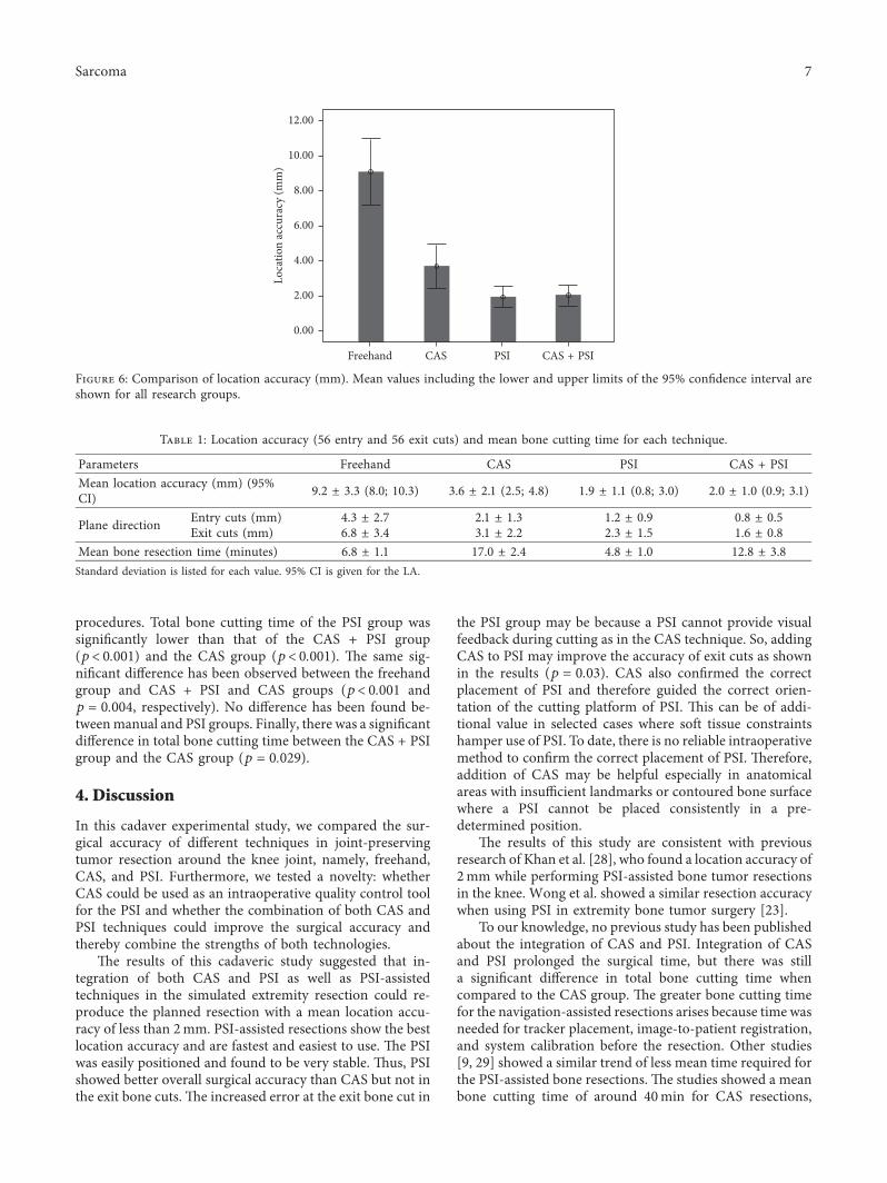
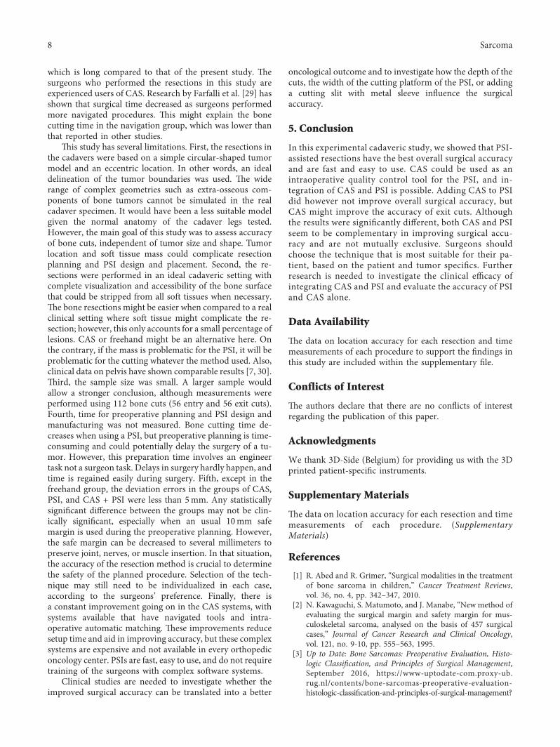
(e freehand group was significantly less accurate thanany other groups (p< 0.001). No significant difference hasbeen observed in location accuracy (LA) between the PSIand the CAS + PSI groups (p � 0.92). (e PSI and CAS +PSI groups were found to be significantly more accuratethan the CAS group (p � 0.034 and p � 0.042, respectively)(Figure 6). (e complete results of the location accuracy foreach resection plane separately are available in Supple-mentary File 1. (e mean entry and exit cut distances aresignificantly larger in the freehand group than any othergroups (p< 0.05). (e distance (deviation error) of theentry bone cuts was significantly smaller than that of theexit bone cuts in all of the four techniques (p< 0.05). Whencompared to the CAS group, the PSI technique showeda significantly better accuracy at the entry cut (p � 0.003)but not at the exit cut (p � 0.16). (e CAS + PSI techniquedemonstrated a significantly better accuracy at the exit cut(p � 0.03) compared to PSI but not at the entry cut(p � 0.07).
Mean timing for each method is summarized in Table 1.(e positioning of the PSI took less than one minute in all 8
(a) (b)
(c) (d)
Figure 4: (e techniques used in this experiment. (a) (e freehand procedure. (b) (e CAS-assisted technique. A pointer (in the surgeonsright hand) is used to determine what the direction of the plane is and how to align the saw (in the surgeons left hand). (c) How the PSI ispinned onto the bone is shown. (d) How the position of the guide is checked by using CAS is shown.
KerfMeasured plane
Target planeAchieved plane
Figure 5: Definition of the kerf and location accuracy. (e thickgrey line shows the kerf, which is the bone loss that arises during anosteotomy.(e saw is aligned on the target plane (black), a thin line inthe middle of the kerf. (e blue line defines the achieved plane of theperformed resections. Location accuracy is the maximum distance(mm) between the target (black line) and achieved (blue line) planes.When the measured error or difference was on the tumor side, theerror was corrected minus the kerf; when the measured error was onthe healthy side, the error was corrected plus the kerf.
6 Sarcoma
procedures. Total bone cutting time of the PSI group wassigni�cantly lower than that of the CAS + PSI group(p< 0.001) and the CAS group (p< 0.001). �e same sig-ni�cant di�erence has been observed between the freehandgroup and CAS + PSI and CAS groups (p< 0.001 andp � 0.004, respectively). No di�erence has been found be-tweenmanual and PSI groups. Finally, there was a signi�cantdi�erence in total bone cutting time between the CAS + PSIgroup and the CAS group (p � 0.029).
4. Discussion
In this cadaver experimental study, we compared the sur-gical accuracy of di�erent techniques in joint-preservingtumor resection around the knee joint, namely, freehand,CAS, and PSI. Furthermore, we tested a novelty: whetherCAS could be used as an intraoperative quality control toolfor the PSI and whether the combination of both CAS andPSI techniques could improve the surgical accuracy andthereby combine the strengths of both technologies.
�e results of this cadaveric study suggested that in-tegration of both CAS and PSI as well as PSI-assistedtechniques in the simulated extremity resection could re-produce the planned resection with a mean location accu-racy of less than 2mm. PSI-assisted resections show the bestlocation accuracy and are fastest and easiest to use. �e PSIwas easily positioned and found to be very stable. �us, PSIshowed better overall surgical accuracy than CAS but not inthe exit bone cuts. �e increased error at the exit bone cut in
the PSI group may be because a PSI cannot provide visualfeedback during cutting as in the CAS technique. So, addingCAS to PSI may improve the accuracy of exit cuts as shownin the results (p � 0.03). CAS also con�rmed the correctplacement of PSI and therefore guided the correct orien-tation of the cutting platform of PSI. �is can be of addi-tional value in selected cases where soft tissue constraintshamper use of PSI. To date, there is no reliable intraoperativemethod to con�rm the correct placement of PSI. �erefore,addition of CAS may be helpful especially in anatomicalareas with insu�cient landmarks or contoured bone surfacewhere a PSI cannot be placed consistently in a pre-determined position.
�e results of this study are consistent with previousresearch of Khan et al. [28], who found a location accuracy of2mm while performing PSI-assisted bone tumor resectionsin the knee. Wong et al. showed a similar resection accuracywhen using PSI in extremity bone tumor surgery [23].
To our knowledge, no previous study has been publishedabout the integration of CAS and PSI. Integration of CASand PSI prolonged the surgical time, but there was stilla signi�cant di�erence in total bone cutting time whencompared to the CAS group. �e greater bone cutting timefor the navigation-assisted resections arises because time wasneeded for tracker placement, image-to-patient registration,and system calibration before the resection. Other studies[9, 29] showed a similar trend of less mean time required forthe PSI-assisted bone resections. �e studies showed a meanbone cutting time of around 40min for CAS resections,
Freehand CAS PSI CAS + PSI
0.00
2.00
4.00
6.00
8.00
10.00
12.00
Loca
tion
accu
racy
(mm
)
Figure 6: Comparison of location accuracy (mm). Mean values including the lower and upper limits of the 95% con�dence interval areshown for all research groups.
Table 1: Location accuracy (56 entry and 56 exit cuts) and mean bone cutting time for each technique.
Parameters Freehand CAS PSI CAS + PSIMean location accuracy (mm) (95%CI) 9.2 ± 3.3 (8.0; 10.3) 3.6 ± 2.1 (2.5; 4.8) 1.9 ± 1.1 (0.8; 3.0) 2.0 ± 1.0 (0.9; 3.1)
Plane direction Entry cuts (mm) 4.3 ± 2.7 2.1 ± 1.3 1.2 ± 0.9 0.8 ± 0.5Exit cuts (mm) 6.8 ± 3.4 3.1 ± 2.2 2.3 ± 1.5 1.6 ± 0.8
Mean bone resection time (minutes) 6.8 ± 1.1 17.0 ± 2.4 4.8 ± 1.0 12.8 ± 3.8Standard deviation is listed for each value. 95% CI is given for the LA.
Sarcoma 7
which is long compared to that of the present study. (esurgeons who performed the resections in this study areexperienced users of CAS. Research by Farfalli et al. [29] hasshown that surgical time decreased as surgeons performedmore navigated procedures. (is might explain the bonecutting time in the navigation group, which was lower thanthat reported in other studies.
(is study has several limitations. First, the resections inthe cadavers were based on a simple circular-shaped tumormodel and an eccentric location. In other words, an idealdelineation of the tumor boundaries was used. (e widerange of complex geometries such as extra-osseous com-ponents of bone tumors cannot be simulated in the realcadaver specimen. It would have been a less suitable modelgiven the normal anatomy of the cadaver legs tested.However, the main goal of this study was to assess accuracyof bone cuts, independent of tumor size and shape. Tumorlocation and soft tissue mass could complicate resectionplanning and PSI design and placement. Second, the re-sections were performed in an ideal cadaveric setting withcomplete visualization and accessibility of the bone surfacethat could be stripped from all soft tissues when necessary.(e bone resections might be easier when compared to a realclinical setting where soft tissue might complicate the re-section; however, this only accounts for a small percentage oflesions. CAS or freehand might be an alternative here. Onthe contrary, if the mass is problematic for the PSI, it will beproblematic for the cutting whatever the method used. Also,clinical data on pelvis have shown comparable results [7, 30].(ird, the sample size was small. A larger sample wouldallow a stronger conclusion, although measurements wereperformed using 112 bone cuts (56 entry and 56 exit cuts).Fourth, time for preoperative planning and PSI design andmanufacturing was not measured. Bone cutting time de-creases when using a PSI, but preoperative planning is time-consuming and could potentially delay the surgery of a tu-mor. However, this preparation time involves an engineertask not a surgeon task. Delays in surgery hardly happen, andtime is regained easily during surgery. Fifth, except in thefreehand group, the deviation errors in the groups of CAS,PSI, and CAS + PSI were less than 5mm. Any statisticallysignificant difference between the groups may not be clin-ically significant, especially when an usual 10mm safemargin is used during the preoperative planning. However,the safe margin can be decreased to several millimeters topreserve joint, nerves, or muscle insertion. In that situation,the accuracy of the resection method is crucial to determinethe safety of the planned procedure. Selection of the tech-nique may still need to be individualized in each case,according to the surgeons’ preference. Finally, there isa constant improvement going on in the CAS systems, withsystems available that have navigated tools and intra-operative automatic matching. (ese improvements reducesetup time and aid in improving accuracy, but these complexsystems are expensive and not available in every orthopediconcology center. PSIs are fast, easy to use, and do not requiretraining of the surgeons with complex software systems.
Clinical studies are needed to investigate whether theimproved surgical accuracy can be translated into a better
oncological outcome and to investigate how the depth of thecuts, the width of the cutting platform of the PSI, or addinga cutting slit with metal sleeve influence the surgicalaccuracy.
5. Conclusion
In this experimental cadaveric study, we showed that PSI-assisted resections have the best overall surgical accuracyand are fast and easy to use. CAS could be used as anintraoperative quality control tool for the PSI, and in-tegration of CAS and PSI is possible. Adding CAS to PSIdid however not improve overall surgical accuracy, butCAS might improve the accuracy of exit cuts. Althoughthe results were significantly different, both CAS and PSIseem to be complementary in improving surgical accu-racy and are not mutually exclusive. Surgeons shouldchoose the technique that is most suitable for their pa-tient, based on the patient and tumor specifics. Furtherresearch is needed to investigate the clinical efficacy ofintegrating CAS and PSI and evaluate the accuracy of PSIand CAS alone.
Data Availability
(e data on location accuracy for each resection and timemeasurements of each procedure to support the findings inthis study are included within the supplementary file.
Conflicts of Interest
(e authors declare that there are no conflicts of interestregarding the publication of this paper.
Acknowledgments
We thank 3D-Side (Belgium) for providing us with the 3Dprinted patient-specific instruments.
Supplementary Materials
(e data on location accuracy for each resection and timemeasurements of each procedure. (SupplementaryMaterials)
References
[1] R. Abed and R. Grimer, “Surgical modalities in the treatmentof bone sarcoma in children,” Cancer Treatment Reviews,vol. 36, no. 4, pp. 342–347, 2010.
[2] N. Kawaguchi, S. Matumoto, and J. Manabe, “New method ofevaluating the surgical margin and safety margin for mus-culoskeletal sarcoma, analysed on the basis of 457 surgicalcases,” Journal of Cancer Research and Clinical Oncology,vol. 121, no. 9-10, pp. 555–563, 1995.
[3] Up to Date: Bone Sarcomas: Preoperative Evaluation, Histo-logic Classification, and Principles of Surgical Management,September 2016, https://www-uptodate-com.proxy-ub.rug.nl/contents/bone-sarcomas-preoperative-evaluation-histologic-classification-and-principles-of-surgical-management?
8 Sarcoma
source�search_result&search�bone%20sarcomas&selectedTitle�1∼74.
[4] L. E. Ritacco, F. E. Milano, G. L. Farfalli, M. A. Ayerza,D. L. Muscolo, and L. A. Aponte-Tinao, “Accuracy of 3-Dplanning and navigation in bone tumor resection,” Ortho-pedics, vol. 36, no. 7, pp. e942–e950, 2013.
[5] R. J. Grimer, “Surgical options for children with osteosar-coma,” :e Lancet Oncology, vol. 6, no. 2, pp. 85–92, 2005.
[6] P. S. Young, S. W. Bell, and A. Mahendra, “(e evolving role ofcomputer-assisted navigation in musculoskeletal oncology,”Bone and Joint Journal, vol. 97-B, no. 2, pp. 258–264, 2015.
[7] O. Cartiaux, L. Paul, B. G. Francq, X. Banse, andP. L. Docquier, “Improved accuracy with 3D planning andpatient-specific instruments during simulated pelvic bonetumor surgery,” Annals of Biomedical Engineering, vol. 42,no. 1, pp. 205–213, 2014.
[8] R. L. Deijkers, R. M. Bloem, P. C. Hogendoorn, J. J. Verlaan,H. M. Kroon, and A. H. Taminiau, “Hemicortical allograftreconstruction after resection of low-grade malignant bonetumours,” Journal of Bone and Joint Surgery-British Volume,vol. 84-B, no. 7, pp. 1009–1014, 2002.
[9] O. Cartiaux, L. Paul, P. L. Docquier, B. Raucent, E. Dombre,and X. Banse, “Computer-assisted and robot-assisted tech-nologies to improve bone-cutting accuracy when integratedwith a freehand process using an oscillating saw,” Journal ofBone and Joint Surgery-American Volume, vol. 92, no. 11,pp. 2076–2082, 2010.
[10] P. Merloz, J. Tonetti, P. Cinquin, S. Lavallee, J. Troccaz, andL. Pittet, “Computer-assisted surgery: automated screwplacement in the vertebral pedicle,” Chirurgie, vol. 123, no. 5,pp. 482–490, 1998.
[11] C. W. Jones and S. A. Jerabek, “Current role of computernavigation in total knee arthroplasty,” Journal of Arthroplasty,vol. 33, no. 7, pp. 1989–1993, 2018.
[12] J. G. Gerbers, M. Stevens, J. J. Ploegmakers, S. K. Bulstra, andP. C. Jutte, “Computer-assisted surgery in orthopedic on-cology,” Acta Orthopaedica, vol. 85, no. 6, pp. 663–669, 2014.
[13] K. C. Wong and S. M. Kumta, “Use of computer navigation inorthopedic oncology,” Current Surgery Reports, vol. 2, no. 4,p. 47, 2014.
[14] L. Aponte-Tinao, L. E. Ritacco, M. A. Ayerza, D. L. Muscolo,J. I. Albergo, and G. L. Farfalli, “Does intraoperative navi-gation assistance improve bone tumor resection and allograftreconstruction results?,” Clinical Orthopaedics and RelatedResearch, vol. 473, no. 3, pp. 796–804, 2015.
[15] M. Ieguchi, M. Hoshi, J. Takada, N. Hidaka, andH. Nakamura, “Navigation-assisted surgery for bone and softtissue tumors with bony extension,”Clinical Orthopaedics andRelated Research, vol. 470, no. 1, pp. 275–283, 2012.
[16] K. C. Wong and S. M. Kumta, “Computer-assisted tumorsurgery in malignant bone tumors,” Clinical Orthopaedics andRelated Research, vol. 471, no. 3, pp. 750–761, 2013.
[17] J. Li, Z. Wang, Z. Guo, G. J. Chen, M. Yang, and G. X. Pei,“Irregular osteotomy in limb salvage for juxta-articular os-teosarcoma under computer-assisted navigation,” Journal ofSurgical Oncology, vol. 106, no. 4, pp. 411–416, 2012.
[18] K. C. Wong and S. M. Kumta, “Joint-preserving tumor re-section and reconstruction using image-guided computernavigation,” Clinical Orthopaedics and Related Research,vol. 471, no. 3, pp. 762–773, 2013.
[19] J. Jiang, X. Kang, Q. Lin et al., “Accuracy of patient-specificinstrumentation compared with conventional instrumentationin total knee arthroplasty,” Orthopedics, vol. 38, no. 4,pp. e305–e313, 2015.
[20] J. J. Jauregui, J. J. Cherian, B. H. Kapadia et al., “Patient-specific instrumentation in total knee arthroplasty,” Journal ofKnee Surgery, vol. 27, no. 3, pp. 177–183, 2014.
[21] J. C. Yang, C. F. Chen, C. A. Luo et al., “Clinical experienceusing a 3D-printed patient-specific instrument for medialopening wedge high tibial osteotomy,” BioMed Research In-ternational, vol. 2018, Article ID 9246529, 9 pages, 2018.
[22] L. Bellanova, L. Paul, and P. L. Docquier, “Surgical guides(patient-specific instruments) for pediatric tibial bone sar-coma resection and allograft reconstruction,” Sarcoma,vol. 2013, Article ID 787653, 7 pages, 2013.
[23] K. C. Wong, S. M. Kumta, K. S. Leung, K. W. Ng, E. W. Ng,and K. S. Lee, “Integration of CAD/CAM planning intocomputer assisted orthopaedic surgery,” Computer AidedSurgery, vol. 15, no. 4–6, pp. 65–74, 2010.
[24] K. C. Wong, K. Y. Sze, I. O. Wong, C. M. Wong, andS. M. Kumta, “Patient-specific instrument can achieve sameaccuracy with less resection time than navigation assistance inperiacetabular pelvic tumor surgery: a cadaveric study,” In-ternational Journal of Computer Assisted Radiology andSurgery, vol. 11, no. 2, pp. 307–316, 2016.
[25] ISO and IOfS, Geometrical Product Specification (GPS)—Geometrical Toplerancing—Tolerances of Form, Orientation,Location and Run-Out, ISO Standard 1101:2004, Geneva,Switzerland, 2004.
[26] O. Cartiaux, L. Paul, P. L. Docquier et al., “Accuracy in planarcutting of bones: an ISO-based evaluation,” InternationalJournal of Medical Robotics and Computer Assisted Surgery,vol. 5, no. 1, pp. 77–84, 2009.
[27] J. A. Bailey, Y. Wang, F. R. W. van de Goot, andR. R. R. Gerretsen, “Statistical analysis of kerf mark mea-surements in bone,” Forensic Science, Medicine, and Pathol-ogy, vol. 7, no. 1, pp. 53–62, 2011.
[28] F. A. Khan, J. D. Lipman, A. D. Pearle, P. J. Boland, andJ. H. Healey, “Surgical technique: computer-generated customjigs improve accuracy of wide resection of bone tumors,”Clinical Orthopaedics and Related Research, vol. 471, no. 6,pp. 2007–2016, 2013.
[29] G. L. Farfalli, J. I. Albergo, L. E. Ritacco, M. A. Ayerza,F. E. Milano, and L. A. Aponte-Tinao, “What is the expectedlearning curve in computer-assisted navigation for bone tu-mor resection?,” Clinical Orthopaedics and Related Research,vol. 475, no. 3, pp. 668–675, 2017.
[30] A. Sallent, M. Vicente, M. M. Reverte et al., “How 3D patient-specific instruments improve accuracy of pelvic bone tumourresection in a cadaveric study,” Bone and Joint Research, vol. 6,no. 10, pp. 577–583, 2017.
Sarcoma 9

Stem Cells International
Hindawiwww.hindawi.com Volume 2018
Hindawiwww.hindawi.com Volume 2018
MEDIATORSINFLAMMATION
of
EndocrinologyInternational Journal of
Hindawiwww.hindawi.com Volume 2018
Hindawiwww.hindawi.com Volume 2018
Disease Markers
Hindawiwww.hindawi.com Volume 2018
BioMed Research International
OncologyJournal of
Hindawiwww.hindawi.com Volume 2013
Hindawiwww.hindawi.com Volume 2018
Oxidative Medicine and Cellular Longevity
Hindawiwww.hindawi.com Volume 2018
PPAR Research
Hindawi Publishing Corporation http://www.hindawi.com Volume 2013Hindawiwww.hindawi.com
The Scientific World Journal
Volume 2018
Immunology ResearchHindawiwww.hindawi.com Volume 2018
Journal of
ObesityJournal of
Hindawiwww.hindawi.com Volume 2018
Hindawiwww.hindawi.com Volume 2018
Computational and Mathematical Methods in Medicine
Hindawiwww.hindawi.com Volume 2018
Behavioural Neurology
OphthalmologyJournal of
Hindawiwww.hindawi.com Volume 2018
Diabetes ResearchJournal of
Hindawiwww.hindawi.com Volume 2018
Hindawiwww.hindawi.com Volume 2018
Research and TreatmentAIDS
Hindawiwww.hindawi.com Volume 2018
Gastroenterology Research and Practice
Hindawiwww.hindawi.com Volume 2018
Parkinson’s Disease
Evidence-Based Complementary andAlternative Medicine
Volume 2018Hindawiwww.hindawi.com
Submit your manuscripts atwww.hindawi.com


![Ballistic research techniques: visualizing gunshot ... · for ballistic research including, but not limited to, soap, gelatine, cadaveric human tissue, cadaveric animal tissue, andliveanimaltissue[13].](https://static.fdocuments.in/doc/165x107/60655ea8929453602f722c04/ballistic-research-techniques-visualizing-gunshot-for-ballistic-research-including.jpg)