UNIVERSITI PUTRA MALAYSIA CHROMOSOMAL REARRANGEMENT AND...
25
UNIVERSITI PUTRA MALAYSIA CHROMOSOMAL REARRANGEMENT AND LOSS OF HETEROZYGOSITY IN CERVICAL CANCER AMONGST PATIENTS IN HOSPITAL KUALA LUMPUR SITI NORLASIAH BINTI ISMAIL. IB 2004 2
Transcript of UNIVERSITI PUTRA MALAYSIA CHROMOSOMAL REARRANGEMENT AND...

UNIVERSITI PUTRA MALAYSIA
CHROMOSOMAL REARRANGEMENT AND LOSS OF HETEROZYGOSITY IN CERVICAL CANCER AMONGST PATIENTS
IN HOSPITAL KUALA LUMPUR
SITI NORLASIAH BINTI ISMAIL.
IB 2004 2

CHROMOSOMAL REARRANGEMENT AND LOSS OF HETEROZYGOSITY STUDIES IN CERVICAL CANCER AMONGST
PATIENTS IN HOSPITAL KUALA LUMPUR
BY
SIT1 NORLASIAH BINTI ISMAIL
Thesis Submitted to the School o f Graduate Studies, Universiti Putra Malaysia in Fulfilment o f the Requirement for the Degree o f
Doctor o f Philosophy
January 2004
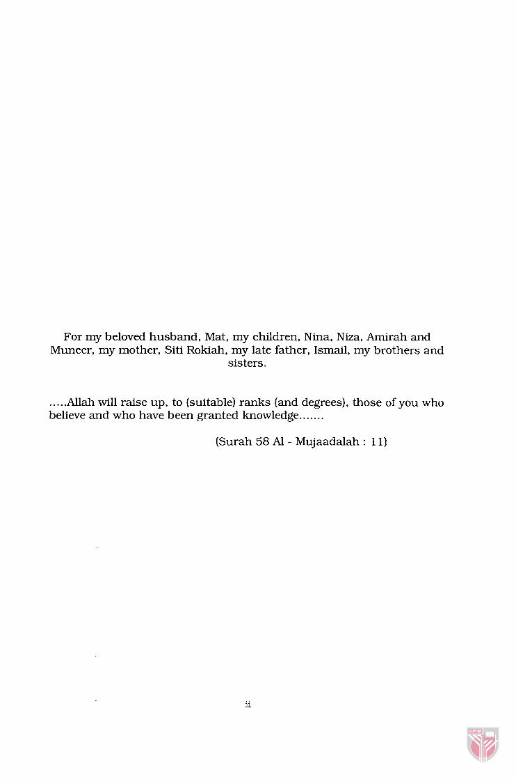
For my beloved husband, Mat, my children, Nina, Niza, Arnirah and Muneer, my mother, Siti Rokiah, my late father, Ismail, my brothers and
sisters.
. . . . .Allah will raise up, to (suitable) ranks (and degrees), those of you who believe and who have been granted knowledge.. . . . . .
(Surah 58 Al - Mujaadalah : 11)

Abstract of thesis submitted to the senate of Universiti Putra Malaysia in fulfilment of the requirement for the degree of Doctor of Philosophy
CHROMOSOMAL REARRANGEMENT AND LOSS OF HETEROZYGOSITY STUDIES IN CERVICAL CANCER AMONGST
PATIENTS IN HOSPITAL KUALA LUMPUR
SIT1 NORLASIAH BINTI ISMAIL
January 2004
Chairman: Professor Abdul Manaf Ali, Ph.D
Institute: Bioscience
C e ~ c a l carcinoma is the second most common malignancy among
women worldwide. The highest incidence rates are observed in
developing countries. The susceptibility to cervical carcinoma in high
incidence populations may result from several factors including human
papillomavirus (HPV) exposure and both inherited and acquired genes.
HPV infection does not always led to cervical cancer. In cervical
carcinoma the other common genetic characteristic of cancer is the
presence of several recurrent genetic alterations, not related to HPV. The
type of recurrent genetic damage might take different forms such as gene
amplification, chromosomal translocation, deletions, loss of
heterozygosity, point mutation, transcriptional silencing, and in some
tumors viral DNA integration.
To deter-mine the possible genetic alterations amongst the Malaysian
women with cervical cancer, this study was conducted on 50 cervical
tumor biopsies received from the Department of Obstetrics and
Gynecology of the Hospital Kuala Lumpur. The objectives of the study
were; i) the establishment of short-term primary cell culture of human
cervical epithelial cells derived from cervical tumors for the
determination of the prevalence and the type of chromosomal
aberrations, ii) characterization of the allelic losses of the chromosomes
3p, 5p, 11 and 17p (p53), subsequently identification of a possible site of
candidate tumor suppressor gene(s) and iii) to determine the HPV status
of the cervical cancers. Primary cell culture and cytogenetic techniques
were performed on the cervical tumor biopsies. G-banding was employed
for the identification of the chromosomes. To define the localization of
the tumor suppressor genes, loss of heterozygosity study was performed
on 37 cervical cancer cells. Twenty-four microsatellite polymorphic
markers for the chromosomes 3p, 5p, 11 and p53 were chosen, the
normal and tumor DNAs from each patient were analyzed for the allelic
loss using PCR-based microsatellite analysis. The status for HPV 16 E6
and HPV 18 E6 was detected by PCR method.
Twenty-five cervical cancer biopsies were successfully karyotyped and
near-diploid was the modal number, with a majority of them being
hypodiploid (35-45). About 50% of the metaphases obtained in the 25
tumors were hypodiploids, 12.1% were hyperdiploids, and 36.7% were
diploids. Numerical abnormalities were predominantly observed in the
patients, with monosomies of chromosomes 17, 22, X, 1 1, 18, 19, 13,
and 6. Fluorescence in situ hybridization using centromeric iv

PERPUSTAKAAN SULTAN A M UHWRClW f w i A MAUYSM
probes 1 1, 17 and 18 confirmed the presence of monosornies 1 1, 17, 18
in a low percentage, 12.0%. 16.2% and 26.4% respectively. Several
clones of cells were observed, with possibility of 45,)(X,-22; 45,XX.-11;
45,=,- 19 and 45,XX.- 18. Twenty-six of the 36 informative individuals
exhibited LOH at one locus or more. The highest incidence was observed
in chromosome 3p with the frequency of 48.6%. while a low frequency of
2.7% was detected in the short-arm of chromosome 17 at position
17~13.1, whereby lies the p53 tumor suppressor. LOH was confined to
four prominent regions, 1 1q23.3, 3p 14.2-3p 14.1, 3p2 1.32 and 3p25.3-
3p25.1. No signiacant correlation was found between the LOH and the
grade of cancer differentiation. The difference between the LOH
frequency in cervical carcinomas with early stage and those with
advanced stage was not statistically si@icant. Only 5 1.4% of the 35
tumors were positive for HPV 16 E6 and 17.2% was HPV 18 E6 positive.
HPV 16 was found to be positive in 64.7% of the Chinese, 41.7% in
Malays and 50% in the Indians. Both the Malays and the Indians were
observed to harbor the HPV 18 at a higher frequency (40 and 33.3%
respectively) than the Chinese. In conclusion, besides HPV infection,
other genetic abnormalities play a role in cervical carcinogenesis. LOH is
a better method than chromosomal analysis in searching for possible
tumor suppressor gene(s) that is responsible for c e ~ c a l tumorigenesis.
Mapping of the smallest region of LOH in these tumors and analysis of
candidate genes present in the region of LOH will be continued.

Abstrak tesis yang dikemukakan kepada Senat Universiti Putra Malaysia sebagai memenuhi keperluan bagi mendapatkan Ijazah Doktor Falsafah
PENYUSUNAN SEMUM KROMOSOM DAN KAJIAN KEHILANGAN HETEROZIGOSITI DALAM BARAH SERVIKS DI KALANGAN PESAKIT
DI HOSPITAL KUALA LUMPUR
Oleh
SIT1 NORLASIAH BINTI ISMAIL
Pengerusi: Professor Abdul Manaf Ali, Ph.D
Institut: Biosains
Karsinoma s e ~ k s adalah malignan kedua yang kerap berlaku di
kalangan wanita di seluruh dunia. Insiden yang paling tinggi kerap
dilihat di negara yang sedang membangun. Populasi yang mempunyai
insiden tinggi mungkin mudah mendapat karsinoma serviks akibat
pendedahan terhadap papilomavirus manusia (HPV) dan gen yang
diwarisi atau diperolehi. Infeksi HPV bukan selalu menyebabkan kanser
serviks. Bagi kanser serviks, ciri genetik yang kerap dilihat adalah
beberapa pengubahan genetik yang berulang-ulang, yang tak berkaitan
dengan HPV. Jenis kerosakan genetik yang berulang-ulang adalah
seperti amplifikasi gen, translokasi kromosom, delesi, kehilangan
heterozigositi, mutasi titik, transcriptional silencing dan integrasi virus.
Kajian ini dijalan untuk mengetahui pengubahan gen di kalangan
wanita yang mempunyai kanser serviks.
Kajian ini melibatkan 50 biopsi barah serviks dari wanita yang

menjalani rawatan barah senriks di Jabatan Obstetrik dan Ginekologi,
Hospital Kuala Lumpur. Tujuan kajian ini adalah; i) membangunkan
kultur sel primer daripada sel barah s e ~ k s bagi menentukan prevalen
dan jenis aberasi kromosom, ii) pencirian kehilangan alel (allelic loss)
pada kromosom 3p, 5p, 11 dan 17p (p53), seterusnya pengenalpastian
lokasi gen penumpas barah (tumor suppressor genes) dan iii) untuk
menentukan status HPV dalam kanser s e ~ k s . Kaedah kultur sel primer
dan sitogenetik telah diperlakukan ke atas sampel biopsi barah s e ~ k s .
Penjaluran-G telah digunakan untuk mengenalpasti kromosom. Untuk
menentukan lokasi gen penumpas barah, kajian kehilangan
heterozigositi telah dilakukan ke atas 37 sampel barah s e ~ k s . Dua
puluh empat penanda polimorfik mikrosatelit pada kromosom 3p, 5p, 11
dan p53 telah dipilih. Pelet DNA normal dan barah dari setiap 37 sampel
telah dianalisis menggunakan kaedah analisis mikrosatelit berasaskan
PCR (PCR-based microsatellite analysis) untuk mengesan kehilangan
alel.
Dua puluh lima biopsi barah serviks telah berjaya dikariotip dan near-
diploid telah dilihat sebagai modal number dengan majoriti adalah
hipoploid (35-45). Lebih kurang 50% daripada taburan metafasa adalah
hipoploid, 12.1% hiperploid dan 36.7% adalah diploid. Keabnormalan
pada jumlah kromosom dilihat lebih utarna di kalangan pesakit,
terutamanya monosomi kromosom 17, 22, X, 11, 18, 19, 13, dan 6.
Penghibridan pendarfluor in situ ~uorescence in situ hybridization)
menggunakan prob sentromerik bagi kromosom 1 1, 17 dan 18 telah vii

mengesahkan kehadiran monosomi kromosom tersebut tetapi pada
kadar yang rendah, iaitu, 12.0%, 16.2% dn 26.4% masing-masing.
Beberapa klon sel telah dijumpai, berkemungkinan 45,XX, -22; 45,XX, -
11; 45,XX,-19 dan 45,XX,-18. Dua puluh enam daripada 36 individu
yang informatif menunjukkan LOH pada satu lokus atau lebih. Insiden
LOH yang tertinggi dikesan pada kromosom 3p dengan kekerapan pada
kadar 48.6%, manakala LOH yang terendah adalah pada kekerapan
2.7% yang dilihat pada kromosom 17p di posisi 17p 13.1. LOH telah
dikesan pada empat kawasan yang utama, 1 1q23.3, 3p 14.2-3p 14.1,
3p2 1.32 dan 3p25.3-3~25.1. Tiada korelasi yang signifikan ditemui di
antara LOH dan gred pembezaan kanser. Perbezaan di antara kekerapan
LOH dalam karsinoma s e ~ k s di peringkat awal dan karsinoma di
peringkat akhir adalah tidak signifikan. Hanya 5 1.4% daripada 35 barah
serviks positif bagi HPV 16 E6 dan 17.2% positif untuk HPV 18 E6. HPV
16 didapati positif di dalam 64.7% kaum Cina, 4 1.7% Melayu dan 50%
di kalangan kaum India. Peratusan positif HPV 18 didapati lebih tinggi
di kalangan kaum Melayu dan India berbanding kaum Cina. Sebagai
rumusan, selain infeksi HPV, keabnormalan genetik juga berperanan
dalarn proses karsinogenesis s e ~ k s . LOH adalah kaedah yang lebih
baik daripada kaedah analisis kromosom untuk mencari gen penumpas
barah yang bertanggungjawab bagi proses barah s e ~ k s . Kehadiran
LOH dalarn barah s e ~ k s mungkin boleh digunakan sebagai indikator
terhadap prognosis penyakit tersebut. Pemetaan kawasan terkecil bagi
LOH dalam barah ini serta analisis calon gen dalam kawasan LOH akan
diteruskan.

ACKNOWLEDGEMENTS
First of all praise to the Almighty Allah for giving me the ability to learn
and strength to complete this research. This study would never have
materialized without the contribution and support of many people to
whom I have the pleasure of expressing my appreciation and gratitude. I
would like to express my sincere gratitude to Professor Dr. Abdul Manaf
Ali for his guidance, understanding, patience and encouragement that
lead to the completion of this thesis, and my s u p e ~ s o r y committee
members: Professor Datin Dr. Khatijah Mohd. Yusoff, for providing the
facility for molecular work, monitoring the progress of research, helpful
comments and editing of the thesis; Associate Professor Dr. Sabariah
Abdul Rahrnan and Associate Professor Dr. Siti Aishah Mohd. Ali, for
their invaluable comments and Dr. Noorjahan Banu Mohd. Ali-Theen for
proofreading and editing this thesis.
I am greatly indebted to the full cooperation and support of many
individuals in the Department of Obstetrics and Gynecology, Hospital
Kuala Lumpur, for providing the samples, without them, this research
would have not materialized. Thanks are extended to the ex-Director Dr.
Alex Mathews and Dr. Wong Sum Keong and his medical and nursing
team.

My sincere gratitude to the Director of Institute for Medical Research
(IMR) and the staff of the Genetics Laboratory for allowing the use of
their laboratory for the Interphase in situ hybridization technique.
Special thanks to Dr. Puteri JamilatulNoor Megat Baharuddin for
sharing her experience and knowledge in this technique and her
continuous encouragement towards the completion of this postgraduate
program, Ms. Roshidah Ishak for the valuable tips on PCR technique,
Mrs. Wang Lily for her technical assistance in FISH technique, Mr.
Roslaini Majid for the simplified Hematoxylin and Eosin (H & E)
technique and Ms. Ten Sew Keoh for her input. I am grateful to Ms
Subha Bhassu of the Department of Biochemistry and Microbiology,
UPM for the microsatellite analysis technique. My sincere thanks to Dr.
Norhayati Zainal Abidin of Malaya University for providing the CaSki cell
line.
I would like to extend my sincere thanks to the staff of the Genetics
Laboratory of the National Population and Family Development Board
(NPFDB) for their technical assistance during the study period, including
taking messages from the doctors for specimen collection. My gratitude
to my organization, NPFDB, for allowing me to pursue the postgraduate
program, and the Public Service Department, as on behalf for the
Government of Malaysia, for sponsoring me throughout this program.
I greatly appreciated my colleagues from the Animal Cell Culture

Laboratory and Biochemistry Laboratory for their unduly support and
encouragement during the hard times. Lastly, but not the least, to my
husband and children for their patience, sacrifices, support and
understanding throughout this postgraduate program, my beloved
mother and my siblings for their unconditional love and encouragement
throughout the trylng period.
I certify that an Examination Committee met on 13th January 2004 to conduct the final examination of Siti Norlasiah binti Ismail on her Doctor of Philosophy thesis entitled "Chromosomal Rearrangement and Loss of Heterozygosity Studies in Cervical Cancer Amongst Patients in Hospital Kuala Lumpur" in accordance with Universiti Pertanian Malaysia (Higher Degree) Act 1980 and Universiti Pertanian Malaysia (Higher Degree) Regulations 198 1. The Committee recommends that the candidate be awarded the relevant degree. Members of the Examination Committee are as follows:
Rozita Rosli, Ph.D. Associate Professor Faculty of Medicine and Health Sciences Universiti Putra Malaysia (Chairman)
Abdul Manaf Ali, Ph.D. Professor Institute Biosciences Universiti Putra Malaysia (Member)
Datin Khatijah Mohd Yusoff, Ph.D. Professor Faculty of Sciences and Environmental Studies Universiti Putra Malaysia (Member)
Sabariah Abdul Rahman, Ph.D. Associate Professor Faculty of Medicine and Health Sciences Universiti Putra Malaysia (Member)
Noorjahan Banu Mohd Ali-Theen, Ph.D. Faculty of Food Science and Biotechnology Universiti Putra Malaysia (Member)
Sit Aishah Mohd Ali, M.D Associate Professor Faculty of Medicine Universiti Kebangsaan Malaysia (Member)
Mohd Nizam Hj. Musa, Ph.D. Professor International Medical University (Independent Examiner)
~rofessor/ ~ e b u t ? Dean School of Graduate Studies Universiti Putra Malaysia
Dare: 2 1 APR Ioo4 sii

This thesis submitted to the Senate of Universiti Putra Malaysia has been accepted as fulfilment of the requirement for the degree of Doctor of Philosophy. The members of the Supervisory Committee are a s follows:
Abdul Manaf Ali, Ph.D, Professor, Institute of Bioscience, Universiti Putra Malaysia. (Chairman)
Datin Khatijah Mohd Yusoff, Ph.D, Professor, Faculty of Environmental Sciences, Universiti Putra Malaysia. (Member)
Sabariah Abdul Rahman, M.D Associate Professor, Faculty of Medical Sciences, Universiti Putra Malaysia. (Member)
Noorjahan Banu Mohd Ali-Theen, Ph.D, Faculty of Food Science and Biotechnology, Universiti Putra Malaysia. (Member)
Sit i Aishah Mohd Ali, M.D, Associate Professor, Faculty of Medicine, Universiti Kebangsaan Malaysia. (Member)
- -
AINI IDERIS, Ph.D, Professor/ Dean School of Graduate Studies Universiti Putra Malaysia.
Date: 1 7 MAY 2004
... Xll l

DECLARATION
I hereby declare that the thesis is based on my original work except for quotations and citations which have been duly acknowledged. I also declare that it has not been previously or concurrently submitted for any degree at UPM or other institutions.
SIT1 NORLASIAH BINTI ISMAIL
Date: 12 March 2004
TABLE OF CONTENTS
Page
DEDICATION ABSTRACT ABSTRAK ACKNOWLEDGEMENTS APPROVAL DECLARATION LIST OF TABLES LIST OF FIGURES LIST OF ABBREVIATIONS GLOSSARY
CHAPTER
INTRODUCTION
LITERATURE REVIEW The multistep nature of cancer Cervical cancer
Etiology of cervical cancer and its precursors Classification and staging of cervical cancer Human papillomavirus and cervical cancer Screening and testing for HPV Oncogenes in cervical cancer Tumor suppressor genes
Genetic alterations in cervical cancer Methods of localizing tumor suppressor genes Cytogenetic studies of carcinoma of the cervix Interphase cytogenetics of carcinoma of the cervix Loss of heterozygosity and microsatellite instability in cervical cancer Loss of heterozygosity (LOH) in cervical cancer Microsatellite instability (MI/MSI) in cervical cancer
I11 MATERlALS AND METHODS Sample population of women with cervical cancer Clinical specimen of cervical tumor biopsies Cytogenetic analysis of cervical cancer
ii iii vi ix xii xiv xviii 2cxi d v XXVi

Primary culture Subculturing of monolayer cells by trypsinization Harvesting and slide preparation Banding and chromosome analysis Freezing of cells and storage Cell recovery Interphase cytogenetics by fluorescence in situ hybridization (FISH) Preparation of specimen and pretreatment of cells Prehybridization Post hybridization washing, counterstaining and visualization
Loss of heterozygosity (LOH) or allelic loss analysis Preparation of genomic DNA Microsatellite polyrnorphisms Amplification of microsatellite loci Assessment of loss of heterozygosity (LOH)
Detection of human papillomavirus DNA 1 6 and 1 8 Culture of established cell lines Isolation of DNA from cultured cells Detection of Human Papillomavirus HPV DNA 1 6 and 1 8 sequences
IV RESULTS AND DISCUSSION Sample population of women with cervical cancer Clinical specimen of cervical tumor biopsies Morphology of the original tumor Morphology of the cultured cells Cytogenetic analysis of cervical cancer Interphase Cytogenetics analysis of cervical imprints Loss of heterozygosity (LOH) or allelic loss analysis Loss of heterozygosity in the short-arm of chromosome 3 Loss of heterozygosity in the short-arm of chromosome 5 Loss of heterozygosity on chromosome 1 1 Loss of heterozygosity at the region 17p 13.1 Detection of HPV DNAs 1 6 and 1 8
V GENERAL DISCUSSION AND CONCLUDING REMARKS Further research
xvi

REFERENCES APPENDICES
Appendix A Appendix B Appendix C
BIODATA OF THE AUTHOR
xvii

LIST OF TABLES
Table Page
1995 Modification of FIG0 Staging of Carcinoma of the Cervix
Frequency of LOH in Cervical Cancer and SIL ( C N
Incidence of allelic losses of markers on 3p
Definition of terms and recommendations related to abnormalities commonly seen in neoplasia
Microsatellite markers on 3p selected for the study
Microsatellite markers on 5p selected for the study
Microsatellite markers on 1 l p and 1 l q selected for the study
Parameters for polymerase chain reaction in the amplification of microsatellite polyrnorphisms on chromosome 3p, 5p, and 1 1
Sequences of oligonucleotide primer pairs for the detection of HPV 16 and 18
FIGO's classification of patients
Number of primary cell cultures successfully established according to clinical stages in the women diagnosed with cervical cancer
Number of successfully karyotyped cases according to clinical stages in 32 successfully established primary cultures
Cytogenetic findings in the twenty-five women diagnosed with cervical cancer
The incidence of chromosome abnormalities seen in the 25 patients diagnosed with cervical cancer
Distribution of successfully karyotyped cases according to age and type of chromosomal abnormalities
Clinical data and DNA ISH results for chromosome 1 1 in 23 women with cervical carcinoma
Clinical data and DNA ISH results for chromosome 17 in 23 women with cervical carcinoma
Clinical data and DNA ISH results for chromosome 18 in 13 women with cervical carcinoma
4.10 DNA ISH results for chromosome 1 1, 17 and 18 according to clinical stages
4.11 Loss of heterozygosity in 37 cervical carcinomas using 2 1 microsatellite polymorphic markers
4.12 Frequency of LOH with 2 1 microsatellite markers in 37 informative individuals with cervical carcinomas
4.13 Correlation of LOH with clinical characteristics in cervical carcinomas
4.14 Frequency of loss of heterozygosity a t 9 loci on the short arm of chromosome 3 in 36 informative individuals with cervical carcinomas
4.15 Frequency of LOH according to clinical stages (FIGO) on the short arm of chromosome 3 in the informative individuals with cervical carcinomas
4.16 Frequency of loss of heterozygosity at 4 loci on the short arm of chromosome 5 in 37 patients with cervical carcinomas
4.17 Frequency of LOH according to clinical stages (FIGO) a t the 4 loci on the short arm of chromosome 5 in 30 informative individuals with cervical carcinomas
4.18 Frequency of LOH a t 7 loci on the chromosome 11 in 37 patients with cervical carcinomas
xix

4.19 Frequency of LOH according to clinical stages (FIGO) at the 6 loci on the chromosome 11 in 37 informative individuals with cervical carcinomas
4.20 Frequency of LOH at locus for p53 in patients with cervical carcinomas
4.2 1 Frequency of LOH at 1 7p 13.1 according to clinical stages (FIGO) in 11 informative individuals with cervical carcinomas
4.22 Outcomes of Human papillomavirus (HPV 16 and 18 detection in 37 women with cervical carcinomas
4.23 The distribution of HPV 16 status according to clinical stage (FIGO) in 33 women with cervical caicinomas
LIST OF FIGURES
Figure
Events in neoplastic transformation
Genome organization of HPV
Flow chart of the process of primary tissue culture and karyotyping
Overview of fluorescence in situ hybridization technique (FISH)
Protocol for DNA extraction from blood and tumor tissue
DNA extraction procedure for cultured cells
G4. Cervical imprint stained with H & E from the patient clinically diagnosed as having a stage IB 1 cervical cancer
G19. Cervical imprint stained with H & E from the patient clinically diagnosed as having a stage IB cervical cancer
G9. Cervical imprint stained with H & E from the patient clinically diagnosed a s having a stage IIA
G47. Cervical imprint stained with H & E from the patient clinically diagnosed as having a stage IIB
G5. Cervical imprint stained with H & E from the patient clinically diagnosed as having a stage IIIA
G15. Cervical imprint stained with H & E from the patient clinically diagnosed as having a stage IIIB
.Cytological smear displaying cells suggestive of invasive squarnous cell carcinoma
Cytological smear of a patient indicative of invasive squamous cell carcinoma
G4. Phase-contrast features of the monolayered cultured cells revealing a sheet of polygonal cells with a pavement-like arrangement (100X) cultured from a stage IB 1 c e ~ c a l tumor
G33. Phase-contrast features of the monolayered cultured cells revealing a sheet of polygonal cells with a pavement-like arrangement (100X) cultured from a stage IIB cervical tumor
Phase-contrast features of the established cervical cell line, CaSki cell, displaying a sheet of polygonal cells with a pavement-like akangement (200X)
G11. Phase-contrast of the cultured cervical carcinoma exhibiting another type of cell morphology
4.13 G32.Phase-contrast microscopy of the cervical cell culture showing the presence of contamination of fibroblasts (1 00X)
4.14 G33. In situ hybridization of the interphase nuclei from cervical imprints
G37. In situ hybridization of the interphase nuclei from cervical imprints
4.16 G40. In situ hybridization of the interphase nucleus from cervical imprints
Patterns of LOH on 3p.
Ethidium bromide stained MetaphorTM gel of PCR products using primer flanking microsatellite polymorphism at D3S 1228 locus on 3p.
Ideogram showing pattern of LOH on 5p .using microsatellite polyrnorphisms in cancerous lesions of cervix.
Ideogram showing pattern of LOH on chromosome 1 1 in cancerous lesions of cervix.
Ethidium bromide stained agarose gel (1.5%) of PCR products of cervical cancer patients tested for HPV type 16 E6

LIST OF ABBREVIATIONS
CIN
CGH
dATP
dCTP
dGTP
DMSO
dTTP
FISH
G-band
HC1
H & E
HPV
ISCN
ISH
KC1
LOH
mar
MOH
N/C
NPFDB
PBS
PCR
Cervical intraepithelial neoplasia
Comparative genomic hybridization
Deoxyadenosine triphosphate
Deoxycytidine triphosphate
Deoxyguanosine triphosphate
Dimethylsulfoxide
Deoxythymidine triphosphate
Fluorescence in situ hybridization
Giemsa band
Hydrogen chloride
Hematoxylin and Eosin
Human papillomavirus
International System for Human Cytogenetic Nomenclature
In situ hybridization
Potassium chloride
Loss of heterozygosity
Marker chromosome
Ministry of Health
Nucleus and cytoplasmic ratio
National Population and Family Development Board
Phosphate buffered-saline
Polymerase chain reaction
xxiv