UNIVERSITI PUTRA MALAYSIA APPLICATION OF …psasir.upm.edu.my/7195/1/FPSK(P)_2008_1a.pdf ·...
25
UNIVERSITI PUTRA MALAYSIA APPLICATION OF PROTEOMICS APPROACHES IN THE IDENTIFICATION OF NEW MARKERS AND THERAPEUTIC TARGETS FOR BREAST CANCER LAMA ABDEL QADER MOH’D HAMADNEH FPSK(P) 2008 1
Transcript of UNIVERSITI PUTRA MALAYSIA APPLICATION OF …psasir.upm.edu.my/7195/1/FPSK(P)_2008_1a.pdf ·...

UNIVERSITI PUTRA MALAYSIA
APPLICATION OF PROTEOMICS APPROACHES IN THE
IDENTIFICATION OF NEW MARKERS AND THERAPEUTIC
TARGETS FOR BREAST CANCER
LAMA ABDEL QADER MOH’D HAMADNEH
FPSK(P) 2008 1

APPLICATION OF PROTEOMICS APPROACHES IN THE IDENTIFICATION OF NEW MARKERS AND THERAPEUTIC TARGETS FOR BREAST CANCER
BY
LAMA ABDEL QADER MOH’D HAMADNEH
Thesis Submitted to the School of Graduate Studies, Universiti Putra Malaysia, in Fulfilment of the Requirements for the Degree of Doctor of Philosophy
June 2008

Abstract of thesis presented to the Senate of Universiti Putra Malaysia in fulfillment of the requirements for the degree of Doctor of Philosophy
APPLICATION OF PROTEOMICS APPROACHES IN THE IDENTIFICATION OF NEW MARKERS AND THERAPEUTIC TARGETS FOR BREAST CANCER
By
LAMA ABDEL QADER MOH’D HAMADNEH
June 2008
Chair: Associate Professor Rozita Rosli, PhD Faculty: Medicine and Health Sciences
Breast cancer is the most common cancer in most parts of the world and is a leading
cause of death among women. Even though the incidence of the disease is increasing
each year, early detection and improved treatments have increased the survival rates.
Currently, only a few markers are used for either early diagnosis, treatment response or
for survival of breast cancer. In this study, two-dimensional gel electrophoresis (2DGE)
was used in the quest for new potential biomarkers for the disease. Breast cancer cell
lines and normal breast cell line were used to optimize the conditions to produce the
respective proteome maps. Fresh frozen samples representing tumor and adjacent normal
tissues were then collected from patients who underwent breast surgery at HUKM, HKL
and Hospital Putrajaya. A total of sixty samples representing tumor and adjacent normal
tissues were collected from June 2005 to December 2006 and were screened using
2DGE. Subsequently, 24 samples representing the different stages of infiltrating ductal
carcinoma were used for further analysis using 17 cm IPG strips with 2 pH ranges 3-10
ii

and 4-7, and the gels were analyzed using PDQuest 7.3 software. Several protein spots
of interest were then excised and analyzed using MALDI-TOF spectrometer. Tumor
rejection antigen (gp96), heat shock protein 90α, nucleosome assembly protein 1-like 1
and opioid-binding cell adhesion molecule precursor were identified and found to be up-
regulated in breast cancer cell lines when compared to the normal breast cell line.
Calreticulin, tumor rejection antigen (gp96), heat shock protein 60 and cytokine induced
apoptosis inhibitor 1 were found to be up-regulated by 2 folds or more in tumor tissues
when compared to the adjacent normal tissues. On the other hand, actin γ 2 and protein
tyrosine phosphatase were found to be down-regulated in tumor tissues. Since
Calreticulin, a calcium binding protein was more intense at different stages of the
disease with its expression confirmed by Western blotting, it was chosen for further
investigations. Quantitative RT-PCR with GAPDH as a house keeping gene was used to
monitor the level of gene expression and to correlate the mRNA levels with calreticulin
levels. In the samples that represent later stages of the disease, mRNA levels were found
to be highly expressed in tumor tissues when compared to the adjacent normal tissues
where in average more than 18 folds increase was observed. The mRNA level was also
found to be decreased in stage IV sample where 12 folds increase was observed,
indicating a possible role of calreticulin in the progression of the disease. In conclusion,
the proteomics approaches were utilized in this study and was found to be valuable in
the search for potential new biomarkers for breast cancer.
.
iii

Abstrak tesis yang dikemukakan kepada Senat Universiti Putra Malaysia sebagai memenuhi keperluan untuk ijazah Doktor Falsafah Sains
PENGGUNAAN PENDEKATAN PROTEMIKS DALAM PENEMUAN PENANDA DAN SASARAN TERAPI BARU UNTUK KANSER PAYUDARA
Oleh
LAMA ABDEL QADER MOH’D HAMADNEH
Jun 2008
Pengerusi: Profesor Madya Rozita Rosli, PhD
Fakulti: Perubatan dan Sains Kesihatan
Kanser payudara merupakan penyakit kanser yang paling banyak dihidapi sebahagian
besar penduduk dunia dan menjadi penyebab utama kematian di kalangan wanita.
Walaupun insiden penyakit ini meningkat setiap tahun, namun pengesanan awal dan
peningkatan dalam bidang pengubatan telah berjaya meningkatkan kekal hidup pesakit.
Buat masa ini, hanya beberapa penanda yang digunakan pada penyakit kanser samada
untuk diagnosis awal, mengesan tindak balas rawatan ataupun untuk menyelamatkan
pesakit kanser payudara. Dalam kajian ini, elektroforesis gel dua-dimensi (2DGE) telah
digunakan dalam usaha mencari penanda baru yang berpotensi untuk penyakit ini.
Kultur sel kanser payudara dan sel normal telah digunakan untuk mengoptimumkan
keadaan bagi menghasilkan peta proteom masing-masing kultur sel tersebut. Sampel
tisu segar beku dari tumor serta tisu normal yang berdekatan telah dikumpulkan dari
pesakit-pesakit yang menjalani pembedahan di HUKM, HKL dan Hospital Putrajaya.
Sejumlah enam puluh sampel telah berjaya dikumpulkan dari bulan Jun 2005 sehingga
iv

Disember 2006, dan telah disaringkan dengan 2DGE. Seterusnya, 24 sampel mewakili
peringkat yang berbeza merupakan jenis karsinoma duktal infiltrasi (IDC) telah
dianalisis dengan lebih lanjut menggunakan 17cm lajur IPG pada dua tahap pH yang
berlainan iaitu lingkungan 3-10 dan 4-7. Kesemua gel kemudian dianalisis dengan
menggunakan perisian PDQuest 7.3. Beberapa bintik protein penting yang berjaya
dicerap telah dikeluarkan dari gel dan dianalisis dengan menggunakan spektrometer
MALDI-TOF. Antigen penghambat tumor (gp96), protein kejutan haba 90 alfa (HSP
90α), himpunan nukleosom-1 menyerupai protein-1(NAP1L1) dan molekul pemula
pelekat sel penambat opioid (OBCAM) telah dikenalpasti dan diekspresikan secara
berlebihan dalam kultur sel kanser payudara, berbanding dengan kultur sel payudara
normal. Kalretikulin, antigen penghambat tumor (gp96), protein kejutan haba 60 (HSP
60), dan sitokin penghambatan aruhan apoptosis-1 (CIAPIN1) telah didapati juga
diekspresikan secara dua kali ganda atau lebih berlebihan dalam tisu tumor berbanding
dengan tisu normal yang berdekatan. Sebaliknya, aktin γ 2 dan enzim pemfosfatan
tirosin protein (PTPs) pula telah didapati ekspresinya ditekan dalam tisu tumor.
Memandangkan ekspresi kalretikulin, sejenis protein pengikat kalsium, adalah lebih
ketara pada peringkat penyakit yang berbeza yang mana ekspresinya disahkan dengan
western blotting dan ianya telah dipilih untuk analisis yang lebih mendalam. Kuantitatif
RT-PCR dengan GAPDH sebagai gen kekal perumah telah digunakan sebagai kawalan
aras ekspresi gen dan hubung kait antara aras mRNA dan aras protein. Pada sampel yang
mewakili peringkat lewat penyakit ini, aras mRNA didapati diekspresikan amat tinggi
pada tisu tumor berbanding tisu normal yang berdekatan di mana secara purata lebih
daripada 18 kali ganda peningkatan didapati. Aras mRNA juga didapati menurun di
v

dalam sampel peringkat ke IV di mana peningkatan sebanyak 12 kali ganda didapati,
menandakan adanya peranan yang munasabah bagi protein ini di dalam perkembangan
penyakit tersebut. Sebagai kesimpulan, pendekatan proteomik telah digunakan dalam
kajian ini dan didapati berpotensi tinggi sebagai teknik dalam usaha mencari penanda
yang baru untuk kanser payudara.
vi

ACKNOWLEDGEMENTS
My greatest and ultimate debt and gratitude is due to Allah, the Most Beneficent and the
Most Merciful. May He pardon and forgive my weaknesses and endow me with
knowledge and help.
My deepest gratitude to Associate Professor Dr. Rozita Rosli for giving me the
opportunity to pursue my PhD studies under her supervision, and for her guidance,
suggestions, patience and encouragement throughout the project. I am also grateful to
my co-supervisors Associate Professor Dr. Sabariah Abdul Rahman and Dr. Chong Pei
Pei, for their help, support and valuable discussions.
I thank my friends in the Molecular Genetics Laboratory; Radha, Michael, Chan, Chin,
Kak Nurma, Zam, Kak Sal, Wendy, Reza, Razieh, Nadine and Eunice for being there for
me. I also thank Dr Syahrilnizam Abdullah and Dr Norshariza Nordin for all the
discussions. I also appreciate the help of the staff from Institute Bioscience, Phelim from
Biochemistry lab, and the staff in the Deputy Dean’s office for post graduate studies.
I am grateful to my family for all the support, encouragement and love they gave me and
for raising me the way I am today. Finally, I am most grateful to my husband Imad, and
my kids Yazan, Leen and Noor for all the patience and understanding.
vii

I certify that an Examination Committee has met on to conduct the final examination of Lama Abdel Qader Moh’d Hamadneh on her Doctor of Philosophy thesis entitled “Application of Proteomics Approaches in the Identification of New Markers and Therapeutic Targets for Breast Cancer” in accordance Universiti Pertanian Malaysia (Higher Degree) act 1980 and Universiti Pertanian Malaysia (Higher Degree) Regulations 1981. The committee recommends that candidate be awarded the Doctor of Philosophy. Members of the Examination Committee are as follows: Fauziah Othman, Ph.D. Professor Faculty of Medicine and Health Sciences Universiti Putra Malaysia (Chairman) Abdul Rahman Omar Ph.D. Associate Professor Faculty of Veterinary Medicine Universiti Putra Malaysia (Internal Examiner) Noorjahan Banu bt Mohamad Alitheen , Ph.D. Lecturer Faculty of Biotechnology and Biomolecular Sciences Universiti Putra Malaysia (Internal Examiner) Sheila Nathan, Ph.D. Professor Faculty of Science and Technology Universiti Kebangsaan Malaysia (External Examiner) _______________________________________ HASANAH MOHD. GHAZALI, Ph.D.
Professor and Deputy Dean, School of Graduate Studies, Universiti Putra Malaysia Date:
viii

This thesis has submitted to the Senate of Universiti Putra Malaysia and was accepted as fulfilment of the requirements of the degree of Doctor of Philosophy. The members of the Supervisory Committee were as follows: Rozita Rosli, PhD Associate Professor Faculty of Medicine and Health Sciences Universiti Putra Malaysia (Chairman) Sabariah Abdul Rahman, PhD Associate Professor Faculty of Medicine and Health Sciences Universiti Putra Malaysia (Member) Chong Pei Pei, PhD Lecturer Faculty of Medicine and Health Sciences Universiti Putra Malaysia (Member)
_____________________ AINI IDERIS, PhD Professor and Dean, School of Graduate Studies, Universiti Putra Malaysia Date:
ix

DECLARATION
I declare that the thesis is my original work except for quotations and citations which have been duly acknowledged. I also declare that it has not been previously and is not concurrently submitted for any other degree at Universiti Putra Malaysia or at any other institutions.
___________________________________________
LAMA ABDEL QADER MOH’D HAMADNEH
Date: 7th August 2008
x

TABLE OF CONTENTS
Page ABSTRACT ii ABSTRAK iv ACKNOWLEDGEMENTS vii APPROVAL SHEETS viii DECLARATION x LIST OF TABLES xiv LIST OF FIGURES xv LIST OF ABBREVIATIONS xviii CHAPTER
1 INTRODUCTION 1
2 LITERATURE REVIEW 6 The Normal Breast 6
Breast Cancer 8 Screening and Detection of Breast Cancer 8 Risk Factors 9 Treatment 11 Survival 13 Histological Types of Breast Cancer Carcinomas 14
Histological Grading and Staging of Breast Carcinomas 16 Prognostic factors 18
Genetics of Breast Cancer 20 Breast Cancer Biomarkers 21
Breast Cancer Cell Lines 24 Proteomics 25
Proteomics Methods 25 Two Dimensional Gel Electrophoresis 28 Sample Preparation 28 First and Second Dimensions 29 Detection Methods 30 Advantages and Limitations of 2DGE 31 2 dimensional Gel Electrophoresis in
Breast Cancer Research 32
3 MATERIALS AND METHODS 34 Breast Cancer Cell Lines 34 Sample Preparation for 2DGE 35 Sample Preparation from Breast Cancer Cell Lines 36
xi

Sample Quantification 37 Standard Curve 38 First Dimension Gel Electrophoresis 38 Second Dimension Gel Electrophoresis 39 Silver Staining and Image Acquisition 41 Fresh Frozen Samples Screening For Protein Content 42 Fresh Frozen Samples 42
Protein Extraction from Fresh Frozen Samples 42 Two-Dimensional Gel Electrophoresis 43
Comparative Expression Analysis between Tumor and Normal Tissues 44
Silver Staining and Image Acquisition 45 Mass Spectrometer Analysis 45 Fresh Frozen Samples from Infiltrating Ductal Carcinoma 46
Hematoxylin and Eosin (H&E) Staining 47 Western Blotting 48
Sample Preparation for Western Blot 49 SDS-PAGE Analysis 49 Detection of Calreticulin Gene Expression Using
Real-Time Polymerase Chain Reaction 51 RNA Extraction from Tissues 52
Determination of Total RNA Purity and Concentration 53 cDNA Synthesis 53 Gradient Polymerase Chain Reaction 54 Real-Time Polymerase Chain Reaction 56 Real-Time Relative Quantification
Polymerase Chain Reaction 56 Calreticulin and GAPDH Gene Expression
Standard Curves 57 Calreticulin Gene Amplification from Fresh
Tumor and Normal Samples 57
4 RESULTS AND DISCUSSIONS 59 Choice of Lysis Buffer 59 Optimization of 2DGE Conditions using Cell Lines 63 Fresh Frozen Samples 65 Screening of Tissue Samples 66
Differential Analysis of Cell Lines 71 Hematoxylin and Eosin (H & E) Staining 81 Comparative Differential Analysis between Tumor and Adjacent Normal Breast Tissues 83 Calretiulin as a Disease Progress Marker 113 Calreticulin Gene Expression using Real Time Polymerase Chain Reaction 115 Relative Quantitative Real Time Polymerase Chain Reaction 121
xii

5 CONCLUSIONS AND FUTURE DIRECTIONS 130 REFERENCES 134 APPENDICES 147 BIODATA OF STUDENT 185 LIST OF PUBLICATIONS AND PROCEEDINGS 186
xiii
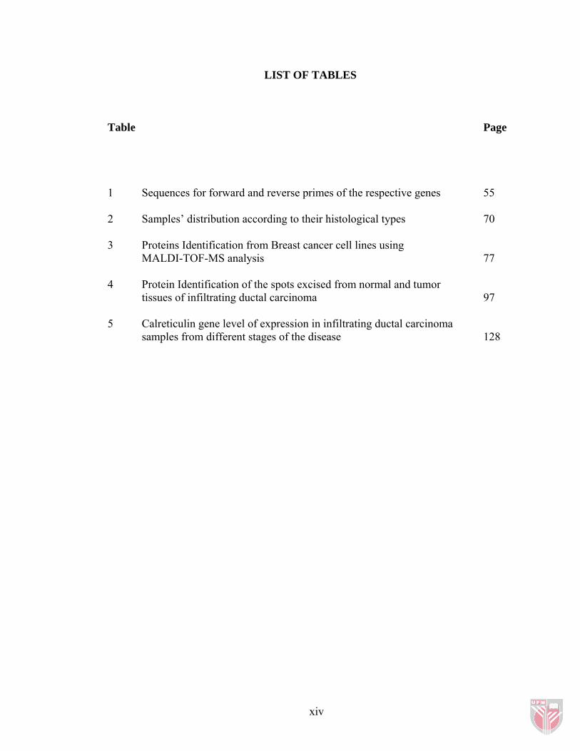
LIST OF TABLES
Table Page
1 Sequences for forward and reverse primes of the respective genes 55 2 Samples’ distribution according to their histological types 70 3 Proteins Identification from Breast cancer cell lines using
MALDI-TOF-MS analysis 77 4 Protein Identification of the spots excised from normal and tumor
tissues of infiltrating ductal carcinoma 97 5 Calreticulin gene level of expression in infiltrating ductal carcinoma
samples from different stages of the disease 128
xiv

LIST OF FIGURES
Figure Page
1 Anatomy of the human mammary gland 7 2 Breast cancer stages 17 3 Protein standard curve 61 4 Protein map from MDA-MB-231 cell line 62 5 2DGE map for (a) MCF-7 and (b) MCF-10A 64 6 2DGE protein maps for samples (a) T36 and (b) N36 67 7 2DGE protein maps for samples (a) T87 and (b) N87 68 8 2DGE protein maps of MCF-10A cell line 72 9 2DGE protein maps of MCF-7 cell line 73 10 2DGE protein maps of MDA-MB-231 cell line 74 11 Mass spectra for the tryptic peptides of spots excised from breast cancer cell lines 75 12 Mass spectra for tryptic peptides of the spots excised from breast cancer cell lines 76 13 Results of H & E staining 82 14 2D gel electrophoresis for the normal tissue from sample 158. 85 15 2D gel electrophoresis for the tumor tissue from sample 158 86 16 2D gel electrophoresis for the normal tissue from sample 89 88 17 2D gel electrophoresis for the tumor tissue from sample 89 89 18 2D gel electrophoresis for the normal tissue from sample 126 90
xv

19 2D gel electrophoresis for the tumor tissue from sample 126 91 20 2D gel electrophoresis for the normal tissue from sample 127 92 21 2D gel electrophoresis for the tumor tissue from sample 127 93 22 Mass spectra for the tryptic peptides of spots excised from breast tissues 94 23 Mass spectra for the tryptic peptides of spots excised from breast tissues 95 24 Mass spectra for the tryptic peptides of spots excised from breast tissues 96 25 Expression of gp96 in different IDC samples from different stages 100 26 Expression of calreticulin in different IDC samples from different stages of the disease 103 27 Expression of HSP60 in different IDC samples from different stages of the disease 106 28 Expression of TCP1 in different IDC samples from different stages of the disease 108 29 Calreticulin’s level of expression in breast cell lines and in different stages of invasive ductal carcinoma using Western blotting 114 30 Schematic representation of calreticulin gene in human and mouse together with calreticulin mRNA 116 31 Gradient amplification of calreticulin. 118 32 Gradient amplification of GAPDH 118 33 Amplification of GAPDH at 56 °C annealing temperature 119 34 Amplification of β actin at 56 °C annealing temperature 120 35(a) Calreticulin’s standard curve acquired using serial dilutions of cDNA sample 122 35(b) GAPDH standard curve acquired using serial dilutions of cDNA sample 122
xvi

35(c) The standard curves of calreticulin and GAPDH genes detected using FAM and TAMRA labeled probes 123 36 A graph represents ∆ CT plotted against the logarithmic template amount 124 37 Amplification plot of fluorescence intensity (RN) versus number of cycles obtained from Rotor-Gene (RG 3000) 126 38 A logarithmic plot of ∆RN versus number of cycles 126 39 A graph represents the folds of increase in calreticulin expression level in different stages of infiltrating ductal carcinoma 129
xvii

LIST OF ABBREVIATIONS
2D two-dimensional 2DGE two dimensional gel electrophoresis ACTB Actin β ACTG2 Actin γ 2 AJCC American Joint Committee on Cancer ASR age standardized incidence rate ATCC American Type Culture Collection CBB Coomassie Brilliant Blue cDNA complementary deoxyribonucleic acid CHAPS 3-[(3-Cholamidopropyl)dimethylammonio]-2-hydroxy-1-
propanesulfonate CIAPIN1 cytokine induced apoptosis inhibitor 1 DCIS Ductal carcinoma in situ DIGE differential in-gel electrophoresis DMEM Dulbecco’s modified Eagle’s medium DTE dithioerythreitol DTT dithiothreitol ECL Enhanced chemiluminescence ER estrogen receptor ER endoplasmic reticulum FNA fine needle aspiration GAPDH glyceraldehydes-3-phosphate dehydrogenase gp96 tumor rejection antigen H&E hematoxylin and eosin HER2 human growth factor receptor 2 HKL Hospital Kuala Lumpur HSP heat shock protein HSP60 Chaperonin / heat shock protein 60 HUKM Hospital Universiti Kebangsaan Malaysia IDC infiltrating ductal carcinoma IHC immunohistochemistry ILC Invasive lobular carcinoma IPG immobilized pH gradient LCIS Lobular carcinoma in situ LCM laser capture microdissection MALDI-TOF Matrix-assisted laser desorption/ionization time-of-flight mass
spectrometer MHC major histocompatibility complex MMP-2 matrix metalloprotease-2 mRNA messenger ribonucleic acid Mr molecular mass NAP1L1 Nucleosome assembly protein 1-like 1 NOS not otherwise specified
xviii

xix
OPCAML opioid-binding cell adhesion molecule precursor PBS phosphate buffered saline PCR polymerase chain reaction PgR progesterone receptor pI isoelectric points PTK protein tyrosine kinase PTP protein tyrosine phosphatase RT-PCR reverse transcription polymerase chain reaction SDS sodium dodocyl sulphate SDS-PAGE sodium dodecyl sulfate polyacrylamide gel electrophoresis SELDI-TOF surface-enhanced laser desorption/ionization time-offlight SERM selective estrogen receptor modulator TCP1 T-complex protein 1 TEMED Tetramethylethylenediamine TMA tissue microarray

CHAPTER 1
INTRODUCTION
Breast cancer is the most frequent cancer in most parts of the world and is a leading
cause of death among women. In the year 2002, 1.15 million new cases were estimated
to have occurred globally (Parkin et al., 2005). Since more than half of the cases were
reported from developed countries, it suggests that screening programs adopted in the
these countries led to the detection of early invasive tumors (Jemal et al., 2007), which
on the other hand would have been missed or detected at later stages in developing
countries (Parkin et al., 2005).
In 2008, a total of 184, 450 new cases from both sexes is estimated to be reported in the
United States of America. Among them, 182, 460 new cases are expected be reported in
females with 40,930 deaths (40,480 women and 450 men) (Jemal et al, 2008). It is also
noted that breast cancer death rates have been decreasing gradually since 1990 due to
early detection and improved treatments (ACS, 2007).
In Malaysia, the national cancer registry in 2003 reported breast cancer as the most
common cancer in females from all age groups above 15, and in all ethnic groups with
3738 new cases. The overall age standardized incidence rate (ASR) was 46.2 per
100,000 of population and 64.1 % of the cases were diagnosed in women between 40
1

and 60 years old. Chinese had the highest incidence followed by Indians and Malays
with ASR of 59.7, 55.8 and 33.9 per 100,000 of population, respectively (NCR, 2004).
However, the cultural and social beliefs of breast cancer in Malaysia are the most
important contributors to the advanced stage of disease presentation (Hisham and Yip,
2003). Also, in a study performed on patients from 1998 to 2001, the average size of
tumor was 5.4 cm in diameter and Malay women had larger tumors and a later stage of
disease at presentation than other ethnic groups. This was due to a strong confidence in
traditional medicine, poverty and poor education, the negative perception of the disease
together with fear and denial (Hisham and Yip, 2004).
Current detection methods for breast cancer depend on mammography, but even though
tumors detected by screening are significantly smaller than those non-screened ones,
only 90% of tumors can be detected (Celis et al., 2005) and a tumor should be at least a
few millimeters in size to be discovered, causing an important limitation to
mammography (Hondermarck, 2003).
After diagnosis, patients with primary breast tumors are often offered surgery followed
by adjuvant therapy. Nevertheless, factors like tumor size, auxiliary lymph node
involvement, steroid receptor status and metastasis to other organs affect the 5 year
survival rates (Celis et al., 2005), in which lymph node negative patients have
approximately 25 % recurrence rate while around 40% with lymph node positive will
experience a relapse and they often die from metastatic tumor. Also, in the quest to
2

increase the survival rates among patients, systemic adjuvant therapy has led to the
improvement in the prognosis of the disease but carried an important side effect of over-
treatment (Bergh and Holmquist, 2001).
Consequently, there is a critical need to identify specific predictive and prognostic
factors to determine the groups that can benefit from individual treatment and also
diagnostic markers that will aid in detecting the disease at very early stages and thus
increase the survival rates.
Therefore, a better understanding of the changes that occur at the molecular and protein
levels in breast cancer will facilitate the disease early detection and intervention by
identifying new drug targets. The completion of human genome project has led to the
rapid development in the technologies for the rapid and efficient analysis of the genes
and their products, in which gene expression arrays and proteomics research particularly,
are expected to provide important information to identify and characterize regulatory
and functional networks of genes and proteins within cells (Celis et al., 2003) that are
expected to accelerate the translation of basic research findings into clinical applications.
Even though studies of protein properties and their relation with diseases started in the
1970s, the field regained attention in the 1990s and a new name was applied
“Proteomics” in 1995, that was defined as the large-scale characterization of the entire
protein complement of a cell line, tissue, or organism (Kellner, 2000). A more inclusive
definition was then introduced to combine the protein and genetic analyses (Pandey and
3

Mann, 2000) which has led to the emerging of different areas of research under
proteomics, including protein-protein interaction studies, protein function, protein
modifications, and protein location studies (Graves and Haystead, 2002). Accordingly,
the involvement of different research disciplines such as biochemistry and molecular
biology together with advances in mass spectrometry and bioinformatics resulted in the
growth of proteomics as a powerful field towards the characterization of new markers
and therapeutic targets (Hondermarck et al., 2002).
In breast cancer research, cell lines are widely used experimental models to obtain a
better understanding of the disease. There are a number of established cell lines such as
MCF-7 and MDA-MB-231 that are well characterized and commonly used (Clarke et
al., 1996). Even though breast cancer cell lines are easy to handle and have a high
degree of homogeneity, they are mostly obtained from pleural effusions (Burdall et al.,
2003). Consequently, the results obtained from in vitro studies represent the aggressive
tumors rather than primary lesions (Burdall et al., 2003). Thus, studying primary lesions
of different tumor grades is more clinically relevant because most therapies are directed
to these tumors.
In this study, two dimensional gel electrophoresis (2DGE) coupled with mass
spectrometric analysis was applied in search of new prognostic markers and therapeutic
targets for breast cancer. Breast cancer cell lines, tumors and adjacent normal tissues
surgically obtained from Malaysian patients diagnosed with primary breast cancer
between 2005 and 2006 were used.
4

5
Thus, the objectives of this study are:
1. To optimize the 2DGE conditions using breast cancer cell lines (MCF-7 and
MDA-MB-231) and normal breast cell line (MCF-10A).
2. To screen and select protein samples extracted from surgically obtained tumor
and their adjacent normal tissues.
3. To perform comparative differential analysis between tumor and normal tissues
from infiltrating ductal carcinoma (IDC) samples.
4. To confirm the expression of potential markers in IDC samples using Western
blotting.
5. To correlate the potential marker levels with their gene expression levels using
real-time PCR.