UNIVERSITÀ DEGLI STUDI DI TRIESTE - openstarts.units.it · indicating a more complex mode of...
120
UNIVERSITÀ DEGLI STUDI DI TRIESTE Sede Amministrativa Università di Trieste UNIVERSITÀ DEGLI STUDI DI TRIESTE, DIPARTIMENTO SCIENZE DELLA VITA XXII CICLO DEL DOTTORATO DI RICERCA IN BIOMEDICINA MOLECOLARE INTERACTION OF HOST DEFENCE PEPTIDES WITH MODEL AND BIOLOGICAL MEMBRANES Settore scientifico-disciplinare BIO/10 DOTTORANDO RESPONSABILE DOTTORATO DI RICERCA FRANCESCA MORGERA PROF. GIANNINO DEL SAL RELATORE PROF. ALESSANDRO TOSSI UNIVERSITA’ di TRIESTE SUPERVISORE DOTT. MAURO DALLA SERRA CNR-FBK TRENTO CORRELATORE DOTT. LISA VACCARI SINCROTRONE ELETTRA TRIESTE ANNO ACCADEMICO 2008/2009
Transcript of UNIVERSITÀ DEGLI STUDI DI TRIESTE - openstarts.units.it · indicating a more complex mode of...

UNIVERSITÀ DEGLI STUDI DI TRIESTE Sede Amministrativa Università di Trieste
UNIVERSITÀ DEGLI STUDI DI TRIESTE, DIPARTIMENTO SCIENZE DELLA VITA
XXII CICLO DEL
DOTTORATO DI RICERCA IN
BIOMEDICINA MOLECOLARE
INTERACTION OF HOST DEFENCE PEPTIDES WITH MODEL
AND BIOLOGICAL MEMBRANES
Settore scientifico-disciplinare BIO/10
DOTTORANDO RESPONSABILE DOTTORATO DI RICERCA
FRANCESCA MORGERA PROF. GIANNINO DEL SAL
RELATORE PROF. ALESSANDRO TOSSI
UNIVERSITA’ di TRIESTE
SUPERVISORE DOTT. MAURO DALLA SERRA
CNR-FBK TRENTO
CORRELATORE DOTT. LISA VACCARI
SINCROTRONE ELETTRA TRIESTE
ANNO ACCADEMICO 2008/2009

Supervisor:
Prof. Alessandro Tossi,
University of Trieste, Life Sciences Dept.,
v. Giorgeri 1,
34127 Trieste, Italy
Co-advisor:
Dott. Lisa Vaccari,
SISSI Beamline (Source for Imaging and Spectroscopic Studies in the Infrared)
Elettra Synchrotron Light Source
S.S. 14, Km 163.5
34149 Basovizza, Trieste, Italy
External Supervisor:
Dott. Mauro Dalla Serra
CNR-IBF
Povo - Via Sommarive 18,
I-38100 Trento, Italy
Members of the thesis committee:
Prof. Enrico Tonin,
University of Trieste, Life Sciences Dept.,
v. Flemimg 22,
34127 Trieste, Italy
Prof. Maria Luisa Mangoni,
University of Rome “La Sapienza”, Biochemistry Dept.,
v. degli Apuli 9,
00185 Roma, Italy
Prof. Alessandro Pini,
University of Siena, Molecular Biology Dept.,
v. Fiorentina 1,
53100 Siena, Italy
Prof. Skjak Braek Gudmund
Norwegian University of Science and Technology (NTNU)
Dept. of Biotechnology N- 7491
Trondheim, Norway
Prof. Gaio Paradossi,
University of Rome “Tor Vergata”, Chemical Sciences Dept.,
v. della Ricerca Scientifica snc,
00133 Roma, Italy

To my family

1
TABLE OF CONTENTS
Abstract 5
List of papers relevant for this thesis 6
List of other papers 6
List of abbreviations 7
1. INTRODUCTION 9
1.1 Host Defence and Host Defense Peptides (HDPs) 9
1.2 HDPs – structure and function 10
1.3 Mammalian HDPs 14
1.3.1 Cathelicidins 15
1.3.2 Defensins 17
1.4 Interaction of HDPs with model membranes 19
1.4.1 HDP structuring in model membranes 19
1.4.2 Methods for probing HDP – model membrane interactions 24
1.5 Interaction of HDPs with cellular membranes: a multi-technique 31
approach
1.5.1 SR-FTIR micro-spectroscopy on bio-systems 32
2. AIMS OF THE STUDY 36
3. MATERIALS AND METHODS 37
3.1 Peptide synthesis and characterization 37
3.1.1 Peptide synthesis and purification 37
3.1.2 Circular Dichroism 38
3.1.3 Transmission FTIR 38
3.1.4 Fluorescence spectroscopy 39
3.2 Peptide-membrane interactions 39
3.2.1 Preparation of model membranes (liposomes, 39
supported mono-multi bilayers)
3.2.2 ATR-FTIR on peptide-model membrane 40

2
3.2.3 AFM on peptide-model membranes
3.2.4 Dye release form pre-loaded liposomes
3.3 Biological activity on prokaryotic cells
3.3.1 Inhibition of bacterial growth
3.3.2 Membrane permeabilization/depolarization kinetic
3.4 Biological activity of HDPs on eukaryotic cells
3.4.1 Hemolytic activity
3.4.2 Generation of monocyte-derived dendritic cells
(MDDCs) from peripheral blood
3.4.3 Determination of iDC cell cycle and protein content
3.4.4 SR-FTIR micro-spectrometric cell mapping
4. RESULTS
I - HDP- model membrane interactions
4.1 Primate cathelicidin - structures and membrane interactions
4.1.1 Peptide structuring in bulk solution and
membrane-like environments
4.1.2 Peptide structuring and aggregation on supported lipid
bilayers
4.1.3 Permeabilization of model and biological membranes
4.1.3.1 Model membranes
4.1.3.2 Biological membranes
4.2 Probing peptide structure and aggregation by using an
IR and fluorescence environmental probe: p-cyano-phenylalanine
4.2.1 Design of CN-analogues of selected cathelicidins
4.2.2 CN-analogues – CD and fluorescence
4.2.3 CN-analogues – FTIR spectroscopy
in solution and model membranes
4.3 Membrane interactions of human !-defensin 2 and 3
4.3.1 Structure predictions and design of human !-defensin
analogues
4.3.2 Probing !-defensin structures with CD and FTIR
spectroscopy
4.3.3 Biological activities
II - HDP-cell membrane interactions - a multi-technique approach
4.4 Effects of short-term interaction of hBD2 with antigen presenting cells
40
41
42
42
42
43
43
43
44
44
46
46
46
46
50
53
53
54
58
60
61
63
65
65
67
71
74
74

3
4.4.1 Peptide cytotoxicity, binding and internalization into APC
4.4.2 Flow-cytometric and TEM analyses of hBD2
interaction with iDC
4.4.3 SR-FTIR micro-spectroscopy of iDC
4.4.4 Biological responses of iDC to short-term
exposure to hBD2
5. DISCUSSION 84
HDP- membrane interactions 84
5.1 Primate cathelicidins - structuring and membrane interactions 84
5.1.1 Peptide structuring in bulk solution and membrane-like 85
environments
5.1.2 Peptide structuring and aggregation on 87
supported lipid bilayers
5.1.3 Permeabilization of model and biological membranes 88
5.1.3.1 Permeabilization of model membranes 88
5.1.3.2 Permeabilization of biological membranes 88
5.2 Probing peptide structure and aggregation by using 90
an IR and fluorescence environmental probe: p-cyano-phenylalanine.
5.2.1 Design of CN-analogues of selected cathelicidins 90
5.2.2 CN-Phe analogues – CD and fluorescence 91
5.2.3 CN-Phe analogues – FTIR spectroscopy in solution and 92
in presence of model membranes
5.3 Membrane interactions of human !-defensin 2 and 3 93
5.3.1 Design and structure prediction of human !-defensin 94
analogues
5.3.2 Probing !-defensin structure with CD and FTIR spectroscopy 95
5.3.3 Biological activities 97
HDP-host cell interactions - a multi-technique approach
5.4 Effects of short-term interaction of hBD2 with antigen presenting cells 98
5.4.1 Peptide cytotoxicity, binding and internalization into APC 99
5.4.2 Flow-cytometric and TEM analyses of hBD2 interaction 99
with iDC
5.4.3 SR-FTIR micro-spectroscopy of iDC 100
6. CONCLUSIONS 103
7. ACKNOWLEDGEMENTS 105
74
75
77
83

4
8. REFERENCES 106
Appendix: Reprints of papers and manuscripts 117

5
Abstract
Host Defence Peptides (HDPs) have multiple roles in immunity, acting
either as direct antimicrobial molecules or as immuno-modulating agents. Their role
at the interface between innate and adaptive immunity makes them potential tools
for combating infectious diseases. The aim of this work was to evaluate functional
and structural features of primate Cathelicidin and Defensin HDPs in order to
correlate them with the selective activities on prokaryotic and eukaryotic cells.
Biophysical and biochemical techniques were applied to the human cathelicidin
LL37 and selected analogues (primate ortholgues or rationally designed peptides) to
study their structural and aggregational behavior in presence of real and model
membranes (i). A similar study was carried out also for the human !-defensins
hBD2, hBD3 and analogues. Moreover, complementary microscopy techniques,
such as TEM, Synchrotron Radiation-Fourier Transform IR (SR-FTIR) µ-
spectroscopy and flow cytometry, were applied to define the short-term interaction
of hBD2 with specific antigen presenting cells, such as immature dendritic cells
(iDCs) (ii).
(i) The study on the structuring and membrane interaction of primate
cathelicidin orthologues allowed us to assess that evolution has worked on peptide
sequences leading to different structuring capacities for the different orthologues.
This in turn implies a differentiated and specific interaction with cell membranes
and different modes of membrane lysis on bacterial cells and may also imply
different modes of interacting and affecting host cells.
(ii) We found that hBD2 is able to interact rapidly with antigen presenting
cells, triggering a general lipid rearrangement in iDCs that involves an increase in
plasma membrane fluidity and a cytoplasmic endo-membrane system re-
organization. The former would lead to changes in cell morphology and promote
cell motility in response to environmental stimuli. This may provide an alternative
mechanism for the induced cellular motility than receptor-mediated chemotaxis,
indicating a more complex mode of action for hBD2 activity on iDCs, than
previously suspected.
.

Abstract
I peptidi in difesa dell’ospite (HDPs) esercitano molteplici ruoli
nell’immunità, agendo sia come molecole antimicrobiche ad azione diretta sia come
agenti immuno-moduatori. Il ruolo all’interfaccia tra immunità innata ed adattativa li
rende molecole ideali per la futura applicazione nel trattamento di malattie infettive.
Lo scopo di questo lavoro è stato quello di valutare le caratteristiche funzionali e
strutturali di Catelicidine e Defensine selezionate al fine di correlare queste proprietà
con l’ attività selettiva su cellule eucariotiche e procariotiche.
Metodi biofisici e biochimici sono stati applicati alla catelicidina umana LL37
ed alcuni analoghi (ortologhi di primate e peptidi artificiali) con lo scopo di studiare
la loro struttura e aggregazione in contatto con membrane biologiche e modello (i).
Lo stesso approccio è stato anche applicato alle defensine umane hBD2 e 3 ed a loro
analoghi. Inoltre, tecniche di microscopia, quali la microscopia a trasmissione
elettronica (TEM), la microspettroscopia infrarossa in trasformata di fourier
accoppiata ad una sorgente di sicrotrone (µSR-FTIR) e la citofluorimetria, sono state
utilizzate in modo complementare al fine di studiare l’interazione a breve termine di
hBD2 con cellule presentanti l’antigene, in particolare le cellule dendritiche immature
(ii).
(i) Lo studio strutturale e l’interazione di membrana di catelicidine ortologhe
ci ha permesso di scoprire come l’evoluzione abbia lavorato sulla sequenza dei
peptidi inducendo una diversa capacità di strutturare nei diversi ortologhi. Questo ha
portato ad un’interazione differenziata e specifica con le membrane e a diversi
meccanismi di lisi di membrana cellulare e probabilmente diversi modi di interagire
con le cellule dell’ospite.
(ii) Inoltre, abbiamo individuato una rapida interazione di hBD2 con le cellule
presentanti l’antigene. hBD2 sembra indurre un riarrangiamento generale dei lipidi
cellulari che sembra comportare un aumento nella fluidità di membrana e una ri-
organizzazione del sistema endomembranoso. Queste variazioni potrebbero essere
responsabili di un cambiamento morfologico delle cellule che promuoverebbe la
mobilità cellulare in risposta a stimuli esterni. Questo studio dimostra l’esistenza di un
posibile meccanismo alternativo di motilità cellulare rispetto alla chemotassi
recettore-mediata, indicando un meccanimo di azione di hBD2 sulle iDC più
complesso rispetto quanto riportato fino ad oggi.

6
List of papers relevant for this thesis
1. Morgera F and Vaccari L, Creatti L, Antcheva N, Pacor S, Tossi A,
Functional and biological implications of short-term interaction of hBD2 with
antigen presenting cells (in preparation)
2. Morgera F, Vaccari L, Antcheva N, Scaini D, Pacor S, Tossi A. Primate
cathelicidin orthologues display different structures and membrane interactions.
Biochem J. 2009 Feb 1;417(3):727-35.
3. Morgera F, Antcheva N, Pacor S, Quaroni L, Berti F, Vaccari L, Tossi A.
Structuring and interactions of human beta-defensins 2 and 3 with model
membranes. J Pept Sci. 2008 Apr;14(4):518-23.
4. Morgera F., Vaccari L, Creatti L, Antcheva N, Tossi A. “Structure and
activities of antimicrobial peptides at the bacterial membrane” in “Membrane-
Active Peptides: Methods and Results on Structure and Function” edited by M.A.
Castanho, IUL Biotechnology Series 9, 2009.
List of other papers
1. Antcheva N, Morgera F, Creatti L, Vaccari L, Pag U, Pacor S, Shai Y,
Sahl HG, Tossi Artificial beta-defensin based on a minimal defensin template.
Biochem J. 2009
2. Tomasinsig L, Morgera F, Antcheva N, Pacor S, Skerlavaj B, Zanetti M,
Tossi A. Structure dependence of biological activities for primate cathelicidins. J
Pept Sci. 2009 Sep;15(9):576-82.3. Jul 15;421(3):435-47.

7
List of abbreviations
AFM: atomic force microscopy
AMP: antimicrobial peptides
APC: antigen presenting cells
ATR: attenuated total reflection
CCR6: CC receptor 6
CD: circular dichroism
CN: cyano
DiBAC4(3): (bis-(1,3-dibarbituric acid)-trimethine oxanol)
FITC: fluorescein isothiocyanate
FTIR: fourier transformed infrared
hBD: human beta defensin
HDP: host defence peptide
Hss-LL37: homo sapiens sapiens LL37
iDC: immature dendritic cells
IR: infrared
IRMS: infrared microspectroscopy
LPS: lipopolysaccharide
LTA: lipoteichic acid
LUV: large unilamellar vesicle
Mmu RL37: mamcaca mulatta RL37
PAMPs: pathogen-associated molecular patterns
PBS: Phosphate buffered saline
PC/SM/Ch: phosphatidylcholine/sphyngomyelin/cholesterol
PG/dPG: phosphatidylglycerol/di-phosphatidylglycerol
PI: propidium iodine

8
PRRs: pattern recognition receptors
Ppy LL37: Pongo pygmaeus LL37
SDS: sodium dodecyl sulfate
SPB: sodium phosphate buffer
SR-FTIR: synchrotron radiation –FTIR
TA: teichoic acid
TFE: trifluoroethanol

9
1 INTRODUCTION
1.1. Host Defense and Host Defense Peptides (HDPs)
Infectious diseases are a major cause of mortality worldwide and the
development of vaccines and therapeutics is considered a current biomedical
challenge, as it requires the detailed understanding of the host immune system. A
mammalian host provides a numbers of niches that are heavily colonized by
microorganisms, such as the skin and the mucosal epithelia of the respiratory,
intestinal and urogenital tracts, which are normally kept under control. However,
injury or a depressed immune system can allow these microorganisms to elude this
control and become pathogenic, compromising health. Exposure to exogenous
pathogens can also lead to infection. Continued bacteria-host interactions in
animals, over hundreds of millions of years, have led to the evolution of several
different host-defense mechanisms that, in vertebrates, are comprised in either the
innate or adaptive immune systems. In effect, the former represents a first line of
protection, alongside the physical barrier of epithelia, and is immediately triggered
upon infection by pathogens, providing the necessary time for the more
sophisticated and specific acquired defense system to mobilize (Medzhitov et al,
1999).
These two defence systems are highly interconnected in mammals, although
they make use of different types of receptors and effectors to recognize the presence
of pathogens and deal with them. In innate immunity, recognition is mediated by
pattern recognition receptors (PRRs), generally the Toll-like receptors present on
host immune cells, that have a broad range of specificities towards conserved and
invariant molecules unique to microbes. Lipopolysaccharide, peptidoglycan and
lipoteichoic acid, as well as other cell wall components are thus termed “pathogen-
associated molecular patterns” (PAMPs). Adaptive immune activation is, instead,
mediated by antigen receptors, highly specific recognition molecules distributed on
both B- and T-lymphocytes (Janeway et al, 2001).
Toll-like receptors (TLRs), the best characterized pattern recognition
receptors within innate immunity, are transmembrane proteins that can recognize
several different bacterial products, as well as viral nucleic acids, and can trigger

10
two different types of responses: (i) the activation of tissue resident macrophages to
produce pro-inflammatory cytokines and (ii) the induction of specific cells to
release antimicrobial peptides/proteins, both of which finally lead to the overall
antimicrobial and inflammatory responses (Janeway et al, 2001; Medzhitov et al,
2000). Antimicrobial peptides (AMPs) are small molecules that are usually
expressed in skin keratinocytes and mucosal epithelial cells and may represent the
main interface between the host and the microbial biota, including both pathogens
and commensal microorganisms. The production of these molecules, induced by the
engagement of TLRs and, presumably, other PRRs, limits the multiplication and
viability of bacteria in those sites. Moreover, several other activities have recently
been associated to AMPs, indicating that they act at the interface between innate
and adaptive immunity. This has lead to the introduction of a new and wider
definition of these peptides that considers their multifunctional role in immunity:
Host Defence Peptides (HDPs).
1.2 HDPs – structure and function
Host defense peptides have been widely investigated in the last three
decades, initially mainly for their role in innate immunity, as direct antimicrobial
agents, acting on a wide range of bacteria, fungi and viruses, although the
importance of these activities may have been underestimated. Their emerging
multifunctional role in both mammalian innate and adaptive immunity is now
strongly stimulating interest in them also as promising tools for the future
development of multipurpose drugs. So far, over 1200 HDPs have been identified
or predicted, and they display a bewildering variability in terms of sequence,
structure, activities and expression (Lai et al, 2009). Nonetheless, they share a few
common features, such as a relatively short length (less than 100 residues, mostly
between 12-50), a net positive charge and the spatial separation of polar and
hydrophobic residues on their surface, often leading to amphipathic structures.
Their classification is based on general primary structural or conformational
features and comprises (i) linear peptides with amphipathic "-helical domains; (ii)
linear peptides with extended conformations and rich in specific amino acids (e.g.
Pro, His, Gly or Trp); (iii) !-hairpin peptides stabilized by 1 or 2 disulphide bonds;

11
(iv) !-sheet structures stabilized by 3 or 4 disulfide bridges, most often defined as
‘defensins’ (Fig.1.1).
Linear "-helical
e.g. magainins
Linear Pro-rich
e.g. bactenecins
Linear Trp-rich
e.g. indolicidin
2 Cys: !-hairpin
e.g. dodecapeptide
4 Cys: !-hairpin
e.g. protegrins
8 Cys !-sheet
plant defensin
6 Cys !-sheet,
!-defensin
6 Cys !-sheet
"-defensin
Fig.1.1 Common secondary structure conformations of HDPs. Adapted from Tossi & Sandri 2000.

12
Accompanying this variety of HDP structures, there is also a wide
variability in expression regarding both producing cell types and tissues, which in
vivo usually consists of the co-expression of multiple HDPs, whose release is
induced upon infection (Lai et al, 2009). The presence of HDPs in a particular
milieu can be constitutive but more commonly is induced by microbes, microbial
components or inflammatory signals, and can be regulated both at the
transcriptional or post-transcriptional level. Constitutive peptides are rarer, and in
general seem to be under a strict development control, being influenced by age or
sexual maturation. In adults, HDPs can be stored in a highly concentrated form,
within cellular granules or vesicles, and then rapidly released upon external stimuli,
although their production can also be increased as a result of infection (Lai et al,
2009). HDPs are normally expressed and stored as pro-peptides and proteolytically
processed to generate the active cationic molecule only on release. Their release at
the site of infection can involve signaling by the pattern recognition receptors, such
as TLRs, or be induced by specific cytokines.
Although surprisingly little information is available on the precise
immunological functions of HDPs in vivo, a common theme in HDP activity is their
capacity to interact with cellular membranes, and in particular those of microbial
cells. Interaction with microbial membranes can then lead to either direct killing or
inhibition of the bacterial growth. HDPs act in vitro against a wide spectrum of
gram-positive and gram-negative bacteria, as well as fungi and some viruses. The
mechanism of microbial killing is not completely understood, but in bacteria it is
evident that these peptides make use of diverse modes of action that share a
common initial step: the electrostatic attraction of these cationic molecules with the
highly anionic bacterial surface. This derives from the fact that the main
constituents of the outer bacterial cell wall, such as lipopolysaccharide (LPS) in
Gram-negatives and lipoteichic acid (LTA) or teichoic (TA) acids and
peptidoglycan (PG) in gram-positive bacteria, are anionic, and also that the
preponderance of anionic phospholipids, such as phosphatidylglycerol, in the outer
leaflet of the cytoplasmic membrane, are also anionic. The presence of HDPs on the
bacterial surface subsequently results in both the perturbation or disruption of the
cytoplasmic membrane, and interference with membrane located metabolic

13
machinery, in a concentration dependent manner, leading to catastrophic events
such as the leakage of cytoplasmic content and membrane depolarization, and
inhibition of vital metabolic processes, that cause the final bacterial killing. A few
HDPs do not induce membrane lysis, but somehow penetrate the cellular membrane
and reach possible intracellular targets (Brogden et al, 2005).
Several different modes of action have been proposed for antimicrobial
peptides, and in fact specific HDP can likely act in several different manners at the
microbial membrane, depending on the membrane type and their concentration. The
“carpet model” proposes that peptides accumulate on the membrane surface and
upon reaching a threshold concentration lead to membrane disruption in a
detergent-like manner. The “toroidal pore model”, instead, suggests the formation
of transient cavitations, where the lipid bilayer bends in onto itself in the manner of
a toroid or worm-hole (Shai et al, 1999; Zasloff et al, 2002). These pores or
channels could be of varying stabilities and lifetimes. Moreover, interaction with
the membrane can also cause interference with the vital protein machinery residing
in the membrane, as proposed in the “sand-in-the-gearbox” model (Pag et al, 2008).
Finally, even localized and transient membrane permeabilization could lead to
translocation across the membrane so that the peptides could reach and interact with
cytoplasmic target molecules.
Many questions however remain to be addressed as to the mode of action of
HDPs in vivo, and especially on how HDPs can act bacterial membranes without
affecting host cell membranes. Many hypotheses have formulated to explain this
selectivity, mainly concerning such features as the different lipid composition of
host cell membranes (which leads them to be neutral rather tan anionic) and, in
particular, the presence of cholesterol, as well as the asymmetric membrane
architecture (Zasloff et al, 2002; Matsuzaki et al, 1999), the different trans-
membrane potentials and the possibility that serum components can act as HDP
activity modulators (Sorensen et al, 1999).
Interestingly, many lines of evidence suggest that HDPs could also confer
protection to the host by indirect mechanisms, such as by modulating the activities
of cellular components of the immune system and, possibly, even acting as a bridge
between innate and adaptive immunity. This role as immuno-modulators considers
the fact that several HDP appear to alter gene expression in host cells and that some

14
can act as chemokines or induce cytokine production. Moreover, they can also
promote wound healing and angiogenesis, sequester LPS and inhibit sepsis, and
modulate specific responses of immune cells, such as dendritic or T-cells (Lai et al,
2009). These activities may complement the direct antibiotic action of HDPs
making them multifunctional immune effectors. As such they are attractive tools for
the future development of multipurpose drugs.
1.3 Mammalian HDPs
In mammals, the most extensively investigated families of HDPs are the
Defensins and the Cathelicidins, both of which are widely distributed and widely
expressed in many animal species. Several defensins are found in many types of
epithelial cells and phagocytes in mammals, and are also widely expressed in
humans (Ganz et al, 1998; Lehrer et al, 1999; Ouellette et al, 1996). In contrast,
although several different cathelicidins are expressed in some mammals, primates
Fig.1.2 Multiple functions of HDP in immunity. Adapted from Lai et al, 2009.

15
and rodents have only one. This tends to be orthologous in the animals expressing
it, and is known as LL37 in man (Zanetti et al, 1995).
Other mammalian antimicrobial peptides include salivary histatins in
primates (Tsai et al, 1998), skin dermicidin (Schittek et al, 2001), liver hepcidin
(Park et al, 2001) and some anionic peptides (Brogden et al, 1997), but their
expression and distribution is restricted to specific tissues, with respect to the
previously mentioned HDP families.
1.3.1 Cathelicidins
Cathelicidins have been described in many vertebrate species (Wang et al,
2008; Zhao et al, 2008; van Dijk et al, 2005; Uzzel et al, 2003; Zanetti et al, 1995)
and were so named on the basis of the common N-terminal region that represents a
distinctive conserved feature in this peptide family, and which in mammals is
homologous to the protein cathelin. These compounds are expressed as “pre-pro-
peptides” and in mammals are usually stored as “pro-peptides” inside granules
present in specific cell types. The conserved anionic N-terminal segment, the
cathelin-like domain, is cleaved upon stimulation, releasing a mature cationic
peptide to exert its antimicrobial function. Some mammalian species, arteodactyls
in particular, have multiple different cathelicidins, bearing AMPs with distinctly
different sequences and secondary structures, ranging from linear peptides enriched
in specific amino acids, such as tryptophan or proline and arginine, to !-hairpin
structures or linear "-helical peptides. Rodents and primates instead only seem to
carry one cathelicidin gene. All of these adopt an "-helical conformation, among
them the human orthologues LL37 (37 residue peptide beginning with Leu-Leu, see
1.4.1). LL37 is produced by neutrophils, epithelial cells, monocytes, NK cells, as
well as lymphocytes and mast cells (Frohm et al, 1999; Gudmundsson et al, 1996;
Agerberth et al, 2000; Di Nardo et al, 2003). The pro-peptide, known as hCAP18
(from human cationic antimicrobial peptide of 18 kDa), is stored at high
concentration in cytoplasmic granules as an inactive molecule (Sorensen et al,
1997). In neutrophils, hCAP18 is processed, upon release, by the serine proteinase
3, which is stored into peroxidase positive granules. Subject to external stimuli, the
two proteins interact either extracellularly, as a result of degranulation, or in

16
phagolysosomes formed during the phagocytic process, finally producing the active
molecule (Sorensen et al, 1997). Interestingly, other proteinases, found in other
locations such as sweat, skin and seminal plasma, seem to generate different
fragments (shorter or longer) from hCAP18, with different antimicrobial and
immunomodulatory activities when compared to LL37 (Yamasaki et al, 2006;
Sorensen et al, 2003; Murakami et al, 2004). The latter observations suggest that the
post-secretory processing generates an additional diversity of antimicrobial
peptides, with different effectiveness, broadening the functions of the single gene
product (Kai-Larsen et al, 2008).
The human cathelicidin-derived peptide LL37 has been widely investigated
for its direct antimicrobial activity in vitro, but this appears to be rather sensitive to
conditions used in assays, such as the salt concentration, the pH, the particular
culture medium used and the bacterial growth phase. Its killing effect is exerted on
a broad spectrum of Gram-positive and -negative bacteria, as well as on some
viruses (Turner et al, 1998; Bergman et al, 2005; Dorschner et al, 2001). The mode
of action of LL37 at the bacterial membrane has been studied by different
techniques and a “toroidal pore” mechanism was initially proposed by solid-state
NMR studies on model membranes (Henzler et al, 2004). The latter work suggested
that membrane disruption and the leakage of cytoplasmic components would lead to
bacterial killing. On the other hand, the cytotoxicity of this peptide was investigated
on erythrocytes and leukocytes in several in vitro studies, underlining that LL37 is a
selective molecule that affects eukaryotic membrane at a significantly higher
concentration than the antimicrobial ones (Johansson et al, 1998; Oren et al, 1999).
Furthermore, the cytotoxic activity on host cells is likely highly attenuated in vivo
by serum components, such as the apolipoprotein A and other lipoproteins that can
modulate its effect (Wang et al, 1998; Sorenson et al, 1999). Additional functions of
LL37 are related to its direct and indirect chemotactic activities, that it seems to
exert by interacting with FPRL-1 or EGFR receptors (De Yang et al, 2000;
Tjabringa et al, 2003, Tjabringa et al, 2006), favouring migration of monocytes,
neutrophils and T-cells to the site of infection in the first case and indirect
chemotaxis of leukocytes in the second.
The capacity to neutralize bacterial endotoxins, such as lipopolysaccharide
(LPS), is another important feature of LL37 that makes it a promising candidate for

17
treatment of endotoxic shock or sepsis. Two mechanisms have been proposed for
this antiendotoxin effect: LL37 may bind directly to LPS, inhibiting its binding to
LPS binding proteins (LBPs) and the subsequent activation of CD14 receptor, or the
human peptide may interact directly with CD14, hindering LPS-LBP binding to the
receptor (Nagaoka et al, 2002).
It is now evident that LL37 is not just an antibiotic peptide but controls
several other intriguing effects related to host defence, displaying chemotactic,
angiogenic (Tokumaru et al, 2005), endotoxin neutralizing and wound healing
activities (Carretero et al, 2008), and suggesting that it is an effective mediator
between innate and adaptive immunity.
1.3.2 Defensins
Mammalian defensins are 2-6 kDa polypeptides with a cationic and
amphipatic structure. The numerous different members of this peptide family
present highly variable sequences, effectively with only the six cysteine residues
involved in the formation of 3 disulfide bridges, and very few others, being
invariant. The disulfide bridges have defined connectivities within each defensin
family and stabilize the canonical antiparallel triple !-sheet structure (Pardi et al,
1992; Hoover et al, 2001; Hoover et al, 2000; Bauer et al, 2001). On the basis of the
bridging patterns, these HDPs are further classified into three families in mammals:
"-, !- and #-defensins (Ganz et al, 1998; Leher et al, 1999). "-defensins have the
three disulfide bonds linked in a C1-C6, C2-C4 and C3-C5 (Zhang et al, 1992)
pattern and are broadly distributed in humans and other primates as well as rodents,
but not in some other mammals or non-mammalian vertebrates; these molecules are
usually stored in granules of neutrophils, macrophages and Paneth cells of the
intestine (Ganz et al, 1998; Lehrer et al, 1999; Ouellette et al, 1996). Conversely,
the !-defensin disulfide bridges are paired C1-C5, C2-C4, C3-C6 (Tang et al, 1993)
and are prevalently expressed in epithelial cells of various organs, such as the
epidermidis, the bronchial tree and the genitourinary tract. These peptides are
expressed in all mammals as well as birds and reptiles.
#-defensins have been identified only in primate phagocytes (Tang et al,
1999) and are formed by two hemi-"-defensin chains that are covalently linked to

18
form a cyclic peptide backbone, each of which brings three cysteine residues. The
distribution pattern in animals suggests that "-defensins evolved from !-defensins
in some mammals only, and #-defensins from "-defensins in primates.
All defensins, with the exception of #-defensins (Tran et al, 2002), exert in
vitro a direct antimicrobial activity on a specific range of bacteria, fungi and some
enveloped viruses, although this effect is quite sensitive to physiological salt
concentration (I.e 150 mM NaCl) (Ganz et al, 1998; Lehrer et al, 1999; Shröder et
al, 1999). It is proposed that in vivo the antimicrobial activity is likely to be
displayed in phagocytic vacuoles or at the level of skin tissues, where they can
reach quite high concentrations and where the ionic strength is relatively low.
Moreover, it is not certain whether they act by damaging microbial membranes and,
if so, the mechanism of membrane permeabilization is still unknown. It is however
generally supposed that the particular conformation of defensins, with separated
charged/polar and hydrophobic surfaces, could allow them to interact efficiently
with and accumulate onto bacterial membrane, leading to membrane disruption and
thus to the final microbial killing, although other mechanisms could also be acting.
While the direct microbicidal activity of defensins has been intensely
investigated for years, more recently "- and !-defensins have been shown to also
have a potentially important role in promoting and/or enhancing the initiation of
adaptive immune responses. In fact, in vitro observations have indicated that both
defensin families are chemotactic for human memory T-cells and immature
dendritic cells (iDC), in a process possibly involving chemokine receptors (Yang et
al, 1999, Yang et al, 2000). These studies suggest that, in vivo, "-defensins might
be released by exocytosis from infiltrating neutrophils, whereas epithelial cells
might secrete !-defensins and that both types of defensins might then form
chemotactic gradients at the site of infection, thus help recruiting T-cells, iDC and
monocytes from blood circulation. Experiments carried out with human "- and !-
defensins have furthermore shown that they can activate mast cell degranulation,
leading to the release of histamine and prostaglandin D2 and thus possibly
increasing, in vivo, neutrophil influx at the inflammation site (Befus et al, 1999;
Niyosaba et al, 2001).

19
Although many questions remain to be addressed on mammalian defensin
activities, the emerging picture proposes multiple roles for these host defense
peptides in both innate and adaptive immunity, ranging from a direct antimicrobial
effect and the regulation of innate immune responses to a role as enhancers in the
initiation of the acquired immune responses (De Yang et al, 2002).
1.4 Interaction of HDPs with model membranes
A central theme in the mode of action of HDPs is their selective interaction
with eukaryotic and prokaryotic cell membranes, leading to diverse effects and to
their different biological activities. Structural modifications and aggregation of
these peptides at the membrane surface, the possible insertion into the lipid bilayer,
as well as the subsequent membrane disruption or translocation to a surface or
internal target, are essential steps in the definition of their mechanisms of action.
Due to the complexity of the cellular membrane, an effective investigation of these
successive steps requires the preliminary use of suitable model membranes and
biophysical techniques, capable to assess and quantitatively describe them in a
simpler milieu. The availability of structural homologues of the peptides,
orthologues from different mammalian species or rationally designed variants,
whose behaviour may be compared to the molecules under investigation, may also
be essential for individuating the structural features responsible for specific
activities.
In this thesis I have focused on selected human HDPs, such as the LL37 and
the better known human !-defensins, and investigated their mode of action at the
cellular membrane by comparing their behaviour with both native primate
orthologues and rationally designed congeners or derivatives.
1.4.1 HDP structuring in model membranes
Despite extraordinary interest in understanding the mode of action of HDPs
and explaining their selectivity toward eukaryotic and prokaryotic cells, their
structure at the membrane surface is mostly still undetermined. Many attempts have
been made to gain some insights on their active conformations, also with the aim of

20
developing novel molecules of therapeutic value based on specific structural
features of HDPs. Low-resolution methods have been widely applied to study
peptides in bulk solution as well as in membrane-mimetic environments, such as
liposomes, SDS micelles, or folding inducers such as trifluoroethanol. High-
resolution studies are much rarer. The following paragraphs describe the structure
of cathelicidins and !-defensins characterized so far, either in bulk solution or at the
membrane surface, on the basis of high-resolution techniques.
i) Cathelicidins - Human cathelicidin has been observed and characterized
for many years by low-resolution methods such as CD spectroscopy (Johansson et
al, 1998). However, and despite its small size, it seems to have been rather
refractory to higher resolution methods, and it is only very recently that NMR
studies of the peptide and its fragments in model membranes have been reported
(Wang 2008, Porcelli et al, 2008).
CD spectroscopy studies indicated that LL37 adopts an "-helical
conformation in a manner that is anion, pH- and concentration-dependent
(Johansson et al, 1998) and it is suggested that oligomerization may accompany its
structuring. This would be required for entropic reasons, as the resulting
amphipathic helix would need to sequester its hydrophobic surface from the
aqueous environment. However, Oren et al. suggested that LL37 may continue to
act as an oligomer also when in contact with neutral biological membranes, while it
dissociates into monomers upon binding to anionic ones, as shown by membrane
binding experiments carried out with the fluorescently labelled peptide (Oren et al,
1999).
Recent investigations have finally proposed a high-resolution structure of
LL37 in the presence of anionic (SDS and D8PG) and neutral (DPC) micelles, by
means of isotope labelled or 2D NMR respectively (Wang et al, 2008; Porcelli et al,
2008). The structure in the presence of anionic micelles confirmed the presence of a
long helix, covering residues 2-31, while a disordered and flexible C-terminal
segment was detected (Fig.1.3A). This structure is amphipathic and presents a bend
between residues G14 ad E16, with some indication that the hydrophobic packing
near I13 and F17 may favour bend formation (Wang et al, 2008). Porcelli and
colleagues reported a more flexible molecule in the presence of neutral DPC
micelles, with a shorter helical domain (residues 4-33) located between unordered

21
C- and N-termini (Fig.1.3B). This characterization was carried out using synthetic
peptide by a less resolved method, but also indicated the presence of a curved
structure (defined helix-break-helix), at the level of the residue K12 (Porcelli et al,
2008). Although indicating slightly different structural behaviours both studies
concurred in defining an important role of the four phenylalanins (residues
5,6,17,27) in micelle binding, as well as a possible aromatic stacking between the F-
5 and -6 in the N-terminal segment.
In this context, the human peptide’s structure and its active form upon
binding to membranes could be used as a model for understanding the structure and
function of homologous primate cathelicidins, as well as understanding how
evolution works on closely related species, varying structural domains with the aim
of tuning them for specific functions. The laboratory where I carried out my thesis
work has recently reported over a dozen cathelicidin sequences from different
primates, individuating sequence similarity and studying how structure and
aggregation affect membrane interaction and biological activities (Zelezetsky et al,
2006). These studies showed that the primate peptides were subject to variations
principally in the distribution of charged residues, in a manner that did not affect the
overall amphipathicity and hydrophobicity of the molecules, but which modulated
the occurrence of intramolecular salt-bridging that markedly affected helix
formation. Moreover, these variations resulted into two distinct type of behaviours:
one is similar to that of helical antimicrobial peptides found in many vertebrate and
invertebrate animals, that are unstructured and un-aggregated in bulk solution but
undergo a transition to an amphipathic helix at the bacterial membrane surface.
Fig 1.3 NMR structure of LL37 in anionic
and neutral model membranes: (A) isotope
labelled NMR structure of LL37 in anionic
micelles (PDB ID 2k60) (Wang et al., 2008),
(B) 2D NMR structure of LL37 in neutral
micelles (Porcelli et al., 2008).
A
B

22
These peptides normally display a potent and medium insensitive antimicrobial
action and we can refer to these “canonical” helical AMPs. The other type,
including the human LL37, instead displays a salt dependent
structuring/aggregation in bulk solution that leads to a more medium-sensitive
activity. These differences in structuring and aggregation may enhance interaction
with specific membranes, affecting both antimicrobial, cytotoxic and host cell
modulation effects.
Additional insights into cathelicidin structure could be achieved by
complementing high-resolution data coming from LL37 NMR structure in contact
with lipid micelles and biophysical experiments on model membranes, by
comparing human and primate peptides. In fact, CD spectroscopy showed that in
presence of a bacterial-type membrane, all primate peptides folded into an "-helical
conformation and, neglecting possible aggregation, sequence alignment showed that
the helix bend is likely to occur in all homologues, as highly conserved glycine
residues at positions 14-16 are suitable for flexible structures. Moreover, the
phenylalanine residues at the N- and C- terminal segments are also quite conserved,
in particular at position 27, and may be important not only in peptide-membrane but
also in interpeptide interactions.
ii) Defensins – The RCSB Protein Data Bank reports numerous solution and
crystal structures of "- and !-defensins, although there are no high-resolution data
available in a membrane-like environment. Among human !-defensins, both hBD1
and hBD2 have been characterized both by NMR in solution and by X-ray
crystallography (Schibli et al, 2002; Hoover et al, 2001; Sawai et al, 2001; Hoover
et al, 2000), whereas the structure of hBD3 has been determined only in solution
(Shibli et al, 2002). All three molecules share a triple stranded !-sheet structure,
while the N-terminal segment, that forms a quite stable "-helix for hBD2, is less
structured in hBD1 or hBD3. In fact, while hBD2 crystal and NMR structures are
very similar, hBD1 shows a defined helical segment only in the crystallographic
structure, suggesting a different conformational stability compared to hBD2.
Moreover, these studies highlighted a different aggregational behaviour for these
related molecules. In particular, hBD2 has been reported to be a monomer in
solution, up to the concentration of 3.5 mM used for NMR studies, while well-
defined multimers are observed in the crystal. Furthermore, in the later case the N-

23
terminal "-helical region participates in hBD2 octamer assembly, along with !1-!1
interpeptide interactions. The possibility that multimeric structure may form at the
lipid-water interface has been proposed, as the pseudo-concentration effect at the
cell membrane may favour the formation of oligomeric peptide platforms (Hoover
et al, 2000). The structure of human !-defensin 3 has been observed only in
solution, as a monomer. However, native PAGE studies indicate it dimerises or
oligomerises even at micromolar concentrations, so that a symmetrical dimeric
structure has been modelled and proposed to occur (Schibli et al. 2002, Boniotto et
al, 2003).
The crystal structure of hBD1 also suggests the presence of oligomeric
forms at higher concentrations, but the dimerization pattern is different with respect
to either hBD2 or hBD3, highlighting that although the low sequence homology
between the peptides does not markedly alter the tertiary structure, it may be
important for the subsequent oligomerization.
In this context, this PhD thesis focuses on the study of
structural/aggregational behaviour of the human HDP peptides in model
Fig.1.4 NMR and X-ray structure of human !-defensins: solution structure of hBD1, -2 and –3
determined by means of NMR in solution (C, D, E respectively) and crystal structure of hBD1 and -2
(A and B). The 3D images derive form the following PDB files: 1E4R, 1E4Q ,1KJ5, 1FD3, 1KJ6.
A B
C D E

24
membranes, also using insertion of non-proteinogenic amino acids or rationally
designed variants in order to identify structural features responsible for specific
activities.
1.4.2 Methods for probing HDP - model membrane interaction
The purpose of investigating and finally understanding the effective
mode of action of HDPs at the bacterial membrane is quite challenging, as it
requires dissecting the peptide trajectory from the bulk solution to the bacterial
membrane into the several steps that permit it to reach the membrane surface and
eventually generate the “interaction”. In fact, as shown in Fig.1.5, HDPs have to
reach the microbial surface from bulk solution (A), by crossing the outer cell-wall
components (B), so that they can interact with the cytoplasmic membrane (C) and
possibly translocate across it into the cytoplasm (D). Although the membrane lysis
or permeabilization are the most studied phases of the HDP mode of action, the
steps that precede them, such as the structuring in bulk solution or the interaction
with the outer cell wall components, can significantly affect the peptides’ trajectory,
inducing structural variations necessary for the attachment to or insertion into the
membrane, or simply modifying the final peptide concentration at the membrane
surface. Within this context, this section aims at describing the main biophysical
techniques that can be applied to address HDP structural behaviour during its
approach to the bacterial membrane. Several of the following methods have been
applied also to our HDP-model systems, as will be described in section 2.
Aspects that need to be taken into account include conformational features
of HDPs and their interactions with each other or other species, at the following
locations:
• bulk solution (A)
• outer cell wall (B)
• membrane (C)
• cytoplasm (D)

25
Bulk solution
HDPs in bulk solution can be unstructured, pre-structured or in equilibrium
between these conformational forms (Fig. 1.5 1), or between monomeric and
aggregational forms (Fig. 1.5 2) and can also bind non productively to serum or
interstitial components (SC, Fig. 1.5 3).
The techniques that are usually applied to access peptide conformation in
bulk solution are circular dichroism, transmission FTIR and solution NMR
spectroscopies.
CD spectroscopy is commonly used to evaluate peptide/protein secondary
structure in different surroundings, such as buffer solutions or membrane like
environments. The low peptide quantity required and the relatively simplicity of the
technique make this a method of choice for probing peptide structure (Kelly et al,
A
B
C
D
Fig.1.5 Modes of action of HDPs. HDPs act at the microbial surface, which they have to reach
from bulk solution (A), by crossing the outer cell-wall components (B) so that they can interact with the cytoplasmic membrane (C) and possibly translocate across it into the cytoplasm (D).
1 2 SC 3

26
2000). Although it does not provide high-resolution data, it is very sensitive at
picking up structural variations over a wide range of experimental conditions,
including different temperature, pH, and solution compositions (Kelly et al, 2005;
Martin et al, 2008). A disadvantage is that it does not well resolve contributions
from different structural forms in equilibrium, especially for the minority forms. On
the other hand, in the case of amphipathic helices, CD can provide information on
the monomeric or oligomeric state of the helices by considering the ratio between
the two spectral minima at 222 nm and 208 nm, as initially reported by Lau et al.
(Lau et al, 1984; Cooper et al, 1990; Wagschal et al, 2008).
Transmission FTIR measurements are complementary to CD analysis in
solution, giving insight on both the conformational and aggregational states of
HDPs, by monitoring peptide amide I band (Table 1.1). The main drawback of this
technique is the high absorbance of water in the amide region (amide I band ranges
from 1590 to 1700 cm-1
) which can be partly resolved by using deuterium oxide
instead of water for transmission measurements (Arrondo et al, 1993; Byler et al,
1986). Deuterium oxide gives rise to IR bands at lower wavenumbers, providing a
valuable IR window for peptide weak bands and allowing the use of relatively low
peptide concentration to obtain well-resolved spectra. In any case, the required
peptide concentration is around 2-3 mM, so peptide aggregational states may be
favoured.
Solution NMR is a high-resolution method that allows the determination of
the 3D structure of peptides and proteins and the results are complementary to those
obtained to X-ray diffraction analysis of protein crystals (Wider et al, 2000). NMR
requires large amount of highly pure proteins and, as all high-resolution techniques,
the availability of expensive equipment as well as expert personal are necessary,
greatly restricting its use.
Outer cell wall components
HDP activity can be modulated by the particular form they assume (random
coil, structured, self-aggregated or bound to other molecules) and by the interaction
with outer membrane lipopolysaccharide (LPS) or peptidoglycan (PG) components
or bacterial exopolisaccharides (EPS). The interaction with these compounds is

27
usually monitored by CD, which allows using peptide concentrations similar to
those used for functional assays, whereas FTIR requires, as stated previously, much
higher amount of peptide. Moreover, the FTIR data analysis could be really quite
complex due to the presence of carbohydrate vibrational bands that may overlap
with peptide ones, leading difficulties in spectral subtraction and possible
misinterpretations.
Cytoplasmic membrane
At the membrane, interaction may be modulated by the conformational or
aggregational form and can consist of an initial surface electrostatic binding
followed by insertion, membrane compromising, and cellular inactivation by one or
more of several possible mechanisms (as discussed in chapt.1.3).
Methods applied to study peptide-membrane interactions require a
membrane-like system suitable for the biophysical method that is to be applied.
Common lipid model systems comprise either very simple lipid-like environments,
such as trifluoroethanol, a folding inducer, or SDS micelles, or more sophisticated
systems, such as liposomes or supported lipid bilayers. The latter systems take
advantage from the possibility to select specific lipid mixtures that are similar to
those reported to constitute real membrane systems, such as the cytoplasmic
bacterial membranes. Moreover, their physico-chemical properties, such as lipid
bilayer architecture or lipid curvature, are closer to bio-membranes (Jelinek et al,
2005). Furthermore, they are compatible with commonly available spectroscopic
techniques such as circular dichroism, FTIR and fluorescence, as well as techniques
such as plasmon resonance and Atomic Force Microscopy (AFM).
As mentioned earlier, CD spectroscopy is commonly used also to evaluate
peptide/protein secondary structure in membrane like environments, such as SDS
micelles or large unilamellar liposomes (LUVs).
Solid state NMR is a high-resolution technique that has been widely applied
to HDP-membrane systems. It allows addressing details of both peptide and lipid
structure and orientation, individuating also if the interaction occurs preferentially
at the level of the lipid head groups or requires insertion into the hydrophobic core
of the membrane (Lu et al, 2005; Salnikov et al, 2009). Unfortunately it is a highly
specialized technique not commonly available to most researchers.

28
Attenuated Total Reflection (ATR) FTIR spectroscopy is a sensitive
technique for the determination of conformation and orientation of membrane
associated peptides and proteins. In particular, ATR-FTIR has become increasingly
popular as i) information can be simultaneously obtained not only on protein
secondary structure but also on membrane phase, by focusing on both the peptide
amide bands and lipid acyl chain IR bands (see Tab.1.1); ii) ATR with polarized
light provides information on both peptide and membrane orientation; iii)
measurements can be carried out also in the presence of an aqueous environment;
iv) sample conditions, salt concentrations and buffer types, can easily be varied in
situ (Tamm & Tatulian 1997; Vigano et al, 2000).

29
Tab.1.1: IR bands of proteins and lipids (A) and band components of the amide I band (B) with the
respective protein secondary structure assignments [Adapted from Tamm & Tatulian 1997; Arrondo
et al, 1993].
A. Band name Assignment
Position
H2O
(cm-1
)
Amide I $ 80% C=O stretching,
(10% C-N st, 10% N-H def) 1600-1700
Amide II 60% N-H deformation
(40 C-N stretching)
1510-1580
Methyl asymmetric
stretching
C-H asymmetric stretching
of CH3 2956
Methylen
asymmetric
stretching
C-H asymmetric stretching
of CH2 2920
Methyl symmetric
stretching
C-H symmetric stretching of
CH3 2870
Methylen symmetric
stretching
C-H symmetric stretching. of
CH2 2850
Phospholipids C=O stretching of
phospholipids 1730
B. Amide I - Band components
Antiparallel !-sheet 1675-1695
Turns 1660-1685
310-helix 1660-1670
"-helix 1648-1660
Unordered 1652-1660
(deuterated
1640-48)
! -sheet 1625-1640
Aggregated
! -structure
1610-1628
1680-1690

30
Fluorescence spectroscopy is a highly sensitive technique commonly
applied to the study of polypeptide folding or membrane interaction. The intrinsic
fluorescence of native aromatic amino acids, such as tryptophan or tyrosine, or of
non proteinogenic ones or fluorescent markers, specifically introduced for the
purpose, could be used to probe either protein oligomerization or binding to
synthetic membranes, by following the fluorescence enhancement or quenching
(Ladokhin et al, 2000). Moreover, fluorescent dyes encapsulated in liposomes can
be used to easily monitor lytic effects of peptides on membranes as a function of
concentration, time of exposure, membrane composition or structural changes and
allow investigating peptide lytic effect on different lipid systems (Ladokhin et al,
1995; Ladokhin et al, 1997).
As will be described in section 2, we have applied a non-proteinogenic
amino acid that has allowed us to couple fluorescence and FTIR experiments. In
fact, cyano-phenylalanine is considered an excellent environmental probe since it
allows to evaluate peptide structure and aggregation or membrane insertion in a
relatively un-invasive manner. In fact, this residue can replace native aromatic
amino acids (e.g. Phe or Tyr residues already present in proteins, including HDPs
such as cathelicidins or defensins), providing a five-fold increase in fluorescence to
the peptide (Tucker et al, 2005), as well as the possibility to monitor the residue
microenvironment by following the CN stretching band by FTIR spectroscopy
(Getahun et al, 2003; Tucker et al, 2004). As a matter of fact, the C-N stretching of
cyan group produces a quite strong absorption band in a region of the spectrum
almost free of other signals (~ 2250-2240 cm-1), characterized by a pronounced
shift to lower wavenumbers when inserted in a apolar environment.
Atomic Force Microscopy (AFM) can provide high-resolution images of
peptide-membrane systems, allowing the direct visualization of membrane
perturbations at a nanometric spatial resolution, which can permit distinguishing
between formation of pores or other types of defined lesions from general
membrane disaggregation (Czajkowsky et al, 2002). Beside the traditional contact
(or “dragging”) mode, most biophysical samples are usually scanned by using the
non contact (or “tapping”) mode that minimizes damage to soft samples (Santos et
al, 2004). Moreover, the phase shift of the tip oscillation relative to the driving
signal induced by the interaction with a heterogeneous sample can be used as an

31
alternative imaging mode, named phase imaging (Stark et al, 2001). This method is
informative for probing chemical heterogeneities (e.g. polar and apolar regions or
regions of different charges) and particularly useful in the investigation of cationic
peptide on phospholipids membrane, and of the membrane perturbations they cause,
which can be easily detected, as will be described in section 4.
Cytoplasmic targets
It is also likely that peptides translocate across the membrane, to interfere
with vital cytoplasmic components (e.g., proteins and/or DNA). Biophysical
techniques are not the most suitable methods to detect this kind of interactions,
since specific targets should be identified, leading to the choice of alternative and
more specific techniques, such as those used in microbiology or molecular biology.
In any case, biophysical techniques on model systems often focus on
specific parameters involved in HDP-membrane interaction, so that the
interpretation of results absolutely requires being corroborated and complemented
by results from biological assays carried out on real systems. In both cases, the
availability of a set of synthetic variants of the naturally occurring antimicrobial
peptides, with defined alterations, can be a very informative tool for probing the
role of specific residues or structural features in peptide activity and membrane
interactions.
1.5. Interactions of HDPs and cellular membranes – a multi-technique
approach
The exploration of HDP-cell membrane interactions in a real system is
evidently complicated by the presence both of multiple biological components and
multiple events that can occur simultaneously or subsequently within the cell. As
previously described, while in a model system variations occurring in the structure
of either peptide or membrane can be easily controlled and studied without
interferences, investigations in real systems require a different approach. In fact,
structural/chemical alterations in molecules interacting at the cellular membrane can
trigger a cascade of intracellular signals, trafficking of molecules at the cell surface
and other consequent events that make observations really quite complex and often

32
confusing. It is therefore necessary to identify and focus on specific targets involved
in the interaction under investigation, by coupling the use of appropriate
dyes/probes with sensitive techniques. In general, a main problem is obtaining
sufficient spatial or temporal resolution. Imaging techniques, such as confocal
fluorescence microscopy, allow to monitor cells or tissues continuously and to
obtain spatially resolved information, but they require first identifying and marking
a specific target. Other techniques, such flow cytometry, only provide general
information over a whole cell, but permit to evaluate high amount of cells in a
relatively short time period, and also to simultaneously analyze a higher number of
different fluorescent markers then conventional imaging methods. Flow cytometry
is a quantitative method, but has a main drawback in that cells cannot be re-
analyzed or investigated continuously over a period of time, because once analyzed
a cell is lost. A good compromise is the concerted use of imaging techniques with
high spatial resolution, such as fluorescence confocal microscopy and transmission
electron microscopy with flow cytometry, on samples prepared under standardized
conditions. In this thesis, for example, the short-term effect of hBD2 was evaluated
on antigen presenting cells by combining the above mentioned conventional
methods also with Synchrotron Radiation (SR) Fourier Transform IR micro-
spectroscopy (IRMS). This new technique allows evaluating the chemical
distribution of the main cellular components without requiring dye staining, as is
described in detail in the following paragraph.
1.5.1 SR-FTIR micro-spectroscopy on bio-systems
Although FTIR spectroscopy is a well-established analytical method used in
a variety of applications, ranging from organic to pharmaceutical chemistry, FTIR
micro-spectroscopy has yet to be widely applied to biomedicine. Compared with
conventional IR spectroscopic approach, IRMS allows acquiring IR spectra
selectively from the region of interest, correlating visible morphological features of
the sample with its specific vibrational pattern. This imaging technique has recently
been applied to tissues and cells, with the objective of detecting variations in the
chemical/biochemical features of analyzed specimens, usually comparing a
control/normal with a treated/diseased sample (Petter et al., 2009; Boskey et al.,
2005). It aims at the individuation, at relatively high spatial resolution, of subtle

33
spectral variations that can be associated to biochemical differences between target
and control samples, likely induced by disease or drug treatment. The IR spectrum
measures the sample chemical composition and molecular structure, so this analysis
of bio-systems provides insight into what occurs at the level the main cellular
components, such as proteins, lipids and phospholipids, nucleic acids and other
molecules whose IR bands are comprised in a typical IR spectrum (see Figure 1.6).
Despite the huge amount of macromolecules that contribute to the IR spectral
pattern, on the basis of the specific vibrational characteristics of each most
fundamental cellular component and on their relative abundance, IR bands can be
assigned to different chemical groups that belong specifically to different bio-
macromolecules, as listed below in table 1.2 (Miller et al, 2003; Jackson & Mantsch
et al, 1995).
A main advantage of IR microspectroscopy is that it does not require any
staining of the sample so as to discriminate between normal and diseased areas of
Fig.1.6 Typical IR spectrum of a biological sample: main absorbance bands of lipids, phospholipids,
proteins and nucleic acids are reported.

34
tissues and cells (Holman et al., 2003). Nevertheless, since IR technique is non-
selective, IR analyses of tissues are always presented along with the respective
histopathological results and, in general, the presence of other complementary
biological assays is essential for a correct exploration of the bio-system under
investigation. Moreover, another important issue is related to the IR spatial
resolution, that is determined by the diffraction limit (about # of the wavelength of
IR radiation). The IR region of biochemical interest is the middle infrared, which
ranges from 4000 to 400 cm-1
, so spatial resolution is 1.25 to 12.5 µm. However, to
fully accede to this spatial resolution, FTIR microscopes are coupled with a brilliant
synchrotron radiation IR source. This allows the technique also to be applied to sub-
cellular investigations, where it could be very informative to compare different cell
populations. Many approaches can be applied in this respect; in the following are
reported the ones we chose to pursue at the SISSI (Synchrotron Infrared Source for
Spectroscopy and Imaging) infrared beamline of Elettra Synchrotron (Trieste, Italy)
(Lupi et al, 2007):
! Entire cell measurements: the spatial resolution is set in order to
match the cell diameter (it could range from 10x10µm to many microns, depending
on the cell type) in order to collect mean IR spectra over the entire area of the cell.
This procedure is repeated for a statistically relevant number of cells in both the
control and treated samples. Collected spectra are subject to correction, as required,
and then evaluated by cluster analysis, a mathematical procedure that can
individuate similarities and differences amongst a large amount of spectra and
express the result in terms of “distance” or “heterogeneity”, usually by means of
dendograms. Moreover, mean spectra or mean derivative spectra of the individuated
spectral classes can be also compared, identifying the main biochemical and
biological divergences. Spectral differences between the normal and treated (or
diseased) groups is further associated to biological divergences, by using tables of
standard literature IR band assignments, although this procedure must be applied
with cognition, requiring experience in both the spectroscopic technique and
biological processes underlying the observed variations.
! Single cell mapping: the spatial resolution is set to higher values,
such as 10x10 µm or less, in order to collect IR spectra over adjacent segments of

35
the cell. IR maps are obtained by collecting a spectrum at each of the sampling
points, previously fixed by creating a virtual grid on the selected cell. Highly
performing software packages are required to assemble all the IR spectra, and
integrate the bands of interest, such as protein amide, acyl chains, phosphate and
carbonyl groups. This procedure finally gives rise to the relative chemical
distribution maps, in which integrated spectral intensities are converted into a
colour scale that can be directly compared to optical images of the analysed cells.
Tab.1.2 FTIR bands of main cellular components: band position, assignment and relative cell
constituent are listed. Abbreviations: % = stretching; s = symmetric; as= asymmetric. Adapted from
Naumann et al, 1998 & Jackson, M. & Mantsch, H. 1995.

36
2. AIMS OF THE STUDY
This work aims at defining the mode of action of selected human HDPs,
focusing on peptide-membrane interactions, which are considered essential in
determining both the peptides’ activities and the selectivity of these for eukaryotic
or prokaryotic cells. The differentiated action exerted on bacterial and host cells is a
key feature of these immune effectors that has been closely investigated also in
order to develop novel drugs for the cure of infectious diseases. The parameters that
regulate this selectivity are however not linked only to physical or structural
features of the peptides, such as critical active concentrations or structure-function
relationships. Several other critical issues have recently emerged, such as the role of
their environment or the physico-chemical characteristics of cell walls and
membranes, and possible interactions with membrane-associated receptors or
internal targets. This makes the study of endogenous HDPs, and the design of
analogues for use as potential drugs, quite complex. In this context, this thesis
proposes to study peptide-membrane interaction by:
i) using several different and complementary biological,
biochemical and biophysical techniques concerning real or model
membranes, in order to identify structural or environmental features that
can affect or regulate peptide activity;
ii) comparing structural and functional features of primate
orthologues or synthetic peptide analogues with those of the native
human peptides, in order to try understanding how evolution has worked
on peptides from closely related species so as to tune them for specific
functions, and rationally test these conclusions;
iii) rationally designing, synthesizing and testing peptide variants
in order to corroborate structural studies and eventually propose new
simplified molecules with enhanced and selective antibiotic properties;

37
3 MATERIALS AND METHODS
3.1 Peptide synthesis and characterization
3.1.1 Peptide synthesis and purification
Peptides used in this work were either endogenous primate
cathelicidins/defensins or artificially designed analogues. Synthesis/folding
protocols are quite different for these two peptide families, due to the diversity in
secondary and tertiary structures. All peptides analogues were synthesized by
Fmoc-solid phase peptide synthesis, performed initially on a Pioneer® automated
synthesizer (PE Biosystems), and subsequently on a CEM Liberty microwave-
assisted peptide synthesizer. Cathelicidins were generally synthesized using PEG-
PS resin (substitution 0.25 mmol/g), whereas defensins were synthesized on the
more acid-labile 2-chlorotrityl chloride resin (substitution $ 0.2 mmol/g). A version
of the anti-aggregation solvent “magic mixture” (dimethylfomamide/N-
methylpyrrolidone (3:1 v/v), 1% tritonX-100 and 1M ethylene carbonate) was used
in both cases. For longer, more complex peptides, synthesis was interrupted
regularly to check synthesis quality on cleaved aliquots, using ESI-MS (Bruker
Esquire 4000).
On synthesis completion, peptides were cleaved from resins using
appropriate versions of the reagent K. Crude linear peptides were then purified
using a preparative C18 column (e.g. Waters Delta Pak®, 5%m, 300 A°, 25 mm x
100 mm). The quality and correctness of the peptides was confirmed using ESI-MS.
The crude linear defensins obtained from synthesis were directly oxidatively folded
for 24-48h in N2-saturated aqueous buffer (1M guanidinium chloride/0.1 M
ammonium acetate/2 mM EDTA, pH 8.5), in which 1mM cysteine and 0.1 mM
cystine were dissolved immediately before use, at a final peptide concentration of
10 µM. Oxidation was monitored by analytical RP-HPLC and ESI-MS until
completion, before final purification on the preparative C18 column.
All peptide solutions for subsequent assays were prepared from stock
solutions of purified peptide in water, in which the concentration was quantified by
at least three independent methods, including i) accurately weighing the peptide
used to prepare solutions, ii) the Waddle method (Waddell, 1956) and iii) aromatic

38
residue absorption, using reported extinction coefficients. In the case of peptides
bearing cyanophenylalanine analogues, concentration were also evaluated by
measuring CN-Phe absorption at 240 nm (&=13,000 M-1
cm-1
).
3.1.2 Circular Dichroism
CD spectroscopy was performed on a J-715 spectropolarimeter (JASCO
Corp. Japan), using 2-mm quartz cells and 20 %M peptide. Peptide conformation
was investigated in aqueous buffers, such as 0.150 M NaCl and 0.010 M sodium
phosphate (PBS buffer) at pH 7.0, or 0.010 M sodium phosphate (SPB), as well as
in the presence of the folding inducer trifluoroethanol (50% TFE in SPB buffer),
SDS micelles (10 mM SDS in SPB buffer) or large unilamellar phospholipid
vesicles (LUVs, 0.4 mM w/v phospholipids in either SPB or PBS buffers).
Peptide/LUV suspensions (molar ratio 1:20) were incubated for 30 min at 37 °C
before use. Spectra are the average of at least two independent experiments, each
with the accumulation of three scans.
3.1.3 Transmission FTIR
Transmission spectra were collected with a Vertex 70 Bruker interferometer
equipped with a Mid-band Mercury-Cadmium-Telluride (MCT) detector, averaging
512 scans acquired with a spectral resolution of 2 cm-1. 2 mM peptide solutions in
D2O or in H2O were measured using a dismountable liquid cell (Harrick Scientific
Products, Inc.): CaF2 13X2 mm windows were chosen and mounted closely spaced
in order to minimize solvent absorption (path length of 25 µm and 5 µm
respectively for D2O or H2O solutions). Deuterium exchange was carried out for
24h, ensuring sufficient time for the amide II band to shift from 1550 to 1460 cm-1.
Nonlinear least-square curve fitting with Gaussians bands was used to
identify the components of the amide I band. Starting parameters for the fitting
process were obtained by second-derivative of spectra (9-data-point Savitzky-
Golay algorithm).

39
3.1.4 Fluorescence spectroscopy
Structure and aggregation of primate cathelicidins were probed by
performing fluorescence spectroscopy on analogues in which selected
phenylalanine residues present in the native sequences were replaced with
cyanophenilalanine residues (PheCN). The fluorescence intensity of 10µM of the
CN-analogues was measured under different conditions, such as in H2O solution, in
100 mM phosphate buffer, in 100 mM NaCl solution, in the presence of LUVs (e.g.
50 µM 9.5:0.5 PG/dPG or 4:4:2 PC/SM/Ch LUVs), at 25°C using a Perkin Elmer
Spectrofluorimeter. For all these environments, fluorescence emission intensity was
followed for the different CN-analogues (5-, 17- or 27-PheCN analogues of human
LL37, or 5- or 27-PheCN analogues of mmuRL37) by exciting at 240 nm and
monitoring at 300 nm. An unstructured and membrane-inactive reference
tetrapeptide (GFCNKG), concurrently tested in the presence of all the described
environments, was used as control.
3.2 Peptide-membrane interactions
3.2.1 Preparation of model membranes
Liposomes. LUVs were prepared by the extrusion of anionic PG/dPG (95:5,
w/w, from egg yolk lecithin and bovine heart respectively) or zwitterionic
PC/SM/Ch (40:40:20, w/w from chicken egg yolk) phospholipid dispersions. Dry
lipid mixtures were dissolved in chloroform, evaporated under a stream of nitrogen
and vacuum-dried for 3 h. The lipid cake was re-suspended to a concentration of 3
mg lipid/ml in the appropriate buffer by spinning the flask at a temperature above
the Tc. The resulting multilammellar vesicle suspensions were then disrupted by
several freeze-thaw cycles prior to extrusion through polycarbonate filters with 100
nm pores using a Mini-Extruder (Avanti Polar Lipids, Inc.). LUVs were always
used within one or two days of preparation.
Supported mono/multibilayers. Supported lipid multi-bilayers are obtained
by means of the spinning method (Mennicke et al., 2002). In brief, pure PG lipid
(10 mg/ml) dissolved in chloroform was directly pipetted onto a clean Germanium
crystal (50 mm x 10 mm x 2 mm), which was then accelerated to a rotation speed of

40
2000 rpm, for 2 minutes, using a spin-coater, and then vacuum-dried for 3 h. This
procedure resulted in relatively homogeneous and ordered multibilayers of about 50
nm thickness (& 10 bilayers), as confirmed by AFM performed directly on the
crystal (data not shown), and further backed by ATR-FTIR spectra.
3.2.2 ATR-FTIR on peptide-model membrane
ATR spectra were collected using a HORIZON TM multiple reflectance
accessory (Harrick Scientific Products, Inc.) mounted on a Bruker Vertex 70 FTIR
spectrometer, equipped with a Mid Band Mercury-Cadmium-Telluride detector
(MCT D316), by acquiring 128 scans with a spectral resolution of 4 cm-1
. The
supported lipid multilayer on ATR crystal was placed into a custom-built, sealed
ATR cell and hydrated by first increasing the relative humidity of the system and
then by coating it with a thin aqueous film. In presence of this film, the lipid
multiple bilayers tends to exfoliate via the formation of small vesicles that peel off
the surface, as confirmed by AFM, leaving behind only one or two bilayers coating
the crystal, as indicated by a 10-fold decrease in ATR-FTIR absorbance after the
hydration process. Peptide solution was then injected onto the residual hydrated
bilayers. After 1h, unbound peptide was gently washed away, the membrane
dehydrated and the spectrum of the dried, membrane-bound peptide was recorded.
The background was collected directly on a clean internal reflection element (IRE).
Any contribution of water vapor to the absorbance spectra in the amide I peak
region was corrected by spectral subtraction.
In order to obtain an estimate of the peptide/lipid molar ratio, ATR-FTIR
spectra of peptide-lipid multibilayers were also recorded at different known peptide
concentrations, following the method of Bechinger and co workers (Bechinger et
al., 1999). For multi-bilayers thinner than the penetration depth of the evanescent
field, such as those used in this experiment, a direct comparison of the amide I/acyl
chain integral ratio could be calculated, allowing a final peptide/lipid ratio
estimation of about 1:20.
3.2.3 AFM on peptide-model membrane
Dried PG bilayers, supported on the ATR crystal, obtained as described
above (3.2.2), were characterized by atomic force microscopy (AFM) before and

41
after exfoliation by successive hydration/surface washing cycles, so as to ensure that
single bilayer formation occurred. The dried bilayers were morphologically
characterized immediately prior to hydration and use in IR experiments, and then
after peptide addition and dehydration, using a Solver PRO AFM instrument (NT-
MDT Co., Moscow). Both topographic (height) and phase (chemical nature) images
were acquired in semi-contact mode (Martin et al., 1987) using golden-silicon AFM
tips (NT-MDT Co., NSG10 rectangular cantilevers, spring constant 11.5 N/m,
resonant frequency 255 kHz, probe tip radius 10 nm). All measurements were
carried out under atmospheric conditions. All measurements were carried out in
collaboration with personnel of the Nanostructure Laboratory at the Elettra
Synchrotron facility.
3.2.4 Dye release form pre-loaded liposomes
Liposomes were pre-loaded with the dye/quencher pair ANTS/DPX
(Ladokhin et al., 1997) (9mM/25mM), by preparing them according to normal
procedures, in the presence of the dyes, in PBS buffer. After extrusion through
polycarbonate filter, free dyes were removed using a Sephadex G-75 gel filtration
column, eluting with PBS. Fluorimetric analyses of peptide-treated liposomes were
carried in 96 well plates (black plastic, flat bottom plates), using increasing peptide
concentrations (1, 5, 10, 20 %M). LUVs were added to wells containing appropriate
peptide dilutions, to a final lipid concentration of 300 %M in each well, and
fluorescence was then monitored continuously over time on a Hidex Chameleon
reader, ('ex = 360 nm, 'em = 515 nm). LUVs added to wells containing TritonX-
100 solution (final detergent concentration of 0.2% leading to 100% disruption) or
just PBS buffer (no disruption) were respectively used as positive (maximal
fluorescence- fmax) and negative (minimal fluorescence-f0) controls. The
fluorescence intensities were subsequently corrected for both background and
dilution and data are reported as fractional fluorescence, according to the formula:
ffrac = ( fmeas - f0) / ( fmax - f0)
The reported value is the mean of at least three independent experiments run
in triplicate.

42
3.3 Biological activity on prokaryotic cells
3.3.1 Inhibition of bacterial growth
The concentration and medium dependent antimicrobial activities of HDPs
were evaluated by following the inhibition of bacterial growth. Suspensions of 106
CFU/ml E.coli ML35 or S.aureus 710Acells were placed in the wells of a 96-well
plate (transparent polypropylene with round bottomed wells), under different
medium conditions [5% - 50% v/v tryptic soy broth (TSB), 5% v/v TSB +10% v/v
foetal bovine serum (FBS), in 10 mM SPB] and in the presence of increasing
peptide concentrations [0.5-10 µM]. Growth was followed by monitoring turbidity
at 600 nm for 4 hours at 37°C, using a Tecan Sunrise reader. Percent growth
inhibition is according to the formula:
I %
= (A°-Ap)/A° ' 100
where A° is the absorption intensity in the absence of peptide at 4 hours, and
Ap the absorption intensity in the presence of a given concentration of peptide.
Plates were then incubated overnight at 37°C to check for long-term growth
inhibition, as determined by visual inspection.
3.3.2 Membrane permeabilization and depolarization kinetics
For flow cytometric studies, 106
colony forming units (CFU)/mL of
bacterial-cell suspension in an appropriate volume were incubated with peptide at
the desired concentration (e.g., 1 µM) for fixed times at 37°C using media and
buffers all passed through a 0.2µm syringe filter. A 0.2 mM pre-filtered solution of
propidium iodide (PI) was then added to monitor for membrane damage, at a final
concentration of 10 mg/mL, and samples are analyzed after a further 4-min
incubation at 37°C. Similarly, a solution of DiBAC4(3) was added to a final
concentration of 1 µM to monitor for membrane depolarization, and samples
analyzed after 4-min incubation at 37°C, as described for PI. Filtered medium and
suspensions of bacteria in saline buffer were used as blank and negative controls,
respectively, before each set of analyses; bacteria treated with a known lytic
peptide, such as SMAP-29, were used as a positive control. Our analyses were

43
performed using a Cytomics FC 500 (Beckman-Coulter, Inc., Fullerton, Calif.,
USA) equipped with an argon ion laser (488 nm, 5 mW).
3.4 Biological activity of HDPs on eukaryotic cells
3.4.1 Haemolytic activities
Haemolysis assays were performed by adding different concentrations of
peptides to 0.5 % erythrocyte suspensions from healthy donors in PBS, and
incubating for 30 min at 37°C. Lysis was then blocked by adding a 5 fold excess of
ice-cold PBS and immediately centrifuging at 5000 rpm for 5 min. Haemoglobin
absorption in the supernatant was measured at 405 nm. Percent hemolysis was
calculated according to the formula:
H%
= (Ap-A°)/(A
100-A°) ' 100,
where Ap is the absorption in the presence of peptide, A° is that in the
absence of peptide (blank) and A100
the absorption of the erythrocyte suspension
treated with 1% Triton-X to achieve complete lysis.
3.4.2 Generation of monocyte-derived dendritic cells (MDDCs) from
peripheral blood
Monocytes were prepared from buffy coat obtained from informed, healthy
donors (Banca del Sangue, Ospedale Maggiore di Trieste) and isolated by
Histopaque® density gradient centrifugation. Plastic adherent cells (monocytes)
were maintained at 37°C in a humidified atmosphere of 5% CO2 in RPMI-1640
supplemented with 10% human serum NHS, 2 mM L-glutamine, 25 mM HEPES,
100 U/ml penicillin, and 100 %g/ml streptomycin (complete medium). iDCs were
induced with GM-CSF (25 ng/ml) and IL-4 (0-44 ng/ml) for 7 days (Sallusto et al.,
1994). These were then phenotypically characterized using antibodies for HLA-DR,
CD11c, DC-SIGN, or CCR6 as markers to confirm differentiation. While HLA-DR,
CD11c, DC-SIGN were persistently highly expressed (80-90% positive in all cell
preparation), the percentage of cells positive for membrane exposed CCR6 was
quite variable in different preparations (ranging from 0-70%), despite the fact that

44
cytosolic CCR6, as revealed by treatment of cells with ethanol and saponin, was
persistently highly expressed (> 90% positive cells in all preparations). This
indicates that while the induction conditions were appropriate, a considerable
variability in membrane exposure occurs amongst different donors, as also reported
in the literature (Conti et al., 2008). By altering the concentration of IL-4 in the
culture medium it was possible to somewhat modulate receptor exposition (Yang et
al., 1999; Soruri et al., 2007; Dieu-Nosjean et al., 2001).
3.4.3 Determination of iDC cell cycle stage and protein content
Treated and untreated iDC7 were diluted to 5x10⁵
cells/ml in RPMI, fixed
with ice-cold 70% EtOH and stored at 4°C overnight. Cells were washed twice in
PBS, centrifuged at 400 rpm at 4°C, before incubation with RNAse (4µg/ml), PI
(0.01 mg/ml) and FITC (0.05 µg/ml) in PBS buffer. Measurements were carried out
with a Cytomics FC 5000 flow cytometer (Beckman Coulter, Inc., Fullerton, CA),
equipped with an Argon ion laser (488 nm, 5 mW), supported by forward and side
light scatter detectors and five PMT fluorescence detectors. Generated histograms
were analyzed with the WinMDI software (Dr J. Trotter, Scripps Research Institute,
La Jolla, CA, U.S.A.).
3.4.4 SR-FTIR micro-spectrometric cell mapping
hBD2-treated and untreated iDC7 were deposited on CaF2 windows and cell
adhesion to the substrate was obtained with 30 minute incubation at 37°C. Cells
were washed with PBS and then fixed in 4% formalin in PBS for 20 minutes at
room temperature (Gazi et al., 2005). Samples were then washed with distilled
water and dried under ambient conditions and stored under vacuum until FTIR
measurements.
SR-FTIR spectral maps were collected at the Elettra Synchrotron Light
Laboratory, using the SISSI beamline (Source for Imaging and Spectroscopic
Studies in the Infrared). For these experiments a Bruker Hyperion 3000 Vis-IR
microscope was used, equipped with nitrogen cooled MCT (Mercury-Cadmium-
Telluride) in transmission mode (15X condenser-objective).

45
Single cells were first selected using the visible path of the Hyperion
microscope, and SR-FTIR spectra of the entire selected cells were then collected
setting the spatial resolution at 50 x 50 µm (a minimum of 30 cells were analyzed
for each sample), in order to have information on the general state and homogeneity
of the sample. FTIR spectra were analyzed using the OPUS 6.5 software (Bruker
Optics GmbH, Ettlingen, Germany). Atmospheric compensation was applied to all
the spectra before the application of the multivariate statistical method of cluster
analysis. Single cell spectra were analyzed through cluster analysis, in the spectral
ranges 3600-1100, 3000-2800 cm-1
and 1760-1475 cm-1
. In order to enhance
spectral band resolution and minimize baseline variations, vector normalized first
derivatives of FTIR cell spectra were processed by the OPUS software using
Euclidean distances and Ward’s dendrogram algorithm.
Subsequently, selected iDCs were also subject to higher resolution FTIR
mapping, using a 10 x 10 µm sampling resolution, collecting sample points 5-µm
spaced (over sampling 1:2). These parameters allowed achieving a good
compromise between S/N in the range of interest (3600-1100 cm-1
) and adequate
spatial resolution for resolving biomolecular macro-domains, without be affected by
diffraction artefacts. A background spectrum was recorded every 5 or 6 sample
points, depending on the grid size, in order to correct for the beam current decay
during the experiment’s course. For both background and sample spectra, 256 scans
at 4 cm-1
spectral resolutions were averaged. FTIR spectra were analyzed using the
OPUS 6.5 software (Bruker Optics GmbH, Ettlingen, Germany). Atmospheric
compensation and baseline correction (rubber band mode, 64 points) algorithms
were applied to all the spectra before assembling IR maps. The integrated bands
used to create FTIR maps were the Amide I band (1707-1590 cm-1
) for proteins, the
lipid ester band (1760-1720 cm-1
) for phospholipids and the acyl chain band (3000-
2800 cm-1
) for lipids in general.
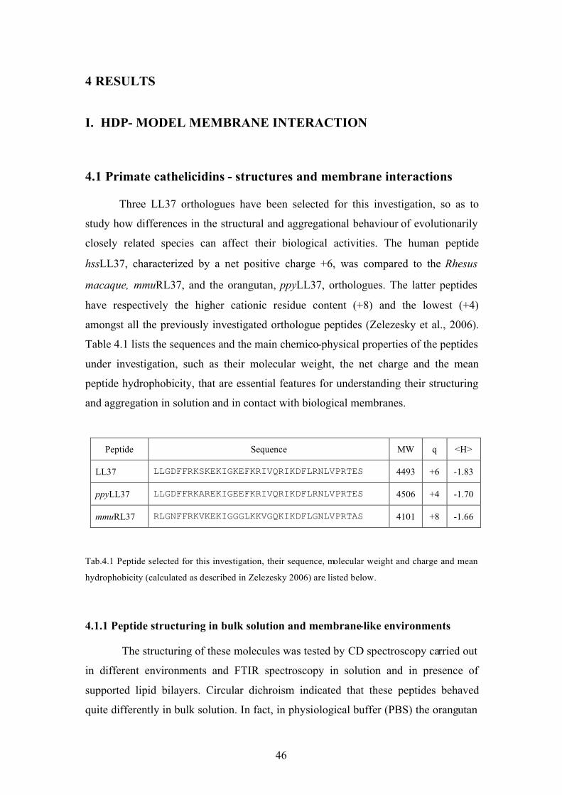
46
4 RESULTS
I. HDP- MODEL MEMBRANE INTERACTION
4.1 Primate cathelicidins - structures and membrane interactions
Three LL37 orthologues have been selected for this investigation, so as to
study how differences in the structural and aggregational behaviour of evolutionarily
closely related species can affect their biological activities. The human peptide
hssLL37, characterized by a net positive charge +6, was compared to the Rhesus
macaque, mmuRL37, and the orangutan, ppyLL37, orthologues. The latter peptides
have respectively the higher cationic residue content (+8) and the lowest (+4)
amongst all the previously investigated orthologue peptides (Zelezesky et al., 2006).
Table 4.1 lists the sequences and the main chemico-physical properties of the peptides
under investigation, such as their molecular weight, the net charge and the mean
peptide hydrophobicity, that are essential features for understanding their structuring
and aggregation in solution and in contact with biological membranes.
Peptide Sequence MW q <H>
LL37 LLGDFFRKSKEKIGKEFKRIVQRIKDFLRNLVPRTES 4493 +6 -1.83
ppyLL37 LLGDFFRKAREKIGEEFKRIVQRIKDFLRNLVPRTES 4506 +4 -1.70
mmuRL37 RLGNFFRKVKEKIGGGLKKVGQKIKDFLGNLVPRTAS 4101 +8 -1.66
Tab.4.1 Peptide selected for this investigation, their sequence, molecular weight and charge and mean
hydrophobicity (calculated as described in Zelezesky 2006) are listed below.
4.1.1 Peptide structuring in bulk solution and membrane-like environments
The structuring of these molecules was tested by CD spectroscopy carried out
in different environments and FTIR spectroscopy in solution and in presence of
supported lipid bilayers. Circular dichroism indicated that these peptides behaved
quite differently in bulk solution. In fact, in physiological buffer (PBS) the orangutan

47
peptide gave rise to a higher !-helical content (>65%) with respect to the human
peptide ("30%), as estimated by the Chen method (Chen et al, 1974), while the
macaque peptide remained essentially unstructured (<5% helical content) (Fig.4.1-A).
Interestingly, in presence of the folding inducer trifluorethanol (50% TFE) all the
peptides showed a comparably high content of helix (" 70%, not shown), which can
be considered the maximum achievable in further considerations on peptide
structuring and aggregation. Moreover, peptide conformation in presence of model
membranes was explored by using large unilamellar vesicles (LUVs), whose lipid
compositions mimic those of selected prokaryotic and eukaryotic cell membranes. In
particular, we chose as model for the bacterial membrane
phosphatidylglicerol/cardiolipin LUVs (PG/dPG) at a molar ratio 95:5, whereas for
the eukaryotic cell membrane phosphatidyl choline/sphingomyelin/cholesterol
(PC/SM/Ch) at a ratio of 40:40:20, mimicking the lipid composition of the external
leaflet of the erythrocyte cells. CD results in presence of LUV dispersions were quite
revealing, as in the presence of anionic LUVs all the tested peptides folded into a
clearly !-helical conformation (Fig.4.1-B), similar to that observed in the presence of
TFE, suggesting a strong interaction with this type of bacterial-like model membrane,
linked to a transition to a helical structure. In contrast, the interaction with the less
charged (zwitterionic) eukaryotic-like membranes resulted in a quite different
behaviour (Fig.4.1-C). The human and the orangutan peptides showed helical contents
comparable to those in bulk solution, whereas the macaque peptide remained
prevalently unordered, also resembling its behaviour in physiological buffer.
However, apart from CD band intensities, used to determine such helical contents, the
shape of the spectra is informative. The !-helical spectrum has two typical ellipticity
minima, located at about 208 and 222 nm, and it is reported that the ratio between
their intensities is indicative of the aggregational state, with lone helices displaying an
#222
/#208
ratio visibly < 1, while stacked helices show such a ratio $ 1 (See Table 4.2)
(Lau et al., 1984).

48
On the basis of this type of analysis, the rhesus peptide appears to be in a
monomeric form upon binding to, and presumably inserted into anionic model
membranes, with a !222
/!208
ratio clearly less than 1. It does not instead seem to insert
into zwitterionic membranes, remaining unstructured. In contrast, the human and
orangutan peptides show a ratio close to one in both saline buffer and anionic LUVs,
suggesting the presence of a state of equilibrium between the monomeric and the
aggregated helices, and while they do not show enhanced negative ellipticity, they
switch to a !222
/!208
ratio visibly greater than 1 in presence of zwitterionic PC/SM/Ch
model membranes, suggesting that while they may not insert into these, they interact
in a manner that enhances aggregation.
Further structural experiments were then focused on the human and rhesus
peptides performing an analysis in bulk solution using FTIR in transmission mode,
another informative method for probing peptide and protein conformation (see section
1.4.2). This technique requires high peptide concentrations (2mM) and, if carried out
in deuterated water (after 24h of H/D exchange so as to observe completely
deuterated peptide), allows individuating peptide secondary structure band
components.
Fig.4.1 CD spectra of LL37 orthologues in bulk solution and membrane-like environments. CD
spectra of human LL37 (black), ppyLL37 (green) and mmuRL37 (red) in saline buffer PBS (A) and
in presence of anionic (B) and neutral (C) LUV dispersions.
A B C

49
The region of interest was the amide I band, assigned to peptide bond
vibrations, giving information on both secondary structure and possible aggregation
states. The spectrum of LL37 showed a stable !-helical conformation even in absence
of salt ions, possibly favoured by the high peptide concentration, which would trigger
aggregation of structured peptides. The rhesus peptide, instead, showed a more
composite amide I band, in line with a more disordered conformation (Fig.4.2). The
second derivative (Fig.4.2, bottom panels) also highlighted the contribution from
deuterated arginine side chains in both peptides (symmetric and anti-symmetric
arginine side chains are respectively located at 1605 and 1585 cm-1
, human LL-37 has
5 Arg residues, mmuRL-37 has 3).
Peptide PBS TFE PG/dPG
LUVs
PC/SM/Ch
LUVs
% helix* "
222/208 %
helix "
222/208 %
helix "
222/208 %
helix "
222/208
hssLL37 30 # 1 70 < 1 60 # 1 35 > 1
ppyLL37 60 # 1 70 < 1 75 # 1 60 > 1
mmuRL37 5 _ 65 < 1 70 < 1 10 _
Tab.4.2 CD spectra analysis: percent of helix conformation estimated at 20µM peptide concentration,
using the method of Chen, and ellipticity ratio between spectral minima, "222
/"208
, (R > 1= oligomeric
helices; R < 1= monomeric helix) using the method of Lau.
Fig. 4.2 Transmission FTIR spectra in the amide I region of LL37 and mmuRL37 in solution: amide I
band of LL37 (black) and mmuRL37 (red) (2mM) in deuterated water after 24h H/D exchange. The
bottom panels show the relative second derivative spectra.

50
4.1.2 Peptide structuring and aggregation on supported lipid
bilayers.
Structural variations in the peptides induced at the membrane surface were
further evaluated by ATR-FTIR spectroscopy on supported lipid bilayers, coupled
with atomic force microscopy on the same, performed in non-contact mode. This
combined method allowed both the investigation of peptide structure and lipid order
through IR spectroscopy and morphological characterization of the bilayer upon
peptide interaction by means of AFM. Both measurements were performed on the
same substrate (germanium crystal), which acts as internal reflection element for
carrying out the ATR-FTIR. Single supported lipid bilayers were prepared by firstly
spinning the selected lipid mixture on the IR substrate and then by following a
specific hydration and washing protocol that leads to the presence of a single lipid
bilayer (see materials and methods 3.2.1). Initially, a very simple membrane system
(anionic PG only) was used, and when characterized in the absence of the peptide,
was found to provide quite stable supported bilayers. Human LL37 and the rhesus
orthologue were then evaluated with this simple model membrane, with which they
seemed to interact strongly. Peptide structure was monitored by focusing on the amide
bands, while variations in membrane order and phase, upon peptide binding, were
monitored by focusing on the acyl chain IR bands located in the 2800-3000 cm-1
range.
Figure 4.3 shows the FTIR spectra for the amide I and II bands, which are
quite susceptible to the peptides’ secondary structure. The human peptide’s amide I
band is centred at 1652 cm-1
, indicating a prevalently !-helical conformation,
according to accepted literature assignments (Martinez et al., 1995). Moreover, the
second derivative spectrum indicates that two other components are present at 1630
and 1683 cm-1
, possibly due to the symmetric and antisymmetric stretching band of
arginine side chains involved in salt bridging. This would occur with acidic residues,
on helix formation, as they would all be on the polar face of the helix, spaced i+3 or
i+4 residues from each other. This also appears to occur in a relatively hydrophobic
environment, as suggested by the red-shifted position with respect to the usual band
assignment at 1633 and 1672 cm-1
respectively, consistent with the helix being partly
inserted into the lipid bilayer. The rhesus peptide also gave rise to a sharp amide I
band, and it was even more red-shifted when compared to the human orthologue

51
(1656 cm-1
). This band position is typical of membrane proteins that are deeply
inserted into the lipid bilayer and so suggests a deeper insertion of mmuRL37 into the
membrane than the human peptide. This assumption is corroborated by the membrane
acyl chain band analysis upon peptide interaction (Fig.4.4), which are broadened and
red-shifted by 3-4 wavenumbers (CH2 and CH3 band shift from 2820 to 2824 cm-1
and from 2950 to 2954 cm-1
respectively), in line with a more massive membrane
perturbation than for human LL37. The second derivative spectrum indicates that also
the rhesus peptide presents salt-bridged arginine side chain contributions.
Morphological variations and perturbations in the membrane upon peptide
interaction were made evident by atomic force microscopy in tapping mode on the
same substrate as used for FTIR analyses. The AFM technique allows measuring
either height images (Fig. 4.5 left panel) that show morphological aspects of the
membrane at high resolution (nanometric scale) or phase images that, instead, indicate
Fig. 4.3 ATR-FTIR spectra of
LL37 and mmuRL37 in
presence of supported lipid
bilayer: amide I and II band
of LL37 (black) and
mmuRL37 (red) in contact
with PG supported bilayer.
The bottom panels show the
relative second derivative
spectra.
Fig. 4.4 ATR-FTIR spectra
of supported PG bilayer
before (black dashed line)
and after exposure to LL37
(black) and mmuRL37
(red).

52
the chemical homogeneity of the analyzed substrate (Fig.4.5, right panel). The AFM
images obtained were consistent with IR results, as mmuRL37 seemed to cause
extensive and generalized membrane perturbations, with the presence of many lesions
of different size and with disruption of the membrane’s chemical homogeneity. LL37
induced a more defined pattern of lesions, consistent with the presence of holes of
roughly the same size (< 20 nm) that were visible only at higher resolution, studded
within a chemically and morphologically homogeneous membrane background.
Interaction with the anionic model membrane was thus quite revealing, highlighting a
differentiated interaction for the human and the monkey peptides. This encouraged a
continued investigation as to whether the different modes of structuring and
aggregation, and different effects at the membrane surface corresponded to
differentiated behaviours in permeabilizing biological membranes, and thus also to
differentiated activities and selectivities.
Fig. 4.5: Atomic force
microscopy imaging of LL37
and mmuRL37 on supported
PG bilayer: AFM height (1-
left panels) and phase (2-right
panels) images of PG
supported bilayer before (A)
and after treatment with LL37
(B) or mmuRL37 (C).

53
4.1.3 Permeabilization of model and biological membranes
4.1.3.1 Model membranes
The capacity of primate cathelicidins to permeabilise biological membranes
was first investigated using model systems and then correlated to the permeabilization
of selected bacterial and host cell membranes. In particular, anionic and neutral large
unilamellar vesicles, as previously used for circular dichroism studies, were pre-
loaded with the fluorescent dye/quencher pair ANTS/DPX (molar ratio 9:25) and dye
leakage was monitored following fluorescence emission at 512 nm. Peptide
concentrations used in these analyses were chosen on the basis of typical peptide/lipid
ratios commonly used in biophysical studies on antimicrobial peptides (1 - 20 µM in
the presence of 300 µM LUVs). At a lower peptide/lipid ratio the less cationic
ppyLL37 seemed the most effective in permeabilizing both anionic PG/dPG and
zwitterionic PC/SM/Ch vesicles, followed by the human and the rhesus peptides,
whereas, at higher ratios all the peptides behaved in a similar manner inducing 80-
90% of dye leakage.
These results were surprising. CD spectroscopy had shown that the peptides
did not appear to insert significantly into zwitterionic membranes. The rhesus peptide
mmuRL37, in particular, did not assume a regular structure in presence of neutral
LUVs, which would be required for insertion. However, this did not seem to preclude
a capacity to permeabilize neutral vesicles (Fig.4.6-B). In order to better understand a
lytic effect without membrane insertion, vesicle fusion was investigated by
monitoring the turbidity at 400 nm at increasing peptide concentrations. Results,
shown in Fig.4.6-D-E, indicated that zwitterionic membranes were in fact less stable
and more prone to aggregate or fuse in presence of both the human and the rhesus
peptide. Globally, these results may indicate that the cathelicidin peptides all have a
capacity to insert into anionic membranes, by assuming helical conformations, and
cause lesions that compromise membrane integrity, although according to AFM
experiments, these are of quite different types. It appears that they can all also bind to
the surface of zwitterionic membranes, without necessarily increasing their helical
structure, in a manner that can in any case cause efficient dye release, at least in the
model systems analysed, possibly also through fusion events.

54
4.1.3.2 Biological membranes
Membrane lysis is likely an initial step towards cellular inactivation, and as
reported previously, is regulated by different parameters. It seemed likely, considering
the quite different modes of membrane interaction for the different cathelicidin
orthologues, that environmental conditions could differentially affect their biological
effects on real systems. The inhibition of bacterial growth exerted by these peptides
was thus evaluated on a selected gram-negative and gram-positive bacterium, at
various concentrations, using medium different conditions. After exposure to the
peptides, growth inhibition was measured continuously over a period of 4h, and an
end-point effect was then determined at 24h. As listed in Tab.4.3, under permissive
medium conditions (5% medium in 10 mM phosphate buffer) the human and rhesus
cathelicidin peptides seemed equally effective in inhibiting E.coli, both short- and
long-term. The less charged orangutan peptide ppyLL37 seemed less effective,
A B C
E D
Fig. 4.6 Permeabilization and fusion of LUVs in the presence of LL-37 orthologues: A-C)
ANTS/DPX leakage from anionic and neutral LUVs in the presence of different peptide
concentrations. ANTS fractional fluorescence due to permeabilization of (--!--) PG/dPG (95:5)
LUVs, and (!!!) of PC/SM/Ch (40:40:20) LUVs [300µM lipid], exerted by LL-37 (A), mmuRL37
(B), ppyLL-37 (C) at different peptide concentrations. D-E) Fusion of LUVs determined by
monitoring the increased turbidity at 400 nm exerted by LL-37 (D) and mmuRL37 (E) at different
peptide concentrations, and 300 µM lipid.

55
requiring a higher active concentration (> 5µM). However, human LL37 and rhesus
RL37 showed quite differentiated activities at increased medium concentrations. The
latter peptide maintained a robust activity even in full medium, or in the presence of
fetal calf serum, resulting in long-term bacterial inhibition even at relatively low
concentrations. The human peptide instead required a two fold higher concentration to
achieve a short-term inhibition, and a ten-fold excess to exert a long-term antibacterial
activity, even in just 20% TSB medium, and was more susceptible to the presence of
serum. All cathelicidins were somewhat less effective against the gram-positive
bacterium, in line with literature reports (Johansson et al, 1999), requiring higher
concentrations to exert their bactericidal action, but also in this context the human and
orangutan peptides were less effective that the rhesus one.
It is evident that the different capacities to structure in bulk solution and
different modes of membrane interaction for hssLL37 and mmuRL37 result in
different capacities to affect bacterial membranes, and possibly to subsequently effect
bacterial killing. Further experiments were thus required to define their different
trajectories from bulk solution to the membrane and subsequent lytic events. These
investigations were carried out in permissive environmental conditions, such as
physiological buffer, where both peptides had been shown to be comparably effective
affecting biological membranes.
The effect of the peptides on the cytoplasmic membrane of E.coli was
investigated by monitoring the time course of propidium iodide (PI) uptake and
Tab 4.3 Inhibition of bacterial growth by LL37 orthologues determined over a period of 4 hours as
[(AaA
p)/A
a]*100, where A is the absorption at 600nm in the absence (
a) or presence (
p) of peptide; long-
term inhibition was checked after 24 hours incubation at 37°C (° no growth; • growth).

56
DiBAC fluorescence, by means of flow cytometry. These fluorescent dyes
respectively reported membrane permeabilization and depolarization upon exposure
to an active concentration of the peptides under standardized conditions, comparable
to those used in antibacterial assays (pH 7.4; 10 mM phosphate, 150 mM NaCl)
(Fig.4.7-A). Membrane lysis was rapid, but the observed lytic efficiency of the
peptides followed a distinctly different trend when compared to permeabilization of
naked vesicles: mmuRL37 > hssLL37 >> ppyLL37. This indicates that membrane
lysis is regulated by parameters that are not only linked to peptide properties and
membrane lipid composition. At selected time points (4 and 30 minutes) the
transmembrane potential was assessed using the fluorescence emitted by DiBAC, so
as to correlate permeabilization and depolarization effects (Fig.4.7-B1-2). Rhesus RL-
37 resulted in rapid permeabilization, with 23% of PI positive cells after only 10
minutes of treatment and reached 90% PI positive cells at 30 minutes. However,
depolarization appeared to be considerably faster, with significant depolarization
already at 4 minutes of exposure to the peptide, so it seemed to precede massive
permeabilisation. For the human peptide, which gave rise to a more moderate
bacterial permeabilization, reaching 60% of PI positive cells only after 40 minutes,
the effect of membrane depolarization instead seemed to accompany the primary
effect of lysis, becoming evident after 30 minutes of treatment. The orangutan
ppyLL37, appeared less effective in both permeabilization and depolarization, even
under these quite permissive conditions. Taken together these results may be in line
with the different types of lesions observed on supported PG bilayers, with the rhesus
peptide resulting in generalized and heterogeneous membrane damage, while the
human one produced discrete hole-like lesions.

57
The effect of the peptides on eukaryotic cells was evaluated by performing a
haemolysis assay on freshly prepared erythrocytes obtained from several different
healthy donors. Results from this assay could then be correlated to the previously
performed experiments on vesicles with zwitterionic membrane, whose lipid
composition resembled that of red blood cells.
Haemolytic activity was assessed by monitoring haemoglobin release from
0.5% erythrocyte suspensions at 405 nm. Peptides were tested at increasing
concentrations (from 1 up to 100 µM) and their effect related to that of exposure to
1% triton-X which causes 100% release. In general, and also considering that
haemolysis can vary significantly among cells deriving from different donors, the
tested peptides did not show significant differences in their activity (see Fig. 4.8). The
Fig. 4.7 Membrane permeabilization and depolarization of E.coli ML35 cells investigated by flow
cytometry: % PI positive (permeabilized) cells after exposure to peptide treatment for different times
(A); % PI (permeabilized) and % DiBAC (depolarized) positive E.coli ML35 cells at minute 4 (B1) and
30 (B2) after exposure to 0.1µM peptide in PBS.

58
human peptide hssLL37 was possibly the most haemolytic at low concentrations, but
the estimated HC50 values for all the orthologues hssLL37, mmuRL37 and ppyLL37
were overlapping, being respectively 75± 25, 70 ±15 and 85± 15 µM. Thus, in line
with results from model membranes, all three peptides have comparable capacities to
interact with and lyse this type of eukaryotic membrane, which remains surprising due
to the apparent lack of insertion into zwitterionic membranes.
4.2 Probing peptide structure and aggregation by using an IR and fluorescence
environmental probe: p-cyano-phenylalanine.
As reported in the previous sections, the structural and aggregational
behaviour of primate cathelicidins is differentiated and may determine the subsequent
mode of action at biological membranes. In an attempt to further probe peptide
structuring and aggregation, and the interaction with membranes, a non-proteinogenic
amino acid was introduced into the peptides, to act as an “environmental probe”. The
cyano analogue of the phenylalanine; p-cyano-phenilalanine, has been applied to the
study of peptide folding and binding in the recent years (Getahun et al., 2003, Tucker
et al., 2004; Tucker et al., 2006; Konstantinos et al., 2007), by monitoring both the
fluorescence emission and the IR absorption of the nitrile group in different
conditions. It was likely that it could also effectively report on membrane interactions.
FIG. 4.8 Haemolytical activity of
LL37 (!), mmuRL37 (") and
ppyLL37 (#). The data are mean ±
SD of at least three independent
experiments.

59
CN-Phe as a fluorescence and IR probe
Studies of protein folding and binding by means of fluorescence quenching or
enhancement usually require the use of intrinsic fluorophores, such the tryptophan
and tyrosine, or the attachment of an external fluorophore that, however, is usually a
quite large molecule that can perturb the native structure of peptides and proteins or
their interactions with other biomolecules. The nitrile group in CN-Phe is a relatively
small modification, making it roughly isosteric with tyrosine, and interestingly with
an intermediate polarity between that of an amide group and a methylene. This
residue is so compatible with both the hydrophobic interior of membrane proteins or
the hydrophilic surface of water soluble ones. The presence of the cyano results in a
quantum yield five-fold higher than that of the native phenylalanine side-chain, and a
fluorescence very sensitive to interaction with solvents, leading to a marked decrease
in fluorescence in organic solvents relative to aqueous solution. It is also strongly
quenched by chloride ions (Fig.4.9-A). Although the fluorescence of this amino acid
has been proposed to be particularly sensitive to hydrogen bonding interactions, a
recent work has indicated that several other parameters can affect it, so that
considerable caution must be taken when interpreting fluorescence data (Taskent-
Sezgin et al., 2009). This suggests that the use of a standard to test all conditions prior
to use is appropriate.
Getahun et al. initially proposed the use of the CN-analogue of phenylalanine
as a micro-environmental probe in FTIR spectroscopy (Getahun et al., 2002). In fact,
Fig. 4.9 Chemico-physical properties of p-cyano-phenylalanine. Fluorescence emission at 300 nm of p-
cyano-Phe in presence of water, methanol and chloride ions (A), p-cyano-phenylalanine chemical
structure (B) and cyano IR stretching band in water and tetrahydrofuran (THF) (C).
C B A

60
the vibrational mode of the nitrile derivatized amino acid is sensitive to the local
environment due to the to hydrogen bonding (Huang et al., 2003) and the local
electric field (Suydam et al., 2003; Dalosto et al., 2004). Moreover, the CN stretching
band is placed in an IR window relatively free from competing absorptions, also
having a high extinction coefficient in that region. It gives rise to an IR band centred
around 2230-40 cm-1
in a polar environment that shifts to lower wavenumbers (2220-
2228 cm-1
) in a hydrophobic or organic environment (Fig.4.9-C).
4.2.1 Design of CN-analogues of selected cathelicidins
The human and rhesus cathelicidins, hssLL37 and mmuRL37, respectively
contain 4 and 3 phenylalanine residues suitable for structural investigation (see
Tab.4.4). Some of these may have a central role in peptide aggregation and peptide
binding to and insertion into lipid bilayers, as suggested by the recent NMR studies of
the human peptide, carried out on model membranes (see Chapt. 1.4.1). With respect
to the human LL37, it was thus decided to synthesize three different analogues,
strategically replacing Phe with CN-Phe at positions 5 or 17 or 27 in the N-terminal,
central and C-terminal regions. Only two analogues of mmuRL37, F5 or F27 could be
synthesized, since this peptide doesn’t natively possess the central phenylalanine.
Furthermore, a short reference peptide was also designed and produced with the aim
of achieving a standard for a feasible quantitative fluorimetric analysis. This tetra-
peptide, with sequence GF*KG, is positively charged (+2) and therefore quite soluble,
but unstructured and un-aggregated in all the tested conditions, as well as unlikely to
insert into model membranes, and therefore seems to be a good reference system.
These physico-chemical properties were confirmed by CD spectroscopy.

61
Tab. 4.4: cyano-analogues investigated in this study, their molecular weight and charge are listed. Red
phenylalanine (F) represents the position of the cyano-phenylalanine in the peptide sequence.
Peptide Sequence MW q
Reference GFKG 432 +2
5CN-LL37 LLGDFFRKSKEKIGKEFKRIVQRIKDFLRNLVPRTES 4520 +6
17CN-LL37 LLGDFFRKSKEKIGKEFKRIVQRIKDFLRNLVPRTES 4520 +6
27CN-LL37 LLGDFFRKSKEKIGKEFKRIVQRIKDFLRNLVPRTES 4520 +6
5CN-mmuRL37 RLGNFFRKVKEKIGGGLKKVGQKIKDFLGNLVPRTA 4128 +8
27CN-mmuRL37 RLGNFFRKVKEKIGGGLKKVGQKIKDFLGNLVPRTA 4128 +8
4.2.2 CN-analogues - CD and fluorescence
The choice of different buffers to perform fluorescence experiments was based
on the considerations that in water all peptides are prevalently unstructured and that
Cl- ions quench phenylalanine fluorescence. The selected buffers are: (i) 100 mM
phosphate buffer, SPB in Fig. 4.10, where the human but not the rhesus peptide, is
partly structured, so it should be possible to assess the effect of intra-molecular
quenching deriving from peptide structuring/aggregation; (ii) 100 mM NaCl in
phosphate buffer, Cl- in Fig. 4.10, to determine if structuring/aggregation affords
protection from quenching; (iii) anionic LUVs (PG/dPG 95:5) or neutral LUVs
(PC/SM/Ch 40:40:20) LUVs in phosphate buffer, to determine the effect of
membrane binding/insertion; (iv) LUVs in the presence of 100 mM NaCl to
determine if membrane binding/insertion affords protection from quenching. The
effect of external quenching by NaCl, applied to probe exposure of the CN-
phenylalanine residue to the solvent environment, was carried out by directly adding
the quencher to the tested environment; in all cases, the GF*KG peptide was used as
reference.
The capacity of the labelled peptides to structure in the different environment
was concurrently evaluated by circular dichroism in order to ensure that the presence
of the nitrile group could affect structural and aggregational behaviour of the

62
analogues, and, as expected, indicated that the presence of the cyano group did not
affect peptide conformation either in the case of LL37 or mmuRL37 (data not shown).
The reference peptide gave rise to a comparatively high level of fluorescence
in all the non-quenched conditions (water, 100 mM phosphate buffer or LUV
suspensions), while 100 mM NaCl dramatically quenched its FCN
emission under all
tested conditions (Fig. 4.10). The probe residue was thus completely exposed to the
solvent, unaffected by the intra- or inter- molecular quenching, and did not interact
with model membranes. For human LL-37, 5FCN
was clearly quenched by the
neighbouring phenylalanine at position 6, as its fluorescence emission appeared lower
than for other positions in aqueous solution buffers, and in presence of liposomes. In
presence of anionic liposomes the 17FCN
emission was particularly low whether
compared to FCN
placed in the other positions (5 or 27) and, upon addition of the
external quencher, the intensity did not change, suggesting that this aromatic residue
was fully buried into the hydrophobic core of the lipid bilayer. The 27FCN
residue
placed at a C-terminal position had an intermediate behaviour to that of the N-
terminal and central positions (Fig.4.10).
Fig.4.10: Fluorescence emission values at 300 nm of the reference peptide (GFKG), LL37 CN-analogues
and mmuRL37 CN-analogues. Histograms represent fluorescence emission in the following conditions: SPB
phosphate buffer (blu), NaCl quencher buffer (green), anionic PG/dPG (upward patterned lines) or neutral
PC/SM/Ch (horizontal patterned lines) liposomes dispersed in SPB (blu) or NaCl quencher buffer (green).

63
It was in any case evident that all three CN-analogues interacted differently
with anionic and neutral membranes LUVs. In the presence of the former vesicles, the
middle position (17FCN
) appeared to be the most inserted and least accessible to the
external quencher, whereas in contact with neutral LUVs all the CN-analogues
behaved in a similar manner. In particular, all the analogues were more effectively
quenched by chloride ions in the presence of neutral than anionic LUVs, suggesting
surface binding on host-like membranes.
Unfortunately, the rhesus peptide lacked the phenylalanine residue in the
central position, to allow probing the interaction of this key segment of the peptide
with membranes. Fluorescence results were quite similar for the 5- and 27-CN
analogues with those of LL37. The N-terminal position was probably also partly
internally quenched due to the presence of the neighbouring phenylalanine F-6.
4.2.3 CN-analogues - FTIR spectroscopy in solution and model membranes
FTIR measurements were carried out either in water, using transmission mode
and high peptide concentrations (3 mM), as required (see 1.4.2), and in presence of
anionic and neutral supported lipid multilayers in ATR mode. The former experiment
was carried out in order to assess the cyano stretching band position in bulk solution,
whereas the effect of possible insertion into lipid bilayers was evaluated by the latter
method at a peptide lipid ratio (1:20) commonly used in the study of peptide-model
membrane interactions (Shai et al., 1999). It should be noted that the particular ATR
experimental setup and the relatively low absorbance of the cyano group required to
carry out this experiment in dehydrated membrane conditions, likely means that only
the signal from membrane-bound peptide can be picked up. This should be taken into
account in interpreting results, as only part of the CN micro-environment may be
evident.
Transmission experiments indicated that in LL37 the labelled phenylalanine
residues are not particularly shielded on aggregation, as their IR absorptions remain in
the range expected for an aqueous environment (2230-40 cm-1
). The residue in the
central position (F-17) was possibly partly protected from the aqueous environment
due to peptide aggregation, since the cyano group stretching band position is slightly
blue-shifted (! 2234 cm-1
, see Tab.4.5). The bandwidth for all the three analogues was

64
however similar and quite broad (bandwidth > 11 cm-1
), indicating a heterogeneous
environment. The band position determined by ATR in the presence of supported
lipid multilayers shifted to lower wavenumbers (! 2229) consistent with partial
insertion into a more hydrophobic environment. However, no significant difference
was observed amongst the different LL37 CN-analogues, or on interaction with either
anionic or neutral membranes. The main difference emerges considering the
bandwidths of LL37 analogues, which decreased from >10 cm-1
to !8 cm-1
on passing
from PC/SM/Ch to PG/dPG model membranes, indicating that insertion may be
deeper into the anionic membrane. In the case of mmu RL37, the residue at position 5
gave rise to more red-shifted and sharper bands under all the tested conditions, but
there was no evidence of different behaviour in contact with anionic or neutral
membranes. This method, in contrast to CD spectroscopy, thus indicates a significant
interaction of the peptides with zwitterionic membranes. However, while CD records
structural modifications on the whole population of peptides, this method may only
pick up those that are intimately associated with the supported bilayer at the
germanium crystal surface, and which fall within the evanescent IR field used by
ATR. It may therefore selectively report on peptides tightly bound to, or inserted into,
the membrane, while missing more loosely associated ones, which according to CD
spectroscopy may be the majority.
Tab.4.5: FTIR cyano stretching band positions and bandwidths of LL37 and mmuRL37 CN-analogues
in bulk solution or in presence of supported lipid multilayers.
CN analogue H20
" (#")
PG multilayers
" (#")
PC/SM/Ch
multilayers
" (#")
5 CN-LL37 2235.6 (11.5) 2229.4 (8.5) 2229 (10.4)
17 CN-LL37 2234.4 (11.4) 2229.7 (8.4) 2230 (10)
27 CN-LL37 2235.15 (11) 2229.0 (7.3) 2229.4 (10.4)
5 CN-MMU 2235 (7.6) 2227.9 (8) 2228.4 (8)
27 CN-MMU 2235.15 (11.5) 2228.9 (8.5) 2229.3 (9)

65
4.3 Membrane interactions of human !-defensin 2 and 3
The structures of primate !-defensins are quite different to those of
cathelicidins, as described in chapter 1.4.2, and are characterized by a conserved
scaffold consisting of a compact, antiparallel, triple-stranded !-sheet stabilized by the
presence of three invariant disulfide bridges, and supporting quite variable sequences.
This results in quite variable oligomerization patterns in the human !-defensin, as
indicated by the NMR and crystallographic structures observed so far. Human !-
defensin 2 and 3 were selected for this study, and were chemically synthesized along
with rationally designed variants to probe the roles of different structural elements in
mediating membrane interaction.
4.3.1 Structure predictions and design of human !-defensin analogues
The stable !-structure of hBD2 was dissected by designing different variants
that probed salient structural elements. In particular, this peptide displays a short and
unusually stable "-helical N-terminal segment (it is similar in both crystal and
solution structures), covering positions Pro5 to Gly
12 (Table 4.6), which is considered
to be functionally important (Crovella et al, 2005). Stabilizing factors may be the
formation of a fourth short strand in the !-sheet by residues 1-5, anchoring the helix
to the sheet, and H-bond capping interactions between Asp4
and Thr7
(Fig.4.11 top
left). The relevance of these segments was probed by synthesizing the variant (-
D4)hBD2, in which the key aspartic acid residue was deleted and the truncated
analogue (5-41)hBD2 in which the putative N-terminal strand was removed (Tab.4.6)
also destabilizing the helix, that is not present in the solution structure of this peptide
(Bauer et al., 2001). Moreover, in order to gain further insight on the role of the
conserved !-structure, an artificial variant, brevi-hBD2, was designed to include the
N-terminal and C-terminal stretches of hBD2, while removing the central part (see
Fig. 4.11 C)

66
Tab.4.6 !-defensins selected for this structural investigation, their sequence, molecular weight and
charge are listed. The F = p-cyano-phenylalanine.
Peptide Sequence MW
(Da)
q
|---:----|----:----|----:----|----:----|----:
hBD2 GIGDPVTCLKSGAICHPVFCPRRYKQIGTCGLPGTKCCKKP 4328 +6
(-D)hBD2 GIG-PVTCLKSGAICHPVFCPRRYKQIGTCGLPGTKCCKKP 4213 +7
(5-41)hBD2 ----PVTCLKSGAICHPVFCPRRYKQIGTCGLPGTKCCKKP 3986 +7
brevi hBD2 GIGDPVTCLKS----------------GTCGLPGTKCCKKP 2483 +5
hBD3 GIINTLQKYYCRVRGGRCAVLSCLPKEEQIGKCSTRGRKCCRRKK 5155 +11
Chimera hBD3 GIGDPVTCRVRGGRCAVLSCLPKEEQIGKCSTRGRKCCRRKK 4600 +10
CN-hBD3 GIINTLQKFYCRVRGGRCAVLSCLPKEEQIGKCSTRGRKCCRRKK 5169 +11
Only the solution NMR structure of human hBD3 is available in the
Protein Data Bank and it describes a !-sheet structure with a less well-defined N-
terminal helical segment than hBD2 (Fig. 4.11-B). However, the N-terminal stretch is
considerably longer than in hBD2 (see Table 4.6) and, when projected on a helical-
wheel (Fig. 4.11), would give rise to a distinctly amphipathic helix. In analogy to
helical AMPs (such as for example mmuRL-37), it would only acquire such a
conformation in the presence of biological membranes, and this could be a key
structural element in mediating peptide-membrane binding. As indicated in Fig.4.11,
the helical wheel projection comprising residues 1-14 suggests that the amphipatic
helix might be suitable for membrane insertion (Fig.4.11-D). To investigate the role
of this structural element, a chimeric peptide was synthesized in which this segment
of hBD3 was replaced by the shorter stretch of hBD2 (Table 4.6). Furthermore, a
cyano-phenylalanine analogue of hBD3, termed CN-hBD3, was prepared by
exploiting the presence of tyrosine residues in the N-terminal segment of hBD3. The
p-cyano-phenylalanine residue was strategically placed at the center of the putative
amphipathic helix (in position 9) (Fig.4.11-D).

67
4.3.2 Probing !-defensin structures with CD and FTIR spectroscopy
The secondary structures of hBD2 and hBD3 and their rationally designed
variants were firstly evaluated using circular dichroism, at peptide concentrations
comparable to the active ones on bacteria (20µM) in different environments such as
low-salt or physiological buffers, SDS micelles or liposomes, resembling lipid
composition of bacterial- and host cell-like membranes (see Fig.4.12). hBD2 showed
a CD spectrum typical of a !-sheet structure in either aqueous saline buffer or in the
presence of anionic or neutral LUVs, as indicated by a single spectrum maximum at
Fig. 4.11 Human !-defensin structures: (A) hBD2 NMR structure (PDB file:1FD3) and the intra
molecular interactions that involve the aspartic residue in position 4 (left) ; (B) hBD3 NMR structure
(PDB file:1KJ6). C) hBD2 segments that compose brevi-hBD2 (included sequence as solid ribbon,
excluded sequence as line ribbon) – the structure is just indicative as it is unlikely that the residual
sequence maintain the native structure; D,E) Edmuson projection of the "-helical segments in hBD2
and hBD3 respectively, showing the site of cyano-phenylalanine replacement for hBD3.
egment of hBD2 (C) and the putative one of hBD3 (D).
hBD2 hBD3
hBD3
G
C
!
?
L
I
Y
R
T
Q
1
2
3
4
5
6
7
8
9
10
11
12
1 4
13 V
I Y
R
K
N
hBD2
1
2
3
4
5
6
7
8
9
10
11
12
G
G
C
!
?
P
L V
K
G
T
S D
I
N
A B
D E C
Asp4

68
196 nm and minimum at 218 nm. In all tested environments a shoulder at 222 nm may
account for the short !-helical segment. hBD2 analogues, (-D)hBD2 and (5-41)
hBD2, showed spectra consistent with more disordered structures in bulk solution,
and no sign of a possible !-helical segment. Only (-D)hBD2 seemed to partially
recover "-structure and also possibly the N-terminal helix, in presence of a
membrane-like environment, such as the anionic SDS micelles or the anionic LUVs
(see Fig.4.12), but it did not do so with zwitterionic LUVs. brevihBD2 showed a
prevalently random coil conformation in aqueous buffer, but underwent a transition to
a partly helical conformation in the presence of SDS micelles or trifluorethanol
(Fig.4.12). However, this conformational transition was not detected in presence of
either anionic or zwitterionic vesicles, in the presence of which peptide remained
prevalently unstructured.
The structural analysis carried out on hBD3 and its analogues indicated a
somewhat different behaviour to hBD2, as the CD spectrum of this human defensin
indicated that it was predominantly unordered in bulk solution. However, it
underwent a clear coil-helix transition in presence of anionic LUVs or also SDS
micelles, and the CD spectra in presence of lipid vesicles displayed the two
characteristic minima of a well-defined !-helical conformation (at 208 and 222 nm).
This indicates that the hypothesis that the long N-terminal segment folded into an !-
helix upon interaction with bacterial-like membranes was correct. This aspect was
further investigated by performing ATR-FTIR measurements on the CN-hBD3
analogue in contact with anionic (PG) supported multilayers, monitoring the
stretching band of the cyano group and comparing it with the same band measured by
transmission IR in solution (Fig.4.13-C). The cyano band shifted significantly, from
2235 cm-1
in water to 2229 cm1 in the presence of PG multilayers, and also became
narrower, consistent with the insertion of the N-terminal segment into a hydrophobic
environment (Tucker et al., 2004). These results indicate that the N-terminal segment
of hBD3 is essentially unordered in bulk solution, swamping the contribution from the
"-sheet core in CD experiments, but that it undergoes a transition to a helical
conformation in the presence of anionic membranes, and simultaneously inserts into
the bilayer.

69
FTIR in transmission mode was applied to hBD2 and hBD3 and their analogues
dissolved in deuterated water, after 24h H/D exchange, and was used to gain insights
on peptide structure and aggregation in bulk solution. The concentration required for
this experiment (2 mM) may be sufficiently high for human !-defensin 2
oligomerization (see 1.4.1). It gave rise to a narrow amide I band, with two shoulders
at 1673 and 1630 cm-1
, consistent with a stable !-structure and possible !-
aggregation, possibly occurring via !-sheet interactions, highlighted by two further
components at 1610 and 1690 cm-1
(Fig.4.13-A and Tab.4.7). Moreover, the two
hBD2 analogues in which the helical segment had been destabilized showed broader
amide I bands, in line with a less defined !-conformation and the lack of the N-
terminal helical segment (usually indicated by the presence of a band component
centered at 1650 cm-1
). (-D)hBD2 seemed the more structured of the two, also
presenting band components assigned to !-aggregated structures (1610 and 1690 cm-
1), while completely lacking any indication of the helical segment. The brevihBD2
Fig.4.12 CD spectra of !-defensins: spectra of hBD2 and hBD3 (top) and their analogues (bottom) at
the concentration of 20 µM in presence of — phosphate buffer; — SDS (10mM); — TFE 50% in
SPB; — PG-dPG LUVs (95:5), — PC/SM/Ch LUVs (40:40:20) [0.4mM lipids].

70
FTIR spectrum appeared even broader, and was consistent with a random coil
conformation with no sign of possible aggregation, as listed in Tab.4.7.
hBD3 and its chimeric analogue, unlike hBD2 and consistent with CD studies,
appeared unordered, and both presented two bands at 1605 and 1585 cm-1
associated
with the symmetric and antisymmetric stretching bands of the side chains of the
arginine, which are abundant in this peptide (!15%) (Fig.4.13-B). There was no
evidence of "-aggregated bands, which is in line with a mode of oligomerization that
occurs via salt bridging rather than sheet extension (Boniotto et al, 2003).
Fig.4.13 FTIR transmission spectra of ! -defensins in deuterated water, amide I band. FTIR spectra
of hBD2 (____
), ("D)hBD2 (- - - ), (5 – 41)hBD2 ( – · – ·) and brevi hBD2 (. . .) (A) and hBD2 (____
),
hBD3 (- - -), hBD2/3 chimera ( – – ) (B) [2 mM peptide in D2O, after complete H/D exchange]. (C)
Cyano stretching band of CN-hBD3 in water (____
) and in presence of PG/dPG supported multilayers
(- - -).
A B C

71
Tab.4.7 Peptide secondary structure components determined by FTIR curve fitting: integrated areas of
the amide I component bands and relative secondary structure assignments.
4.3.3 Biological activities
The direct antimicrobial activities of hBD2, hBD3 and their variants were
evaluated in vitro by the minimum inhibitory concentration (MIC) assay on selected
reference bacterial and yeast strains (c.f. E.coli ML37, S.aureus 710A, P. aeruginosa
ATCC and C.albicans). These tests were carried out under specific medium
conditions, since the antibacterial activity exerted by defensins in general is well
established to be highly salt-sensitive by this laboratory and others groups (Boniotto
Peptide !-structure "-helix turns Random
coil
Aggregate
d
structure
Arg
side
chains
#
(cm-1
)
Area
(%)
#
(cm-
1)
Area
(%)
#
(cm-
1)
Area
(%)
#
(cm-
1)
Area
(%)
#
(cm-1
)
#
(cm-1
)
hBD2 1628 32
1672
1651 3 1662 24 1644 41 1612
1680
hBD2
(-D)
1628 33
1670
1660 21 1643 45 1613
1681
hBD2
(5-41)
1623 33
1673
1659 21 1642 45
Brevi-
hBD2
1625 27
1674
1661 19 1643 54
hBD3 1624 24
1673
1653 4 1661 11 1641 60 1585
(#a)
1607
(#s)
hBD3
chimera
1623 26
1673
1654 6 1663 8 1641 60
1585
(#a)
1607
(#s)

72
et al, 2003; Antcheva et al, 2004). It was thus decided to perform the assay in either
5% Triptic Soy Broth (TSB) (or 5% SAB for yeast) and in 50% NaCl in phosphate
buffer to explore the effect of salt concentration. As listed in Tab.4.8, hBD2, as
previously reported, showed a direct, salt sensitive antimicrobial activity on both
Gram-positive and Gram-negative bacteria, being more effective against the latter. It
is interesting that destabilizing the N-terminal helix of hBD2 did not markedly affect
antimicrobial activity, with (-D)hBD2 and (5-41)hBD2 having a comparable activity
and salt sensitivity to the native peptide. Only brevi hBD2 was completely inactive.
hBD3 was more potent than hBD2, and less sensitive to medium conditions, and in
this case also, replacing the N-terminal stretch in the chimeric peptide did not greatly
affect the MIC values.
Tab. 4.8 Antimicrobial activity of !-defensins and analogues. Minimum Inhibitory Concentration
(MIC) assay in presence of 105 CFU bacterial cells in 5% TSB (or SAB) in phosphate buffer or 50 mM
NaCl .
To further investigate the antibacterial activities of the analogues, a detailed
study was performed on E.coli cells, which seemed sensitive to relatively low
concentrations of most of the peptides under investigation.
MIC (µM)
S. aureus
710A
E. coli
ML35
P. aeruginosa
ATCC 27853
C. albicans
ATCC 90029
!-defensin
5% TSB
50 mM
NaCl
5%
TSB
50 mM
NaCl 5% TSB
50 mM
NaCl 5% SAB
hBD2 16 >32 4 16 4 16 4 4
hBD2(-D) 8 16 4 16-8 8 16 1-2 1-2
brevi-hBD2 >32 >32 >32 >32 >32 >32 >32 >32
hBD2(5-41) 16-32 >32 8 32 16 >32 4 4
h3Nh2 1 2 1 >32 2-4 16 0,5-1 0,5-1
hBD3 1 1-2 1 16-32 2 8 1 1

73
Flow cytometric analysis was carried out to determine how efficiently exposure
to the peptides resulted in membrane permeabilization. 106 CFU/ml of E. coli were
exposed to selected analogues over a period of 40 minutes and the internalization of
the fluorescent probe propidium iodine (PI) monitored at different times using flow
cytometry. As shown in Fig. 4.14, the chimera-hBD3 seemed the most efficient and
rapid in permeabilizing E.coli membrane, followed by the native human !-defensin 3,
that also induced forward and side scatter variations, compatible with morphological
changes induced on bacterial cells (not shown). hBD2 showed a moderate
permeabilizing activity, and was in this case discernibly more effective than both
variants in which the N-terminal helix is destabilized, which displayed both a slower
and weaker effect. Brevi hBD2 was completely inactive. Permeabilization was also
studied by following exposure of cytoplasmic !-galactosidase to the impermeant
substrate ONPG, which unlike PI is uncharged, using a conventional colorimetric
assay. It was slower than that monitored for PI in flow cytometry, becoming
significant only after a considerable lag-time (not shown).
These methods for assessing the effects of the peptides on the bacterial
membrane can be compared to the killing kinetics, which were also more rapid for
hBD3 and its analogue than for hBD2. Taken together, these results may indicate a
rapid compromising of the membrane, which is favoured by the higher cationicity of
hBD3, and which initiates an irreversible killing process, but which may not be the
only or primary killing effect. A subsequent slower alteration of the bacteria may
result in a later, more massive damage to the membrane.
Fig. 4.14: Flow cytometric study of
peptide permeabilization of E.Coli ML35
cells by defensin peptides. (A) Percent of
PI positive cells exposed to 1µM hBD2
(light blu), hBD3 (blu), (5-41)hBD2
(pink), brevi hBD2 (green) and chimera-
hBD3 (red).
A
Time (min)
% P
I p
osi
tive
cell
s 80
40
20
20 40 60 0

74
II. HDP-CELL MEMBRANE INTERACTIONS - a multi-technique
approach
4.4 Effects of short-term interaction of hBD2 with antigen presenting cells
Human !-defensins were considered for years to be just antimicrobial peptides
exerting a moderate and curiously medium dependent activity against a broad
spectrum of bacteria and fungi, as reported in the previous chapter. However, there is
growing evidence that they also play a role in alerting and enhancing other
components of innate and adaptive immunity and their interaction with the biological
membranes of the host cells involved in these processes plays a central role this type
of action. hBD2, amongst the human defensins, seems to be particularly active in this
respect (Pazgier et al., 2006), including induction of chemokine responses form
PBMC (Boniotto et al., 2006) and chemotaxis of lymphocytes and dendritic cells. For
this reason, it was decided to investigate the interaction of hBD2 with immature
dendritic cells, by observing different cell activities upon peptide treatment, coupling
conventional and innovative microscopy techniques to explore the functional and
biological implications of these interactions. This study originates from a close
collaboration of laboratories at the Dept. of Life Sciences at Trieste University and
the SISSI beamline of the Elettra Synchrotron Light Laboratory.
4.4.1 Peptide cytotoxicity, binding and internalization into APC
Prior to performing microscopy and flow cytometry on hBD2-treated immature
dendritic cells (iDC), an extensive analysis was carried out to determine whether
hBD2 was cytotoxic towards cells of the mononuclear-phagocytic lineage (CD14+) at
the concentration range to be used (< 4µM). This was done by monitoring possible
membrane damage by assessing permeabilisation to propidium iodide (PI) and the
effect on cellular vitality as assessed by the MTT test. Moreover, binding and
internalization of a fluorescently labelled analogue of hBD2 (carboxyfluorescein-
labelled hBD2) into macrophages, monocytes and immature dendritic cells was
explored by both flow cytometry and confocal microscopy, to determine whether the

75
peptide acted exclusively at the level of the cellular membrane or could internalize
and act also intracellularly. This essential investigation is described and discussed in
detail also in the Phd thesis of L. Creatti (University of Trieste, School of Molecular
Biomedicine, 2010).
4.4.2 Flow-cytometric and TEM analyses of hBD2 interaction with iDC
Short-term effects exerted by hBD2 on immature dendritic cells were
investigated by correlating data from flow cytometry, TEM, and optical and FTIR
micro-spectroscopies (see below), carried out under standardized conditions (1!M
peptide, 30 minutes exposure in complete medium). iDCs were firstly phenotypically
characterized for specific co-stimulatory molecules and chemokine receptors in order
to confirm that the correct cell differentiation had occurred (Fig.4.15-A,F). Flow
cytometric analyses indicated the presence of two subpopulations, R1 and R2,
respectively being 70%±10 and 30%±10 of the total (Fig.4.15-A). R1 cells were
morphologically more heterogeneous, with higher SS and FS values, indicating
greater size and internal complexity than R2, which likely consisted of incompletely
differentiated cells. Phenotypical analysis confirmed a lower content of iDC markers
for R2 (Fig.4.15-B,F). This was also in accordance with the different chemical
fingerprints for these two populations, as revealed by infrared microspectroscopy (see
below).
Naïve and hBD2-exposed cells of both subpopulations were treated with FITC
so as to monitor variations in protein content in terms of variations in mean
fluorescence intensity. A statistically significant decrease in total protein was
observed for the R1 subpopulation (10±5%; p<0.001) with respect to the untreated
control. Moreover, significant interaction of fluorescently labelled hBD2 was
observed only with the R1 population (Fig.4.16 ).

76
[email protected]@units.it
Fig.4.16 iDC protein content variations upon
hBD2 treatment. Frequency histogram
displays relative fluorescence to FITC for
R1- and R2-gated treated (dashed grey line)
and untreated (black line) cells, plotted
against the number of events
Fig.4.15 Morphological and phenotypical characterization of immature dendritic cells by flow
cytometry. A) Dot plot displaying forward and side scatter parameters. The R1 and R2
subpopulations are gated separately as shown by ellipses; B-F) Frequency histograms display relative
fluorescence to the indicated specific markers for R1-gated (grey-filled curve) and R2-gated (empty
curves) cells, plotted against the number of events
R1
R2

77
Naïve and untreated iDC samples were then investigated by transmission
electron microscopy (Fig.4.17). Untreated cells (control) were characterized by the
presence of numerous, dark and electron-rich cytoplasmic vesicles with an estimated
size of ~ 200 nm, as shown in Fig. 4.17-A. Upon 30 min exposure to the peptide, they
seemed to release their content, while maintaining their size and integrity. At higher
resolution, vesicle membranes in peptide treated cells appeared swollen and presented
10-20 nm discontinuities, consistent with the formation of some kind of large
opening.
4.4.3 SR-FTIR microspectroscopy of iDC
As reported in section 1.5.1, Synchrotron Radiation FTIR microspectroscopy
(SR-IRMS) is a quite new technique that provides valuable information on the
biochemical state of cells and tissues without the use of dyes or staining. It requires a
synchrotron source to achieve the necessary spatial resolution so the experiments
Fig. 4.17 TEM images of immature dendritic cells unexposed (A) or treated with 1µM hBD2 (B) for 30’.
Images for untreated (A) and peptide-treated (B) cells are shown at increasing resolution (respectively 1.8
Kx, 11 Kx and 56 Kx), highlighting the presence of dark, electron dense vesicles in the cytoplasm of
control iDC (A-1, 2, 3) that seem to release their content upon peptide treatment (B-1, 2, 3). The images
were selected from a large set of electron scans showing similar features.

78
were carried out at Elettra Synchrotron Light Laboratory, at the Infrared beamline
(SISSI).
Immature dendritic cells were treated with hBD2 for 30 minutes and were then
immediately deposited on calcium fluoride windows and fixed in 4% formalin on this
IR transparent substrate. This type of fixation does not interfere significantly with IR
absorbances, while ensuring that fundamental biochemical and structural cellular
aspects are maintained (Gazi et al, 2005). The infrared experimental design (Fig.
4.18) consisted of two different stages: acquiring single-cell spectra in order to obtain
the mean IR spectrum of a statistically relevant number of cells (1) and then mapping
selected cells in both the control and the treated samples to achieve images
corresponding to the relative biochemical distribution of the main cellular
components (2).
Fig.4.18 Experimental layout of SR-FTIR micro-spectroscopy. FTIR experiment was divided in two
parts:(1) single-cell spectra of a statistically relevant number of cells of the control and treated
samples and then (2) selected cells were mapped, obtaining biochemical distribution maps of the
main cellular components.

79
Single-cell spectra were acquired for between 30 to 60 cells from each sample,
setting the microscope aperture to a sufficient dimension so as to cover the entire
cellular area (Fig. 4.18, top). Multivariate analysis was then applied over the entire IR
spectral range (3600-1100 cm!1
) and fully succeeded in distinguishing two sub-
populations of cells, within both naïve and peptide-treated samples (Fig. 4.19). Visual
inspection by optical microscopy of the fixed cells corresponding to the two sub-
populations confirmed substantial morphological differences. For both control and
treated cells, the minor population (~30%) appeared smaller, with lack of
pseudopodia and a heterochromatic nucleus, entirely consistent with the R2
population observed in flow-cytometric experiments. The main population (~70%)
was formed by larger cells, with an extended cytoplasm and paler euchromatic
nucleus, consistent with the R1 sub-population.
Multivariate analysis allowed individuating subtle spectral differences either
within cells from the same sample (e.g. difference among cells from the two
populations in untreated control) or between cells from the same population in the
control and the peptide treated sample. In the first case divergences were used to
characterize biochemical differences in the R1 and R2 sub-populations (Fig.4.19).
Analysis was restricted to specific spectral ranges of 3000-2800 cm-1
and 1755-1475
cm-1
, so as to respectively focus on lipid, phospholipid, nucleic acid and protein
bands, according to literature band assignments (For more details see Table 1.5.1).
Comparisons carried out between the first derivatives of the averaged spectra of the
control R1 and R2 cells indicated that differences occurred principally in bands
centered at 1713 cm-1
and 1600 cm-1
, respectively associated with carbonyl stretching
vibrations of nucleic acids and to variation in DNA base-pairing (German et al, 2006;
Tagoh et al, 2004; Georgopoulos et al, 2002) (Fig.4.19).
Cluster analysis carried out between untreated and hBD2-treated cells from
the R2 sub population did not indicate any particular response to hBD2 treatment, as
spectra from both groups clustered together (Ct-R2 and T-R2 respectively in
Fig.4.20). Cells from the R1 population instead showed distinct differences between
untreated control (Ct-R1) and peptide-treated samples (T-R1) (Fig. 4.20).

80
Single cell spectra of untreated and hBD2-treated R1 cells were then averaged
and compared (Fig.4.20-B,C). Subtraction of averaged spectra for the bands located in
the ranges 1750-1600 and 3020-2800 cm-1
, indicated significant alterations in the
general lipid composition on exposure to hBD2 (Fig. 4.20-B and C). In particular, a
reduced intensity for the band centered at 1736 cm-1
(Fig.4.20-B) is consistent with a
relative decrement in phospholipid concentration upon hBD2 treatment, while the
relative changes in the bands at 2850 and 2920 cm-1
indicate a variation in the
CH3/CH2 ratio (Dreissig et al., 2009). The subtraction spectrum instead indicated that
the band centered at 3012 cm-1
(Fig. 4.20-C) did not change upon treatment, ruling out
significant variation in acyl chain saturation (Fig. 4.20-C).
Spatial resolution was then increased to an irradiation area of just 5 µm, exploiting the
SR brilliance, so as to obtain chemical distribution maps of selected treated and
untreated R1 cells (Fig.4.21). Optical images of the same cells were also recorded and
are shown for comparison. In these maps, protein signals are derived from the Amide
Fig.4.19 FTIR single cell analysis on untreated iDC. A) Cluster analysis on first derivatives of spectra
(Euclidean distances, Ward’s algorithm) discriminates between R1 (Ct-R1) and R2 (Ct-R2) cell groups in
untreated control cells in the 1755-1475 cm-1
range. First derivatives of the mean spectra of control iDCs
form R1 and R2 groups are shown in panel B. Spectrum line thickness is proportional to standard
deviation of averaged spectra.

81
I and II bands at 1590-1705 cm-1
, lipid signals from the acyl chain stretching modes at
2800-3000 cm-1
, and phospholipid signals from their carbonyl ester absorptions at
1720-1760 cm-1
. Signal intensities were converted to an appropriate RGB scale to
allow the drawing of the maps. These maps allow visualizing biochemical alterations
in the cells both in terms of intensity variations and spatial shifts within the cells.
Fig.4.20 FTIR single cell analysis on treated iDC: A) discrimination between R1 and R2 cell groups in
control and treated cells (Ct R1, Ct R2 and T-R1, T-R2 respectively) by cluster analysis over the 1755-
1475 cm-1
range. FTIR mean spectra representing carbonyl esters and amide region (B) and acyl chains
region (C) of untreated (black line) and treated (grey line) iDCs and their relative subtraction spectra
(B&C bottom).

82
Fig. 4.21 FTIR maps of hBD2-treated iDC: A) selected untreated cells from the R1 population and B)
selected peptide-treated cells. The optical microscope images (A, B) correspond to the same cell for
which the biochemical distribution maps of major cellular macromolecule absorption was obtained:
protein Amide I (P) phospholipid carbonyl esters (Ph), lipid acyl chains (L). In these maps the color
corresponds to signal intensity. In the RGB images the red channel (P- red) relates to the protein amide
I signal which is proportional to color intensity; the green channel (Ph-green) relates to the carbonyl
ester groups, while their superposition (colocalization - blue) shows areas of co-localization.

83
4.4.4 Biological responses of iDC to short-term exposure to hBD2
Findings from synchrotron radiation FTIR microspectroscopy were compared with
those form concomitant biological experiments carried out at Trieste University in
order to explore if the observed biochemical changes occurring on short-term
interaction between hBD2 and iDC could be correlated to effects on cellular activities
or responses. In particular, with the previously described flow-cytometric and TEM
studies, and the well-established chemotactic effect exerted by hBD2, investigated
under comparable conditions. These are described in detail in the Phd thesis of L.
Creatti (University of Trieste, School of Molecular Biomedicine, 2010).

84
5 DISCUSSION
HDP-MEMBRANE INTERACTION
5.1 Primate cathelicidin orthologues - structuring and membrane interaction
The three evolutionarily related peptides investigated in this work; human
hssLL-37, orangutan ppyLL-37 and rhesus mmuRL-37, are characterized by
significantly different chemico-physical properties (Tab.4.1) and it was hypnotized
that these features could influence their structural and aggregational behaviour both in
solution and in membrane-like environments, thus also affecting their biological
activities. In particular, the varied distribution of positively and negatively charged
residues leads to different networks of intra-molecular electrostatic
attraction/repulsion interactions (salt-bridges) on helix formation for each peptide
(Tab.4.1, Fig. 5.1), when these residues are spaced i+3 or i+4 positions apart in the
polar sector of the helix. This affects i) the propensity for the formation of the helical
conformation, ii) the stability of the helical conformation in a given environment and
iii) as the helix is amphipathic and presents a hydrophobic face, the necessity to mask
this face from the aqueous environment either by self-aggregating or interacting with
a hydrophobic surface (e.g. the membrane bilayer or another molecular complex).
These processes depend on environmental factors and thus result in a concentration,
medium and salt dependence of peptide activities.
Human and orangutan LL37, which present an excess of intra-chain
electrostatic attractions on helix formation, are in fact more prone to fold into a stable
!"helical conformation than the macaca RL-37, which presents an excess of intra-
chain repulsions and is less prone to structure and possibly aggregate in solution.
The behaviour of the HDPs in bulk solution determines their approach-
binding-insertion trajectory to the bacterial membrane, affecting the sequential
interactions with medium or cell wall components encountered on the way, and thus
leading to a differentiated mode of action at the bacterial or host cell membrane. The
equilibrium between monomeric/unstructured (free or F-form) and helical/aggregated
(A-form) states in bulk solution, as determined by the pattern of intra-molecular salt-
bridges, affects how they interact with other molecules in their environment (e.g. in
vitro to components of the medium used in assays, in vivo to serum or interstitial fluid

85
components or to bacterial products such as exopolisaccharides) and could lead to
sequestering of the peptides (S-form). Each of these three possible forms could
interact differently with bacterial cell wall components (e.g. LPS in the outer
membrane of Gram-positive bacteria or teichoic acid and peptidoglycan in Gram-
positive bacteria), and could also have a different capacity to interact with and then
insert into the bacterial membrane, leading to differentiated antimicrobial activities. It
is within this conceptual framework that we have planned, executed and interpreted
our experiments to probe the mode of action of LL-37 and its orthologues.
5.1.1 Peptide structuring in bulk solution and membrane-like environments
Our data indicates that the primate cathelicidins have a differential propensity
for helix formation under physiological salt conditions, following the trend ppyLL-37
> hssLL-37 > mmuLL37. These conclusions are backed both by CD spectroscopy in
aqueous buffer and transmission FTIR. For the orangutan peptide ppyLL-37 the
helical structure appears to be the most stable, due to the potential formation of more
intramolecular salt-bridges (Fig. 5.1), and it is likely in the A-form, as suggested also
by an [!]222
/[!]208
ratio " 1 observed in solution CD spectra. The human peptide LL-
37 seems to undergo a partial transition to a helical conformation in bulk solution and
Fig. 5.1 Network of electrostatic attractions and repulsions for the human #-helical cathelicidin
LL-37, based on the NMR structure (Porcelli et al., 2008) (A) and plot of possible electrostatic
attractions and repulsions for its primate orthologues peptides, based on a fully helical
conformation (adapted from Zelezesky 2006).

86
may thus either be in a salt and concentration dependent equilibrium between the free
(F-form) and aggregated (A-form) forms, or to have a less well-formed helix, or both
of these. The NMR structure of LL-37 in DPC micelles has recently been reported,
and does show a helix-break-helix conformation with unstructured N- and C- termini
(Porcelli et al, 2008). mmuRL-37 clearly is primarily unstructured in physiological
solution and thus in the monomeric F-form.
CD spectra in the presence of anionic model membranes (Sec.4.1.2) indicate
that human and orangutan LL-37 interact with these, as indicated by an increased
intensity of negative CD bands, and may remain in the aggregated A-form, as
indicated by a [!]222
/[!]208
ratio"1. mmuRL-37 also interacts strongly with these
membranes and likely inserts into the lipid bilayer, as it undergoes a transition to a
helical structure, but judging from the [!]222
/[!]208
ratio, which is distinctly > 1, binds
as a monomeric helix.
In the presence of neutral model membrane, the CD spectra for the human and
orangutan peptides have a similar band intensity as in bulk solution, but the
[!]222
/[!]208
ratio alters in a manner suggesting increased aggregation. Conversely, the
spectrum for mmuRL-37 suggests it remains substantially unstructured. This may
indicate a selective capacity of A-form peptides to bind to zwitterionic membrane in a
manner that enhances stacking. The fact that peak intensities do not change with
respect to saline buffer and aggregation increases may be in line with a more surface
type interaction. The latter alters the conformation less than the insertion into the lipid
bilayer. Regarding the F-form peptide, a slight alteration in the typically random coil
spectrum may also be consistent with a surface interaction that does not lead to
structuring and insertion, or with an equilibrium that strongly favours a
surface/unstructured form to a membrane-inserted, helically structured form.
These observations could be quite relevant also to the capacity of A-form
peptides to modulate the activities of host cells. It has in fact recently been reported
that LL-37 and other A-form orthologues, including ppyLL-37, can induce fibroblast
proliferation, acting through receptor P2X7, whereas the macaque and Presbytis
obsucura RL-37 F-form orthologues were inactive (Tomasinsig et al, 2008).

87
5.1.2 Peptide structuring and aggregation on supported lipid bilayers.
The structural and aggregational behaviour of human LL37 and mmuRL37,
belonging respectively to the A-form and F-form peptides accordingly to the previous
investigations, were further explored by using a coupled ATR-FTIR/AFM system that
allowed to jointly investigate their interaction with a supported membrane in terms of
effect on peptide secondary structure, effect on lipid order and also effect on the lipid
bilayer morphology.
ATR-FTIR experiments (Sec.4.1.3) in the presence of supported PG bilayers
confirmed a strong interaction of both orthologues with anionic model membranes,
indicating that both peptides are substantially folded into a !-helical conformation.
Moreover, ATR spectra suggested a deeper insertion of the initially F-form mmuRL-
37 into the lipid bilayer than the initially A-form LL-37, and a more disruptive effect
on the membrane order (see amide I and acyl chain ATR spectra). An explanation
could be that mmuRL-37 can sink more deeply into the lipid layer as a log-like
monomeric helix, also by virtue of the so-called ‘snorkel’ effect, while LL-37 may
tend to remain more on the surface as it interacts as a helical bundle. This laboratory
has shown previously, using model helical peptides, that the ‘snorkel’ effect can have
a considerable impact on the capacity of helical peptides to permeabilise biological
membranes (Zelezetsky et al, 2006).
In any case, IR spectra indicated that they were both in an intimate association
with the membrane surface, as specific stretching frequencies associated with Arg
residues side-chains involved in intra-molecular salt-bridging are red-shifted in a
manner indicative of a less hydrated environment than would be provided by
exposure to bulk solution (Braiman et al, 1999).
The effect of the different modes of interaction with, and insertion into the
bacterial membrane of RL-37 and LL-37 were probed by AFM, performed on the
same supported bilayer as used in ATR-FTIR, and strongly suggested quite different
effects on the membrane itself. In the presence of mmuRL-37, the morphology of the
membrane suggested the presence of large and heterogeneous surface lesions
compatible with the carpet mechanism of membrane disruption, while for LL-37
smaller discrete perforations are evident, more in agreement with the formation of
toroidal pores or worm-holes.

88
5.1.3 Permeabilization of model and biological membranes
5.1.3.1 Permeabilization of model membranes
Fluorophore/quencher loaded anionic or neutral liposomes in the presence of
increasing peptide concentrations were used in an attempt to correlate the observed
differential interactions of the cathelicidin peptides with model membranes and their
capacity to permeabilise them. Results (Sec.4.1.4.1) were however surprising, as
under the conditions used, all three peptides not only resulted in efficient release from
anionic LUVs, as expected, but also from neutral ones, for which only a surface
interaction was indicated (see above). In particular, mmuRL37 showed an effective
permeabilizing activity on both types of LUVs, which either suggests that the
formation of a helical structure was not a prerequisite for exerting an efficient lytic
activity on naked zwitterionic membranes, or that a small proportion of molecules,
unobservable by CD, can attain this conformation and insert into the membrane in
sufficient quantities to cause efficient leakage. Preliminary ATR-FTIR experiments
with supported neutral lipid membranes does indicate the presence of structured
mmuRL37 also with this type of membrane, although it is difficult to quantify them.
However, a third explanation was suggested by vesicle fusion experiments carried out
at increasing peptide concentrations, which indicated that neutral LUVs are highly
prone to fuse in presence of both LL37 and mmuRL37, suggesting that dye release in
this case might also derive from mechanisms related to the induction of liposome
aggregation or fusion (Ladokhin et al, 1997). This in any case indicates that all three
peptides, including the F-form RL-37, must be interacting with the surface of neutral
LUVs, as also suggested by CD experiments.
A relevant observation regarding dye release from anionic LUVs was that it
appeared to be quite similar for all three peptides, whereas their capacity to
permeabilise bacterial membranes or to inactivate bacteria were quite different (see
later). This is a strong indication that their biological activity is not just a consequence
of membrane lysis, and that other factors need to be taken into account.
5.1.3.2 Permeabilization of biological membranes
Peptide activity on bacterial membranes was evaluated by exploring different
parameters that are involved in the final membrane permeabilization and/or lysis

89
(Sec.4.1.4.2). The effect of the medium on the short- and long-term inhibition of
bacterial growth was investigated testing several different medium conditions and
peptide concentrations. The capacity of the human and primate peptides to effect
membrane permeabilization and/or depolarization was then tested on E.coli cells,
under standardised conditions, by means of flow cytometry. Growth inhibition of
selected bacteria indicated that mmuRL-37 displays the overall more potent
antimicrobial activity in vitro, covering both Gram-positive and Gram-negative
bacteria, and had a better capacity to permeabilise the bacterial membrane, displaying
a significantly lower medium and salt sensitivity than human or orangutan LL-37. The
F-form macaque peptide has a lower tendency to bind to medium or serum
components than the A-form LL-37 peptides, thus avoiding the less active medium
segregated form (S-form). For the same reasons it may also interact less strongly with
outer cell wall components such as the peptidoglycan, explaining the higher activity
against both E. coli and S. aureus, even under more permissive (low medium
concentration) conditions. Apart from the different form, mmuRL-37 also has an
increased cationicity with respect to the other peptides, and this in general also
correlates with increased potency for helical peptides (Oren et al, 1999; Chen et al,
1974, Zelezetski & Tossi, 2006), and is another factor that has to be taken into
account.
Considering that the three peptides lysed model anionic membranes (LUVs)
with comparable efficiency, their different activities in the presence of ‘real’
membrane systems indicates that the requirements for efficiently approaching the
membrane, from bulk solution through the outer cell-wall layers to the membrane
surface itself, are different to those for membrane insertion and lysis, and the former
may dominate in deciding the antimicrobial potency. Flow cytometric experiments
allowed a further assessment of the differentiated mode of action, in terms of
membrane permeabilisation and depolarisation, by concurrently monitoring the
fluorescence of two dyes, propidium iodine and DiBAC. mmuRL-37 resulted in a
very rapid depolarisation and efficient permeabilisation of E.coli bacterial membrane.
LL-37 acted more slowly, although the extent of permeabilisation was comparable on
the long term, and depolarisation seemed to be concurrent to it. These data are quite in
agreement with a rapid, detergent-like carpet mechanism for the macaque peptide and
a slower pore formation for the human one, and may relate to the different killing

90
efficiencies. However, it should be considered that membrane disruption is only one
factor contributing to cellular inactivation. Other concurrent factors need to be taken
into account, such as interference with essential membrane-bound proteic complexes
(as proposed by the ‘sand-in-a-gearbox’ mechanism), or translocation to cytoplasmic
targets, which may also be carried out in different manners by the two types of
peptides.
The effects of lysis on eukaryotic cells were investigated by carrying out
haemolysis assays on freshly prepared erythrocytes from healthy donors. Haemolysis
levels varied somewhat amongst different donors, but overall, the peptides did not
show a marked difference in their activities. In general, A-form peptides such as
human LL-37 may be somewhat more haemolytic at lower concentrations that F-form
ones such as mmuRL-37, but HD50 values were comparable at about 70 µM for a
0.5% erythrocyte suspension. These values are significantly higher than the in vitro
antimicrobial, although measurable haemolysis (15-30%) occurs at 10 µM peptide.
These assays with a ‘real’ membrane system are thus in agreement with those
obtained using model membranes (neutral LUVs) in indicating that all the peptides
can cause significant lysis, irrespective of whether they are A- or F-form. This
suggests the need to further investigate how F-form peptides in particular can affect
such lysis without generalized structuring and insertion into the membrane, as
indicated by CD experiments.
5.2 Probing peptide structure and aggregation by using an IR and fluorescence
environmental probe: p-cyano-phenylalanine.
5.2.1 Design of CN-analogues of selected cathelicidins
The investigation of protein folding benefits from fluorescent probes that are
not invasive and so don’t perturb protein structure or activity. Cyano-analogues of
phenylalanine are a good tool for probing protein structuring or aggregation as they
are sterically not too different from phenylalanine or tyrosine and allow the
simultaneous application of FTIR and fluorescence techniques. In this study, the
synthesis of three cyano-analogues of the naturally occurring peptide LL37 is

91
described, singly replacing three of the native phenylalanine residues, respectively in
the N-terminal, middle and C-terminal regions of the peptide. Only two CN-Phe
analogues of the rhesus cathelicidin mmuRL37 were prepared, by replacing the N-
and C-terminal native phenylalanines, as this peptide lacks the central Phe residue.
All the analogues were confirmed to behave similarly to the native peptides by CD
spectroscopy, and then analyzed by using FTIR and fluorescence spectroscopy in
different environments, such as in aqueous solution or buffers (representative of bulk
solution), organic solvent or model membranes (anionic or neutral LUVs), with the
aim of understanding how peptides interacted with each other and the membrane
environment, also with respect to different segments of the peptides (Sec.4.2.1). This
investigation required the design and preparation of a reference peptide, GFCN
KG for
use in fluorescence experiments. It did not appear to aggregate or interact with model
membranes, and was partially quenched by organic solvent and very effectively
quenched by Cl- ions, in line with literature-derived expectations, providing a handle
to interpret environmental effects in the cathelicidin analogues.
5.2.2 CN-Phe analogues - CD spectroscopy and fluorescence
The cyano-phenylalanine (CN-Phe) analogues of LL37 and mmuRL37 were
initially evaluated by means of circular dichroism under different conditions, such as
physiological buffer (PBS) and membrane-like environments, with the aim of
exploring whether the presence of the cyano group affected peptide structuring or
aggregation. All analogues of both cathelicidins behaved quite similarly to the native
peptides, confirming that this functional group is relatively non-invasive and could
thus be a valuable tool to study peptide and proteins without affecting their structure
(Sec.4.2.2). Fluorescence experiments were carried out under different conditions,
using the reference GFCN
KG as an example of an unstructured, monomeric peptide
devoid of inter or intra-molecular self-quenching, and was thus used as the 100%
fluorescence value under each condition. This peptide showed in fact similar
fluorescence in phosphate buffer or the presence of anionic or neutral LUVs (no
interaction) and was equally quenched by the presence of Cl- ions under all conditions
(no protection). The fluorescence studies on CN-Phe analogues of LL-37 (Sec.4.2.2)
indicated that 17-CN-Phe, in the middle segment of the peptide, was in a more
conformationally stable and shielded environment. As a matter or fact, this residue is

92
partially shielded from Cl-
quenching in bulk solution, consisted with a peptide
aggregate that partly shields the hydrophobic face. The fluorescence is markedly
decreased in the presence of anionic LUVs, indicating that the residue may be
inserted in the hydrophobic membrane environment, in a manner that renders it
unsusceptible also to Cl- quenching. These findings are in agreement with the recent
NMR structure of LL37 in presence of anionic micelles (see 1.4.1) that describes an
!-helical central segment in which a bend may protect the middle position, that
anchored the peptide to the micelle (Wang et al, 2008). Conversely, it is not affected
by interaction with zwitterionic LUVs, indicating a more surface interaction, but Cl-
quenching decreases markedly, compatible with a surface interaction that either
shields the hydrophobic sector, or increases aggregation, or both. The 5-CN-Phe and
27-CN-Phe residues may instead be in a more flexible environment, compatible with
their position in the N- and C-terminal stretches. They are somewhat less protected
from Cl- quenching in bulk solution, indicating they may be less involved in peptide
aggregation, and also less quenched in the presence of anionic membranes, possibly
indicating they are not so intimately associated with the membrane, even though some
protection from Cl-
quenching is afforded. They are partially protected from Cl-
quenching in the presence of neutral LUVs also, but less so than the more central 17-
CN-Phe, compatible with a surface peptide binding that enhances aggregation.
The rhesus peptide mmuRL-37 gave less clear results, probably due to the
lack of the key labelled residue in the middle position. The N-terminal Phe residue in
particular behaved in a manner more similar to the human one than the C-terminal
one, in presence of both anionic and zwitterionic LUVs. Partial protection from Cl-
quenching suggested the peptide could bind to both types of membrane in a manner
that partly shields the Phe residues.
5.2.3 CN-Phe analogues - FTIR spectroscopy in solution and in the presence of
model membranes
In the case of the human peptide LL-37, FTIR experiments confirmed
fluorescence results, indicating that the 17CN-Phe residue, in the central position, was
more affected by both peptide-peptide interactions in solution and interaction with

93
anionic vesicles (Sec.4.2.3). Transmission FTIR in aqueous solution however
indicated that none of the residues was in a fully organic environment, compatible
with only a partial self-shielding of hydrophobic residues, as also indicated by
fluorescence quenching experiments. ATR experiments in the presence of supported
PG/dPG membranes indicated that LL37 interacts strongly with the anionic
membrane, placing its Phe residues in a more organic environment, but was less able
to differentiate this interaction than fluorescence spectroscopy. mmuRL37 CN-
analogues behaved analogously, and the shift in the presence of anionic membranes
was more marked, consistent with a deeper insertion as indicated by conventional
FTIR experiments. Results accomplished by applying this method in the presence of
supported PC/SM/Ch lipid membranes indicate that both peptides are capable of
interacting with neutral membranes. For human LL-37, slightly smaller shifts and
wider bands may be compatible with a more surface binding, but for the rhesus
peptide, results are similar to those in the presence of anionic membranes. In ATR-
FTIR, however, membrane inserted peptides, being close to the surface and within the
evanescent IR field, could be over-represented, and further refinements are required to
determine what proportion of peptides are in this state.
In conclusion, the combination of CD, IR and fluorescence spectroscopies,
coupled with the use of internal probe residues turned out to be very useful to obtain
data on the interactions of the cathelicidin peptides with membranes, and can thus be
considered a valuable methodology for studying peptide folding, self-interactions and
membrane binding that avoids possible misinterpretation of data coming from the use
of single techniques.
5.3 Membrane interactions of human !-defensins 2 and 3
This part of the work herein described aimed at systematically evaluating
structural variations induced by the interactions of hBD2 and hBD3 with model
membranes, and attempted to correlate these to their effects on bacterial or host cells.
Within this framework, rationally designed variants were synthesized in order to
probe the roles of different structural elements in mediating membrane interactions.
!-defensins have quite variable sequences, but share a common !-sheet scaffold often
bearing an N-terminal "-helical domain. In particular, hBD2 has a quite stable, mildly

94
amphipathic N-terminal helical segment and is mostly monomeric in bulk solution,
but may dimerize at higher concentrations or at the membrane surface (see section
1.4.1). For hBD3 the N-terminal stretch appears to be more disordered in bulk
solution but may fold to an extended amphipathic helix in the presence of a
membrane-like environment. These features result in a considerably less
conformationally defined structure than hBD2, in aqueous solution, but nonetheless,
formation of stable oligomers has been proposed to occur, possibly via the formation
of intermolecular salt bridges (Schibli et al., 2002; Antcheva et al, 2004).
5.3.1 Design and structure prediction of human !-defensin analogues
The molecular scaffold of hBD2 was dissected by designing (i) a variant, (-
D)hBD2, lacking the key Asp4 residue required for stabilization of the stable N-
terminal helix via capping interactions; (ii) a truncated peptide, (5-41)hBD2, in which
an N-terminal stretch required for helix stabilization and also possibly for the
formation of an extra !-strand was deleted and (iii) brevi hBD2 in which the N-
terminal helical domain and last strand was maintained, while removing the central
part of the !-platform. To gain insight on hBD3 structural elements we also designed
and tested (i) a chimeric peptide obtained by grafting part of the stable "-helix of
hBD2 onto the highly cationic hBD3 !-sheet core and (ii) a variant, CN-hBD3, in
which the Tyr9 residue, placed at the center of the putative N-terminal amphipathic
helix was replaced with the isosteric p-cyanophenylalanine, to act as an IR and
fluorescence environmental probe (Sec.4.3.1).
It was hypnotized that, apart from the characteristic !-sheet core, the presence
of the helical N-terminal segment could be an important key element in peptide mode
of action. Moreover, assuming that oligomerization occurs, as has been proposed, at
the membrane surface, this could lead to the formation of extended !-sheet platforms.
Considered in the simplest form, dimers occurring in the manner suggested by X-ray
or NMR studies of these two peptides would result in the platforms presenting two
antiparallel, amphipathic helices for insertion into or anchoring to the lipid bilayer. In
this context, computer models of hBD2 and hBD3 dimers were built in order to assess
how the possible helices could serve to direct the !-platform, mentioned above, to the
membrane. This hypothesis was then systematically tested in this work.

95
5.3.2 Probing !-defensin structure with CD and FTIR spectroscopy
Structural features of the two human !-defensins in bulk solution and in
presence of neutral or anionic membranes were investigated by CD and transmission
or ATR FTIR spectroscopy, comparing their behaviours with that of the rationally
designed variants (Sec.4.3.2).
CD spectra of hBD2 were consistent with a quite stable !-sheet structure, with
visible evidence of the short "-helical segment, in both physiological buffers and
membrane-like environments, and these seemed to dominate over coil or loop regions.
Both analogues in which the helical segment was destabilized, (-D)hBD2 and (5-
41)hBD2, apart from loosing this conformation, also showed a reduction of !-sheet
content, indicating the entire structure was destabilized. Only the less radically altered
(-D)hBD2 seemed to be able to partly recover both !-sheet and helical conformation
in presence of anionic LUVs. Both native hBD2 and (-D)hBD2 behaved similarly also
in the presence of neutral vesicles. brevi hBD2 was disordered in aqueous buffer, and
underwent a transition to spectrum typical of a helical conformation in presence of
TFE but not in contact with LUVs, indicating the inherent propensity of an
amphipathic helix to form, but the possible incapacity to interact with biological
Fig.5.2 Proposed dimeric structures for hBD2 (A) and hBD3 (B). The hBD2 dimeric structure was
derived directly from the crystallographic structure (PDF file:1FD3), whereas the structure of hBD3
was derived from molecular modeling of the NMR structure (PDB file:1KJ6), carrying out the
dimerisation scheme proposed by Schibli et al, manually imposing a helical conformation on the N-
terminal segment and then relaxing it using molecular dynamics.
A B

96
membranes without an underlying !-sheet platform.
The CD spectra of hBD3 (Sec.4.3.2) indicated a higher content of disordered
coil/loop structure than hBD2, but in the presence of TFE or SDS indicated the
propensity for the formation of an extended helical structure. They also indicated a
strong interaction with anionic membranes (PG/dPG LUVs), resulting in increased !-
sheet and helix content. For this peptide however, a strong interaction with neutral
membrane was not indicated. Furthermore, its chimeric analogue, with the shorter
hBD2 helix replacing the native one, seemed to have an even less stable structure,
which was partially stabilised in the presence of anionic but not neutral phospholipid
vesicles.
The use of a CN analogue allowed us to better understand the role of the N-
terminal segment of hBD3. Correlating the transition in the CD spectrum and shift in
the CN stretching IR band on going from an aqueous to a membrane environment
suggested that the membrane interaction triggers a coil-helix transition in hBD3 and
the N-terminal segment undergoes a transition to a helical structure on contact with
the membrane and is then inserted into the lipid bilayer.
FTIR in transmission were carried out at high peptide concentration (2mM) so
that hBD2 and hBD3 were both likely in an aggregated form. This technique allowed
us to explore the different peptide oligomerization patterns, since hBD2 showed FTIR
bands typically assigned to !-aggregated structures possibly occurring via !-
interactions, consistent with its X-ray structure, whereas hBD3 showed no sign of !-
aggregation, compatible with a proposed dimerisation occurring via salt bridging
(Boniotto at al., 2003). In any case, results are globally compatible with the
hypothesis that hBD3 and hBD2 may act as dimeric platforms, forming in solution or
at the anionic membrane surface, with antiparallel helical segments for insertion into
the membrane. However, interaction with neutral membranes appears more
differentiated, with only hBD2 appearing to interact significantly, according to CD
studies. This may be significant in explaining, at least in part, why it has a greater
effect on host cells than hBD3 (Boniotto et al., 2006).

97
5.3.3 Biological activities
Antimicrobial activity assays, carried out on selected Gram-positive and
negative bacteria, indicated that this activity is principally correlated with cationicity,
since hBD3 and its analogues were the most effective in both short- and long-term
bacterial inhibition (Sec.4.3.3). Apart from this, the several available hBD2 and 3
analogues allowed to probe the specific roles of the !-sheet core and the N-terminal
"-helix in the antimicrobial action. The former appears to be the primary structural
element required for antimicrobial activity for both peptides, whereas the presence of
the N-terminal helical, although not strictly essential for bacterial killing, did seem to
modulate it, affecting membrane selectivity and killing kinetics. In particular, in both
cases it may serve both to stabilize the !-sheet core, and to help anchor the peptide to
the bacterial membrane (see previous section).
The interaction with membranes led to a relatively rapid compromising of
membrane integrity, as indicated by anionic PI uptake in flow cytometric studies,
followed by a slower and more massive compromising, as indicated by uptake of the
neutral ONPG after a significant lag time (Antcheva et al., 2004), and this also
correlated with cationicity. In any case, an interaction triggers an irreversible killing
process as bacterial inactivation is observed already after few minutes exposure. It
still remains to be determined how central membrane compromising is to killing, in
the light of other experiments indicating it is only partial (Pag et al., 2008), and it is
likely that other factors to do with membrane interaction, including interference with
protein machinery in the membrane, also contribute to the final killing effect.

98
II. HOST CELL INTERACTIONS - a multi-technique approach
In 1999, a seminal paper in Science indicated that the human defensin hBD2
could interact with dendritic cells and induce chemotaxis, putatively through the
CCR6 receptor (Yang et al., 1999). Although there were some earlier indications that
HDPs could modulate the activities of host cells, this work produced a paradigm shift
from these peptides being considered only directly antimicrobial, to molecules that
could significantly alter cellular immune responses, also acting as signal molecules
between innate and adaptive immunity. Considering the literature, hBD2 seems to be
more frequently reported to play such a role than the other human defensins, and
indeed this laboratory has collaborated in a study indicating it causes a more robust
release of cytokines from PBMC than hBD1 or 3 (Boniotto et al., 2006). There are
also strong indications in the literature that the human cathelicidin LL-37 plays an
analogous role, and this laboratory, for example, has participated in a study showing a
form-dependent capacity to stimulate fibroblast proliferation through the P2X7
receptor. Given that hBD2 may have an important role as signal molecule in host
defence, and in view of its apparent capacity to interact with eukaryotic type
membranes, it was decided to probe these interactions and these effects using an
integrated approach combining biochemical methods with and different types of
microscopies.
5.4 Effects of short-term interaction of hBD2 with antigen presenting cells
In this work the effect of short-term exposure to a non-cytotoxic concentration
of hBD2 (1 µM), under standardized conditions, with selected APC was investigated
by using an integrated approach combining conventional biological assays such as
flow cytometry and different microscopy techniques, including transmission, optical
and confocal microscopy, as well as SR-FTIR microspectroscopy. This investigation
firstly explored the different capacities of binding and internalization into different
APC, such as monocytes, macrophages and dendritic cells and then focused on the
latter, evaluating how this interaction affects different cellular parameters.

99
5.4.1 Peptide citoxitcity, binding and internalization into APC
As described in the thesis by L. Creatti Phd, presented jointly with this one,
hBD2 did not seem to be cytotoxic to the selected APC at concentrations up to 4µM,
as for example shown by negative PI uptake and MTT tests. Moreover, data from
experiments of binding and internalization of the fluorescently labelled peptide into
macrophages, monocytes and dendritic cells indicated that these three types of APC
displayed a differentiated capacity to interact with the peptide. Macrophages showed
the highest relative capacity to internalize the peptide, whereas iDCs bound the largest
amount on the surface.
5.4.2 Flow-cytometric and TEM analyses of hBD2 interaction with iDC
Given the efficient binding and internalization into these APC, a detailed
investigation was carried out on immature dendritic cells from several healthy donors
(Sec.4.4.2), under standardized conditions (1 µM hBD2, 30 minutes exposure, in
complete medium). Data from flow cytometry indicated both a morphological and
functional divergence between naïve and hBD2-treated cells from different donors,
individuating two main sub-populations (termed R1 and R2). The more
morphologically heterogeneous population (R1) was completely differentiated to iDC,
being positive to specific markers, whereas the less populated and homogeneous
population, R2, was incompletely differentiated and unable to respond to hBD2
exposure. Moreover, R1 cells showed a significant decrease in protein content upon
peptide treatment, as indicated by FITC fluorescence assay, although cells remained
viable. Taken together, these data indicated that only fully differentiated iDCs were
responsive to short exposure to hBD2, maintaining their state of immaturity and
possibly activating a process of exocytosis or degranulation.
Transmission electron microscopy highlighted the presence of dark, electron
dense vesicles in untreated iDCs that appeared to release their content upon peptide
exposure. The presence of defined discontinuities in the membrane vesicles upon
peptide interaction could account for an osmotic swelling, which would favour
expulsion of intracellular content. This could be an alternative mechanism to calcium-
mediated degranulation through vesicle fusion, since it has been reported that hBD2
does not mobilize cellular calcium (Niyonsaba et al, 2004).

100
5.4.3 SR infrared microspectroscopy of iDC
The clear alterations triggered by hBD2 in iDC were further investigated by
SR FTIR microspectroscopy (Sec.4.4.3). A multivariate analysis was applied to the
whole cell spectra of both control and peptide exposed cells, in order to group them
according to subtle spectral similarities and differences related to the absorptions of
key molecular components of the cells. The algorithm allowed to clearly
distinguishing between the two previously individuated sub-populations, within both
the control and hBD2 treated cells, and confirmed that while the population
corresponding to R1 (as indicated by the relative abundance and morphological
features) was more affected by exposure to hBD2 than the one corresponding to R2.
Optical microscopy in fact highlighted the significant difference in their appearance,
consisting of a minor population of smaller cells, lacking pseudopodia and with
heterochromatic nucleus (R2) and a major population of larger cells with extended
cytoplasm and paler euchromatic nucleus (R2). Spectral analyses, concentrated on
lipid, phospholipid, nucleic acid and protein absorptions, indicated that R1 and R2
populations differed significantly at the level of IR bands assigned to carbonyl
stretching vibrations of nucleic acids and variations in DNA base-pairing, suggesting
changes in the chromatin structure that are usually required for transcription and thus
linked to the different differentiation state of the sub-populations (German et al, 2006;
Tagoh et al, 2004; Georgopoulos et al, 2002). These data are in agreement with a
differentiated R1 and undifferentiated R2 population, as indicated by flow cytometric
analyses.
Upon hBD2 treatment, the R2 population did not show significant variations
in the average spectrum, clustering into a heterogeneous family with control cells,
whereas for R1 population two clearly distinct spectral sub-classes were observed,
corresponding to peptide-treated and untreated cells, further confirming the flow-
cytometric evidences that the incompletely differentiated cells, belonging to R2 sub-
population, were unable to respond to peptide treatment, whereas the R1 cells did
respond.
On the basis of these observations, further investigation focused exclusively
on the R1 cell sub-population of completely differentiated and responsive iDCs.
Single cell spectra of untreated and hBD2-treated cells were averaged and compared,
and indicated that exposure to hBD2 induced significant alterations in the general

101
lipid composition, suggesting a critical role of lipids and phospholipids in peptide
induced cellular activation. In particular, the shift in bands indicates an increased lipid
disorder. This seems insufficient to indicate a compromising effect on membranes,
given also that cells remain PI negative, but it could be consistent with an increased
membrane fluidity, and might also be related to the observed degranulation events
induced by hBD2. Data did not support alteration in the degree of lipid insaturation,
also associated with variations in membrane fluidity, but this would require
considerably longer times to be implemented than the maximum of 60 min exposure
to peptide before fixation (Alberts et al, 1994). Variations in the CH2 and CH3 ratio
were instead consistent with a possible increase in cholesterol and sphyngomyelin
enriched regions in the plasma membrane of treated iDCs (Casal et al, 1984; Mantsch
et al., 1991).
Synchrotron radiation FTIR mapping was then applied to R1 cells in both the
control and the peptide treated classes. R1 cells (T-R1) showed a significant variation
in their maps upon treatment with the peptide: the phospholipids signal seemed to
appreciably decrease in the cytoplasmic region with respect to the control-R1 (Ct-R1).
This effect was verified in all treated cells regardless of their derivation (buffies from
3 different donors), and is consistent with processes affecting granular vesicles,
suggesting a general rearrangement of the endomembrane system (Alberts et al, 1994)
upon short exposure to hBD2. Moreover, they are in agreement with the results from
whole-cell IR spectra, which highlighted a change in plasma membrane composition.
Flow-cytometric, optical microscopy and FTIR microscopy data thus confirm
significant changes in cell morphology and membrane composition of iDC exposed to
a relatively low concentration for short periods, which might be consistent with either
or both the observed degranulation events, and an altered cell motility in response to
peptide treatment, features that could be linked to the reported chemotactic effect of
hBD2 on iDCs upon short term exposure and that were simultaneously investigated
and are described in the Phd thesis of L. Creatti (Creatti L., 2009).
In conclusion, the data coming from several different biochemical, biophysical
and microscopy studies are summarised in Fig. 5.3, and would indicate that hBD2 has
a strong capacity to bind to the surface of iDC and be efficiently internalised into
them. Either or both of these processes seems to eventually result in a significant
alterations of the cellular membranes, with a possible increase in fluidity of the

102
plasma membrane and an enrichment of cholesterol/sphingomyelin with respect to
phospholipid, and causing a rearrangement of the endomembrane system and
inducing the release of material from cytoplasmic granules (Casal et al, 1984;
Mantsch et al., 1991). These processes might then lead to, or aid, subsequent
biological effects involved in the host-defence activities of iDC, including the
observed chemotaxis in the presence of hBD2, either due to interaction of the peptide
with specific surface receptors, such as CCR6, or via induced alteration of membrane
properties.
Fig.5.3.Proposed short-term effect of hBD2 on immature dendritic cells. hBD2 acts at low concentrations
on iDCs, maybe inducing an increased plasma membrane fluidity and triggering directly or indirectly the
activation of chemokine receptors. The peptide may internalize into iDC, leading to a general
rearrangement of the endomemrane system.

103
6 CONCLUSIONS
This Phd thesis aimed at analyzing and interpreting the mode of action of
human HDPs on both prokaryotic and eukaryotic cells, attempting to discern common
or selective features of their action. The multifunctional roles of these molecules
make them interesting actors in host defence and quite promising and attractive tools
for the future development of multipurpose anti-infective agents, but at the same time
it makes the study of their specific functions rather complicated. This complexity has
required the use of both simplified model systems and rationally designed peptide
variants to better understand structure-function relationships and the possible role of
specific peptide structural features in triggering selective biological responses.
Moreover, we have proposed to study human !-defensin-membrane interaction in a
real system, by correlating experiments from different and complementary
biochemical and microscopy techniques.
In particular, the study on the structuring and membrane interaction of primate
cathelicidin orthologues allowed us to assess that evolution has worked on peptide
sequences leading to different structuring capacities for the different orthologues (i).
This in turn implies a differentiated and specific interaction with cell membranes and
different modes of membrane lysis on bacterial cells (ii) and may also imply different
modes of interacting with and affecting host cells (iii). We have identified two main
structural families; one behaved similarly to the canonical helical antimicrobial
peptides, being unstructured in physiological conditions and folding only in presence
of a bacterial membrane, while the second displayed a salt sensitive capacity to
structure as a helix also in bulk solution, which induces aggregation and the tendency
to interact with other medium components. This diversity also results in quite
different antimicrobial activities (iv) with the former type of peptide being more
effective and less medium-sensitive than the latter type, which includes the human
peptide LL37. This differentiated antimicrobial activity might be offset the greater
capacity of the latter type of peptide to affect host-cell and modulate their action.
The investigation of the interaction of human !-defensins with model
membranes indicated that both the human !-defensins 2 and 3 may act at the bacterial
membrane in a oligomeric forms by forming extended !-platforms presenting

104
amphipatic helices for the insertion into the membranes (i). In vitro activity assays
indicated that while the !-sheet platform seems to be the primary structural element
for exerting the antimicrobial activity, the helical segment, although not strictly
essential, may modulate the interaction, possibly affecting membrane selectivity (ii).
These observations may also be relevant for the emerging and differentiated immune
cell modulating activities of hBD2 and hBD3.
These were evaluated for hBD2 by investigating its short-term effects at low
concentrations on APC, by performing an integrated study that combined the
expertise of different laboratories. This work highlighted that hBD2 is able to interact
rapidly with antigen presenting cells, leading to differential binding and
internalization (i), with immature dendritic cells being the most effective in peptide
binding, whereas the macrophages in internalization (ii). This activity didn’t
compromise cell viability or membrane integrity and lead to a general lipid
rearrangement in iDC, consistent with endomembrane reorganization and a possible
increase in membrane fluidity (iii). These effects point to as yet undescribed effects of
hBD2 on iDC, and may also provide an alternative mechanism for the induced
cellular motility than receptor mediated chemotaxis, indicating a more complex mode
of action for hBD2 activity on antigen presenting cells, and in particular on iDCs,
than previously suspected.

105
7 ACKNOWLEDGEMENTS
The work in this thesis wouldn't have been possible without the support of many
people.
First of all, I would like to thank Alex Tossi, my mentor, for coordinating the whole
project, for supporting my research and most of my scientific ideas, for his
enthusiasm and for all the stimulating and fruitful discussions.
I'm also grateful to my “Bulgarian friends”. Maja Kiskinova for a myriad of
opportunities and for her entertaining but especially precious guidance. Nina
Antcheva, from whom I learned so many things and with whom spending my time
was always fun.
An affectionate thanks goes also to my co-advisor and friend Lisa Vaccari, always
resolute and supportive.
I'm also indebted to Sabrina Pacor, who very kindly taught me on the biological side
of my research.
I would also like to thank the good friends I made at BBCM and Elettra for the nice
time spent together.
Finally, my deepest gratitude goes to Andrea and to my Family.

106
REFERENCES
Agerberth, B., Charo, J., Werr, J., Olsson, B., Idali, F., Lindbom, L., Kiessling, R.,
Jörnvall, H., Wigzell, H., Gudmundsson, GH. (2000). The human antimicrobial and
chemotactic peptides LL-37 and alpha-defensins are expressed by specific
lymphocyte and monocyte populations. Blood. 96(9):3086-3093.
Alberts B, Bray D, Lewis, J Raff, M Roberts, K and James D. Watson. (1994).
Molecular Biology of the Cell, Garland Inc, New York, USA.
Allen, S., Rigby-Singleton, S.M., Harris, H., Davies, M.C., O'Shea, P. (2003).
Measuring and visualizing single molecular interactions in biology. Biochem Soc
Trans. 31(Pt 5):1052-1057.
Antcheva, N., Boniotto, M., Zelezetsky, I., Pacor, S., Verga Falzacappa, M.V.,
Crovella, S., Tossi, A. (2004). Effects of positively selected sequence variations in
human and Macaca fascicularis beta-defensins 2 on antimicrobial activity. Antimicrob
Agents Chemother. 48(2):685-688.
Arrondo, J.L.R., Muga, A., Castresana, J., and Goni, F.M. (1993). Quantitative
studies of the structure of proteins in solution by Fourier transform infrared
spectroscopy. Prog. Biophys. Mol. Bio. 59:23–56.
Bauer, F., Schweimer, K., Klüver, E., Conejo-Garcia, J.R., Forssmann, W.G., Rösch,
P., Adermann, K., Sticht, H. (2001). Structure determination of human and murine
beta-defensins reveals structural conservation in the absence of significant sequence
similarity. Protein Sci. 10(12):2470-2479.
Bechinger, B., Ruysschaert, J.M. and Erik Goormaghtigh (1999). Membrane Helix
Orientation from Linear Dichroism of Infrared Attenuated Total Reflection Spectra.
Biophysical Journal 76, 552– 563.
Befus, A.D., Mowat, C., Gilchrist, M., Hu, J., Solomon, S., Bateman, A. (1999).
Neutrophil defensins induce histamine secretion from mast cells: mechanisms of
action. J Immunol. 163(2):947-953.
Bergman, P., Johansson, L., Asp, V., Plant, L., Gudmundsson, G.H., Jonsson, A.B.,
Agerberth, B. (2005). Neisseria gonorrhoeae downregulates expression of the human
antimicrobial peptide LL-37. Cell. Microbiol. 7(7):1009-1017.
Boniotto, M., Antcheva, N., Zelezetsky, I., Tossi, A., Palumbo, V., Verga Falzacappa
M.V., Sgubin, S., Braida, L., Amoroso, A., Crovella, S. (2003). A study of host
defence peptide beta-defensin 3 in primates. Biochem J. 374(Pt 3):707-714.

107
Boskey, A., Mendelsohn, R. (2005). Infrared analysis of bone in health and disease. J
Biomed Opt. 10(3):031102.
Boniotto, M., Jordan, W.J, Eskdale, J., Tossi, A., Antcheva, N., Crovella, S., Connell,
N.D., Gallagher, G. (2006). Human beta-defensin 2 induces a vigorous cytokine
response in peripheral blood mononuclear cells. Antimicrob Agents Chemother.
50(4):1433-1441.
Braiman, M. S., Briercheck, D. M. and Kriger, K. M. (1999). Modeling Vibrational
Spectra of Amino Acid Side Chains in Proteins: Effects of Protonation State,
Counterion, and Solvent on Arginine C-N Stretch Frequencies. J. Phys. Chem. 103,
4744-4750.
Brogden, K.A. (2005). Antimicrobial peptides: pore formers or metabolic inhibitors in
bacteria? Nat Rev Microbiol. 3(3):238-250.
Brogden, K.A., Ackermann, M., Huttner, K.M. (1997). Small, anionic, and charge-
neutralizing propeptide fragments of zymogens are antimicrobial. Antimicrob Agents
Chemother. 41(7):1615-1617.
Byler, D.M. and Susi H. (1986). Examination of the secondary structure of proteins
by deconvolved FTIR spectra. Biopolymers. 25:469–487.
Carretero, M., Escámez, M.J., García, M., Duarte, B., Holguín, A., Retamosa, L.,
Jorcano, J.L., Río, M.D., Larcher, F. (2008). In vitro and in vivo wound healing-
promoting activities of human cathelicidin LL-37. J Invest Dermatol. 128(1):223-236.
Casal, H.L. and Mantsch, H.H. (1984). Polymorphic Phase Behaviuor of phospholipid
membranes studied by infrared spectroscopy. Biochimica et Biophysica Acta, 779
381-401 381.
Chen, Y.H., Yang, J.T. and Chau, K.T. (1974). Determination of the helix and beta
form of proteins in aqueous solution by circular dichroism. Biochmistry 13, 3350-
3359.
Conti, L., Cardone, M., Varano, B., Puddu, P., Belardelli, F., Gessani, S. (2008). Role
of the cytokine environment and cytokine receptor expression on the generation of
functionally distinct dendritic cells from human monocytes. Eur J.Immunol.
38(3):750-762.
Cooper, T.M., Woody, R.W. (1990). The effect of conformation on the CD of
interacting helices: A theoretical study of tropomyosin. Biopolymers 30 :657– 676.
Crovella, S., Antcheva, N., Zelezetsky, I., Boniotto, M., Pacor, S., Verga Falzacappa
M.V., Tossi, A. (2005). Primate beta-defensins--structure, function and evolution.
Curr Protein Pept Sci. 6(1):7-21.
Czajkowsky, D.M., Shao, Z. (2002). Supported lipid bilayers as effective substrates
for atomic force microscopy. In Atomic Force Microscopy in Cell Biology, Methods

108
in Cell Biology, vol. 68, Jena BP, Horber JKH (eds). Academic Press: San Diego, 231.
De Yang, C.Q., Schmidt, A.P., Anderson, G.M., Wang, J.M., Wooters, J.,
Oppenheim, J.J., Chertov, O. (2000). LL-37, the neutrophil granule- and epithelial cell-derived cathelicidin, utilizes formyl peptide receptor-like 1 (FPRL1) as a receptor
Fto chemoattract human peripheral blood neutrophils, monocytes, and T cells. J Exp Med. 192(7):1069-1074.
Dalosto, S.D., Vanderkooi, J. M., and Kim A. Sharp (2004). Vibrational Stark Effects
on Carbonyl, Nitrile, and Nitrosyl Compounds Including Heme Ligands, CO, CN, and NO, Studied with Density Functional Theory J. Phys. Chem. B. 108(20): 6450–6457.
Di Nardo, A., Vitiello, A., Gallo, R.L. (2003). Cutting edge: mast cell antimicrobial
activity is mediated by expression of cathelicidin antimicrobial peptide. J Immunol.
170(5):2274-2278.
Dieu-Nosjean, M. C., Massacrier, C., Vanbervliet, B., Fridman, W.H., Caux, C.
(2001). IL-10 induces CCR6 expression during Langerhans cell development while
IL-4 and IFN-gamma suppress it. J Immunol 15;167(10):5594-602.
Dorschner, R.A., Pestonjamasp, V.K., Tamakuwala, S., Ohtake, T., Rudisill, J., Nizet,
V., Agerberth B., Gudmundsson, G.H., Gallo, R.L. (2001). Cutaneous injury induces the release of cathelicidin anti-microbial peptides active against group A
Streptococcus. J Invest Dermatol. 117(1):91-97.
Dreissig, I., Machill, S., Salzer, R., Krafft, C. (2009). Quantification of brain lipids by FTIR spectroscopy and partial least squares regression Spectrochimica Acta Part A
71: 2069–2075.
Frohm, N.M, Sandstedt, B., Sørensen, O., Weber, G., Borregaard, N., Ståhle-Bäckdahl M. (1999). The human cationic antimicrobial protein (hCAP18), a peptide
antibiotic, is widely expressed in human squamous epithelia and colocalizes with interleukin-6. Infect Immun. 67(5):2561-2566.
Ganz, T., Lehrer, R.I. (1998). Antimicrobial peptides of vertebrates. Curr Opin
Immunol. 10(1):41-44.
Gazi, E., Dwyer, J., Lockyer, N.P., Miyan, J., Gardner, P., Hart, C., Brown, M., Clarke, N.W. (2005). Fixation protocols for subcellular imaging by synchrotron-based
Fourier transform infrared microspectroscopy. Biopolymers. 77(1):18-30.
Georgopoulos, K. (2002). Haematopoietic cell-fate decisions, chromatin regulation and ikaros. Nat Rev Immunol. 2:162–174.
German, M. J., Pollock, H. M., Zhao, B., Tobin, M. J., Hammiche, A., Bentley, A,
Cooper L. J., Martin F. L., and N. J. Fullwood. (2006). Characterization of Putative
Stem Cell Populations in the Cornea Using Synchrotron Infrared Microspectroscopy.

109
JOVS 47(. 6): 2417-2421.
Getahun, Z., Huang, C.Y., Wang, T., De León, B., DeGrado, W.F., Gai, F. (2003).
Using nitrile-derivatized amino acids as infrared probes of local environment. J Am
Chem Soc. 125(2):405-411.
Gudmundsson, G.H., Agerberth, B., Odeberg, J., Bergman, T., Olsson, B., Salcedo,
R.Fhuang
(1996). The human gene FALL39 and processing of the cathelin precursor to the
antibacterial peptide LL-37 in granulocytes. Eur J Biochem. 238(2):325-332.
Henzler-Wildman, K.A., Martinez, G.V., Brown, M.F., Ramamoorthy, A. (2004).
Perturbation of the hydrophobic core of lipid bilayers by the human antimicrobial
peptide LL-37. Biochemistry. 43(26):8459-8469.
Holman, H.-Y.N. Martin, M.C., McKinney, W.R. (2003) Synchrotron-Based FTIR
Spectromicroscopy: Cytotoxicity and Heating Considerations, Journal of Biological
Physics 29: 275–286.
Hoover, D.M., Rajashankar, K.R., Blumenthal, R., Puri, A., Oppenheim, J.J., Chertov,
O., Lubkowski, J. (2000). The structure of human beta-defensin-2 shows evidence of
higher order oligomerization. J Biol Chem. 275(42):32911-3298.
Hoover, D.M., Chertov, O., Lubkowski, J. (2001). The structure of human beta-
defensin-1: new insights into structural properties of beta-defensins. J Biol Chem.
276(42):39021-39026.
Huang, C.Y., Wang, T., and F. Gai. (2003). Temperature dependence of the CN
stretching vibration of a nitrile-derivatized phenylalanine in water Chem. Phys. Lett.
371, 731-738.
Jackson, M. & Mantsch, H. (1995). The use and Misuse of FTIR Spectroscopy in the
Determination of Protein structure. Critical Reviews in Biochemistry and Molecular
Biology 30, 95-120.
Janeway, C.A. Jr, Medzhitov, R. (2002). Innate immune recognition. Annu Rev
Immunol.;20:197-216.
Jelinek, R., Kolusheva, S. (2005). Membrane interactions of host-defense peptides
studied in model systems. Curr Protein Pept Sci. 6(1):103-114.
Johansson, J., Gudmundsson, G.H., Rottenberg, M.E., Berndt, K.D., Agerberth, B.
(1998). Conformation-dependent antibacterial activity of the naturally occurring
human peptide LL-37. J Biol Chem. 273(6):3718-3724.
Kai-Larsen, Y., Agerberth, B. (2008). The role of the multifunctional peptide LL-37
in host defense. Front Biosci. 13:3760-3767.

110
Kelly, S.M., Price, N.C. (2000). The use of circular dichroism in the investigation of
protein structure and function. Curr Protein Pept Sci.1(4):349-84.
Kelly, S.M., Jess, T.J., Price, N.C. (2005). How to study proteins by circular
dichroism. Biochim Biophys Acta. 1751(2):119-139
Konstantinos, N.A., Taskent, H. and Raleigh, D.P. (2007). Use of the Novel
Fluorescent Amino Acid p-Cyanophenylalanine Offers a Direct Probe of
Hydrophobic Core Formation during the Folding of the N-Terminal Domain of the
Ribosomal Protein L9 and Provides Evidence for Two-State Folding Biochemistry,
46, 12308-12313.
Ladokhin, A.S., (2000). Fluorescence spectroscopy in peptide and protein analysis. In
Encyclopedia of Analytical Chemistry R.A. Meyers (Ed.) John Wiley & Sons Ltd.
Ladokhin, A.S., Wimley, W.C., White, S.H. (1995). Leakage of membrane vesicle
contents: determination of mechanism using fluorescence requenching. Biophys J.
69(5):1964-1971.
Ladokhin, A.S., Selsted, M.E., White, S.H. (1997). Sizing membrane pores in lipid
vesicles by leakage of co-encapsulated markers: pore formation by melittin. Biophys
J. 72(4):1762-1766.
Ladokhin, A. S., Wimley, W. C., Hristova, K., and White, S. H. (1997). Mechanism
of leakage of contents of membrane vesicles determined by fluorescence requenching.
Meth Enzymol 278:474-485.
Lai, Y., Gallo, R.L. (2009). AMPed up immunity: how antimicrobial peptides have
multiple roles in immune defense.Trends Immunol. 30(3):131-141.
Lau, S.Y.M, Taneja, A.K., Hodges, R.S. (1984). Synthesis of a model protein of
defined secondary and quaternary structure: Effect of chain length on the stabilization
and formation of two-stranded a-helical coiled-coils. J Biol Chem 259 :13253–13261.
Lehrer, R.I., Ganz, T. (1999). Antimicrobial peptides in mammalian and insect host
defence. Curr Opin Immunol. 11(1):23-27.
Lu, J.X., Damodaran, K., Blazyk, J., Lorigan, G.A. (2005). Solid-state nuclear
magnetic resonance relaxation studies of the interaction mechanism of antimicrobial
peptides with phospholipid bilayer membranes. Biochemistry. 44(30):10208-10217.
Lupi, S., Nucara, A., Perucchi, A., Calvani, P., Ortolani, M., Quaroni, L. &
Kiskinova, M. (2007). Performance of SISSI, the infrared beamline of the ELETTRA
storage ring. J. Opt. Soc. Am. B 24, 959-964.
Mantsch, H.H. and McElhaney, R.N. (1991) Phospholipid phase transitions in model
and biological membranes as studied by infrared spectroscopy. Chemistry and Physics
of Lipids, 57 213--226 213.

111
Martin, S.R., Schilstra, M.J. (2008). Circular dichroism and its application to the study of biomolecules. Methods Cell Biol.;84:263-293.
Martin, Y., Williams, C. C. and Wickramasinghe, H. K. (1987). Atomic force microscope–force mapping and profiling on a sub 100-Å scale. J. Appl. Phys. 61, 4723-4729.
Martinez, G., Millhause, G. (1995). FTIR spectroscopy of alanine-based peptides: assignment of the amide I' modes for random coil and helix. J Struct Biol. 114, 23-27. Matsuzaki, K., (1999). Why and how are peptide-lipid interactions utilized for self-defense? Magainins and tachyplesins as archetypes. Biochim Biophys Acta. 1462(1-2):1-10. Medzhitov, R., Janeway, C.A. Jr (1999). Innate immune induction of the adaptive immune response. Cold Spring Harb Symp Quant Biol.;64:429-435.
Medzhitov, R., Janeway, C.Jr (2000). The Toll receptor family and microbial recognition.Trends Microbiol. 8(10):452-456.
Mennicke, U. and Salditt, T. (2002). Preparation of Solid-Supported Lipid Bilayers by
Spin-Coating. Langmuir 18, 8172-8177. Miller, L., Smith, G. D. & Carr, G. L. (2003). Synchrotron-based Biological microspectroscopy: from the Mid-Infrared through the Far-Infrared Regimes .Journal of Biological Physics 29, 219-230. Murakami, M., Lopez-Garcia, B., Braff, M., Dorschner, R.A., Gallo, R.L. (2004). Postsecretory processing generates multiple cathelicidins for enhanced topical antimicrobial defense. J Immunol. Mar 1;172(5):3070-3077. Nagaoka, I., Hirota, S., Niyonsaba, F., Hirata, M., Adachi, Y., Tamura, H., Tanaka, S., Heumann, D. (2002). Augmentation of the lipopolysaccharide-neutralizing activities of human cathelicidin CAP18/LL-37-derived antimicrobial peptides by replacement with hydrophobic and cationic amino acid residues. Clin Diagn Lab Immunol. 9(5):972-982. Naumann, D. (1998). FT-IR and FT-NIR Raman Spectroscopy in Biomedical Research. AIP Conference Proceedings 430, 96-109. Niyonsaba, F., Someya, A., Hirata, M., Ogawa, H., Nagaoka, I. (2001). Evaluation of the effects of peptide antibiotics human beta-defensins-1/-2 and LL-37 on histamine release and prostaglandin D(2) production from mast cells. Eur.J.Immunol. 31(4):1066-1075. Oren, Z., Lerman, J.C., Gudmundsson, G.H., Agerberth, B., Shai, Y. (1999). Structure and organization of the human antimicrobial peptide LL-37 in phospholipid

112
membranes: relevance to the molecular basis for its non-cell-selective activity.
Biochem J. 341 ( Pt 3):501-513.
Ouellette, A.J., Selsted, M.E. (1996). Paneth cell defensins: endogenous peptide
components of intestinal host defense. FASEB J. 10(11):1280-1289.
Pag, U., Oedenkoven, M., Sass, V., Shai, Y., Shamova, O., Antcheva, N., Tossi, A.,
Sahl, H.G., (2008). Analysis of in vitro activities and modes of action of synthetic
antimicrobial peptides derived from an alpha-helical 'sequence template'. J
Antimicrob Chemother. 61(2):341-352.
Pardi, A., Zhang, X.L., Selsted, M.E., Skalicky, J.J., Yip PF. (1992). NMR studies of
defensin antimicrobial peptides. 2. Three-dimensional structures of rabbit NP-2 and
human HNP-1. Biochemistry. Nov 24;31(46):11357-11364.
Park, C.H., Valore, E.V., Waring, A.J., Ganz, T.J (2001). Hepcidin, a urinary
antimicrobial peptide synthesized in the liver. Biol Chem. 276(11):7806-7810.
Pazgier, M., Hoover, D.M., Yang, D., Lu, W., Lubkowski, (2006.) J. Human beta-
defensins. Cell. Mol. Life. Sci. 63: 1294–1313.
Petter, C.H., Heigl, N., Rainer, M., Bakry, R., Pallua, J., Bonn, G.K., Huck, C.W.
(2009). Development and application of Fourier-transform infrared chemical imaging
of tumour in human tissue. Curr Med Chem.;16(3):318-326.
Porcelli, F., Verardi, R., Shi, L., Henzler-Wildman, K.A., Ramamoorthy, A., Veglia,
G. (2008). NMR structure of the cathelicidin-derived human antimicrobial peptide
LL-37 in dodecylphosphocholine micelles. Biochemistry. 47(20):5565-5572.
Salnikov, E.S., Mason, A.J., Bechinger, B. (2009). Membrane order perturbation in
the presence of antimicrobial peptides by (2)H solid-state NMR spectroscopy.
Biochimie. Jun;91(6):734-743.
Santos, N.C., Castanho, M.A. (2004). An overview of the biophysical applications of
atomic force microscopy. Biophys Chem. 107(2):133-149.
Sallusto, F. and Lanzavecchia, A. (1994). Efficient presentation of soluble antigen by
cultured human dendritic cells is maintained by granulocyte/macrophage colony-
stimulating factor plus interleukin 4 and downregulated by tumor necrosis factor
alpha Journal of Experimental Medicine. 179, 1109-1118.
Sawai, M.V., Jia, H.P., Liu, L., Aseyev, V., Wiencek, J.M., McCray, P.B. Jr, Ganz, T,
Kearney, W.R., Tack, B.F. (2001). The NMR structure of human beta-defensin-2
reveals a novel alpha-helical segment. Biochemistry. 40(13):3810-3816.
Schibli, D.J., Hunter, H.N., Aseyev, V., Starner, T.D., Wiencek, J.M., McCray, P.B.
Jr, Tack, B.F., Vogel, H.J. (2002). The solution structures of the human beta-
defensins lead to a better understanding of the potent bactericidal activity of HBD3
against Staphylococcus aureus. J Biol Chem. 277(10):8279-8289.

113
Schittek, B., Hipfel, R., Sauer, B., Bauer, J., Kalbacher, H., Stevanovic, S., Schirle,
M., Schroeder, K., Blin, N., Meier, F., Rassner, G., Garbe, C. (2001). Dermcidin: a
novel human antibiotic peptide secreted by sweat glands. Nat Immunol. 2(12):1133-
1137.
Schröder, J.M. (1999). Epithelial antimicrobial peptides: innate local host response
elements. Cell Mol Life Sci. 56(1-2):32-46.
Shai, Y. (1999). Mechanism of the binding, insertion and destabilization of
phospholipid bilayer membranes by alpha-helical antimicrobial and cell non-selective
membrane-lytic peptides. Biochim Biophys Acta. 1462(1-2):55-70.
Stark, M., Moller, C., Muller, D.J., Guckenberger, R., (2001). From images to
interactions: high-resolution phase imaging in tapping-mode atomic force
microscopy, Biophys. J. 80 3009–3018.
Sørensen, O., Bratt, T., Johnsen, A.H., Madsen, M.T., Borregaard, N. (1999). The
human antibacterial cathelicidin, hCAP-18, is bound to lipoproteins in plasma. J Biol
Chem. 274(32):22445-22451.
Sørensen, O.E., Gram, L., Johnsen, A.H., Andersson, E., Bangsbøll, S., Tjabringa,
G.S., Hiemstra, P.S., Malm, J., Egesten, A., Borregaard, N. (2003). Processing of
seminal plasma hCAP-18 to ALL-38 by gastricsin: a novel mechanism of generating
antimicrobial peptides in vagina. J Biol Chem. 278(31):28540-2856.
Sørensen, O., Arnljots, K., Cowland, J.B., Bainton, D.F., Borregaard, N. (1997). The
human antibacterial cathelicidin, hCAP-18, is synthesized in myelocytes and
metamyelocytes and localized to specific granules in neutrophils. Blood. 90(7):2796-
2803.
Sørensen, O., Bratt, T., Johnsen, A.H., Madsen, M.T., Borregaard, N. (1999). The
human antibacterial cathelicidin, hCAP-18, is bound to lipoproteins in plasma. J Biol
Chem. 274(32):22445-22451.
Suydam, I. T. and Steven, G. (2003). Vibrational Stark Effects Calibrate the
Sensitivity of Vibrational Probes for Electric Fields in Proteins Biochemistry. 42 (41)-
12050–12055.
Soruri, A., Grigat, J., Forssmann, U., Riggert, J., Zwirner, J. (2007). beta-Defensins
chemoattract macrophages and mast cells but not lymphocytes and dendritic cells:
CCR6 is not involved. Eur. J. Immunol. 37, 2474–2486.
Tagoh, H., Melnik, S., Lefevre, P., Chong, S., Riggs, A.D., Bonifer, C. (2004).
Dynamic reorganization of chromatin structure and selective DNA demethylation
prior to stable enhancer complex formation during differentiation of primary
hematopoietic cells in vitro. Blood 15:103:2950 –2955.
Tamm, L.K., Tatulian SA. (1997). Infrared spectroscopy of proteins and peptides in
lipid bilayers. Q Rev Biophys. 30(4):365-429.

114
Tang, Y.Q., Selsted, M.E. (1993). Characterization of the disulfide motif in BNBD-
12, an antimicrobial beta-defensin peptide from bovine neutrophils. J Biol Chem.
268(9):6649-56653.
Tang, Y.Q., Yuan, J., Osapay, G., Osapay, K., Tran, D., Miller, C.J., Ouellette, A.J.,
Selsted, M.E. (1999). A cyclic antimicrobial peptide produced in primate leukocytes
by the ligation of two truncated alpha-defensins. Science. 286(5439):498-502.
Taskent-Sezgin, H., Chung, J., Patsalo, V., Miyake-Stoner, S.J., Miller, A.M.,
Brewer, S.H., Mehl, R.A., Green, D.F., Raleigh, D.P., Carrico, I. (2009).
Interpretation of p-cyanophenylalanine fluorescence in proteins in terms of solvent
exposure and contribution of side-chain quenchers: a combined fluorescence, IR and
molecular dynamics study. Biochemistry. 48(38):9040-9046.
Tjabringa, G.S., Ninaber, D.K., Drijfhout, J.W., Rabe, K.F., Hiemstra, P.S. (2006).
Human cathelicidin LL-37 is a chemoattractant for eosinophils and neutrophils that
acts via formyl-peptide receptors. Int Arch Allergy Immunol.;140(2):103-112.
Tjabringa, G.S., Aarbiou, J., Ninaber, D.K., Drijfhout, J.W., Sørensen, O.E.,
Borregaard, N., Rabe, K.F., Hiemstra, P.S. (2003). The antimicrobial peptide LL-37
activates innate immunity at the airway epithelial surface by transactivation of the
epidermal growth factor receptor. J Immunol. 171(12):6690-6696.
Tomasinsig, L., Pizzirani, C, Skerlavaj, B., Pellegatti, P., Gulinelli, S., Tossi, A., Di
Virgilio, F., Zanetti, M. (2008). The human cathelicidin LL-37 modulates the
activities of the P2X7 receptor in a structure-dependent manner. J Biol Chem.
283(45):30471-30481.
Tokumaru, S., Sayama, K., Shirakata, Y., Komatsuzawa, H., Ouhara, K., Hanakawa,
Y., Yahata, Y., Dai, X., Tohyama, M., Nagai, H., Yang, L., Higashiyama, S.,
Yoshimura, A., Sugai, M., Hashimoto, K. (2005). Induction of keratinocyte migration
via transactivation of the epidermal growth factor receptor by the antimicrobial
peptide LL-37. J Immunol. 175(7):4662-4668.
Tossi, A., Sandri, L. (2002) Molecular diversity in gene-encoded, cationic
antimicrobial polypeptides. Curr Pharm Des. 8(9):743-61.
Tran, D., Tran, P.A., Tang, Y.Q., Yuan J., Cole, T., Selsted, M.E. (2002).
Homodimeric theta-defensins from rhesus macaque leukocytes: isolation, synthesis,
antimicrobial activities, and bacterial binding properties of the cyclic peptides. J Biol
Chem. 277(5):3079-3084.
Tsai, H., Bobek, L.A. (1998). Human salivary histatins: promising anti-fungal
therapeutic agents. Crit Rev Oral Biol Med. 9(4):480-497.
Tucker, M. J., Oyola, R., and Gai, F. (2006). A novel fluorescent probe for protein
binding and folding studies: p-Cyano-pheny- lalanine, Biopolymers 83, 571-576.
Tucker, M. J., Oyola, R., and Gai, F. (2005). Conformational distribution of a 14-

115
residue peptide in solution: A fluorescence resonance energy transfer study, J. Phys.
Chem. B 109, 4788- 4795.
Tucker, M.J., Gai, F. (2005). A novel fluorescent probe for protein binding and
folding studies: p-cyano-phenylalanine. Biopolymers 83 (6):571–576.
Tucker, M.J., Getahun, Z., Nanda, V., DeGrado, W.F., Gai, F. (2004). A new method
for determining the local environment and orientation of individual side chains of
membrane-binding peptides. J Am Chem Soc. 126(16):5078-5079.
Turner, J., Cho, Y., Dinh, N.N., Waring, A.J., Lehrer, R.I. (1998). Activities of LL-
37, a cathelin-associated antimicrobial peptide of human neutrophils. Antimicrob
Agents Chemother. 42(9):2206-2214.
Uzzell, T., Stolzenberg, E.D., Shinnar, A.E., Zasloff, M. (2003). Hagfish intestinal
antimicrobial peptides are ancient cathelicidins. Peptides. 24(11):1655-1667.
Van Dijk, A., Veldhuizen, E.J., van Asten, A.J., Haagsman, H.P. (2005). CMAP27, a
novel chicken cathelicidin-like antimicrobial protein. Vet Immunol Immunopathol.
106(3-4):321-327.
Vigano, C., Manciu, L., Buyse, F., Goormaghtigh, E., Ruysschaert, J.M. (2000).
Attenuated total reflection IR spectroscopy as a tool to investigate the structure,
orientation and tertiary structure changes in peptides and membrane proteins.
Biopolymers. 55(5):373-380.
Waddell, W.J. (1956). A simple ultraviolet spectrophotometric method for the
determination of protein. J Lab Clin Med. 48(2):311-314.
Wagschal, K., Tripet, B., Lavigne, P., Mant, C. and R.S. Hodges. (1999). The role of
position a in determining the stability and oligomerization state of alpha-helical coiled
coils: 20 amino acid stability coefficients in the hydrophobic core of proteins Protein
Sci. 8: 2312-2329.
Wang, G. (2008). Structures of human host defense cathelicidin LL-37 and its
smallest antimicrobial peptide KR-12 in lipid micelles. J Biol Chem. 283(47):32637-
32643.
Wang, Y., Agerberth, B., Löthgren, A., Almstedt, A., Johansson, J. (1998).
Apolipoprotein A-I binds and inhibits the human antibacterial/cytotoxic peptide LL-
37. J Biol Chem. 273(50):33115-33118.
Wang, Y., Hong, J., Liu, X., Yang, H., Liu, R., Wu, J., Wang, A., Lin, D., Lai, R.
(2008). Snake cathelicidin from Bungarus fasciatus is a potent peptide antibiotics.
PLoS One. 3(9):e3217.
Wider, G. (2000). Structure determination of biological macromolecules in solution
using nuclear magnetic resonance spectroscopy. Biotechniques. 29(6):1278-82, 1284-
90, 1292.

116
Yamasaki, K., Schauber, J., Coda, A., Lin, H., Dorschner, R.A., Schechter, N.M.,
Bonnart, C., Descargues, P., Hovnanian, A., Gallo, R.L. (2006). Kallikrein-mediated
proteolysis regulates the antimicrobial effects of cathelicidins in skin. FASEB J.
20(12):2068-2080.
Yang, D., Chertov, O., Bykovskaia, S.N., Chen, Q., Buffo, M.J., Shogan, J.,
Anderson, M., Schröder, J.M., Wang, J.M., Howard, O.M., Oppenheim, J.J. (1999).
Beta-defensins: linking innate and adaptive immunity through dendritic and T cell
CCR6. Science. 286(5439):525-528.
Yang, D., Chen, Q., Chertov, O., Oppenheim, J.J. (2000). Human neutrophil
defensins selectively chemoattract naive T and immature dendritic cells. J Leukoc
Biol. 68(1):9-14.
Yang, D., Biragyn, A., Kwak, L.W., Oppenheim, J.J. (2002). Mammalian defensins in
immunity: more than just microbicidal. Trends Immunol. 23(6):291-296.
Yang, D., Chertov, O., Bykovskaia, S.N., Chen, Q., Buffo, M.J., Shogan, J.,
Anderson, M., Schröder, J.M., Wang, J.M., Howard, O.M., Oppenheim, J.J. (1999).
Beta-defensins: linking innate and adaptive immunity through dendritic and T cell
CCR6. Science 286, 525–528.
Zasloff, M. (2002). Antimicrobial peptides in health and disease. N Engl J Med.
347(15):1199-1200.
Zanetti, M., Gennaro, R., Romeo, D. (1995). Cathelicidins: a novel protein family
with a common proregion and a variable C-terminal antimicrobial domain. FEBS
Lett. 374(1):1-5.
Zhang, X.L., Selsted, M.E., Pardi, A. (1992). NMR studies of defensin antimicrobial
peptides. 1. Resonance assignment and secondary structure determination of rabbit
NP-2 and human HNP-1. Biochemistry. 31(46):11348-11356.
Zhao, H., Gan, T.X., Liu, X.D., Jin, Y., Lee, W.H., Shen, J.H., Zhang, Y. (2008).
Identification and characterization of novel reptile cathelicidins from elapid snakes.
Peptides. 29(10):1685-1691.
Zelezetsky, I., Pontillo, A., Puzzi, L., Antcheva, N., Segat, L., Pacor, S., Crovella, S.,
Tossi, A. (2006). Evolution of the primate cathelicidin. Correlation between structural
variations and antimicrobial activity. J Biol Chem. 281(29):19861-1971.
Zelezetsky, I., Tossi, A. (2006) Alpha-helical antimicrobial peptides--using a
sequence template to guide structure-activity relationship studies. Biochim Biophys
Acta. 1758(9):1436-49.


![USER INSTRUCTIONS SECTION - VoIP Lines & … iDCS 500/iDCS 500 Tech...USER INSTRUCTIONS SECTION ... Default is 1234 [201] STN LOCK UNLOCKED 3. ... SAMSUNG 3. Press the transfer key](https://static.fdocuments.in/doc/165x107/5b05baae7f8b9a5c308be99f/user-instructions-section-voip-lines-idcs-500idcs-500-techuser-instructions.jpg)