Universidade Estadual de Campinas Faculdade de Odontologia ... · É sobre escalar e sentir Que o...
78
Universidade Estadual de Campinas Faculdade de Odontologia de Piracicaba IRLAN DE ALMEIDA FREIRES O PAPEL DA GLICOPROTEÍNA CNM DE STREPTOCOCCUS MUTANS NA INVASÃO INTRACELULAR, ADESÃO A TECIDOS CARDÍACOS HUMANOS E CARIOGENICIDADE IN VIVO THE ROLE OF STREPTOCOCCUS MUTANS CNM IN INTRACELLULAR INVASION, ADHESION TO HUMAN CARDIAC TISSUES, AND CARIOGENICITY IN VIVO PIRACICABA 2017
Transcript of Universidade Estadual de Campinas Faculdade de Odontologia ... · É sobre escalar e sentir Que o...
Universidade Estadual de Campinas
Faculdade de Odontologia de Piracicaba
IRLAN DE ALMEIDA FREIRES
O PAPEL DA GLICOPROTEÍNA CNM DE STREPTOCOCCUS
MUTANS NA INVASÃO INTRACELULAR, ADESÃO A TECIDOS
CARDÍACOS HUMANOS E CARIOGENICIDADE IN VIVO
THE ROLE OF STREPTOCOCCUS MUTANS CNM IN
INTRACELLULAR INVASION, ADHESION TO HUMAN
CARDIAC TISSUES, AND CARIOGENICITY IN VIVO
PIRACICABA 2017

IRLAN DE ALMEIDA FREIRES
O PAPEL DA GLICOPROTEÍNA CNM DE STREPTOCOCCUS
MUTANS NA INVASÃO INTRACELULAR, ADESÃO A TECIDOS
CARDÍACOS HUMANOS E CARIOGENICIDADE IN VIVO
THE ROLE OF STREPTOCOCCUS MUTANS CNM IN
INTRACELLULAR INVASION, ADHESION TO HUMAN
CARDIAC TISSUES AND CARIOGENICITY IN VIVO
PIRACICABA 2017
Tese de Doutorado apresentada à Faculdade de Odontologia de Piracicaba da Universidade Estadual de Campinas como parte dos requisitos exigidos para obtenção do titulo de Doutor em Odontologia, na Área de Farmacologia, Anestesiologia e Terapêutica.
Thesis presented to the Piracicaba Dental
School of the University of Campinas in partial
fulfilment of the requirements for the degree of
Doctor in Dentistry, in Pharmacology,
Anesthesiology and Therapeutics area.
Orientador: Prof. Dr. Pedro Luiz Rosalen
Este exemplar corresponde à versão final da tese defendida por Irlan de Almeida Freires, e orientada por Pedro Luiz Rosalen.


DEDICATÓRIA
À DEUS.
Agradeço, em plenitude, pelas benções sobre mim derramadas, pela sapiência nas tomadas de
decisão e construção deste trabalho, e pelas pessoas maravilhosas que fizeram parte do meu
caminho. Agradeço, sobretudo, por compreender de forma edificante o intrigante elo entre
ciência e fé.
À MINHA AMADA MÃE (in memoriam).
Dedico-lhe a minha trajetória de vida. Espero ser reflexo do seu sorriso, dos ensinamentos e do
ser singelo que esteve comigo por 14 (lindos) anos. Eu não vivo sem você, eu vivo para você,
pois quando um amor é muito forte permanece vivo em suas lembranças.

AGRADECIMENTOS
À Universidade Estadual de Campinas (UNICAMP) e à Faculdade de Odontologia de
Piracicaba (FOP) pelo apoio institucional.
À Profª Drª Cinthia P. M. Tabchoury, coordenadora dos cursos de Pós-graduação da
FOP/UNICAMP e ao Prof. Dr. Marcelo de Castro Meneghim, coordenador do Programa
de Pós-Graduação em Odontologia da FOP/UNICAMP.
Ao meu orientador, Prof. Dr. Pedro Luiz Rosalen, pelo brilhante exemplo de vida pessoal
e profissional. Foram cinco anos frutíferos de convivência diária, um intenso período de
aprendizagem, construção e amadurecimento. Juntos, formamos uma verdadeira
equipe em prol da ciência e sociedade. Obrigado pelo legado de integridade,
compromisso, afinco e competência. Sempre fui admirador de sua capacidade crítica e
resolutiva, da inteligência emocional e da criatividade, e tantos outros atributos
essenciais para um pesquisador de sucesso. Agradeço o privilégio de sua orientação e,
acima de tudo, por tê-lo em minha história de vida como alguém inesquecível.
Aos professores da Área de Farmacologia, Anestesiologia, e Terapêutica, Profª Drª Maria
Cristina Volpato, Prof. Dr. Eduardo Dias de Andrade, Prof. Dr. Francisco Carlos Groppo e
Profª Drª Karina Cogo, pelos ensinamentos, convívio harmonioso e amizade.
Aos queridos Profs. Drs. Jacqueline Abranches e José Lemos (orientadores no exterior)
por terem me recebido em seu laboratório no Center for Oral Biology (COB), University
of Rochester Medical Center, Rochester, New York, EUA. O doutorado sanduíche junto a
pesquisadores tão renomados foi importantíssimo para consolidar o meu
desenvolvimento profissional e acadêmico e, não menos importante, crescimento como
ser humano. Acima de tudo, esta oportunidade me abriu portas promissoras para o
futuro próximo com este grupo de sucesso.
Ao grande colaborador do meu projeto na University of Rochester, Dr. Alejandro Áviles-
Reyes. Agradeço pela ajuda imprescindível na condução dos experimentos e análise dos
dados, alguém com quem aprendi muito ao longo de 12 meses. Sua proatividade,
capacidade crítica e competência são admiráveis.
Aos amigos do laboratório na University of Rochester, Cindy Góes Dodo, Cristina
Colomer-Winter, Jessica K. Kajfasz e seu marido Mark, James H. Miller, Kathleen Scott-
Anne (Kathy), Beatriz Bezerra e Robert Quivey (diretor do COB). Obrigado pelo
companheirismo, pela ajuda no laboratório e pela comunhão de momentos
inesquecíveis.
Ao Dr. William H. Bowen (in memoriam) por sua contribuição inestimável com tudo o
que sabemos sobre cárie. Tive a oportunidade incrível de ser treinado por ele para os

procedimentos de dessalivação de ratos. Seu legado é eterno e seus ensinamentos
sobrevivem ao tempo.
Aos colaboradores do meu projeto na FOP/UNICAMP, Prof. Dr. Marcelo Meneghim e,
em especial, a Emílio Prado, pela enorme ajuda e solicitude na coleta das amostras.
Também agradeço à Secretaria Municipal de Saúde do Município de Piracicaba e à
Unidade Básica de Saúde (UBS) 1º de Maio, na pessoa da Enfermeira Ana Paula, pelo
acolhimento da nossa equipe na UBS e por incentivar a nossa pesquisa.
Aos professores das bancas de qualificação primeira e segunda fases: Profª Drª Renata
Mattos-Granner, Prof. Dr. Fábio Mialhe, Profª Drª Marlise Klein, Profª Drª Cinthia P. M.
Tabchoury. Aos professores titulares da banca de defesa: Prof. Dr. Pedro Luiz Rosalen,
Profª Drª Janaína de Cássia Orlandi Sardi, Profª Drª Ana Paula Souza Pardo, Profª Drª
Maria Regina Lorenzetti Simionato e Profª Drª Luciana Kfouri Siriani. Aos professores
suplentes da banca de defesa: Profª Drª Maria Cristina Volpato, Profª Drª Carina Denny
e Prof. Dr. Marcos Guilherme da Cunha. Obrigado a todos pela criteriosa avaliação e
estimadas contribuições a fim de enriquecer o nosso trabalho.
À Fundação de Amparo à Pesquisa do Estado de São Paulo (FAPESP) por me conceder
uma bolsa de doutorado (2013/25080-7) e uma Bolsa Estágio em Pesquisa no Exterior
(BEPE) (2014/07231-0).
À Eliane Melo pela amizade de cinco anos. Sua presença faz a diferença no laboratório.
Obrigado por me ensinar a ser prático, a focar nos meus objetivos e a seguir em direção
ao que faz me feliz. Sentirei muita falta da sua companhia, das nossas risadas e
confidencialidades, mas estarei sempre disposto a voltar e lhe abraçar.
A José Carlos, pela ajuda imprescindível na realização do estudo animal e pela
convivência diária.
À Elisa dos Santos, que desde o raiar do dia estampa um sorriso contagiante. Obrigado
por sua boa vontade em ajudar ao próximo, bem como dedicação e competência.
Obrigado, também, por fazer o melhor café do mundo!
Aos meus amigos companheiros de jornada na FOP: Aline Castilho, Bruno Nani, Bruno
Muniz (Bigode), Bruno Bueno-Silva, Bruna Benso, Diego Romário, Luiz Eduardo, Livia
Galvão, Jonny Burga, Marcos Cunha e Marcelo Franchin. Agradeço em especial à Janaína
Sardi e Josy Goldoni, pessoas incríveis com quem tive o privilégio de conviver e aprender.
Obrigado pelo companheirismo e pela comunhão de momentos alegres e difíceis. Tive
uma relação muito peculiar com cada um de vocês ao longo deste período e estou
convicto de que semeamos uma amizade para toda a vida.

Aos alunos de iniciação científica, Gabriel Wilson e Pedro Andrade, por todo o apoio na
realização dos experimentos.
Aos técnicos do Biotério da FOP/UNICAMP, Wanderlei Francisco Vieira e Rafael Soares
de Sousa, pela colaboração e apoio na condução do experimento animal.
Ao Prof. Ricardo Dias de Castro, meu orientador na UFPB, pela prestimosa amizade e
parceria científica.
À Juliano Marçal Lopes, por dividir comigo a sua vida e agregar os seus sonhos aos meus.
Obrigado por descobrir o mundo ao meu lado e ser o melhor companheiro de viagens
que alguém pode ter. Obrigado pela paciência – quando tive que trabalhar aos fins de
semana ou quando fiquei 365 dias distante em busca do meu sonho. Obrigado por me
ensinar tantas coisas e, ainda, me presentear com a sua família.
À Lívia Araújo Alves, pela amizade de longa data, incentivo, pela força incrível que tem
dentro de si e irradia a todos que a conhecem.
Às minhas ‘mães’ Madrinha Nininha e Tia Edézia pelo carinho materno, pela
preocupação, pelo zelo, incentivo e orgulho.
Aos meus irmãos queridos, Tatiana, Itamara e Clóvis, pelo amor incondicional, que é
combustível para que eu possa seguir meus projetos.
À minha família, pela atuação basilar nas decisões mais importantes da minha vida.
Vocês me incentivaram a estar aqui e buscar sempre crescimento pessoal e profissional.
À família Elias Faustense: Tiana, Jandir, Jeisson e Jan. Obrigado por me acolherem como
um verdadeiro filho e por me mostrarem o valor da família.
Às famílias Piracicabanas: Nice, Marcelo, Cristina, Pedro, Marcelo Filho, Natália,
Guilherme, Vinicius, Sérgio, Marilena, Esther e Rafael. Obrigado pelo acolhimento e pelo
amor fraterno.
À minha amada terra Livramento, PB, onde cresci e, com simplicidade, aprendi a ser
feliz.
Enfim, sou profundamente grato a todos que, direta ou indiretamente, contribuíram
para a concretização deste ideal.
EPÍGRAFE
Não é sobre chegar no topo do mundo E saber que venceu
É sobre escalar e sentir Que o caminho te fortaleceu.
É saber se sentir infinito
Num universo tão vasto e bonito É saber sonhar
E, então, fazer valer a pena cada verso Daquele poema Sobre acreditar.
Trecho da Canção “Trem Bala”, de autoria de Ana Vilela

RESUMO
Um dos principais agentes etiológicos da cárie dentária, Streptococcus mutans, dispõe de
um sofisticado repertório de mecanismos de sobrevivência e virulência, pelos quais pode
causar doenças orais e extra-orais. A capacidade de S. mutans interagir com componentes
de matriz extracelular tecidual, tais como colágeno, permite a colonização de diferentes
tecidos do hospedeiro. Previamente, demonstramos que a proteína ligante de colágeno
Cnm (~120 kDa) promove adesão ávida de S. mutans à dentina radicular ex vivo e
aumenta cárie coronária in vivo. Todavia, a influência de Cnm sobre o desenvolvimento
de cárie radicular, onde o colágeno é abundante, permanece desconhecida. No Capítulo
I, utilizamos um modelo de ratos dessalivados sob alto desafio cariogênico para investigar
o papel de Cnm no desenvolvimento de cárie radicular. Ratos infectados com a cepa
OMZ175Δcnm (knockout) apresentaram menos cárie coronária com envolvimento inicial
de dentina quando comparados aos infectados com a cepa selvagem OMZ175 e a
complementada OMZ175Acnm::pcnm (p<0,05) após 5 semanas. No entanto, não foram
observadas diferenças significativas quanto ao desenvolvimento de cárie radicular.
Interessantemente, os ratos infectados com OMZ175 ou OMZ175Δcnm::pcnm
apresentaram maior exposição radicular (até cerca de 18,5% mais elevada, P <0,05) do
que aqueles infectados com OMZ175Δcnm ou UA159 (cepa naturalmente negativa para
Cnm). Em síntese, este capítulo descreve a contribuição de Cnm no desenvolvimento da
cárie, particularmente nos estágios iniciais de lesão dentinária, e destaca seu potencial
impacto no desenvolvimento de doenças periodontais e destruição óssea alveolar, a ser
investigado em estudos futuros. Há também evidências de que Cnm desempenha um
papel importante, e ainda subestimado, no desenvolvimento de doenças cardiovasculares.
Contudo, ainda não se conhecem os mecanismos pelos quais patógenos orais Cnm+
colonizam e persistem em tecidos cardíacos humanos. No Capítulo II, utilizamos um
sistema de expressão heteróloga em Lactococcus lactis NZ9800 para investigar o papel
de Cnm na invasão intracelular, adesão a tecidos cardíacos ex vivo e desenvolvimento de
endocardite infecciosa (EI) in vivo. Semelhante a S. mutans, a expressão de Cnm em L.
lactis mediou ligação ao colágeno e laminina, invasão de células endoteliais da artéria
coronária humana e aumentou a virulência em larvas de Galleria mellonella (p<0,05).
Utilizando um modelo de colonização da valva aórtica humana ex vivo, demonstramos
que as cepas Cnm+ de S. mutans ou L. lactis colonizaram tecidos da valva aórtica de 10 a
100 vezes mais que as cepas Cnm- (p<0,001). Finalmente, um ensaio de EI em coelhos
revelou que a cepa Cnm+ de L. lactis foi recuperada de tecidos cardíacos com uma

frequência significativamente mais elevada (~ 70%, p=0,0001) em relação à cepa Cnm-.
Em síntese, este capítulo descreve que a presença de Cnm é suficiente para conferir
virulência a um organismo não patogênico e confirma a utilidade do sistema heterólogo
de L. lactis para caracterização adicional de fatores de virulência bacterianos.
Coletivamente, os nossos resultados fornecem evidência de que a ligação a componentes
da matriz extracelular tecidual mediada por Cnm é um atributo de virulência importante
de S. mutans para o desenvolvimento de doenças orais e sistêmicas.
Palavras-chave: Streptococcus mutans, adesinas bacterianas, cárie radicular, doenças
cardiovasculares, matriz extracelular.

ABSTRACT
One of the major etiological pathogens of dental caries, Streptococcus mutans, utilizes a
sophisticated repertoire of survival and virulence mechanisms, thereby potentially
causing oral and extra-oral diseases. The ability of S. mutans to interact with tissue
extracellular matrix components, such as collagen, enables colonization of different host
tissues. Previously, we demonstrated that the 120-kDa surface collagen-binding protein
Cnm promotes stringent adhesion of S. mutans to root dentin ex vivo and increases coronal
caries in vivo. Nevertheless, the impact of Cnm on root caries development, where
collagen is abundant, remains unknown. In Chapter I, we used a desalivated rat model
under a highly cariogenic challenge to investigate whether Cnm contributes to root caries
development. Rats infected with the cnm-knockout strain OMZ175Δcnm showed lower
levels of slight dentinal smooth-surface caries when compared to the wild type OMZ175
and complemented strain OMZ175Δcnm::pcnm (P<0.05) after 5 weeks. Nevertheless, no
significant differences were observed for root caries development. Surprisingly, rats
infected with OMZ175 or OMZ175Δcnm::pcnm showed increased root surface exposure
(up to ~18.5% higher, P<0.05) than those infected with OMZ175Δcnm or UA159
(naturally defective for Cnm). In summary, this chapter describes the contribution of Cnm
to caries development, particularly in early stages of lesion severity, and highlights its
potential impact on periodontal diseases and alveolar bone destruction. Cnm has also been
reported to play an important and underestimated role in the onset of cardiovascular
diseases. Yet, the mechanisms employed by Cnm+ oral pathogen to colonize and persist
in human heart tissues are still poorly understood. In Chapter II, we used a heterologous
expression system in Lactococcus lactis NZ9800 to investigate the role of Cnm in
intracellular invasion, adhesion to cardiac tissues ex vivo and development of infective
endocarditis (IE) in vivo. Similar to S. mutans, expression of Cnm in L. lactis enabled
robust binding to collagen and laminin, invasion of human coronary artery endothelial
cells and increased virulence in Galleria mellonella larvae (P<0.05). Using an ex vivo
human heart tissue colonization model, we showed that Cnm+ strains of either S. mutans
or L. lactis outcompete their Cnm- counterparts by 10-100 fold for tissue colonization
(P<0.001). Finally, a rabbit IE assay showed that the L. lactis Cnm+ strain was recovered
from heart tissues at significantly higher frequencies (~70%, p=0.0001) than the L. lactis
Cnm- strain. In summary, this chapter describes that Cnm is enough to confer virulence
to an otherwise non-pathogenic organism and confirms the usefulness of the L. lactis

heterologous system for further characterization of bacterial virulence factors.
Collectively, our results provide evidence that binding to extracellular matrix
components mediated by Cnm is an important virulence attribute of S. mutans to cause
oral and systemic diseases.
Keywords: Streptococcus mutans, bacterial adhesins, root caries, cardiovascular
diseases, extracellular matrix.

LISTA DE ABREVIATURAS E SIGLAS1
BHI, brain heart infusion;
CBPs, collagen-binding proteins;
CFU, colony-forming unit;
CI, competition/competitive index;
EB, electroporation buffer;
EBM, endothelial basal medium;
ECM, endothelial cell medium;
ECM, extracellular matrix;
EPS, extracellular polysaccharides
FBS, fetal bovine serum;
FIB-FE-SEM, focused ion beam field emission scanning electron microscope;
GM17, M17 broth supplemented with 0.5% glucose;
GM17-Erm or GM17-Chlor, M17 broth supplemented with 0.5% glucose plus
erythromycin or chloramphenicol (Chlor);
GM17S, GM-17 containing 1% glycine and 0.5 M sucrose;
GTF, glycosyltransferase
HBSS, Hank's balanced salt solution;
HCAEC, human coronary artery endothelial cells;
IE, infective endocarditis;
PVDF, polyvinylidene fluoride;
SDS-PAGE, sodium dodecyl sulphate-polyacrylamide gel electrophoresis;
SEM, scanning electron microscopy;
TSA, tryptic soy agar;
VS, viridans streptococci.
1 Abreviaturas correspondem à versão na língua inglesa.

SUMÁRIO
1 INTRODUÇÃO…………………………………………………………………….....................................15
2 ARTIGOS
2.1 Artigo: Streptococcus mutans Cnm contributes to caries development and
alveolar bone loss in desalivated rats…………………………………………………………………………….19
2.2 Artigo: Heterologous Expression of Streptococcus mutans Cnm in Lactococcus
lactis Promotes Intracellular Invasion, Adhesion to Human Cardiac Tissues and
Virulence 36
3 DISCUSSÃO 64
4 CONCLUSÃO...............................................................................................................................68
REFERÊNCIAS..................................................................................................................................69
ANEXOS............................................................................................................................................73
Anexo 1. Informação CPG/001/2015. Trata do Formato Alternativo das........... ....................73
Dissertações de Mestrado e Teses de Doutorado da UNICAMP.
Anexo 2. Artigo científico publicado referente ao Capítulo II.............. .....................................74
Anexo 3. Certificado de aprovação do Comitê de Ética no Uso de Animais................ ......76
CEUA/UNICAMP.
Anexo 4. Certificado de aprovação para atividades em contenção com......................... ....77
organismos geneticamente modificados e seus derivados (CIBio – FOP/UNICAMP)

15
INTRODUÇÃO
Streptococcus mutans é um dos principais microrganismos associados à
etiopatogenia da cárie dentária, a doença infecciosa mais prevalente em todo o mundo
(Kassebaum et al., 2015). É sabido que a virulência de S. mutans reside principalmente
em três atributos essenciais: (i) capacidade de aderir e formar biofilmes nas superfícies
dentárias pela produção de polissacarídeos extracelulares (PECs) via glicosiltransferases
(Gtfs); (ii) produção de grandes quantidades de ácidos orgânicos a partir de uma ampla
gama de carboidratos e sobrevivência em meio ácido (Banas, 2004); e (iii) tolerância a
estresses ambientais (Kajfasz et al., 2010; Galvão et al., 2015). A maioria dos estudos na
literatura têm sido conduzidos seguindo um modelo clássico, sacarose-dependente, para
compreender o desenvolvimento e a progressão da doença cárie. Entretanto, novos fatores
de virulência, não diretamente relacionados à produção de polímeros e quedas de pH,
também têm sido atribuídos à capacidade de S. mutans em causar doenças orais, como a
cárie dentária e, sobretudo, infecções sistêmicas (Nobbs et al., 2009; Abranches et al.,
2011; Avilés-Reyes et al., 2016), como será discutido mais adiante.
Diversas proteínas ancoradas ou associadas à superfície celular e capazes de se
ligar à componentes da matriz extracelular (MEC) do hospedeiro já foram descritas em
S. mutans (Nobbs et al., 2009), dentre as quais se destacam: SpaP, WapA, Cnm, Cbm,
SmFnB, entre outras (Avilés-Reyes et al., 2016). A produção de ácidos orgânicos por
bactérias cariogênicas, em consequência da fermentação de carboidratos da dieta,
associada ao acúmulo de biofilme oral podem desencadear o processo carioso, que se
inicia pela perda de componente mineral do esmalte dentário (Krzyściak et al., 2014). O
avanço do desequilíbrio bioquímico e posterior destruição do esmalte ou cemento expõem
estruturas colagenosas da dentina coronária ou radicular, respectivamente, as quais
constituem substratos altamente favoráveis à ligação ávida por adesinas bacterianas

16
(Miller et al., 2015). Assim sendo, torna-se imprescindível compreender não apenas os
mecanismos dependentes de sacarose para o estabelecimento de S. mutans na cavidade
oral, mas, sobretudo, sua capacidade pouco conhecida de persistir no hospedeiro e invadir
tecidos por meio da aderência direta a proteínas da MEC ou outros componentes
celulares.
Cnm é uma glicoproteína ligadora de colágeno e laminina encontrada em cepas
invasivas de S. mutans (Nakano et al., 2010; Abranches et al., 2011). A proteína Cnm
tem aproximadamente 120 kDa e é composta de um domínio conservado de ligação ao
colágeno (domínio A) e um domínio único rico em treonina (domínio B) de função ainda
desconhecida, com ancoragem à parede celular por meio de um motif LPXTG (sortase
recognition sequence). Aproximadamente 10 a 20% dos isolados de S. mutans
apresentam o gene cnm, localizado entre os genes SMU.2067 and SMU.2069, o qual
codifica a proteína Cnm (Sato et al., 2004; Nomura et al., 2009; Nakano et al., 2010).
Um estudo recente do nosso grupo de pesquisa demonstrou que Cnm promove
adesão firme a tecidos da dentina coronária e radicular ex vivo bem como medeia a
invasão de S. mutans a fibroblastos gengivais (HGF-1) e queratinócitos do epitélio oral
(HOK) (Miller et al., 2015). O estudo também apontou o potencial cariogênico de cepas
Cnm+ em superfícies lisas e sulco de ratos, e demonstrou que a inativação do gene cnm
não alterou parâmetros relacionados à produção de PECs, aciduricidade e acidogênese do
microrganismo. Não obstante, a contribução de Cnm para o desenvolvimento de cárie de
raiz, onde há abundância de colágeno, bem como para a perda óssea adjacente, permanece
desconhecida.
No Capítulo I, utilizamos um modelo com ratos dessalivados sob alto desafio
cariogênico para investigar a contribuição da proteína Cnm de S. mutans para o
desenvolvimento e severidade de cárie e perda óssea alveolar in vivo.

17
O papel de Cnm no desenvolvimento de endocardite infecciosa
S. mutans dispõe de um sofisticado arsenal de mecanismos de sobrevivência e
virulência (Krzyściak et al., 2014), que permitem a este microganismo se estabelecer no
hospedeiro e favorecer o desenvolvimento de doenças orais, como a cárie dentária, e
doenças sistêmicas, como a endocardite infecciosa (EI) (Nakano et al., 2010a).
A EI é uma infecção grave do endocárdio, com incidência anual crescente
variando entre 2-7 casos/100.000 indivíduos em diferentes países (Yiu et al., 2007;
Fernández-Hidalgo & Almirante, 2012) e taxa de mortalidade entre 15 e 30% dos casos
(Murdoch et al., 2009), sendo, portanto, um grave problema de saúde pública mundial. A
patogênese do EI é caracterizada pela formação de vegetações no endocárdio resultantes
de um processo inflamatório. Essas vegetações consistem em um coágulo de fibrina
contendo plaquetas e células inflamatórias por entre as quais se aderem os
microrganismos (Shun et al., 2005). Estafilococos, enterococos e estreptococos do grupo
Viridans são os grupos microbianos mais frequentemente associados ao desenvolvimento
de EI em humanos (Werdan et al., 2014). Nesse cenário, S. mutans é responsável por
aproximadamente 20% dos casos de EI causada por microrganismos do grupo Viridans
(Banas, 2004; Que & Moreillon, 2011). Há relatos na literatura de que DNA genômico
de S. mutans foi detectado com alta frequência em lesões de valvas cardíacas e placas de
ateroma (Nakano et al., 2010). Entretanto, informações sobre os mecanismos moleculares
pelos quais esse patógeno oral sobrevive na corrente sanguínea e infecta os tecidos
cardíacos ainda são escassas.
O gene cnm foi encontrado com frequência nos sorotipos e, f e k, e raramente no
sorotipo c (Sato et al., 2004; Nomura et al., 2009; Nakano et al., 2010).
Interessantemente, os isolados de S. mutans que pertencem aos sorotipos e, f e k são raros
na cavidade oral (20%, 5% e 5%, respectivamente), mas comumente detectados em

18
pacientes com EI, aterosclerose e outras infecções sistêmicas (Nakano et al., 2010). Além
disto, estudos in vivo e transversais sugerem uma associação entre a presença de S. mutans
Cnm-positivo dos sorotipos e, f e k e patologias extra-orais, como acidente vascular
encefálico hemorrágico, micro-hemorragias cerebrais e esteatohepatite não alcoólica
(Nakano et al., 2011; Naka et al., 2014; Miyatani et al., 2015), indicando que Cnm pode
contribuir para a virulência e patogenicidade de S. mutans em infecções sistêmicas.
A bactéria Gram-positiva Lactococcus lactis é um microrganismo não patogênico
utilizado na indústria de laticínios para a produção de produtos lácteos fermentados
(Mierau & Kleerebezem, 2005). Em pesquisa, L. lactis tem sido utilizado na produção de
proteínas heterólogas (Morello et al., 2008) e vacinas (Wyszynska et al., 2015). A
presença de um baixo número de genes que codificam proteínas secretadas e ancoradas à
superfície torna L. lactis um hospedeiro desejável para a caracterização de fatores de
virulência de superfície de bactérias Gram-positivas patogênicas, visto que em outros
microrganismos há redundância de função de diversas proteínas de parede celular (Que
et al., 2005; Piroth et al., 2008; Danne et al., 2011).
No Capítulo II, utilizamos um sistema de expressão heteróloga em L. lactis para
investigar o papel de Cnm na invasão intracelular, adesão a tecidos cardíacos ex vivo e
desenvolvimento de endocardite infecciosa em modelo animal.
Coletivamente, os resultados desta tese fornecem evidência de que Cnm é um
importante fator de virulência de S. mutans, até então pouco conhecido, relacionado à
aderência, invasão e persistência deste patógeno no hospedeiro.

19
Capítulo I 1,2
Streptococcus mutans Cnm contributes to caries development and alveolar
bone loss in desalivated rats
Irlan Almeida Freiresa, Jacqueline Abranchesb, José A. Lemosb, Pedro Luiz Rosalena,*
a Department of Physiological Sciences, Piracicaba Dental School, University of Campinas,
Piracicaba, SP, Brazil. b Department of Oral Biology, University of Florida College of Dentistry, Gainesville, Florida,
USA.
*Corresponding author:
Pedro Luiz Rosalen
Department of Physiological Sciences, Piracicaba Dental School, University of Campinas
(UNICAMP), 13414-018, Piracicaba, SP, Brazil. Phone +55 19 2106-5208; fax: +55 19 2106-
5250, [email protected].
Keywords: dental caries; bacterial adhesins; collagen-binding proteins; extracellular
matrix components; alveolar bone loss.
1 Esta tese está apresentada no formato alternativo, conforme informação da CPG/001/2015 (Anexo 1).
2 Este manuscrito será submetido ao periódico Molecular Oral Microbiology (2015 Impact Factor:
3.061, 2016 Thomson Reuters, Journal Citation Reports).

20
SUMMARY
In addition to glucan-mediated binding, S. mutans utilizes sucrose-independent
mechanisms (e.g. expression of collagen-binding proteins - CBPs) to thrive and persist in
oral and extra-oral sites. Previously, we demonstrated that the CBP Cnm promotes
stringent adhesion of S. mutans to root dentinal tissues ex vivo. Here, we used a
desalivated rat model under a highly cariogenic challenge to investigate whether Cnm
contributes to the extension and severity of coronal and root caries in vivo. SPF female
Wistar rats were infected with Cnm+ or Cnm- strains. To stimulate root caries
development, animals were desalivated and fed diet 2000 (56% sucrose) plus 5% sucrose-
water ad libitum for 5 weeks. Rats infected with the cnm-knockout strain OMZ175Δcnm
showed significantly lower average levels of slight dentinal smooth-surface caries
(57.1±19.6) when compared to the wild type OMZ175 (79.7±17.7) and complemented
strain OMZ175Δcnm::pcnm (81.3±15.4) (P<0.05). Nevertheless, no significant
differences were observed between Cnm+ and Cnm- strains for root surface caries
(P>0.05). Surprisingly, rats infected with OMZ175 or OMZ175Δcnm::pcnm showed
increased root surface exposure, which reflects bone loss (up to ~18.5% higher, P<0.05),
than those infected with OMZ175Δcnm or UA159 (naturally Cnm defective strain). This
study reports on the contribution of S. mutans Cnm to caries development, particularly in
early stages of lesion progression, and highlights the potential impact of Cnm on alveolar
bone destruction in desalivated rats.

21
INTRODUCTION
One of the major etiological pathogens of dental caries, Streptococcus mutans,
utilizes a sophisticated repertoire of survival and virulence mechanisms, thereby
triggering the development and progression of oral (Krzyściak et al., 2014) and extra-oral
diseases (Nakano et al., 2010a; Freires et al., 2017). Most studies in the literature have
focused on classical sucrose-dependent pathways of S. mutans associated with caries
development, including glycosyltransferase (Gtf) activity (Bowen & Koo, 2011),
exopolysaccharide (EPS) synthesis (Ren et al., 2016), and high tolerance to pH
fluctuations as well as acid production (Kim & Burne, 2017). Nevertheless, recent
evidence has also shed light on some non-classical sucrose-independent mechanisms as
a key strategy used by S. mutans to thrive and persist either inside or outside the oral
cavity (Nobbs et al., 2009; Abranches et al,. 2011; Avilés-Reyes et al., 2016).
A number of cell wall-anchored or surface-associated adhesins able to bind
extracellular matrix (ECM) proteins have been described in S. mutans, including SpaP,
WapA, Cnm, Cbm, SmFnB, among others (Nobbs et al., 2009; Avilés-Reyes et al., 2016).
In the oral millieu, lactic acid production by cariogenic bacteria due to fermentation of
dietary carbohydrates associated with biofilm buildup may initiate the carious process
(Krzyściak et al., 2014). If continuously repeated, demineralization and further
destruction of enamel structures expose underlying collagenous tissues of the coronal or
root dentin, thereby creating a favorable substrate for bacterial adhesins. Hence, it
becomes critical to understand not only the glucan-related mechanisms for the
establishment of S. mutans in oral biofilms, but also its potential capacity to persist in and
invade tissues through a direct binding to ECM proteins or other host components.
Cnm is a 120-kDa cell-surface collagen- and laminin-binding protein found in
approximately 10-20% of S. mutans strains (Nakano et al., 2010). Several studies have

22
linked the presence of Cnm with extra-oral diseases, such as infective endocarditis
(Nakano et al., 2010b; Freires et al., 2017), haemorrhagic stroke (Nakano et al., 2011),
cerebral microbleeds (Miyatani et al., 2015) and non-alcoholic steatohepatitis (Naka et
al., 2014). However, the study of such a glycoprotein as a bolstering factor for the onset
of oral diseases, where S. mutans naturally occurs, is very recent. Approximately 20% of
S. mutans strains recovered from inflamed dental pulp were Cnm+, all of which being able
to adhere to human dental pulp fibroblasts (Nomura, Ogaya and Nakano, 2016). In
addition, we previously demonstrated that Cnm promotes stringent adhesion to dentinal
and root tissues ex vivo as well as to collagen-coated surfaces, and increases cariogenicity
in vivo (Miller et al., 2015). Nevertheless, the impact of Cnm on root caries development,
where collagen is abundant, remains unknown.
Rodent models have been considered the gold standard for the study of caries
development in vivo (Larson & Fitzgerald, 1964; Hana et al., 2017). Moreover, it is well
known that humans and rodents with greatly reduced salivary flow are more prone to
develop coronal and root surface caries than are those with intact salivary function
(Bowen, Madison and Pearson, 1988). The diminished salivary buffering capacity allied
to the cariogenic stress of a sucrose-rich diet promote a highly acidic plaque environment,
which impairs tooth remineralization, exposes adjacent collagenous tissues, and promotes
bone loss (Humphrey & Wiliamson, 2001). Here, we used a desalivated rat model under
a highly cariogenic challenge to investigate whether S. mutans Cnm contributes to the
extension and severity of coronal and root caries in vivo.
MATERIAL AND METHODS
Animals. Fifty-six specific pathogen-free (SPF) female Wistar rats aged 19 days were
purchased from the Multidisciplinary Center for Biological Research (CEMIB) at

23
University of Campinas, SP, Brazil. All procedures followed the ethical principles for the
use and care of animals. This study had prior approval of the Ethics Committee on Animal
Use (CEUA) and of the Institutional Biosafety Board due to the use of genetically
modified organisms (GMOs), University of Campinas, SP, Brazil, under protocols nº.
4232-1/2016 and A006/2016, respectively.
Bacterial strains and growth conditions. Wild type and defective strains and plasmids
used in this study originated from the study by Abranches et al. (2011). The S. mutans
strains UA159 (non-invasive, Cnm-, used as a reference due to its known cariogenicity),
OMZ175 (invasive, native Cnm+), OMZ175Δcnm (non-invasive, Cnm-) and
OMZ175Δcnm/pMSP3535::cnm (complemented, invasive, Cnm+) were routinely
cultured in brain heart infusion (BHI) (Difco®) medium at 37°C in a 5% CO2 atmosphere.
To enhance infectivity, the strains were previously grown for 72 h on vertically positioned
glass rods in the presence of BHI plus 2% sucrose, with fresh medium replacement once
a day. Then biofilm cells were scrapped from the rods and allowed to grow overnight in
fresh sucrose-free BHI at 37°C, 5% CO2, to serve as a starter culture.
Experimental caries model. The contribution of Cnm to dental caries was assessed using
methods described elsewhere (Bowen, Pearson and Young, 1988), with modifications. At
the age of 19 days, pups were weaned and randomly assigned into four groups of 14
animals each, as follows: Group 1 (S. mutans UA159), Group 2 (S. mutans OMZ175),
Group 3 (S. mutans OMZ175Δcnm) and Group 4 (S. mutans
OMZ175Δcnm/pMSP3535::cnm). Pups were submitted to confirmatory screening for the
presence of specific pathogens and total cultivable bacteria. Then starter cells from a 72-
h rod-grown biofilm culture, as described above, were grown to exponential phase (~0.5

24
OD600nm) in BHI and swabbed onto all hard and soft surfaces of the oral cavity of the
animals (teeth, marginal gingiva, tongue, mouth floor, palate, and jugal mucosa) for 4
consecutive days (approximate volume of 120 µL per swab). The animals were screened
for strain implantation at the age of 25 days by plating oral swabs onto mitis salivarius
agar (Difco®) (MSA) plus 200 U bacitracin/liter (Sigma-Aldrich, St. Louis, USA) (MSB).
To bolster caries development, pups were anesthetized and submitted to desalivation
procedures at the age of 26 days. First, both parotid ducts were ligated by passing a #4
silk suture beneath the ducts. Then the submandibular and sublingual glands were excised
following blunt dissection through a midline incision. Wounds were approximated using
surgical clips (Clay Adams, Parsippany, NJ) and removed after a 4-day recovery period.
At this point, rats were housed in pairs in screen-bottomed cages and fed a high-sucrose
diet (Diet 2000, 56% sucrose) plus 5% sucrose water ad libitum for 5 weeks. All rats were
weighed weekly and monitored daily for behavioral and physical appearance alterations.
Microbiological analysis. After 5 weeks, the animals were killed. Their lower left jaws
were aseptically dissected, placed in sterile saline solution (0.9%, w/v) and sonicated
(three 10-s pulses at 5-s intervals, at 30 W; Vibracell, Sonics and Material Inc., Newtown,
CT). Then the solution was serially diluted and plated for CFUs (colony-forming units)
onto MSB for S. mutans counts and onto blood agar (5% sheep blood) for total bacterial
counts. The proportion of S. mutans over the total cultivable microbiota was calculated.
Coronal caries scoring. Smooth-surface caries were scored by a single calibrated blind
examiner, who showed excellent intra-rater agreement according to Intraclass Correlation
Coefficient (0.93). Scores were set based on their extension and severity (E, enamel
lesion; Ds, slight dentinal caries; Dm, moderate dentinal caries – 3/4 of the dentin

25
affected; Dx, extensive dentinal caries – all dentin affected), according to Larson’s
modification of Keyes’ system (Keyes, 1958; Larson, 1981).
Root surface exposure and root caries scoring. Root surface exposure, which indirectly
reflects alveolar bone loss, and root surface caries were estimated by a modification of
the Rosen et al.’s method (1981) described by Bowen, Pearson and Young (1988).
Briefly, roots were divided into 4 areas, except in the area of furcation, which was divided
into 2 sections. Caries and/or root exposure in each area was given a value of 1. Root
exposure was estimated by the distance from the cemento-enamel junction to the tip of
the alveolar bone. According to this method, each maxillary first molar could theoretically
have a maximum root surface score of 44. All values obtained were expressed as average
score (± standard deviation).
Statistical analysis. Coronal and root caries score and root exposure data were checked
for normality using Shapiro-Wilk test and submitted to one-way analysis of variance
(ANOVA) followed by Tukey-Kramer HSD (Honest Standard Deviation) test. Intra- and
inter-group pairwise comparisons of animal weight gains were performed using paired-t
test and ANOVA with Tukey’s post-hoc test, respectively. Statistical analysis was carried
out on GraphPad Prism 6.0 software (San Diego, California, USA), with a 5%
significance level.
RESULTS
All rats gained weight (P<0.001) and remained active throughout the duration of
the study. Consistent with this, the average weight gains did not differ among groups in
any of the 5 experimental weeks (P>0.05, Table S1, supplementary material).

26
Figure 1 shows the total cultivable microbiota and S. mutans counts after oral
infection of rats with the selected microorganisms. Overall, no significant difference was
observed in total flora (Figure 1A) and S. mutans (Figure 1B) carriage among groups
(P>0.05), except for rats infected with OMZ175Δcnm::pcnm, which showed lower total
flora counts than those infected with UA159 (P<0.01), although these numbers did not
differ from those of OMZ175 (P>0.05). Likewise, similar proportions of S. mutans per
total flora were found among the reference (UA159) and experimental groups (P>0.05),
revealing that all strains successfully implanted in the oral cavity of the rats.
Figure 1. Total cultivable microbiota (A) and Streptococcus mutans (B) recovered from
left jaws of animals 5 weeks after infection with UA159, OMZ175, OMZ175Δcnm or
OMZ175Δcnm::pcnm (complemented). (C) Proportion (%) of S. mutans per total
microbiota. The results represent the mean and standard deviations of one experiment
(n=14). Asterisks indicate statistically significant differences at 0.05 when compared with
UA159 (One-way analysis of variance with Tukey’s post-hoc).
Table 1 shows the amount of smooth-surface caries developed upon infection with
Cnm+ and Cnm- strains. In all cases, rats infected with OMZ175Δcnm showed lower
average levels of enamel (E) and dentinal (Ds, Dm, Dx) smooth-surface caries when
compared to OMZ175 or OMZ175Δcnm::pcnm, with statistical significance in slight
dentinal (Ds) lesions (P<0.05). Importantly, the reference strain UA159 produced a high
amount of coronal caries, with no significant difference from Cnm+-infected rats in (E)

27
and (Ds) lesions (P>0.05). These findings reveal that Cnm plays an important role in
coronal caries development, particularly in early stages of lesion progression.
Table 1. The influence of S. mutans Cnm on the development and severity of smooth-surface
caries in desalivated Wistar rats.
Strain Lesion
extension (E)
Lesion severity
Ds Dm Dx
UA159 114.7 (± 18.9)a 97.8 (± 20.7)a 69.2 (± 24.8)a 32.2 (± 28.7)a
OMZ175 104.0 (± 16.5)a 79.7 (± 17.7)a 33.5 (± 25.3)b 5.2 (± 10.0)b
OMZ175Δcnm 91.9 (± 28.8)a 57.1 (± 19.6)b 33.0 (± 22.2)b 2.6 (± 4.3)b
OMZ175Δcnm::pcnm 102.9 (± 17.5)a 81.3 (± 15.4)a 43.1 (± 22.0)b 8.6 (± 11.5)b The results are given in averages (± standard deviations). Different letters in the same column indicate statistically
significant differences at 0.05. Analysis of variance using Tukey–Kramer honest significant difference test for all
pairs was used to determine statistical significance. E, enamel; Ds, slight dentinal caries; Dm, moderate dentinal
caries; Dx, extensive dentinal caries.
Despite the potential role of Cnm in the progression of smooth-surface caries, no
significant differences were observed between Cnm+ and Cnm- strains for root surface
caries (P>0.05), as shown in Table 2. Nevertheless, rats infected with OMZ175 or
OMZ175Δcnm::pcnm surprisingly showed increased root surface exposure in most cases
(up to ~18.5%) than those infected with OMZ175Δcnm or UA159 (P<0.05). Interestingly,
Cnm+ strains promoted a significantly higher amount of mandibular bone destruction than
the reference strain UA159 (P<0.05).
Table 2. The influence of S. mutans Cnm on root surface exposure and root caries in desalivated Wistar rats.
Strain Root surface exposure Root surface caries
Mandible Maxilla Total Mandible Maxilla Total
UA159 96.7 (± 10.0)a 96.3 (± 12.4)a 96.5 (± 11.0)a 10.4 (± 9.0)a 5.6 (± 8.6)a 8.0 (± 8.9)a
OMZ175 116.3 (± 17.3)bc 106.4 (± 7.5)ab 111.3 (± 13.9)b 5.2 (± 8.5)a 0.2 (± 0.9)a 2.8 (± 6.4)ab
OMZ175Δcnm 94.7 (± 13.9)a 90.0 (± 12.1)c 92.4 (± 12.9)a 3.0 (± 5.3)a 0.1 (± 0.3)a 1.5 (± 3.9)b
OMZ175Δcnm::pcnm 103.3 (± 9.0)ac 97.3 (± 9.3)ac 100.3 (± 9.5)a 6.8 (± 6.6)a 2.0 (± 4.5)a 4.4 (± 6.0)ab
The results are given in averages (± standard deviations). Different letters in the same column indicate statistically
significant differences at 0.05. Analysis of variance using Tukey–Kramer honest significant difference test for all pairs
was used to determine statistical significance.
DISCUSSION
Dental caries remains one of the most costly and widespread chronic diseases
worldwide, with an enormous economic and social burden to healthcare systems

28
(Kassebaum et al., 2015). Despite the extensively studied mechanisms related to
etiopathogenesis, new virulence traits of S. mutans, including Cnm expression, have been
unraveled (Avilés-Reyes et al., 2014; Miller et al., 2015). Here, we studied for the first
time the contribution of S. mutans Cnm to root caries development and, potentially,
alveolar bone loss, indirectly measured by root surface exposure.
The caries model used in our study with desalivated Wistar rats revealed that the
presence of Cnm did not seem to be a prerequisite for oral colonization, since both Cnm+
and Cnm- strains successfully implanted in the oral cavity of the rats after 5 weeks.
However, these findings disagree with those reported by Miller et al. (2015), who used a
Sprague Dawley conventional (non-desalivated) caries model. The authors showed that
carriage of OMZ175Δcnm was significantly lower than that of the wild type OMZ175.
Several reasons may explain such divergence from our study: (i) the highly acidic
environment promoted by desalivation, which may have favored glucan-binding
mechanisms for oral colonization, given that S. mutans biofilms form more efficiently at
low pH (Li & Burne, 2001) and that there is no difference in EPS production between
wild type and Cnm defective strains (Miller et al., 2015); (ii) reduced salivary clearance
of the microorganisms; (iii) microbiome shifting caused by desalivation, which may have
facilitated oral colonization by OMZ175Δcnm; and, to a minor extension, (iv) the
constitutional variability of the animal species and their susceptibility to caries (Van Reen
et al., 1962). We note that the caries model used herein poses a substantially more
aggressive cariogenic challenge than that used by Miller et al. (2015), as our major aim
now was to produce carious lesions on the root surfaces.
As for smooth-surface caries, OMZ175 and OMZ175Δcnm::pcnm consistently
developed greater amounts of enamel and dentinal lesions than OMZ175Δcnm. The caries
scores reported herein are higher and more aggresive than those previously reported by

29
Miller et al. (2015), probably due to the diminished salivary function of the animals. All
groups in our study showed high Dm and Dx scores, which indicates severe coronal
destruction regardless of the presence of Cnm. It is likely, however, that an endpoint
shorter than five weeks could have revealed Cnm-related differences in advanced dentinal
lesions. The experimental period of five weeks was selected based on the study by Bowen,
Pearson and Young (1988), in which authors showed that a six-week challenge is very
destructive – and thus unnecessary and unethical – while a 3 to 4-week challenge is
insufficient to promote a fair amount of root caries in desalivated animals. It is also worth
noting that intrinsic host factors play a protective role against caries, for instance, saliva.
The desalivation procedure may alter some of its several functions, including innate (non-
immunoglobulin factos) and acquired (specific sIgA immunoglobulin) immunity,
buffering capacity, sugar clearance, among others, which directly make animals more
prone to caries (Leone & Oppenheim, 2001).
Other well known surface-associated adhesins of S. mutans have also been
implicated in coronal caries development in addition to Cnm. The 185-kDa protein SpaP,
also known as antigen I/II or P1, binds to components of the salivary pellicle, e.g.
agglutinins, and plays a key role in primary colonization of teeth, particularly under low
sucrose conditions. Deletion of spaP gene decreased the amount of enamel and dentinal
buccal, sulcal and proximal caries in Fischer rats (Crowley et al., 1999). WaP-A is another
wall-associated protein (47-kDa) found in S. mutans, which has been postulated to bind
collagen and contribute to caries development (Han, Zhang and Dao, 2006). Due to its
binding properties, WaP-A was considered as a potential target for a dental caries vaccine,
especially because it is widely conserved in human pathogenic strains of S. mutans (Han
& Dao, 2005).

30
Collagen type I constitutes the most abundant component of the dentin organic
matrix, accounting for 90% of the organic matter (Ho et al., 2007). Hence, the collagen-
binding capacity of S. mutans may confer enhanced capacity to colonize and impair root
surfaces (Han, Chang and Dao, 2006). Based on this and on the previously reported
stringent adhesion to dentinal and root tissues ex vivo (Miller et al., 2015), we
hypothesized that Cnm expression in S. mutans would bestow this microorganism an
increased ability to develop root lesions in vivo. Our data showed no significant difference
in Cnm+ and Cnm- strains as for the amount of mandibular and maxillary root lesions. On
the other hand, the presence of Cnm seemed to aggravate alveolar bone loss resulting
from the carious process, indirectly measured by root exposure. Cnm+ strains were found
to promote more root surface exposure than their Cnm- counterpart, particularly in the
mandible. Furthermore, in some Cnm+ samples of our study, root surface was highly
exposed even in the absence of lesions on the roots, which may indicate activation of an
inflammatory response leading to osteoclast activation and bone destruction
(Hajishengallis et al., 2016). Hence, Cnm-related host immune cell recruitment and
release of proinflammatory cytokines (e.g., IL-17, IL-1β, TNF-α), chemokines (e.g.,
CXCR2) and other signaling molecules (e.g., RANK/RANK-L) (Hajishengallis, 2014;
Hajishengallis et al., 2016) merit further research and are now being investigated by our
research group.
In summary, S. mutans Cnm contributes to further dentinal caries development on
smooth surfaces, but not on the roots, and seems to aggravate alveolar bone loss in
desalivated rats. Future research should focus on understanding the molecular
mechanisms of alveolar bone destruction upon infection with Cnm+ strains.

31
ACKNOWLEDGEMENTS
The authors would like to dedicate this study to Dr. William H. Bowen (in
memoriam) for his prestigious contribution and assistance with the desalivation training
procedures. This work was supported by São Paulo Research Foundation (FAPESP,
Brazil, grants no. 2014/07231-0 and no. 2013/25080-7) and by the National Council for
Scientific and Technological Development (CNPq, Brazil, grant no. 310522/2015-3).

32
SUPPLEMENTARY MATERIAL
Table S1. Average weight gains (± SD) of Wistar rats infected with Cnm+ and Cnm- strains and fed a highly cariogenic diet for five weeks.
Group Average weight gain in grams (± SD)
1st week 2nd week 3rd week 4th week 5th week p-value2
UA159 17.8 ± 11.6 20.2 ± 5.7 11.8 ± 9.7 31.1 ± 12.2 14.4 ± 6.6 p<0.0001
OMZ175 15.9 ± 7.8 21.4 ± 5.8 11.1 ± 5.5 32.2 ± 8.5 13.7 ± 7.3 p<0.0001
OMZ175Δcnm 15.7 ± 14.3 20.3 ± 11.0 13.9 ± 16.3 31.4 ± 16.6 13.8 ± 10.9 p<0.0001
OMZ175Δcnm::pcnm 17.8 ± 9.9 17.5 ± 6.4 14.1 ± 9.0 28.9 ± 11.4 14.1 ± 4.5 p<0.0001
p-value1 0.9349 0.5941 0.8743 0.9142 0.9957 1 Analysis of variance (ANOVA) with Tukey-Kramer test was used to determine statistical significance among groups each week. 2 Paired-t test was used to determine statistical significance of average weight in each group individually from baseline (1st week) to the endpoint
(5th week).

33
REFERENCES
Abranches J, Miller JH, Martinez AR, Simpson-Haidaris PJ, Burne RA, Lemos JA. The
collagen-binding protein Cnm is required for Streptococcus mutans adherence to
and intracellular invasion of human coronary artery endothelial cells. Infect Immun
2011; 79:2277-84.
Avilés-Reyes A, Miller JH, Lemos JA, Abranches J. The Collagen Binding Proteins of
Streptococcus mutans and Related Streptococci. Mol Oral Microbiol. 2016;
32(2):89-106. doi: 10.1111/omi.12158.
Aviles-Reyes A, Miller JH, Simpson-Haidaris PJ, Lemos JA, Abranches J. Cnm is a
major virulence factor of invasive Streptococcus mutans and part of a conserved
three-gene locus. Mol Oral Microbiol 2014; 29:11-23.
Bowen WH, Koo H. Biology of Streptococcus mutans-derived glucosyltransferases: role
in extracellular matrix formation of cariogenic biofilms. Caries Res. 2011; 45:69-
86. doi: 10.1159/000324598.
Bowen WH, Madison KM, Pearson SK. Influence of Desalivation in Rats on Incidence
of Caries in Intact Cagemates. J Dent Res. 1988; 67:1316. doi:
10.1177/00220345880670101401.
Bowen WH, Pearson SK, Young DA. The Effect of Desalivation on Coronal and Root
Surface Caries in Rats. J Dent Res. 1988; 67:21. doi:
10.1177/00220345880670010301.
Crowley PJ, Brady LJ, Michalek SM, Bleiweis AS. Virulence of a spaP Mutant of
Streptococcus mutans in a Gnotobiotic Rat Model. Infect Immun. 1999; 67:1201-
06.
Freires IA, Avilés-Reyes A, Kitten T, Simpson-Haidaris PJ, Swartz M, Knight PA, et al.
Heterologous expression of Streptococcus mutans Cnm in Lactococcus lactis
promotes intracellular invasion, adhesion to human cardiac tissues and virulence.
Virulence. 2017; 8(1):18-29. doi: 10.1080/21505594.2016.1195538.
Hajishengallis G, Moutsopoulos NM, Hajishengallis E, Chavakis T. Immune and
regulatory functions of neutrophils in inflammatory bone loss. Seminars Immunol.
2016; 28:146-58.
Hajishengallis, G. Immunomicrobial pathogenesis of periodontitis: keystones,
pathobionts, and host response. Trends Immunol. 2014; 35:3-11.
Han TK, Dao ML. Differential immunogenicity of a DNA vaccine containing the
Streptococcus mutans wall-associated protein A gene versus that containing a
truncated derivative antigen A lacking in the hydrophobic carboxyterminal region,
DNA Cell Biol. 2005; 24:574-81.
Han TK, Zhang C, Dao ML .Identification and characterization of collagen-binding
activity in Streptococcus mutans wall-associated protein: a possible implication in

34
dental root caries and endocarditis. Biochem Biophys Res Commun. 2006; 343:
787-92.
Hana S, Fana Y, Zhoub Z, Tua H, Lia D, Lva X, et al. Promotion of enamel caries
remineralization by an amelogenin-derived peptide in a rat model. Arch Oral Biol.
2017; 73:66-71.
Ho SP, Marshall SJ, Ryder MI, Marshall GW. The tooth attachment mechanism defined
by structure, chemical composition and mechanical properties of collagen fibers in
the periodontium. Biomaterials. 2007; 28:5238-45. doi:
10.1016/j.biomaterials.2007.08.031.
Humphrey SP, Williamson RT. A review of saliva: normal composition, flow, and
function. J Prosthet Dent. 2001; 85:162-9.
Keyes PH. Dental caries in the Syrian hamster. VIII. The induction of rampant caries
activity in albino and golden animals. J Dent Res. 1959; 38:525-33.
Kim JN, Burne RA. CcpA and CodY Coordinate Acetate Metabolism in Streptococcus
mutans. Appl Environm Microbiol. 2017, doi: 10.1128/AEM.03274-16 [Epub
ahead of print].
Krzyściak W, Jurczak A, Kościelniak D, Bystrowska B, Skalniak A. The virulence
of Streptococcus mutans and the ability to form biofilms. Eur J Clin Microbiol
Infect Dis. 2014; 33:499-515. doi:10.1007/s10096-013-1993-7.
Larson RH, Fitzgerald RJ. Caries development in rats of different ages with controlled
flora. Arch Oral Biol. 1964; 9:705-12.
Larson RH. Merits and modifications of scoring rat dental caries by Keye’s method. In:
Tanzer JM ed. Animals Models in Cariology. Washington, DC: Information
Retrieval, pp. 195–203, 1981.
Leone CW, Oppenheim FG. Physical and chemical aspects of saliva as indicators of risk
for dental caries in humans. J Dent Educ. 2001; 65:1054-62.
Li Y, Burne RA. Regulation of the gtfBC and ftf genes of Streptococcus mutans in
biofilms in response to pH and carbohydrate. Microbiology. 2001; 147:2841-8.
Miller JH, Aviles-Reyes A, Scott-Anne K, Gregoire S, Watson GE, Sampson E, et al. The
collagen binding protein Cnm contributes to oral colonization and cariogenicity of
Streptococcus mutans OMZ175. Infect Immun. 2015; 83:2001-10.
Miyatani F, Kuriyama N, Watanabe I, Nomura R, Nakano K, Matsui D, et al. Relationship
between Cnm-positive Streptococcus mutans and cerebral microbleeds in humans.
Oral Dis. 2015; 21:886-93.
Naka S, Nomura R, Takashima Y, Okawa R, Ooshima T, Nakano K. A specific
Streptococcus mutans strain aggravates non-alcoholic fatty liver disease. Oral Dis.
2014; 20:700-6.

35
Nakano K, Hokamura K, Taniguchi N, Wada K, Kudo C, Nomura R, et al. The collagen-
binding protein of Streptococcus mutans is involved in haemorrhagic stroke. Nat
Commun. 2011; 2:485.
Nakano K, Nomura R, Matsumoto M, Ooshima T. Roles of oral bacteria in cardiovascular
diseases—from molecular mechanisms to clinical cases: cell-surface structures of
novel serotype k Streptococcus mutans strains and their correlation to virulence. J
Pharmacol Sci. 2010a 113:120-125. Doi: 10.1254/jphs.09R24FM.
Nakano K, Nomura R, Taniguchi N, Lapirattanakul J, Kojima A, Naka S, et al. Molecular
characterization of Streptococcus mutans strains containing the cnm gene encoding
a collagen-binding adhesin. Arch Oral Biol. 2010b; 55:34-9.
Nobbs AH, Lamont RJ, Jenkinson HF. Streptococcus adherence and colonization.
Microbiol Mol Biol Rev. 2009; 73:407-50. doi: 10.1128/MMBR.00014-09.
Nomura R, Ogaya Y, Nakano K. Contribution of the Collagen-Binding Proteins of
Streptococcus mutans to Bacterial Colonization of Inflamed Dental Pulp. PLoS
ONE. 2016; 11:e0159613. doi:10.1371/journal.pone.0159613.
Ren Z, Chen L, Li J, Li Y. Inhibition of Streptococcus mutans polysaccharide synthesis
by molecules targeting glycosyltransferase activity. J Oral Microbiol. 2016; 8. doi:
10.3402 doi:10.3402/jom.v8.31095.
Rosen S, Doff RS, App G, Rotile JA. Topographical Scoring System for Evaluating Root
Surface Caries. In: Proceedings, Symposium on Animal Models in Cariology, J.M.
Tanzer, Ed., Sp. Supp. Microbiology Abstracts, Washington, D.C.: Information
Retrieval, Inc., pp. 175-182, 1981.
Van Reen R, Koenig KG, Ostrom CA, Mcclure FJ. Evaluation of dental caries in the rat.
A comparison of grinding and slicing techniques in two strains of rats fed a purified
diet of high cariogenic capacity with orthophosphate supplements. Arch Oral Biol.
1962; 7:481-9.

36
Capítulo II 3,4
Heterologous Expression of Streptococcus mutans Cnm in Lactococcus
lactis Promotes Intracellular Invasion, Adhesion to Human Cardiac Tissues
and Virulence
Irlan A. Freiresa,b, Alejandro Avilés-Reyesc, Todd Kittend, P. J. Simpson-Haidarise,
Michael Swartzf, Peter A. Knightf, Pedro L. Rosalena, José A. Lemosc, Jacqueline
Abranchesc,*
a Department of Physiological Sciences, Piracicaba Dental School, University of Campinas,
Piracicaba, SP, Brazil. b Center for Oral Biology, University of Rochester Medical Center, Rochester, NY, USA. c Department of Oral Biology, University of Florida College of Dentistry, Gainesville, Florida,
USA. d Philips Institute for Oral Health Research, Virginia Commonwealth University, Richmond,
Virginia, USA e Department of Medicine/Hematology-Oncology Division and Department of Pathology and
Laboratory Medicine, University of Rochester Medical Center, Rochester, NY, USA f Department of Surgery Cardiac Division, University of Rochester School of Medicine and
Dentistry, Rochester, New York, USA.
* Corresponding author
Department of Oral Biology, 1395 Center Drive, Box 100424, University of Florida
College of Dentistry, Gainesville, FL 32610
Tel: (362) 273-6672; Fax: (352) 273-8290
Email: [email protected]
Keywords: Collagen-binding protein, infective endocarditis, bacterial pathogenesis,
Streptococcus mutans, Lactococcus lactis.
3 Esta tese está apresentada no formato alternativo, conforme informação da CPG/001/2015 (Anexo 1).
4 Este manuscrito encontra-se publicado no periódico Virulence, 8(1) 2017 (2015 Impact Factor: 5.418,
2016 Thomson Reuters, Journal Citation Reports), conforme consta no Anexo 2. A formatação do
manuscrito e das referências segue os padrões da revista. O artigo publicado foi capa do vol 8, n.1, 2017
(issue cover) e também tema de um editorial escrito pela Dr. Angela Nobbs, University of Bristol, Bristol,
UK, conforme Anexo 2.1.

37
ABSTRACT
In S. mutans, the expression of the surface glycoprotein Cnm mediates binding to
extracellular matrix proteins, endothelial cell invasion and virulence in the Galleria
mellonella invertebrate model. To further characterize Cnm as a virulence factor, the cnm
gene from S. mutans strain OMZ175 was expressed in the non-pathogenic Lactococcus
lactis NZ9800 using a nisin-inducible system. Despite the absence of the machinery
necessary for Cnm glycosylation, Western blot and scanning electron microscopy
analyses demonstrated that Cnm was effectively expressed and translocated to the cell
wall of L. lactis. Similar to S. mutans, expression of Cnm in L. lactis enabled robust
binding to collagen and laminin, invasion of human coronary artery endothelial cells and
increased virulence in G. mellonella. Using an ex vivo human heart tissue colonization
model, we showed that Cnm-positive strains of either S. mutans or L. lactis outcompete
their Cnm-negative counterparts for tissue colonization. Finally, Cnm expression
facilitated L. lactis adhesion and colonization in a rabbit model of infective endocarditis.
Collectively, our results provide unequivocal evidence that binding to extracellular
matrices mediated by Cnm is an important virulence attribute of S. mutans and confirm
the usefulness of the L. lactis heterologous system for further characterization of bacterial
virulence factors.
INTRODUCTION
The association of Streptococcus mutans with dental caries, the most prevalent
and costly infectious disease worldwide, is well established.1, 2 The capacity of S. mutans
to form biofilms in the presence of sucrose, to produce organic acids upon fermentation
of dietary carbohydrates, and to tolerate large fluctuations in pH are considered the major
cariogenic traits of this bacterium.3, 4 However, the medical implications of S. mutans are

38
not limited to the oral cavity as this organism can cause extra-oral infections and may
play an important and underestimated role in the onset of cardiovascular diseases.5-9 This
facet of S. mutans pathogenesis is likely associated with its ability to interact with
extracellular matrix (ECM) components such as collagen, which enables colonization of
different host tissues.10, 11
Infective endocarditis (IE) is a life-threatening bacterial infection of the
endocardium with approximately 45,000 occurrences per year in the U.S.12 and mortality
rates around 15-20%.9, 13, 14 Along with staphylococci and enterococci, viridans
streptococci (VS) are a major bacterial group associated with IE12, and S. mutans is
estimated to account for ~ 20% of all VS-associated IE cases.9, 15 In addition, S. mutans
DNA has been detected at a high frequency in cardiovascular tissues from patients that
underwent valve replacement surgery and atherosclerotic plaque removal.16, 17 Yet, the
mechanisms employed by S. mutans to colonize and persist in human heart tissues are
still poorly understood.
The S. mutans surface protein Cnm is a collagen- and laminin- binding
glycoprotein shown to mediate adhesion to collagen and laminin, invasion of endothelial
cells, and virulence in an invertebrate model.18-20 Epidemiological studies have shown
that cnm is present in approximately 15% of S. mutans isolates, being found at higher
frequency among serotype e, f and k strains but rarely present in serotype c strains.18, 21-23
Interestingly, isolates of S. mutans that belong to serotypes e, f and k are rare in the oral
cavity but commonly detected in patients with IE, atherosclerosis and other systemic
infections.1, 22 Moreover, in vivo and cross-sectional studies suggest an association
between S. mutans serotypes e, f and k expressing Cnm and extra-oral pathologies such
as haemorrhagic stroke, cerebral microbleeds and non-alcoholic steatohepatitis, 5, 6, 24

39
indicating that Cnm may contribute to the pathogenic potential of S. mutans during
systemic infections
The Gram-positive bacterium Lactococcus lactis is a non-pathogenic organism
used in the dairy industry for the production of fermented milk products.25 In addition, L.
lactis is used for the production of heterologous proteins,26 and in live mucosal vaccines.27
The presence of secretory and protein anchoring enzymes (e.g. SecA and SrtA) combined
with the low number of genes encoding secreted and surface-anchored proteins makes L.
lactis a desirable host for the characterization of surface virulence factors of pathogenic
Gram-positive bacteria.26, 28-30 For example, systemic or mucosal delivery of a
recombinant L. lactis strain expressing the Streptococcus agalactiae pilus 1 genes was
shown to protect mice from subsequent challenges with S. agalactiae.31 Other bacterial
proteins successfully expressed in L. lactis include Staphylococcus aureus clumping
factor A (ClfA) and fibronectin-binding protein A (FnBPA),30 Staphylococcus
epidermidis SdrF32, and Listeria monocytogenes LnlA.33 In each of these studies,
heterologous expression of these foreign factors was sufficient to increase the pathogenic
potential of this otherwise harmless microorganism. Here, the contribution of Cnm to
bacterial virulence was further investigated using the L. lactis heterologous expression
system. Our results provide unequivocal evidence that Cnm is a major virulence factor of
S. mutans that contributes to systemic infections.
RESULTS
Optimization of Cnm expression in L. lactis
After successful electroporation of pMSP3535::cnm into L. lactis NZ9800, the
concentration of nisin that conferred optimal expression of Cnm without causing growth
inhibitory effects was determined. The growth of L. lactis was only noticeably affected
at 25 ng ml-1 nisin or higher concentrations (data not shown). Western blot analysis

40
showed a concentration-dependent induction of Cnm from pMSP3535::cnm by nisin with
strong induction observed at 10 ng ml-1 and above (Fig. 1A). Non-induced cultures of L.
lactis pMSP3535::cnm showed basal expression of Cnm, which is in line with previous
reports demonstrating that the pMSP3535 system is leaky.34, 39 Immunofluorescence
labeling of intact cells confirmed expression and surface localization of Cnm (Fig. 1B).
Based on these results, we chose 10 ng ml-1 nisin for all subsequent experiments as it
yielded robust protein production without interfering with cell growth.
Figure 1. Expression of Cnm from pMSP3535::cnm in recombinant L. lactis. (A)
Western blot analysis of protein lysates was performed with a polyclonal antibody
specific to Cnm. A nisin concentration-dependent induction of Cnm was observed from
pMSP3535::cnm, and the uninduced culture showed basal protein expression. (B)
Immunofluorescence labeling analysis revealing Cnm expression on the surface of L.
lactis.
41
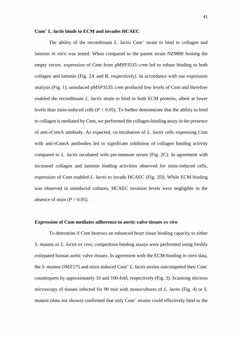
Cnm+ L. lactis binds to ECM and invades HCAEC
The ability of the recombinant L. lactis Cnm+ strain to bind to collagen and
laminin in vitro was tested. When compared to the parent strain NZ9800 hosting the
empty vector, expression of Cnm from pMSP3535::cnm led to robust binding to both
collagen and laminin (Fig. 2A and B, respectively). In accordance with our expression
analysis (Fig. 1), uninduced pMSP3535::cnm produced low levels of Cnm and therefore
enabled the recombinant L. lactis strain to bind to both ECM proteins, albeit at lower
levels than nisin-induced cells (P < 0.05). To further demonstrate that the ability to bind
to collagen is mediated by Cnm, we performed the collagen-binding assay in the presence
of anti-rCnmA antibody. As expected, co-incubation of L. lactis cells expressing Cnm
with anti-rCnmA antibodies led to significant inhibition of collagen binding activity
compared to L. lactis incubated with pre-immune serum (Fig. 2C). In agreement with
increased collagen and laminin binding activities observed for nisin-induced cells,
expression of Cnm enabled L. lactis to invade HCAEC (Fig. 2D). While ECM binding
was observed in uninduced cultures, HCAEC invasion levels were negligible in the
absence of nisin (P > 0.05).
Expression of Cnm mediates adherence to aortic valve tissues ex vivo
To determine if Cnm bestows an enhanced heart tissue binding capacity to either
S. mutans or L. lactis ex vivo, competition binding assays were performed using freshly
extirpated human aortic valve tissues. In agreement with the ECM-binding in vitro data,
the S. mutans OMZ175 and nisin-induced Cnm+ L. lactis strains outcompeted their Cnm-
counterparts by approximately 10 and 100-fold, respectively (Fig. 3). Scanning electron
microscopy of tissues infected for 90 min with monocultures of L. lactis (Fig. 4) or S.
mutans (data not shown) confirmed that only Cnm+ strains could effectively bind to the

42
collagenous fibrils of damaged valve tissues (Figs. 4A and 4C). Notably, Cnm+ strains
could also adhere to tissue areas where no apparent damage was observed (Fig. 4B).
Figure 2. Cnm promotes binding to ECM proteins and invasion of endothelial cells.
(A and B) Binding of bacterial cells to extracellular matrix proteins: (A) collagen type I
and (B) laminin. (C) Inhibition of binding to collagen type I by anti-rCnmA antibody
(1:20). (D) HCAEC invasion assay showing the normalized percentage of invasion for
each strain after 3 h of co-incubation (*P < 0.05, **P < 0.01, ***P < 0.0001, ANOVA
followed by Tukey’s post-test). The S. mutans OMZ175 (Cnm+) strain was used as a
positive control in all assays. The symbols “+” and “–“ in the graphs denote induction or
no induction with nisin, respectively.

43
Figure 3. Expression of Cnm mediates bacterial adherence to aortic valve sections.
A competition assay was developed to test the contribution of Cnm toward bacterial
adherence to human aortic valves ex vivo. A 1:1 mixed infection of the valve sections was
allowed for 90 min using Cnm+ and Cnm- strains. The competitive index (CI) was
calculated based on the initial inocula, where a value of “=1” represents no difference and
“>1” represents preferred binding by the numerator strain.

44
Figure 4. Cnm mediates binding to the collagenous fibrils of damaged and
apparently undamaged infected human valve sections. SEM analysis of valve sections
incubated with bacterial monocultures showed Cnm+ L. lactis binding to bundles of
collagenous fibrils on the rough edges of the valves whereas no Cnm- L. lactis were found
attached to this area (A). Cnm+ L. lactis could also adhere to tissue areas where no
apparent damage was observed in contrast with their Cnm- counterparts (B).

45
Figure 4. (C) Computer-colored SEM photomicrograph (5,000x) showing Cnm+
bacterial cells (green) attached to the collagenous fibrils (light brown) of damaged human
aortic valve sections ex vivo.
Cnm contributes to virulence in Galleria mellonella
The ability of the L. lactis strains to kill the larvae of G. mellonella, a model of
systemic infection was assessed. As shown in Figure 5, the mortality rates of G.
mellonella were significantly higher in larvae infected with the Cnm+ strain compared to
those infected with the wild-type strain harboring the empty vector control (P < 0.05).
Remarkably when considering the extremely low virulence potential of L. lactis, the
recombinant Cnm+ strain showed a similar pattern of killing when compared to the Cnm+
S. mutans OMZ175 strain.

46
Figure 5. Cnm contributes to virulence in the G. mellonella systemic infection model.
The Kaplan-Meier curves indicate the percent survival at selected intervals of larvae
infected with S. mutans OMZ175 and L. lactis strains for 96 h. The mortality rates of G.
mellonella were significantly higher in larvae infected with Cnm+ L. lactis compared to
those infected with the wild-type NZ9800 harboring the empty vector control (pMSP3535)
(P < 0.05).
Cnm contributes to endocardium adherence in vivo
A rabbit model of IE 38 was employed to examine the effect of Cnm expression
on L. lactis adherence to endocardium and vegetations. In one experiment, a mixed
inoculum of NZ9800 (pCIE) and NZ9800 (pMSP3535::cnm) was injected into rabbits
intravenously 1 h after surgery. Two hours later, the rabbits were euthanized and the
hearts removed. This time was chosen so as to allow enough time for clearance of bacteria
from the bloodstream,40 but not so long as to have excessive loss of Cnm due to growth
without nisin. Catheters were clamped into place during necropsy to preserve their
positions. At this early stage, vegetations were not visible, and the entire endocardial
surface in apparent contact with the catheter was excised and homogenized. Following
sonication, both the mixed inoculum and the homogenates were plated on selective media

47
to determine strain ratios. In the second experiment, animals were inoculated 2 days after
surgery. Vegetations, which were visible in all animals, were removed and homogenized.
Because vegetations are composed primarily of platelets and fibrin rather than collagen,41
the endocardium beneath each vegetation was also collected, homogenized, and plated
separately.
As shown in Figure 6, the CI value of the cells harvested from the endocardium
were similar in animals inoculated 1 hour or 2 days after catheterization (geometric mean
= 1.68 and 1.66, respectively), indicating a ~67% increase in infectivity of the Cnm-
expressing strain relative to the control strain. These values were significantly greater
from 1.0 — the value indicating no change in infectivity — whether the two experiments
were analyzed separately or together. In contrast, there was no significant difference in
the CI value for bacteria recovered from vegetations.
Figure 6. Cnm contributes to bacterial endocardium adherence in vivo. Rabbits were
co-inoculated with Cnm-expressing and control L. lactis strains 1 hour or 2 days after
insertion of an intracardiac catheter as indicated and then sacrificed 2 hours later. Bacteria
were recovered from endocardial surfaces (End.), and in the 2-day samples, from adjacent

48
vegetations (Veg.). CI values (for L. lactis Cnm+ over L. lactis Cnm-) are indicated for
each sample, with mean CIs indicated by a horizontal line. Means significantly different
from 1.0 (dashed line) are indicated: *P < 0.05; ***P < 0.001. Like symbols in the 2-day
experiment indicate samples recovered from the same animal. Red circle, sample from
which one of the two strains was not recovered (L. lactis Cnm-); the limit of detection
was used for the calculation.
DISCUSSION
The presence of Cnm has been correlated with an increased ability of S. mutans
to invade and persist intracellularly, and to cause disease.6, 18, 20, 21, 42, 43 In the present
study, by expressing Cnm in the non-pathogenic L. lactis host we provided unequivocal
evidence that Cnm contributes to bacterial virulence.
Various properties of L. lactis have justified its attractiveness for heterologous
expression of surface proteins. For instance, the fermentative metabolism of L. lactis is
relatively simple and well known, the genomes of various strains are available and known
to encode a very low number of genes encoding secreted and surface-anchored proteins.25,
44, 45 Here, we showed that Cnm expression increased the ability of L. lactis to bind to
type I collagen and laminin in vitro, adhere to aortic valve tissues ex vivo, and colonize
endocardial surfaces in vivo. Of note, type I collagen is the most abundant ECM
component in human46, 47 and rabbit48 aortic valves. Although ex vivo experiments cannot
reproduce the immunologic responses and constant shear force of blood flow, our
findings highlighted the contribution of Cnm in binding of organisms to collagen-rich
tissues in both humans and rabbits.
Recently, our group reported that Cnm is a glycoprotein and identified the
glycosylation machinery (Pgf) responsible for its post-translational modification. In

49
SDS-PAGE, the native Cnm migrates at approximately 120 kDa whereas the variant from
the deletion of the pgfS gene was found to migrate at ~ 90 kDa.19 When analyzed by
Western blot, the Cnm variant produced by L. lactis was found to migrate at ~ 90 kDa,
the same size as the unglycosylated version of Cnm produced in S. mutans.19 Moreover,
like the unglycosylated Cnm produced by the pgfS strain in S. mutans, the Cnm
expressed by L. lactis was highly susceptible to proteinase K degradation (data not
shown).19 These findings are supported by in silico analysis that indicated the absence of
Pgf homologs in the available genomes of L. lactis. Most importantly, the lack of Cnm
glycosylation may explain the lesser effect of Cnm expression on adhesion to endocardial
tissue in vivo compared to in vitro or ex vivo. Whether this difference is due to the
considerable shear forces and host proteases present in the in vivo model, or to some other
factor(s) remains to be determined. Expression of Cnm had no effect on colonization of
preexisting vegetations, which is not surprising considering these vegetations are
composed primarily of platelets and fibrin.41 Nevertheless, adhesins for collagen and
other matrix proteins have been identified previously as virulence factors for IE.29, 49, 50
As with these other adhesins, Cnm may allow adherence to damaged endocardium prior
to vegetation formation, thereby providing another route for infection in IE.51 The
increased adherence to endocardium observed is also consistent with previous reports of
association of Cbm-expressing strains, with both IE and non-IE cardiovascular diseases.7,
52
Collectively, our results clearly show that expression of Cnm alone confers
virulence to an otherwise non-pathogenic bacterium, and promotes bacterial adherence to
cardiac tissue in vivo and ex vivo. Thus, Cnm could be considered a target for the
development of antimicrobial strategies to mitigate virulence of invasive S. mutans strains.

50
MATERIAL AND METHODS
Bacterial strains and growth conditions
All strains used in this study are listed in Table 1. The L. lactis strains were
routinely cultured aerobically at 30°C in M17 broth (Difco) supplemented with 0.5% (w/v)
glucose (GM17). When necessary, 10 µg ml-1 erythromycin or 15 µg ml-1
chloramphenicol (Sigma-Aldrich) was added to the growth media (GM17-Erm or GM17-
Chlor, respectively). The S. mutans strains were routinely cultured in brain heart infusion
(BHI) medium at 37°C in a 5% CO2 atmosphere.
Table 1. Strains used in this study.
Strain Relevant
phenotype Reference
Streptococcus mutans
OMZ175 Cnm+ Lab collection
OMZ175 (pBGE) Cnm+ Ermr (21)
∆cnm Cnm– Kanr (21)
Lactococcus lactis subsp. lactis
NZ9800 WT, Cnm– Lab collection
NZ9800 (pMSP3535) Cnm– Ermr This study
NZ9800 (pMSP3535::cnm) Cnm+ Ermr This study
NZ9800 (pCIE) Cnm–, Chlorr This study
Note: WT (wild type); Kan (kanamycin); Erm (erythromycin); Chlor
(chloramphenicol).
Expression of Cnm in L. lactis
The nisin-inducible plasmid pMSP3535 34 harboring the cnm gene
(pMSP3535::cnm) from S. mutans OMZ175,21 was used to express Cnm in L. lactis. The
pMSP3535::cnm plasmid as well as the empty pMSP3535 vector 34 and the pCIE
plasmids (Dunny et al., unpublished) were electroporated into L. lactis NZ9800 as
detailed elsewhere 35 with some modifications. The pCIE plasmid, conferring resistance
to chloramphenicol, was generated from shuttle plasmid pCI13340 36 and introduced into
L. lactis for selection purposes in competition assays. Briefly, cells were grown overnight

51
at 30oC in M17 broth supplemented with 0.5% glucose (GM-17) and sub-cultured (1:100)
in fresh GM-17 containing 1% glycine and 0.5 M sucrose (GM17S). The culture was
grown to OD600 of 0.2, centrifuged at 5,000 rpm for 10 min at 4oC and washed 3 times
with cold electroporation buffer (EB) (0.5 M sucrose, 1 mM MgCl2, 7 mM K2HPO4-
KH2PO4 [pH 7.4]). Electrocompetent cells were resuspended in EB, quickly frozen on
dry ice/ethanol bath and stored at -80oC until use. A micro-pulser electroporation
apparatus (Bio-Rad Laboratories) (0.1cm cuvette, 1.4 kV, 600 Ω, 10 µF) was used to
electroporate the desired plasmids into L. lactis. After electroporation, cells were
incubated in GM17S for 3 h at 30oC and spread on GM17 plates containing the desired
antibiotic. Resulting colonies were screened for the presence of the desired plasmid by
PCR. For induction of Cnm, cells were grown overnight in GM17-Erm in the presence of
increasing concentrations of nisin (Sigma-Aldrich). The production and cell surface
localization of Cnm was confirmed by Western blotting and by immunofluorescence
labeling as detailed below.
Western Blot (WB) analysis
Overnight cultures of S. mutans OMZ175 and recombinant L. lactis strains were
used to prepare protein lysates by homogenization with 0.1 mm glass beads using a bead
beater (Biospec). Protein lysates were separated on a 10% SDS-PAGE and transferred to
a polyvinylidene fluoride (PVDF) membrane (Millipore) using standard protocols. The
presence of Cnm was detected using rabbit anti-rCnmA polyclonal antibody 19 diluted
1:2000 in 1 x phosphate buffered saline (PBS) with 0.1% Tween 20 pH 7.2 and anti-IgG
(goat anti-rabbit) horseradish peroxidase-coupled antibody diluted 1:10000 (Sigma-
Aldrich). Membranes were developed using an enhanced chemiluminescent detection kit
(GE Life Sciences).

52
Immunofluorescence labeling of Cnm
Cultures of recombinant L. lactis and S. mutans OMZ175 were used for cellular
localization of Cnm. Cells grown to mid-exponential phase were washed in PBS and
incubated in the presence of anti-rCnmA antibody (1:100) followed by anti-rabbit Alexa-
488 conjugate antibody (1:100) with three washes between each antibody exposure. Cell-
antibody complexes were visualized using an Olympus BX41 fluorescent microscope and
images were captured with the QCapture Software.
Binding to extracellular matrix (ECM) proteins
Collagen and laminin binding assays were performed as previously described.20,
21 Briefly, 96-well plates were coated with 40 µg ml−1 type I collagen from rat tail (Sigma-
Aldrich), or 50 µg ml−1 mouse laminin (Becton-Dickinson) for 18 h at 4oC. Individual
wells were washed and blocked with 5% bovine serum albumin for 1 h at 37oC. Overnight
bacterial cultures were washed twice in PBS and 100 μl aliquots of each bacterial
suspension added to the wells, followed by incubation for 3 h at 37oC. Loosely bound
bacteria were removed by washing with PBS and adhered cells were stained with 0.05%
crystal violet solution and then quantified at 575 nm. Experiments were also performed
with bacteria pre-incubated with anti-rCnmA antibody (1:20 dilution in PBS) for 30 min
before incubation with collagen. To confirm the requirement of Cnm in collagen binding,
experiments were performed as described above but bacteria were pre-incubated with
anti-rCnmA or pre-immune serum (1:100) for 30 min before exposure to collagen.19
Invasion of human coronary artery endothelial cells (HCAEC)
Cultivation of primary HCAECs and invasion assays were performed as
previously described.18, 21 The HCAEC monolayer was incubated with 1 ml of 2% fetal
bovine serum (FBS)-endothelial basal medium (EBM-2) containing 1-2 x 10⁷ CFU m-1

53
of S. mutans OMZ175 or ∆cnm, or recombinant L. lactis strains for 2 h at a multiplicity
of infection of 100:1, followed by 3 h of incubation in 2% FBS-EBM-2 medium
containing 300 μg ml−1 gentamicin and 50 μg ml−1 penicillin G to kill extracellular
bacteria. After incubation with antibiotics, HCAEC were lysed with 1 ml of sterile water
and the mixture of lysed HCAEC and bacteria serially diluted and plated onto tryptic soy
agar (TSA) plates to determine the number of intracellular bacteria. The percentage of
invasion for each strain was calculated based on the total colony forming units (CFU)
number present in the initial inoculum and the number of intracellular bacteria recovered
from HCAEC lysates.
Adherence to human cardiac valve sections ex vivo
The ability of S. mutans and L. lactis strains to bind to human aortic valve tissues
ex vivo was assessed by adapting a protocol originally described for porcine heart
tissues.37 Human aortic valve tissues were obtained from patients undergoing valve
replacement surgery due to calcific stenosis (Institutional Review Board protocol no.
RSRB00053016, University of Rochester). Valvular tissues with no signs of calcification,
bleeding or inflammation were selected and 3-mm diameter valve sections were obtained
using a biopsy punch tool (Acuderm). Valve sections were incubated overnight in 12-
well plates with endothelial cell medium (ECM) supplemented with 5% FBS, 1000 mg
L-1 hydrocortisone, 12 mg L-1 bovine brain extract, 0.1% human recombinant epidermal
growth factor (Lonza) plus 300 µg ml-1 gentamicin. On the following day, sections were
washed twice with Hank's Balanced Salt Solution (HBSS, Corning Inc.) to remove
residual antibiotic and then transferred to a new plate. A competition assay was then
performed to determine the capacity of Cnm+ strains to compete with Cnm- strains. For
this, valve sections were infected with approximately 5 x 10⁷ CFU ml-1 of a 1:1
suspension of Cnm+ and Cnm- strains bearing distinct antibiotic markers for CFU

54
determination of each strain. Plates were incubated for 90 min at 37°C (for S. mutans) or
30°C (for L. lactis) with gentle rocking. Valve sections were transferred to sterile
centrifuge tubes containing 1 ml of HBSS and vigorously agitated three times for 5
seconds using a vortex to remove loosely bound bacteria from the tissues. Washed valve
sections were homogenized in 500 µl PBS for 2 min using a motorized pestle (Fisher
Scientific), serially diluted and plated onto TSA plates with the corresponding antibiotics.
The competitive index (CI) was calculated based on the CFUs present in the initial
inoculum and the final enumeration of bacteria recovered from the tissue.
Scanning Electron Microscopy (SEM) analysis of valve sections
To visualize bacterial attachment to heart valve sections, tissue samples were
prepared and incubated in the presence of 5 x 10⁷ CFU ml-1 S. mutans or L. lactis
monocultures as described in the previous section. Valve samples were washed and fixed
with 4% paraformaldehyde, 2.5% glutaraldehyde and 0.1 M sodium cacodylate for 2 days
at 4oC, with slow rocking. The samples were analyzed using a Zeiss-Auriga focused ion
beam field emission scanning electron microscope (FIB-FE-SEM) and the images
captured using the attached Gatan Erlangshen digital camera at the University of
Rochester Medical Center Electron Microscopy Core Facility.
Galleria mellonella infection
The systemic infection of G. mellonella has been previously detailed elsewhere.19,
21 Briefly, 5 µl aliquots containing 1-1.5x10⁶ CFU of S. mutans OMZ175 (positive
control), or recombinant L. lactis were injected in the larvae hemocoel via the last left
proleg. Larvae injected with heat-killed bacteria (45 min, 80°C) were used as negative

55
controls. After injection, larvae were kept in the dark at 37°C, and survival was
periodically monitored for up to 4 days.
In vivo model of infective endocarditis in rabbits
The rabbit model of IE employed for this study was modified from a previously
used protocol.38 Specific-pathogen-free, male New Zealand White Rabbits 2 to 3 kg in
size were purchased from Robinson Services Inc. After acclimation, rabbits were injected
with acepromazine as a sedative, a cocktail of ketamine, xylazine, and buprenorphine for
general anesthesia, glycopyrrolate for control of respiratory secretions, and bupivacaine
at the incision site. An incision was then made in the neck region, and a 19-gauge catheter
(Intracath, BD Bioscience) was inserted through the right internal carotid artery,
contacting or passing through the aortic valve to cause minor damage. The catheter was
tied off, trimmed, and sutured into place, and the incision closed. Buprenorphine SR Lab
was used for post-surgical analgesia. The day prior to inoculation, L. lactis strains were
cultured overnight in GM17 and antibiotics as described above with 10 ng ml-1 nisin.
Twenty hours later, the cultures were diluted 10-fold into pre-warmed media of the same
composition and cultured another 3 h. Cells were harvested by centrifugation, washed
twice with cold PBS, diluted in PBS, and combined to achieve a concentration of 3 – 5 x
108 CFU ml-1 for each strain. One hour or 2 days after the surgeries, rabbits were
inoculated with 0.5 ml of bacterial cells via a peripheral ear vein. Remaining cells were
sonicated for 1.5 min at 50% power in a Biologics Model 150 sonicator (Biologic Inc.)
to break up clumps and chains and plated with an Eddy Jet spiral plater (Neutec Group
Inc.) on GM17 plates containing erythromycin or chloramphenicol to establish the ratio
of the two strains in the inoculum. Two hours after inoculation, each rabbit was
euthanized with intravenous Euthasol (Virbac) and was then checked for proper

56
placement of the catheter tip in the valve region or beneath it in the left ventricle. Any
rabbit in which the catheter was not correctly situated was excluded from the study. For
rabbits inoculated on the day of surgery, the aortic valves and surrounding endocardium
were removed and homogenized in 5 ml PBS using an electric homogenizer (Omni
International). For rabbits inoculated 2 days post-surgery, visible vegetations and the
endocardium underlying each vegetation were harvested separately and homogenized in
2 ml PBS each using a disposable 15-ml tissue grinder. In all cases, homogenates were
sonicated and plated as above for enumeration.
Statistical analysis
All results represent a minimum of three independent experiments. Descriptive
and inferential statistics were used to analyze the data on Graphpad Prism version 5.0.
One-way ANOVA followed by Tukey’s post-test was used to analyze the significance of
binding and invasion, with type I error set as 0.05. Analysis of log10-transformed
competitive indices was performed using one-way ANOVA with Bonferroni post hoc
comparisons to determine substrate preferences among strain pairs. For the G. mellonella
studies, Kaplan-Meier killing curves were plotted and estimation of differences in
survival was compared using the log-rank test. Log10-transformed competitive indices
were compared to 0 using a one-sample T test for rabbit studies.
Disclosure of Potential Conflicts of Interest
No potential conflicts of interest were disclosed.
Acknowledgements
This work was supported by the National Institute of Dental and Craniofacial
Research R01 DE022559 to JL and JA, National Heart, Lung, and Blood Institute F31

57
HL124952 to AAR, São Paulo Research Foundation (FAPESP, Brazil, grants no.
2014/07231-0 and no. 2013/25080-7) to IAF, and the National Council for Scientific and
Technological Development (CNPq, Brazil, grant no. 308644/2011-5) to PLR. The
authors thank Dr. Gary Dunny (University of Minnesota) for kindly providing L. lactis
NZ9800 and the pCIE plasmid.
REFERENCES
1. National Institutes of Health. Diagnosis and management of dental caries
throughout life. NIH Consens Statement 2001; 18:1-23.
2. Bagramian RA, Garcia-Godoy F, Volpe AR. The global increase in dental
caries. A pending public health crisis. Am J Dent 2009; 22:3-8.
3. Bowen WH. The Stephan Curve revisited. Odontology 2013; 101:2-8.
4. Lemos JA, Burne RA. A model of efficiency: stress tolerance by Streptococcus
mutans. Microbiology 2008; 154:3247-55.
5. Miyatani F, Kuriyama N, Watanabe I, Nomura R, Nakano K, Matsui D, Ozaki
E, Koyama T, Nishigaki M, Yamamoto T, et al. Relationship between Cnm-positive
Streptococcus mutans and cerebral microbleeds in humans. Oral Dis 2015; 21:886-93.
6. Nakano K, Hokamura K, Taniguchi N, Wada K, Kudo C, Nomura R, Kojima A,
Naka S, Muranaka Y, Thura M, et al. The collagen-binding protein of Streptococcus
mutans is involved in haemorrhagic stroke. Nat Commun 2011; 2:485.
7. Nomura R, Naka S, Nemoto H, Inagaki S, Taniguchi K, Ooshima T, Nakano K.
Potential involvement of collagen-binding proteins of Streptococcus mutans in infective
endocarditis. Oral Dis 2013; 19:387-93.
8. Nomura R, Nakano K, Nemoto H, Fujita K, Inagaki S, Takahashi T, Taniguchi
K, Takeda M, Yoshioka H, Amano A, et al. Isolation and characterization of

58
Streptococcus mutans in heart valve and dental plaque specimens from a patient with
infective endocarditis. J Med Microbiol 2006; 55:1135-40.
9. Que YA, Moreillon P. Infective endocarditis. Nat Rev Cardiol 2011; 8:322-36.
10. Nakano K, Nomura R, Nakagawa I, Hamada S, Ooshima T. Demonstration of
Streptococcus mutans with a cell wall polysaccharide specific to a new serotype, k, in
the human oral cavity. J Clin Microbiol 2004; 42:198-202.
11. Shun CT, Lu SY, Yeh CY, Chiang CP, Chia JS, Chen JY. Glucosyltransferases
of viridans streptococci are modulins of interleukin-6 induction in infective
endocarditis. Infect Immun 2005; 73:3261-70.
12. Pant S, Patel NJ, Deshmukh A, Golwala H, Patel N, Badheka A, Hirsch GA,
Mehta JL. Trends in infective endocarditis incidence, microbiology, and valve
replacement in the United States from 2000 to 2011. J Am Coll Cardiol 2015; 65:2070-
6.
13. Murdoch DR, Corey GR, Hoen B, Miro JM, Fowler VG, Jr., Bayer AS,
Karchmer AW, Olaison L, Pappas PA, Moreillon P, et al. Clinical presentation,
etiology, and outcome of infective endocarditis in the 21st century: the International
Collaboration on Endocarditis-Prospective Cohort Study. Arch Intern Med 2009;
169:463-73.
14. Yew HS, Murdoch DR. Global trends in infective endocarditis epidemiology.
Curr Infect Dis Rep 2012; 14:367-72.
15. Banas JA. Virulence properties of Streptococcus mutans. Front Biosci 2004;
9:1267-77.
16. Nakano K, Ooshima T. Serotype classification of Streptococcus mutans and its
detection outside the oral cavity. Future Microbiol 2009; 4:891-902.

59
17. Oliveira FA, Forte CP, Silva PG, Lopes CB, Montenegro RC, Santos AK,
Sobrinho CR, Mota MR, Sousa FB, Alves AP. Molecular Analysis of Oral Bacteria in
Heart Valve of Patients With Cardiovascular Disease by Real-Time Polymerase Chain
Reaction. Medicine (Baltimore) 2015; 94:e2067.
18. Abranches J, Zeng L, Belanger M, Rodrigues PH, Simpson-Haidaris PJ, Akin D,
Dunn WA, Jr., Progulske-Fox A, Burne RA. Invasion of human coronary artery
endothelial cells by Streptococcus mutans OMZ175. Oral Microbiol Immunol 2009;
24:141-5.
19. Aviles-Reyes A, Miller JH, Simpson-Haidaris PJ, Hagen FK, Abranches J,
Lemos JA. Modification of Streptococcus mutans Cnm by PgfS contributes to adhesion,
endothelial cell invasion, and virulence. J Bacteriol 2014; 196:2789-97.
20. Aviles-Reyes A, Miller JH, Simpson-Haidaris PJ, Lemos JA, Abranches J. Cnm
is a major virulence factor of invasive Streptococcus mutans and part of a conserved
three-gene locus. Mol Oral Microbiol 2014; 29:11-23.
21. Abranches J, Miller JH, Martinez AR, Simpson-Haidaris PJ, Burne RA, Lemos
JA. The collagen-binding protein Cnm is required for Streptococcus mutans adherence
to and intracellular invasion of human coronary artery endothelial cells. Infect Immun
2011; 79:2277-84.
22. Nakano K, Nomura R, Taniguchi N, Lapirattanakul J, Kojima A, Naka S,
Senawongse P, Srisatjaluk R, Gronroos L, Alaluusua S, et al. Molecular
characterization of Streptococcus mutans strains containing the cnm gene encoding a
collagen-binding adhesin. Arch Oral Biol 2010; 55:34-9.
23. Nomura R, Nakano K, Taniguchi N, Lapirattanakul J, Nemoto H, Gronroos L,
Alaluusua S, Ooshima T. Molecular and clinical analyses of the gene encoding the
collagen-binding adhesin of Streptococcus mutans. J Med Microbiol 2009; 58:469-75.

60
24. Naka S, Nomura R, Takashima Y, Okawa R, Ooshima T, Nakano K. A specific
Streptococcus mutans strain aggravates non-alcoholic fatty liver disease. Oral Dis 2014;
20:700-6.
25. Mierau I, Kleerebezem M. 10 years of the nisin-controlled gene expression
system (NICE) in Lactococcus lactis. Appl Microbiol Biotechnol 2005; 68:705-17.
26. Morello E, Bermudez-Humaran LG, Llull D, Sole V, Miraglio N, Langella P,
Poquet I. Lactococcus lactis, an efficient cell factory for recombinant protein production
and secretion. J Mol Microbiol Biotechnol 2008; 14:48-58.
27. Wyszynska A, Kobierecka P, Bardowski J, Jagusztyn-Krynicka EK. Lactic acid
bacteria--20 years exploring their potential as live vectors for mucosal vaccination. Appl
Microbiol Biotechnol 2015; 99:2967-77.
28. Danne C, Entenza JM, Mallet A, Briandet R, Debarbouille M, Nato F, Glaser P,
Jouvion G, Moreillon P, Trieu-Cuot P, et al. Molecular characterization of a
Streptococcus gallolyticus genomic island encoding a pilus involved in endocarditis. J
Infect Dis 2011; 204:1960-70.
29. Piroth L, Que YA, Widmer E, Panchaud A, Piu S, Entenza JM, Moreillon P. The
fibrinogen- and fibronectin-binding domains of Staphylococcus aureus fibronectin-
binding protein A synergistically promote endothelial invasion and experimental
endocarditis. Infect Immun 2008; 76:3824-31.
30. Que YA, Haefliger JA, Piroth L, Francois P, Widmer E, Entenza JM, Sinha B,
Herrmann M, Francioli P, Vaudaux P, et al. Fibrinogen and fibronectin binding
cooperate for valve infection and invasion in Staphylococcus aureus experimental
endocarditis. J Exp Med 2005; 201:1627-35.
31. Buccato S, Maione D, Rinaudo CD, Volpini G, Taddei AR, Rosini R, Telford
JL, Grandi G, Margarit I. Use of Lactococcus lactis expressing pili from group B

61
Streptococcus as a broad-coverage vaccine against streptococcal disease. J Infect Dis
2006; 194:331-40.
32. Arrecubieta C, Toba FA, von Bayern M, Akashi H, Deng MC, Naka Y, Lowy
FD. SdrF, a Staphylococcus epidermidis surface protein, contributes to the initiation of
ventricular assist device driveline-related infections. PLoS Pathog 2009; 5:e1000411.
33. Innocentin S, Guimaraes V, Miyoshi A, Azevedo V, Langella P, Chatel JM,
Lefevre F. Lactococcus lactis expressing either Staphylococcus aureus fibronectin-
binding protein A or Listeria monocytogenes internalin A can efficiently internalize and
deliver DNA in human epithelial cells. Appl Environ Microbiol 2009; 75:4870-8.
34. Bryan EM, Bae T, Kleerebezem M, Dunny GM. Improved vectors for nisin-
controlled expression in gram-positive bacteria. Plasmid 2000; 44:183-90.
35. Holo H, Nes IF. Transformation of Lactococcus by electroporation. Methods
Mol Biol 1995; 47:195-9.
36. Hayes F, Daly C, Fitzgerald GF. Identification of the Minimal Replicon of
Lactococcus lactis subsp. lactis UC317 Plasmid pCI305. Appl Environ Microbiol 1990;
56:202-9.
37. Chuang-Smith ON, Wells CL, Henry-Stanley MJ, Dunny GM. Acceleration of
Enterococcus faecalis biofilm formation by aggregation substance expression in an ex
vivo model of cardiac valve colonization. PLoS One 2010; 5:e15798.
38. Crump KE, Bainbridge B, Brusko S, Turner LS, Ge X, Stone V, Xu P, Kitten T.
The relationship of the lipoprotein SsaB, manganese and superoxide dismutase in
Streptococcus sanguinis virulence for endocarditis. Mol Microbiol 2014; 92:1243-59.
39. Fu RY, Chen J, Li Y. Heterologous leaky production of transglutaminase in
Lactococcus lactis significantly enhances the growth performance of the host. Appl
Environ Microbiol 2005; 71:8911-9.

62
40. Durack DT, Beeson PB. Experimental bacterial endocarditis. I. Colonization of a
sterile vegetation. Br J Exp Pathol 1972; 53:44-9.
41. Durack DT. Experimental bacterial endocarditis. IV. Structure and evolution of
very early lesions. J Pathol 1975; 115:81-9.
42. Miller JH, Aviles-Reyes A, Scott-Anne K, Gregoire S, Watson GE, Sampson E,
Progulske-Fox A, Koo H, Bowen WH, Lemos JA, et al. The collagen binding protein
Cnm contributes to oral colonization and cariogenicity of Streptococcus mutans
OMZ175. Infect Immun 2015; 83:2001-10.
43. Kojima A, Nomura R, Naka S, Okawa R, Ooshima T, Nakano K. Aggravation of
inflammatory bowel diseases by oral streptococci. Oral Dis 2014; 20:359-66.
44. Kunji ER, Slotboom DJ, Poolman B. Lactococcus lactis as host for
overproduction of functional membrane proteins. Biochim Biophys Acta 2003;
1610:97-108.
45. Wegmann U, O'Connell-Motherway M, Zomer A, Buist G, Shearman C,
Canchaya C, Ventura M, Goesmann A, Gasson MJ, Kuipers OP, et al. Complete
genome sequence of the prototype lactic acid bacterium Lactococcus lactis subsp.
cremoris MG1363. J Bacteriol 2007; 189:3256-70.
46. Latif N, Sarathchandra P, Taylor PM, Antoniw J, Yacoub MH. Localization and
pattern of expression of extracellular matrix components in human heart valves. J Heart
Valve Dis 2005; 14:218-27.
47. Rodriguez KJ, Piechura LM, Porras AM, Masters KS. Manipulation of valve
composition to elucidate the role of collagen in aortic valve calcification. BMC
Cardiovasc Disord 2014; 14:29.

63
48. Hinton RB, Jr., Lincoln J, Deutsch GH, Osinska H, Manning PB, Benson DW,
Yutzey KE. Extracellular matrix remodeling and organization in developing and
diseased aortic valves. Circ Res 2006; 98:1431-8.
49. Singh B, Fleury C, Jalalvand F, Riesbeck K. Human pathogens utilize host
extracellular matrix proteins laminin and collagen for adhesion and invasion of the host.
FEMS Microbiol Rev 2012; 36:1122-80.
50. Singh KV, La Rosa SL, Somarajan SR, Roh JH, Murray BE. The Fibronectin-
Binding Protein EfbA Contributes to Pathogenesis and Protects against Infective
Endocarditis Caused by Enterococcus faecalis. Infect Immun 2015; 83:4487-94.
51. Bashore TM, Cabell C, Fowler V, Jr. Update on infective endocarditis. Curr
Probl Cardiol 2006; 31:274-352.
52. Nakano K, Nomura R, Nemoto H, Mukai T, Yoshioka H, Shudo Y, Hata H,
Toda K, Taniguchi K, Amano A, et al. Detection of novel serotype k Streptococcus
mutans in infective endocarditis patients. J Med Microbiol 2007; 56:1413-5.

64
DISCUSSÃO
A habilidade de S. mutans interagir com componentes da MEC por meio da
expressão de proteínas ligantes de colágeno, como Cnm, confere a este patógeno oral uma
capacidade aumentada para o desenvolvimento de cárie (Miller et al., 2015). Além disto,
devido à ampla distribuição e abundância de colágeno no corpo humano, outros sítios
onde S. mutans não ocorre naturalmente também encontram-se susceptíveis à infecção
por cepas invasivas (Avilés-Reyes et al., 2016). No presente estudo, nós utilizamos
abordagens orais (Capítulo I) e extra-orais (Capítulo II) para elucidar o papel desta
glicoproteína no desenvolvimento e progressão de doenças humanas, como a cárie e a EI.
Estas doenças geram alto ônus econômico direto e indireto, por exemplo, a cárie dentária
é responsável por um gasto anual de cerca de 90 bilhões de dólares (Listl et al., 2015) e
doenças cardiovasculares de 863 bilhões de dólares (World Heart Federation, 2010), bem
como elevada taxa de morbidade e/ou mortalidade em nível global (Werdan et al., 2014).
No Capítulo I, demonstramos que a presença de Cnm está associada a um
incremento de lesões iniciais de cárie em dentina coronária, corroborando relatos
anteriores (Miller et al., 2015). Em um período experimental de cinco semanas, todos os
grupos apresentaram altos escores de lesões avançadas em dentina (Dm e Dx), o que
indica a ocorrência de destruição coronária severa e independente da presença de Cnm.
Entretanto, é possível que diferenças relacionadas ao potencial cariogênico de Cnm em
lesões avançadas tenham ocorrido em um menor tempo experimental, mas não foram
detectadas devido ao avançar da doença nos demais grupos como resultado da
dessalivação. A escolha pelo período experimental de 5 semanas baseou-se no estudo de
Bowen, Pearson e Young (1988), no qual os autores concluíram que um período de 5
semanas de desafio cariogênico é ideal, visto que um desafio de 6 semanas é bastante

65
destrutivo – e, portanto, desnecessário – enquanto que um período de 3 a 4 semanas é
insuficiente para o desenvolvimento apropriado de cárie de raiz nos animais dessalivados.
Considerando que a matriz orgânica dentinária é composta por 90% de colágeno
tipo I, a capacidade de se ligar a este substrato favorece a colonização e desenvolvimento
de cárie na superfície radicular (Han, Chang and Dao, 2006; Ho et al., 2007). Em um
estudo prévio (Miller et al., 2015), demonstrou-se que OMZ175 apresenta uma
capacidade aumentada de se ligar à dentina radicular ex vivo quando comparada à cepa
com deleção do gene cnm. Com base no exposto, hipotetizamos que a expressão de Cnm
poderia contribuir para o desenvolvimento de cárie radicular in vivo. Nenhuma diferença
significante foi verificada entre cepas Cnm+ e Cnm-, incluindo a cepa referência UA159
(Cnm-), no que concerne ao desenvolvimento de cárie radicular na mandíbula e maxila.
Todavia, observamos que OMZ175 e OMZ175Δcnm::pcnm promoveram perda óssea
alveolar expressiva, até 18.5% maior do que a cepa knockout OMZ175Δcnm,
especialmente na região mandibular. Do ponto de vista clínico, este percentual de perda
óssea é significativo (Lima et al., 2000) e deve ser melhor investigado para
compreendermos a patogenicidade conferida pela expressão de Cnm não apenas na
progressão da cárie, mas também em doenças periodontais, pela perda de suporte ósseo.
A literatura aponta que a interação de S. mutans com o hospedeiro pode interferir
com a função imunológica, desencadear a produção de citocinas proinflamatórias e,
consequentemente, retardar o reparo e a homeostase teciduais (Nagata, Toleto e Oho,
2011). Em algumas amostras do nosso estudo, a superfície radicular encontrava-se
altamente exposta, mesmo na ausência de lesões extensas de cárie na raiz, o que pode
indicar a ativação de vias inflamatórias que promovem ativação osteoclastogênica e
destruição óssea (Hajishengallis et al., 2016). Portanto, estudos futuros devem investigar
a influência de microrganismos expressando Cnm sobre o recrutamento de células imunes

66
do hospedeiro, liberação de citocinas (ex.: TNF-α , IL-1β) e quimiocinas (CXCR2)
proinflamatórias, assim como outros mediadores de reabsorção alveolar (ex.:
RANK/RANK-L) (Hajishengallis, 2014; Hajishengallis et al., 2016).
A presença de Cnm também tem sido fortemente correlacionada à maior
capacidade de S. mutans em invadir células endoteliais do hospedeiro e persistir no
citoplasma (Abranches et al., 2011). No Capítulo II, nós expressamos Cnm em um
microrganismo não patogênico, L. lactis, e demonstramos evidência inequívoca de que
esta glicoproteína contribui para a virulência bacteriana, visto que L. lactis recombinante
mostrou-se consistentemente invasivo e patogênico.
Diversas propriedades tornam L. lactis um hospedeiro (host) atrativo para a
expressão heteróloga de proteínas de superfície. Por exemplo, o metabolismo de L. lactis
é relativamente simples e bem conhecido (Kunji, Slotboom e Poolman, 2003; Mierau &
Kleerebezem, 2005); e o genoma de cepas desta espécie já foi sequenciado e aponta para
um baixo número de genes que codificam secreção de proteínas e ancoragem à superfície
(Wegmann et al., 2007). Neste estudo, nós demonstramos que a expressão de Cnm
aumentou a habilidade de L. lactis em se ligar ao colágeno tipo I e à laminina in vitro,
bem como aderir à valva aórtica humana ex vivo e, interessantemente, colonizar
superfícies do endocárdio in vivo. É importante notar que o colágeno tipo I é o
componente da MEC mais abundante nas valvas cardíacas humanas (Latif et al., 2005;
Rodriguez et al., 2014) e de coelhos (Hinton et al., 2006). Muito embora os experimentos
ex vivo não consigam reproduzir a resposta imunológica e forças mecânicas do fluxo
sanguíneo, os nossos resultados apontam para uma forte aderência de organismos Cnm+
com tecidos ricos em colágeno, tanto em humanos quanto em coelhos, condição que
predipõe a etiopatogenia de distúrbios cardiovasculares (Hinton et al., 2006).

67
A expressão de Cnm não afetou a colonização de vegetações preexistentes, o que
era esperado, visto que vegetações desta natureza são compostas primariamente por
plaquetas e fibrina (Durack, 1975; Werdan et al., 2014). Entretanto, Cnm pode favorecer
a aderência ao endocárdio danificado anteriormente à formação da vegetação,
propiciando, assim, uma outra rota para EI (Bashore, Cabell and Fowler, 2006).
Em síntese, os nossos resultados mostram que a expressão de Cnm conferiu
virulência a um microrganismo não patogênico, promoveu aderência bacteriana à tecidos
cardíacos in vivo e ex vivo, e aumentou a cariogenicidade e absorção óssea em modelo
com ratos dessalivados. Os nossos achados abrem caminhos inovadores para o
desenvolvimento de terapias alvo-específicas contra doenças que ainda geram um
considerável impacto social e econômico, como a cárie dentária e a endocardite
infecciosa.

68
CONCLUSÃO
1) A expressão de Cnm em S. mutans contribuiu para o desenvolvimento de cárie de
superfície lisa em um modelo animal dessalivado sob alto desafio cariogênico, mas
não incrementou o desenvolvimento de cárie de raiz após cinco semanas. Entretanto,
a presença de Cnm foi relacionada a uma maior perda óssea alveolar nos animais
dessalivados.
2) A expressão heteróloga de Cnm em Lactococcus lactis permitiu concluir que esta
glicoproteina confere capacidade aumentada de ligação bacteriana ao colágeno e à
laminina (componentes da matrix extracelular), invasão intracelular, aderência à
valva aórtica humana ex vivo e, finalmente, desenvolvimento de endocardite
infecciosa in vivo. Além disto, o presente estudo confirma a efetividade do sistema
heterólogo L. lactis para caracterização de fatores de virulência de superfície em
bactérias Gram-positivas.
Com base no exposto, a proteína ligante de colágeno Cnm pode ser considerada
um alvo em potencial para o desenvolvimento de novas terapias antimicrobianas que
visam mitigar a colonização e infecção de valvas cardíacas por cepas invasivas e, na
cavidade oral, onde S. mutans ocorre naturalmente, controlar o desenvolvimento e
progressão de cárie dentária por mecanismos alternativos aos disponíveis atualmente.
REFERÊNCIAS 5
Abranches J, Miller JH, Martinez AR, Simpson-Haidaris PJ, Burne RA, Lemos JA. The
collagen-binding protein Cnm is required for Streptococcus mutans adherence to and
intracellular invasion of human coronary artery endothelial cells. Infect Immun. 2011;
79: 2277-84.
5 De acordo com as normas da UNICAMP/FOP, baseadas na padronização do International Committee of
Medical Journal Editors – Grupo de Vancouver. Abreviatura dos periódicos em conformidade com o
PubMed.
69
Avilés-Reyes A, Miller JH, Lemos JA, Abranches J. The Collagen Binding Proteins of
Streptococcus mutans and Related Streptococci. Mol Oral Microbiol. 2016;
32(2):89-106. doi: 10.1111/omi.12158.
Banas JA. Virulence properties of Streptococcus mutans. Front Biosci. 2004; 9:1267-77.
Bashore TM, Cabell C, Fowler V, Jr. Update on infective endocarditis. Curr Probl Cardiol.
2006; 31:274-352.
Bowen WH, Pearson SK, Young DA. The Effect of Desalivation on Coronal and Root
Surface Caries in Rats. J Dent Res. 1988 67: 21. doi:
10.1177/00220345880670010301.
Danne C, Entenza JM, Mallet A, Briandet R, Debarbouille M, Nato F, et al. Molecular
characterization of a Streptococcus gallolyticus genomic island encoding a pilus
involved in endocarditis. J Infect Dis. 2011; 204:1960-70.
Durack DT. Experimental bacterial endocarditis. IV. Structure and evolution of very early
lesions. J Pathol. 1975; 115:81-9.
Fernández-Hidalgo N, Almirante B. La endocarditis infecciosa en el siglo xxi: cambios
epidemiológicos, terapéuticos y pronósticos. Enferm Infecc Microbiol Clin. 2012;
30:394-406.
Galvão LCC, Miller JH, Kajfasz JK, Scott-Anne K, Freires IA, Franco GCN, et al.
Transcriptional and Phenotypic Characterization of Novel Spx-Regulated Genes in
Streptococcus mutans. PLoS ONE. 2015; 10(4):e0124969.
doi:10.1371/journal.pone.0124969.
Hajishengallis, G. Immunomicrobial pathogenesis of periodontitis: keystones,
pathobionts, and host response. Trends Immunol. 2014; 35:3-11.
Hajishengallis G, Moutsopoulos NM, Hajishengallis E, Chavakis T. Immune and
regulatory functions of neutrophils in inflammatory bone loss. Seminars Immunol.
2016; 28:146-158.
Han TK, Zhang C, Dao ML. Identification and characterization of collagen-binding
activity in Streptococcus mutans wall-associated protein: a possible implication in
dental root caries and endocarditis. Biochem Biophys Res Commun. 2006;
343:787-92.
Hinton RB, Jr., Lincoln J, Deutsch GH, Osinska H, Manning PB, Benson DW, et al.
Extracellular matrix remodeling and organization in developing and diseased aortic
valves. Circ Res. 2006; 98:1431-8.

70
Ho SP, Marshall SJ, Ryder MI, Marshall GW. The tooth attachment mechanism defined
by structure, chemical composition and mechanical properties of collagen fibers in
the periodontium. Biomaterials. 2007; 28:5238-45. doi:
10.1016/j.biomaterials.2007.08.031.
Kajfasz JK, Rivera-Ramos I, Abranches J, Martinez AR, Rosalen PL, Derr AM, et al.
Two Spx Proteins Modulate Stress Tolerance, Survival, and Virulence in
Streptococcus mutans. J. Bacteriol. 2010; 192:2546-56.
Kassebaum NJ, Bernabé E, Dahiya M, Bhandari B, Murray CJ, Marcenes W. Global
burden of untreated caries: a systematic review and metaregression. J Dent Res.
2015; 94:650-58. doi: 10.1177/0022034515573272.
Krzyściak W, Jurczak A, Kościelniak D, Bystrowska B, Skalniak A. The virulence
of Streptococcus mutans and the ability to form biofilms. Eur J Clin Microbiol Infect
Dis. 2014; 33:499-515. doi:10.1007/s10096-013-1993-7.
Kunji ER, Slotboom DJ, Poolman B. Lactococcus lactis as host for overproduction of
functional membrane proteins. Biochim Biophys Acta. 2003; 1610:97-108.
Latif N, Sarathchandra P, Taylor PM, Antoniw J, Yacoub MH. Localization and pattern
of expression of extracellular matrix components in human heart valves. J Heart
Valve Dis. 2005; 14:218-27.
Listl S, Galloway J, Mossey PA, Marcenes W. Global Economic Impact of Dental
Diseases. J Dent Res. 2015; 94(10):1355-61. doi: 10.1177/0022034515602879. Epub
2015 Aug 28.
Mierau I, Kleerebezem M. 10 years of the nisin-controlled gene expression system
(NICE) in Lactococcus lactis. Appl Microbiol Biotechnol. 2005; 68:705-17.
Miller JH, Aviles-Reyes A, Scott-Anne K, Gregoire S, Watson GE, Sampson E, et al. The
collagen binding protein Cnm contributes to oral colonization and cariogenicity of
Streptococcus mutans OMZ175. Infect Immun. 2015; 83:2001-10.
Miyatani F, Kuriyama N, Watanabe I, Nomura R, Nakano K, Matsui D, et al. Relationship
between Cnm-positive Streptococcus mutans and cerebral microbleeds in humans.
Oral Dis. 2015; 21:886-93.
Morello E, Bermudez-Humaran LG, Llull D, Sole V, Miraglio N, Langella P, et al.
Lactococcus lactis, an efficient cell factory for recombinant protein production and
secretion. J Mol Microbiol Biotechnol. 2008; 14:48-58.

71
Nagata E, Toledo A, Oho T. Invasion of human aortic endothelial cells by oral viridans
group streptococci and induction of inflammatory cytokine production. Mol Oral
Microbiol. 2011; 26:78-88.
Naka S, Nomura R, Takashima Y, Okawa R, Ooshima T, Nakano K. A specific
Streptococcus mutans strain aggravates non-alcoholic fatty liver disease. Oral Dis.
2014; 20:700-6.
Nakano K, Hokamura K, Taniguchi N, Wada K, Kudo C, Nomura R, et al. The collagen-
binding protein of Streptococcus mutans is involved in haemorrhagic stroke. Nat
Commun. 2011; 2:485.
Nakano K, Nomura R, Taniguchi N, Lapirattanakul J, Kojima A, Naka S, Senawongse P,
Srisatjaluk R, Gronroos L, Alaluusua S, et al. Molecular characterization of
Streptococcus mutans strains containing the cnm gene encoding a collagen-binding
adhesin. Arch Oral Biol. 2010; 55:34-9.
Nobbs AH, Lamont RJ, Jenkinson HF. Streptococcus adherence and colonization.
Microbiol Mol Biol Rev. 2009; 73:407-50. doi: 10.1128/MMBR.00014-09.
Nomura R, Nakano K, Taniguchi N, Lapirattanakul J, Nemoto H, Grönroos L, et al.
Molecular and clinical analyses of the gene encoding the collagen-binding adhesin
of Streptococcus mutans. J Med Microbiol. 2009; 58:469-75.
Piroth L, Que YA, Widmer E, Panchaud A, Piu S, Entenza JM, et al. The fibrinogen- and
fibronectin-binding domains of Staphylococcus aureus fibronectin-binding protein
A synergistically promote endothelial invasion and experimental endocarditis. Infect
Immun. 2008; 76:3824-31.
Que YA, Haefliger JA, Piroth L, Francois P, Widmer E, Entenza JM, et al. Fibrinogen
and fibronectin binding cooperate for valve infection and invasion in Staphylococcus
aureus experimental endocarditis. J Exp Med. 2005; 201:1627-35.
Que YA, Moreillon P. Infective endocarditis. Nat Rev Cardiol. 2011; 8:322-36.
Rodriguez KJ, Piechura LM, Porras AM, Masters KS. Manipulation of valve composition
to elucidate the role of collagen in aortic valve calcification. BMC Cardiovasc
Disord. 2014; 14:29.
Sato Y, Okamoto K, Kagami A, Yamamoto Y, Igarashi T, Kizaki H. Streptococcus
mutans strains harboring collagen-binding adhesin. J Dent Res. 2004; 83:534-39.

72
Shun CT, Lu SY, Yeh CY, Chiang CP, Chia JS, Chen JY. Glucosyltransferases of
viridans streptococci are modulins of interleukin-6 induction in infective
endocarditis. Infect Immun 2005; 73:3261-70.
Yiu KH, Siu CW, Lee KL, Fong YT, Chan HW, et al. Emerging trends of community
acquired infective endocarditis. Int J Cardiol 2007; 121:119-122.
Wegmann U, O'Connell-Motherway M, Zomer A, Buist G, Shearman C, Canchaya C, et
al. Complete genome sequence of the prototype lactic acid bacterium Lactococcus
lactis subsp. cremoris MG1363. J Bacteriol. 2007; 189:3256-70.
Werdan K, Dietz S, Löffler B, Niemann S, Bushnaq H, Silber RE, et al. Mechanisms of
infective endocarditis: pathogen-host interaction and risk states. Nat Rev Cardiol.
2014; 11(1):35-50. doi: 10.1038/nrcardio.2013.174.
World Heart Federation. The costs of CVD. Disponível em:
http://www.championadvocates.org/en/champion-advocates-programme/the-costs-
of-cvd. Acesso em 15/02/2017.
Wyszynska A, Kobierecka P, Bardowski J, Jagusztyn-Krynicka EK. Lactic acid bacteria
-20 years exploring their potential as live vectors for mucosal vaccination. Appl
Microbiol Biotechnol. 2015; 99:2967-77.

73
ANEXOS
Anexo 1. Informação CPG/001/2015. Trata do Formato Alternativo (Art 2º) das
Dissertações de Mestrado e Teses de Doutorado da UNICAMP.
INFORMAÇÃO CCPG/001/2015
Substitui Informação CCPG/002/2013
E altera a redação da versão aprovada pela CCPG em 17/06/2015).
(Nova redação dos itens; 2, 3 e 4, Inciso I, do Art. 1º)

74
Anexo 2. Artigo científico publicado referente ao Capítulo II.
Por questões de direitos autoriais da revista, o artigo final publicado na íntegra não será
inserido nesta tese, mas será disponibilizado aos componentes da banca, e pode ser
acessado no link: Virulence 8(1), 2017:
http://www.tandfonline.com/eprint/3UvVRJurMhk7XbvuHyIN/full.
(2015 Impact Factor: 5.418, 2016 Thomson Reuters, Journal Citation Reports)

75
Anexo 2.1. O artigo publicado foi tema de um editorial escrito pela Dr. Angela Nobbs,
University of Bristol, Bristol, UK.
Por questões de direitos autoriais da revista, o editorial não será inserido nesta tese, mas
será disponibilizado aos componentes da banca, e pode ser acessado no link: Virulence
8(1), 2017: http://www.tandfonline.com/doi/full/10.1080/21505594.2016.1212157.

76
Anexo 3. Certificado de aprovação do Comitê de Ética no Uso de Animais
CEUA/UNICAMP.

77
Anexo 4. Certificado de aprovação para atividades em contenção com organismos
geneticamente modificados e seus derivados (CIBio – FOP/UNICAMP).


![Hidden Abuses Against Detained Youths in Rio de Janeiro1].pdf · procedimento que leva a gente a se sentir de uma inutilidade profunda?” 2 “Realidade de hoje é diferente da de](https://static.fdocuments.in/doc/165x107/5c05a69209d3f2e6218b8baa/hidden-abuses-against-detained-youths-in-rio-de-janeiro-1pdf-procedimento.jpg)