UNIVERSIDADE ESTADUAL DE CAMPINAS FACULDADE DE...
52
ALAN RODRIGO MUNIZ PALIALOL INFLUÊNCIA DO PROTOCOLO ADESIVO NAS PROPRIEDADES FÍSICO- QUÍMICAS E RESISTÊNCIA DE UNIÃO À CERÂMICA DE CIMENTOS RESINOSOS EXPERIMENTAIS CONTENDO SISTEMA FOTOINICIADOR TERNÁRIO INFLUENCE OF ADHESIVE PROTOCOL ON PHYSICOCHEMICAL PROPERTIES AND CERAMIC BOND STRENGTH OF EXPERIMENTAL RESIN CEMENTS CONTAINING TERNARY PHOTOINITIATOR SYSTEM Piracicaba 2017 UNIVERSIDADE ESTADUAL DE CAMPINAS FACULDADE DE ODONTOLOGIA DE PIRACICABA
Transcript of UNIVERSIDADE ESTADUAL DE CAMPINAS FACULDADE DE...

ALAN RODRIGO MUNIZ PALIALOL
INFLUÊNCIA DO PROTOCOLO ADESIVO NAS PROPRIEDADES FÍSICO-QUÍMICAS E RESISTÊNCIA DE UNIÃO À CERÂMICA DE CIMENTOS
RESINOSOS EXPERIMENTAIS CONTENDO SISTEMA FOTOINICIADOR TERNÁRIO
INFLUENCE OF ADHESIVE PROTOCOL ON PHYSICOCHEMICAL PROPERTIES AND CERAMIC BOND STRENGTH OF EXPERIMENTAL
RESIN CEMENTS CONTAINING TERNARY PHOTOINITIATOR SYSTEM
Piracicaba
2017
UNIVERSIDADE ESTADUAL DE CAMPINAS
FACULDADE DE ODONTOLOGIA DE PIRACICABA

INFLUÊNCIA DO PROTOCOLO ADESIVO NAS PROPRIEDADES FÍSICO-QUÍMICAS E RESISTÊNCIA DE UNIÃO À CERÂMICA DE CIMENTOS
RESINOSOS EXPERIMENTAIS CONTENDO SISTEMA FOTOINICIADOR TERNÁRIO
INFLUENCE OF ADHESIVE PROTOCOL ON PHYSICOCHEMICAL PROPERTIES AND CERAMIC BOND STRENGTH OF EXPERIMENTAL
RESIN CEMENTS CONTAINING TERNARY PHOTOINITIATOR SYSTEM
Tese apresentada à Faculdade de Odontologia de Piracicaba,
da Universidade Estadual de Campinas, como parte dos
requisitos exigidos para obtenção do Título de Doutor em
Clínica Odontológica, Área de Concentração em Dentística.
Thesis presented to the Piracicaba Dental School of the
University of Campinas in partial fulfillment of the
requirements for the degree of Doctor in Clinical Dentistry,
in Restorative Dentistry area.
Orientador: Profa. Dra. Giselle Maria Marchi Baron ESTE EXEMPLAR CORRESPONDE À VERSÃO
FINAL DA TESE DE DOUTORADO DEFENDIDA
PELO ALUNO ALAN RODRIGO MUNIZ PALIALOL,
ORIENTADO PELA PROFA. DRA. GISELLE MARIA
MARCHI BARON.
Piracicaba
2017
ALAN RODRIGO MUNIZ PALIALOL

Agência(s) de fomento e nº(s) de processo(s): FAPESP, 2014/02898-7
Ficha catalográficaUniversidade Estadual de Campinas
Biblioteca da Faculdade de Odontologia de PiracicabaMarilene Girello - CRB 8/6159
Palialol, Alan Rodrigo Muniz, 1984- P176i PalInfluência do protocolo adesivo nas propriedades físico-químicas e
resistência de união à cerâmica de cimentos resinosos experimentais contendosistema fotoiniciador ternário / Alan Rodrigo Muniz Palialol. – Piracicaba, SP :[s.n.], 2017.
PalOrientador: Giselle Maria Marchi Baron. PalTese (doutorado) – Universidade Estadual de Campinas, Faculdade de
Odontologia de Piracicaba.
Pal1. Cerâmica. 2. Cimentos de resina. 3. Adesivos. 4. Fotopolimerização. I.
Marchi, Giselle Maria,1970-. II. Universidade Estadual de Campinas. Faculdadede Odontologia de Piracicaba. III. Título.
Informações para Biblioteca Digital
Título em outro idioma: Influence of adhesive protocol on physicochemical properties andceramic bond strength of experimental resin cements containing ternary photoinitiatorsystemPalavras-chave em inglês:CeramicResin cementsAdhesivesPhotopolymerizationÁrea de concentração: DentísticaTitulação: Doutor em Clínica OdontológicaBanca examinadora:Giselle Maria Marchi Baron [Orientador]Rafael Pino VittiWilliam Cunha BrandtVanessa Cavalli GobboLuis Roberto Marcondes MartinsData de defesa: 23-02-2017Programa de Pós-Graduação: Clínica Odontológica
Powered by TCPDF (www.tcpdf.org)


DEDICATÓRIA
Dedico este trabalho ao meu pai Antônio, à minha mãe Rosimeiri, ao meu
irmão Thiago e à toda minha família pelo apoio, amor e compreensão durante toda esta
jornada.
AGRADECIMENTOS ESPECIAIS
A Deus, por estar sempre presente iluminando minha vida, guiando meus
passos e me dando força para seguir em frente.
Aos meus avós João, Maria, Antônio e Yolanda pela torcida, apoio, amor e
carinho mesmo estando ausente na maior parte do tempo.
A todos os familiares que sempre torceram e incentivaram minha caminhada
nessa jornada. Obrigado pelo apoio!
Ao irmão crossfitero que Piracicaba me deu, Diogo Dressano e sua família.
Só tenho a agradecer pela parceria e palavras de incentivo e apoio nos momentos bons e
ruins. Meu muito obrigado! Tamo junto mlk!
Aos amigos e parceiros de pesquisa Adriano Lima e Luciano Gonçalves,
pela parceria, paciência, ensinamentos e convivência que se estende até hoje! Muito
obrigado!
À minha orientadora Profa. Giselle Maria Marchi Baron, exemplo de
pessoa e profissional a ser seguido. Serei sempre grato pelas oportunidades, pela
confiança no meu trabalho, pelos ensinamentos acadêmicos e conselhos na vida pessoal.
Sinto orgulho de ter sido seu orientado! Muito obrigado!
AGRADECIMENTOS
À direção da Faculdade de Odontologia de Piracicaba da Universidade
Estadual de Campinas, na pessoa do Diretor Prof. Dr. Guilherme Elias Pessanha
Henriques e do Diretor Associado Prof. Dr. Francisco Haiter Neto.
Ao Conselho Nacional de Desenvolvimento Científico e Tecnológico
(CNPq), pelo auxílio financeiro na concessão de bolsa e à Fundação de Amparo à
Pesquisa do Estado de São Paulo (FAPESP), pela concessão de bolsa de Doutorado
(2014/02898-7) e de Estágio e Pesquisa no Exterior (2014/23317-2) que possibilitaram a
realização desse trabalho.
À Coordenadoria Geral da Pós-Graduação da FOP/UNICAMP, em
nome da Profa. Dra. Cinthia Correia Machado Tabchoury, do secretário Leandro
Viganó e da secretária Alessandra Pinho Sinhoreti, por toda atenção.
À minha Supervisora durante estágio de pesquisa no exterior (2015) na
Oregon Health and Science University, Profa. Dra. Carmem Silvia Pfeifer, uma pessoa
ímpar, outro exemplo a ser seguido! Obrigado pelos desafios que me fizeram crescer, por
toda atenção, paciência e ensinamentos transmitidos com a maior boa vontade e
dedicação.
Aos professores da Oregon Health and Science University, Prof. Dr. Jack
Ferracane e Prof. Dr. Luiz E. Bertassoni pelos ensinamentos e convivência durante o
estágio de pesquisa no exterior.
Aos Profs. Flavio Henrique Baggio Aguiar, Fernanda Miori Pascon e
Anderson Catelan pelas sugestões e idéias que colaboraram para o enriquecimento desse
trabalho no exame de qualificação.
Aos Profs. Luis Roberto Marcondes Martins, Vanessa Cavalli, Rafael
Pino Vitti e William Cunha Brandt pelas correções, sugestões e idéias que colaboraram
para o enriquecimento desse trabalho na defesa de Tese.

Aos Profs. Luis Roberto Marcondes Martins, Flávio Henrique Baggio
Aguiar, Larissa Sgarbosa de Araújo Matuda e Anderson Catelan pelo aceite do
convite para membro suplente durante os exames de qualificação e defesa de tese.
Aos Profs. Da Área de Dentística, Profa. Dra. Giselle Maria Marchi Baron,
Profa. Dra. Débora Alves Nunes Leite Lima, Profa. Dra. Vanessa Cavalli, Prof. Dr.
Flávio Henrique Baggio Aguiar, Prof. Dr. Marcelo Giannini, Prof. Dr. Luis
Alexandre Maffei Sartini Paulillo, Prof. Dr. Luis Roberto Marcondes Martins, Prof.
Dr. José Roberto Lovadino, pelos conhecimentos transmitidos na área clínica e
acadêmica que contribuíram para o meu crescimento profissional.
Aos amigos de pós-graduação Ailla Lancellotti, Maria Beatriz D’Arce,
Maria Humel, Ciça, Hugo, Anderson Catelan, Livia Aguilera, Larissa Sgarbosa,
Kamila Andrade, Juliana Públio, Felipe Nogueira Anacleto, Bruno Vilela Muniz,
Thais Mageste, pela convivência e por tornar os dias mais agradáveis.
Aos amigos de República durante a graduação, Guilherme Ramos Costa,
Vitor Hugo, Leonardo Filizzola, Anderson de Souza, Fernando Domingos, Rafael
Kinouti, Fabrício Fonseca, pela convivência, parceria e amizade acima de tudo!
Aos amigos de República durante a pós-graduação, Adriano Lima, Lucas
Moura, Armando Kaieda, Leonardo Santos e Anderson Catelan pela convivência nas
horas boas e difíceis e por tornar mais fácil a vida longe da minha família. Muito obrigado
pela parceria e amizade!
Aos amigos que a FOP me deu na graduação e continuam presentes até hoje
Thatiana de Vicente Leite, Daniel Sunfeld e Diogo Silva. Obrigado pela convivência e
amizade.
Aos colegas de mestrado e doutorado deste e de outros programas.
À funcionária da Área de Dentística Mônica, pela convivência e por se
mostrar sempre solícitas e prestativa quando precisei.
Ao Engenheiro Mecânico Marcos Blanco Cangiani, sempre solícito e pronto
para ajudar na hora do aperto.

Ao Biólogo e técnico do setor de Microscopia Eletrônica de Varredura
Adriano Martins pela ajuda e auxílio quando precisei.
Ao protético Carlos Donato pela comprometimento e colaboração com este
trabalho.
A todos os funcionários da limpeza por tornarem o ambiente de trabalho
limpo e agradável para todos.
À Suha (in memoriam) pelo companheirismo, fidelidade e proteção,
conquistados com muito esforço e persistência, devido o seu valor.

RESUMO
No presente estudo foi avaliado a influência do protocolo adesivo nas propriedades físico-
químicas e resistência de união (RU) à cerâmica de cimentos resinosos experimentais
(CREs) contendo um sistema fotoiniciador ternário. Foram preparados cinco formulações
de CREs contendo como base os monômeros Bis-GMA e TEGDMA (na proporção 1:1
em massa). O sistema fotoiniciador foi composto por 1 mol% de canforoquinona, 2 mol%
de amina terciária 2-(dimetilamino) etil metacrilato e diferentes concentrações do sal
hexafluorofosfato de difeniliodôno (DFI): 0; 0,25; 0,5; 1 e 2 mol%, definindo assim, cinco
formulações de CREs. Como inibidor, foi acrescido à mistura 0,1 mol% de hidroxitolueno
butilado e 60% em peso de partículas silanizadas de vidro de bário-alumínio-silicato
foram incorporadas como carga inorgânica. Diante disso, dez grupos foram estabelecidos
de modo que metade foi confeccionado sem a mistura com adesivo Adper Scotchbond
Multi-Purpose - bond (G1 – G5) e a outra metade com a mistura de 3 µl de adesivo (G6
- G10). A fotoativação, para todos os testes, foi realizada através de um bloco de cerâmica
IPS e.max com LED Bluephase G2 a 1265.5 mW/cm2 por 60 segundos. Para o teste de
RU foram confeccionados setenta espécimes de cerâmica IPS e.max (n=7; 10 mm x 10
mm x 3 mm). Os espécimes foram cimentados em blocos de resina fotopolimerizável
Filtek Z250 (10 mm x 10 mm x 5 mm). Após armazenamento em estufa por 24 horas a
37oC, as amostras foram seccionadas para obtenção de palitos (25 a 30 por bloco). Metade
dos palitos de cada bloco foram submetidos ao ensaio de microtração imediato em
máquina de ensaio universal EMIC (velocidade de 0,5 mm/min). A outra metade foi
separada para o teste de microtração após armazenamento em água destilada pelo período
de um ano. Foi realizado análise do padrão de fratura em lupa estereoscópica (Leica
MZ75). Adicionalmente, foi avaliado a resistência coesiva (RC) dos CREs (n=10),
cinética de polimerização e grau de conversão (GC) em espectrômetro de infravermelho
transformado de Fourier (FTIR) (n=5) e teste de sorção e solubilidade (SS) (n=5). Os
dados foram analisados através do teste de análise de variância (medidas repetidas para
RU e 2 fatores para os demais testes) e teste Tukey, a um nível de significância de 5%.
Para os valores de RU, não houve diferença estatísitca entre os grupos, tanto no tempo
imediato quanto após o armazenamento (p>0,05). Da mesma forma, não houve diferença
estatística entre os tempos imediato e após armazenamento (p>0,05). O grupo 0 mol%
DFI apresentou os maiores valores de solubilidade e os menores valores para GC e RC

(p<0,05). Os demais grupos apresentaram valores semelhantes entre si (p>0,05). A adição
do adesivo aumentou os valores de GC apenas para o grupo 0 mol% DFI (p<0,05). A
adição do sal DFI melhorou a reatividade e as propriedades físico-químicas dos CREs,
porém, não foi capaz de aumentar a RU à cerâmica. O uso do adesivo não exerceu
influência nas propriedades físico-químicas dos CREs contendo sal de DFI. O
armazenamento em água destilada por um ano não reduziu a RU à cerâmica.
Palavras-chave: Cerâmica. Cimento resinoso. Adesivo. Hexafluorofosfato de
difeniliodônio.

ABSTRACT
The present study evaluated the influence of an adhesive protocol on physicochemical
properties and ceramic bond strength (BS) of experimental resin cements (ERCs)
containing a ternary photoinitiator system. For this purpose, five ERCs were prepared
using a Bis-GMA/TEGDMA (1:1 molar ratio) base compound. The photoinitiator system
was composed by 1 mol% of camphorquinone, 2 mol% of dimethylaminoethil
methacrylate and different diphenyliodonium hexafluorophosphate (DPI) salt different
concentrations of 0, 0.25, 0.5, 1 or 2 mol%, resulting in five ERCs. As an inhibitor,
0.1mol% of hydroxyl butyl toluene was used and a 60% in weigth of silanated barium-
aluminum-silicate glass for fillers particles. Thus, ten groups were established so that one
half was performed without adhesive mixture (G1-G5) and the other half (G6-G10) with
adhesive mixture of 3 µl of Adper Scotchbond Multi-Purpose – Bond. For all laboratory
tests, the light curing was performed through an IPS e.max ceramic block with Bluephase
G2 light source at 1265.5 mW/cm2 for 60 seconds. Seventy IPS e.max Press ceramic
specimens (10 mm x 10 mm x 3 mm) were fabricated and randomly divided among the
ten groups (n=7) previously established. The specimens were fixed to light curing
composite resin Filtek Z250 blocks (10 mm x 10 mm x 5 mm). After storage for 24 hours
at 37oC, the specimens were sectioned perpendicular to the bond interface (25 to 30 beams
per block) and half of the beams of each block were submitted to immediate microtensile
bond strength test. The other half was separated for the microtensile test after storage in
distilled water for one year. The failure mode was analyzed by a stereomicroscope (Leica
MZ75) at 40x magnification. Additionally, it was evaluated the ultimate tensile strength
(UTS) of ERCs using hourglass shape specimens (n=10). Degree of conversion (DC) and
real-time polymerization kinetics were measured in disc shape specimens (n=5) using
near-infrared spectroscopy (NIRS). Water up take and solubility (WS) were assessed too
using disc shape specimens (n=5). Data were analyzed using analysis of variance
(repeated measures for BS test and two criteria for the other tests) and Tukey test at a
significance level of 5%. For immediate and after water storage BS values, there was no
statistical difference between groups (p>0,05). Similarly, there was no statistical
difference between immediate and after one year of water storage (p>0,05). The 0 mol%
DPI ERC showed the higher values of WS and the lower values for DC, maximum rate
of polymerization and UTS (p<0,05). The others ERCs presented similar values and were

not statistically different from each other (p>0,05). The addition of adhesive led to higher
DC values only for 0 mol% DPI ERC (p<0,05). The addition of DPI salt improve the
reactivity and physicochemical properties of ERCs. The adhesive had no influence on the
physicochemical properties of ERCs containing DPI salt. Addition of a DPI salt to light-
activated ERCs increased its DC, but was not able to increase the BS between the ceramic
and resin materials. For this study, one year of water storage was not capable of
jeopardizing ceramic BS.
Keywords: Ceramics. Resin cement. Adhesive system. Diphenyliodonium
hexafluorophosphate.

SUMÁRIO
1 INTRODUÇÃO 15
2 ARTIGO: Influence of adhesive protocol on physicochemical properties and ceramic
bond strength of experimental resin cements containing ternary photoinitiator system 18
3 CONCLUSÃO 39
REFERÊNCIAS 40
APÊNDICE 1 – Detalhamento das metodologias 45
ANEXO 1 – Comprovante de submissão do artigo 52

151 INTRODUÇÃO
As cerâmicas são materiais utilizados rotineiramente em procedimentos
restauradores estéticos na odontologia devido as suas excelentes propriedades como alta
resistência à compressão, estabilidade química, baixa condutibilidade elétrica,
difusibilidade térmica, alta translucidez, alta fluorescência, biocompatibilidade, estética
favorável e coeficiente de expansão térmica similar ao da estrutura dental (Anusavice,
1996; Borges, et al., 2003). De acordo com sua composição química, tais cerâmicas
podem ser classificadas em: cerâmicas com óxidos metálicos e cerâmicas vítreas (Tian et
al., 2014). As cerâmicas com óxidos metálicos são constituídas em maior parte pela sua
fase cristalina (cristais de alumina ou zircônia), não apresentando mais do que 15% de
matriz vítrea, ou praticamente sem matriz vítrea (Kern, 2009). Já as cerâmicas vítreas
(feldspática, leucita, dissilicato de lítio) são compostas por uma matriz de vidro reforçada
por cristais dispersos na sua estrutura (Hölland, 2000; Conrad et al., 2007; Sundfeld Neto
et al., 2015).
A alteração da superfície de cerâmicas vítreas pelo condicionamento com
ácido fluorídrico, resulta no aumento da área de superfície de contato proporcionando
melhor interação entre cerâmica e agente cimentante (Kukiattrakoon & Thammasitboon,
2007; Zogheib et al., 2011). Para melhor aproveitamento do aumento da área de
superfície, o cimento deve apresentar uma capacidade de molhamento da superfície
adequada que consiga infiltrar nas irregularidades promovidas pelo condicionamento
ácido (Oh et al., 2002). Apesar do uso do silano na união à cerâmica ser bem consolidado
na literatura (Guarda et al., 2013; Yavuz et al., 2012), ainda existem poucos estudos
avaliando a necessidade de aplicação do adesivo antes do uso do cimento resinoso (Oh et
al., 2002; Naves et al., 2010) e muitos fabricantes recomendam a aplicação do cimento
resinoso diretamente sobre a superfície silanizada da cerâmica. No entanto, é questionável
se a combinação entre apenas agente silano e cimento resinoso seria capaz de promover
a melhor combinação entre o cimento e as irregularidades promovidas pelo
condicionamento ácido da cerâmica.
Devido à natureza frágil das cerâmicas, indica-se preferencialmente o uso da

16técnica adesiva de cimentação. Cimentos resinosos são eleitos como primeira escolha no
procedimento de cimentação de restaurações em cerâmica pura, pois proporcionam
vantagens como maior qualidade do selamento marginal, boa retenção, menor
solubilidade e aumento da resistência à fratura da cerâmica (Burke et al., 2002; Guazzato
et al., 2004a). Quanto ao modo de ativação, os cimentos resinosos podem ser classificados
como quimicamente ativados, fotoativados e de dupla ativação (Pedreira et al., 2009;
Baena et al., 2012). Dentre as principais características, os cimentos fotoativados
apresentam algumas vantagens frente aos cimentos com ativação química, como maior
controle do tempo de trabalho e melhor estabilidade de cor (Hekimoglu et al., 2000; Good
et al., 2008). Entretanto, diante do efeito de atenuação da luz ativadora promovido pela
cerâmica (Moraes et al., 2008; Arrais et al., 2009; Pick et al., 2010), a polimerização
adequada deste tipo de cimento torna-se mais difícil. Sabe-se que as propriedades
mecânicas de um compósito dependem da estrutura polimérica formada (smussen &
Peutzfeldt, 2001) e do grau de conversão (Lovell et al., 2001), que estão estritamente
relacionados com uma polimerização eficaz (Elliott et al., 2001). Sendo assim, a
polimerização inadequada do material de cimentação pode resultar em propriedades
mecânicas deficientes, maior sorção de água, maior solubilidade e menor estabilidade de
cor (Watts, 2005; Ferracane, 2006; Ruttermann, et al., 2010).
Outra opção para este tipo de procedimento seria a utilização de cimentos de
dupla ativação, uma vez que possuem o auxílio da ativação química para obtenção de
adequada conversão, mesmo com menor incidência de luz (Prieto et al., 2013). Porém, os
sistemas de dupla ativação possuem maior quantidade de amina terciária, molécula que
sofre oxidação ao longo do tempo acarretando em alteração da cor do cimento, fator que
pode comprometer a estética obtida inicialmente em restaurações anteriores de cerâmica
pura (Lu & Powers, 2004; Ghavam et al., 2010; Kilinc et al., 2011).
Encontra-se, na literatura, uma possibilidade para melhorar a polimerização
de compósitos fotoativados através do aumento de sua reatividade por meio da utilização
de sistemas fotoiniciadores mais eficazes. Ogliari e colaboradores (2007) observaram a
efetividade da adição de diferentes concentrações de um sal de ônio (hexaflurfosfato de
difeniliodônio) na cinética de polimerização de adesivos dentinários. O estudo

17demonstrou que a utilização conjunta com canforquinona, o fotoiniciador mais
comumente utilizado em compósitos odontológicos, pode promover a decomposição de
sais de ônio, permitindo que o mesmo atue na geração de radicais livres e,
consequentemente, no aumento da reatividade de polimerização de metacrilatos.
Baseado nos resultados obtidos, Gonçalves e colaboradores (2013),
avaliaram a influência da incorporação do sal de hexafluorfosfato de difeniliodônio na
reatividade de cimentos resinosos experimentais fotoativados. A adição do sal nos
cimentos resinosos elevou os valores das propriedades mecânicas do material, além de
aumentar a reatividade e melhorar os valores de conversão monomérica dos cimentos em
questão.
Entretanto, sabe-se que durante o procedimento de cimentação de facetas ou
laminados cerâmicos é comum a fotoativação da camada de adesivo (aplicada no dente e
na peça) e cimento resinoso ao mesmo tempo (Gresnigt & Özcan, 2011; Rigolin et al.,
2014) e não se sabe qual seria o efeito dessa combinação/mistura nas propriedades do
compósito formado na interface dente/cerâmica.
Sendo assim, o objetivo do presente estudo foi analisar a resistência de união
a longo prazo destes cimentos experimentais com e sem adesivo, sob desafio de
degradação hidrolítica, bem como o estudo de suas propriedades físico-químicas nas
condições de cimentação adesiva.

182 ARTIGO: Influence of adhesive protocol on physicochemical properties and
ceramic bond strength of experimental resin cements containing ternary
photoinitiator system
Artigo submetido ao periódico The Journal of Prosthetic Dentistry (Anexo)
Alan R. M. Palialol, Flávio H. B. Aguiar, Hugo F. do Vale, Diogo Dressano, Luciano
S. Gonçalves, Adriano F. Lima, Giselle M. Marchi
Abstract
The aim of this study was to evaluate the effect of an adhesive on ceramic bond strength
and physicochemical properties of experimental resin cements (ERCs) containing a
ternary photoinitiator system. ERCs were prepared using a Bis-GMA/TEGDMA (1:1
molar ratio) base monomers with silanated glass fillers (60% wt). The photoinitiator
system used was camphorquinone (CQ-1mol%), dimethylaminoethyl methacrylate
(DMAEMA-2mol%) and different DPI concentrations (0, 0.25, 0.5, 1 or 2 mol%). Ten
groups were established so that one half was performed with addition of 3 µl of adhesive
(G6-G10) and the other half with no adhesive addition (G1-G5). Light curing was
performed through an IPS e.max ceramic block with Bluephase G2 light source at 1200
mW/cm2 for 60 seconds. Seventy IPS e.max Press ceramic specimens (10 mm x 10 mm
x 3 mm) were fabricated for the ceramic bond strength (BS) test. The specimens were
fixed to light curing composite resin Filtek Z250 blocks (10 mm x 10 mm x 5 mm).
Ultimate tensile strength (UTS) was performed using hourglass specimens (n=10).
Degree of conversion (DC) and real-time polymerization kinetics were carried out using
near-infrared (NIR) spectroscopy in disc shape specimens (n=5). Water sorption and

19solubility (WS) test were performed using disc shape specimens (n=5). Data were
analyzed using analysis of variance (two criteria and repeated measures) and Tukey test
(α=0.05). All groups presented similar BS to the ceramic. Similarly, there was no
statistical difference between immediate BS and after one year of water storage. The 0
mol% DPI ERC showed higher values of WS and lower values of DC, maximum rate of
polymerization and UTS. The others ERCs presented similar values and were not
statistically different from each other. The addition of adhesive led to higher DC values
only for 0 mol% DPI ERC. The addition of DPI salt improves the reactivity and
physicochemical properties of ERCs. The adhesive had no influence on the
physicochemical properties of ERCs containing DPI salt. Addition of a DPI salt to light
curing ERCs increased its DC, but was not able to increase the BS between the ceramic
and resin composite materials. For this study, one year of water storage was not capable
of jeopardizing ceramic BS.
Key Words: Diphenyliodonium hexafluorophosphate. Resin Cement. Ceramics. Dental
Adhesive.
Introduction
Dental ceramics are used for aesthetic restorative procedures due to excellent
properties, such as a similar coefficient of thermal expansion to the tooth structure,
chemical stability, low electrical conductivity, high compressive strength, thermal
diffusivity, translucence and fluorescence1. For effective adhesion, the surface of glassy
dental ceramics must be effectively etched using a strong acid, e.g. hydrofluoric, which
increases surface free energy and contact area, improving the interaction between the
20resin cement and ceramic2, 3. However, resinous material must present suitable wettability
to infiltrate the ceramic surface irregularities promoted by the etching procedure4. It has
been questioned if only resin cement is sufficient to promote suitable adhesion, due to
relatively high viscosity that restricts adequate flow properties, and previous studies have
demonstrated the positive influence of resin cement to ceramic adhesion following the
additional application of an adhesive layer4, 5.
Resin cements are the first choice for cementation of all-ceramic restorations
due to good marginal sealing, retention and increase of fracture strength of ceramic6-10.
Regarding the curing mode, resin cements can be classified as self curing, light curing,
and dual curing11, 12. Light-cured resin cements exhibit more control of working and
setting time and improved color stability compared with self-cured types13, 14.
Previous studies have reported the effect of thickness, crystalline structure,
and general opacity on light absorption through ceramics15-17 and it is well known that
non-optimal polymerization of the resin cement can result in inferior mechanical
properties, reduced color stability and greater water sorption and solubility18-20.
Therefore, dual curing cements are considered the most appropriate for all-ceramic
restorations, since a maximum degree of conversion is more likely to be achieved, even
under lower light exposure21. However, dual curing systems present high concentrations
of tertiary amine compared with entirely photoactivated materials, since it must have
sufficient quantity to react with the chemical initiator (usually, benzoyl peroxide).
Nevertheless, the high amount and type of tertiary amine (aromatic) leads to oxidation of
this molecule over time, resulting in color change of resin cement, compromising
aesthetic quality, mainly in anterior full ceramic restorations22-24. Other negative aspects

21presented by the dual resin cements are the requirement for mechanically mixing, which
may increase air inclusions and decrease mechanical properties25, as well as reduced
working time due and less temporal control of the setting reaction, critical for cementation
procedures.
A possibility to improve cure and potentially the effectiveness of resin cement
may be realized by the use of alternative, or modified photoinitiator systems that improve
polymerization efficiency in low light situations. The use of iodonium salts in resinous
materials containing camphorquinone (CQ) has been previously reported to improve
degree of conversion, flexural strength and flexural modulus, due to increased reactivity
of adhesive systems and resin cements26, 27. It follows that a more reactive system with
higher quantum yield may assist in providing adequate degree of conversion in lower
light conditions through opaque ceramics.
The use of iodonium salts such as diphenyliodonium hexafluorphosphate
(DPI) in combination with conventional binary CQ-amine photoinitiator systems
increases the degree and rate of polymerization of dental resins by facilitating an
increased free radical concentration compared with the binary system alone26, 27. Despite
the maximum absorption of DPI at ~200-250 nm, its interaction with the ketone radical
formed by activation of CQ-amine system, generates additional active phenyl radicals
improving the efficiency of the reaction. The influence of the DPI on mechanical
properties of experimental resin cements were demonstrated in previous investigations28-
30.
Moreover, it is known that during veneering cementation procedure, it is

22common to light cure the adhesive layer (applied to the tooth and ceramic) and resin
cement at the same time40,41. However, it is not known what would be the effect of this
combination/mixture of adhesive and cement on the properties of the composite formed
at the tooth/ceramic interface.
Therefore, the aim of this study was to evaluate the influence of an adhesive
and different DPI concentrations on the long-term ceramic bond strength of light curing
experimental cements under hydrolytic degradation challenge, as well as their
physicochemical properties. Study hypotheses were: 1) Water storage will decrease
ceramic bond strength. 2) Addition of an adhesive to experimental resin cement will not
improve physicochemical properties of experimental resin cements. 3) DPI salt will
improve ceramic bond strength and physicochemical properties of experimental resin
cements.
Material and methods
Transmission of light through ceramic
In order to evaluate the attenuation of light transmitted through ceramic, the
irradiance of the LED source (Bluephase G2, Ivoclar-Vivadent, Schaan, Liechtenstein)
on ‘High Power’ mode was measured with the calibrated spectrometer (MARC® Resin
Calibrator, BlueLight analytics Inc., Halifax, Canada). Three measurements were
performed using the bottom surface sensor either without the ceramic at a distance of 0
mm or beneath the ceramic block (3 mm).
Preparation of experimental resin cements

23A monomer blend with 1:1 mass ratio of 2,2-bis[4-(2-hydroxy-3-
methacryloxypropoxy) phenyl] propane (Bis-GMA) and triethyleneglycol dimethacrylate
(TEGDMA) (Esstech Inc., Essington, PA, USA) was prepared. Camphorquinone
(1mol%) (Esstech) and 2-(dimethylamino) ethyl methacrylate (2mol%) (Sigma–Aldrich,
St. Louis, MO, USA) were added as the photoinitiator system. For the model blends, five
experimental resin cement (ERC)s were prepared by incorporation of 0 (control), 0.25,
0.5, 1 or 2 mol% of a diphenyliodonium hexafluorophosphate (DPI) salt (Sigma–
Aldrich). The monomers were mixed with 60% mass fraction of silanated barium-
aluminum-silicate glass fillers (Esstech Inc., 0.7 µm average size). All the compounds
were blended using a centrifugal mixing device (SpeedMixer, DAC 150.1 FVZ-K,
Hauschild Engineer- ing, Hamm, North Rhine-Westphalia, Germany).
Microtensile bond strength and failure pattern analysis
For the bond strength test, 70 lithium disilicate reinforced ceramic blocks (10
mm length, 10 mm width, 3 mm thickness) (IPS E.max Press, Ivoclar Vivadent, Schaan,
Liechtenstein) shade MO1 and 70 microhybrid composite resin blocks (Filtek Z250, 3M
ESPE, St. Paul, MN, USA) shade A2 (10 mm length, 10 mm width, 5 mm thickness) were
fabricated (n=7). Resin composite blocks were made with an elastomer mold (Express,
3M ESPE, St. Paul, MN, USA) filled with 2 layers, each one light cured for 20 s each
using a third generation LED source (Bluephase G2, Ivoclar Vivadent, Schaan,
Liechtenstein) with an irradiance of 1266 mW/cm2, measured using a calibrated
spectrometer (MARC® Resin Calibrator, BlueLight analytics Inc., Halifax, Canada).
Ceramic blocks were etched with 10% hydrofluoric acid (Dentsply, Petrópolis, Brazil)
for 20 s, rinsed with air/water spray and air-dried. Then, a silane-coupling agent (RelyX

24Ceramic Primer, 3M ESPE, St. Paul, MN, USA) was applied on the ceramic surface and
air-dried for 1 min. In groups G6 to G10, a coat of adhesive (Adper Scotchbond Multi-
Purpose -Bond, 3M ESPE, St. Paul, MN, USA) was applied after the silane and light
cured together with the cement. A Microman 25 M pipette (Gilson, Villiers-le-Bel,
France) was used to standardize the amount of cement (10 µl) and adhesive (3 µl). ERCs
were applied on the ceramic surface, and bonded to the composite block. Light-curing
was performed through the ceramic for 60 s using the LED source (Bluephase G2) with
an irradiance of 1266 mW/cm2. After light curing procedure, the specimens were storage
in distilled water at 37°C for 24 h. Following 24 h storage, the specimens were cross-
sectioned perpendicularly to the ceramic-composite interface with a low-speed saw
(Isomet, Buehler Ltd., Lake Bluff, IL, USA) under water-cooling, to obtain beams (25 to
30 peer block). Half of beams of each block were submitted to immediate microtensile
bond test performed in an universal testing machine (EMIC DL 2000, São José dos
Pinhais, PR, Brazil) at a crosshead speed of 0.5 mm/min. The other half was submitted
for the storage in distilled water (changed every 15 days) for one year and subsequent
microtensile test.
After the microtensile bond-strength test, the fractured interfaces were
analyzed under a stereomicroscope (Leica MZ75, Heerbrugg, Switzerland) at 40x
magnification and the failure modes were classified into four types: adhesive failure,
cohesive failure in ceramic, cohesive failure in composite and mixed failure involving
ceramic, cement and composite.
Polymerization kinetics and degree of conversion
25Real-time polymerization was analyzed using a Fourier Transform near
infrared spectroscopy (FT-NIRS; Nicolet 6700, Thermo Scientific, Hemel Hemstead,
UK) monitoring the peak at 6164 cm-1, which corresponded with the vinyl bond -CH2
absorbance. Specimens (6 mm diameter x 0.8 mm thick, n=5) were prepared in a disc
shape rubber mold using only the ERCs (G1-G5) or mixing the ERCs with 3 µl of
adhesive (Adper Scotchbond Multipurpose Bond, 3M ESPE) (G6-G10). A Microman 25
M pipette (Gilson, Villiers-le-Bel, France) was used to standardize the amount of cement
(10 µl) and adhesive (3 µl). Light curing of the ERC specimens was performed using the
same LED unit and time of curing used for bond strength test, beneath a IPS E.max Press
ceramic block, to simulate the attenuation of light during the cementation procedure. Rate
of polymerization was obtained by taking the first derivative between time (s) and
conversion (%).
Ultimate tensile strength
Hourglass shape specimens (3 mm base, 0.8 mm thick and constricted area
of 1mm2, n=10) were prepared using an elastomer mold (Express, 3M ESPE, St. Paul,
MN, USA). For standardize the cement (10 µl) and adhesive (3 µl) quantity, it was used
a Microman 25 M pipette (Gilson, Villiers-le-Bel, France). Specimens were light cured
using the same Bluephase G2 LED source (Ivoclar Vivadent, Schaan, Liechtenstein) for
60s through an IPS E.max Press ceramic block (Ivoclar Vivadent, Schaan, Liechtenstein)
measuring 10 mm in length x10 mm in wide x 3 mm in thickness. After light curing, all
specimens were storage in distilled water at 37°C for 24 h and submitted to ultimate tensile
strength test in an universal testing machine (EMIC DL 2000) at a crosshead speed of 0.5
mm/min. The crosssectional area of the fractured zone of each specimen was measured

26using a digital caliper (Mitutoyo Corporation, Tokyo, Japan) and the ultimate tensile
strength (UTS, MPa) of each specimen was calculated as the maximum force at tensile
failure, divided by the cross-sectional area of the specimen.
Water sorption and solubility
The water sorption and solubility test was carried out according to Gonçalves
et al., 2013, but with changes in the specimens dimension of specimen and light curing
process. Disc shape specimens (n=5) measuring 6 mm of diameter and 0,5 mm of
thickness were made using an elastomer mold (Express, 3M ESPE, St. Paul, MN, USA).
The light curing was performed using the same LED source Bluephase G2 (Ivoclar
vivadent, Lichtenstein) for 60 s and through an IPS E.max Press ceramic block (10 mm
in length x10 mm in wide x 3 mm in thickness), as described before. Afterward, the
specimens were dry-stored at 37°C for 24h and repeatedly weighed using an analytical
balance (Discovery DV215CD; Ohaus Corporation, Pine Brook, NJ, USA) accurate to
0.01mg until achieving a constant mass (m1). Specimens were then individually placed
in sealed vials, immersed in distilled water (changed every period of 7 days), and stored
at 37°C. After 21 days, the surface water of the specimens was removed by blotting with
absorbent paper, and the weighing process was repeated to obtain a new mass (m2). The
specimens were then dry-stored again at 37°C and reweighed after 24h intervals until the
obtainment of a constant final dry mass (m3). Water sorption and solubility were
calculated in µg/mm3 from the percentage differences in mass gain or loss during the
sorption and desorption cycles by the formulas; sorption = m2-m3(µg) / V(mm3);
solubility = m1-m3(µg) / V(mm3).

27Statistical analysis
For the bond strength test, the experimental unit considered was the
ceramic/composite blocks, not the sticks. Normality and homoscedasticity of data were
evaluated using Shapiro Wilk test. Bond strength values were submitted to repeated
measures 3-way ANOVA and Tukey’s test (α=0.05). The other tests results were
submitted to 2-way ANOVA and Tukey’s test (α=0.05).
Results
Atenuation of light promoted by ceramic
Table 1 shows the irradiance of the light source (1265.6 mW/cm2) that was
reduced to 62 mW/cm2 when the exposure was performed through the ceramic block. This
reduction lead to a reduction of 95.1% of the energy delivered to ERCs (75.9 J/ cm2 to
3.7 J/ cm2 after 60s).
Table 1. Irradiance values in different light-curing modes.
Irradiance (mW/cm2)
Radiant exposure after
60s (J/ cm2)
Loss of energy/irradiance
(%)
Without ceramic 1265.6 75.9 -
Beneath ceramic 62 3.7 95.1
Microtensile bond strength and failure pattern
Values for ceramic bond strength are described on table 2. According to
values, addition of DPI salt to ERCs did not improve the bond strength values (p>0,05).

28Groups with and without adhesive addition were not statistically different (p>0,05).
Similarly, there was no statistical difference between immediate bond strength and after
one year of water storage (p>0,05).
Table 2. Microtensile bond strength means and standard deviations (MPa) for all groups
according to time, use of adhesive and DPI concentration.
Time Adhesive DPI salt (mol%) 0 0.25 0.5 1 2
immediate with 32.30 (6.11) 36.00 (5.14) 36.24 (8.36) 37.22 (8.74) 34.19 (6.19)
without 37.50 (7.92) 39.04 (4.09) 33.88 (7.82) 39.32 (8.31) 34.33 (6.03)
after 1 year with 31.62 (3.86) 32.09 (4.95) 36.46 (6.66) 34.55 (5.26) 31.38 (1.99) without 37.64 (3.31) 32.36 (3.52) 32.39 (4.65) 38.06 (6.14) 35.36 (4.58)
All groups were statistically similar. ANOVA repeated measures (α=0.05).
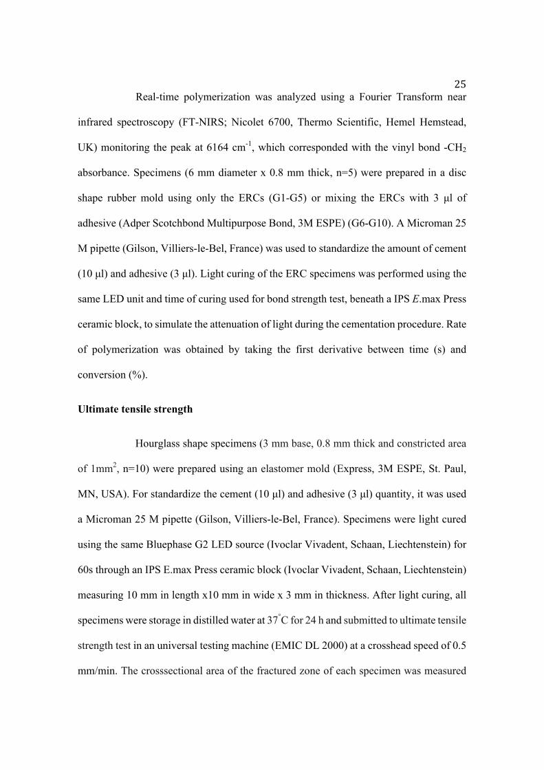
Failure pattern evaluation was performed for both times of microtensile bond
strength test (immediate and after 1 year of storage). In both times, predominantly, it can
be observed mixed failure (Figure 1 and 2) and almost none adhesive failure. Still, it can
be noticed similar performance of ERCs comparing both times.
Figure 1. Distribution (%) of failure pattern observed for ERCs after immediate microtensile bond strength.
29
Figure 2. Distribution (%) of failure pattern observed for ERCs after 1 year of water
storage.
Ultimate tensile strength
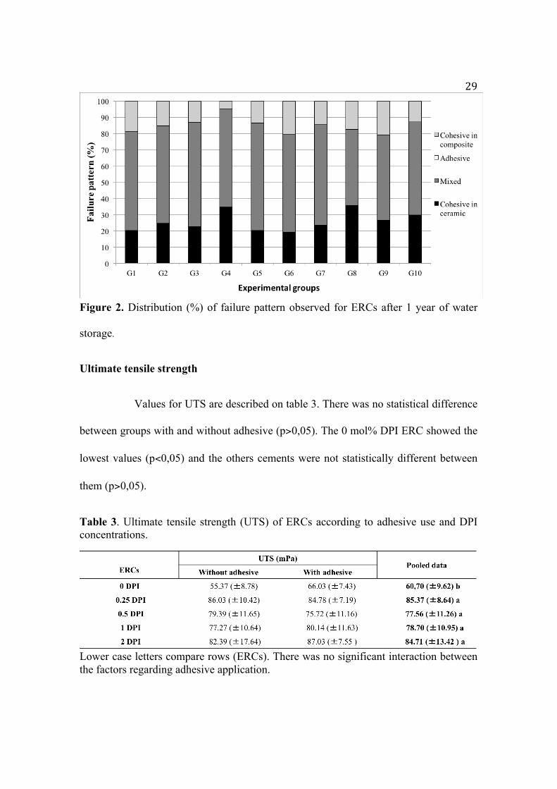
Values for UTS are described on table 3. There was no statistical difference
between groups with and without adhesive (p>0,05). The 0 mol% DPI ERC showed the
lowest values (p<0,05) and the others cements were not statistically different between
them (p>0,05).
Table 3. Ultimate tensile strength (UTS) of ERCs according to adhesive use and DPI concentrations.
Lower case letters compare rows (ERCs). There was no significant interaction between the factors regarding adhesive application.

30
Polymerization kinetics and Degree of conversion
Degree of conversion was significantly lower for ERCs without DPI
(p<0.0001; Table 4). The mixing of adhesive with the ERC without DPI promoted an
increase in DC values, statistically higher compared to ERC (0%DPI) without adhesive
addition (p<0.0001). For other ERCs, the adhesive did not influence DC values (p>0.05).
Observing the rate of polymerization (Figure 3), it could be noted that the ERC without
DPI presented inferior results, with best values observed when the adhesive was mixed
with cement. The cements containing DPI presented similar rate of polymerization
regardless the addition of adhesive.
Figure 3. Real time polymerization kinetics and rate of polymerization beneath a lithium disilicate ceramic (3mm thickness, MO1 shade) of the experimental groups.
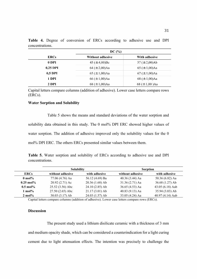
31 Table 4. Degree of conversion of ERCs according to adhesive use and DPI concentrations.
Capital letters compare columns (addition of adhesive). Lower case letters compare rows (ERCs).
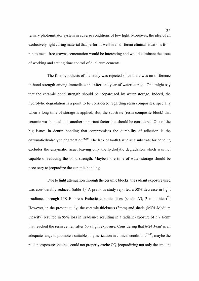
Water Sorption and Solubility
Table 5 shows the means and standard deviations of the water sorption and
solubility data obtained in this study. The 0 mol% DPI ERC showed higher values of
water sorption. The addition of adhesive improved only the solubility values for the 0
mol% DPI ERC. The others ERCs presented similar values between them.
Table 5. Water sorption and solubility of ERCs according to adhesive use and DPI concentrations.
Solubility Sorption ERCs without adhesive with adhesive without adhesive with adhesive
0 mol% 77.06 (4.76) Aa 56.12 (4.69) Ba 40.36 (3.44) Aa 50.36 (6.82) Aa 0.25 mol% 20.92 (2.71) Ac 20.36 (1.68) Ab 31.36 (2.71) Aa 36.68 (1.27) Ab 0.5 mol% 25.52 (3.56) Abc 24.10 (2.85) Ab 36.65 (4.53) Aa 43.05 (6.18) Aab 1 mol% 27.50 (2.65) Abc 21.17 (3.01) Ab 40.83 (9.13) Aa 35.94 (3.03) Ab 2 mol% 30.03 (3.17) Ab 24.03 (1.57) Ab 33.05 (4.24) Aa 40.97 (4.14) Aab
Capital letters compare columns (addition of adhesive). Lower case letters compare rows (ERCs).
Discussion
The present study used a lithium disilicate ceramic with a thickness of 3 mm
and medium opacity shade, which can be considered a counterindication for a light curing
cement due to light attenuation effects. The intention was precisely to challenge the
32ternary photoinitiator system in adverse conditions of low light. Moreover, the idea of an
exclusively light curing material that performs well in all different clinical situations from
pin to metal free crowns cementation would be interesting and would eliminate the issue
of working and setting time control of dual cure cements.
The first hypothesis of the study was rejected since there was no difference
in bond strength among immediate and after one year of water storage. One might say
that the ceramic bond strength should be jeopardized by water storage. Indeed, the
hydrolytic degradation is a point to be considered regarding resin composites, specially
when a long time of storage is applied. But, the substrate (resin composite block) that
ceramic was bonded to is another important factor that should be considered. One of the
big issues in dentin bonding that compromises the durability of adhesion is the
enzymatic/hydrolytic degradation38,39. The lack of tooth tissue as a substrate for bonding
excludes the enzymatic issue, leaving only the hydrolytic degradation which was not
capable of reducing the bond strength. Maybe more time of water storage should be
necessary to jeopardize the ceramic bonding.
Due to light attenuation through the ceramic blocks, the radiant exposure used
was considerably reduced (table 1). A previous study reported a 58% decrease in light
irradiance through IPS Empress Esthetic ceramic discs (shade A3, 2 mm thick)32.
However, in the present study, the ceramic thickness (3mm) and shade (MO1-Medium
Opacity) resulted in 95% loss in irradiance resulting in a radiant exposure of 3.7 J/cm2
that reached the resin cement after 60 s light exposure. Considering that 6-24 J/cm2 is an
adequate range to promote a suitable polymerization in clinical conditions33-35, maybe the
radiant exposure obtained could not properly excite CQ, jeopardizing not only the amount

33of photons reaching the composite but also the supply of electrons needed to interact with
DPI and create the additional free radicals. However, if we consider the size of a cement
film between 50-100 µm42, it might be considered that the low radiant exposure (3.7
J/cm2) was sufficient to polymerize the composite and allow suitable bond strength, even
in the control group (0 mol% DPI).
It is known that adequate polymerization and degree of conversion are
required to obtain good mechanical properties43. However, there are no reports in the
literature showing a linear correlation between degree of conversion and ceramic bond
strength, or minimum values of degree of conversion required to obtain suitable bond
strength. This is due to the fact that ceramic bonding depends not only on degree of
conversion but also on other factors such as surface treatment, cementation protocol and
chemical composition of ceramics44,45. Moreover, some studies have shown that,
depending on the material and condition, surface treatment of ceramic plays a more
important role than the chemical composition of resin cement46,47. According to the
results of the present study, it is possible to obtain a stable ceramic/resin composite
bonding against hydrolytic degradation challenge, even with a low conversion degree
(46% of the control group).
Further, the distribution of failure modes suggests similar bond quality even
without the use of adhesive, which was not able to improve the bond strength values. The
results of bond strength test corroborate with the values of failure pattern analysis, where
all groups showed similar performance regarding the amount and type of failure in both
times (immediate and after one year of storage). The same can be considered for groups
without adhesive (G1-G5) and with adhesive (G6-G10), where there was no difference in
34performance either, leading to believe that use of the adhesive was not able to promote
better wettabilty of ceramic surface, nor better bond strength values. Probably the
monomeric composition (BisGMA / TEGDMA ratio 1: 1) added to the amount of
inorganic filler (60% by mass) gave the CREs a viscosity capable of promoting good
surface wetting. Addition of an adhesive was able to improve DC only for the control
group (0mol% DPI), while for the other cements (containing DPI salt) it had no effect.
This result leads to rejection of our second hypothesis. Possibly the adhesive reduced the
viscosity of the system (adhesive + resin cement), resulting in higher mobility of the
reacting chains, increasing DC of the cement. This effect was not observed in the ERCs
containing DPI, possibly due to the higher reactivity already established of this system.
The third hypothesis was also rejected once DPI salt improved
physicochemical properties of ERCs, but it did not improve ceramic bond strength.
Physicochemical properties of ERCs with same composition of materials tested in the
present study were demonstrated in previous studies26,28. The addition of DPI salt was
able to reduce the sorption and solubility of the cements, as well as increase the flexural
strength and modulus of experimental materials tested26. The same effect of DPI salt on
the physicochemical properties could be observed in this study. The DC for all ERCs
containing DPI were similar and significantly higher compared with the control (without
DPI). Increased reactivity and improved polymerization of methacrylate-based dental
materials as consequence of DPI addition associated to camphorquinone has been
reported in previous studies26, 27. The salt intercepts the ketone radical anion, preventing
back electron transfer from occurring and then irreversibly decomposes to an aryl radical
and an aryl iodide, providing more free radicals and improving the curing of the

35CQ/amine system27, 31. If the DC of ERCs improve, the others properties will also get
better as a consequence, once physical/mechanical properties depends directly on DC of
composites18-20,36,37. This could be observed in this study for water sorption and solubility
and ultimate tensile strength.
It was obtained no difference on bond strength values of light cured ERCs to
overlying ceramic, despite low irradiance, low radiant exposure, ceramic opacity and
thickness, and water storage. It is important to highlight that the present results were
obtained “in vitro” and using experimental materials, which might not be fully
extrapolated to the clinical situation. Further aspects such as more time of water storage,
different concentration and composition of initiator systems as well as use of dentin as a
substrate for bonding warrant future investigation in order to improve the properties of
resin cement materials.
Conclusion
According to obtained results, it can be concluded that:
• One year of water storage was not sufficient to decrease ceramic bond strength of light
curing ERCs.
• Adhesive addition did not influence bond strength to ceramic, neither physicochemical
properties for ERCs with DPI salt, but it was able to increase DC only for no DPI ERC.
• DPI salt is capable of increasing rate of polymerization and DC of light curing ERCs even
when cured beneath thick lithium disilicate ceramic, although it did not affect ceramic
bond strength.

36Acknowledgments
The authors are grateful for grants (2014/02898-7) provided by FAPESP. It was used by
the corresponding author in partial fulfillment of the requirements to obtain the degree of
Doctor in Clinical Dentistry (PhD) and as part of his PhD thesis.
References
1. Borges GA, Sophr AM, de Goes MF, Sobrinho LC, Chan DC. Effect of etching and airborne particle abrasion on the microstructure of different dental ceramics. J Prosthet Dent. 2003 May; 89(5):479-88. 2. Kukiattrakoon B, Thammasitboon K. The effect of different etching times of acidulated phosphate fluoride gel on the shear bond strength of high-leucite ceramics bonded to composite resin. J Prosthet Dent. 2007 Jul; 98(1):17-23. 3. Zogheib LV, Bona AD, Kimpara ET, McCabe JF. Effect of hydrofluoric acid etching duration on the roughness and flexural strength of a lithium disilicate-based glass ceramic. Braz Dent J. 2011 22(1):45-50. 4. Oh WS, Shen C, Alegre B, Anusavice KJ. Wetting characteristic of ceramic to water and adhesive resin. J Prosthet Dent. 2002 Dec; 88(6):616-21. 5. Naves LZ, Soares CJ, Moraes RR, Goncalves LS, Sinhoreti MA, Correr-Sobrinho L. Surface/interface morphology and bond strength to glass ceramic etched for different periods. Oper Dent. 2010 Jul-Aug; 35(4):420-7. 6. Addison O, Marquis PM, Fleming GJ. Adhesive luting of all-ceramic restorations--the impact of cementation variables and short-term water storage on the strength of a feldspathic dental ceramic. J Adhes Dent. 2008 Aug; 10(4):285-93. 7. Burke FJ, Fleming GJ, Nathanson D, Marquis PM. Are adhesive technologies needed to support ceramics? An assessment of the current evidence. J Adhes Dent. 2002 Spring; 4(1):7-22. 8. Fleming GJ, Hooi P, Addison O. The influence of resin flexural modulus on the magnitude of ceramic strengthening. Dent Mater. 2012 Jul; 28(7):769-76. 9. Guazzato M, Albakry M, Ringer SP, Swain MV. Strength, fracture toughness and microstructure of a selection of all-ceramic materials. Part I. Pressable and alumina glass-infiltrated ceramics. Dent Mater. 2004 Jun; 20(5):441-8. 10. Isgro G, Addison O, Fleming GJ. The deformation and strength of a dental ceramic following resin-cement coating. J Dent. 2011 Feb; 39(2):122-7. 11. Baena E, Fuentes MV, Garrido MA, Rodriguez J, Ceballos L. Influence of post-cure time on the microhardness of self-adhesive resin cements inside the root canal. Oper Dent. 2012 Sep-Oct; 37(5):548-56. 12. Pedreira AP, Pegoraro LF, de Goes MF, Pegoraro TA, Carvalho RM. Microhardness of resin cements in the intraradicular environment: effects of water storage and softening treament. Dent Mater. 2009 Jul; 25(7):868-76.

3713. Good ML, Orr JF, Mitchell CA. In vitro study of mean loads and modes of failure of all-ceramic crowns cemented with light-cured or dual-cured luting cement, after 1 and 30 d of storage. Eur J Oral Sci. 2008 Feb; 116(1):83-8. 14. Hekimoglu C, Anil N, Etikan I. Effect of accelerated aging on the color stability of cemented laminate veneers. Int J Prosthodont. 2000 Jan-Feb; 13(1):29-33. 15. Arrais CA, Giannini M, Rueggeberg FA. Effect of sodium sulfinate salts on the polymerization characteristics of dual-cured resin cement systems exposed to attenuated light-activation. J Dent. 2009 Mar; 37(3):219-27. 16. Moraes RR, Brandt WC, Naves LZ, Correr-Sobrinho L, Piva E. Light- and time-dependent polymerization of dual-cured resin luting agent beneath ceramic. Acta Odontol Scand. 2008 Oct; 66(5):257-61. 17. Pick B, Gonzaga CC, Junior WS, Kawano Y, Braga RR, Cardoso PE. Influence of curing light attenuation caused by aesthetic indirect restorative materials on resin cement polymerization. Eur J Dent. 2010 Jul; 4(3):314-23. 18. Ferracane JL. Hygroscopic and hydrolytic effects in dental polymer networks. Dent Mater. 2006 Mar; 22(3):211-22. 19. Ruttermann S, Dluzhevskaya I, Grosssteinbeck C, Raab WH, Janda R. Impact of replacing Bis-GMA and TEGDMA by other commercially available monomers on the properties of resin-based composites. Dent Mater. 2010 Apr; 26(4):353-9. 20. Watts DC. Reaction kinetics and mechanics in photo-polymerised networks. Dent Mater. 2005 Jan; 21(1):27-35. 21. Prieto LT, Souza EJ, Jr., Araujo CT, Lima AF, Dias CT, Paulillo LA. Knoop hardness and effectiveness of dual-cured luting systems and flowable resin to bond leucite-reinforced ceramic to enamel. J Prosthodont. 2013 Jan; 22(1):54-8. 22. Ghavam M, Amani-Tehran M, Saffarpour M. Effect of accelerated aging on the color and opacity of resin cements. Oper Dent. 2010 Nov-Dec; 35(6):605-9. 23. Kilinc E, Antonson SA, Hardigan PC, Kesercioglu A. Resin cement color stability and its influence on the final shade of all-ceramics. J Dent. 2011 Jul; 39 Suppl 1:e30-6. 24. Lu H, Powers JM. Color stability of resin cements after accelerated aging. Am J Dent. 2004 Oct; 17(5):354-8. 25. Runnacles P, Correr GM, Baratto Filho F, Gonzaga CC, Furuse AY. Degree of conversion of a resin cement light-cured through ceramic veneers of different thicknesses and types. Braz Dent J. 2014 Jan-Feb; 25(1):38-42. 26. Gonçalves LS, Moraes RR, Ogliari FA, Boaro L, Braga RR, Consani S. Improved polymerization efficiency of methacrylate-based cements containing an iodonium salt. Dent Mater. 2013 Dec; 29(12):1251-5. 27. Ogliari FA, Ely C, Petzhold CL, Demarco FF, Piva E. Onium salt improves the polymerization kinetics in an experimental dental adhesive resin. J Dent. 2007 Jul; 35(7):583-7. 28. Andrade KM, Palialol AR, Lancellotti AC, Aguiar FH, Watts DC, Gonçalves LS, et al. Effect of diphenyliodonium hexafluorphosphate on resin

38cements containing different concentrations of ethyl 4-(dimethylamino)benzoate and 2-(dimethylamino)ethyl methacrylate as co-initiators. Dent Mater. 2016 Jun; 32(6):749-55. 29. Dressano D, Palialol AR, Xavier TA, Braga RR, Oxman JD, Watts DC, et al. Effect of diphenyliodonium hexafluorophosphate on the physical and chemical properties of ethanolic solvated resins containing camphorquinone and 1-phenyl-1,2-propanedione sensitizers as initiators. Dent Mater. 2016 Jun; 32(6):756-64. 30. Song L, Ye Q, Ge X, Misra A, Spencer P. Tris(trimethylsilyl)silane as a co-initiator for dental adhesive: Photo-polymerization kinetics and dynamic mechanical property. Dent Mater. 2016 Jan; 32(1):102-13. 31. Guo X, Wang Y, Spencer P, Ye Q, Yao X. Effects of water content and initiator composition on photopolymerization of a model BisGMA/HEMA resin. Dent Mater. 2008 Jun; 24(6):824-31. 32. Moraes RR, Correr-Sobrinho L, Sinhoreti MA, Puppin-Rontani RM, Ogliari FA, Piva E. Light-activation of resin cement through ceramic: relationship between irradiance intensity and bond strength to dentin. J Biomed Mater Res B Appl Biomater. 2008 Apr; 85(1):160-5. 33. Cunha LG, Alonso RC, Correr GM, Brandt WC, Correr-Sobrinho L, Sinhoreti MA. Effect of different photoactivation methods on the bond strength of composite resin restorations by push-out test. Quintessence Int. 2008 Mar; 39(3):243-9. 34. Lohbauer U, Rahiotis C, Kramer N, Petschelt A, Eliades G. The effect of different light-curing units on fatigue behavior and degree of conversion of a resin composite. Dent Mater. 2005 Jul; 21(7):608-15. 35. Seth S, Lee CJ, Ayer CD. Effect of instruction on dental students’ ability to light-cure a simulated restoration. J Can Dent Assoc. 2012 78:c123. 36. Lovell LG, Lu H, Elliott JE, et al: The effect of cure rate on the mechanical properties of dental resins. Dent Mater. 2001; 17(6): 504-11. 37. Elliott JE, Lovell LG, Bowman CN: Primary cyclization in the polymerization of bis-GMA and TEGDMA: a modeling approach to understanding the cure of dental resins. Dent Mater. 2001;17(3): 221-29. 38. Liu R, Fang M, Xiao Y, Li F, Yu L, Zhao S, Shen L, Chen J. The effect of transient proanthocyanidins preconditioning on the cross-linking and mechanical properties of demineralized dentin. J Mater Sci Mater Med 2011 Nov;22(11): 2403-1.
39. Hashimoto M, Ohno H, Sano H, Kaga M, Oguchi H. In vitro degradation of resin-dentin bonds analyzed by microtensile bond test, scanning and transmission electron microscopy. Biomaterials. 2003 Sep;24(21):3795-803.
40. Gresnigt M, Ozcan M. Esthetic rehabilitation of anterior teeth with porcelain laminates and sectional veneers. J Can Dent Assoc. 2011;77: b143. 41. Rigolin FJ, Miranda ME, Flório FM, Basting RT. Evaluation of bond strength between leucite-based and lithium disilicate-based ceramics to dentin after cementation with conventional and self-adhesive resin agents. Acta Odontol Latinoam. 2014;27(1):16-24. 42. Cekik-Nagas I, Canay S, Sahin E. Bonding of resin core materials to lithium disilicate ceramics: The effect of resin cement film thickness. Int J Prosthodontics.

392010; 23(5): 469-471. 43. Calheiros FC, Kawano Y, Stansbury JW, Braga RR. Influence of radiant exposure on contraction stress, degree of conversion and mechanical properties of resin composites; Dent Mater, 22 (2006), pp. 799–803. 44. Guarda GB, Correr AB, Gonçalves LS, Costa AR, Borges GA, Sinhoreti MAC, Correr-Sobrinho L. Effects of surface treatments, thermocycling, and cyclic loading on the bond strength of a resin cement bonded to a lithium disilicate glass ceramic. Oper Dent. 2013; 38(2): 208-17. 45. Sundfeld Neto D, Naves LZ, Costa AR, Correr AB, Consani S, Borges GA, et al. The Effect of Hydrofluoric Acid Concentration on the Bond Strength and Morphology of the Surface and Interface of Glass Ceramics to a Resin Cement. Oper Dent. 2015; 40(5): 470-479. 46. Aboushelib MN, Sleem D. Microtensile bond strength of lithium disilicate ceramics to resin adhesives. J Adhes Dent. 2014; 16(6): 547-52. 47. Lise DP, Perdigão J, Van Ende A, Zidan O, Lopes GC. Microshear bond strength of resin cements to lithium disilicate substrates as a function of surface preparation. Oper Dent. 2015; 40(5): 524-32.
3 CONCLUSÃO
De acordo com os resultados obtidos, pode-se concluir que:
• O tempo de armazenamento de um ano em água destilada não foi capaz de reduzir os
valores de resistência de união à cerâmica dos CREs fotoativados.
• O adesivo não exerceu influência na resistência de união à cerâmica, tampouco nas
propriedades físico-químicas dos CREs contendo sal de DFI, mas foi capaz de aumentar
o grau de conversão apenas para o CRE sem o sal de DFI.
• O sal de DFI foi capaz de aumentar a taxa de polimerização e o grau de conversão dos
CREs fotoativados, mesmo utilizando uma cerâmica vítrea de grande espessura como
barreira para luz. Porém, nenhuma concentração do sal exerceu efeito na resistência de
união à cerâmica, tanto imediata quanto após armazenamento em água.
* De acordo com as normas da UNICAMP/FOP, baseadas na padronização do International Committee of Medical Journal Editors – Vancouver Group. Abreviatura dos periódicos em conformidade com o PubMed.
40REFERÊNCIAS
Aboushelib MN, Sleem D. Microtensile bond strength of lithium disilicate
ceramics to resin adhesives. J Adhes Dent. 2014; 16(6): 547-52.
Aguiar TR, Andre CB, Arrais CAG, Bedran-Russo AK, Giannini M.
Micromorphology of resin-dentin interfaces using self-adhesive and conventional resin
cements: A confocal laser and scanning electron microscope analysis. Int J Adhesion &
Adhesives. 2012; 38: 69-74.
Anusavice KJ (1996) Philips Science of Dental Materials, ed. 10. Philadelphia:
Saunders.
Borges GA, Spohr AM, De Goes MF, Correr-Sobrinho L, Chan DNC. Effect of
etching particle abrasion in the microstructure of different dental ceramics. J Prost Dent.
2003; 89(5): 479-88.
Burke FJ, Flemming GI, Nathanson D, Marquis PM. Are adhesive technologies
needed to support ceramics? An assessment of the current evidence. J Aesthet Dent. 2002;
4: 7-22.
Cekik-Nagas I, Canay S, Sahin E. Bonding of resin core materials to lithium
disilicate ceramics: The effect of resin cement film thickness. Int J Prosthodontics. 2010;
23(5): 469-471.
Conrad HJ, Seong WJ, Pesun IJ. Current ceramic materials and systems with
clinical recommendations: a systematic review. J Prost Dent. 2007; 98(5): 389-404.
Crivello JV, Lam JH. Diaryliodonium salts - new class of photo-initiators for
cationic polymerization. Macromolecules 1977; 10: 1307-1315.
Crivello, J. V.; Dietliker, K. In Photoinitiators for Free Radical Cationic and
Anionic Photopolymerisation; Bradley, G., Ed.; Wiley: New York, 1998; Vol. 3, p 374.
Cunha LG, Alonso RCB, Correr GM, Brandt WC, Correr-Sobrinho L, Sinhoreti

41MAC. Effect of different photoactivation methods on the bond strength of
composite resin restorations by push-out test. Quintessence Int. 2008; 39(3): 243-249.
el-Mowafy OM, Rubo MH, el-Badrawy WA. Hardening of new resin cements
cured through a ceramic inlay. Oper Dent. 1999; 24: 38-44.
Ferracane JL. Hygroscopic and hydrolytic effects in dental polymer networks.
Dent Mater. 2006; 22: 211-222.
Gómez ML, Avila V, Montejano HA, Previtali CM. A mechanistic and laser flash
photolysis investigation of acrylamide polymerization photoinitiated by the three
component system safranine-T/triethanolamine/diphenyliodonium chloride. Polymer.
2003; 44: 2875-2881.
Gonçalves LS, Moraes RR, Ogliari FA, Boaro L, Braga RR, Consani S. Improved
polymerization efficiency of methacrylate-based cements containing an iodonium salt.
Dent Mater. 2013 Dec; 29(12):1251-1255.
Groten M, Probster L. The influence of different cementation modes on the
fracture resistance of feldspathic ceramic crowns. Int J Prosthodont 1997; 10:169-177.
Guarda GB, Correr AB, Gonçalves LS, Costa AR, Borges GA, Sinhoreti MAC,
Correr-Sobrinho L. Effects of surface treatments, thermocycling, and cyclic loading on
the bond strength of a resin cement bonded to a lithium disilicate glass ceramic. Oper
Dent. 2013; 38(2): 208-217.
Hashimoto M, Ohno H, Sano H, Kaga M, Oguchi H. In vitro degradation of resin-
dentin bonds analyzed by microtensile bond test, scanning and transmission electron
microscopy. Biomaterials. 2003 Sep;24(21):3795-803.
Höland W, Schweiger M, Frank M, Rheinberger V. A comparison of the
microstructure and properties of the IPS Empress 2 and the IPS Empress glass-ceramics.
J Biomed Mater Res. 2000; 53(4): 297-303.

42Kern M. Resin bonding to oxide ceramics for dental restorations. J Adhes Sci
Technol. 2009; 23(7-8):1097-111.
Kukiattrakoon B, Thammasitboon K. The effect of different etching times of
acidulated phosphate fluoride gel on the shear bond strength of high-leucite ceramics
bonded to composite resin. J Prosthet Dent. 2007; 98(1): 17-23.
Lee JH, Um CM, Lee IB. Rheological properties of resin composites according to
variations in monomer and filler composition. Dent Mater. 2006; 22: 515-526.
Lise DP, Perdigão J, Van Ende A, Zidan O, Lopes GC. Microshear bond strength
of resin cements to lithium disilicate substrates as a function of surface preparation. Oper
Dent. 2015; 40(5): 524-32.
Liu R, Fang M, Xiao Y, Li F, Yu L, Zhao S, Shen L, Chen J. The effect of transient
proanthocyanidins preconditioning on the cross-linking and mechanical properties of
demineralized dentin. J Mater Sci Mater Med 2011 Nov;22(11): 2403-1.
Lohbauer U, Rahiotis C, Kramer N, Petschelt A, Eliades G. The effect of different
light-curing units on fatigue behavior and degree of conversion of a resin composite. Dent
Mater. 2005; 21: 608-615.
Lu H, Powers JM. Color stability of resin cements after accelerated aging.
American J of Dent. 2004; 17: 354–358.
Moraes RR, Brandt WC, Naves LZ, Correr-Sobrinho L, Piva E. Light- and time-
dependent polymerization of dual-cured resin luting agent beneath ceramic. Acta Odontol
Scand. 2008; 66: 257-261.
Moraes RR, Correr-Sobrinho L, Sinhoreti MA, Puppin-Rontani RM, Ogliari FA,
Piva E. Light-activation of resin cement through ceramic: relationship between irradiance
intensity and bond strength to dentin. J Biomed Mater Res B Appl Biomater 2008;
85:160-165.
Naves LZ, Soares CJ, Moraes RR, Gonçalves LS, Sinhoreti MAC, Correr-

43Sobrinho L. Surface/Interface morphology and bond strength to glass ceramic etched for
different periods. Oper Dent. 2010; 35(4): 420-427.
Ogliari FA, Ely C, Petzhold CL, Demarco FF, Piva E. Onium salt improves the
polymerization kinetics in an experimental dental adhesive resin. J Dent. 2007; 35: 583-
587.
Oh WS, Shen C, Alegre B, Anusavice KJ. Wetting characteristic of ceramic to
water and adhesive resin. J Prosthet Dent. 2002; 88(6): 616-21.
Ozturk N, Usumez A, Usumez S, Ozturk B. Degree of conversion and surface
hardness of resin cement cured with different curing units. Quintessence Int. 2005; 36:
771-777.
Prieto LT, Souza-Junior EJ, Araújo CTP, Lima AF, Dias CTS, Paulillo LAMS.
Knoop hardness and effectiveness of dual –cured luting systems and flowable resin to
bond leucite-reinforced ceramic to enamel. J Prosthodont. 2013; 22: 54–58.
Rigolin FJ, Miranda ME, Flório FM, Basting RT. Evaluation of bond strength
between leucite-based and lithium disilicate-based ceramics to dentin after cementation
with conventional and self-adhesive resin agents. Acta Odontol Latinoam. 2014;27(1):16-
24.
Rosenstiel SF, Land MF, Crispin BJ. Dental luting agents: A review of the current
literature. J Prosthet Dent. 1998; 80: 280-301.
Ruttermann S, Dluzhevskaya I, Grosssteinbeck C, Raab WH, Janda R. Impact of
replacing Bis-GMA and TEGDMA by other commercially available monomers on the
properties of resin-based composites. Dent Mater. 2010; 26: 353-359.
Sakaguchi RL, Berge HX. Reduced light energy density decreases post-gel
contraction while maintaining degree of conversion in composites. J Dent. 1998; 26: 695-
700.
Santos-Daroz CB, Oliveira MT, Góes MF, Nikaido T, Tagami J, Giannini M.
Bond strength of a resin cement to dentin using the resin coating technique. Braz Oral

44Res. 2008; 22(3): 198-204.
Soares CJ, da Silva NR, Fonseca RB. Influence of the feldspathic ceramic
thickness and shade on the microhardness of dual resin cement. Oper Dent. 2006; 31:
384-389.
Sundfeld Neto D, Naves LZ, Costa AR, Correr AB, Consani S, Borges GA, et al.
The Effect of Hydrofluoric Acid Concentration on the Bond Strength and Morphology of
the Surface and Interface of Glass Ceramics to a Resin Cement. Oper Dent. 2015; 40(5):
470-479.
Tian T, Tsoi JK, Matinlinna JP, Burrow MF, Aspects of bonding between resin
luting cements and glass ceramic materials, Dent Mater. 2014; 30: 147-162.
Uctasli S, Hasanreisoglu U, Wilson HJ. The attenuation of radiation by porcelain
and its effect on polymerization of resin cements. J Oral Rehabil. 1994; 21: 565-575.
Watts DC. Reaction kinetics and mechanics in photo-polymerised networks. Dent
Mater. 2005; 21: 27-35.
Watts DC, Cash AJ. Analysis of optical transmission by 400-500 nm visible light
into aesthetic dental biomaterials. J Dent. 1994; 22: 112-117.
Yavuz T, Dilber E, Kara HB, Tuncdemir AR, Ozturk AN. Effects of different
surface treatments on shear bond strength in two different ceramic systems. Lasers Med
Sci. 2012; in press.
Zogheib LV, Bona AD, Kimpara ET, McCabe JF. Effect of hydrofluoric acid
etching duration on the roughness and flexural strength of a lithium disilicate-based glass
ceramic. Braz Dent J. 2011; 22(1): 45-50.

45APÊNDICE – METODOLOGIA ILUSTRADA
1 Preparo dos cimentos resinsos experimentias
Inicialmente, foi obtido um composto base a partir da mistura dos monômeros
2,2 - bis [4 - (2 – hidróxi - 3 - metilacriloxipropoxi) fenil] - propano (Bis-GMA) (Sigma-
Aldrich, Millwaukee, WI, EUA) e dimetacrilato de trietilenoglicol (TEGDMA) (Sigma-
Aldrich) na proporção 1:1 em massa (Figura 1, A e B). Ao composto base foi adicionado
como sistema fotoiniciador 1 mol% de canforoquinona (CQ) (Esstech Inc., Essington,
PA, EUA) e 2 mol% de metacrilato de dimetilaminoetil (DMAEMA) (Sigma-Aldrich)
(Figura 1 D). Como inibidor, foi acrescido à mistura uma quantidade de 0,1 mol% de
hidroxitolueno butilado (BHT) (Sigma-Aldrich). A partir do composto base, foram
preparados cinco cimentos experimentais, contendo crescentes concentrações de
hexafluorfosfato de difeniliodônio (DPIHFP - Sigma-Aldrich) (0; 0,25; 0,5; 1 e 2 mol%).
Posteriormente, foram adicionados 60% em peso de partículas silanizadas de vidro bário-
alumínio-silicato com diâmetro médio de 0,7 µm (Esstech Inc., Essington, Pensilvânia,
USA) (Figura 1, C e E).
D E
CBA

46Figura 1. Reagentes utilizados para confecção dos cimentos resinosos: (A) Bis-GMA;
(B) TEGDMA; (C) Sal de hexafluorofosfato de difeniliodônio; (D) DMAEMA; (E) Carga
de vidro de bário alumínio silicato.
2 Confecção dos espécimes de cerâmica
Setenta blocos da cerâmica vítrea IPS e.max Press (Ivoclar Vivadent, Schaan,
Liechtenstein) com dimensões de 10 mm x 10 mm x 3mm foram utilizados neste estudo.
Após o processo de fabricação das amostras segundo o fabricante, o revestimento
aglutinado por fosfato foi removido com auxílio de jato de óxido de alumínio de 50 µm
(Oxyker Dry; F.lli Manfredi, Sofia, Itália), sob pressão inicial de 4 bar, e posteriormente,
2 bar para remoção do revestimento próximo das amostras da cerâmica. O conduto de
alimentação foi removido com um disco diamantado (KG Sorensen, Cotia, SP, Brasil)
acoplado em peça reta em baixa rotação e os espécimes submetidos a acabamento com
ponta cilíndrica de diamante. (Figura 2).
Figura 2. (A) Jateamento com óxido de alumínio; (B) Remoção dos espécimes de
cerâmica; (C) Aspecto dos espécimes após acabamento.

473 Resistência de união
Para o ensaio de resistência de ligação, os blocos cerâmicos reforçados com
disilicato de lítio (10 mm de comprimento, 10 mm de largura, 3 mm de espessura, cor
MO1) foram condicionados com ácido fluorídrico a 10% (Dentsply, Petrópolis, Brasil)
durante 20 s, lavados com jato de ar/água e secos. Em seguida, foi aplicado agente silano
(RelyX Ceramic Primer, 3M ESPE, St. Paul, MN, EUA) sobre a superfície cerâmica e
deixado em repouso por 1 min. Nos grupos G6 a G10, foi aplicado adesivo (Adper
Scotchbond Multi-Purpose-Bond, 3M ESPE, St. Paul, MN, EUA) após o silano e
fotoativado juntamente com o cimento. Os blocos cerâmicos foram cimentados aos
blocos de resina composta. A fotoativação foi realizada através da cerâmica durante 60 s
usando LED Bluephase G2 com irradiância de 1266 mW/cm2. Após a cimentação, os
espécimes foram armazenados em água destilada a 37 ° C durante 24 h. Feito isso, os
blocos foram seccionados perpendicularmente à interface de união cerâmica/cimento com
disco diamantado em baixa velocidade (Isomet, Buehler Ltd., Lake Bluff, IL, EUA)
(figura 3, A e B). Metade dos palitos obtidos de cada bloco foram submetidos ao ensaio
imediato de microtração realizada em máquina de ensaio universal (EMIC DL 2000, São
José dos Pinhais, PR, Brasil) com velocidade de 0,5 mm/min (figura 4, A e B). A outra
metade foi submetida ao processo de armazenamento em água destilada por um ano e
subsequente teste de microtração.
Figura 3. (A) Amostra fixada em placa de acrílico para realização dos cortes na máquina
ISOMET; (B) Cortes realizados perpendicularmente à interface de união para obtenção de palitos.

48
Figura 4. (A) Palito posicionado no dispositivo de microtração acoplado à máquina de ensaio
universal EMIC DL 2000; (B) Palito logo após a fratura com a interface de união livre de
cianoacrilato.
4 Cinética e polimerização e grau de conversão
A cinética de polimerização polimerização e grau de conversão final foram
realizados em espectrômetro de infravermelho transformação de Fourier (FT-NIRS,
Nicolet 6700, Thermo Scientific, Hemel Hemstead, UK) (figura 5, A). Os espécimes (6
mm de diâmetro x 0,8 mm de espessura, n=5) foram preparados em molde de borracha
com formato de disco utilizando apenas os cimentos (G1-G5) ou misturando-os com 3 µl
de adesivo (Adper Scotchbond Multipurpose Bond, 3M ESPE) (G6-G10). Utilizou-se
uma pipeta Microman 25 M (Gilson, Villiers-le-Bel, França) para padronizar a
quantidade de cimento (10 µl) e adesivo (3 µl). Para reproduzir a atenuação da luz durante
o processo de cimentação, foi realizado a fotopolimerização dos espécimes utilizando a
mesma fonte de LED e tempo de fotoativação utilizados para o teste de resistência de
união, através de um bloco de cerâmica IPS E.max Press (figura 5, B).
49
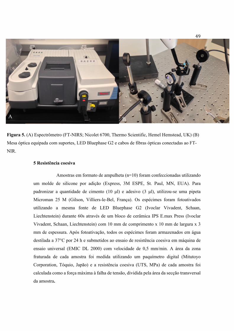
Figura 5. (A) Espectrômetro (FT-NIRS; Nicolet 6700, Thermo Scientific, Hemel Hemstead, UK) (B)
Mesa óptica equipada com suportes, LED Bluephase G2 e cabos de fibras ópticas conectadas ao FT-
NIR.
5 Resistência coesiva
Amostras em formato de ampulheta (n=10) foram confeccionadas utilizando
um molde de silicone por adição (Express, 3M ESPE, St. Paul, MN, EUA). Para
padronizar a quantidade de cimento (10 µl) e adesivo (3 µl), utilizou-se uma pipeta
Microman 25 M (Gilson, Villiers-le-Bel, França). Os espécimes foram fotoativados
utilizando a mesma fonte de LED Bluephase G2 (Ivoclar Vivadent, Schaan,
Liechtenstein) durante 60s através de um bloco de cerâmica IPS E.max Press (Ivoclar
Vivadent, Schaan, Liechtenstein) com 10 mm de comprimento x 10 mm de largura x 3
mm de espessura. Após fotoativação, todos os espécimes foram armazenados em água
destilada a 37°C por 24 h e submetidos ao ensaio de resistência coesiva em máquina de
ensaio universal (EMIC DL 2000) com velocidade de 0,5 mm/min. A área da zona
fraturada de cada amostra foi medida utilizando um paquímetro digital (Mitutoyo
Corporation, Tóquio, Japão) e a resistência coesiva (UTS, MPa) de cada amostra foi
calculada como a força máxima à falha de tensão, dividida pela área da secção transversal
da amostra.

50
Figura 6. (A) Moldes de silicone por adição das amostras em formato de ampulheta;
(B) Amostra posicionada na máquina de ensaio para realização do teste de resistência
coesiva; (C) Amostra logo após falha de tensão reproduzida pelo teste.
6 Sorção e solubilidade em água
O teste de sorção e solubilidade foi realizado de acordo com o estudo de
Gonçalves e colaboradores (2013), exceto pelas dimensões das amostras e procedimento
de fotoativação. Foram confeccionadas, a partir de matrizes de silicone por adição,
amostras em forma de disco (n=5) medindo 6 mm de diâmetro e 0,5 mm de espessura.
Após fotoativadas durante 60s através de um bloco de cerâmica IPS E.max
Press (Ivoclar Vivadent, Schaan, Liechtenstein), as amostras foram armazenados em
tubos de plástico secos por 24 h a 37°C. Em seguida, foram submetidas ao dessecador
(figura 7 C) contendo sílica gel e pesadas diariamente em balança analítica digital
(Discovery DV215CD; Ohaus Corporation, Pine Brook, NJ, USA) (figura 7 B) com
precisão de 0,01 mg até a obtenção de massa constante (m1). A espessura e o diâmetro
iniciais foram medidos em quatro pontos equidistantes utilizando um paquímetro digital
(Mitutoyo, Tóquio, Japão) para o cálculo do volume das amostras em mm3 no final do
teste. Após a estabilização da massa m1, as amostras foram imersas em 1,5 ml de água
destilada (figura 7 A) e armazenadas em estufa a 37°C por 21 dias, sendo a água trocada
a a cada quinze dias. Após 21 dias, as amostras foram levemente secadas com papel
absorvente e foram pesadas para obtenção da massa após imersão em água (m2). Em
seguida, as amostras foram novamente levadas ao dessecador, a 37°C, para novo processo
de pesagens consecutivas até se obter o valor da massa constante final (m3). Os valores

51de sorção e solubilidade foram calculados em µg/mm3, através das seguintes fórmulas:
sorção = m2-m3(µg) / V(mm3); solubilidade = m1-m3(µg) / V(mm3).
Figura 7. (A) Amostra em forma de disco inserida em água destilada; (B) Balança
analítica digital utilizada durante as três fases de pesagem das amostras; (C) Dessecador
contendo sílica gel para remoção da umidade das amostras.
A B C

52ANEXO
Comprovante de submissão do artigo
De: [email protected]: Submission Confirmation
Data: January 22, 2017 at 20:58Para: [email protected]
Dear Mr. Alan Palialol,
We have received your article "Influence of adhesive system on physicochemical properties and ceramic bond strength of experimental resincements containing ternary photoinitiator system" for consideration for publication in The Journal of Prosthetic Dentistry.
Your manuscript will be given a reference number once an editor has been assigned.
To track the status of your paper, please do the following:
1. Go to this URL: http://ees.elsevier.com/jpd/
2. Enter the login details.Your username is: [email protected] you need to retrieve password details, please go to: http://ees.elsevier.com/JPD/automail_query.asp.
3. Click [Author Login]This takes you to the Author Main Menu.
4. Click [Submissions Being Processed]
Thank you for submitting your work to this journal.
Kind regards,
Elsevier Editorial SystemThe Journal of Prosthetic Dentistry
******************************************Please note that the editorial process varies considerably from journal to journal. To view a sample editorial process, please click here:http://ees.elsevier.com/eeshelp/sample_editorial_process.pdf
For further assistance, please visit our customer support site at http://help.elsevier.com/app/answers/list/p/7923. Here you can search forsolutions on a range of topics, find answers to frequently asked questions and learn more about EES via interactive tutorials. You will also findour 24/7 support contact details should you need any further assistance from one of our customer support representatives.