Universidade de Lisboarepositorio.ul.pt/bitstream/10451/9456/1/ulfc105399_tm_Carolina... ·...
72
Universidade de Lisboa Faculdade de Ciências Departamento de Química e Bioquímica “Research of polyphenols with neuroprotective potential in a yeast model of degeneration” Carolina Emanuel Carreira Gomes Jardim Dissertação Mestrado em Bioquímica Bioquímica Médica 2012
Transcript of Universidade de Lisboarepositorio.ul.pt/bitstream/10451/9456/1/ulfc105399_tm_Carolina... ·...

Universidade de Lisboa
Faculdade de Ciências
Departamento de Química e Bioquímica
“Research of polyphenols with neuroprotective
potential in a yeast model of degeneration”
Carolina Emanuel Carreira Gomes Jardim
Dissertação
Mestrado em Bioquímica
Bioquímica Médica
2012
“Research of polyphenols with neuroprotective potential in a yeast model of degeneration”
ii
Universidade de Lisboa
Faculdade de Ciências
Departamento de Química e Bioquímica
“Research of polyphenols with neuroprotective
potential in a yeast model of degeneration”
Carolina Emanuel Carreira Gomes Jardim
Dissertação orientada por: Doutora Cláudia Nunes dos Santos, iBET
Profª Doutora Ana Ponces Freire, FCUL
Mestrado em Bioquímica
Bioquímica Médica
2012
“Research of polyphenols with neuroprotective potential in a yeast model of degeneration”
iii
O trabalho desenvolvido na presente tese decorreu no âmbito do projecto “Polifenoís como agentes
protectores em modelos celulares de alfa-sinucleinopatias, em particular na doença de Parkinson”,
financiado pela FCT (PTDC/BIA-BCM/111617/2009)

“Research of polyphenols with neuroprotective potential in a yeast model of degeneration”
iv
Acknowledgements
I would like to acknowledge to everyone that one way or another helped me to conclude this
stage of my life, contributing for the “scientist” that I am today.
First of all to Cláudia Nunes dos Santos, PhD, my supervisor, a many thanks for all the
moments in which you were capable of simplify my ideas during brain storming and for the patient and
support during this work. It makes me realize how lucky I was to being part of this group.
My sincerely gratitude to Professor Ricardo Boavida Ferreira for allowing me to developed this
work on his laboratory and for all the knowledge transmitted during this year.
I cannot forget all my Disease and Stress Biology laboratory colleagues, which made my lost
experiences easier to face. Without them would not be possible to finish this work. Thank you for all
the time spent discussing results, for all the encouraging and support. In special I want to
acknowledge to Diana Macedo for introducing me to “the yeast world” giving me all the important
teachings, to Inês Figueira for helping me with the industrial quantity of samples for GSH/GSSG and
to Rui Pimpão for providing me photographs of Corema album. I would also like to thank to Regina for
being always available for spent some time clarifying scientific questions that were really important for
me to do my work better every day.
To Professor Ana Ponces Freire for having accepted to be my supervisor and for support the
development of my work.
At last but also important I want to thank to my family. To my parents that deserve the biggest
thanks of all since that without them is completely impossible to keep studying and reaching my
objectives in live. THANK YOU for EVERYTHING!! To Pedro Benevides that always remember me
that “no one told that is easy to do an MsC thesis” and for his capacity of makes me furious saying
“don’t you have work to do?”. He never let me give up. Finally to my cousin Sofia for providing “free
nights”.
“Research of polyphenols with neuroprotective potential in a yeast model of degeneration”
v
Abstract
Polyphenols are the most abundant phytochemicals in the human diet. It is known that the
consumption of polyphenol-rich foods, especially fruits and vegetables, translates into benefits for
human health. Polyphenols besides to be free radical scavengers, are now considered to invoke a
spectrum of cellular mechanisms of action, such as induction of endogenous antioxidants; modulation
of genes related to cell survival/death; cell signaling pathway regulatory activity and regulation of the
mitochondrial function. Increasing production of free radicals is one of the major causes of
degeneration that can occur via redox-cycling, metabolism of xenobiotics, ageing, environmental
toxins and mutant proteins. Agents that prevent these phenomena could be of particular therapeutic
interest.
In this study a yeast model of degeneration composed by four Saccharomyces cerevisiae
strains with different sensitivities to oxidative stress, was implemented. Extracts from fruits and leaves
of Corema album and Arbutus unedo were submitted to an in vitro procedure that mimics the gastro-
intestinal digestion to obtain the Polyphenol-digested fractions (PDFs). After chemical characterization
and toxicity, the PDFs protective effect was evaluated in the yeast model of degeneration. Protective
effects were observed for yap1 and sod1 strains due to the treatment with C. album fruit and A.
unedo fruit and leaf PDFs. At the same time alterations on GSH/GSSG ratio for these two strains were
also observed in the presence of the PDFs with protective effects. However, it was not observed an
effect in the production of reactive oxygen species (ROS) mediated by the tested PDFs, indicating that
ROS scavenging is not the mechanism of action of PDFs.
This work provided valuable information about possible mechanisms of protection of
polyphenols after gastrointestinal digestion that will be further evaluated in a yeast model of
Parkinson’s disease already implemented in DSB laboratory to provide more insights.
Keywords: Ageing, ROS, Degeneration, S. cerevisiae, Polyphenol-digested fractions.
“Research of polyphenols with neuroprotective potential in a yeast model of degeneration”
vi
Resumo
Os polifenóis são os fitoquímicos mais abundantes na dieta humana. O consumo de
alimentos ricos nestes compostos, principalmente frutas e vegetais, tem sido cada vez mais
associado a efeitos benéficos para a saúde humana. Além disso, estudos indicam que há uma forte
associação entre o consumo de alimentos ricos em polifenóis e a diminuição da incidência de
doenças degenerativas. Os polifenóis são compostos conhecidos como sendo capazes de eliminar
espécies reactivas de oxigénio, no entanto em estudos mais recentemente tem-se vindo a constatar
que estes compostos estão também envolvidos num largo espectro de mecanismos de acção
celulares, como é o caso da activação de moléculas antioxidantes endógenas, quelação de iões,
modulação de genes relacionados com a morte/sobrevivência celular, regulação da activação de
genes/proteínas de vias de sinalização celular e ainda na regulação da função mitocondrial.
As reacções redox, que ocorrem no interior das células, estão na base de numerosas vias
metabólicas importantes na homeostasia celular. O aumento na produção celular de espécies
reactivas de oxigénio, ou de azoto, é uma das principais causas associadas ao desenvolvimento de
doenças degenerativas. Este aumento pode ocorrer devido a vários tipos de insultos celulares como o
metabolismo de xenobióticos, toxinas ambientais, proteínas mutantes tóxicas para a célula e mais
importante, devido a processos de envelhecimento.
A levedura Saccharomyces cerevisiae é um dos modelos biológicos mais versáteis para
estudar as doenças degenerativas, devido ao facto dos mecanismos relacionados com a disfunção
mitocondrial ou proteossomal e a desregulação transcricional serem extremamente bem conservados
entre leveduras e humanos, permitindo estudar os mecanismos envolvidos neste tipo de doenças.
Para além disso, os mecanismos inerentes à produção de espécies reactivas de oxigénio e de
apoptose são reproduzidos com sucesso em levedura.
Neste estudo o principal objectivo consiste em avaliar o potencial protector de metabolitos
provenientes da digestão de frutos e folhas de Corema album e Arbutus unedo num modelo de
degeneração em levedura optimizado durante este trabalho. Estudos preliminares do laboratório
Biologia da Doença e do Stresse (resultados não publicados) demostraram que as fracções totais não
digeridas destas duas plantas apresentam elevada capacidade antioxidante e actividade protectora
de células submetidas a stresse.
Para atingir os objectivos propostos para este trabalho foi inicialmente optimizado um modelo
de degeneração em levedura induzido pelo H2O2. Este modelo é composto por 4 estirpes de S.
cerevisiae (BY4714, yap1, sod1 e AD1-8), que apresentam diferentes sensibilidades ao stress
oxidativo. Numa primeira fase foram definidos os tempos de geração (Tg) para cada estirpe, sendo de
2 h para BY4741 e yap1 e de 3 h para sod1 e AD1-8 e as concentrações de H2O2 a usar tanto em
meio sólido como em meio líquido. Em meio solido foram escolhidas as concentrações de 0.15 mM
para yap1 e sod1 e 0.5 mM para BY4741 e AD1-8, para incubações prolongadas. Em meio líquido
foi escolhida a concentração de 2 mM de H2O2 e o Tg de cada estirpe como tempo de incubação. Os
resultados obtidos para as curvas de crescimento indicam que baixas concentrações de H2O2 na
célula podem activar mecanismos de defesa e activação de genes que são importantes para o
crescimento e diferenciação celulares.
“Research of polyphenols with neuroprotective potential in a yeast model of degeneration”
vii
Os extractos hidroetanólicos provenientes de frutos e folhas de C. album e A. unedo foram
submetidos a um processo de digestão in vitro que mimetiza as alterações que ocorrem durante a
digestão gastro intestinal e a um passo final de limpeza, para obtenção das fracções digeridas de
polifenóis (FDPs). Paralelamente foram também digeridos os extractos totais de Rubus ideaus fruto e
Ginkgo biloba folha para fins comparativos, visto que estas duas espécies estão descritas como tendo
capacidade de melhorar o desempenho celular constituindo benefícios para a saúde humana. Numa
segunda fase deste estudo foi efectuada a caracterização química dos extractos obtidos em cada
etapa do processo de digestão in vitro, tendo sido avaliado o conteúdo total em fenóis e a capacidade
antioxidante in vitro. Esta caracterização permitiu a compreensão das alterações e perdas que
ocorrem nos compostos desde que são ingeridos até ao final da digestão no intestino delgado. Em
particular neste estudo foram utilizadas as fracções IN, correspondente ao conteúdo que entra
passivamente na circulação sanguínea e OUT, que diz respeito ao conteúdo que é atinge o colón,
visto que podem ocorrer várias alterações químicas aos compostos.
A terceira tarefa permitiu avaliar a toxicidade inerente a cada FDPs por “spot assays”, tendo
sido escolhida a concentração máxima de 250 g GAE.mL-1
para a qual foi posteriormente testado o
seu efeito protector. Acima desta concentração a FDP de C. album folha já apresenta toxicidade.
Para testar o efeito protector destas fracções no modelo de degeneração em levedura, sob condições
de stresse oxidativo foram utilizadas duas metodologias distintas. Após seleccionadas as FDPs que
apresentam melhorias na viabilidade por spot assays, foi avaliado o estado metabólico geral das
células pelo ensaio do CellTiter-Blue viability assay. Em ambos os métodos, tanto para a yap1 como
para a sod1 foi observada uma capacidade protectora dos metabolitos digeridos do fruto de C.
album e do fruto e folha de A. unedo. Estes resultados permitem concluir que os mecanismos de
defesa celulares são activados ou modelados pelos compostos naturais presentes nestas espécies
como é o caso dos polifenóis. No caso da FDP dos frutos de A. unedo os resultados indicam uma
capacidade de activar mecanismos que permitem á célula defender-se mais eficazmente quando
submetida a um stresse oxidativo posteriormente.
Numa fase final do trabalho foi avaliado o estado redox da célula. A produção exacerbada de
espécies reactivas de oxigénio nas células submetidas está descrita como uma das condições de
maior importância no desenvolvimento de doenças degenerativas, foi avaliada a produção destas
espécies na presença e na ausência das FDPs, após as células serem submetias a um stresse
oxidativo. Estas espécies foram quantificadas pelo método de H2DCFDA e foi observado um aumento
na produção destas espécies quando estas eram incubadas na presença de H2O2. No entanto
contrariamente ao esperado não foram observadas quaisquer alterações na produção de espécies
reactivas de oxigénio, quando as células eram pré-incubadas com os FDPs. Estes resultados
contrariam alguns estudos que apontam a eliminação destas espécies como o mecanismo pelo qual
os polifenóis melhoram a viabilidade celular mas reforça a ideia que estes compostos possam actuar
por outros mecanismos de regulação envolvidos na defesa celular.
Outro parâmetro analisado foi o conteúdo em tióis totais por HPLC. Apenas foram observadas
diferenças nos níveis de GSH reduzido para as estirpes yap1 e sod1, quando submetidas ou não a
stresse oxidativo, não se tendo observado diferenças para a WT. O facto de não se observarem

“Research of polyphenols with neuroprotective potential in a yeast model of degeneration”
viii
diferenças para a WT dever-se-á ao facto de esta estirpe possuir todos os mecanismos antioxidantes
necessários para repor os níveis de GSH reduzido. Avaliou-se então a razão GSH/GSSG para as
estirpes mais afectadas, yap1 e sod1, na presença e na ausência das FDPs. A razão GSH/GSSG
é considerado um indicador do estado redox das células.
Na presença das FDPs com capacidade protectora, foi observada uma diminuição no estado
oxidado das células para a estirpe sod1. No entanto, para a estirpe yap1 apenas o fruto de A.
unedo apresenta capacidade de melhorar significativamente o estado redox da célula, quando esta é
submetida à condição de stresse oxidativo, sendo que ainda assim não é capaz de repor os níveis
normais de oxidação celular para o yap1.
Observou-se ainda através das metodologias aplicadas que a estirpe yap1 é a mais afetada
pelo stresse oxidativo, o que era espectável, uma vez que o factor de transcrição Yap1p para o qual
esta estirpe é mutante, regula vários enzimas antioxidantes como a SOD1 e SOD2. As metodologias
aplicadas neste trabalho parecem indicar que os polifenóis presentes nas fracções digeridas têm a
capacidade de afectar os mecanismos relacionados com sobrevivência e proliferação celular.
Este trabalho fornece informação importante acerca de possíveis mecanismos de protecção
dos polifenóis depois da digestão gastrointestinal, que serão avaliados posteriormente num modelo
de PD em levedura já implementado no laboratório da Biologia da Doença e do Stresse.
Palavras-chave: Envelhecimento, Especies Reactivas de Oxigénio, Degeneração, S.
cerevisiae, Fracções digeridas de polifenóis
“Research of polyphenols with neuroprotective potential in a yeast model of degeneration”
ix
General Index
Acknowledgements .........................................................................................................................iii
Abstract .............................................................................................................................................v
Resumo ............................................................................................................................................vi
General Index ...................................................................................................................................ix
Index of Figures ...............................................................................................................................xi
Index of Tables ................................................................................................................................ xii
Abbreviations ................................................................................................................................. xiii
1. Objectives ..................................................................................................................................1
2. Theoretical Fundaments ...........................................................................................................2
2.1. Degenerative diseases .......................................................................................................2
2.1.1. Oxidative stress and degeneration............................................................................3
2.2. Saccharomyces cerevisiae ................................................................................................5
2.2.1. Yeast as a model of degeneration ............................................................................5
2.2.2. Adaptive response against oxidative stress ..............................................................6
2.2.2.1. Yeast activator protein (Yap) ...................................................................................8
2.2.2.2. Glutathione defense system ....................................................................................9
2.2.2.3. Superoxide dismutase (SOD) ................................................................................ 10
2.3. Natural phytochemicals and human health .................................................................... 10
2.3.1. Polyphenols ........................................................................................................... 11
2.3.2. Berries ................................................................................................................... 11
2.3.3. Polyphenols bioavailability ...................................................................................... 14
3. Material and Methods .............................................................................................................. 15
3.1. Samples preparation ........................................................................................................ 15
3.1.1. Plant material ......................................................................................................... 15
3.1.2. Hydroethanolic extraction ....................................................................................... 16
3.1.3. In vitro digestion (IVD) ............................................................................................ 16
3.1.4. Solid Phase Extraction (SPE) ................................................................................. 16
3.2. Total phenol content ........................................................................................................ 17
3.3. Antioxidant capacity assay .............................................................................................. 17
3.4. Saccharomyces cerevisiae .............................................................................................. 18
3.4.1. Growth curves ........................................................................................................ 19
3.4.2. Spot Assay ............................................................................................................. 19
3.4.3. Resazurin-based assay .......................................................................................... 20

“Research of polyphenols with neuroprotective potential in a yeast model of degeneration”
x
3.4.4. Dichlorofluorescein assay ....................................................................................... 21
3.4.5. Glutathione determination ...................................................................................... 21
3.5. Statistical analysis ........................................................................................................... 22
4. Results and Discussion .......................................................................................................... 22
4.1. Oxidative stress in a yeast model of degeneration ........................................................ 22
4.1.1. Establishment of the yeast model of degeneration and optimization of methods ...... 22
4.2. Characterization of polyphenol-digested fractions (PDFs) ............................................ 30
4.2.1. Fruits...................................................................................................................... 32
4.2.2. Leaves ................................................................................................................... 35
4.3. Selection of polyphenol-digested fractions with protective potential ........................... 38
4.3.1. Viability analysis by Spot Assays ............................................................................ 38
4.3.2. CellTiter-Blue viability assay analysis ..................................................................... 41
4.4. Evaluation of cellular oxidative stress markers .............................................................. 44
4.4.1. Reactive Oxygen Species ...................................................................................... 44
4.4.2. Glutathione ............................................................................................................ 46
5. Final considerations and future perspectives ....................................................................... 49
6. References............................................................................................................................... 52

“Research of polyphenols with neuroprotective potential in a yeast model of degeneration”
xi
Index of Figures
Figure 1. Representation of the phenomena that lead to oxidative stress damage in ageing process. .2
Figure 2. Protein misfolding and ubiquitylation processes induced by an external stress .....................4
Figure 3. Sources of ROS and its influence in the mechanisms of apoptosis in S. cerevisiae ..............6
Figure 4. Cooperative action of the two main enzymatic systems of ROS modulation on S. cerevisiae,
to the mantainance of redox homeostasis ...........................................................................................9
Figure 5. Plants used in this work ..................................................................................................... 12
Figure 6. Bioavailability of polyphenols after alterations occurring during metabolization in the human
body ................................................................................................................................................. 14
Figure 7. Growth curve and growth parameters of the S. cerevisiae strains in presence of H2O2.. ..... 24
Figure 8. Spot assays of S. cerevisiae strains BY41741, yap1, sod1 and AD1-8 grown in the
presence of oxidative stress induced by H2O2.................................................................................... 27
Figure 9. Spot assays of S. cerevisiae strains BY41741, AD1-8, yap1 and sod1 grown in the
presence of a chronic oxidative stress induced by H2O2. ................................................................... 28
Figure 10. Total phenolic content for polyphenol extracts in each step of the IVD process................. 31
Figure 11. Percentage of the original of total phenolic content for polyphenol fruit extracts in each step
of the IVD process ............................................................................................................................ 33
Figure 12. Antioxidant properties of polyphenol fruit extracts in each step of the IVD process ........... 33
Figure 13. PDFs toxicity evaluation by spot assays for S. cerevisiae strains BY4741, yap1 and
sod1 exposure to PDFs from fruits .................................................................................................. 34
Figure 14. Percentage of the original of total phenolic content for polyphenol leaf extracts in each step
of the IVD process ............................................................................................................................ 36
Figure 15. Antioxidant properties of polyphenol leaf extracts in each step of the IVD process ........... 36
Figure 16. PDFs toxicity evaluation by spot assays for S. cerevisiae strains BY4741, yap1 and
sod1 exposure to PDFs from leaves ................................................................................................ 37
“Research of polyphenols with neuroprotective potential in a yeast model of degeneration”
xii
Figure 17. PDFs protective potential evaluation by spot assays for S. cerevisiae strains BY4741,
yap1 and sod1 exposure to PDFs and subjected to an oxidative stress. ........................................ 40
Figure 18. Percentage of viability of S. cerevisiae strains BY4741, yap1 and sod1 growth in the
presence of PDFs and subjected to an oxidative stress. .................................................................... 42
Figure 19. Evaluation of ROS levels in S. cerevisiae strains (BY4741, yap1 and sod1) growth in the
presence of PDFs and subjected to an oxidative stress. .................................................................... 45
Figure 20. GSH determination of S. cerevisiae strains (BY4741, yap1 and sod1) growth in
presence of an oxidative stress ......................................................................................................... 47
Figure 21. GSH/GSSG determination for yap1 and sod1 S. cerevisiae strains growth in presence of
PDFs and subsequently submitted to an oxidative stress................................................................... 47
Index of Tables
Table 1. Representation of the most important antioxidant defenses in S. cerevisiae relationated with
ROS detoxification ..............................................................................................................................7

“Research of polyphenols with neuroprotective potential in a yeast model of degeneration”
xiii
Abbreviations
OH - Hydroxyl radical
AAPH – 2,2’-azobis (2-amidopropane)
dihidrochloride
Ace1p – metallothionein protein
AD – Alzheimer´s disease
AHP1 – Alkyl hydroperoxide reductase
Aif1p – apoptosis induncing factor
ALS – Amyotrophic lateral sclerosis
Cat1p – Peroxisomal catalase enzyme
CCP1 – Cytochrome c peroxidase gene
Ccs1p – Copper chaperon protein
Crm1 – Yap1p exportin
CSM –Complete supplement mixture
Ctt1p – Cytosolic catalase
CUP1 – metallothionein (copper binding
protein) gene
DCF –Dichlorofluorescein
DMSO – Dimetyl sulfoxide
DSB Laboratory – Disease and Stress
Biology Laboratory
DT – Doubling time
EGb 761 – Ginkgo biloba leaf standard extract
ER – Endoplasmatic reticulum
F.U. – Fluorescence units
G-6-PDH – Glucose-6-Phosphate
Dehydrogenase
GAE – Gallic acid equivalents
GIT – Gastrointestinal tract
GLR1 – Glutathione reductase gene
Glr1p – Glutathione reductase
Gpxp – Glutathione peroxidase
Grxp – Glutaredoxins
GSH – Reduced Glutathione
GSH1 – -glutamylcysteine synthetase gene
GSH2 – Glutathione synthetase gene
GSSG – Oxidized glutathione
Gstp – Glutathione transferase
H2DCFDA – 2,7-dichlorofluorescein diacetate
HD – Huntington’s disease
IN – Serum available fraction obtain after IVD
ITQB – Instituto Tecnologia Química e
Biologica
IVD – In vitro digestion
MDR – Multidrug resistance
NADPH – Nicotinamide adenine dinucleotide
phosphate
Ndi1p – internal mitochondria NADH
dehydrogenase
NEM – N-ethyl maleimide
Nrf2p – Mammalian AP-1 transcriptional
factor.
OD 600 nm – Optical density at 600 nm
ONOO – Peroxynitrate
OPA – Orthophtaldehyde
ORAC – Oxygen radical absorbance capacity
OUT – Colon available fraction obtain after IVD
PBS – Phosphate buffer
PD – Parkinson´s disease
PDFs – Polyphenol-digested fractions
Prxp - Peroredoxins
R.F.U. – Relative Fluorescence units
RNS – Reactive nitrogen species
ROS – Reactive oxygen species
SC – Synthetic complete
Skn7p – Transcriptional factor Skn7
SOD – Superoxide dismutase
SOD1 – Cu/Zn Superoxide dismutase gene
Sod1p – Cu/Zn superoxide dismutase protein
SOD2 – Mn superoxide dismutase gene
Sod2p – Mn superoxide dismutase protein
SPE – Solid phase extraction
TE – Trolox equivalents
Tpxp – Thioredoxin peroxidase
Trr1p – Thioredoxin reductase
TRx – Thioredoxins
UPS – Ubiquitin-proteosome system
“Research of polyphenols with neuroprotective potential in a yeast model of degeneration”
xiv
WT – Wild-type
YAP1 – Yap1p gene
Yap1p – Yeast activactor protein 1
Yca1p – Yeast metacaspase
YPD – Yeast extract, peptone, dextrose
YRE – Yap1p recognition element
YCF1 – Glutathione S-conjugated pump, ATP
binding cassette tranporter gene.
“Research of polyphenols with neuroprotective potential in a yeast model of degeneration”
1
1. Objectives
In the developed world, besides the increase of population lifespan, the number of
degenerative diseases has also increased associated with the ageing process; this is the case of
cancer, cardiovascular and neurodegenerative diseases. It has been demonstrated that plants contain
many natural compounds with antioxidant properties, including polyphenols that have been intensively
studied as free radical scavengers. Moreover fruits and vegetables rich in polyphenols have been
studied as protecting agents in many of the age-related diseases.
S. cerevisiae is one of the most versatile biological systems used as a model for the study of
many diseases. In order to evaluate the role of polyphenols with neuroprotective potential from A.
unedo and C. album on the degeneration process, a yeast model was established and optimized for
this study.
The main goal of this work is evaluate the protective potential of polyphenols digested
metabolites from chosen plants, in a yeast model that mimics the degeneration process after an
oxidative insult. To accomplish this goal, 5 tasks were defined with intermediate milestones. The
defined tasks were (i) establishment of a yeast model of degeneration induced by H2O2 insult on 4
strains of S. cerevisiae (BY4741, yap1, sod1 and AD1-8), (ii) chemical characterization of in vitro
digested (IVD) fractions by total phenols content and in vitro antioxidant capacity, (iii) PDFs toxicity, in
the yeast cells, (iv) evaluation of the protective capacity of PDFs in the established yeast model of
degeneration, and (v) ultimately understanding the mechanisms of action by which the PDFs interfere
with the redox state of cells.
This study is a major contribute for degeneration as a preliminary stage of screening for
potential neuroprotective compounds present in plants, to be further tested in a yeast model of
Parkinson´s disease, already implemented in DSB laboratory.

“Research of polyphenols with neuroprotective potential in a yeast model of degeneration”
2
2. Theoretical Fundaments
2.1. Degenerative diseases
In the developed world, the population lifespan is increasing, with a concomitant increased
incidence of many age-related diseases, such as cancer, cardiovascular and neurodegenerative
disorders. However, in what concerns to neurodegeneration, the mechanisms involved in the
behavioural deficits during ageing remain not totally clear [1, 2]. The brain, due to its high metabolic
rate, low enzymatic activity, as well as the high proportion of polyunsaturated fatty acids, becomes a
tissue particularly susceptible to oxidative damage [3, 4].
Several studies have been supporting the influence of environmental and genetic factors in
degeneration processes, becoming these disorders difficult to study. Substantial evidence also
indicates that excessive production of reactive oxygen species (ROS) and reactive nitrogen species
(RNS) in cells plays an important role in the regulation of redox reactions, that in high concentrations
lead to an increase of lipid peroxidation, oxidative damage to DNA and proteins and decreased levels
of GSH. All this phenomena have as consequence the ageing process and finally degeneration. (Fig.
1) [1-3].
In chronic degenerative diseases, apoptosis is the predominant form of cell death. Several
pathways such as protein misfolding and aggregation, alterations of the endoplasmatic reticulum (ER)
and cytoskeleton, altered RNA metabolism, dysfunction of the ubiquitin proteasome system (UPS),
and mitochondrial dysfunction have a role in many of these disorders, leading to programmed cell
death (Fig. 1) [4-6].
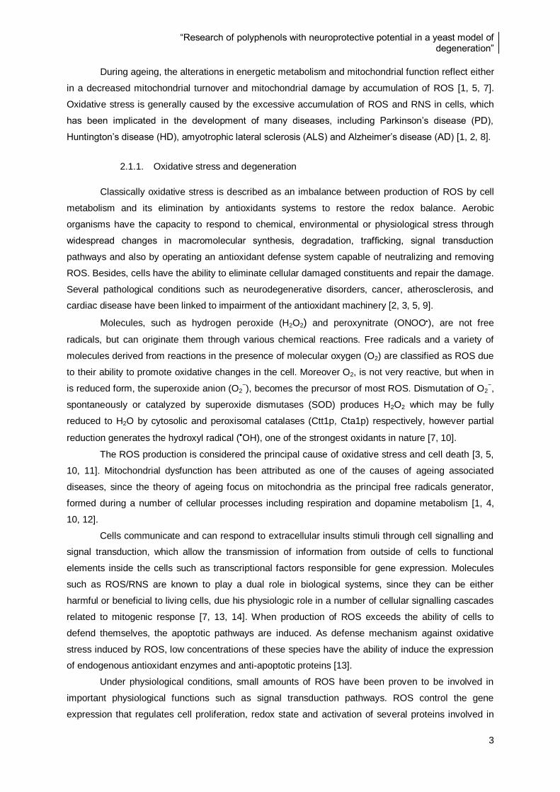
Figure 1. Representation of the phenomena that lead to oxidative stress damage in ageing process. Both genetic
alterations and environmental influences that leads to oxidative stress, characterized by ROS increase and cellular components
damage. The final consequence is the appearance of several diseases associated with ageing. Adapted from Uttara and co-
workers, 2009 [1].
“Research of polyphenols with neuroprotective potential in a yeast model of degeneration”
3
During ageing, the alterations in energetic metabolism and mitochondrial function reflect either
in a decreased mitochondrial turnover and mitochondrial damage by accumulation of ROS [1, 5, 7].
Oxidative stress is generally caused by the excessive accumulation of ROS and RNS in cells, which
has been implicated in the development of many diseases, including Parkinson’s disease (PD),
Huntington’s disease (HD), amyotrophic lateral sclerosis (ALS) and Alzheimer’s disease (AD) [1, 2, 8].
2.1.1. Oxidative stress and degeneration
Classically oxidative stress is described as an imbalance between production of ROS by cell
metabolism and its elimination by antioxidants systems to restore the redox balance. Aerobic
organisms have the capacity to respond to chemical, environmental or physiological stress through
widespread changes in macromolecular synthesis, degradation, trafficking, signal transduction
pathways and also by operating an antioxidant defense system capable of neutralizing and removing
ROS. Besides, cells have the ability to eliminate cellular damaged constituents and repair the damage.
Several pathological conditions such as neurodegenerative disorders, cancer, atherosclerosis, and
cardiac disease have been linked to impairment of the antioxidant machinery [2, 3, 5, 9].
Molecules, such as hydrogen peroxide (H2O2) and peroxynitrate (ONOO), are not free
radicals, but can originate them through various chemical reactions. Free radicals and a variety of
molecules derived from reactions in the presence of molecular oxygen (O2) are classified as ROS due
to their ability to promote oxidative changes in the cell. Moreover O2, is not very reactive, but when in
is reduced form, the superoxide anion (O2−), becomes the precursor of most ROS. Dismutation of O2
−,
spontaneously or catalyzed by superoxide dismutases (SOD) produces H2O2 which may be fully
reduced to H2O by cytosolic and peroxisomal catalases (Ctt1p, Cta1p) respectively, however partial
reduction generates the hydroxyl radical (OH), one of the strongest oxidants in nature [7, 10].
The ROS production is considered the principal cause of oxidative stress and cell death [3, 5,
10, 11]. Mitochondrial dysfunction has been attributed as one of the causes of ageing associated
diseases, since the theory of ageing focus on mitochondria as the principal free radicals generator,
formed during a number of cellular processes including respiration and dopamine metabolism [1, 4,
10, 12].
Cells communicate and can respond to extracellular insults stimuli through cell signalling and
signal transduction, which allow the transmission of information from outside of cells to functional
elements inside the cells such as transcriptional factors responsible for gene expression. Molecules
such as ROS/RNS are known to play a dual role in biological systems, since they can be either
harmful or beneficial to living cells, due his physiologic role in a number of cellular signalling cascades
related to mitogenic response [7, 13, 14]. When production of ROS exceeds the ability of cells to
defend themselves, the apoptotic pathways are induced. As defense mechanism against oxidative
stress induced by ROS, low concentrations of these species have the ability of induce the expression
of endogenous antioxidant enzymes and anti-apoptotic proteins [13].
Under physiological conditions, small amounts of ROS have been proven to be involved in
important physiological functions such as signal transduction pathways. ROS control the gene
expression that regulates cell proliferation, redox state and activation of several proteins involved in

“Research of polyphenols with neuroprotective potential in a yeast model of degeneration”
4
adaptation. Cell homeostasis is achieved through a highly coordinated mechanism of sensing and
transduction stress signal, a genetic reprogramming involving several transcription factors. These
mechanisms leads to cellular changes, such as the decrease in the expression of housekeeping
genes and protein synthesis; increase of the expression of genes encoding stress proteins (including
molecular chaperones responsible for maintaining protein folding), membrane transporters, proteins
involved in DNA repair and regulating the degradation and detoxification pathways and nutrient
metabolism [7, 11, 15].
Endogenous antioxidant systems are essential for the maintenance of the redox balance of
eukaryotic cells. In mitochondria an abnormal increase of ROS can also lead to an increase oxidation
of GSH and consequently to lower GSH/GSSG levels, being GSH the most important non-enzymatic
antioxidant molecule for the maintenance of mitochondrial integrity [1, 5, 16].
Proteins are the main effectors in the cell, playing the most essential functions in all biological
processes. A fine balance between protein synthesis and degradation rate must be maintained.
Normally, the cell is fully equipped with a surveillance system mediated by UPS that rapidly eliminates
unfolded and/or abnormal proteins, which are unwanted in the cell. Proteins with non-native or
aberrant structures are not observed in normal cells, since they are rapidly removed, but if the cell
ability to degrade abnormal, mutated or oxidized proteins is exceeded or a UPS dysfunction occur,
proteins tend to aggregate. Proteolytic stress could be due to the over production of various toxic
molecules such as ROS or mutanted proteins (Fig. 2) [4, 17-19].
Figure 2. Protein misfolding and ubiquitylation processes induced by an external stress. A fine balance between protein
synthesis and degradation rate must be maintained. Proteins with non-native or aberrant structures are rapidly removed inside
the cells, but if the cell ability to degrade abnormal proteins is exceeded they tend to aggregate leading to degeneration
processes (Adapted from Alves-Rodrigues and co-workers, 1998 [20]).
Despite the strong connection between protein misfolding, aggregation and disease, the
mechanism by which these proteins form aggregates and their pathological significance are yet
unknown [4, 17-19].
Ubiquitin-
Proteossome
System (UPS)
Autopahgy-
Lysossome
System
Degeneration
Cell
Recovery
“Research of polyphenols with neuroprotective potential in a yeast model of degeneration”
5
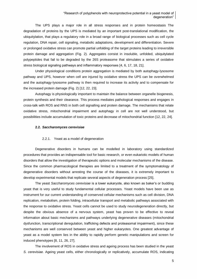
The UPS plays a major role in all stress responses and in protein homeostasis The
degradation of proteins by the UPS is mediated by an important post-translational modification, the
ubiquitylation, that plays a regulatory role in a broad range of biological processes such as cell cycle
regulation, DNA repair, cell signaling, metabolic adaptations, development and differentiation. Severe
or prolonged oxidative stress can promote partial unfolding of the target proteins leading to irreversible
protein damage and aggregation (Fig. 2). Aggregates consist in insoluble, unfolded, ubiquitylated
polypeptides that fail to be degraded by the 26S proteasome that stimulates a series of oxidative
stress biological signaling pathways and inflammatory responses [4, 6, 17, 18, 21].
Under physiological conditions protein aggregation is mediated by both autophagy-lysosome
pathway and UPS, however when cell are injured by oxidative stress the UPS can be overwhelmed
and the autophagy-lysosome pathway is then required to increase its activity and to compensate for
the increased protein damage (Fig. 2) [12, 22, 23].
Autophagy is physiologically important to maintain the balance between organelle biogenesis,
protein synthesis and their clearance. This process mediates pathological responses and engages in
cross-talk with ROS and RNS in both cell signalling and protein damage. The mechanisms that relate
oxidative stress, mitochondrial impairment and autophagy in cell are not well understood, but
possibilities include accumulation of toxic proteins and decrease of mitochondrial function [12, 22, 24].
2.2. Saccharomyces cerevisiae
2.2.1. Yeast as a model of degeneration
Degenerative disorders in humans can be modelled in laboratory using standardized
procedures that provides an indispensable tool for basic research, or even eukariotic models of human
disorders that allow the investigation of therapeutic options and molecular mechanisms of the disease.
Since the common pharmacological therapies are limited to a treatment of the symptomatology of
degenerative disorders without arresting the course of the diseases, it is extremely important to
develop experimental models that replicate several aspects of degeneration process [25].
The yeast Saccharomyces cerevisiae is a lower eukaryote, also known as baker’s or budding
yeast that is very useful to study fundamental cellular processes. Yeast models have been use as
instrument for our current understanding of conserved cellular mechanisms such as cell division, DNA
replication, metabolism, protein folding, intracellular transport and metabolic pathways associated with
the response to oxidative stress. Yeast cells cannot be used to study neurodegeneration directly, but
despite the obvious absence of a nervous system, yeast has proven to be effective to reveal
information about basic mechanisms and pathways underlying degenerative diseases (mitochondrial
dysfunction, transcriptional deregulation, trafficking defects and proteasomal impairment), since these
mechanisms are well conserved between yeast and higher eukaryotes. One greatest advantage of
yeast as a model system lies in the ability to rapidly perform genetic manipulations and screen for
induced phenotypes [8, 11, 26, 27].
The involvement of ROS in oxidative stress and ageing process has been studied in the yeast
S. cerevisiae. Ageing yeast cells, either chronologically or replicatively, accumulate ROS, indicating

“Research of polyphenols with neuroprotective potential in a yeast model of degeneration”
6
that oxidative stress defenses plays a major role in governing the key pathways involved in ageing-
induced apoptosis (Fig. 3) [10, 28-30].
Figure 3. Sources of ROS and its influence in the mechanisms of apoptosis in S. cerevisiae. In red are intracellular
sources of reactive oxygen species. Superoxide dismutases (Sod1p and Sod2p) represent the conversion of O2- to H2O2 and O2
in cytosol and mitochondria respectively. The detoxification of H2O2 is mediated by catalases (Cta1p, Ctt1p) into H2O.
Glutathione systems are represented as fundamental elements for ROS elimination: Glr1p (glutathione reductase), Gpxp
(Glutathione peroxidase), Grxp (Glutaredoxins) and Gstp (Glutathione transferase). Is represented later the oxidation of GSH
into GSSG and consequent reduction of GSSG into GSH by Glr1p. Processes such as protein misfolding, ER stress,
mitochondrial dysfunction and Fenton & Haber-Weiss reaction are all represented. Ndi1p is an internal mitochondria NADH
dehydrogenase, Aif1p is apoptosis inducing factor and Yca1p that is a yeast metacaspase. Adapted from Perrone and co-
workers [30].
2.2.2. Adaptive response against oxidative stress
Adaptation to efficient respiratory maintenance ensures that aged cells still display a functional
replicative capacity. Indeed, yeast longevity is characterized by overexpression of the transcription
factors which increases the expression of numerous respiratory genes, or overexpression of
mitochondrial NAD-dependent dehydrogenases [7, 31]. All enzymes involved in the pentose
phosphate metabolic pathway such as glucose-6-phosphate dehydrogenase (G-6-PDH), are crucial
for the production of cellular reducing power in the form of NADPH. Enzymatic response against ROS,
energetic metabolism and glutathione systems requires NADPH as reductant (Table 1) [28, 32].
Yeast contains two classes of oxygen regulated nuclear genes: aerobic and hypoxic genes.
Transcriptional factors of these genes are altered as a response to ROS increase. In stress conditions
due to increase of ROS, the glycolytic flux is also inhibit being the genes coding for glycololytic
enzymes downregulated [7, 15].
OH
ROS
H2O2
O2-
O2-
“Research of polyphenols with neuroprotective potential in a yeast model of degeneration”
7
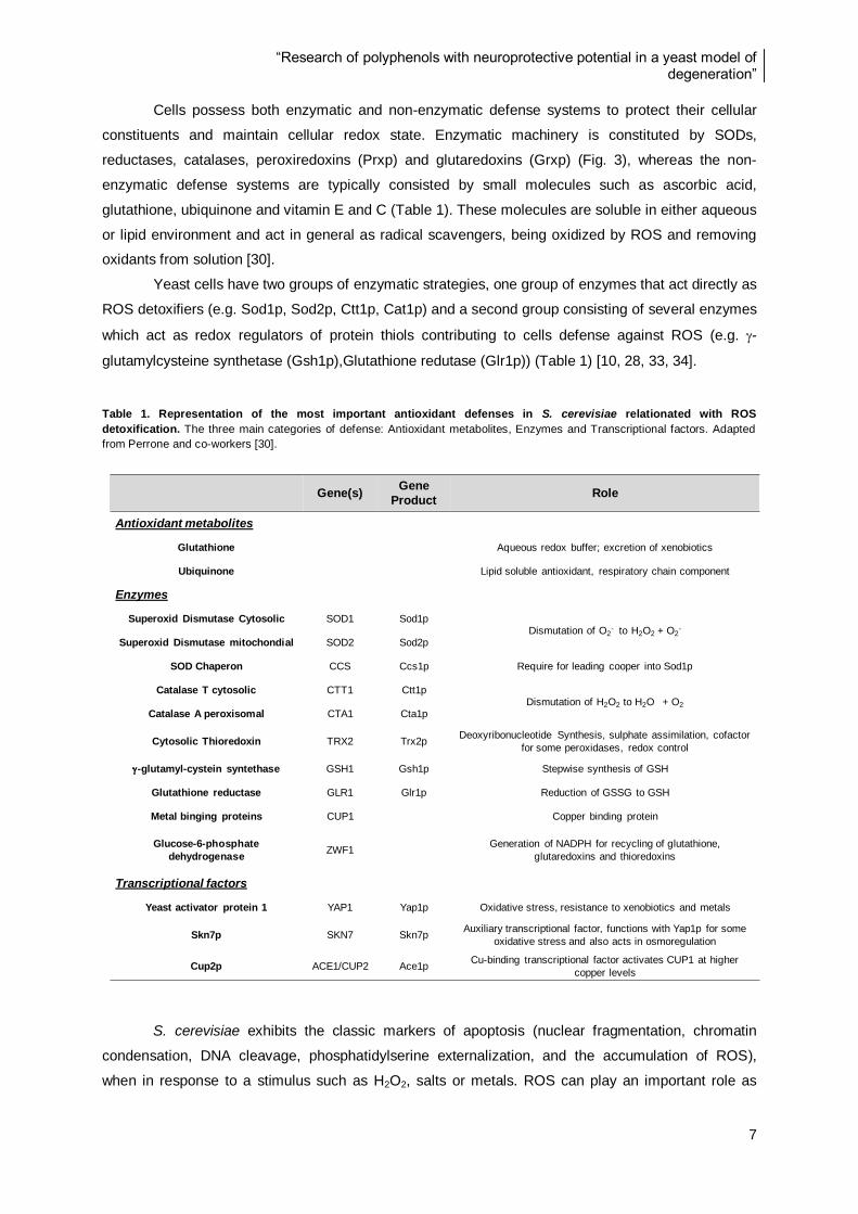
Cells possess both enzymatic and non-enzymatic defense systems to protect their cellular
constituents and maintain cellular redox state. Enzymatic machinery is constituted by SODs,
reductases, catalases, peroxiredoxins (Prxp) and glutaredoxins (Grxp) (Fig. 3), whereas the non-
enzymatic defense systems are typically consisted by small molecules such as ascorbic acid,
glutathione, ubiquinone and vitamin E and C (Table 1). These molecules are soluble in either aqueous
or lipid environment and act in general as radical scavengers, being oxidized by ROS and removing
oxidants from solution [30].
Yeast cells have two groups of enzymatic strategies, one group of enzymes that act directly as
ROS detoxifiers (e.g. Sod1p, Sod2p, Ctt1p, Cat1p) and a second group consisting of several enzymes
which act as redox regulators of protein thiols contributing to cells defense against ROS (e.g. -
glutamylcysteine synthetase (Gsh1p),Glutathione redutase (Glr1p)) (Table 1) [10, 28, 33, 34].
Table 1. Representation of the most important antioxidant defenses in S. cerevisiae relationated with ROS
detoxification. The three main categories of defense: Antioxidant metabolites, Enzymes and Transcriptional factors. Adapted
from Perrone and co-workers [30].
S. cerevisiae exhibits the classic markers of apoptosis (nuclear fragmentation, chromatin
condensation, DNA cleavage, phosphatidylserine externalization, and the accumulation of ROS),
when in response to a stimulus such as H2O2, salts or metals. ROS can play an important role as
Gene(s)Gene
ProductRole
Antioxidant metabolites
Glutathione Aqueous redox buffer; excretion of xenobiotics
Ubiquinone Lipid soluble antioxidant, respiratory chain component
Enzymes
Superoxid Dismutase Cytosolic SOD1 Sod1pDismutation of O2
- to H2O2 + O2-
Superoxid Dismutase mitochondial SOD2 Sod2p
SOD Chaperon CCS Ccs1p Require for leading cooper into Sod1p
Catalase T cytosolic CTT1 Ctt1pDismutation of H2O2 to H2O + O2
Catalase A peroxisomal CTA1 Cta1p
Cytosolic Thioredoxin TRX2 Trx2pDeoxyribonucleotide Synthesis, sulphate assimilation, cofactor
for some peroxidases, redox control
-glutamyl-cystein syntethase GSH1 Gsh1p Stepwise synthesis of GSH
Glutathione reductase GLR1 Glr1p Reduction of GSSG to GSH
Metal binging proteins CUP1 Copper binding protein
Glucose-6-phosphate
dehydrogenaseZWF1
Generation of NADPH for recycling of glutathione,
glutaredoxins and thioredoxins
Transcriptional factors
Yeast activator protein 1 YAP1 Yap1p Oxidative stress, resistance to xenobiotics and metals
Skn7p SKN7 Skn7pAuxiliary transcriptional factor, functions with Yap1p for some
oxidative stress and also acts in osmoregulation
Cup2p ACE1/CUP2 Ace1pCu-binding transcriptional factor activates CUP1 at higher
copper levels

“Research of polyphenols with neuroprotective potential in a yeast model of degeneration”
8
secondary messenger in protein phosphorylation regulation and transcriptional factor activation, to
mediate the early response to oxidative stress [15, 35]. Cells can adapt to become more resistant to a
subsequent lethal exposure, by induction of cell growth and differentiation, but at higher doses of ROS
the cell activates gene expression mediated mainly by Yeast activator protein 1 (Yap1p) and Snk7p
transcription factors, that in normal conditions are negatively regulated by glucose and promotes
cellular growth [30, 36]. These two transcriptional factors control expression of protective genes that
repair DNA damage, stress proteins (e.g. chaperons), arrest the proliferation of damage cells and
induce apoptosis (Table 1) [15, 30, 37].
2.2.2.1. Yeast activator protein (Yap)
In yeast, the basic leucine-zipper transcription factor, Yap1p regulates most of the known
cellular antioxidant genes and plays a major role in the adaptive response to oxidative stress [28, 35,
38]. The adaptive response to oxidative stress involves a change in the expression of sereval proteins,
as well as a rapid and widespread genomic response suggesting the existence of specific control
pathways. Mutants deficient in Yap1p have reduced activities of several enzymes related with
antioxidant defense such as SOD, G-6-PDH, and Glr1p, suggesting that this transcription factor is
involved in the regulation of enzymes which protect against oxidants [10, 28, 39]. Yap1p is a
transcription factor that controls the oxidative stress adaptive response through the expression of
genes encoding most of the yeast endogenous antioxidants and components of the cellular thiol-
reducing pathway, including the SOD1, SOD2, TRx (Thioredoxin), GLR1, CTT1, CCP1 (cytochrome c
peroxidase), TRR1 (Thioredoxin redutase), AHP1 (alkyl hydroperoxide reductase) and GSH1 genes
[10, 37, 39].
The Yap1p target-genes include important activities in the maintenance of cellular redox
homeostasis and in cellular xenobiotic detoxification (ATP-binding cassettes transporters, GSH
transferases, GSH biosynthetic pathway) [28, 39-41]. Yap1p activates an oxidative stress adaptive
response by redox sensory mechanisms which detect changes in the intracellular redox balance
caused by ROS, oxidized thiols and metals. Yap1p can activate gene expression through binding to
Yap1 Recognition Elements (YRE) present in the majority of gene of response to oxidative stress
promoters [10, 28, 39].
Yap1p can be activated by H2O2, thiol oxidants, electrophilic compounds and metals. ROS
(hydroperoxides and the superoxide anion) and chemicals with thiol reactivity (electrophiles and
divalent heavy metals cations) both inhibit Yap1p nuclear export by disrupting its interaction with the
nuclear export receptor Crm1, leading to gene transcription. The response of Yap1p to O2- was
suggested by the identification of SODs as target genes. Yap1p might also protect yeast from the
combined oxidative and phytochemical induced stress, since several phytochemicals have
electrophilic properties [38-40].
The similarity between the cysteine thiol reactivity of Yap1p and the mammalian AP-1, Keap1–
Nrf2 systems makes of Yap1p a good model for investigating the general mechanisms of oxidative
stress response in mammalian cells [42].
“Research of polyphenols with neuroprotective potential in a yeast model of degeneration”
9
2.2.2.2. Glutathione defense system
Sulfhydryl groups are important components of cellular defense against oxidative stress and
for the maintenance of the redox homeostasis of cells. Glutathione is present in most living cells from
microorganisms to humans and can occur in the cells under the form of GSH (reduced glutathione),
GSSG (oxidized glutathione), and mixed disulfides, GSS-CoA and GSS-Cys. GSH is a low-molecular-
mass thiol existing in cells in the concentration range of 1–10 mM and function as a redox buffer in
many cellular processes including protection against xenobiotics, carcinogens and damaged by ROS.
GSH is essential in eukaryotes; studies in yeast have shown that GSH is required as a reductant to
remove endogenously-derived toxic metabolites.Glutathione is the best-known example of a non-
enzymatic defense system that acts as a radical scavenger to maintain redox homeostasis. The redox
active sulphydryl group of this thiol reacts with oxidants to produce GSSG. GSH is synthesized in two
ATP-dependent steps catalyzed by Gsh1p and Gsh2p (Glutathione synthesize) (Table 1). The
biological importance of GSH is dependent upon the redox-active free sulfydryl moiety of its cysteine
residue [9, 31, 41, 43].
ROS detoxification through GSH is catalysed by Glutathione peroxidase (Gpxp), which
converts two molecules of GSH into one molecule of GSSG, while the enzyme Glr1p is responsible for
the reduction of GSSG that is very important to maintain the GSH/GSSG ratio in cells. This process
utilizes NADPH as a reducing power, which is generated by the G-6-PDH and is found not only in the
cytosol of cells but also in the mitochondrial matrix (Fig. 3 and 4). In cells this system is equilibrated by
a GSH/GSSG ratio of approximately 50 [28, 36, 41, 44].
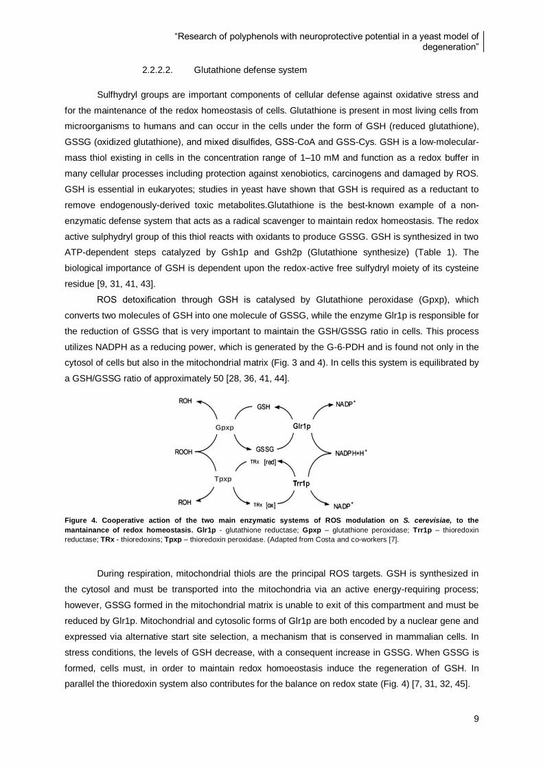
Figure 4. Cooperative action of the two main enzymatic systems of ROS modulation on S. cerevisiae, to the
mantainance of redox homeostasis. Glr1p - glutathione reductase; Gpxp – glutathione peroxidase; Trr1p – thioredoxin
reductase; TRx - thioredoxins; Tpxp – thioredoxin peroxidase. (Adapted from Costa and co-workers [7].
During respiration, mitochondrial thiols are the principal ROS targets. GSH is synthesized in
the cytosol and must be transported into the mitochondria via an active energy-requiring process;
however, GSSG formed in the mitochondrial matrix is unable to exit of this compartment and must be
reduced by Glr1p. Mitochondrial and cytosolic forms of Glr1p are both encoded by a nuclear gene and
expressed via alternative start site selection, a mechanism that is conserved in mammalian cells. In
stress conditions, the levels of GSH decrease, with a consequent increase in GSSG. When GSSG is
formed, cells must, in order to maintain redox homoeostasis induce the regeneration of GSH. In
parallel the thioredoxin system also contributes for the balance on redox state (Fig. 4) [7, 31, 32, 45].
Tpxp
Gpxp
“Research of polyphenols with neuroprotective potential in a yeast model of degeneration”
10
GSH also has strong nucleophilic properties allowing the conjugation with xenobiotics with
electrophilic properties. In living tissues and also in yeast GSH assumes a pivotal role in the regulation
of sulphur metabolism, being most of the excess of sulphur incorporated into GSH. It is also important
in bioreductive reactions, transport, enzyme activity and integrity of mitotic apparatus [41, 43].
2.2.2.3. Superoxide dismutase (SOD)
SODs catalyze the disproportionation of O2- to H2O2 and O2, but their activity requires redox
active metal ions. Yeast cells, like other eukaryotes, possess two SODs, the Cu/ZnSod (Sod1p)
encoded by SOD1 gene and MnSod (Sod2p) encoded by SOD2 gene. Both Sod1p and Sod2p play a
role in detoxification of superoxide anion generated in the mitochondrial respiratory chain. Sod1p
comprises 90% of the total SOD and is widely distributed in the cell being in more quantity in the
cytoplasm. The Sod1p, the major enzyme involved in the remotion of O2- from the cytoplasm and
possibly also from the peroxisome, appears to be a key enzyme involved in the regulation of
intracellular levels of ROS and in protecting cells from the exogenous toxicity of oxidant agents. On
the other hand Sod2p, localized in the mitochondria, has the physiological role of protect the
mitochondria from O2- generated during respiration. These proteins have different metal binding ability,
distribution in cell compartments and sensitivity to various reagents [11, 44, 46].
Sod1p is synthesized in a non-active form; being functional after addition of Cu2+
at the active
site by the copper chaperone protein (Ccs1p) [11, 44, 46]. Oxidation state of sulfhydryl groups is
important for Sod1p activation, essential for the folding and for import to mitochondria. Grxp protect
Sod1p from oxidation by catalyzing both the formation and reduction of intermolecular disulfides
between Sod1p and low molecular weight thiols such as GSH [44, 47].
In eukaryotic cells, MnSOD is strictly a mitochondrial enzyme located in the inner membrane
but is synthesized by nuclear genes. A larger precursor form is produced in the cytosol and
transported into the mitochondria by an energy dependent process. For its translocation CUP1 (that
codifies for metallothionein, a copper binding protein) and SOD1 genes are coordinately regulated
through Ace1p (metallothionein protein) and copper (Table 1) [44, 47].
SODs have great physiological significance and therapeutic potential, being involved in
diseases frequently associated with ageing, being ALS the best study. Mutations in the gene coding
for the antioxidant enzyme Sod1p are the most frequent cause of familial ALS. Oxidative crosslinks at
cysteine residues play an important role in Sod1p aggregate formation, accumulation of this protein in
motor neuronal cells would interfere with axonal transport, UPS and mitochondrial function, resulting in
neuronal cell death of ALS patients [46].
2.3. Natural phytochemicals and human health
Intake of fruit and vegetables, increase cell survival and life span, in mammals through the
reduction of oxidative stress damage Natural phytochemicals present in food prevent oxidation of
proteins, lipid peroxidation and generation of ROS. Significant evidences show beneficial effects on
human health and the reduction of the risk of cardiovascular diseases, cancer, type II diabetes and
other disorders associated with ageing [2, 3, 48].

“Research of polyphenols with neuroprotective potential in a yeast model of degeneration”
11
The protective effect of fruits and vegetables may be due to the biological activities of its
phytochemical compounds, defined as bioactive non-nutrient components. Berry extracts are widely
consumed as dietary supplements due to their potential human health benefits [49, 50]. It has been
demonstrated that plants contain many natural phytochemicals such as carotenoids, polyphenols,
alkaloids and other nitrogen-containing compounds; which have been identified as free radicals or
active oxygen scavengers. In addition, intake of these phytochemicals in the diet may prevent
diseases by modulating signalling pathways, decreasing the DNA damage or through enhanced DNA
repair by genes activation [51-53].
2.3.1. Polyphenols
The primary function of polyphenols is the protection of plants against ROS produced during
photosynthesis, and from herbivores attack. The main dietary sources of polyphenols are berries,
cereals, dry legumes, chocolate, and plant-derived beverages, such as tea, coffee, and wine. Several
hundreds of different polyphenols have been identified in foods, constituting a large group of
phytochemicals such as: flavonoids (anthocyanins, flavonols, and flavanols), tannins,
proanthocyanidins, ellagitannins, gallotannins, stilbenoids, and phenolic acids [3, 48, 53-55]
Total dietary polyphenols intake could be as high as 1 g per day, which is much higher than
that of all other classes of phytochemicals from diet [48, 54, 56]. Flavonoids are themselves distributed
among several classes: flavones, flavonols, flavanols, flavanones, isoflavones, proanthocyanidins, and
anthocyanins. Proanthocyanidins, common in many fruits, such as apple, grape, or cocoa, are
responsible for their characteristic astringency or bitterness [48, 57].
Anthocyanins are polyphenol pigments responsible for the attractive colors red, blue, and
purple of fruits, vegetables, flowers, and other plant tissues that are important to their large role of
bioactivities including antioxidant, anticancer, and anti-inflammatory. Anthocyanins are major dietary
components mainly found in berries [48, 57]. Ellagitannins, which are complex derivatives of ellagic
acid, identified in tea, medicinal plants, and several fruits, also have general scacenging effects [49,
58].
For many years, polyphenols and other phenolic compounds were thought to protect cell
constituents against oxidative damage through scavenging of free radicals. However, phytochemicals
are described as also possesing anti-inflammatory, antimutagenic, anti-atherosclerotic,
anticarcinogenic, antibacterial and antiviral activities and can exert their effects through different
mechanisms. Cells also respond to polyphenols through direct interactions with receptors or enzymes
involved in signal transduction or modulating the activity of enzymes involved in ROS modulation,
which may result in modification of the redox status [50, 58, 59]. Numerous pathways have been
reported as being targets of phenolic compounds, thereby demonstrating the broad spectrum of
targets and strengthening their usefulness in addressing multifactorial diseases [2, 3].
2.3.2. Berries
Berries such as blackberries, strawberries, raspberries and blueberries provide a rich, diverse
and specific source of dietary phytochemicals, especially polyphenols [49, 58]. Levels of

“Research of polyphenols with neuroprotective potential in a yeast model of degeneration”
12
proanthocyanidins or ellagitannins, vary considerably among berries and among commonly consumed
berries, blueberries and cranberries contain predominantly proanthocyanidins whereas blackberries,
raspberries, and strawberries contain predominantly ellagitannins [58, 60, 61].
The A. unedo and C. album berries and leaves were tested in this study for being still poorly
characterized for protective effects in degeneration processes. G. biloba leaf and R. ideaus fruit are
two plant matrixes very well studies that can serve as comparison.
Figure 5. Plants used in this work. a) Arbutus unedo (L.) Ericaceae family commonly known as strawberry tree; b) Corema
album (L.) D. Don Empetraceae family commonly kwon as Portuguese crowberry; c) Red raspberry (Rubus idaeus L.),
Rosaceae family; d) Ginkgo biloba tree (Maidenhair tree, Ginkgoaceae)
Arbutus unedo
Arbutus unedo (L.) commonly known as strawberry tree is an evergreen shrub, a native
Mediterranean species that belongs to Ericaceae family. Its fruits (berries) are spherical and dark red.
These berries are rarely eaten as fresh fruits but have importance in local agricultural communities
which use them for the production of alcoholic beverages, jams, jellies and marmalades. In Portugal,
the strawberry tree is mainly implanted in the south, being however present throughout all of the
country in a dispersed way (Fig. 5 a)) [51, 62, 63].
A. unedo fruit has been used in folk medicine as antiseptics, diuretics and laxatives, while the
leaves have long been employed as an astringent, diuretic, urinary anti-septic agent, depurative and,
more recently, in the therapy of hypertension, inflammatory diseases and diabetes. Studies showed
that fruit and leaf extracts contain several phenolic compounds, like tannins, flavonoids, phenolic (e.g.
anthocyanins), gallic acid derivatives, tannins, vitamins C and E, and carotenoids, which make them a
good source of phytochemicals [51, 62-64].
Corema album
Corema album (L.) D. Don, belongs to Empetraceae family. It is also designated by camarinha
or Portuguese crowberry, is a dioecious shrub endemic from Iberian Peninsula and Azores Islands
a) b)
c) d)
“Research of polyphenols with neuroprotective potential in a yeast model of degeneration”
13
(ssp. azoricum) that grows in sand dunes and coastal cliffs of the Atlantic coast. It produces edible
berries, which are white when ripe and have a sugary and water-rich pulp. The fruits take months to
ripen while flowering occurs from February to April, with fruits ripening from June to September. This
fruit is not currently commercially explored and very few studies were performed with this plant,
however might be a potential important source of nutrients and phytochemicals (Fig. 5 b)) [65, 66].
Rubus ideaus and Ginkgo biloba
Berries belonging to Rosaceae family, namely, raspberry (Rubus idaeus L.), provide delicious
fruits with high content in phenolic compounds. The genus Rubus, one of the most diverse in the plant
kingdom, contains approximately 740 species. These diverse species are native on six continents and
have been found from the tops of mountains to coastal locations at sea level. Subgenus Idaeobatus
includes the European raspberries (Rubus idaeus L.)(Fig. 5 c)) [67, 68].
Raspberries are common in cool temperate regions of the northern hemisphere and its
cultivation only became widespread in European countries by the 16th century. Nowadays raspberries
are of economical importance, this fruits are widely consumed fresh, frozen, or in processed forms
such as jellies, jams, and juices [67, 69]. Raspberry is a source of a variety of potentially healthy
compounds. Studies recognized possible health benefits from their natural phytochemicals. Rubus
fruit are considered a healthy and nutritious food, containing phenolics, vitamin C, α-tocopherol,
carotenoids, linoleic acid and linolenic acid [60, 69, 70].
Red raspberries (Rubus idaeus) presents among fruits one of the highest scavenging capacity
due to a unique phytochemical profile rich in ellagitannins and anthocyanins that distinguishes them
from other fruits. Besides, raspberries contain a variety of beneficial compounds, including essential
minerals, vitamins, fatty acids, and dietary fiber [60, 69-71].
The Ginkgo biloba tree (Maidenhair tree, Ginkgoaceae) is described as a "living fossil" since it
represents the only surviving species of the order Ginkgoales that existed when the dinosaurs roamed
the earth more than 200 million years ago. Modern Chinese pharmacopoeias introduced Ginkgo
leaves for treating dysfunctions of the heart and lungs, and currently extracts of these leaves
represent one of the most common phytomedicines in the world (Fig. 5 d)) [72].
Extracts of G. biloba leaf are widely used in herbal medicine for the treatment of mild to
moderate cognitive disorders and dementia. A standardized extract of leaves from G. biloba named
EGb 761 that is constituted by flavonol glycosides, terpene, lactones flavonoids, inkgolides,
bilobalides, proanthocyanidins and organic acids is efficient in the treatment of clinical disorders with
multifactorial origins. This extracts have been suggested by several studies to possess numerous
beneficial properties, including free radical scavenging, antiapoptotic, antiageing and antigenotoxic.
They also have been described to regulate gene expression and attenuate AD symptoms [3, 72-74].
“Research of polyphenols with neuroprotective potential in a yeast model of degeneration”
14
2.3.3. Polyphenols bioavailability
Phenolic compounds present relatively low levels in circulation due to its reduce bioavailability.
The ability of polyphenols to influence cellular function and affect health is dependent of their
absorption from the gastrointestinal tract (GIT) to the blood and delivery to the target tissues. These
compounds are extensively metabolized in the body tissues and can be absorbed in stomach and at
small intestine level, by diffusion or transport. Variable amounts of flavonoids that are not absorbed in
the upper GIT, reach the colon, where they are subjected to colonic microflora action and the microbial
catabolites could be absorbed into the circulatory system from the large intestine. The real biological
response to these compounds is greatly determined by the bioavailability of active molecules (Fig. 6)
[49, 53, 75].
Figure 6. Bioavailability of polyphenols after alterations occurring during metabolization in the human body.
Representation of the three main steps of polyphenols digestion: Stomach conditions where occur the degradation of sugars,
vitamins and minerals; Small intestine where the majority of phytochemicals are modify and colon where occur the modifications
by microflora. (Adapted from [2, 57, 76-78])
Bioavailability studies determine which are the: best absorbed polyphenols; modifications that
polyphenols suffer after absorption; active metabolites and the polyphenols that lead to the active
metabolites. Therefore, the knowledge of polyphenols bioavailability and metabolization is essential to
identify those most predisposed to exert protective effects. The most absorbed in humans are
isoflavones and gallic acid, followed by catechins, flavanones, and quercetin glucosides. The least
well-absorbed polyphenols are the proanthocyanidins and the anthocyanins [49, 54, 58, 75, 76].
Evaluation of metabolites as they exist in plants or the effect of single purified phenolic
compounds, ignoring the chemical alterations with a consequential impact on bioavailability and
bioefficacy, leads to a loss of possible synergetic/cooperative or competitive activities between
phenolic compounds that affect their final form present in serum (Fig. 6). These compounds may
“Research of polyphenols with neuroprotective potential in a yeast model of degeneration”
15
persist in vivo, accumulate in target tissues, and contribute significantly to the prevention and
treatment of chronic human diseases [2, 48, 58, 68]. In addition, there are also large variations in
polyphenols bioavailability observed among individuals due to nutrigenetic and nutrigenomic effects.
[50, 54, 77]. For these motif is important evaluate the food metabolites in the circulating form in the
body.
In vitro digestion (IVD)
In order to evaluate the potential role of plant phytochemicals in the human body, should take
into account the physiochemical changes occurring in the GIT [2, 48, 58, 68]. Only the compounds
that can reach the serum and be distributed by all organs contribute significantly to the biological
effects [49, 54, 58]. For these reasons is important an IVD process that mimics the physic and
chemical transformations occurring in stomach and gut during disgetion before pass to serum, to
provide essential informations on the relative potential bioavailability of different polyphenolic
components in this specific phase of digestion. [52, 55, 57].
Despite the relatively high amounts of anthocyanins consumed in the diet and the reported
biological activities, little is known about the in vivo biological activity of anthocyanins including
bioabsorption. The instability of anthocyanins in procedures that mimic the physiochemical and
biochemical changes that occur in the upper GIT has been study by Gordon M. and collaborators, that
demonstrate that the major part of anthocyanins is degraded in GIT or goes out into colon, remaining
only a small part in serum. [52, 53, 57, 76, 79].
Chemical and structural modifications due to gastrointestinal absorption and metabolization
have not been taken into account in many previous studies. Information about absorption, distribution,
metabolism, and excretion of individual flavonoids is still scarce and is important to understand the
molecular mechanism(s) underlying the effects of food on human health and their biological activities
[2, 52, 57].
3. Material and Methods
3.1. Samples preparation
3.1.1. Plant material
For this study were selected fruits and leaves, from C. album (Corema album (L.) D. Don) and
A. unedo (Arbutus unedo (L.)). R. idaeus (Rubus idaeus L. cv polka), fruit and Ginkgo biloba leaf were
also chosen due to their well known health benefits, to comparison purposes.
All the berries were harvested, frozen, grinded and then freeze-dried. Fruits and leaves of C.
album and A. unedo were collected by random sampling in Comporta (southern region of Portugal)
and in an extensive area of Arrábida Natural Park, respectively. R. ideaus cv polka fruit was harvested
at Fataca experimental field (Odemira, Portugal). Leaves from G. biloba were from plants keep at
ITQB (Instituto Tecnologia Química e Biológica) green house.

“Research of polyphenols with neuroprotective potential in a yeast model of degeneration”
16
3.1.2. Hydroethanolic extraction
Secondary metabolites were extracted, using clean solvents, as previously described by
Tavares and co-workers [80]. Briefly, to 1 g of lyophilised powder 12 mL of hydroethanolic solution
(50% (v/v) ethanol/water) were added. This mixture was shaken for 30 min at room temperature in the
dark and centrifuged at 12400 g for 10 min at room temperature. The supernatant was then filtered
through 0.2 m cellulose acetate membrane filters and the resulting extracts were stored at - 80 ºC.
3.1.3. In vitro digestion (IVD)
Phytochemical alterations occurring during digestion were mimicked using an adapted IVD
model, accordingly with Tavares and co-workers [2]. This methodology was performed by a colleague
of the Disease & Stress Biology (DSB) Laboratory in collaboration with Gordon McDougall and Derek
Stewart from The James Hutton Institute, Dundee, Scotland.
This method consists in two sequential steps that represent an average time for
gastrointestinal transit; an initial pepsin/HCl digestion for 2 h at 37 ºC, that simulate the gastric
conditions, and a final bile salts/pancreatin digestion for 2 h at 37 ºC, that simulate small intestine
conditions.
The original extract was adjusted to pH 1.7 with 5 M HCl and then the pepsin (Sigma Product
number P6887) was added at 315 units.mL-1
and incubated at 37 ºC in a heated water bath for 2 h
with shaking at 100 rpm. The remainder solution was placed in a glass beaker and 4.5 mL of a mixture
consisting of 4 mg.mL-1
pancreatin and 25 mg.mL-1
bile salts were added. Then a segment of cellulose
dialysis tubing, containing sufficient 0.1 M NaHCO3 to neutralize the samples titratable acidity, was
added and the beaker sealed with parafilm. Diffusion of NaHCO3 out of the dialysis tubing represents
the simplest and most convenient means to mimic the gradual rise in pH that occurs to the stomach
contents when entering the small intestine.
After another 2 h of incubation at 37 ºC, the solution inside the dialysis tubing (IN),
representing the serum available material and the solution outside the dialysis tubing (OUT), that
mimics the material remained in colon, were mixed together to be representative of the total digestion
process and the solution was frozen [76].
3.1.4. Solid Phase Extraction (SPE)
A Solid Phase Extraction (SPE) protocol was carried out to concentrate polyphenols of
digested extracts, using a GIGA tubes 20g/60mL (1000 mg capacity, Phenomenex Ltd.), C18-E units,
after the IVD process. This protocol was performed accordingly with Tavares and co-workers [80] to
afford complete separation of total phenolics compounds from the bile salts presents in the sample
[52].
Briefly, after pre-equilibration of the column with 0.5% (v/v) (CH3COOH/H2O), the soluble
materials from IVD were diluted in the equilibration solution and loading into the column C18-E. A
wash step was performed with two volumes of 0.5% (v/v) (CH3COOH/H2O). Finally the bound fraction
“Research of polyphenols with neuroprotective potential in a yeast model of degeneration”
17
that is enriched in polyphenols was eluted and recovered by adding a solution of 0.5 % (v/v)
(CH3COOH/CH3CN).
Fractions were concentrated in a Speed-Vac to a final suitable phenol concentration and
frozen at -80 ºC. To test in S. cerevisiae, PDFs (polyphenol-digested fractions) obtained after the IVD-
SPE procedure were unfrozen and dissolved to the final concentration to test, in a (v/v)
CH3COOH/CH3CN/H2O (0.05/0.995/99.9) solution.
3.2. Total phenol content
Determination of total phenolic compounds present in PDFs was performed by the Folin–
Ciocalteu method, adapted to a 96 microplate reader, as described by Tavares and co-workers [80].
Phenolic compounds are excellent oxygen radical scavengers because of its electron
reduction potential. Folin-Ciocalteu reagent is a solution of polymeric complex ions, formed by
phosphomolybdic and phosphotungstenic acid. The phenolic compounds in the samples react with
this colorimetric reagent, which allows measurement in the visible portion of the spectrum. The
transfer of electrons in alkaline medium from phenolic compounds to oxidized
phosphomolybdic/phosphotungstic acid complexes forms blue complexes that are determined
spectroscopically at approximately 760 nm, due to its reduction. Sodium carbonate provides the
alkalization [81].
To determine the total phenolic compounds in our samples along all steps of the digestion
protocol, in each well of a 96-well plate, 235 μL of water, 5 μL of sample, 15 μL of Folin-Ciocalteu
reagent (Fluka) and 45 μL of saturated Na2CO3 (Riedel-deHaën), were added. The microplate was
incubated for 30 min at 40 ºC and the absorbance read at 765 nm in the end.
In case of the blank and standards we added 5 μL of water or gallic acid (concentrations
between 0.05 and 0.7 mg.mL-1
), respectively, instead of sample. All measurements were performed
with three replicates and the results were expressed as mg of gallic acid equivalents (mg GAE) and as
% of original fraction.
3.3. Antioxidant capacity assay
Peroxyl radical-scavenging capacity of the samples in study was determined by the ORAC
(Oxygen Radical Absorbance Capacity) method. This assay was carried out following a modified
procedure described by Tavares and co-workers [59], adapted from Cao and co-workers [82], to 96-
well plates.
This assay measures the ability of the antioxidant components in samples to inhibit the decline
in fluorescein fluorescence, induced by a peroxyl radical generator, AAPH (2,2’-azobis (2-
amidopropane) dihydrochloride) (Sigma). The peroxyl radicals generated from thermal decomposition
of AAPH in aqueous buffer, react with fluorescein, to form a non-fluorescent product which allow the
quantification of fluorescein fluorescence decline. [82]
Briefly, the reaction mixture was prepared in a 96-well plate containing 150 μL of sodium
fluorescein (0.2 nM) (Uranine, Fluorescein Sodium Salt TCI Europe), 25 μL of sample and 25 μL of
“Research of polyphenols with neuroprotective potential in a yeast model of degeneration”
18
AAPH (41.4 g.L-1
) per well. All solutions were prepared in 75 mM phosphate buffer (pH 7.4) and
instead of standards/sample, the blank contained 25 μL phosphate buffer (75 mM, pH 7.4). To perform
the standards 25 μL of 6-hydroxy-2,5,7,8-tetramethylchroman-2-carboxylic acid (Trolox, Fluka) with
concentrations ranging between 10 - 50 μM, were assayed.
Fluorescent emission at 515 nm was monitored kinetically during 30 min at 37 ºC, after
excitation at 493 nm using FLx800 Fluorescence Microplate Reader (Biotek). All measurements were
performed with three replicates. The final results were calculated and expressed using the differences
in area under the fluorescence decay curves between the blank and the Trolox/sample, and were
expressed as M trolox equivalents (M TE) and mol of trolox equivalents per phenol unit (mol TE.
mg GAE-1
).
3.4. Saccharomyces cerevisiae
In this study four different strains of S. cerevisiae were used; the WT BY4741 (MATa; his3Δ1;
leu2Δ0; met15Δ0; ura3Δ0) and its isogenic mutants, Δsod1 (MATa; his3Δ1; leu2Δ0; met15Δ0;
ura3Δ0; Δsod1::KanMX4) and Δyap1 (MATa; his3Δ1; leu2Δ0; met15Δ0; ura3Δ0 Δyap1::KanMX4),
which do not possess a functional gene corresponding to the cytosolic superoxide dismutase (Sod1p)
or a critical transcription factor involved in the oxidative stress response (Yeast Activator Protein 1,
Yap1p), respectively. These strains were acquired from Euroscraf (http://web.uni-
frankfurt.de/fb15/mikro/euroscarf/) and carry a KanMx4 deletion cassette replacing the gene in
question.
In addition, AD1-8 strain was also used. This strain features eight deletions in MDR genes
(multi drug resistance genes) (Δyor1, Δsnq2, pdr5-Δ2, Δpdr10, Δpdr11, Δycf1, pdr3 -Δ2, Δpdr15) that
encode ABC transporters involved in xenobiotic efflux capacity. This strain does not have efflux
systems, facilitating the uptake of compounds and allow us to verify the effect of a prolonged
exposition to compounds from PDFs.
All strains used present a different sensitivity to oxidative stress and were routinely maintained
in YPD (Yeast extract, Peptone, Dextrose) solid medium composed of yeast extract 1% (w/v)
(HiMedia), glucose 2% (w/v) (Sigma), peptone 2% (w/v) (HiMedia) and agar 2% (w/v) (HiMedia), at 4
°C, after growth at 30 °C for 48 h [11].
For all experiments, a pre-inoculum was prepared, in syntetic complete (SC) liquid medium,
composed of yeast nitrogen base without amino acids 0.67% (w/v) (Difco), glucose 2% (w/v) and
complete supplement mixture (CSM) 0.79 g.L-1
(QBiogene). A colony was growth until the optical
density at 600 nm (OD 600 nm) reached approximately 0.4-0.6 (which corresponds to the middle of log
phase) at 30 ºC under constant shaking, to a final volume of 3 mL (ratio of 1:3 flask/medium volume).
Afterwards, to prepare the inoculum, cells were diluted in the same medium, in order to obtain all
strains in log phase (OD 600 nm aprox. 0.6) and the same conditions of pre-inoculum were repeated.
Also was taken into account the doubling time (DT) of each strain, using a DT of 2 h for BY4741 and
yap1 and 3 h for sod1 and AD1-8 according with what was defined by previous growth curves.
Therefore OD readings were performed in the plate spectrophotometer Power Wave XS (Biotek) and
all experiments were done with three biological replicates.
“Research of polyphenols with neuroprotective potential in a yeast model of degeneration”
19
For long period storage, stocks of these strains were frozen at - 80° C in a solution consisting
of 50% (v/v) glycerol and YPD liquid medium.
3.4.1. Growth curves
To study the different sensitivities of each strain to H2O2, the growth of all strains (BY4741,
yap1, sod1, AD1-8) was monitored by OD 600 nm reads during 24 h, to achieve the growth curve and
growth parameters corresponding to each strain. The growth curves were performed, in presence and
absence of several concentrations of H2O2.
After inoculum preparation, previously described in section 3.4, cells were diluted in SC liquid
medium to OD 600 nm of 0.03 and H2O2 was added to the yeast culture at final concentrations between 0
and 1 mM in a 96-well plate, to a final volume of 100 L. The H2O2 concentrations used were
determined based in previous unpublished studies of DSB Laboratory.
All strains were incubated at 30 °C for 24 h, with constant shaking and yeast growth was
kinetically monitored hourly by OD 600 nm readings in a spectrophotometer microplate reader
(PowerWave XS Biotek).
All experiments were performed with three replicates. For each strain and condition, in the end
of growth, the final biomass was estimated by OD 600 nm readings at 24 h. DT and lag phase times
were determined based on the linear regression of log growth phase equation, obtained after
logarithmic transformation of OD values.
3.4.2. Spot Assay
Spot assays was the methodology chosen to test three different conditions: 1) The
susceptibility of yeast strains to oxidative stress induced by H2O2; 2) The range of non-toxic
concentration of PDFs and 3) The protective potential of PDFs in the presence of oxidative stress.
After an inoculum preparation, yeast cells were incubated with different concentrations of
H2O2. In a 96-well plate, cells were diluted until OD 600 nm of 0.4 in SC liquid medium and H2O2 was
added in concentrations between 0 - 10 mM, to a final volume of 100 L. The microplate was
incubated during 1 h at 30 ºC, with constant shaking. Subsequently 30 μL of cells were withdrawn to a
new 96-well plate and the OD 600 nm adjusted to 0.1 in PBS (phosphate buffer) pH 7.4. Finally, five-fold
serial dilutions were made, in PBS pH 7.4 and 5 L of each well was spotted in YPD solid medium and
incubated for 48 h at 30 ºC. At the same time 5 L of the same cells growth in the absence of H2O2
were spotted in SC solid medium with glucose supplemented with different concentrations of H2O2,
between 0 and 1 mM.
The PDFs toxicity was evaluated by the same procedure, a 96-well plate was prepared but
instead H2O2, the PDFs were added in final concentrations between 0 and 1000 g GAE.mL-1. The
initial OD 600 nm was 0.2 and the microplate was incubated during 4 h at 30 ºC, with constant shaking.
At the end 5 L of cells were spotted in SC solid medium with glucose.

“Research of polyphenols with neuroprotective potential in a yeast model of degeneration”
20
After PDFs toxicity assay, in order to evaluate the protector potential of PDFs under an
oxidative stress condition, cells were grown first in the presence of PDFs and subsequently were
submitted to an oxidative stress induced by H2O2.
Cells were inoculated in a 96-well plate with an initial OD 600 nm of 0.2 and in presence of PDFs
non-toxic concentrations, previously determined. The microplate was incubated at 30 ºC during 4 h,
with constant shaking. Finally the same serial dilutions described above were performed and 5 L of
cells were spotted in SC solid medium with glucose supplemented with the previously selected H2O2
concentration according with the strain (BY4741 0.5 mM and yap1, sod1 0.15 mM). Cells were
incubated for 48 h at 30 ºC.
Spot assays were prepared in the same technical conditions and always with three biological
replicates, being the most representative showed. Images were acquired using Quantity-one
software from Chemidoc and were obtained in the end of 48 h of growth.
3.4.3. Resazurin-based assay
To evaluate the protective effect of PDFs based on mitochondrial metabolism integrity the
resazurin-based assay methodology was used, adapted from CellTiter-Blue Cell Viability Assay
(Promega) protocol, to S. cerevisiae. This is a sensitivity test that has been used for estimating
viability based on cell energy metabolic activity. Resazurin is a blue weakly fluorescent indicator dye
that changes into highly fluorescent pink resorufin, in response to irreversible chemical reduction
inside the cell [83].
Resazurin can penetrate cells and acts as an intermediate electron acceptor in the electron
transport chain. This allows enzymatic reduction in the mitochondria due to the activity of enzymes
and subsequently a pink resorufin that is also a fluorophor, can diffuse from cells and back to
surrounding medium, which results in fluorescence increase [83].
To establish the number of cells (represented by OD 600 nm readings) to use in this
methodology, in a 96-well plate each strain was inoculated with OD 600 nm between 0.2 and 0.02. To
each 100 L of total volume of cell suspension, 20 L of CellTiter-Blue® reagent were added.
Incubations of 45 min were performed with resazurin at 30 ºC and in the end of each time
fluorescence was measured.
To evaluate if PDFs can affect the metabolic answer of yeast cells in the presence of oxidative
stress, the inoculum was diluted to a final OD 600 nm of 0.2 in 96-well plate, with SC liquid medium with
glucose, in the same conditions described in section 3.4.2. After 4 h incubation with PDFs, yeast cells
were harvested by centrifugation at 1000 g for 5 min, rinsed twice in PBS pH 7.4 and diluted to a final
OD 600 nm of 0.2, with the same medium supplemented with 2 mM H2O2, for all strains, in a new 96-well
plate. The microplate was incubated for 1 DT according with the strain and in the end 30 L of cells
were withdrawn and diluted in SC liquid medium with glucose until OD 600 nm of 0.02 and CellTiter-Blue
reagent was added. Before fluorescence reading the microplate was incubated for 45 min at 30 ºC
with constant shaking.
All fluorescence readings were performed using a FLx800 Fluorescence Microplate Reader
(Biotek) using excitation and emission wavelengths of 560 and 590 nm, respectively. The experiences
“Research of polyphenols with neuroprotective potential in a yeast model of degeneration”
21
were done with three biological and two technical replicates and results were expressed as
fluorescence units (F.U.) and percentage of viable cells relative to the control.
3.4.4. Dichlorofluorescein assay
To determine the levels of ROS produced, intracellular ROS production was measured by
H2DCFDA (2,7-dichlorofluorescein diacetate) method, adapted from Priault and co-workers [84].
H2DCFDA is added to cells, which penetrates easily the plasma membrane and is hydrolyzed inside
the cells by non-specific esterases, being converted to the non-fluorescent H2DCF that in presence of
ROS is oxidized to fluorescent dichlorofluorescein (DCF), a highly fluorescent compound and can be
measured fluorimetrically. Thus, measurement of DCF concentrations is a valid measure of oxidation
processes inside the cells [84-86].
H2DCFDA (Sigma-Aldrich) stock solution was prepared in dimethyl sulfoxide (DMSO Sigma-
Aldrich) and stored at −80 °C.
To test the effect of PDFs in the boost of ROS induced by H2O2, the incubation with PDFs was
performed exactly in the same conditions described for CellTiter-Blue viability assay. Then yeast cells
were harvested by centrifugation at 1000 g during 5 min, diluted to a final OD 600 nm of 0.2 in SC liquid
medium with glucose. H2DCFDA was added in a final concentration of 100 M and cells were
incubated at 30 ºC for 15 min, in the dark with constant shaking, to allow the uptake of the probe by
cells. After the 15 min incubation, OD 600 nm reading was performed and H2O2 added to a final
concentration of 2 mM for all strains.
Fluorescence intensity was always measured at every 10 min, during 1 DT for each strain
with constant shaking, using a FLx800 Fluorescence Microplate Reader (Biotek). Fluorescence
excitation and emission used were of 488 and 520 nm, respectively. All experiments were performed
with three biological replicates and the results were expressed as percentage of increase in
fluorescence per well, normalized for total cell number represented by OD 600 nm.
3.4.5. Glutathione determination
To analyze the redox state of cells, GSH and GSSG were quantified by HPLC after
derivatization with orthophthalaldehyde (OPA), accordingly to the already described in Tavares and
co-workers [2].
Briefly, yeast cells were harvested by centrifugation at 1000 g during 5 min and rinsed twice in
PBS pH 7.4. Then cold 10% (w/v) metaphosphoric acid was carefully added to samples, into 1.5 mL
propylene tubes (50 µL for determination of GSH and 200 µL for determination of GSSG). Glass
beads were added and cells were lysed by vortexing, 6 cycles of 1 min in the vortex followed by 1 min
at 4 ºC. The final mixture was centrifuged at 2700 g during 5 min, the supernatant was removed and
centrifuged again at 2700 g during 30 min and the final supernatant was collected. After samples
preparation the final supernatant was derivatizated accordingly to Kand’ar and co-workers [87],
adapted from Hissin and Hilf [88].

“Research of polyphenols with neuroprotective potential in a yeast model of degeneration”
22
For GSH analysis 1 mL of 0.1% (w/v) EDTA in 0.1 M sodium hydrogenphosphate, pH 8.0, was
added to 50 µL of supernatant. To 20 µL portion of this mixture, 300 µL of 0.1% (w/v) EDTA in 0.1 M
PBS, pH 8.0, and 20 µL of 0.1% (w/v) OPA in methanol, were added. Tubes were incubated at 25 ºC
for 15 min in the dark. The reaction mixture was then stored at 4 ºC until analysis. In parallel, for
GSSG analysis, 200 µL of supernatant was incubated at 25 ºC with 200 µL of 40 mM N-ethyl
maleimide (NEM) for 25 min in dark. To this mixture, 750 µL of 0.1 M NaOH was added. A 20 µL
portion was taken and mixed with 300 µL of 0.1 M NaOH and 20 µL of 0.1% (w/v) OPA. Tubes were
incubated at 25 ºC for 15 min in the dark and stored at 4 ºC until analysis.
Chromatographic analysis was accomplished using isocratic elution on C18 analytical column
(YMC- PACK ODS-A, HPLC 200Å, 5 µm) at 37 ºC on an Acquity TM Ultra performance LC system
(Waters). The mobile phase consisted of 15% (v/v) methanol in 25 mM PBS (v/v), pH 6.0. The flow
rate was kept constant at 0.7 mL.min-1
. The excitation and emission wavelengths were set at 350 and
420 nm, respectively. The amount of GSH and GSSG was quantified from the corresponding peak
area using EmpowerPro 2.0 software [13]. The concentration of GSH and GSSG in the samples was
determined from standard curves with ranges 0 - 250 µM for GSH and 0 - 25 µM for GSSG, prepared
in parallel with the samples. All experiments were performed with three biological and two technical
replicates. The results were expressed as M of GSH normalized for total number of cells (OD 600 nm)
(GSH / OD 600 nm) and as the ratio (GSH/GSSG).
3.5. Statistical analysis
The results reported in this work are the averages of at least three independent experiments
and are represented as the mean ± SD. Regressions presented were obtained using Microsoft®
Excel
2007 tools. Differences among treatments were detected by analysis of variance with Tukey HSD
(Honest Significant Difference) multiple comparison test (α = 0.05) using SigmaStat 3.10 (Systat).
4. Results and Discussion
4.1. Oxidative stress in a yeast model of degeneration
4.1.1. Establishment of the yeast model of degeneration and optimization of methods
Growth curves and parameters
In this work the yeast model of degeneration is constituted by 4 different S. cerevisiae strains
submitted to an oxidative injury that can mimics the metabolic changes that occur during ageing
processes and degeneration. For this model were chosen the WT, BY4741, 2 isogenic mutants
(yap1 and sod1) and AD1-8. The yap1 strain does not possess a critical transcription factor
involved in the oxidative stress response, the sod1 strain does not possess a functional gene
codifying to the cytosolic superoxide dismutase and AD1-8 strain that features eight deletions in MDR
genes, encoding ABC transporters involved in xenobiotic efflux capacity. All these strains together
allow the achievement of important information about their differential behaviour in presence of H2O2
“Research of polyphenols with neuroprotective potential in a yeast model of degeneration”
23
and about the metabolic pathways involved in the deveplment of degeneration process. To implement
the yeat model of degeneration several oxidative stress conditions were induced in the 4 strains.
The facility of mutation isolation, the rapid growth on defined media and the fact of being a well
defined system makes of yeast a model very attractive to study. The most common method to study
microbial growth is through batch culture, allowing the monitorization of yeast growth and the detection
of small phenotypic changes or growth defects based on genetic mutation of strains. This pattern of
changes in the cell growth parameters, either due to mutant restrictions or presence of a stress, could
provide important information concerning the mode of action of tested chemicals [8, 89]. The analysis
of cell growth in liquid cultures are particularly suitable [89].
In a typical cell growth analysis in liquid medium, the number of yeast cells in a growing
population is monitored by the optical density at 600 nm (OD 600 nm) and the relevant growth
parameters, such as Lag phase time, doubling time (DT) and final biomass are extrapolated from the
growth curve. Lag time is determined based on the linear regression of log growth phase equation
obtained after logarithmic transformation of OD 600 nm values, and corresponds to the time of
adaptation required by cells to respond to environmental changes. DT is also obtain based on a
logarithmic transformation of OD 600 nm values of log growth phase and is used to characterize the
kinetic growth of a population under a set of growth conditions and is an indicator of the
microorganism response to a specific environment. The growth rate varies typically with the strain and
is strongly influenced by the growth medium. The rate of growth together with stationary-phase OD
increments reflects quantitatively the minor changes in growth phenotype, and is shown by cell density
or final biomass in the end of growth. [8, 89, 90].
In order to determined growth curves and parameters, as a first approach in this optimization,
tree different H2O2 concentrations were tested in liquid medium. The main propose of this task was to
select the H2O2 concentration that represent about 50% of lost of viability in a chronic exposure and to
define the growth conditions in liquid medium to use in the further tasks. To perform the growth
curves, yeast cells of each strain were grown in SC liquid medium with glucose and supplemented
with 0, 0.25, 0.5 or 1 mM of H2O2 and finally the growth was monitored hourly during 24 h (Fig. 7 A).
The effects of H2O2 on cell growth at low concentration or transient concentrations stimulate
growth and enhanced survival [7, 15]. The results of growth curves (Fig. 7 A) show that the WT strain
(BY4741) is the most resistant to H2O2 as expected. For this strain, when the growth parameters (Fig.
7 B) are considered, the most significant change is denoted on the doubling time when cells are
treated with 0.25 mM of H2O2 or higher. One possible reason for this could be the fact that 0.25 mM of
H2O2 induces retardation on growth, increasing the Lag time (Fig. 7 B). However, cells are capable to
induce mechanism of defense that are reflected by the increase in final biomass (Fig. 7 B), contrarily
to what is observed with 0.5 or 1 mM of H2O2 that reflects in a reduction of final biomass.

“Research of polyphenols with neuroprotective potential in a yeast model of degeneration”
24
Figure 7. Growth curve and growth parameters of the S. cerevisiae strains in presence of H2O2. S. cerevisiae strains
BY4741, yap1, sod1 and AD1-8 were grown in SC liquid medium with glucose and supplemented with 0.25 ( ■ ), 0.5 ( ▲ )
and 1 mM ( ) of H2O2 or without H2O2 as control (), for 48 h at 30 ºC with constant shaking. A) Growth was kinetically
monitored hourly by OD measurements at 600 nm and growth curve represented. B) Growth parameters were determined
based on the growth curve being represented the 0.25 ( ■ ), 0.5 ( ■ ) or 1 mM ( ■ ) of H2O2 or without H2O2 as control ( □ ).
Doubling time (h), representing the time necessary to cells duplicate, determined by logarithmic transformation of raw OD 600 nm
from growth curves. Lag times (h), representing the adaptation time, determined by logarithmic transformation of raw OD 600 nm
from growth curves. Final Biomass (OD 600 nm), representing the final number of cells is calculated based on the measurements
of OD at 24 h of growth. Results represent the mean SD of three independent biologic replicates. *, ** and *** represent
statistically significance for a control of cells without H2O2, for a p<0.05, p<0.01 and p<0.001 respectively.
B)
A)
0
5
10
15
20
25
30
BY4741 Dyap1 Dsod1 AD1-8[H2O2] mM
Tim
e (h
)
Lag Time
******
***
***
***
***
***
*
***
***
***
***
0.25 0.5 10
BY4741
0.25 0.5 10
yap1
0.25 0.5 10
sod1
0.25 0.5 10
AD1-8
0
1
2
3
4
5
6
BY4741 Dyap1 Dsod1 AD1-8[H2O2] mM
Tim
e (h
)
Doubling Time
***
******
***
***
***
******
***
***
***
***
0.25 0.5 10
BY4741
0.25 0.5 10
yap1
0.25 0.5 10
sod1
0.25 0.5 10
AD1-8
0
0.1
0.2
0.3
0.4
0.5
0.6
0.7
0.8
0.9
1
BY4741 Dyap1 Dsod1 AD1-8[H2O2] mM
OD
60
0 n
m
Final Biomass
*
****
***
***
*
**
*
***
0.25 0.5 10
BY4741
0.25 0.5 10
yap1
0.25 0.5 10
sod1
0.25 0.5 10
AD1-8
Time (h)
0
yap1
AD1-8
BY4741
sod1OD
60
0 n
m
Doubling Time Lag Time
Final Biomass
“Research of polyphenols with neuroprotective potential in a yeast model of degeneration”
25
It is possible to observe that for both BY4741 and sod1 cells growth in presence of 1 mM of
H2O2 seem to suffer a strong adaptation that allows these cells to restore the normal viability,
demonstrated by the number of cells in the end of 24 h, e.g. by the final biomasss (Fig. 7 B). These
results may indicate that ROS production have a preconditioning effect on cells at low concentrations.
These results indicate that at high concentrations of H2O2 (1 mM) the sod1 strain, besides present
the longest adaptation phase (Lag phase) can activate mechanisms of defese that allow to growth and
adapt contrarily to yap1 and AD1-8 that present a inhibition of growth in the same conditions (1mM)
(Fig 7 A and B).
In general the most affected mutant by oxidative stress is yap1 (Fig. 7 A and B). This is
revealed based on the increasing amount of time needed by cells to start replication (Lag time) and
reaching the log phase (maximum growth rate). Was also observed that yap1 strain present almost
total growth inhibition at 1 mM of H2O2.
In normal conditions yap1 and sod1 present a lower number of cells in the end of the 24 h
of growth than BY4741, represented by final biomass values. For yap1 and AD1-8 the final biomass
is the more affected at 1 mM of H2O2, while that for BY4741 and sod1 strains the final biomass is
more affected at 0.5 mM of H2O2. BY4741 and sod1 show a similar metabolic adaption in these
conditions, in what regards to final biomass parameter (Fig. 7 B)).
When is induced the oxidative stress, even at 0.25 mM of H2O2 all strains are affected,
however the H2O2 concentration needed to induce more metabolic alterations is different according
with the strain mutation. Furthermore, the pattern of changes in the cell growth parameters could
reveal important information concerning the mode of action of the tested agents and the involved
pathway.
These results were essential to define the incubation times in liquid medium according with the
strain and also the H2O2 concentrations to test in solid medium by spot assays in different type of
exposures. With the growth curves results was possible to define the time of incubation according with
the strain, being defined 2 h of DT for BY4741 and yap1 and 3 h of DT for sod1 and AD1-8 (results
that are corroborated with previous results from DSB laboratory). The definition of at least 1 DT of
incubation in liquid medium for each strain is important for next studies. Based on these results was
also defined the H2O2 concentrations to test in solid medium of until 2 mM or 0.5 mM of H2O2
according with acute or chronic exposures respectively. In the next section was defined the conditions
to use in solid medium by spot assays.
Viability evaluation under oxidative stress by spot assays
In general, phenotypes such as defective growth are used to determine the sensitivity of cells
to chemical compounds. Plating or spotting assays on solid media can be used to screen non-toxic
concentrations of chemicals that affect principally the replicative viability. Although often valuable, the
qualitative interpretation of colony formation does not identify whether changes occured during the lag-
time after the insult or during growth. Spot assays involve the visual inspection of colony size, number
and the thickness of cells, which allows a preliminary visual evaluation of colonies growth and showing
“Research of polyphenols with neuroprotective potential in a yeast model of degeneration”
26
just the cells that are still capable of replication. Therefore, spot assays are used as a methodology to
access total cellular viability [89, 90].
In order to evaluate the strains behaviour to the presence of H2O2 in solid medium was
decided to analyse the impact of incubation time (1 h/1 DT) and most importantly the difference
between a complete medium and a medium constituted only by amino acids (YPD or SC) respectively.
To achieve these insights, were tested the same two different types of exposure to oxidative stress
(chronic and acute) in solid medium. These procedures allow to complete the information obtain based
on growth curves.
Yeast cells were grown in SC liquid medium with glucose supplemented with 0, 1 or 2 mM of
H2O2, during 1 h or 1 DT and in the end of this incubation cells were spotted in both rich solid medium
(YPD) or in SC glucose solid medium (Fig. 8A, 8C and 8E), for 48 h at 30 ºC. In parallel yeast cells
were growth at least during 1 DT in SC liquid medium with glucose and finally spotted in SC solid
medium with glucose supplement with 0, 0.1, 0.2 or 0.4 mM of H2O2 for the mutants yap1 and sod1
and 0, 0.1, 0.2, 0.4 or 0.5 mM for BY4741 (WT) and AD1-8 (Fig. 9A). The used concentrations were
different for BY4741 and AD1-8 since that these 2 strains present more resistance to oxidative stress
induced by H2O2.
The results, demonstrate that in tested conditions, cellular viability of all strains was not
significantly affected by H2O2 until the 2 mM when cells were incubated during for 1 h in YPD medium
(Fig. 8B). Although some mechanisms implied in growth retardation suffer modifications and are
visible by growth curves, by spot assays the effects are not totally clear. This lack of alterations
compared with growth curves can be explained by the fact that submitting the cells to an oxidative
stress in a liquid medium during 24 h represent a chronic exposure, while in this experiment the
oxidant is administrated by a short period of time, representing an acute exposure, and then cells are
incubated during 48 h in solid rich medium (YPD) that allows the recovery of cells. Another curious
observation in this result is that both yap1 and sod1 are almost not affected by stress oxidative to
H2O2 concentrations at 1 h of incubation, since this are the most sensitive strains. These results
indicate that the tested conditions were not the most appropriated to denote the differences of growth.
Since this methodology is based on visual inspection of colonies is also important to have
significant differences in the number of spot between treatments in order to detect possible viability
changes. Besides, these differences are also important in further tasks to analyse viability
improvements after incubation in presence of extracts from selected plants. For these reasons cells
were incubated during 1 DT. The exposure time to H2O2 was rearranged to show significant
differences on viability since that cell division is one of the most important steps on cell cycle,
indicating if the cells are able or not to create a new generation. Based on the results obtain it is
possible to observe that the treatment with 2 mM of H2O2 during 1 DT for each strain already represent
significantly changes in the viability of cells above 1 mM (Fig 8D and 8F), demonstrating that this
exposure maximize the visualization of significant differences in viability, relatively to 1 h of exposure.
The yap1 is the most affected strain followed by sod1, an expected result, in agreement with the
results obtained from growth parameters analysis.

“Research of polyphenols with neuroprotective potential in a yeast model of degeneration”
27
Figure 8. Spot assays of S. cerevisiae strains BY41741, yap1, sod1 and AD1-8 grown in the presence of oxidative
stress induced by H2O2. A) Schematic representation of cells growth for 1 h and spotted in YPD medium. B) Spot assays for
S. cerevisiae strains in presence of 0, 1 or 2 mM of H2O2. Cells were grown in SC liquid medium with glucose supplemented
with the concentrations of H2O2 during 1 h at 30 ºC, with constant shaking. Cells in lag phase of growth were diluted starting with
an OD 600 nm of 0.1 and subsequently 5 L of each dilution were spotted in YPD medium and incubated for 48 h at 30 ºC. C)
Schematic representation of cells growth for 1 DT and spotted in YPD medium. D) Spot assays in the presence of 0, 1 or 2 mM
of H2O2. Cells were grown in SC liquid medium with glucose supplemented with the concentrations of H2O2 during 1 DT
according with the strain: 2 h (BY4741 and yap1) and 3 h (sod1 and AD1-8) at 30 ºC, with constant shaking. Cells in lag
phase of growth were diluted starting with an OD 600 nm of 0.1 and subsequently 5 L of each dilution were spotted in YPD
medium and incubated for 48 h at 30 ºC. E) Schematic representation of cells growth for 1 DT and spotted in SC solid medium
with glucose. F) Spot assays in the presence of 0, 1 or 2 mM of H2O2 in SC solid medium with glucose. Cells were grown in SC
liquid medium with glucose supplemented with the concentrations of H2O2 during 1 DT according with the strain: 2 h (BY4741
and yap1) and 3 h (sod1 and AD1-8) at 30 ºC, with constant shaking. Cells in lag phase of growth were diluted starting with
an OD 600 nm of 0.1 and subsequently 5 L of each dilution were spotted in SC solid medium with glucose and incubated for 48 h
at 30 ºC. a - e represent the serial dilutions. Image acquisition was made in the end of 48 h of growth with Chemidoc XRS and
Quantity-one software and the most representative of the biological replicates is show.
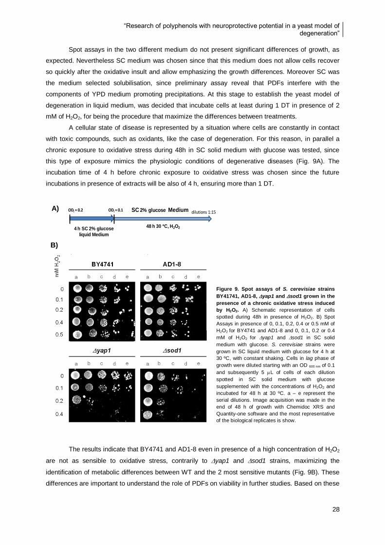
“Research of polyphenols with neuroprotective potential in a yeast model of degeneration”
28
Spot assays in the two different medium do not present significant differences of growth, as
expected. Nevertheless SC medium was chosen since that this medium does not allow cells recover
so quickly after the oxidative insult and allow emphasizing the growth differences. Moreover SC was
the medium selected solubilisation, since preliminary assay reveal that PDFs interfere with the
components of YPD medium promoting precipitations. At this stage to establish the yeast model of
degeneration in liquid medium, was decided that incubate cells at least during 1 DT in presence of 2
mM of H2O2, for being the procedure that maximize the differences between treatments.
A cellular state of disease is represented by a situation where cells are constantly in contact
with toxic compounds, such as oxidants, like the case of degeneration. For this reason, in parallel a
chronic exposure to oxidative stress during 48h in SC solid medium with glucose was tested, since
this type of exposure mimics the physiologic conditions of degenerative diseases (Fig. 9A). The
incubation time of 4 h before chronic exposure to oxidative stress was chosen since the future
incubations in presence of extracts will be also of 4 h, ensuring more than 1 DT.
The results indicate that BY4741 and AD1-8 even in presence of a high concentration of H2O2
are not as sensible to oxidative stress, contrarily to yap1 and sod1 strains, maximizing the
identification of metabolic differences between WT and the 2 most sensitive mutants (Fig. 9B). These
differences are important to understand the role of PDFs on viability in further studies. Based on these
A)
B)
Figure 9. Spot assays of S. cerevisiae strains
BY41741, AD1-8, yap1 and sod1 grown in the
presence of a chronic oxidative stress induced
by H2O2. A) Schematic representation of cells
spotted during 48h in presence of H2O2. B) Spot
Assays in presence of 0, 0.1, 0.2, 0.4 or 0.5 mM of
H2O2 for BY4741 and AD1-8 and 0, 0.1, 0.2 or 0.4
mM of H2O2 for yap1 and sod1 in SC solid
medium with glucose. S. cerevisiae strains were
grown in SC liquid medium with glucose for 4 h at
30 ºC, with constant shaking. Cells in lag phase of
growth were diluted starting with an OD 600 nm of 0.1
and subsequently 5 L of cells of each dilution
spotted in SC solid medium with glucose
supplemented with the concentrations of H2O2 and
incubated for 48 h at 30 ºC. a – e represent the
serial dilutions. Image acquisition was made in the
end of 48 h of growth with Chemidoc XRS and
Quantity-one software and the most representative
of the biological replicates is show.
4 h SC 2% glucose
liquid Medium
48 h 30 ºC, H2O2
ODi = 0.2 ODi = 0.1 dilutions 1:15SC 2% glucose Medium
“Research of polyphenols with neuroprotective potential in a yeast model of degeneration”
29
results was decided that the most appropriated medium to spot cells for further evaluations is SC solid
medium with glucose and with chronic exposure to H2O2.
According with these observations was also decided use a concentration of 0.15 mM of H2O2
for yap1 and sod1 and 0.5 mM of H2O2, for BY4741 and AD1-8, in the further studies. The
concentration of 0.15 mM of H2O2 was chosen for yap1 and sod1 (results not shown) since that in
presence of 0.2 mM of H2O2 yap1 cells viability is too low and sod1 also shows a significant
decrease on viability. For further studies was decided to use the same concentration of H2O2 for the
two mutants directly involved in oxidative stress defense (yap1 and sod1), since that differential
alterations on viability of cells submitted to the same growth conditions could provide important
information about the affected mechanism.
For both BY4741 and AD1-8 was decided to test only until 0.5 mM of H2O2 since that AD1-8
not denoted a significant decrease on viability between 0.1 and 0.5 mM of H2O2 concentrations and
BY4741 is not affected significantly by any tested concentration. As expected the WT strain
demonstrate to be the more resistant in both liquid and solid medium to H2O2. The differences
demonstrated on viability must be due to the fact of BY4741 and AD1-8 has all the antioxidant
defenses that are not present in yap1 and sod1 mutants.
All the experiments described in this section were important to optimize this model already
implemented in DSB laboratory for further studies of this work implicated in cells protection by plant
extracts. Finally the selection of the most appropriate conditions must be taking into account the type
of experiment and the type of exposure (chronic or acute).
The yap1 and sod1 mutants present different responses to a wide range of oxidant
molecules what becomes difficult to determine which events lead to loss of viability of cells following
damage. These responses depend on the dose, at very low doses the cell can adapt to become more
resistant to a subsequent lethal exposure while at higher doses the cell activate various antioxidant
functions including a program of gene expression mediated mainly by several transcription factors, like
for example Yap1p. These distinct responses include expression of proteins, differentially induced by
H2O2 or menadione, as a source of superoxide [21, 29, 37, 40, 42, 91].
CellTiter-Blue viability assay optimization
Cell viability and growth was also monitored by CellTiter-Blue viability assay that is based on a
blue weakly fluorescent indicator dye named resazurin.
Resazurin can enter in viable cells and already inside of cells undergoes enzymatic reduction
into a highly fluorescet pink fluorophor resorufin, in mitochondria, due to the activity of enzymes such
as: flavin mononucleotide dehydrogenase, flavin adenine dinucleotide dehydrogenase, nicotinamide
adenine dehydrogenase, and cytochromes. Resorufin after irreversible reduction reactions is excreted
from cells to the medium.
The CellTiter-Blue viability assay is described to be used in human cell lines. For this reason
was essential the optimization of the methodology parameters and sensibility according with the model
and type of cells, being significantly different when applied to yeast, since that yeast presents a much
faster metabolim. Due to the fact of CellTiter-Blue viability assay being based on the quantification of a
“Research of polyphenols with neuroprotective potential in a yeast model of degeneration”
30
fluorescent indicator produced during cells metabolism is important to obtain linear fluorescence
detection and avoid over metabolization of resazurin in resorufin. In order to accomplish these
requirements, the appropriated number of cells for inocullum and time of incubation were determined.
Both a high or low number of cells cannot be measured due to the limits of detection of the
fluorimetter. When occur an over metabolization inside the cells of resazurin in resorufin is visible a
change on color of the medium from blue to pink.
To determine the appropriate number of cells, S. cerevisiae strains OD 600 nm inoculum of
0.025, 0.05, 0.1 or 0.2 were performed and cells incubated in presence of resazurin and fluorescence
measured. The selected incubation time of 45 min was the time that allowed the maximization of the
differences between treatments.
For BY4741, yap1 and sod1, the results (data not show) demonstrate that the inoculums
with OD 600 nm of 0.025 is the most appropriated, being used in the further studies. The other tested
inoculum present an imediatly change of color of medium to pink, denoting over metabolization. The
viability of AD1-8 strain cannot be evaluated by this methodology, since the OD 600 nm of 0.025 for this
strain presents over metabolization (data not shown) and above 0.02 of OD 600 nm the
spectrophotometer is not capable of detect correctly the number of cells.
Since AD1-8 strain features eight deletions in multidrug resistance (MDR) genes, the efflux of
metabolites is compromised leading to an over metabolization of several compounds. This represents
one possible explanation for the results obtained by CellTiter-Blue viability assay. Besides this
observation, the spot assays results for AD1-8 strain do not present significant changes on viability in
presence of oxidative stress, showing a similar performance to BY4741.
The S. cerevisiae transcription factor Yap1p also plays an important role in MDR by activating
target genes involved in cellular detoxification, such as YCF1, that codifies for an ATP binding
cassette (ABC) transporter and functions as a glutathione S-conjugate pump. Besides, AD1-8 strain
presents the capacity of keep the PDFs inside of cell is an approach to prolong the protective effects
or even an eventually scavenging capacity [40]. For these reasons AD1-8 strain was initially chosen to
be part of this model. However due to this strain behaviour in CellTiter-Blue viability assay and poor
sensibility to H2O2 stress, the AD1-8 strain was discarded in following studies.
4.2. Characterization of polyphenol-digested fractions (PDFs)
In this study polyphenol fractions were chemically characterized before and after an in vitro
digestion process. The total phenolic content and in vitro antioxidant capacity of the fractions obtained
during the main steps of IVD process were performed to understand the role of digestion in
polyphenols protection capacity. This chemical characterization was performed to three different
fractions. The original fraction obtained after hydroethanolic extraction before digestion (Orig.); the
digested fraction obtained in the end of IVD and that is composed by IN fraction representing the
serum available part plus the OUT fraction representing the colon available part of polyphenols
content (Diges.) representing IN+OUT; and finally the fraction that were tested in the yeast model of
degeneration, the digested fraction after a SPE cleaning process to eliminate all digestion compounds
such as bile salts and pancreatin (PDF).

“Research of polyphenols with neuroprotective potential in a yeast model of degeneration”
31
For this study were selected fruits and leaves, from C. album (L.) D. Don (Empetraceae) also
known as Portuguese crowberry and A. unedo (L.) commonly known as strawberry tree (Ericaceae
family), based on previous unpublished results from DSB laboratory. Undigested extracts from both
species reveal neuroprotective portential, in previous studies on a yeast model of Parkinson´s disease
(yeast overexpressing α-synuclein), unpublish results from DSB laboratory. R. idaeus cv polka fruit
(Rosaceae family) or red raspberry and G. biloba leaf (Maidenhair tree, Ginkgoaceae) were also
chosen due to their well known health benefits, to comparison purposes, for fruits and leaves,
respectively [51, 67, 72]
To understand how digestion affects the properties of polyphenols, the PDFs were chemical
characterized and their bioactivity analysed in the yeast model of degeneration established. The
chemical characterization includes the total phenolic content and in vitro antioxidant capacity
performed by Folin-Ciocalteu and ORAC methods respectively, also the LC-MS profile was obtain and
is currently under analysis in collaboration with Derek Stewert from James Hutton Institute, Dundee,
Scotland.
According to Folin-Ciocalteu it is possible to observe that there is a significant loss on total
phenolic content during the 2 main steps of the IVD (Digest. and PDF extracts) compared with the
original (Orig.). The results demonstrate that leaves present higher levels of phenolic content than
fruits for almost all the fractions analysed (Fig. 10). The total content in phenolic compounds is
represented in mg of acid gallic equivalent (mg GAE) normalized for the total volume obtain for each
fraction, to demonstrate the quantity of phenolic compounds that are collected, independently of the
volumes loss during this process.
Figure 10. Total phenolic content for polyphenol extracts in each step of the IVD process. Total phenolic content was
quantified by Folin-Ciocalteu method: Orig. (original fraction before IVD), Diges. (Digested fraction after IVD representing IN and
OUT fractions together) and PDF (polyphenol-digested fraction after SPE column). Values represent the mean SD of three
independent measurements in mg GAE. *** represent statistical significance for p <0.001 compared with the Orig. fraction of the
respective specie.
0
200
400
600
800
1000
1200
Orig
.
Dig
es.
PD
F
Orig
.
Dig
es.
PD
F
Orig
.
Dig
es.
PD
F
Orig
.
Dig
es.
PD
F
Orig
.
Dig
es.
PD
F
Orig
.
Dig
es.
PD
F
R. idaeus Fruit A.unedo Fruit C.album Fruit G. biloba Leaf A.unedo Leaf C.album Leaf
Fruits Leaves
mg
GA
E
Folin mg GAE
R. ideaus C.album A. unedo C.album A. unedo G. biloba
Total Phenolic Content (mg GAE)
*** *** ***
*** *** *** *** ***
***
***
***
***
“Research of polyphenols with neuroprotective potential in a yeast model of degeneration”
32
The highest value was obtained for the orig. fraction of A. unedo leaf, with 1127 33 mg GAE.
Regarding to the fruits, A. unedo presents the highest levels for all fractions, having the Orig. fraction
156 10 mg GAE.
The decrease on total amount of compounds in both Diges. and PDF fractions, comparing to
the Orig. was expected since digestion is an aggressive transformation that implies chemical changes.
For this reason the percentage of phenols lost were also evaluated and compared with Orig. fractions
by specie (Fig. 11 and 14), for fruits and leaves respectively. Due to the fact of digested fraction had
pass through a cleaning step that is always responsible for losses, lower values for PDFs were also
expected.
Folin-Ciocalteu method was an initial screening that allows the comparison between the
fractions chosen for this study with R. ideaus and G. biloba, for fruits and leaves, respectively, since
this species are described as having health benefits. These results show that the chosen fruits for this
study A. unedo and C. album present approximately the same among of phenolic compounds than R.
ideaus (Fig. 10).
Phytochemical studies showed that both fruit and leaf extracts from A. unedo contain several
classes of naturally phytochemicals such as phenolic compounds, like tannins, flavonoids, phenolic
glycosides, anthocyanins and other flavonoids such as gallic acid derivatives. A. unedo berries are
already known as a very good dietary source of phytochemicals based on their chemical composition
and high antioxidant capacity [51, 59, 62]. For C. album a few studies were performed and is not
totally known its composition and biological effects on degeneration.
4.2.1. Fruits
The percentage of original was calculated based on values of mg GAE (Fig. 10), for each
species separetly. The association of total phenol content in Orig. Fraction with the other two (Diges.
and PDF) allows the understanding if the losses of polyphenols is related with the specie and the
different sensibilities of the respective phenolic content to the IVD process.
Relative to the percentage of total phenolic content in original fraction for fruits no more than
19% is recovered, after digestion, being C. album the fruit with the higher percentage of recover for
PDF (Fig.11). However A. unedo fruit PDF do not seem suffer many alterations during IVD since that
Diges. fraction have a high percentage of original, however after SPE procedure the percentage of
phenolic compound recovered in PDF fractions is significantly lower that in Orig. Fraction. Contrarily,
R. ideaus do not show the higher percentage of loss on PDF but is the fraction that represents more
losses due to digestion process since that the Diges. fraction have the lower percentage of original
compared with same fractions of the other tested fruits (Fig. 11). These results can be explained by
the fact of R. ideaus be very rich in anthocyanins, that are very unstable, contrarily to C. album that
have no anthocyanins. All the fruits seem to present a similar decrease on phenolic compounds in end
of the IVD process (PDF fractions) [71].
Other methodology that is used to study the antioxidant properties of extracts is ORAC. This
methodology is based on the antioxidant capacity of metabolites in the presence of a strong oxidant,
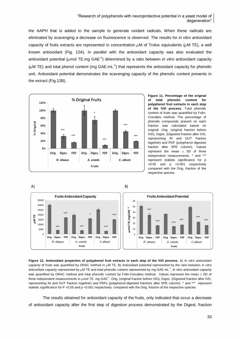
“Research of polyphenols with neuroprotective potential in a yeast model of degeneration”
33
the AAPH that is added to the sample to generate oxidant radicals. When these radicals are
eliminated by scavenging a decrease on fluorescence is observed. The results for in vitro antioxidant
capacity of fruits extracts are represented in concentration M of Trolox equivalents (M TE), a well
known antioxidant (Fig. 12A). In parallel with the antioxidant capacity was also evaluated the
antioxidant potential (mol TE.mg GAE-1
) determined by a ratio between in vitro antioxidant capacity
(M TE) and total phenol content (mg GAE.mL-1
) that represents the antioxidant capacity for phenolic
unit. Antioxidant potential demonstrates the scavenging capacity of the phenolic content presents in
the extract (Fig.12B).
Figure 12. Antioxidant properties of polyphenol fruit extracts in each step of the IVD process. A) In vitro antioxidant
capacity of fruits was quantified by ORAC method in M TE. B) Antioxidant potential represented by the ratio between in vitro
antioxidant capacity represented by M TE and total phenolic content represented by mg GAE.mL-1
. In vitro antioxidant capacity
was quantified by ORAC method and total phenolic content by Folin-Ciocalteu method. Values represent the mean SD of
three independent measurements in mol TE. mg GAE-1
. Orig. (original fraction before IVD), Diges. (Digested fraction after IVD,
representing IN and OUT fraction together) and PDFs (polyphenol-digested fractions after SPE column). * and *** represent
statistic significance for P <0.05 and p <0.001 respectively compared with the Orig. fraction of the respective species.
The results obtained for antioxidant capacity of the fruits, only indicated that occur a decrease
of antioxidant capacity after the first step of digestion process demonstrated by the Digest. fraction
A) B)
0
10
20
30
40
50
60
Orig. Diges. PDF Orig. Diges. PDF Orig. Diges. PDF
Rubus ideaus Arbutus unedo Corema album
Fruits
m
ol T
E.m
gG
AE
-1
Fruits Antioxidant Potential
0
5000
10000
15000
20000
25000
30000
35000
Orig. Diges. PDF Orig. Diges. PDF Orig. Diges. PDF
Rubus ideaus Arbutus unedo Corema album
Fruits
M
TE
Fruits Antioxidant Capacity
R. ideaus A. unedo C. album
Figure 11. Percentage of the original
of total phenolic content for
polyphenol fruit extracts in each step
of the IVD process. Total phenolic
content of fruits was quantified by Folin-
Ciocalteu method. The percentage of
phenolic compounds present on each
fraction was calculated based on
original: Orig. (original fraction before
IVD), Diges. (Digested fraction after IVD,
representing IN and OUT fraction
together) and PDF (polyphenol digested
fraction after SPE column). Values
represent the mean SD of three
independent measurements. * and ***
represent statistic significance for p
<0.05 and p <0.001 respectively
compared with the Orig. fraction of the
respective species.
R. ideaus A. unedo C.album R. ideaus A. unedo C.album
***
***
*
***
***
***
***
***
***
*
***
***
***
*** *** ***
*** ***

“Research of polyphenols with neuroprotective potential in a yeast model of degeneration”
34
(Fig. 12), but it seems to have a recovering after SPE, showed by PDF values for all fruits. The
decrease on antioxidant capacity demonstrated for Diges. fraction can occur due to
loss/transformation of polyphenols during IVD, corroborating the total phenolic content results.
Antioxidant potential results demonstrat that the loss of phenolic compounds in Diges. fraction
is accompanied by a loss of antioxidant capacity, since that antioxidant potential is a value normalized
for the content in phenolic compouds (Fig. 12B). The results obtained for antioxidant potential in each
step of IVD are different according with the species probably due to different compositions in phenolic
compounds. R. ideaus is the fruit that presents the higher antioxidant potential for Orig. and Diges.
fractions, with 50.6 1.4 mol TE.mg GAE-1
and 34.5 3.4 mol TE.mg GAE-1
, respectively.
However, according with the antioxidant potential, R. ideaus is the fraction that suffers more losses
during SPE process, presenting the lower levels for PDF comparing with the other fruits (Fig. 12B).
Figure 13. PDFs toxicity evaluation by spot assays for S. cerevisiae strains BY4741, yap1 and sod1 exposure to
PDFs from fruits. BY4741, yap1 and sod1 were grown in SC liquid medium with glucose supplemented with concentrations
of 0, 62, 125, 250, 500 or 1000 g GAE .mL-1
of PDFs from fruits (R. ideaus fruit, A. unedo fruit and C. album fruit), for 4 h at 30
ºC, with constant shaking. Cells in lag phase of growth were diluted starting with an OD 600 nm of 0.1 and subsequently 5 L of
cells of each dilution spotted in SC solid medium with glucose and incubated for 48 h at 30 ºC. a - e represent the serial
dilutions. Image acquisition was made in the end of 48 h of growth with Chemidoc XRS and Quantity-one software.
The direct correlation between antioxidant potential or phenolic content and the biological role
of extract compounds inside of the cells cannot be done. Because of this it is important to evaluate the
direct influence of extracts in the cellular metabolism through different methodologies. The non-toxic
range of PDFs concentrations in the yeast strains BY4741, yap1 and sod1 were determined, as
well as the concentrations that was used in the protective assays in the degeneration model.
In order to achieve the more appropriated concentrations of PDFs from all characterized
species, to use in further tasks, the PDFs toxicity was first evaluated by spot assays as described in
R. ideaus Fruit
A. unedo Fruit C. album Fruit
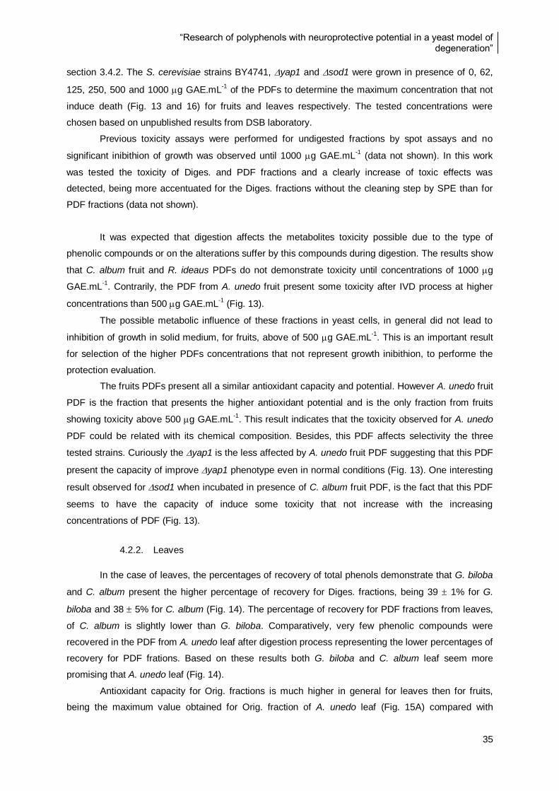
“Research of polyphenols with neuroprotective potential in a yeast model of degeneration”
35
section 3.4.2. The S. cerevisiae strains BY4741, yap1 and sod1 were grown in presence of 0, 62,
125, 250, 500 and 1000 g GAE.mL-1
of the PDFs to determine the maximum concentration that not
induce death (Fig. 13 and 16) for fruits and leaves respectively. The tested concentrations were
chosen based on unpublished results from DSB laboratory.
Previous toxicity assays were performed for undigested fractions by spot assays and no
significant inibithion of growth was observed until 1000 g GAE.mL-1
(data not shown). In this work
was tested the toxicity of Diges. and PDF fractions and a clearly increase of toxic effects was
detected, being more accentuated for the Diges. fractions without the cleaning step by SPE than for
PDF fractions (data not shown).
It was expected that digestion affects the metabolites toxicity possible due to the type of
phenolic compounds or on the alterations suffer by this compounds during digestion. The results show
that C. album fruit and R. ideaus PDFs do not demonstrate toxicity until concentrations of 1000 g
GAE.mL-1
. Contrarily, the PDF from A. unedo fruit present some toxicity after IVD process at higher
concentrations than 500 g GAE.mL-1
(Fig. 13).
The possible metabolic influence of these fractions in yeast cells, in general did not lead to
inhibition of growth in solid medium, for fruits, above of 500 g GAE.mL-1
. This is an important result
for selection of the higher PDFs concentrations that not represent growth inibithion, to performe the
protection evaluation.
The fruits PDFs present all a similar antioxidant capacity and potential. However A. unedo fruit
PDF is the fraction that presents the higher antioxidant potential and is the only fraction from fruits
showing toxicity above 500 g GAE.mL-1
. This result indicates that the toxicity observed for A. unedo
PDF could be related with its chemical composition. Besides, this PDF affects selectivity the three
tested strains. Curiously the yap1 is the less affected by A. unedo fruit PDF suggesting that this PDF
present the capacity of improve yap1 phenotype even in normal conditions (Fig. 13). One interesting
result observed for sod1 when incubated in presence of C. album fruit PDF, is the fact that this PDF
seems to have the capacity of induce some toxicity that not increase with the increasing
concentrations of PDF (Fig. 13).
4.2.2. Leaves
In the case of leaves, the percentages of recovery of total phenols demonstrate that G. biloba
and C. album present the higher percentage of recovery for Diges. fractions, being 39 1% for G.
biloba and 38 5% for C. album (Fig. 14). The percentage of recovery for PDF fractions from leaves,
of C. album is slightly lower than G. biloba. Comparatively, very few phenolic compounds were
recovered in the PDF from A. unedo leaf after digestion process representing the lower percentages of
recovery for PDF frations. Based on these results both G. biloba and C. album leaf seem more
promising that A. unedo leaf (Fig. 14).
Antioxidant capacity for Orig. fractions is much higher in general for leaves then for fruits,
being the maximum value obtained for Orig. fraction of A. unedo leaf (Fig. 15A) compared with
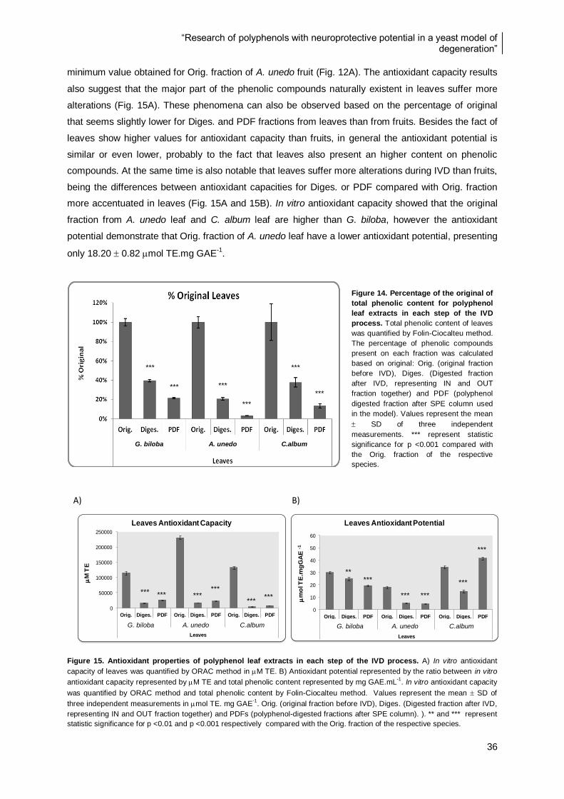
“Research of polyphenols with neuroprotective potential in a yeast model of degeneration”
36
minimum value obtained for Orig. fraction of A. unedo fruit (Fig. 12A). The antioxidant capacity results
also suggest that the major part of the phenolic compounds naturally existent in leaves suffer more
alterations (Fig. 15A). These phenomena can also be observed based on the percentage of original
that seems slightly lower for Diges. and PDF fractions from leaves than from fruits. Besides the fact of
leaves show higher values for antioxidant capacity than fruits, in general the antioxidant potential is
similar or even lower, probably to the fact that leaves also present an higher content on phenolic
compounds. At the same time is also notable that leaves suffer more alterations during IVD than fruits,
being the differences between antioxidant capacities for Diges. or PDF compared with Orig. fraction
more accentuated in leaves (Fig. 15A and 15B). In vitro antioxidant capacity showed that the original
fraction from A. unedo leaf and C. album leaf are higher than G. biloba, however the antioxidant
potential demonstrate that Orig. fraction of A. unedo leaf have a lower antioxidant potential, presenting
only 18.20 0.82 mol TE.mg GAE-1
.
Figure 15. Antioxidant properties of polyphenol leaf extracts in each step of the IVD process. A) In vitro antioxidant
capacity of leaves was quantified by ORAC method in M TE. B) Antioxidant potential represented by the ratio between in vitro
antioxidant capacity represented by M TE and total phenolic content represented by mg GAE.mL-1
. In vitro antioxidant capacity
was quantified by ORAC method and total phenolic content by Folin-Ciocalteu method. Values represent the mean SD of
three independent measurements in mol TE. mg GAE-1
. Orig. (original fraction before IVD), Diges. (Digested fraction after IVD,
representing IN and OUT fraction together) and PDFs (polyphenol-digested fractions after SPE column). ). ** and *** represent
statistic significance for p <0.01 and p <0.001 respectively compared with the Orig. fraction of the respective species.
A) B)
0
10
20
30
40
50
60
Orig. Diges. PDF Orig. Diges. PDF Orig. Diges. PDF
Ginkgo biloba Arbutus unedo Corema album
Leaves
m
ol T
E.m
gG
AE
-1
Leaves Antioxidant Potential
0
50000
100000
150000
200000
250000
Orig. Diges. PDF Orig. Diges. PDF Orig. Diges. PDF
Ginkgo biloba Arbutus unedo Corema album
Leaves
M
TE
Leaves Antioxidant Capacity
Figure 14. Percentage of the original of
total phenolic content for polyphenol
leaf extracts in each step of the IVD
process. Total phenolic content of leaves
was quantified by Folin-Ciocalteu method.
The percentage of phenolic compounds
present on each fraction was calculated
based on original: Orig. (original fraction
before IVD), Diges. (Digested fraction
after IVD, representing IN and OUT
fraction together) and PDF (polyphenol
digested fraction after SPE column used
in the model). Values represent the mean
SD of three independent
measurements. *** represent statistic
significance for p <0.001 compared with
the Orig. fraction of the respective
species.
G. biloba A. unedo C.album
G. biloba A. unedo C.album G. biloba A. unedo C.album
***
*** ***
***
***
***
*** *** *** ***
*** ***
** ***
*** ***
***
***

“Research of polyphenols with neuroprotective potential in a yeast model of degeneration”
37
The antioxidant potential for A. unedo leaf present the lower levels for all the three
characterized fractions, including for Orig. fraction, despite of be the leaf that has the higher in vitro
antioxidant capacity for Orig. fraction.
Fruits and leaves present a similar pattern for antioxidant capacity of PDFs, since these
fractions also present higher values of antioxidant capacity than Diges. frations. Antioxidant potential
results demonstrate that G. biloba is the plant that seems to have this parameter less affected by
digestion process, since that the Diges. fraction for this leaf is the fraction that show less differences
compared with Orig. fraction. However after SPE occur a significantly lost of phenolic compounds (Fig.
15B), result corroborated with percentage of original of total phenol content (Fig. 14). The PDF fraction
of C. album leaf, contrarily to fruit is the only fraction that has higher values than Orig. fraction, for
antioxidant potential, which can indicates that SPE clean substances that could interfere with Folin-
Ciocalteu method (Fig. 15B).
To access the PDFs toxicity from leaves, PDFs were tested by spot assays in the same
concentrations of fruits, between 0 – 1000 g GAE.mL-1
. The results represented show that
independently of possible changes in cellular metabolism of yeast only A. unedo leaf PDF does not
present any toxicity until concentrations of 1000 g GAE.mL-1
(Fig. 13), contrarily to observe for A.
unedo fruit PDF that induces death at concentrations higher than 500 g GAE.mL-1
(Fig. 16).
Figure 16. PDFs toxicity evaluation by spot assays for S. cerevisiae strains BY4741, yap1 and sod1 exposure to
PDFs from leaves. BY4741, yap1 and sod1 were grown in SC liquid medium with glucose supplemented with concentrations
of 0, 62, 125, 250, 500 or 1000 g GAE.mL-1
of PDFs from leaves (G.biloba leaf, A. unedo leaf and C. album leaf), for 4 h at 30
ºC, with constant shaking. Cells in lag phase of growth were diluted starting with an OD 600 nm of 0.1 and subsequently 5 L of
cells of each dilution spotted in SC solid medium with glucose and incubated for 48 h at 30 ºC. a - e represent the serial
dilutions. Image acquisition was made in the end of 48 h of growth with Chemidoc XRS and Quantity-one software.
G. biloba leaf
A. unedo leaf C. album leaf

“Research of polyphenols with neuroprotective potential in a yeast model of degeneration”
38
For this type of matrix C. album leaf PDF is toxic for yeast cells, showing lost of viability for
sod1 at concentrations higher than 250 g GAE.mL-1
and for yap1 and BY4741 higher than 125 g
GAE.mL-1
. The differences on toxicity of analised fractions can indicate that phytochemical
compounds with electrophilic properties can influence the viability of mutants differentially, probably
due to the alteration of redox homeostasis of cells (Fig. 16). Interestingly in the case of leaves, C.
album PDF is the fraction that show higher values for antioxidant potential, comparing with all other
fractions from leaves, chemically characterized.
For G. biloba leaf, these results indicate a slightly decrease on viability with the increase of
PDF concentration, however this changes on viability just become more accentuated for sod1 in
concentrations higher than 500 g GAE.mL-1
(Fig. 16). G. biloba PDF also present a high antioxidant
capacity comparing with A. unedo leaf PDF, however the value of antioxidant potential is not higher
than antioxidant potential of fruits. In general fruits are less toxic than leaves, a result consistent with
the fact of all the results presented in this section.
Based on these results together with the results obtain for the fruits, it was chosen 0, 62, 125
and 250 g GAE.mL-1
of PDFs fractions as the concentrations to use in further protective evaluation
by spot assays for all the species except for C.album leaf PDF. This fraction was tested until the
concentration of 125 g GAE.mL-1, since that 250 g GAE.mL
-1 of this PDF is already too toxic for
yeast cells.
Like in fruits the toxicity analisys was compared for Diges. without SPE and PDF fractions.
The results obtained for this toxicity screen reveal that the Diges. fractions without SPE are very toxic
for yeast cells for all tested fractions demonstrated to be very toxic. The other tested Diges. fractions
present a similar behaviour (Data not shown). The cleaning step by SPE allowed the cleaning of the
digestion compounds keeping only polyphenols that are available in serum and in colon. This
demonstrate to be very important to ensure polyphenol bioactivities analisys without digestion
resiudes that could interfere. After SPE column the PDFs obtained were tested in the yeast model of
degeneration for protection (Fig. 15 and 18), demonstrating that in fact after this procedure the major
part of toxic compounds are eliminated and almost all fractions did not present toxicity until 1000 g
GAE.mL-1
.
4.3. Selection of polyphenol-digested fractions with protective potential
4.3.1. Viability analysis by Spot Assays
The protective potential of PDFs was carried out by two different methods. Viability was
determined by spot assays and the general cellular state evaluated by resazurin-based assay. To
achieve further insight into the bioactivities of PDFs from fruits and leaves in the yeast model of
degeneration, cells were incubated during 4 h with 0, 62, 125 or 250 g GAE.mL-1
of all PDFs except
C. album leaf PDF that was tested only until 125 g GAE.mL-1
, as defined in section 4.1. (Fig. 16).
“Research of polyphenols with neuroprotective potential in a yeast model of degeneration”
39
Finally cells were spotted in SC solid medium with 0.5 mM of H2O2 for BY4741 and 0.15 mM of H2O2
for yap1 and sod1, during 48 h.
Spot assays were performed as a first approach to select the PDFs with protective potential
and to understand the form by each PDF affect the viability according with the strain. Each strain of
this model represents a defect on metabolism and possible changes on growth conditions by the
presence of PDFs can reflect the different roles of PDFs in oxidative metabolic pathways.
In presence of C. album fruit PDF, BY4741 do not show any difference on viability but
contrarily to what was expected was observed that this PDF present a curiously result, since that
seems to affect yap1 and sod1 differentially. This fraction is not able to improve yap1 cells viability
and seems to induce some lost of viability in the yeast model of degeneration that was not
demonstrate to be toxic for yeast cells. However for sod1 is not totally clear if is capable of provide
any viability improvement concretely (Fig. 17). For these reason C. album fruit PDF was evaluated in
further methodologies.
A. unedo fruit and leaf PDFs show a clear increase of viable cells after being subjected to an
oxidative stress for 48 h. A. unedo fruit is the only PDF providing viability improvements for the two
mutant strains yap1 and sod1. The PDF from A. unedo leaf can ameliorate the H2O2 induced
damaged only for yap1 mutant (Fig. 17).
Cells growth in presence of G. biloba PDF seems differently affected according with the strain,
being sod1 the only strain that present a slightly improve on viability (Fig. 17). Contrarily, R. ideaus
PDF does not ameliorate the damage induced by the H2O2 for any strain, an interesting observation
since that several studies based on phytochemical composition of this berries point their benefitial
effects in some ageing disorders including neurodegeneration [69]. Gordon, M. and co-workers
evaluated the effects of IVD in anthocyanins compounds, showing that this polyphenols suffer many
alterations during this process [76]. Since red raspberry is rich in anthocyanins, the observed results
for protecting assay could suggest that the IVD possible alter the protecting capacity of this berries.
With these results is possible to conclude that not all PDFs have the capacity of improve the
viability by protecting cells during a chronic exposure to oxidative stress, such as R. ideaus and C.
album leaf PDFs. On the other hand A. unedo leaf and fruit PDFs, were the fractions with more
potential to be explored. yap1 was the strain that was more responsive to improvements on viability
by PDFs (Fig. 17).
The results obtained for A. unedo leaf PDF are surprising since the antioxidant capacity per
unit of phenol content, shown in section 4.2.1. is lower than the other PDFs. One possible explanation
for these results is that this PDF can improve viability not through antioxidant mechanisms but
functioning thought other molecular mechanisms. To confirme if ROS scanvenging is involved in the
protective mechanisms induced by these fractions, ROS production was evaluated after cells growth
in presence of PDFs with protective potential.
The PDFs from A. unedo fruit and leaf exhibit a rising protecting capacity with the increase of
PDFs concentration until the tested 250 g GAE.mL-1, demonstrating a dose response. This increase
on viability promoted by C. album fruit PDF is not clear and for this reason becomes interestingly to
evaluate its biological role in cells by other methodologies.

“Research of polyphenols with neuroprotective potential in a yeast model of degeneration”
40
Figure 17. PDFs protective potential evaluation by spot assays for S. cerevisiae strains BY4741, yap1 and sod1
exposure to PDFs and subjected to an oxidative stress. BY4741, yap1 and sod1 were grown in SC liquid medium with
glucose supplemented with concentrations of 0, 62, 125 or 250 g GAE.mL-1
of all PDFs, except for C. album leaf tested only
until 125 g GAE.mL-1
, for 4 h at 30 ºC, with constant shaking. Cells in lag phase of growth were diluted starting with an OD 600
nm of 0.1 and subsequently 5 L of cells of each dilution spotted in SC solid medium with glucose supplemented with 0 mM of
H2O2 or 0.5 mM of H2O2 for BY4741 and 0.15 mM of H2O2 for yap1 and sod1; cells were incubated for 48 h at 30 ºC. a – e
represent the serial dilutions. Image acquisition was made in the end of 48 h of growth with Chemidoc XRS and Quantity-one
software.
A. u
ne
do
C. a
lbu
mR
. id
ea
us
Fruits
G. b
ilo
ba
Leaves
A. u
ne
do
C. a
lbu
m
“Research of polyphenols with neuroprotective potential in a yeast model of degeneration”
41
4.3.2. CellTiter-Blue viability assay analysis
Many recent studies have proven that mitochondria are one of the cellular components with
the most impact in ageing disorders influenced by oxidative stress to keep cell homeostasis. To
understand how the most promising PDFs, demonstrated by spot assays results (Fig. 17) influence
the mitochondrial state we also assay for viability evaluation by CellTiter-Blue viability assay that is
based on mitochondrial functional metabolism. CellTiter-Blue viability assay is method that also
reflects the mitochondrial state of cells, since resazurin is reduced by mitochondrial enzymes in viable
cells. The fact of resazurin became reduced due to the activity of mitochondrial enzymes associated
with some of the most signalling pathways on cell, allow the understanding the metabolic sate of cells
and consequently the viability.
Based on protective effect results by spot assays, only A. unedo fruit and leaf and C. album
fruit PDFs were evaluated in the next tasks of this work (Fig. 17). The previous results also allow the
choosing of 0 and 250 g GAE.mL-1 concentrations of the PDFs, since that 250 g GAE.mL
-1 is the
higher concentration of phenolic compounds showing protective capacity. S. cerevisiae strains were
grown in SC liquid medium with glucose supplemented with or without PDFs and subsequently grown
in the same medium supplemented with 2 mM of H2O2 to induce the oxidative stress condition. The
results are shown in percentage of viability, normalized for the number of cells, after cells being
incubated during 45 min in presence of the resazurin compound (Fig.18).
This approach is important to understand the cellular state of each strains according with the
treatment, which can not be inferred by spot assays. The percentage of viability implies a relation
between enzymatic activity of cells in normal conditions, to infer the alterations of this activity in cells
submitted to the tested condition. One the other hand provides information about how the different
mutants lead with oxidative stress and which pathways can be possible more affected by the presence
of PDFs.
When the percentage of viability is calculated for BY4741 it is not observed any differences in
cells viability after incubation with 250 g GAE.mL-1
of PDFs. At the same time a decrease on viability
is observed for cells incubated only in the presence of H2O2 for this strain, suggesting that could occur
mechanism related with mitochondria, such as mitofagy and that is not verify by spot assays.
In these results is also possible to observe a slight improvement for the BY4741 viability in the
presence of PDFs from A. unedo fruit and leaf, when cells are submitted to an oxidative stress. For A.
unedo leaf the improvements observed in the cellular viability demonstrate that this is the most
favourable fraction, presenting higher viability values (Fig. 18). Interestingly the improvements
demonstrated by this methodology for BY4741 in the presence of A. unedo fruit and leaf are not clear
by spot assays (Fig. 17 and 18), suggesting that PDFs beneficial effect may be occur through
direct/indirect amelioration of enzymatic mitochondrial activity. The fact of the percentage of viability
for yap1 do not demonstrate significant difference for BY4741 in presence of H2O2 could indicate a
mechanism of degeneration in commun for both strains that not implies Yap1p activation, such as
autophagic process. However more studies must be performed.
The results indicate that in normal conditions the yap1 strain has the lower viability, as
expected, due to lower levels of percentage of viability when submitted to oxidative stress. These
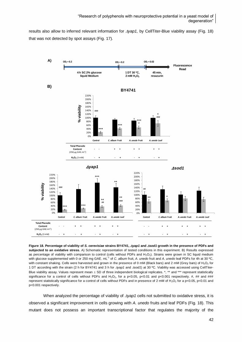
“Research of polyphenols with neuroprotective potential in a yeast model of degeneration”
42
results also allow to inferred relevant information for yap1, by CellTiter-Blue viability assay (Fig. 18)
that was not detected by spot assays (Fig. 17).
Figure 18. Percentage of viability of S. cerevisiae strains BY4741, yap1 and sod1 growth in the presence of PDFs and
subjected to an oxidative stress. A) Schematic representation of tested conditions in this experiment. B) Results expressed
as percentage of viability with comparison to control (cells without PDFs and H2O2). Strains were grown in SC liquid medium
with glucose supplemented with 0 or 250 mg GAE. mL-1
of C. album fruit, A. unedo fruit and A. unedo leaf PDFs for 4h at 30 ºC,
with constant shaking. Cells were harvested and grown in the presence of 0 mM (Black bars) and 2 mM (Grey bars) of H2O2 for
1 DT according with the strain (2 h for BY4741 and 3 h for yap1 and sod1) at 30 ºC. Viability was accessed using CellTiter-
Blue viability assay. Values represent mean SD of three independent biological replicates. *, ** and *** represent statistically
significance for a control of cells without PDFs and H2O2, for a p<0.05, p<0.01 and p<0.001 respectively. #, ## and ###
represent statistically significance for a control of cells without PDFs and in presence of 2 mM of H2O2 for a p<0.05, p<0.01 and
p<0.001 respectively.
When analyzed the percentage of viability of yap1 cells not submitted to oxidative stress, it is
observed a significant improvement in cells growing with A. unedo fruits and leaf PDFs (Fig. 18). This
mutant does not possess an important transcriptional factor that regulates the majority of the
0%
20%
40%
60%
80%
100%
120%
140%
160%
180%
200%
220%
Controlo C. album Fruit
A. unedo Fruit
A. unedo Leaf
sod1
**
##
*
0%
20%
40%
60%
80%
100%
120%
140%
160%
180%
200%
220%
Controlo C. album. Fruit A. unedo Fruit A. unedo Leaf
yap1
***
***
###
#####
**
***
****
0%
20%
40%
60%
80%
100%
120%
140%
160%
180%
200%
220%
Controlo C. album.
Fruit
A. unedo
Fruit
A. unedo
Leaf
% v
iab
ilit
y
*
***
***
***
###
0
BY4741
#
###
Control C. album Fruit A. unedo Fruit A. unedo Leaf
Total PhenolicContent
(250 g.GAE.ml-1)
- - + + + + + +
H2O2 (2 mM) - + - + - + - +
Control C. album Fruit A. unedo Fruit A. unedo Leaf
- - + + + + + +
- + - + - + - +
Control C. album Fruit A. unedo Fruit A. unedo Leaf
Total PhenolicContent
(250 g.GAE.ml-1)
- - + + + + + +
H2O2 (2 mM) - + - + - + - +
% v
iab
ilit
y
A)
4 h SC 2% glucose
liquid Medium
1 DT 30 ºC,
2 mM H2O2
ODi = 0.2 ODi = 0.2 ODi = 0.02
Fluorescence
Read45 min,
resazurin
B)
*** ***
***
*
*** ***
** **
**
***
** *
“Research of polyphenols with neuroprotective potential in a yeast model of degeneration”
43
antioxidant enzymes; consequently the enzymatic activity is normally low, leading to a lower viability.
When yap1 cells are incubated with the PDFs from A. unedo, the percentage o viability values
obtained rise significantly, above the values obtain for cells in normal conditions. These indicate that
the compounds constituents of A. unedo fruit and leaf PDFs may be have the capacity of ameliorate
the yap1 phenotype. After this study could be interesting analyse if in presence of these PDFs the
cell state can reaches the WT phenotype (Fig. 18).
However in the presence of an oxidative stress condition PDFs have not the same capacity to
improve the viability of this mutant to a control condition. Nevertheless is observed a viability increase
when cells are incubated with A.unedo fruit or leaf (Fig. 18).
The sod1 strain show fewer differences with the several treatments applied in comparison to
the control. One important result was obtained for C. album fruit PDF, representing an improvement
on viability in cells when incubated with this PDF. This observation is not clear in spot assays results
(Fig. 17), demonstrating that the small differences observed by CellTiter-Blue viability assays can
represent a general cellular metabolism improvement (Fig. 18). For the other PDFs there are not
visible significant differences on viability for sod1, but interestingly when sod1 cells are incubated in
the presence of A. unedo fruit PDF and submitted to oxidative stress, the percentage of viability values
show that cells have not a much different viability from cells not submitted to H2O2. These results
indicate that A. unedo fruit PDF could have a preconditioning role in sod1 cells, which allow to the
cells be more responsiveness to oxidative stress (Fig. 18).
In general, cells in presence oxidative stress after submitted to A. unedo fruit PDF have an
improvement of viability, demonstrating a protective effect (Fig. 18). Adaptive stress response towards
H2O2 is performed by a complex network of several different regulons. For example, several genes
appear to be regulated directly by the transcription factor Yap1p, whereas others are regulated by
other mechanisms [11, 28, 42].
The fact of sod1 present a similar behaviour to BY4741 in presence of PDFs, is according
with that fact of sod1 mutant possessing different mechanism of defense against oxidative damage
and in absence of SOD1 gene cells can still respond and maintain activity of mitochondrial enzymes.
The improvements observed in metabolism of BY4741 after induction of oxidative stress and PDFs
incubation, which is not observed in sod1, could be explained by the fact of BY4741 containing all
oxidative stress related defenses enabling a better metabolic response [9, 31]. The fact that BY4741
and yap1 presents improvement more notable than in sod1 by this methodology it can be indicated
that the mechanisms of defense related with Sod1p are not equally affected by these compounds and
that exist different stimuli and pathways to control oxidative stress. To understand the main reasons of
these differential changes and confirm the influence of the oxidative species in cellular mechanisms,
important markers were evaluated such as ROS and GSH/GSSG. The CellTiter-Blue viability assay
results (Fig. 18) together with the results of spot assays (Fig. 17) indicated that oxidative stress is
evolved in cellular dysfunctions that lead to mitochondrial disruption and cell death by several
pathways.
The CellTiter-Blue viability assay results suggest the mitochondria as one of the most
important organelles involved in oxidative stress. Based on these arise the necessity of understand
“Research of polyphenols with neuroprotective potential in a yeast model of degeneration”
44
more directly the role of mitochondria, an important evaluation to perform in further studies with more
targeted methodologies.
4.4. Evaluation of cellular oxidative stress markers
4.4.1. Reactive Oxygen Species
Research in the yeast S. cerevisiae has provided considerable information about oxidative
stress and the mechanisms employed by cells in response to increased of ROS [8]. Cells use an
elaborate enzymatic machinery consisting of superoxide dismutases, reductases, catalases,
peroxiredoxins, glutaredoxins, and glutathione transferases to maintain redox balance. ROS
generation is the final step in many cell degeneration pathways and act as a non-specific rather than
specific damage mechanism. This regulation can be mentioned as an additive mechanism of
neuroprotection by polyphenols, claiming a therapeutic effect for polyphenols via ROS regulation as
the only mechanism that seems to be overdrawn. ROS production is counteracted by an intricate
antioxidant defense system that includes antioxidant enzymes, scavengers molecules but also the
activation of signalling pathways that mediates cell growth and differentiation [7, 10, 16, 34].
In this task ROS production was measured by H2DCF (2,7-dichlorofluorescein) in the S.
cerevisiae strains BY4741, yap1 and sod1. This methodology is important to determine how
oxidative compounds, such as H2O2 or menadione, modify cellular molecules and activate defense
mechanisms in the cells. ROS evaluation also allows examining if PDFs act through ROS scavenging
mechanisms.
The increase of ROS are represented in relative fluorescence units (R.F.U.) determined based
on the ROS production during an incubation time (t), normalized for the number of cells per well and
for initial fluorescence read. This allows the removal of the differences related with the initial number of
cells per well and temporal differences between the H2O2 addition and the first fluorescence
measurement. To infer the results of ROS increase, the fluorescence values were measured during 1
DT according with the strain at each 10 min and only were used values that represented linearity.
Contrary to what could be expected for all strains, was not observed any differences on ROS
production after cells being incubated with the PDFs followed by oxidative stress, demonstrating that
scavenging of ROS are not the mechanism by which PDFs protect cells (Fig. 19). The treatment
described in section 4.3.2 was also used to test ROS production. The results obtained for this assay
indicates that yap1 have a similar performance to BY4741, both representing the higher values for
ROS production when cells are incubated with H2O2. For the tested PDFs yap1 is the strain that
represent a higher difference between production of ROS when cells are incubated in the presence or
absence of H2O2, which indicates that this is the strain with more difficulty to eliminate the ROS
produced, an expected result based on its absence of Yap1p transcriptional factor (Fig. 19).
The A. unedo fruit PDF is the only condition in this results that present a significant alteration
on sod1 cells. When sod1 cells are incubated in presence of A. unedo fruit and subsequently
submitted to oxidative stress no difference is observed compared to cells only incubated with the
PDFs. However A. unedo fruit PDF seems capable of induce a slightly increase on ROS production,

“Research of polyphenols with neuroprotective potential in a yeast model of degeneration”
45
demonstrated by the difference between cells only gowth in presence of A. unedo PDFs and cells
growth in normal conditions (Fig. 19). This could suggest that cells prepare themself to respond to
H2O2 injury by several mechanisms, such as the increase of ROS function or by genes activation and
signalling pathways.
Figure 19. Evaluation of ROS levels in S. cerevisiae strains (BY4741, yap1 and sod1) growth in the presence of PDFs
and subjected to an oxidative stress. A) Schematic representation of tested conditions in this experiment. B) Results are
expressed in relative fluorescence units normalized for number of cells and initial fluorescence (R.F.U) representing the
increase of ROS produced during time of exposure to oxidative stress. Strains were grown in SC liquid medium with glucose
supplemented with 0 and 250 g GAE. mL-1
of C. album fruit, A. unedo fruit and leaf PDFs for 4h at 30 ºC, with constant
shaking. Cells were harvested and grown in presence of 0 mM (Grey bars) and 2 mM (Black bars) of H2O2 for 1 DT according
with the strain (2 h for BY4741 and 3 h for yap1 and sod1) at 30 ºC. ROS produced were quantified by Diclhrofluorescein
assay. Values represent mean SD of three independent biological replicates. *, ** and *** represent statistically significance
for a control of cells without PDFs and H2O2, for a p<0.05, p<0.01 and p<0.001 respectively. #, ## and ### represent statistically
significance for a control of cells without PDFs and in presence of 2 mM of H2O2, for a p<0.05, p<0.01 and p<0.001 respectively.
0
0.5
1
1.5
2
2.5
3
3.5
4
4.5
0 C.a.F A.u.F A.u.L
sod1
***
###
***
### *
## *** ***
###
0
0.5
1
1.5
2
2.5
3
3.5
4
4.5
0 C.a.F A.u.F A.u.L
R.F
.U.
yap1
***
### ### ###
###
*** ***
***
0
0.5
1
1.5
2
2.5
3
3.5
4
4.5
0 C.a.F A.u.F A.u.L
R.F
.U.
BY4741
***
###
***
*** ***
### ###
###
Control C. album Fruit A. unedo Fruit A. unedo Leaf
- - + + + + + +
- + - + - + - +
Control C. album Fruit A. unedo Fruit A. unedo Leaf
Total Phenolic
Content
(250 g.GAE.ml-1)
- - + + + + + +
H2O2 (2 mM) - + - + - + - +
Control C. album Fruit A. unedo Fruit A. unedo Leaf
Total Phenolic
Content
(250 g.GAE.ml-1)
- - + + + + + +
H2O2 (2 mM) - + - + - + - +
A)
B)
4 h SC 2% glucose
liquid Medium
1 DT 30 ºC,
2 mM H2O2
ODi = 0.2 ODi = 0.2
Fluorescence
Read
15 min
DCFH-DA
***
***
*** ***
*** *** ***
*** *
*** *** ***
***
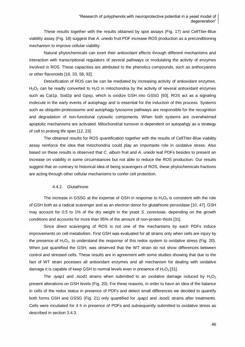
“Research of polyphenols with neuroprotective potential in a yeast model of degeneration”
46
These results together with the results obtained by spot assays (Fig. 17) and CellTiter-Blue
viability assay (Fig. 18) suggest that A. unedo fruit PDF increase ROS production as a preconditioning
mechanism to improve cellular viability.
Natural phytochemicals can exert their antioxidant effects through different mechanisms and
interaction with transcriptional regulators of several pathways or modulating the activity of enzymes
involved in ROS. These capacities are attributed to the phenolics compounds, such as anthocyanins
or other flavonoids [16, 33, 58, 92].
Detoxification of ROS can be can be mediated by increasing activity of antioxidant enzymes.
H2O2 can be readly converted to H2O in mitochondria by the activity of several antioxidant enzymes
such as Cat1p, Sod2p and Gpxp, which is oxidize GSH into GSSG [93]. ROS act as a signaling
molecule in the early events of autophagy and is essential for the induction of this process. Systems
such as ubiquitin-proteossome and autophagy lysosome pathways are responsible for the recognition
and degradation of non-functional cytosolic components. When both systems are overwhelmed
apoptotic mechanisms are activated. Mitochondrial turnover is dependent on autopahgy as a strategy
of cell to prolong life span [12, 23].
The obtained results for ROS quantification together with the results of CellTiter-Blue viability
assay reinforce the idea that mitochondria could play an importante role in oxidative stress. Also
based on these results is observed that C. album fruit and A. unedo leaf PDFs besides to present an
increase on viability in some circumstances but not able to reduce the ROS production. Our results
suggest that on contrary to historical idea of being scavengers of ROS, these phytochemicals fractions
are acting through other cellular mechanisms to confer cell protection.
4.4.2. Glutathione
The increase in GSSG at the expense of GSH in response to H2O2 is consistent with the role
of GSH both as a radical scavenger and as an electron donor for glutathione peroxidase [10, 47]. GSH
may account for 0.5 to 1% of the dry weight in the yeast S. cerevisiae, depending on the growth
conditions and accounts for more than 95% of the amount of non-protein thiols [31].
Since direct scavenging of ROS is not one of the mechanisms by each PDFs induce
improvements on cell metabolism. First GSH was evaluated for all strains only when cells are injury by
the presence of H2O2, to understand the response of this redox system to oxidative stress (Fig. 20).
When just quantified the GSH, was observed that the WT strain do not show differences between
control and stressed cells. These results are in agreement with some studies showing that due to the
fact of WT strain pocesses all antioxidant enzymes and all mechanism for dealing with oxidative
damage it is capable of keep GSH to normal levels even in presence of H2O2 [31].
The yap1 and sod1 strains when submitted to an oxidative damage induced by H2O2
present alterations on GSH levels (Fig. 20). For these reasons, in order to have an idea of the balance
in cells of the redox status in presence of PDFs and detect small differences we decided to quantify
both forms GSH and GSSG (Fig. 21) only quantified for yap1 and sod1 strains after treatments.
Cells were incubated for 4 h in presence of PDFs and subsequently submitted to oxidative stress as
described in section 3.4.3.
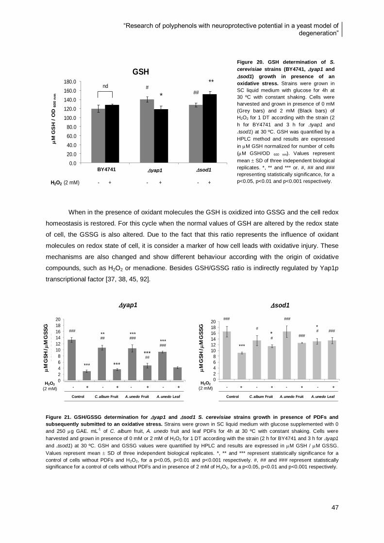
“Research of polyphenols with neuroprotective potential in a yeast model of degeneration”
47
When in the presence of oxidant molecules the GSH is oxidized into GSSG and the cell redox
homeostasis is restored. For this cycle when the normal values of GSH are altered by the redox state
of cell, the GSSG is also altered. Due to the fact that this ratio represents the influence of oxidant
molecules on redox state of cell, it is consider a marker of how cell leads with oxidative injury. These
mechanisms are also changed and show different behaviour according with the origin of oxidative
compounds, such as H2O2 or menadione. Besides GSH/GSSG ratio is indirectly regulated by Yap1p
transcriptional factor [37, 38, 45, 92].
Figure 21. GSH/GSSG determination for yap1 and sod1 S. cerevisiae strains growth in presence of PDFs and
subsequently submitted to an oxidative stress. Strains were grown in SC liquid medium with glucose supplemented with 0
and 250 g GAE. mL-1
of C. album fruit, A. unedo fruit and leaf PDFs for 4h at 30 ºC with constant shaking. Cells were
harvested and grown in presence of 0 mM or 2 mM of H2O2 for 1 DT according with the strain (2 h for BY4741 and 3 h for yap1
and sod1) at 30 ºC. GSH and GSSG values were quantified by HPLC and results are expressed in M GSH / M GSSG.
Values represent mean SD of three independent biological replicates. *, ** and *** represent statistically significance for a
control of cells without PDFs and H2O2, for a p<0.05, p<0.01 and p<0.001 respectively. #, ## and ### represent statistically
significance for a control of cells without PDFs and in presence of 2 mM of H2O2, for a p<0.05, p<0.01 and p<0.001 respectively.
0
2
4
6
8
10
12
14
16
18
20
- + - + - + - +
- C. album fruit A. unedo fruit A. unedo leaf
GS
H /
GS
SG
yap1
***
### ** ##
*** ###
*** ###
##
0
2
4
6
8
10
12
14
16
18
20
- + - + - + - +
- C. album fruit A. unedo fruit A. unedo leaf
GS
H /
GS
SG
sod1
***
###
### # *
###
#
#
###
- + - + - + - +
Control C. album Fruit A. unedo Fruit A. unedo Leaf
- + - + - + - +
Control C. album Fruit A. unedo Fruit A. unedo Leaf
Figure 20. GSH determination of S.
cerevisiae strains (BY4741, yap1 and
sod1) growth in presence of an
oxidative stress. Strains were grown in
SC liquid medium with glucose for 4h at
30 ºC with constant shaking. Cells were
harvested and grown in presence of 0 mM
(Grey bars) and 2 mM (Black bars) of
H2O2 for 1 DT according with the strain (2
h for BY4741 and 3 h for yap1 and
sod1) at 30 ºC. GSH was quantified by a
HPLC method and results are expressed
in M GSH normalized for number of cells
(M GSH/OD 600 nm). Values represent
mean SD of three independent biological
replicates. *, ** and *** or. #, ## and ###
representing statistically significance, for a
p<0.05, p<0.01 and p<0.001 respectively.
*
# **
##
0.0
20.0
40.0
60.0
80.0
100.0
120.0
140.0
160.0
180.0
BY Yap1 Sod1
M
GS
H /
OD
600 n
m
GSH
nd
BY4741 yap1 sod1
H2O2 (2 mM) - + - + - +
yap1 sod1
H2O2
(2 mM)
H2O2
(2 mM)
BY4741
M
GS
H /
M G
SS
G
M
GS
H /
M G
SS
G
***
***
*
*
“Research of polyphenols with neuroprotective potential in a yeast model of degeneration”
48
The results demonstrate that yap1, in all situations of oxidative stress induction a very low
ratio than when in absence of oxidative stress (Fig. 21). The decrease on ratio values indicates that
GSH is oxidized into GSSG. The yap1 results (Fig. 21) illustrate that this mutant even when
incubated with the PDFs in absence of H2O2 undergoes metabolic alterations that lead to GSH
oxidation. This is demonstrated by the significant lower values of the GSH/GSSG for all tested PDFs.
Phytochemicals presents in PDFs are compounds that cells do not possess in normal situations and
that have electrophilic properties, which can act by similar mechanisms of action of ROS, activating
the gene expression of antioxidant enzymes, signalling pathways, growth/differentiation pathways and
adaptation proteins. When yap1 cells are incubated with PDFs it seems that cells suffer a metabolic
adaptation that could later contribute to a better cell performance after injury, by a preconditioning
mechanism. For the other hand the same effect is not so significant for the metabolic answer of
sod1, since that the GSH/GSSG parameter only presence a decrease for cells incubated in presence
of A. unedo leaf with less significance.
The ratio values for both strains incubated with PDFs and consequently submitted to oxidative
stress are above of the GSH/GSSG values of cells only incubated in presence of PDFs, however
sod1 cells incubated only with PDFs present higher GSH/GSSG values than cells only incubated
with H2O2, indicating an amelioration of cell metabolism. A. unedo fruit PDF show values significantly
higher than cells only incubated with H2O2, for yap1 and sod1, suggesting that this fraction is
pobabily involved in redox metabolism and can improve cell state of both strains.
The results obtain for GSH/GSSG (Fig. 21) together with the results obtained for CellTiter-Blue
viability assay (Fig. 18) suggests that both A. unedo fruit and leaf PDFs improve the viability of yap1
cells that seems associated to a decrease on GSH/GSSG ratio. On the other hand when yap1 cells
are submitted to an oxidative stress after being incubated in presence of A. unedo fruit PDF is
observed an increase of GSH/GSSG ratio, which also reflects in an increase of viability (Fig. 17 and
18) compared with cells only incubated in presence of H2O2.
When sod1 is incubated in the presence of C. album fruit or A. unedo leaf PDFs and
submitted to oxidative stress is observed a higher ratio than control of cells only in the presence of
H2O2, which in turn is also equal to the ratio of cell just growth in the presence of this PDFs. This
observation indicates that these fractions are also capable of ameliorate the sod1 phenotype. These
processes to improve cell redox homeostasis can be performed by several mechanisms.
For sod1 cells in general is observed an increase of ratio GSH/GSSG for cells incubated in
presence of PDFs and then submitted to oxidative stress comparing with the control cells only
incubated with H2O2 (Fig. 21), however though the results obtained by CellTiter-Blue viability assay
(Fig. 17) is not clear by each mechanisms these PDFs influence cells viabilty. Only A. unedo fruit PDF
is capable of restore the viability of sod1 cells submitted to oxidative stress to the level of cells
growth only in presence of this PDF.
The GSH/GSSG is very important for cells and beside its cytosolic function, this ratio it can be
also found in mitochondrial matrix. GSH is required for Sod1p activation when under the influence of
oxidant compounds capable of generat O2-
. These results demonstrate that sod1 seem less affected
“Research of polyphenols with neuroprotective potential in a yeast model of degeneration”
49
by oxidative stress than yap1, as a result of higher levels of GSH/GSSG ratio when sod1 cells are
incubated with H2O2, comparing to yap1 (Fig. 21). sod1 cells demonstrate a decrease of
GSH/GSSG ratio after being submitted to oxidative stress even after growth in presence of PDFs,
when compared with the cells in normal conditions. Nevertheless the difference in the ratio relating to
control cells is not as huge as for yap1. A possible explanation for this result can be the fact that
Yap1p regulates Sod1p enzyme and its absence makes yap1 cells more susceptible to oxidative
stress induced by H2O2. The yap1 is also affected in other several pathways involved in oxidative
stress, such as G-6-PDH. Moreover in the absence of SOD1 gene, the cell still possesses the
principal transcriptional factors that activate other molecules such as Grxp, Gpxp, Trxp and Trr1p to
maintain the redox state, became yap1 cells more resistence to oxidative stress.
Besides GSH other molecules and enzymes are involved in redox homeostasis and
GSH/GSSG ratio mantainance, such as glutaredoxins and thioredoxins, important protein sources of
sulfhydryl groups in the cell and are structurally and functionally conserved. Both GSH and TRx can
function as electron donors for thiol-dependent peroxidases. Even the YAP1 gene is regulated by
thioredoxin but not by glutathione. Trxp could be another important components to maintain
mitochondrial activity intact because it is known that oxidative stress and the decrease in glutathione
content leads to the oxidation of Trxp in vivo [16, 28, 32, 94].
Peroxiredoxins (Prxp) are ubiquitous thiol-specific proteins that have multiple functions for
stress protection as antioxidants, molecular chaperones and in the regulation of signal transduction.
The mitochondrial thioredoxin system is important for the detoxification of ROS through the activity of
Prxp [28, 32]. All the results in this study could be an indicator about the biological role of PDFs in
mitochondrial homeostasis.
5. Final considerations and future perspectives
This study was important to select the plant species with protective potential in a general
model of degeneration for further studies in a yeast model of Parkinson’s Disease, already
implemented in DSB laboratory. The evaluation of PDFs with protective potential in yeast model of PD
will allow understanding the influence of these compounds in the molecular mechanisms related with
this disease in particularly. Nevertheless in the end of this study many questions remain to be answer.
The four strains of the yeast model of degeneration, represented different sensibilities to
oxidative stress induced by H2O2, being yap1 the most affected strain. The H2O2 sensibilities of all
strains tested by different exposures of cells to oxidative stress demonstrated to be dependent of the
type of exposure (chronic or acute).
The IVD process is important to access the group of compounds that mimics what can be
recovered after all physic and chemical transformations that plants constituents suffer during human
gastrointestinal digestion [52, 57, 76]. Since that IVD process only mimics the gastrointestinal
digestion, to understand the total bioavailabilty of phenolic compounds will be interesting use a more
complete model that also mimics the microflora action and the metabolization by the cells.
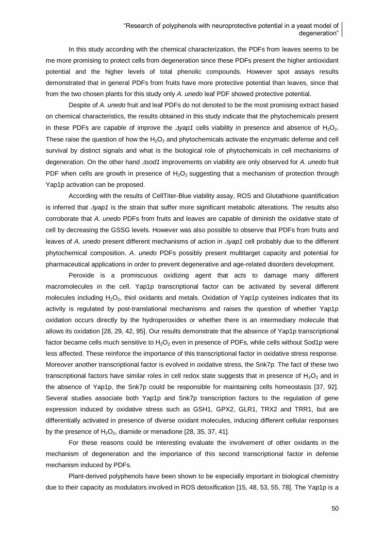
“Research of polyphenols with neuroprotective potential in a yeast model of degeneration”
50
In this study according with the chemical characterization, the PDFs from leaves seems to be
me more promising to protect cells from degeneration since these PDFs present the higher antioxidant
potential and the higher levels of total phenolic compounds. However spot assays results
demonstrated that in general PDFs from fruits have more protective potential than leaves, since that
from the two chosen plants for this study only A. unedo leaf PDF showed protective potential.
Despite of A. unedo fruit and leaf PDFs do not denoted to be the most promising extract based
on chemical characteristics, the results obtained in this study indicate that the phytochemicals present
in these PDFs are capable of improve the yap1 cells viability in presence and absence of H2O2.
These raise the question of how the H2O2 and phytochemicals activate the enzymatic defense and cell
survival by distinct signals and what is the biological role of phytochemicals in cell mechanisms of
degeneration. On the other hand sod1 improvements on viability are only observed for A. unedo fruit
PDF when cells are growth in presence of H2O2 suggesting that a mechanism of protection through
Yap1p activation can be proposed.
According with the results of CellTiter-Blue viability assay, ROS and Glutathione quantification
is inferred that yap1 is the strain that suffer more significant metabolic alterations. The results also
corroborate that A. unedo PDFs from fruits and leaves are capable of diminish the oxidative state of
cell by decreasing the GSSG levels. However was also possible to observe that PDFs from fruits and
leaves of A. unedo present different mechanisms of action in yap1 cell probably due to the different
phytochemical composition. A. unedo PDFs possibly present multitarget capacity and potential for
pharmaceutical applications in order to prevent degenerative and age-related disorders development.
Peroxide is a promiscuous oxidizing agent that acts to damage many different
macromolecules in the cell. Yap1p transcriptional factor can be activated by several different
molecules including H2O2, thiol oxidants and metals. Oxidation of Yap1p cysteines indicates that its
activity is regulated by post-translational mechanisms and raises the question of whether Yap1p
oxidation occurs directly by the hydroperoxides or whether there is an intermediary molecule that
allows its oxidation [28, 29, 42, 95]. Our results demonstrate that the absence of Yap1p transcriptional
factor became cells much sensitive to H2O2 even in presence of PDFs, while cells without Sod1p were
less affected. These reinforce the importance of this transcriptional factor in oxidative stress response.
Moreover another transcriptional factor is evolved in oxidative stress, the Snk7p. The fact of these two
transcriptional factors have similar roles in cell redox state suggests that in presence of H2O2 and in
the absence of Yap1p, the Snk7p could be responsible for maintaining cells homeostasis [37, 92].
Several studies associate both Yap1p and Snk7p transcription factors to the regulation of gene
expression induced by oxidative stress such as GSH1, GPX2, GLR1, TRX2 and TRR1, but are
differentially activated in presence of diverse oxidant molecules, inducing different cellular responses
by the presence of H2O2, diamide or menadione [28, 35, 37, 41].
For these reasons could be interesting evaluate the involvement of other oxidants in the
mechanism of degeneration and the importance of this second transcriptional factor in defense
mechanism induced by PDFs.
Plant-derived polyphenols have been shown to be especially important in biological chemistry
due to their capacity as modulators involved in ROS detoxification [15, 48, 53, 55, 78]. The Yap1p is a
“Research of polyphenols with neuroprotective potential in a yeast model of degeneration”
51
DNA binding protein of the AP-1 family that was identified as a functional homologue of the
mammalian AP-1, the Nrf2p transcriptional factor. The similarity of the cystein thiol reactivity of both
Yap1p and Nrf2p makes from S. cerevisiae a good model to investigate the mechanisms of action
involved in cell defense against oxidative stress. Besides, Nrf2p regulates the chemical stress
response pathways being trigger by ROS such as H2O2 and phytochemicals as a defense to oxidative
stress [96, 97]. The fact of yap1 strain presents cell viability improvements in presence of PDFs
probably indicate that the compounds present in these extracts, such as polyphenols are capable of
regulate the expression of gene associated to cell death/survival and cell signaling through Yap1p.
Moreover, after ROS determination we conclude that ROS scavenging is not the mechanism
by which cells improve their metabolism in presence of PDFs, however the mechanisms inherent to
cellular improvements by this phytochemicals is not totally clear. Besides ROS early mechanisms of
defense based on signaling pathways and gene activation, the autophagy-lysosome pathway has
been recently implicated as one of the most important mechanisms in development of degenerative
diseases. The reduction of mitochondrial oxidative damage could slow or even prevent the
progression of degeneration. It is now considered that constitutive basal autophagic activity is a quality
control process that selectively disposes aberrant protein aggregates and damaged organelles for
degradation [12, 24, 93].
Differences in cellular response based on GSH/GSSG ratio of yap1 and sod1 indicate that
PDFs affect differentially the two mutants, which can indicate the possibility of the phytochemicals
besides to keep redox homeostasis by glutathione systems are also probably capable of activate other
mechanisms such as glutaredoxins and thioredoxins. Further studies could provide more insights
about how these mechanisms interplay in cell degeneration processes.
The cross-talk between autophagy, redox signaling and mitochondrial dysfunction is not totally
clear yet. In conclusion, to understand the impact of natural phytochemicals in degenerative diseases
some key molecules involved in all this steps of metabolism must be evaluated with more targeted
methodologies.
“Research of polyphenols with neuroprotective potential in a yeast model of degeneration”
52
6. References
1. Uttara, B., Singh, A.V., Zamboni, P., and Mahajan, R.T., Oxidative stress and
neurodegenerative diseases: a review of upstream and downstream antioxidant therapeutic
options. (2009). Curr Neuropharmacol, 7(1): 65-74.
2. Tavares, L., Figueira, I., Macedo, D., McDougall, G.J., Leirao, M.C., Vieira, H.L.A., Stewart,
D., Alves, P.M., Ferreira, R.B., and Santos, C.N., Neuroprotective effect of blackberry (Rubus
sp.) polyphenols is potentiated after simulated gastrointestinal digestion. (2012). Food Chem,
131(4): 1443-1452.
3. Zhao, B., Natural antioxidants protect neurons in Alzheimer's disease and Parkinson's
disease. (2009). Neurochem Res, 34(4): 630-638.
4. Betarbet, R., Sherer, T.B., and Greenamyre, J.T., Ubiquitin-proteasome system and
Parkinson's diseases. (2005). Expl Neurol, 191: S17-S27.
5. Emerit, J., Edeas, M., and Bricaire, F., Neurodegenerative diseases and oxidative stress.
(2004). Biomed Pharmacother, 58(1): 39-46.
6. Liu, Y.T. and Connor, J.R., Iron and ER stress in neurodegenerative disease. (2012).
Biometals, 25(4): 837-845.
7. Costa, V. and Moradas-Ferreira, P., Oxidative stress and signal transduction in
Saccharomyces cerevisiae: insights into ageing, apoptosis and diseases. (2001). Mol Aspects
Med, 22(4-5): 217-246.
8. Tenreiro, S. and Outeiro, T.F., Simple is good: yeast models of neurodegeneration. (2010).
FEMS Yeast Res, 10(8): 970-979.
9. Fernandes, P.N., Mannarino, S.C., Silva, C.G., Pereira, M.D., Panek, A.D., and Eleutherio,
E.C., Oxidative stress response in eukaryotes: effect of glutathione, superoxide dismutase and
catalase on adaptation to peroxide and menadione stresses in Saccharomyces cerevisiae.
(2007). Redox Rep, 12(5): 236-244.
10. Herrero, E., Ros, J., Belli, G., and Cabiscol, E., Redox control and oxidative stress in yeast
cells. (2008). Biochim Biophys Acta, 1780(11): 1217-1235.
11. Pereira, M.D., Herdeiro, R.S., Fernandes, P.N., Eleutherio, E.C., and Panek, A.D., Targets of
oxidative stress in yeast sod mutants. (2003). Biochim Biophys Acta, 1620(1-3): 245-251.
12. Mammucari, C. and Rizzuto, R., Signaling pathways in mitochondrial dysfunction and aging.
(2010). Mech Ageing Dev, 131(7-8): 536-543.
13. Esposito, E., Rotilio, D., Di Matteo, V., Di Giulio, C., Cacchio, M., and Algeri, S., A review of
specific dietary antioxidants and the effects on biochemical mechanisms related to
neurodegenerative processes. (2002). Neurobiol Aging, 23(5): 719-735.
14. Calabrese, V., Guagliano, E., Sapienza, M., Panebianco, M., Calafato, S., Puleo, E., Pennisi,
G., Mancuso, C., Butterfield, D.A., and Stella, A.G., Redox regulation of cellular stress
response in aging and neurodegenerative disorders: role of vitagenes. (2007). Neurochem
Res, 32(4-5): 757-773.
15. Valko, M., Rhodes, C.J., Moncol, J., Izakovic, M., and Mazur, M., Free radicals, metals and
antioxidants in oxidative stress-induced cancer. (2006). Chem Biol Interact, 160(1): 1-40.

“Research of polyphenols with neuroprotective potential in a yeast model of degeneration”
53
16. Monteiro, G., Kowaltowski, A.J., Barros, M.H., and Netto, L.E., Glutathione and thioredoxin
peroxidases mediate susceptibility of yeast mitochondria to Ca(2+)-induced damage. (2004).
Arch Biochem Biophys, 425(1): 14-24.
17. Tanaka, K., Suzuki, T., Hattori, N., and Mizuno, Y., Ubiquitin, proteasome and parkin. (2004).
Biochim Biophys Acta, 1695(1-3): 235-247.
18. Ardley, H.C. and Robinson, P.A., The role of ubiquitin-protein ligases in neurodegenerative
disease. (2004). Neurodegener Dis, 1(2-3): 71-87.
19. Outeiro, T.F. and Tetzlaff, J., Mechanisms of disease II: cellular protein quality control. (2007).
Semin Pediatr Neurol, 14(1): 15-25.
20. Alves-Rodrigues, A., Gregori, L., and Figueiredo-Pereira, M.E., Ubiquitin, cellular inclusions
and their role in neurodegeneration. (1998). Trends Neurosci, 21(12): 516-520.
21. Hahn, J.S., Neef, D.W., and Thiele, D.J., A stress regulatory network for co-ordinated
activation of proteasome expression mediated by yeast heat shock transcription factor. (2006).
Mol Microbiol, 60(1): 240-251.
22. Daiber, A., Redox signaling (cross-talk) from and to mitochondria involves mitochondrial pores
and reactive oxygen species. (2010). Biochim Biophys Acta, 1797(6-7): 897-906.
23. Lee, J., Giordano, S., and Zhang, J., Autophagy, mitochondria and oxidative stress: cross-talk
and redox signalling. (2012). Biochem J, 441(2): 523-540.
24. Arduino, D.M., Raquel Esteves, A., Cortes, L., Silva, D.F., Patel, B., Grazina, M., Swerdlow,
R.H., Oliveira, C.R., and Cardoso, S.M., Mitochondrial metabolism in Parkinson's disease
impairs quality control autophagy by hampering microtubule-dependent traffic. (2012). Hum
Mol Genet.
25. von Bohlen Und Halbach, O., Modeling neurodegenerative diseases in vivo review. (2005).
Neurodegener Dis, 2(6): 313-320.
26. Outeiro, T.F. and Muchowski, P., Molecular genetics approaches in yeast to study amyloid
diseases. (2004). J Mol Neurosci, 23(1-2): 49-59.
27. Miller-Fleming, L., Giorgini, F., and Outeiro, T.F., Yeast as a model for studying human
neurodegenerative disorders. (2008). Biotechnol J, 3(3): 325-338.
28. Jamieson, D.J., Oxidative stress responses of the yeast Saccharomyces cerevisiae. (1998).
Yeast, 14(16): 1511-1527.
29. Coleman, S.T., Epping, E.A., Steggerda, S.M., and Moye-Rowley, W.S., Yap1p activates gene
transcription in an oxidant-specific fashion. (1999). Mol Cell Biol, 19(12): 8302-8313.
30. Perrone, G.G., Tan, S.X., and Dawes, I.W., Reactive oxygen species and yeast apoptosis.
(2008). Biochim Biophys Acta, 1783(7): 1354-1368.
31. Mannarino, S.C., Amorim, M.A., Pereira, M.D., Moradas-Ferreira, P., Panek, A.D., Costa, V.,
and Eleutherio, E.C., Glutathione is necessary to ensure benefits of calorie restriction during
ageing in Saccharomyces cerevisiae. (2008). Mech Ageing Dev, 129(12): 700-705.
32. Greetham, D. and Grant, C.M., Antioxidant activity of the yeast mitochondrial one-Cys
peroxiredoxin is dependent on thioredoxin reductase and glutathione in vivo. (2009). Mol Cell
Biol, 29(11): 3229-3240.
“Research of polyphenols with neuroprotective potential in a yeast model of degeneration”
54
33. Amari, F., Fettouche, A., Samra, M.A., Kefalas, P., Kampranis, S.C., and Makris, A.M.,
Antioxidant small molecules confer variable protection against oxidative damage in yeast
mutants. (2008). J Agric Food Chem, 56(24): 11740-11751.
34. Grant, C.M., Perrone, G., and Dawes, I.W., Glutathione and catalase provide overlapping
defenses for protection against hydrogen peroxide in the yeast Saccharomyces cerevisiae.
(1998). Biochem Biophys Res Commun, 253(3): 893-898.
35. Rodrigues-Pousada, C., Menezes, R.A., and Pimentel, C., The Yap family and its role in
stress response. (2010). Yeast, 27(5): 245-258.
36. Witt, S.N. and Flower, T.R., alpha-Synuclein, oxidative stress and apoptosis from the
perspective of a yeast model of Parkinson's disease. (2006). FEMS Yeast Res, 6(8): 1107-
1116.
37. Lee, J., Godon, C., Lagniel, G., Spector, D., Garin, J., Labarre, J., and Toledano, M.B., Yap1
and Skn7 control two specialized oxidative stress response regulons in yeast. (1999). J Biol
Chem, 274(23): 16040-16046.
38. Azevedo, D., Tacnet, F., Delaunay, A., Rodrigues-Pousada, C., and Toledano, M.B., Two
redox centers within Yap1 for H2O2 and thiol-reactive chemicals signaling. (2003). Free Radic
Biol Med, 35(8): 889-900.
39. Rodrigues-Pousada, C.A., Nevitt, T., Menezes, R., Azevedo, D., Pereira, J., and Amaral, C.,
Yeast activator proteins and stress response: an overview. (2004). FEBS Lett, 567(1): 80-85.
40. Nguyen, D.T., Alarco, A.M., and Raymond, M., Multiple Yap1p-binding sites mediate induction
of the yeast major facilitator FLR1 gene in response to drugs, oxidants, and alkylating agents.
(2001). J Biol Chem, 276(2): 1138-1145.
41. Penninckx, M.J., An overview on glutathione in Saccharomyces versus non-conventional
yeasts. (2002). FEMS Yeast Res, 2(3): 295-305.
42. Ouyang, X., Tran, Q.T., Goodwin, S., Wible, R.S., Sutter, C.H., and Sutter, T.R., Yap1
activation by H2O2 or thiol-reactive chemicals elicits distinct adaptive gene responses. (2011).
Free Radic Biol Med, 50(1): 1-13.
43. Penninckx, M., A short review on the role of glutathione in the response of yeasts to
nutritional, environmental, and oxidative stresses. (2000). Enzyme Microb Technol, 26(9-10):
737-742.
44. Mannarino, S.C., Vilela, L.F., Brasil, A.A., Aranha, J.N., Moradas-Ferreira, P., Pereira, M.D.,
Costa, V., and Eleutherio, E.C.A., Requirement of glutathione for Sod1 activation during
lifespan extension. (2011). Yeast, 28(1): 19-25.
45. Menezes, R.A., Amaral, C., Batista-Nascimento, L., Santos, C., Ferreira, R.B., Devaux, F.,
Eleutherio, E.C., and Rodrigues-Pousada, C., Contribution of Yap1 towards Saccharomyces
cerevisiae adaptation to arsenic-mediated oxidative stress. (2008). Biochem J, 414(2): 301-
311.
46. Carri, M.T. and Cozzolino, M., SOD1 and mitochondria in ALS: a dangerous liaison. (2011). J
Bioenerg Biomembr, 43(6): 593-599.
47. Harris, E.D., Regulation of antioxidant enzymes. (1992). FASEB J, 6(9): 2675-2683.
“Research of polyphenols with neuroprotective potential in a yeast model of degeneration”
55
48. Ebrahimi, A. and Schluesener, H., Natural polyphenols against neurodegenerative disorders:
potentials and pitfalls. (2012). Ageing Res Rev, 11(2): 329-345.
49. Del Rio, D., Borges, G., and Crozier, A., Berry flavonoids and phenolics: bioavailability and
evidence of protective effects. (2010). Br J Nutr, 104 Suppl 3: S67-90.
50. Seeram, N.P., Berry fruits for cancer prevention: current status and future prospects. (2008). J
Agric Food Chem, 56(3): 630-635.
51. Oliveira, I., Coelho, V., Baltasar, R., Pereira, J.A., and Baptista, P., Scavenging capacity of
strawberry tree (Arbutus unedo L.) leaves on free radicals. (2009). Food Chem Toxicol, 47(7):
1507-1511.
52. Coates, E.M., Popa, G., Gill, C.I., McCann, M.J., McDougall, G.J., Stewart, D., and Rowland,
I., Colon-available raspberry polyphenols exhibit anti-cancer effects on in vitro models of colon
cancer. (2007). J Carcinog, 6: 4-16.
53. Scalbert, A., Manach, C., Morand, C., Remesy, C., and Jimenez, L., Dietary polyphenols and
the prevention of diseases. (2005). Crit Rev Food Sci, 45(4): 287-306.
54. Scalbert, A., Johnson, I.T., and Saltmarsh, M., Polyphenols: antioxidants and beyond. (2005).
Am J Clin Nutr, 81(1 Suppl): 215S-217S.
55. Crozier, A., Del Rio, D., and Clifford, M.N., Bioavailability of dietary flavonoids and phenolic
compounds. (2010). Mol Aspects Med, 31(6): 446-467.
56. Wang, S.Y. and Lin, H.S., Antioxidant activity in fruits and leaves of blackberry, raspberry, and
strawberry varies with cultivar and developmental stage. (2000). J Agric Food Chem, 48(2):
140-146.
57. McDougall, G.J., Dobson, P., Smith, P., Blake, A., and Stewart, D., Assessing potential
bioavallability of raspberry anthocyanins using an in vitro digestion system. (2005). J Agric
Food Chem, 53(15): 5896-5904.
58. Seeram, N.P., Berry fruits: compositional elements, biochemical activities, and the impact of
their intake on human health, performance, and disease. (2008). J Agric Food Chem, 56(3):
627-629.
59. Fortalezas, S., Tavares, L., Pimpao, R., Tyagi, M., Pontes, V., Alves, P.M., McDougall, G.,
Stewart, D., Ferreira, R.B., and Santos, C.N., Antioxidant properties and neuroprotective
capacity of strawberry tree fruit (Arbutus unedo). (2010). Nutrients, 2(2): 214-229.
60. Beekwilder, J., Jonker, H., Meesters, P., Hall, R.D., van der Meer, I.M., and Ric de Vos, C.H.,
Antioxidants in raspberry: on-line analysis links antioxidant activity to a diversity of individual
metabolites. (2005). J Agric Food Chem, 53(9): 3313-3320.
61. Mazza, G.J., Anthocyanins and heart health. (2007). Ann Ist Super Sanita, 43(4): 369-374.
62. Mendes, L., de Freitas, V., Baptista, P., and Carvalho, M., Comparative antihemolytic and
radical scavenging activities of strawberry tree (Arbutus unedo L.) leaf and fruit. (2011). Food
Chem Toxicol, 49(9): 2285-2291.
63. Tavares, L., Fortalezas, S., Carrilho, C., McDougall, G.J., Stewart, D., Ferreira, R.B., and
Santos, C.N., Antioxidant and antiproliferative properties of strawberry tree tissues. (2010). J
Berr Res, 10: 3-12.
“Research of polyphenols with neuroprotective potential in a yeast model of degeneration”
56
64. Pabuccuoglu, A., Kivcak, B., Bas, M., and Mert, T., Antioxidant activity of Arbutus unedo
leaves. (2003). Fitoterapia, 74(6): 597-599.
65. Marques, E.M.M., Caracterização das populações de Camarinha (Corema album L.) no Cabo
Carvoeiro MsC. 2007, Lisbon: 79.
66. Calviño-Cancela, M., Spatial patterns of seed dispersal and seedling recruitment in Corema
album (Empetraceae): the importance of unspecialized dispersers for regeneration. (2002). J
Ecology, 90: 775-784.
67. Hummer, K.E., Rubus Pharmacology: Antiquity to the Present. (2010). Hortscience, 45(11):
1587-1591.
68. Maatta-Riihinen, K.R., Kamal-Eldin, A., and Torronen, A.R., Identification and quantification of
phenolic compounds in berries of Fragaria and Rubus species (Family rosaceae). (2004). J
Agric Food Chem, 52(20): 6178-6187.
69. Rao, A.V. and Snyder, D.M., Raspberries and human health: a review. (2010). J Agric Food
Chem, 58(7): 3871-3883.
70. Lee, J., Dossett, M., and Finn, C.E., Rubus fruit phenolic research: The good, the bad, and the
confusing. (2012). Food Chem, 130(4): 785-796.
71. Bradish, C.M., Perkins-Veazie, P., Fernandez, G.E., Xie, G., and Jia, W., Comparison of
Flavonoid Composition of Red Raspberries (Rubus idaeus L.) Grown in the Southern United
States. (2012). J Agric Food Chem, 60: 5779-5786.
72. DeFeudis, F.V. and Drieu, K., Ginkgo biloba extract (EGb 761) and CNS functions: basic
studies and clinical applications. (2000). Curr Drug Targets, 1(1): 25-58.
73. Marques, F., Azevedo, F., Johansson, B., and Oliveira, R., Stimulation of DNA repair in
Saccharomyces cerevisiae by Ginkgo biloba leaf extract. (2011). Food Chem Toxicol, 49(6):
1361-1366.
74. Dajas, F., Rivera-Megret, F., Blasina, F., Arredondo, F., Abin-Carriquiry, J.A., Costa, G.,
Echeverry, C., Lafon, L., Heizen, H., Ferreira, M., and Morquio, A., Neuroprotection by
flavonoids. (2003). Braz J Med Biol Res, 36(12): 1613-1620.
75. Manach, C., Williamson, G., Morand, C., Scalbert, A., and Remesy, C., Bioavailability and
bioefficacy of polyphenols in humans. I. Review of 97 bioavailability studies. (2005). Am J Clin
Nutr, 81(1 Suppl): 230S-242S.
76. McDougall, G.J., Fyffe, S., Dobson, P., and Stewart, D., Anthocyanins from red wine--their
stability under simulated gastrointestinal digestion. (2005). Phytochemistry, 66(21): 2540-
2548.
77. McGhie, T.K., Ainge, G.D., Barnett, L.E., Cooney, J.M., and Jensen, D.J., Anthocyanin
glycosides from berry fruit are absorbed and excreted unmetabolized by both humans and
rats. (2003). J Agric Food Chem, 51(16): 4539-4548.
78. Ramassamy, C., Emerging role of polyphenolic compounds in the treatment of
neurodegenerative diseases: a review of their intracellular targets. (2006). Eur J Pharmacol,
545(1): 51-64.
“Research of polyphenols with neuroprotective potential in a yeast model of degeneration”
57
79. Seeram, N.P., Adams, L.S., Zhang, Y., Lee, R., Sand, D., Scheuller, H.S., and Heber, D.,
Blackberry, black raspberry, blueberry, cranberry, red raspberry, and strawberry extracts
inhibit growth and stimulate apoptosis of human cancer cells in vitro. (2006). J Agric Food
Chem, 54(25): 9329-9339.
80. Tavares, L., Carrilho, D., Tyagi, M., Barata, D., Serra, A.T., Duarte, C.M., Duarte, R.O.,
Feliciano, R.P., Bronze, M.R., Chicau, P., Espirito-Santo, M.D., Ferreira, R.B., and dos
Santos, C.N., Antioxidant capacity of Macaronesian traditional medicinal plants. (2010).
Molecules, 15(4): 2576-2592.
81. Ainsworth, E.A. and Gillespie, K.M., Estimation of total phenolic content and other oxidation
substrates in plant tissues using Folin-Ciocalteu reagent. (2007). Nat Protoc, 2(4): 875-877.
82. Cao, G., Alessio, H.M., and Cutler, R.G., Oxygen-radical absorbance capacity assay for
antioxidants. (1993). Free Radic Biol Med, 14(3): 303-311.
83. Jamnik, P., Goranovic, D., and Raspor, P., Antioxidative action of royal jelly in the yeast cell.
(2007). Exp Gerontol, 42(7): 594-600.
84. Priault, M., Bessoule, J.J., Grelaud-Coq, A., Camougrand, N., and Manon, S., Bax-induced
cell death in yeast depends on mitochondrial lipid oxidation. (2002). Eur J Biochem, 269(22):
5440-5450.
85. Wang, H. and Joseph, J.A., Quantifying cellular oxidative stress by dichlorofluorescein assay
using microplate reader. (1999). Free Radic Biol Med, 27(5-6): 612-616.
86. Bussche, J.V. and Soares, E.V., Lead induces oxidative stress and phenotypic markers of
apoptosis in Saccharomyces cerevisiae. (2011). Appl Microbiol Biotechnol, 90(2): 679-687.
87. Kand'ar, R., Zakova, P., Lotkova, H., Kucera, O., and Cervinkova, Z., Determination of
reduced and oxidized glutathione in biological samples using liquid chromatography with
fluorimetric detection. (2007). J Pharm Biomed Anal, 43(4): 1382-1387.
88. Hissin, P.J. and Hilf, R., A fluorometric method for determination of oxidized and reduced
glutathione in tissues. (1976). Anal Biochem, 74(1): 214-226.
89. Toussaint, M. and Conconi, A., High-throughput and sensitive assay to measure yeast cell
growth: a bench protocol for testing genotoxic agents. (2006). Nat Protoc, 1(4): 1922-1928.
90. Toussaint, M., Levasseur, G., Gervais-Bird, J., Wellinger, R.J., Elela, S.A., and Conconi, A., A
high-throughput method to measure the sensitivity of yeast cells to genotoxic agents in liquid
cultures. (2006). Mutat Res, 606(1-2): 92-105.
91. Maeta, K., Izawa, S., Okazaki, S., Kuge, S., and Inoue, Y., Activity of the Yap1 transcription
factor in Saccharomyces cerevisiae is modulated by methylglyoxal, a metabolite derived from
glycolysis. (2004). Mol Cell Biol, 24(19): 8753-8764.
92. Morgan, B.A., Banks, G.R., Toone, W.M., Raitt, D., Kuge, S., and Johnston, L.H., The Skn7
response regulator controls gene expression in the oxidative stress response of the budding
yeast Saccharomyces cerevisiae. (1997). Embo Journal, 16(5): 1035-1044.
93. Szeto, H.H., Mitochondria-targeted peptide antioxidants: novel neuroprotective agents. (2006).
AAPS J, 8(3): E521-531.

“Research of polyphenols with neuroprotective potential in a yeast model of degeneration”
58
94. Eriksen, J.L., Wszolek, Z., and Petrucelli, L., Molecular pathogenesis of Parkinson disease.
(2005). Arch Neurol, 62(3): 353-357.
95. Carmel-Harel, O., Stearman, R., Gasch, A.P., Botstein, D., Brown, P.O., and Storz, G., Role of
thioredoxin reductase in the Yap1p-dependent response to oxidative stress in Saccharomyces
cerevisiae. (2001). Mol Microbiol, 39(3): 595-605.
96. Tufekci, K.U., Civi Bayin, E., Genc, S., and Genc, K., The Nrf2/ARE Pathway: A Promising
Target to Counteract Mitochondrial Dysfunction in Parkinson's Disease. (2011). Parkinsons
Dis: 1-14.
97. Davinelli, S., Sapere, N., Zella, D., Bracale, R., Intrieri, M., and Scapagnini, G., Pleiotropic
protective effects of phytochemicals in Alzheimer's disease. (2012). Oxid Med Cell Longev: 1-
11.