UNIVERSIDADE DE SÃO PAULO FACULDADE DE ODONTOLOGIA … · 2018. 7. 13. · Te amo!! À Karina...
148
UNIVERSIDADE DE SÃO PAULO FACULDADE DE ODONTOLOGIA DE BAURU JULIANA GONÇALVES PIRES Effect of Myracrodruon urundeuva and Qualea grandiflora extracts on viability and activity of microcosm biofilm and prevention of enamel demineralization in vitro Efeito de extratos de Myracrodruon urundeuva All. e Qualea grandiflora Mart. sobre a viabilidade e atividade de biofilme microcosmo e na prevenção da desmineralização do esmalte in vitro BAURU 2018
Transcript of UNIVERSIDADE DE SÃO PAULO FACULDADE DE ODONTOLOGIA … · 2018. 7. 13. · Te amo!! À Karina...

UNIVERSIDADE DE SÃO PAULO
FACULDADE DE ODONTOLOGIA DE BAURU
JULIANA GONÇALVES PIRES
Effect of Myracrodruon urundeuva and Qualea grandiflora extracts
on viability and activity of microcosm biofilm and prevention of
enamel demineralization in vitro
Efeito de extratos de Myracrodruon urundeuva All. e Qualea
grandiflora Mart. sobre a viabilidade e atividade de biofilme
microcosmo e na prevenção da desmineralização do esmalte in
vitro
BAURU
2018

JULIANA GONÇALVES PIRES
Effect of Myracrodruon urundeuva All. and Qualea grandiflora Mart.
extracts on viability and activity of microcosm biofilm and
prevention of enamel demineralization in vitro
Efeito de extratos de Myracrodruon urundeuva All. e Qualea
grandiflora Mart. sobre a viabilidade e atividade de biofilme
microcosmo e na prevenção da desmineralização do esmalte in
vitro
Thesis presented to the Bauru School of Dentistry of the University of São Paulo to obtain the degree of PhD in Science in the Applied Dental Science Program, Stomatology and Oral Biology concentration area.
Supervisor: Prof. Dr. Ana Carolina Magalhães
Tese apresentada à Faculdade de Odontologia de Bauru da Universidade de São Paulo para obtenção do título de Doutor em Ciências no Programa de Ciências Odontológicas Aplicadas, área de concentração Estomatologia e Biologia Oral.
Orientadora: Profa. Dra. Ana Carolina Magalhães
Versão Corrigida
BAURU
2018
Nota: A versão original desta tese encontra-se disponível no Serviço de Biblioteca e
Documentação da Faculdade de Odontologia de Bauru – FOB/USP.
Autorizo exclusivamente para fins acadêmicos e científicos, a reprodução total ou parcial desta dissertação/tese, por processos fotocopiadores e outros meios eletrônicos.
Assinatura:
Data:
Comitê de Ética em Pesquisa em Seres Humanos e Animais FOB-USP
Protocolo nº CAAE 43948115200005417 Registro nº CEEPA 007/2016 Registro nº CEEPA 008/2017 Data: 29/07/2015, 20/12/2016 e 05/04/2017
Pires, Juliana Gonçalves
Effect of Myracrodruon urundeuva and Qualea grandiflora
extracts on viability and activity of microcosm biofilm and
prevention of enamel demineralization in vitro / Juliana
Gonçalves Pires – Bauru, 2018.
132 p. : il. ; 31cm.
Tese (Doutorado) – Faculdade de Odontologia de Bauru. Universidade de São Paulo
Orientadora: Prof. Drᵃ Ana Carolina Magalhães
P666e
FOLHA DE APROVAÇÃO


DEDICATÓRIA
A Deus,
Pela sua bondade indiscutível.
Aos meus pais,
Moacir Duarte Pires e Rosangela Gonçalves Pires,
Pelo amor grandioso, todo apoio, respeito, dedicação e por ensinarem o verdadeiro
significado da Família.
Ao meu irmão,
Rafael Gonçalves Pires,
Por todo companheirismo, amor, respeito e por tornar nossas conversas engraçadas
e leves.
Aos meus avós,
Mauro Duarte Pires e Teresa Menzatto Pires,
Pessoas especiais com humildade incomparável.
Aos meus avós,
Manoel Jesus Gonçalves (in memorian) e Aparecida Leonetti (in memorian),
Sinto uma saudade imensa, mas sei que estão zelando por nós.
Aos meus amigos,
Anahi Ricaldes, Daniela Razera, Eduardo Galeskas, Guilherme Fernandes, Paulo
Henrique Souza, Leticia Calarga, Karina Tozze, Valéria Guedes, Nadya Melhen,
Aline Braga, Daiana Moreli, Vinícius Taioqui e Priscila Salomão.
Verdadeiros amigos que levarei para a vida toda e que sempre terão um espaço em
especial no meu coração.


A minha família da Bioquímica e FOB,
Ana Carolina Magalhães, André Luís da Silva, Dalva Ribeiro de Oliveira, Lívia Maria
de Melo, Gabriela Neubern, Nádia Amôr, Aline Dionízio, Talita Ventura, Beatriz
Souza, Carlos, Even Taira, Luiza, Tatiana Martini, Natália Mello, Tamara, Maria
Aparecida, Thelma Silva, Larissa, Sara Zabini, Flávia Levy, Cíntia Tokuhara, Mariana
Santesso, Isabela Tomazini, João Paulo Domezi, Adriana Matos, Flávia Amadeu,
Mileni Fernandez, Cíntia Souza, Carlos, Zohaib, Polliana Scaffa, Tamara Araújo.
Minha segunda família, por toda paciência e respeito, toda alegria vivida nos dias de
laboratório e torcida para os experimentos darem certos.


AGRADECIMENTOS
A Deus,
Só tenho a agradecer, por sempre ter me dado forças nos momentos que
achei que iria fraquejar e que não iria conseguir. Por permitir que minha vida seja
rodeada de pessoas abençoadas e por eu ter amigos bons.
Aos meus pais Moacir e Rosangela,
Vocês sempre foram e sempre serão meus exemplos de vida e de pessoas.
São meus heróis, amigos e conselheiros. Nossa família já passou por muitas coisas
e todas elas fizeram a gente permanecer unidos e fortes. Vocês sempre criaram
Rafael e eu com muito amor e carinho, além da vasta educação que nos deram.
Tudo isso só me fez tornar uma pessoa madura com muita consciência, respeito ao
próximo. Toda essa alegria que carrego dentro de mim, o amor, a paz e sentimento
de estar de “bem com a vida” é com certeza devido a vocês. Obrigada por serem tão
maravilhosos. O amor e respeito que tenho por vocês é imensurável. Amo vocês!!
Ao meu irmão Rafael,
Irmããããõoo, você é a pessoa que mais me faz rir e também a única pessoa
que me tira do sério com tanta facilidade, mas você sabe que eu te amo muito!!
Obrigada por todo o carinho, amor e respeito que você tem por mim. Obrigada por
ser meu companheiro. Lembro de todos os apelidos que colocou em mim e dou
risada todas as vezes. Te amo!!
À Karina Tozze,
Ka, você é com certeza um dos motivos da minha alegria. Obrigada por ser
minha amiga e companheira de todas as horas. Obrigada por ter toda paciência do
mundo nos dias que chorei ou que fiquei brava e também por comemorar e sorrir
nos dias das vitórias e conquistas. Obrigada por ser essa mulher maravilhosa que
você é e por ter aceitado entrar na minha vida.


À Aline Braga,
Amiga do céu, quantos perengues passamos, hein?! Mas todos foram
superados com muito louvor, alguns foram chorando, outros foram sorrindo. Em
todos, no final, estavámos nós lá rindo (as vezes rindo das desgraças, mas rindo!).
Todos esses anos vão deixar saudade e vou me recordar só de coisas boas que
passamos juntas. Foram muitos dias no fluxo, com muitas placas e as nossas
músicas no Fluxo FM, muitas noites no laboratório, pedindo pizza, ou comida
japonesa para o jantar. Muitos congressos e histórias para contar e muitos almoços
no bandejão. Mas também foram várias conversas, conselhos e opiniões. Além dos
trabalhos e perengues passados, foram também muitas jantinhas, algumas baladas
(porque não tenho muito pique) e raras caminhadas (porque tenho menos pique
ainda). Obrigada por toda paciência e por toda ajuda que me deu em todos esses
anos, sem você essas pesquisas não teriam saído com tanta leveza. Nossa amizade
levarei por toda a vida!
À Daiana Moreli,
Daia, também passamos muitos dias no fluxo, mas eles foram gostosos e
divertidos, com várias risadas e conversas. Entrou para o time da Micro, com esse
seu jeito todo especial de ser e jeito brava também. Nossa amizade, que começou
na FOB, levarei por toda a vida.
Aos funcionários do Centro Integrado de Pesquisa - CIP,
Márcia, Rafaela e Marcelo, obrigada por toda ajuda nos experimentos e por
serem sempre tão pacientes e prestativos.
As professoras Profa. Dra. Marília Afonso Rabelo Buzalaf e Profa. Dra. Flaviana
Bombarda de Andrade,
Obrigada por compartilharem o conhecimento de vocês, sempre com muita
prontidão, carinho, competência e atenção.


À Dalva Ribeiro de Oliveira,
Dalva, a sua calma e carinho são contagiosos. Obrigada por toda a dedicação
e por toda atenção dada não só para mim, mas para todos os alunos.
Ao André Silva,
André, meu amigo! Muito obrigada por toda ajuda que me deu nesses anos
todos de FOB. Obrigada por ser prestativo e por sanar todas as minhas dúvidas.
Você sempre me recebeu de braços abertos, com um sorriso no rosto, independente
da hora ou do lugar que eu te procurava. Sem você os dias de sufoco teriam sido
bem piores. Muito obrigada!!
Às secretárias da Pós-graduação Fátima, Leila, Letícia, Meg e Maristela,
Ao Prof. Dr. Rodrigo Cardoso de Oliveira,
Professor, muito obrigada por ter me concedido os extratos das plantas e por
toda paciência e prontidão em me receber.
A Profa. Dra. Anne L. Dokkedal e Leonardo Saldanha,
Obrigada por terem preparado e cedido todos os extratos utilizados nas
pesquisas.
Aos voluntários desta pesquisa,
Vocês foram fundamentais para que esse trabalho fosse realizado.
À Faculdade de Odontologia de Bauru- FOB/USP, na pessoa da diretora Prof.ª
Drª Maria Aparecida Moreira Machado e vice-diretor Carlos Ferreira dos Santos,
É uma honra realizar minha pós-graduação, em nível de Mestrado e Doutorado,
nesta grande instituição, que nos dá total suporte e estrutura indiscutível.
À Fundação de Amparo à Pesquisa do Estado de São Paulo (FAPESP),
Pela concessão da bolsa de Iniciação Ciêntífica à aluna Sara Salustiano
Zabini, relacionada ao 3o artigo desta tese. Esta experiência foi importante para meu
aprimoramento profissional e pessoal (Processo 2016/20212-0)


À Sara Zabini,
Sara, você chegou tímida, mas logo consegui conquistar sua confiança e
entrar um pouquinho na sua vida! Você se mostrou uma pessoa muito responsável,
prestativa, esforçada. Uma aluna de Iniciação Científica rara! Agradeço por toda
ajuda que me deu, mesmo nos momentos de correria. Obrigada!
À Coordenação de Aperfeiçoamento de Pessoal de Nível Superior (CAPES),
Pela concessão da minha bolsa de Doutorado, a qual foi importante para meu
aprimoramento profissional e pessoal.
À Profa. Dra. Ana Carolina Magalhães
Carol, agradeço por você ter sempre me recebido tão bem e agradeço pela
oportunidade que me deu, sempre confiando no meu trabalho, me abrindo todas as
portas, me passando confiança e me deixando voar. Sou muito grata! Te admiro
muito como profissional e como pessoa e espero ter um dia pelo menos um
pouquinho de toda a sua didática. Você sempre foi muito receptiva com todos os
alunos, independente do que está fazendo, você pára e ajuda quem precisa. E dar
conta de tudo que você dá, não é para qualquer pessoa não! Porque além de
professora e orientadora, você também é mãe, esposa e filha. Te admiro demais por
você ser essa super mulher! Pode ter certeza que termino meu doutorado com
muitas lições e com um grande conhecimento. A aluna que entrou, não é a mesma
que está saindo. Sei que ainda tenho muito a aprender, mas tenho certeza que
nesses anos de doutorado, aprendi com a melhor. Muito obrigada!


“A tarefa não é tanto ver aquilo que ninguém
viu, mas pensar o que ninguém ainda pensou
sobre aquilo que todo mundo vê”
Arthur Schopenhauer


ABSTRACT
Effect of Myracrodruon urundeuva All. and Qualea grandiflora Mart. extracts on
viability and activity of microcosm biofilm and prevention of enamel
demineralization in vitro
The objective of this study was to evaluate the antimicrobial and anti-caries effects of
two plant extracts. The first chapter dealt with a review of the literature whose
objective was to discuss the antimicrobial potential of Brazilian natural agents on the
biofilm related to dental caries and gingivitis/periodontal disease. The research of the
articles was carried out using PubMed. We found a total of 23 papers. Most of the
studies were performed using planktonic microorganisms or under clinical trials.
Nineteen articles were focused on cariogenic bacteria. From these nineteen articles,
eleven were also about periodontopathogenic bacteria. Four studies addressed only
periodontopathogenic bacteria. The most tested Brazilian natural agents were green
propolis, essential oils of Lippia sidoides and Copaifera sp. Most of the tested agents
showed similar results when compared to positive control (essential oils and extracts)
or better effect than negative control (green propolis). More studies involving
protocols closer to the clinical condition and the use of response variables that allows
understanding the mechanism of action of natural agents are necessary before the
incorporation of these natural agents into dental products. The second chapter aimed
to test the effect of the hydroalcoholic extracts of Myracrodruon urundeuva All. and
Qualea grandiflora Mart. leaves on the viability of the microcosm biofilm and on the
prevention of enamel demineralization. The microcosm biofilm was produced on
bovine enamel, using human saliva pool mixed with McBain saliva (0.2% sucrose) for
14 days. The biofilm was treated daily with the extracts for 1 min. M. urundeuva at
100, 10 and 0.1 µg/ml and Q. grandiflora at 100 and 0.1 µg/ml reduced cell viability
similarly to the positive control and significantly more than negative control. M.
urundeuva at 1000, 100 and 0.1 µg/ml were able to reduce the counting formation
unit-CFU counting of lactobacilli sp. and Streptococcus mutans, while Q. grandiflora
at 1000 and 1.0 µg/ml significantly reduced the S. mutans CFU counting. On the
other hand, the natural extracts did not reduce the production of extracellular
polyssacharides, lactic acid and the development of enamel caries lesions. The third


chapter aimed to evaluate the effect of hydroalcoholic extracts of M. urundeuva and
Q. grandiflora (alone or combined) on the viability of S. mutans biofilm and the
prevention of enamel demineralization. S. mutans strain (ATCC 21175) was
reactivated in BHI broth. Minimum inhibitory concentration, minimum bactericidal
concentration, minimum biofilm inhibitory concentration and minimum biofilm
eradication concentration were determined to choose the concentrations to be tested
under the biofilm model. S. mutans biofilm (5x10⁵ CFU/ml) was produced on bovine
enamel using McBain saliva with 0.2% sucrose for 3 days. The biofilm was treated
daily with the extracts for 1 min. M. urundeuva (isolated or combined) at
concentrations equal or higher than 0.625 mg/ml was able to reduce the bacteria
viability, whereas Q. grandiflora extract alone showed antimicrobial effect at 5 mg/ml
only (p<0.05). On the other hand, none of the extracts was able to reduce the
development of enamel caries lesions. Despite the tested natural extracts have
antimicrobial effect; they are unable to prevent caries in enamel.
Keywords: Antimicrobial agents; dental biofilm; enamel caries; microcosm biofilm;
oral disease; phytotherapy.


RESUMO
Avaliação de extratos de Myracrodruon urundeuva All. e Qualea grandiflora
Mart. sobre a viabilidade e atividade de biofilme microcosmo e na prevenção
da desmineralização do esmalte in vitro O objetivo foi avaliar os efeitos antimicrobiano e anti-cárie de dois extratos de
plantas. O primeiro capítulo se referiu a uma revisão da literatura cujo objetivo foi
discutir o potencial antimicrobiano dos agentes naturais brasileiros sobre o biofilme
relacionado à cárie dentária e à gengivite/doença periodontal. A pesquisa dos artigos
foi realizada usando o PubMed. Foram encontrados 23 trabalhos. A maioria dos
estudos foi realizada utilizando microorganismos na fase planctônica ou ensaios
clínicos. Dezenove artigos foram focados em bactérias cariogênicas. Dos dezenove
artigos, onze também eram sobre bactérias periodontopatogênicas. Quatro estudos
abordaram apenas bactérias periodontopatogênicas. Os agentes naturais brasileiros
mais testados foram própolis verde, óleos essenciais de Lippia sidoides e Copaifera
sp. Os agentes testados apresentaram resultados similares quando comparados ao
controle positivo (óleos essenciais e extratos) ou melhor efeito que o controle
negativo (própolis verde). Mais estudos próximos da condição clínica e o uso de
variáveis de resposta que permitam entender o mecanismo de ação são
necessários, para permitir a incorporação desses agentes naturais em produtos
odontológicos. O segundo capítulo teve como objetivo testar o efeito dos extratos
hidroalcoólicos de Myracrodruon urundeuva All. e Qualea grandiflora Mart. sobre a
viabilidade do biofilme microcosmo e na prevenção da desmineralização do esmalte.
O biofilme microcosmo foi produzido em esmalte bovino, utilizando pool de saliva
humana misturada à saliva de McBain (0,2% de sacarose) durante 14 dias. O
biofilme foi tratado diariamente com os extratos durante 1 min. M. urundeuva a 100,
10 e 0,1 µg/ml e Q. grandiflora a 100 e 0,1 µg/ml reduziram a viabilidade dos
microrganismos de forma semelhante ao controle positivo e significativamente maior
do que o controle negativo. M. urundeuva a 1000, 100 e 0,1 µg/ml foi capaz de
reduzir a contagem de Unidade formadora de colônia-UFC para Lactobacilos totais e
Streptococcus mutans, enquanto a Q. grandiflora a 1000 e 1,0 µg/ml reduziu
significativamente a contagem de UFC para S. mutans. Os extratos naturais não
conseguiram reduzir a produção de polissacarídeos extracelulares-PEC, ácido lático

e o desenvolvimento da lesão cariosa em esmalte. O terceiro capítulo teve como
objetivo avaliar o efeito dos extratos hidroalcoólicos de M. urundeuva. e Q.
grandiflora (sozinhos ou combinados) sobre a viabilidade do biofilme de S. mutans e
na prevenção da desmineralização do esmalte. Cepa de S. mutans (ATCC 21175)
foi reativada em caldo BHI. Concentração inibitória mínima, concentração bactericida
mínima, concentração inibitória mínima de biofilme e concentração de erradicação
mínima de biofilme foram determinadas para escolher as concentrações a serem
testadas sob o modelo de biofilme. O biofilme de S. mutans (5x10⁵ CFU/ml) foi
produzido em esmalte bovino, utilizando saliva de McBain com 0,2% de sacarose
durante 3 dias. O biofilme foi tratado diariamente com os extratos durante 1 min. M.
urundeuva (isolada ou combinada) nas concentrações iguais ou superiores a 0,625
mg/ml foi capaz de reduzir a viabilidade das bactérias, enquanto que o extrato da Q.
grandflora apresentou efeito antimicrobiano somente a 5 mg/ml (p<0,05). Nenhum
dos extratos reduziu o desenvolvimento da lesão da cárie. Apesar dos extratos
naturais terem efeito antimicrobiano, são incapazes de prevenir o desenvolvimento
da lesão cariosa em esmalte.
Palavras-chave: Agentes antimicrobianos; biofilme dental; biofilme microcosmo;
cárie dentária; doenças orais; fitoterapia.

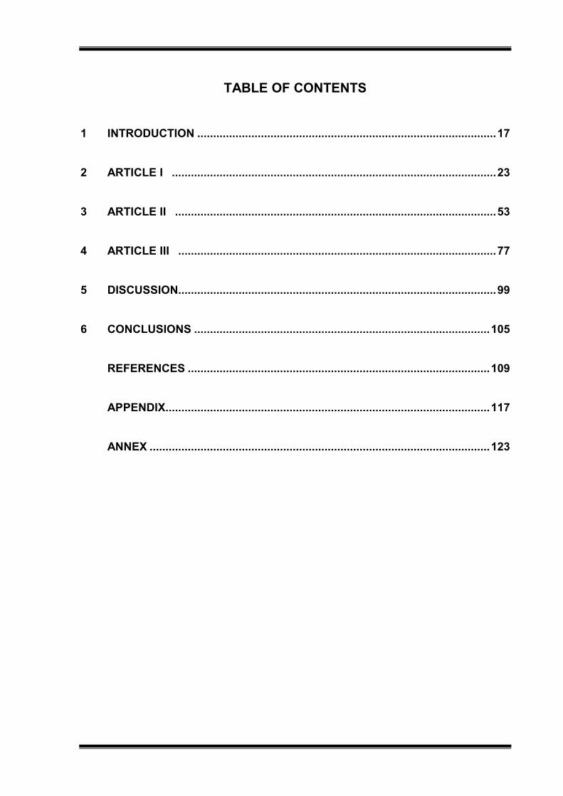
TABLE OF CONTENTS
1 INTRODUCTION .............................................................................................. 17
2 ARTICLE I ...................................................................................................... 23
3 ARTICLE II ..................................................................................................... 53
4 ARTICLE III .................................................................................................... 77
5 DISCUSSION .................................................................................................... 99
6 CONCLUSIONS ............................................................................................. 105
REFERENCES ............................................................................................... 109
APPENDIX...................................................................................................... 117
ANNEX ........................................................................................................... 123


1-Introduction

Introduction 17
1 INTRODUCTION
Dental caries is a tooth disease related to the presence of supragingival
biofilm, whose metabolism is dependent on the frequency of exposure to sugar from
diet, especially sucrose (ERIKSSON et al., 2017). It is one of the most important oral
health problems (PETERSEN, 2003), since 60-90% of school age children and adults
have dental caries experience wordwile (PETERSEN et al., 2005). The first sign of
the disease is the appearance of a white-spot lesion in enamel, which may progress
to cavitation and reach dentin according to the severity of the acid challenges
(CAVALCANTI et al., 2014; FERNANDEZ; TENUTA; CURY, 2016). When caries
affects dentin, it can cause negative impact in quality of life due to the consequences
such as pain, lack of appetite, weight loss and high cost for treatment (ABANTO et
al., 2011; RAMOS-JORGE et al., 2015).
The human oral cavity has more than hundreds microbial species and only
some of them take part of dental biofilm (PALMER, 2010). Among the
microorganisms present in biofilm, the most known ones are Streptococcus mutans,
Streptococcus mitis, Streptococcus salivarius, Lactobacillus, Veillonella,
Actinomyces, bifidobacteria and fungi (TAKAHASHI; WASHIO; MAYANAGI, 2010;
KOO; FALSETTA; KLEIN, 2013; ABUSLEME et al., 2013). The cariogenic
microorganisms have the ability to metabolize sugar, especially sucrose from the
diet, producing extracellular polysaccharides (EPS) and acids that alter the biofilm
pH, inducing tooth demineralization (KEYES, 1960; MARSH; MOTER; DEVINE,
2011; PITTS et al., 2017). Besides, they are able to survive in acidic environment
(TAKAHASHI; NYVAD, 2011; ZHAO et al. 2014).
Streptococcus mutans is considered one of the most important
microorganisms involved in the etiology of dental caries (MARSH, 2003). They are
known to be acidogenic and aciduric bacteria and highly producers of EPS, which are
glycan responsible for the development and protection of dental biofilm (KOO;
FALSETTA; KLEIN, 2013; ZHAO et al., 2014).
In order to control dental biofilm and avoid the disease, mechanical practice of
brushing and reduction of sugar consume are advised (RUGG-GUNN, 2013).
However, for patients under high risk for caries, chemical agents may be needed as
co-adjuvant (JAMES et al., 2017). Chlorhexidine (CHX) is known in dentistry as a

18 Introduction
gold standard antimicrobial agent (JONES, 1997; PARWANI et al., 2013), however, it
may induce undesired side effects (JAMES et al., 2017; ZHENG; WANG, 2011) such
as tooth staining, supragingival calculus formation, changes in taste perception,
parotid gland swelling and irritation of the oral mucosa, when applied for periods
higher than 2 weeks (JAMES et al., 2017). Therefore, inhibition of cariogenic bacteria
growth by alternative antimicrobial agents has been extensively investigated in an
attempt to obtain an agent with effectiveness and with low incidence of side effects.
Accordingly, public and private research institutes are engaged in testing the effect of
plant extracts and natural compounds against dental pathogens (PATRA et al.,
2014).
Many people believe in the effectiveness of herbal medicines; it is estimated
that most population has applied plants or natural agents as the sole source for the
treatment of various health problems (WHO, 1998) including oral diseases
(PALOMBO, 2011). In dentistry, the phytotherapy is a subject of growing interest
(BAKRI; DOUGLAS, 2005; FANI; KOHANTEB; DAYAGHI, 2007). The use of
medicinal plants is dated in manuscripts since 1.500 years B.C (BETTEGA et al.,
2011). Civilizations throughout history have used plants or parts of plants (roots,
stems, leaves and/or bark) to treat toothache, gingivitis, dental abscesses and mouth
sores (HENLEY-SMITH; BOTHA; LALL, 2013).
There are about 500.000 species of plants available wordwile, but only 1% has
been investigated as phytochemical (PALOMBO, 2011). Brazil houses a larger
number of plant species (32,086 native Angiosperms and 23 native Gymnosperms)
(ZAPPI et al., 2015) than any other country in the world (MITTERMEIER et al., 2005;
FORZZA et al., 2012), allocated mainly in Cerrado and Atlantic Rainforest (FORZZA
et al., 2012).
Myracrodruon urundeuva All. (Anacardiaceae Family) and Qualea grandiflora
Mart. (Vochysiaceae Family) are examples of plants from Cerrado. M. urundeuva has
antimicrobial (MONTANARI et al., 2012) including against cariogenic bacteria
(ALVES et al., 2009; MENEZES et al., 2010), analgesic, hepatoprotective,
antidiarrheal, colonic anastomotic wound healing, anti-ulcerogenic effects as well as
protective effect on the gastric mucosa (CARLINI et al., 2010). Q. grandiflora exhibits
anti-ulcerogenic action from the ethanol extract of its bark (HIRUMA-LIMA et al.,

Introduction 19
2006). Besides, the ethanolic extract of the leaves has antioxidant effect (SOUSA et
al., 2007), analgesic, anticonvulsive potential (GASPI et al., 2006) and antibacterial
action (MOURA; NASCIMENTO; PINTO, 2012). However, its antimicrobial effect
against cariogenic bacteria is unknown. Furthermore, no information about the anti-
caries effect of both plants is available in the literature.
Considering the search for natural agents able to prevent oral diseases and
the high prevalence of dental caries in specific populations that are under
unfavorable social-economic conditions (MARCENES et al., 2013), this study was
divided in three parts. The first chapter is reffered to a review of literature with the aim
to discuss the antimicrobial potential of different Brazilian plants with respect to the
control of dental caries and periodontal disease. In the second chapter, the
antimicrobial and anti-caries effects of the hydroalcoholic extracts of M. urundeuva
All. and Q. grandiflora Mart. leaves were tested under microcosm biofilm (second
chapter), while in the third chapter their effects were tested under S. mutans biofilm
model.

20 Introduction

2-Article I

Article I 23
2 ARTICLE I – Review of literature
Article under review in Oral Health & Preventive Dentistry (ANNEX 1).
Brazilian natural antimicrobial agents on caries and periodontitis-related
biofilm: a literature review
Authors: Juliana Gonçalves Piresa, Aline Silva Bragab, Ana Carolina Magalhãesc
a- PhD student, Department of Biological Sciences, Bauru School of Dentistry,
University of São Paulo, Bauru, SP, Brazil. [email protected]
b- Master student, Department of Biological Sciences, Bauru School of Dentistry,
University of São Paulo, Bauru, SP, Brazil. [email protected]
c- DDS, MS, PhD, Associate Professor, Department of Biological Sciences, Bauru
School of Dentistry, University of São Paulo, Bauru, SP, Brazil. [email protected]
Author for correspondence: Ana Carolina Magalhães, Department of Biological
Sciences, Bauru School of Dentistry, University of São Paulo, Al. Octávio Pinheiro
Brisolla 9-75 Bauru, São Paulo, Brazil. Phone: +551432358497. E-mail: [email protected]

24 Article I
Abstract
Purpose: This review aims to discuss the antimicrobial potential of Brazilian natural
agents on the biofilm related to tooth decay and gingivitis/periodontal disease.
Methods: The survey was conducted using PubMed for the selection of papers
published in English, in journals with impact factor greater than 0.8.
Results: We found twenty-three articles, which tested numerous Brazilian plants,
essential oils, propolis or crude extracts. The majority of studies were conducted
using planktonic microorganisms or under clinical trial. Nineteen articles were
focused on cariogenic bacteria; from these nineteen articles, eleven were also about
periodontopathogenic bacteria. Four studies addressed only periodontopathogenic
bacteria. The most tested Brazilian natural agents were green propolis, essential oils
of Lippia sidoides and Copaifera sp. Most of the tested agents showed similar results
when compared to positive control (essential oils and extracts) or better effect than
negative control (green propolis).
Conclusions: More studies involving protocols closer to the clinical condition and
using response variables that allow understanding the mechanism of action of the
natural agents are needed, to thus allow the incorporation of these natural agents in
dental products.
Key words: Antimicrobials; Brazilian natural agents; dental biofilm; dental caries;
periodontitis.
Introduction
Worldwide there are about 500.000 species of plants available, but only 1%
has been investigated as phytochemical49. Brazil houses a larger number of plant
species (32,086 native Angiosperms and 23 native Gymnosperms)69 than any other
country in the world24,40.
Many people believe in the effectiveness of herbal medicines and it is
estimated that most population use these plants as the sole source for the treatment
of various health problems47 including oral diseases49. In dentistry, the phytotherapy
is a study object of growing interest4,22,27. Civilizations throughout history have used
Article I 25
plants or parts of plants, such as fresh or dried roots, stems, leaves and/or bark, to
treat toothache, gingivitis, dental abscesses and mouth sores28.
Dental caries and periodontal disease are among the most important oral
health problems in the world54. Accordingly, 60-90% of school age children and
adults have dental caries experience and 5-15% of adults have tooth loss due to
periodontitis53.
Both oral diseases are caused by bacteria. Approximately more than hundreds
bacterial species from the oral cavity have been isolated and named and some of
them can be organized in supragingival or subgingival biofilm48. Dental biofilm is
found naturally in health, but in disease there is a shift in its composition inducing
damage of the tissues (periodontal inflammation, gingivitis and tooth decay)39.
The global demand for alternative preventive and treatment measurements of
oral diseases with safe, efficiency and low cost has allowed the development of
phytotherapy for caries and periodontitis control11. Accordingly, plants with medicinal
importance have been included as antimicrobial agents into toothpaste, capsules,
tablets, gels and ointments2,21,49.
Therefore, the aim of this review was to highlight the studies about Brazilian
natural agents used as antimicrobial agent to promote oral health, under laboratorial
and clinical models, focused in the prevention of dental caries and
gingivitis/periodontal diseases.
2. METHODS
For the selection, the papers should have been written in English language
and published in journals with impact factor higher than 0.8. The search was made
using PubMed website, and the key-words applied for the search were: Brazilian
plants extracts, Brazilian natural antimicrobials agents, oral biofilm, dental caries,
periodontal diseases, gingivitis and oral bacteria. All selected articles involved in vivo,
in situ or in vitro (mono or multispecies biofilm or planktonic bacteria) models (Table
1).
3. RESULTS
Twenty-three papers were select for this review. All the selected articles were
related with the use of Brazilian native plants against bacteria involved in oral
diseases (dental caries, gingivitis and/or periodontitis). Nineteen articles were

26 Article I
focused on cariogenic bacteria, in which one addressed in situ12, five in
vivo33,35,41,42,44, eleven in vitro models6-8,12,15,20,25,29,60,61,64 and two studies were in
vitro/in vivo32,63. From these nineteen articles, eleven also studied periodontal
bacteria, in which four addressed in vivo35,42,44, five in vitro7,14,25,29,64 and two in
vitro/in vivo models32,63. Four studies addressed only periodontopathogenic bacteria:
three using in vivo51,52,55 and one using in vitro models68.
3.1 Control of dental caries and gingivitis/periodontal disease
Microbial communities in biofilms have been found to be 1000 times more
resistant to antibiotics compared to their equivalents planktonic cultures19,59.
Accordingly, great efforts have been done to find alternative treatments. Several
herbal medicines have been tested to treat or prevent oral diseases that are linked
with oral pathogens9,44,51.
The development of dental caries involves gram-positive, acidogenic and
aciduric bacteria such as Streptococcus mutans, Streptococcus sobrinus and
Lactobacillus sp.36, which accumulate in dental biofilm and grow under high sugar
exposure, resulting in tooth demineralization38. Periodontitis it is gum infection that
damages the soft tissue and the bone that supports the tooth. It involves subgingival
biofilm rich in anaerobic gram-negative bacteria including Aggregatibacter
actinomycetemcomitans, Eikenella corrodens, Fusobacterium nucleatum,
Porphyromonas gingivalis, Prevotella intermedia, Tannerella forsythia, Enterococcus
faecalis and Treponema denticola1. Gingivitis is a mild and reversible form of
periodontal disease (bleeding of gum tissue), while periodontitis causes permanent
damage of tooth-supporting tissues and may lead to tooth loss (loss of collagen,
tooth insertion and bone mass)10.
Oral problems can contribute to the development of other serious diseases
and, therefore, their prevention is a subject of growing interest. Recent researches
suggest that periodontal disease may contribute to the development of heart disease
and endanger patients with diabetes, respiratory disease and osteoporosis5,46.
Nowadays, the demand for plants and extracts that are able to control the
pathogens Porfiromonas gingivalis and S. mutans, are growing exponentially18,29,50,
since the indiscriminate use of antibiotics has led the emergence of multi-resistant
pathogens23,56. Additionally, the conventional antimicrobial agents (ampicillin,
chlorhexidine, sanguinarine, metronidazole, phenolic antiseptics, and quaternary

Article I 27
ammonium-antiseptics) can alter oral microbiota and cause undesirable side effects
such as vomiting, diarrhea, taste alteration, teeth and restoration staining, increasing
calculus formation and disruption of the oral and intestinal microbiota26,67.
Chlorhexidine, for example, may cause teeth stain, astringent sensation, changes in
taste and mucosal desquamation after 2 weeks of use23,56. Therefore, scientists are
shifting their attention to folk medicine, in order to find new and better anti-plaque
agents against oral microbial infections28.
3.2 Brazilian plant species used in oral health
Brazil is the country harboring the highest plant diversity, including two
(Cerrado and Atlantic Rainforest) of the 34-recognized global hotspots24,40. The
majority of Brazilian plant species are terrestrial, ranging from 83.7% in the Amazon
Rainforest through to 75.6% in the Atlantic Rainforest. The Brazilian states with the
largest number of Angiosperm species are: Bahia (1,284 species), Minas Gerais (849
species), Amazonas (733 species), São Paulo (604 species), Rio de Janeiro (586
species), Pará (652 species) and Paraná (629 species). In respect to Gymnosperms,
the highest species number is found in the Atlantic Rainforest (15,001 native
species), Cerrado (12,097 total species), Amazon Rainforest (11,896 total species),
Caatinga (4,657 total species), Pampa (1,685 total species) and Pantanal (1,277 total
species)69.
In their review, Chinsembu16 documented the potential use of plant extracts
and other natural products (poly-herbal and plant compounds) in oral health
according to the country (Africa, Asia, Brazil, Mexico, Europe, and the Middle East).
In respect to Brazil, the most known natural agents are propolis (green propolis),
essential oils (Lippia sidoides, Mammea americana, etc), crude extracts (Schinus
terebinthifolius and Croton urucurana), organic extracts (Cordia sp., Psychotria sp.,
etc), hydroalcoholic (Copaifera trapezifolia, etc) and aqueous extracts (Psidium
cattleianum), among others.
3.2.1 Propolis
Propolis is a resinous product collected by honeybees from various plant
sources31. Its composition varies according to the geographic region37. The color of
propolis (green, red, brown, or almost black) also varies depending on the plants
from which the resinous substance is collected34. The green propolis, for example, is

28 Article I
derived mainly from Baccharis dracunculifolia and is highly applied as antibacterial30,
antifungal57, antiviral58, anti-inflammatory62 and antitumor65 agent. With all these
benefits, green propolis has been incorporated into oral care products (toothpastes,
mouthwashes, and prophylactic gels) and tested in dentistry63,66.
The ethanolic extract of green propolis was tested against S. mutans,
Streptococcus sanguinis, Staphylococcus aureus, Lactobacillus acidophilus, P.
gingivalis, A. actinomycetemcomitans, Candida albicans in planktonic phase63 and S.
mutans biofilm15. S. mutans and S. sanguinis were more sensitive than L. acidophilus
to 50 mg/ml of the extract and P. gingivalis and A. actinomycetemcomitans were the
most resistant species63. Cardoso et al15 determined MIC (2.08 mg/ml) and MBC
(8.33 mg/ml) of green propolis for S. mutans. The S. mutans CFU for green propolis
was statistically lower than the negative controls; however, in respect to enamel
microhardness, green propolis did not differ from controls15.
Skaba et al63 also tested toothpaste containing 3% ethanolic extract of propolis in
patients with and without periodontitis, while similar toothpaste was also tested in
patients with nonsyndromic complete unilateral or bilateral cleft lip and palate treated
with fixed appliances35 and patients who underwent implant-prosthetic therapy41. All
authors presented the same research method, in which they evaluated the
Approximate Plaque Index (API), Oral Hygiene Index (OHI), Sulcus Blood Index (SBI)
or Gingival Index (GI), and isolated and identified, through kits, bacteria from biofilm.
In general, API, OHI and SBI/GI values decreased for propolis-treated patients when
compared to control. The number of identified strains from the gingival sulcus also
decreased for propolis-treated patients63. Streptococcus spp. and Neisseria spp.
were the most found bacteria. On the other hand, Actinomyces israelii,
Capnocytophaga spp., Fusobacterium, Bacteroides and Eubacterium, the pathogenic
species for periodontal tissue, were found for both groups at the baseline35. The
number of C. albicans did not change with the treatment. The patients who used
propolis toothpaste had lower levels of Actinomyces spp., A. israelii and
Capnocytophaga spp. at the final compared to the baseline analysis. There was a
10% decrease in A. israelii level accompanied by an increase in Actinomyces spp.
among the control patients35. In the study of Morawiec et al41, the number of
microorganisms isolated from dental implants after the use of toothpaste with
propolis increased. With bacterial identification, the researchers observed that the

Article I 29
pathogenic bacteria were eliminated, resulting in benefic changes of the
microorganisms.
Two years later, Morawiec et al42, tested gel containing 3% ethanolic propolis
extract in patients under postoperative process of oral soft tissue and observed the
elimination of Streptococcus acidominimus, Streptococcus oralis, Staphylococcus
epidermidis, Veillonella parvula, Bifidobacterium breve and L. acidophilus of mucosal
surface of the region that underwent the surgery. In 2016, Niedzielska et al44, tested
the same gel in patients with mandible fractures. They isolated and identified the
microorganisms and evaluated the values of API, OHI and SBI. The authors obtained
significant reduction of API, OHI and SBI and the elimination of the bacteria
Clostridium perfringens, Actinomyces naeslundii, Prevotella bivia, B. breve and S.
epidermidis.
Considering the above studies, we infer that propolis (especially the green one)
may be incorporated into toothpaste, gels, mouthwashes and other dental products
as promising agent to control oral microorganisms; however, we have few information
about its effect on the prevention of tooth decay and the reduction of bone loss under
long-term clinical trials, highlighting the need of further studies.
3.2.2 Essential oils
Essential oils (EO) are complex mixtures of low molecular weight compounds
extracted in various ways (steam distillation, hydrodistillation or solvent extraction)43.
They are applied in health, agriculture, cosmetic and food industries, because they
are known to have antimicrobial, antiviral, antimutagenic, anticancer, antioxidant,
anti-inflammatory, immunomodulatory and antiprotozoal activities3.
Essential oil of Lippia sidoides has been extensively studied, as they present
phenolic compounds (carvacrol and thymol) that have antimicrobial activities against
yeasts and bacteria45. Through a clinical trial, da Silva Pereira et al17, and Lobo et
al33, evaluated the activities of this essential oil. The EO (10%) was incorporated into
the gel and applied 3 times a day for 90 days in healthy volunteers. In one of the
cited studies, the authors evaluated Bleeding index (BI) and Plaque index (PI) and,
as a result, they observed that the volunteers who applied the gel with essential oil
had a significant reduction in BI and PI when compared to the negative control
(placebo gel) and similar results when compared to the positive control (2%
chlorhexidine gel)17. Lobo et al33, also tested the EO of Lippia sidoidos. The

30 Article I
treatment was focused on dental caries in children (aged 6-12 years) and the
essential oil was incorporated into toothpaste (1.4%), gel (1.4%) and mouthwash
(0.8%). The researchers observed that only the toothpaste showed a protective
result, significantly reducing S. mutans from the saliva during the first 5 days of use.
This result was maintained throughout the study and did not return to the baseline
values during the follows up (30, 60, 180 and 365 days). This essential oil was also
tested against E. faecalis at concentrations of 2.5% and 10% in vitro68. The biofilm
was formed for 3 days in nitrocellulose membrane filters and then, dipped in the
treatments for 30 or 60 minutes. E. faecalis counts were significantly reduced when
the essential oil (2.5%) was compared to the negative control and thymol (positive
control). The same results were found for the concentration of 10%68.
The concentrations of the constituents present in the plants vary according to the
season of the year in which they are collected (dry or rainy) and the extracted parts
(stem, root, leaves, fruits, bark). These differences were well noted by Furtado et al25,
when authors analyzed the essential oil of leaf and stem bark of Inga laurina (dry and
rainy season). MIC values obtained were similar for leaf and stem bark (rainy
season) with respect to aerobic bacteria (S. mutans, S. sobrinus, S. sanguinis, S.
salivarius and S. mitis, the values ranged from 25 to 50 µg/ml). MIC values increased
when essential oils of leaf and stem bark were tested against anaerobic bacteria (A.
naeslundii, P. gingivalis, Prevotella nigrescens, Bacteroides fragilis and F.
nucleatum), regardless of the season (50 to> 400 µg/ml). MIC values were higher for
anaerobic compared to aerobic bacteria during dry than rainy season (100 to 200
µg/ml). Regarding toxicity, Furtado et al25 applied VERO cells and observed that the
essential oils had low cytotoxic activity at concentrations that were able to inhibit
bacterial growth.
EO and ethanolic extract of the fruits of Mammea americana were evaluated on
S. mutans and P. gingivalis. For both bacteria, the MIC values for EO were lower
when compared to the ethanolic extract. MBC was found only for S. mutans and its
value was lower for EO than ethanolic extract29. Sousa et al64, tested the EO and the
fractions of Eugenia calycina Cambess against cariogenic bacteria (S. mutans, S.
sanguinis and S. sobrinus) and periodontopathogenic bacteria (P. nigrescens, P.
gingivalis, A naeslundii and B. fragilis). The lowest MIC values found for cariogenic
bacteria were 100 to 200 µg/ml for S. mutans treated with fractions F2 to F4. P.
nigrescens and P. gingivalis were more sensitive to the plant, showing MIC values

Article I 31
varying from 50 to 100 µg/ml (EO and fractions). They also tested the cytotoxic
potential of the plant and showed low effect on HeLa cells.
Pimentel et al55, induced experimental periodontitis in Wistar rats and tested the
effect of EO of Cordia verbenacea. The animals were treated 3 times daily for 11
days. The authors observed that the animals treated had lower loss of alveolar bone,
lower IL-1α concentration and higher IL-10 levels compared to the control (without
treatment). A. actinomycetemcomitans (7% treated rats x 36% control rats) and P.
gingivalis (13.5% treated rats x 60% control rats) were found in higher percentage in
control rats compared to the treated ones. In 2015, Pedrazzi et al51, formulated a
mouthwash containing 0.04% of essential oil and 0.16% of hydroethanolic extract of
Baccharis dracunculifolia DC and compared them with mouthwash without active
component, Plax (Colgate™), and Listerine (Johnson & Johnson™). The volunteers
applied the mouthwashes 4 times a day for 4 months. All volunteers completed the
study showing similar reduction in dental biofilm among them compared to the
baseline. No treatment was able to improve the effect.
3.2.3 Crude, hydroalcoholic, aqueous extracts, fractions and derivatives
Psidium cattleianum, a Brazilian native plant, in previous studies showed activity
against oral bacteria13, however, only in 201212, an in situ study was carried out, in
which the effect of aqueous extract of leaves (100 g/600 ml) was tested against the
formation of biofilm on bovine enamel for 14 days. The authors observed that the
extract reduced enamel demineralization, acidogenic potential, microorganism
viability, and extracellular polysaccharide production.
Silva et al61, tested the hydroalcoholic extracts of Acanthospermum hispidum DC.,
Annona coriacea L., Schinopsis brasiliensis Engl., Ximenia americana L. and
Hibiscus mutabilis Briq. (10%, 20%, 30%, 50% and 70%) against Pseudomonas
aeruginosa, S. mutans, S. salivarius, S. oralis, Lactobacillus casei, E. faecalis, S.
aureus, C. albicans, C. tropicalis and C. krusei. The extract of S. brasiliensis Engl.
inhibited the growth of S. oralis and S. aureus when compared to chlorhexidine and
the MIC values were 0.004 µl/µl (P. aeruginosa), 1000 µl/µl (E. faecalis), 0.063 µl/µl
(S. aureus) and 0.500 µl/µl (S. oralis). For the extract of X. americana L., the MIC
was only found for S. aureus (0.063 µl/µl). The other extracts had no antimicrobial
effect.

32 Article I
The susceptibility of bacteria and fungi (S. aureus, S. epidermidis, S. mutans, C.
albicans, C. tropicalis and C. glabrata) to the extracts of Equisetum arvense L.,
Glycyrrhiza glabra L., Punica granatum L. and Stryphnodendron barbatimam Mart.
were evaluated20. The authors demonstrated that all extracts possessed bactericidal
activity (MBC) against all bacteria; however, the values ranged from 3.13 to 100
mg/ml. There was an increase in IL-1β production in cultures treated with P.
granatum L. and S. barbatimam Mart.; while no difference was found between the
cultures treated with the extracts E. arvense L. and G. glabra L. with respect to the
production of TNF-α. The complete inhibition of TNF-α occurred for culture treated
with the extract of P. granatum L. With respect to cytotoxicity, G. glabra L. and E.
arvense L. were the least and the most cytotoxic agent against mouse macrophage.
In 2014, some aqueous and organic extracts of aerial organs of Cordia sp.,
Psychotria sp., Cordia nodosa Lam., Solanum sp., Ipomea alba L., Casearia
javitensis Kunth, Dioscorea altissima Lam., leaves of Casearia spruceana Benth. Ex
Eichler, Symphonia globulifera L.f., Moronobea coccinea Aubl., stem of Zanthoxylum
sp., Psychotria sp., Annona hypogauca Mart., Cordia cf. exaltata, Gnetum leyboldii
Tul. and flowers of Moronobea coccinea Aubl, were evaluated (CFU) against S.
mutans biofilm cultivated on hydroxyapatite for four days. Only Dioscorea altissima
and Annona hypoglaucase showed to be able to reduce the number of S. mutans 8.
In the same year, Silva et al60, evaluated similar extracts (aerial organs of Cordia sp.,
Psychotria sp., Cordia nodosa Lam., Solanum cf. lanceifolium, Ipomea alba L.,
Casearia javitensis, Smilax sp., leaves of Casearia spruceana Benth. Ex Eichler,
Moronobea coccinea Aubl. and stem of Zanthoxylum compactum, Diospyros
guianensis, Psychotria sp., Annona hypogauca Mart., Cordia cf. exaltata, Annona
hypoglauca, Cordia sp.) against S. mutans in their planktonic form. Casearia
spruceana and Psychotria sp. showed significant activity against S. mutans and
Ipomoea alba L. presented the lowest MIC and MBC values.
Barbieri et al6, tested methanol fraction, ethyl acetate-methanol fraction and crude
methanol extract from the leaves of S. terebinthifolius and stem bark of C. urucurana
against S. mutans and C. albicans. The researchers diluted the fractions in two
different solutions: hydroalcoholic solution-HA (50%) and DMSO solution (10%). The
crude extract of S. terebinthifolius showed an adherence inhibitory activity against C.
albicans with a reduction in biofilm formation about 29% using HA and 14% using
DMSO. The ethyl acetate–methanol extract and the methanol extract fractions in HA

Article I 33
showed C. albicans biofilm reduction of 49% and 47%, respectively. The crude
extract of C. urucurana (HA) reduced the C. albicans biofilm in 35%, while its
methanolic fraction (HA) reduced in 46%. With respect to S. mutans, the crude
extract of S. terebinthifolius (HA) showed to reduce biofilm formation in 44%, while
the methanolic fraction reduced biofilm formation in 41%. The crude extract of C.
urucurana (HA) reduced S. mutans biofilm formation in 16%, while its methanolic and
ethyl acetate–methanol fractions reduced S. mutans biofilm in 17% and 34%,
respectively. The best solvent for the tested plants was HA. Furthermore, the
fractions had antimicrobial effect similar or higher than the crude extract of the tested
plants.
Brighenti et al14, tested different ways for extraction of 10 plants (Jatropha
weddelliana, Attalea phalerata, Buchenavia tomentosa, Croton doctoris, Mouriri
elliptica, Mascagnia benthamiana, Senna aculeate, Unonopsis guatterioides,
Allagoptera leucocalyx and Bactris glaucescens) as following: 70° ethanol 72h/25°C
(A), water 5min/100°C (B), water 1h/55°C (C), water 72h/25°C (D), hexane 72h/25°C
(E) and 90° ethanol 72h/25°C (F). They observed that only A. naeslundii and S. mitis
were susceptible to the agents by using agar diffusion. Among the tested plants, the
only one that showed lower MIC and MBC activity against all microorganisms was
Croton doctoris (extraction A, E and F). The cytotoxicity of the hydroalcoholic extract
of Croton doctoris was tested on human epithelial cells, and the results showed no
cytotoxic potential at MIC values.
Two species of Copaifera sp. were studied against oral bacteria. Bardají et al7,
tested Copaifera reticulata (oleoresin) against oral bacteria (11 ATCC and 8 clinical
isolated). Lower MIC and MBC values were found against P. gingivalis and lower
MICB (minimal inhibitory concentration against biofilm) were found against S.
mutans, P. gingivalis and F. nucleatum. S. mitis and F. nucleatum had the best time-
kill after 4 h. Cytotoxic effect against human lung fibroblasts was found at
concentrations higher than 39 µg/ml31. In the same year, Leandro et al32, tested the
hydroalcoholic extract of the leaves of another species, Copaifera trapezifolia. MIC
and MBC values ranged from 100 to 400 µg/ml for all types of bacteria tested (11
ATCC and 14 clinical isolated). The authors also tested the mutagenic potential of
the extract in Swiss mice and cytotoxicity against fibroblasts, showing no dangerous
potential at the concentrations lower than 156.2 µg/ml (fibroblasts).

34 Article I
4. DISCUSSION
There are a lot of studies on the use of Brazilian natural agents in dentistry. In
general, the effect found is positive against cariogenic and periodontopathogenic
bacteria compared to negative control (green propolis) or similar to positive controls
(essential oils and extracts). However, the focus of the most studies was the
antimicrobial effect rather than the gingival status or caries prevention.
It is also important to keep in mind that there is a wide variety of ways of
acquiring the natural agents (essential oils, propolis, crude extracts) varying
according to season and the collected parts of the plants (leaves, root) that may
contain different fractions of active components (polyphenols, terpene, monoterpene,
dimeric chalcones and carvacrol) with antibacterial potential. This review shows a lot
of in vitro studies and few clinical trials, being most of them about bacteria involved in
periodontitis/gingivitis. There is a lack of clinical trials on caries prevention.
Unfortunately, the in vitro studies are still done using bacteria in planktonic phase
and not under more complex environment, as biofilm.
Clinical studies are able to provide information about the periodontal tissues
responses, the anti-inflammatory and antimicrobial effects and the prevention of
caries lesions by the natural agents. However, before in vivo studies, there is a need
to ascertain whether natural agents have cytotoxic effects and few of the cited
studies (26.10%) attempted to test both antimicrobial and cytotoxic effects.
Finally, studies on this topic should be stimulated, since Brazil is a country of
great biodiversity, suggesting a leadership opportunity for the production of new
medicines and oral hygiene products from natural sources. The Brazilian plants have
awakened interest not only of Brazilian, but also of researchers worldwide, as shown
in this review where 26.10% of publications have been produced abroad Brazil.
However, even with the increase in research about Phytotherapy for Dentistry in
recent years, we still need to identify new Brazilian species and their fractions and to
perform characterization of chemical compounds that have antimicrobial activity on
oral pathogens, as well as, to apply better laboratory models to study them.
5. CONCLUSIONS
More studies involving protocols closer to the clinical condition and using
response variables that allow understanding the mechanism of action of the natural

Article I 35
agents are needed, to thus allow the incorporation of these natural agents in dental
products.
ACKNOWLEDGEMENTS
The authors thank CAPES for the concession of scholarship to the first author.
The funders had no role in study design, data collection and analysis, decision to
publish, or preparation of the manuscript.
REFERENCES
1. Alireza RGA, Afsaneh R, Hosein MSS, Siamak Y, Afshin K, Zeinab K, et al. Inhibitory activity of Salvadora persica extracts against oral bacterial strains associated with periodontitis: An in-vitro study. J Oral Biol Craniofac Res 2014;4:5.
2. Allaker RP, Douglas CW. Novel anti-microbial therapies for dental plaque-related
diseases. Int J Antimicrob Agents 2009;33(1):8-13. 3. Bakkali F, Averbeck S, Averbeck D, Idaomar M. Biological effects of essential oils
– a review. Food Chem Toxicol 2008;46: 446–475. 4. Bakri IM, Douglas CW. Inhibitory effect of garlic extract on oral bacteria. Arch Oral
Biol 2005;50(7):645-651. 5. Bandela V, Munagapati B, Karnati RK, Venkata GR, Nidudhur SR. Osteoporosis:
Its Prosthodontic Considerations - A Review. J Clin Diagn Res 2015;9(12):ZE01-4. 6. Barbieri DS, Tonial F, Lopez PV, Sales Maia BH, Santos GD, Ribas MO, et al.
Antiadherent activity of Schinus terebinthifolius and Croton urucurana extracts on in vitro biofilm formation of Candida albicans and Streptococcus mutans. Arch Oral Biol 2014;59(9):887-896.
7. Bardaji DK, da Silva JJ, Bianchi TC, de Souza Eugenio D, de Oliveira PF, Leandro
LF, et al. Copaifera reticulata oleoresin: Chemical characterization and antibacterial properties against oral pathogens. Anaerobe 2016;40:18-27.
8. Barnabé M, Saraceni CH, Dutra-Correa M, Suffredini IB. The influence of Brazilian
plant extracts on Streptococcus mutans biofilm. J Appl Oral Sci 2014;22(5):366-372.
9. Bersan SM, Galvao LC, Goes VF, Sartoratto A, Figueira GM, Rehder VL, et al.
Action of essential oils from Brazilian native and exotic medicinal species on oral biofilms. BMC Complement Altern Med 2014;14:451.

36 Article I
10. Bonifait L, Marquis A, Genovese S, Epifano F, Grenier D. Synthesis and antimicrobial activity of geranyloxy- and farnesyloxy-acetophenone derivatives against oral pathogens. Fitoterapia 2012;83(6):996-999.
11. Borhan-mojabi K, Azimi S. Antimicrobial Natural Products in Oral Health. Microb
Pathog Strateg Combat Them Sci Technol Educ: 2013:8. 12. Brighenti FL, Gaetti-Jardim E, Jr., Danelon M, Evangelista GV, Delbem AC.
Effect of Psidium cattleianum leaf extract on enamel demineralisation and dental biofilm composition in situ. Arch Oral Biol 2012;57(8):1034-1040.
13. Brighenti FL, Luppens SBI, Delbem ACB, Deng DM, Hoogenkamp MA, Gaetti-
Jardim Jr et al. Effect of Psidium cattleianum leaf extract on Streptococcus mutans viability, protein expression and acid production. Caries Res 2008;42: 148–154.
14. Brighenti FL, Salvador MJ, Delbem AC, Delbem AC, Oliveira MA, Soares CP, et
al. Systematic screening of plant extracts from the Brazilian Pantanal with antimicrobial activity against bacteria with cariogenic relevance. Caries Res 2014;48(5):353-360.
15. Cardoso JG, Iorio NL, Rodrigues LF, Couri ML, Farah A, Maia LC, et al. Influence
of a Brazilian wild green propolis on the enamel mineral loss and Streptococcus mutans' count in dental biofilm. Arch Oral Biol 2016;65:77-81.
16. Chinsembu KC. Plants and other natural products used in the management of
oral infections and improvement of oral health. Acta Trop 2016;154:6-18. 17. da Silva Pereira SL, Praxedes YCM, Bastos TC, Alencar PNB, da Costa FN.
Clinical effect of a gel containing Lippia sidoides on plaque and gingivitis control. Eur J Dent 2013; 7:28.
18. Dagli N, Dagli R, Mahmoud RS, Baroudi K. Essential oils, their therapeutic
properties, and implication in dentistry: A review. J Int Soc Prev Community Dent 2015;5(5):335-340.
19. Davies D. Understanding biofilm resistance to antibacterial agents. Nat Rev Drug
Discov 2003;2(2):114-122. 20. de Oliveira JR, de Castro VC, das Gracas Figueiredo Vilela P, Camargo SE,
Carvalho CA, Jorge AO, et al. Cytotoxicity of Brazilian plant extracts against oral microorganisms of interest to dentistry. BMC Complement Altern Med 2013;13:208.
21. De Rossi A, Ferreira DC, da Silva RA, de Queiroz AM, da Silva LA, Nelson-Filho
P. Antimicrobial activity of toothpastes containing natural extracts, chlorhexidine or triclosan. Braz Dent J 2014;25(3):186-190.

Article I 37
22. Fani MM, Kohanteb J, Dayaghi M. Inhibitory activity of garlic (Allium sativum) extract on multidrug-resistant Streptococcus mutans. J Indian Soc Pedod Prev Dent 2007;25(4):164-168.
23. Flotra L, Gjermo P, Rolla G, Waerhaug J. Side effects of chlorhexidine mouth
washes. Scand J Dent Res 1971;79(2):119-125. 24. Forzza RC, Baumgratz JFA, Bicudo CEM, Canhos DAL, Jr. AAC, Coelho MAN,
et al. New brazilian floristic list highlights conservation challenges. BioScience 2012;62(1):7.
25. Furtado FB, de Aquino FJ, Nascimento EA, de MMC, de Morais SA, Chang R, et
al. Seasonal variation of the chemical composition and antimicrobial and cytotoxic activities of the essential oils from Inga laurina (Sw.) Willd. Molecules 2014;19(4):4560-4577.
26. Haffajee AD, Yaskell T, Socransky SS. Antimicrobial effectiveness of an herbal
mouthrinse compared with an essential oil and a chlorhexidine mouthrinse. J Am Dent Assoc 2008;139(5):606-611.
27. Hamill FA, Apio S, Mubiru NK, Bukenya-Ziraba R, Mosango M, Maganyi OW, et
al. Traditional herbal drugs of Southern Uganda, II: literature analysis and antimicrobial assays. J Ethnopharmacol 2003;84(1):57-78.
28. Henley-Smith C, Botha F, Lall N. The use of plants against oral pathogens.
Microb Pathog Strateg Combat Them Sci Technol Educ: 2013:10. 29. Herrera Herrera A, Franco Ospina L, Fang L, Diaz Caballero A. Susceptibility of
Porphyromonas gingivalis and Streptococcus mutans to Antibacterial Effect from Mammea americana. Adv Pharmacol Sci 2014;2014:384815.
30. Koru O, Toksoy F, Acikel CH, Tunca YM, Baysallar M, Uskudar Guclu A, et al. In
vitro antimicrobial activity of propolis samples from different geographical origins against certain oral pathogens. Anaerobe 2007;13(3-4):140-145.
31. Kujumgiev A, Tsvetkova I, Serkedjieva Y, Bankova V, Christov R, Popov S.
Antibacterial, antifungal and antiviral activity of propolis of different geographic origin. J Ethnopharmacol 1999;64(3):235-240.
32. Leandro LF, Moraes Tda S, de Oliveira PF, Alves JM, Senedese JM, Ozelin SD,
et al. Assessment of the antibacterial, cytotoxic and mutagenic potential of the phenolic-rich hydroalcoholic extract from Copaifera trapezifolia Hayne leaves. J Med Microbiol 2016;65(9):937-950.
33. Lobo PL, Fonteles CS, Marques LA, Jamacaru FV, Fonseca SG, de Carvalho
CB, et al. The efficacy of three formulations of Lippia sidoides Cham. essential oil in the reduction of salivary Streptococcus mutans in children with caries: a randomized, double-blind, controlled study. Phytomedicine 2014;21(8-9):1043-1047.

38 Article I
34. Machado BA, Silva RP, Barreto Gde A, Costa SS, Silva DF, Brandao HN, et al. Chemical Composition and Biological Activity of Extracts Obtained by Supercritical Extraction and Ethanolic Extraction of Brown, Green and Red Propolis Derived from Different Geographic Regions in Brazil. PLoS One 2016;11(1):e0145954.
35. Machorowska-Pieniazek A, Morawiec T, Mertas A, Tanasiewicz M, Dziedzic A,
Krol W. Influence of propolis on hygiene, gingival condition, and oral microflora in patients with cleft lip and palate treated with fixed orthodontic appliances. Evid Based Complement Alternat Med 2013;2013:183915.
36. Maeda H, Hirai K, Mineshiba J, Yamamoto T, Kokeguchi S, Takashiba S.
Medical microbiological approach to Archaea in oral infectious diseases. Jpn Dent Sci Rev 2013;49:7.
37. Marcucci MC, Ferreres F, Garcia-Viguera C, Bankova VS, De Castro SL, Dantas
AP, et al. Phenolic compounds from Brazilian propolis with pharmacological activities. J Ethnopharmacol 2001;74(2):105-112.
38. Marsh PD. Are dental diseases examples of ecological catastrophes?
Microbiology 2003;149(Pt 2):279-294. 39. Marsh PD. Role of the oral microflora in health. Microb Ecol Health Dis
2000;12:8. 40. Mittermeier RA, Gil PR, Hoffmann M, Pilgrim J, Brooks T, Mittermeier CG, et al.
Hotspots revisited: Earth’s biologically richest and most endangered terrestrial ecoregions. 2005. 200 p.
41. Morawiec T, Dziedzic A, Niedzielska I, Mertas A, Tanasiewicz M, Skaba D, et al.
The biological activity of propolis-containing toothpaste on oral health environment in patients who underwent implant-supported prosthodontic rehabilitation. Evid Based Complement Alternat Med 2013;2013:704947.
42. Morawiec T, Mertas A, Wojtyczka RD, Niedzielska I, Dziedzic A, Bubilek-Bogacz
A, et al. The Assessment of Oral Microflora Exposed to 3% Ethanolic Extract of Brazilian Green Propolis Preparation Used for Hygiene Maintenance following Minor Oral Surgeries. Biomed Res Int 2015;2015:869575.
43. Nakatsu T, Lupo AT, Chinn JW, Kang RKL. Biological activity of essential oils
and their constituents. Stud Nat Prod Chem 2000;21:571–631. 44. Niedzielska I, Puszczewicz Z, Mertas A, Niedzielski D, Rozanowski B, Baron S,
et al. The Influence of Ethanolic Extract of Brazilian Green Propolis Gel on Hygiene and Oral Microbiota in Patients after Mandible Fractures. Biomed Res Int 2016;2016:9190814.
45. Nostro A, Roccaro AS, Bisignano G, Marino A, Cannatelli MA, Pizzimenti FC, et
al. Effects of orégano, carvacrol and thy-mol on Staphylococcus aureus and Staphylococcus epidermidis biofilms. J Med Microbiol 2007;56:519–523.

Article I 39
46. Oliveira FA, Forte CP, Silva PG, Lopes CB, Montenegro RC, Santos AK, et al.
Molecular Analysis of Oral Bacteria in Heart Valve of Patients With Cardiovascular Disease by Real-Time Polymerase Chain Reaction. Medicine (Baltimore) 2015;94(47):e2067.
47. Organization WH. Regulatory situation of herbal medicines: a worldwide rewiew.
Geneva, 1998. 48. Palmer RJ Jr. Supragingival and subgingival plaque: paradigm of biofilms.
Compend Contin Educ Dent. 2010;31(2):104-106. 49. Palombo EA. Traditional Medicinal Plant Extracts and Natural Products with
Activity against Oral Bacteria: Potential Application in the Prevention and Treatment of Oral Diseases. Evid Based Complement Alternat Med 2011;2011:68035.
50. Patra JK, Kim ES, Oh K, Kim HJ, Kim Y, Baek KH. Antibacterial effect of crude
extract and metabolites of Phytolacca americana on pathogens responsible for periodontal inflammatory diseases and dental caries. BMC Complement Altern Med 2014;14:343.
51. Pedrazzi V, Leite MF, Tavares RC, Sato S, do Nascimento GC, Issa JP. Herbal
mouthwash containing extracts of Baccharis dracunculifolia as agent for the control of biofilm: clinical evaluation in humans. ScientificWorldJournal 2015;2015:712683.
52. Pereira SL, Praxedes YC, Bastos TC, Alencar PN, da Costa FN. Clinical effect of
a gel containing Lippia sidoides on plaque and gingivitis control. Eur J Dent 2013;7(1):28-34.
53. Petersen PE, Bourgeois D, Ogawa H, Estupinan-Day S, Ndiaye C. The global
burden of oral diseases and risks to oral health. Bull World Health Organ 2005;83(9):661-669.
54. Petersen PE. The World Oral Health Report 2003: continuous improvement of
oral health in the 21st century--the approach of the WHO Global Oral Health Programme. Community Dent Oral Epidemiol 2003;31 Suppl 1:3-23.
55. Pimentel SP, Barrella GE, Casarin RC, Cirano FR, Casati MZ, Foglio MA, et al.
Protective effect of topical Cordia verbenacea in a rat periodontitis model: immune-inflammatory, antibacterial and morphometric assays. BMC Complement Altern Med 2012;12:224.
56. Sajjan PLN, Kar PP, Sajjanar M. Chlorhexidine as an Antimicrobial Agent in
Dentistry – A Review. Oral Health Dent Manag 2016;15:8. 57. Santos VR, Pimenta FJ, Aguiar MC, do Carmo MA, Naves MD, Mesquita RA.
Oral candidiasis treatment with Brazilian ethanol propolis extract. Phytother Res 2005;19(7):652-654.

40 Article I
58. Schnitzler P, Neuner A, Nolkemper S, Zundel C, Nowack H, Sensch KH, et al.
Antiviral activity and mode of action of propolis extracts and selected compounds. Phytother Res 2010;24 Suppl 1:S20-8.
59. Sedlacek MJ, Walker C. Antibiotic resistance in an in vitro subgingival biofilm
model. Oral Microbiol Immunol 2007;22(5):333-339. 60. Silva JP, Castilho AL, Saraceni CH, Diaz IE, Paciencia ML, Suffredini IB. Anti-
Streptococcal activity of Brazilian Amazon Rain Forest plant extracts presents potential for preventive strategies against dental caries. J Appl Oral Sci 2014;22(2):91-97.
61. Silva MS, Brandao DO, Chaves TP, Formiga Filho AL, Costa EM, Santos VL, et
al. Study bioprospecting of medicinal plant extracts of the semiarid northeast: contribution to the control of oral microorganisms. Evid Based Complement Alternat Med 2012;2012:681207.
62. Simoes LM, Gregorio LE, Da Silva Filho AA, de Souza ML, Azzolini AE, Bastos
JK, et al. Effect of Brazilian green propolis on the production of reactive oxygen species by stimulated neutrophils. J Ethnopharmacol 2004;94(1):59-65.
63. Skaba D, Morawiec T, Tanasiewicz M, Mertas A, Bobela E, Szliszka E, et al.
Influence of the toothpaste with brazilian ethanol extract propolis on the oral cavity health. Evid Based Complement Alternat Med 2013;2013:215391.
64. Sousa RMF, Morais SALd, Vieira RBK, Napolitano DR, Guzman VB, Moraes TS,
et al. Chemical composition, cytotoxic, and antibacterial activity of the essential oil from Eugenia calycina Cambess. leaves against oral bacteria. Industrial Crops and Products 2015;65:8.
65. Szliszka E, Zydowicz G, Janoszka B, Dobosz C, Kowalczyk-Ziomek G, Krol W.
Ethanolic extract of Brazilian green propolis sensitizes prostate cancer cells to TRAIL-induced apoptosis. Int J Oncol 2011;38(4):941-953.
66. Tanasiewicz M, Skucha-Nowak M, Dawiec M, Krol W, Skaba D, Twardawa H.
Influence of hygienic preparations with a 3% content of ethanol extract of Brazilian propolis on the state of the oral cavity. Adv Clin Exp Med 2012;21(1):81-92.
67. Tsui VW, Wong RW, Rabie AB. The inhibitory effects of naringin on the growth of
periodontal pathogens in vitro. Phytother Res 2008;22(3):401-406. 68. Veras HN, Rodrigues FF, Botelho MA, Menezes IR, Coutinho HD, da Costa JG.
Antimicrobial effect of Lippia sidoides and thymol on Enterococcus faecalis biofilm of the bacterium isolated from root canals. ScientificWorldJournal 2014;2014:471580.

Article I 41
69. Zappi DC, Filardi FLR, Leitman P, Souza VC, Walter BMT, Pirani JR, et al. Growing knowledge: an overview of Seed Plant diversity in Brazil. Rodriguésia 2015;66(4):30.

42 Article I
Table 1. Effect of different Brazilian natural agents against cariogenic and periodontopatogenic bacteria
Authors/Year Type of medicinal herb Oral Microorganism Experimental
design Main results
Brighenti et al, 201212
- Leaves of Psidium cattleianum (aqueous extract; 167 mg/ml) NC: deionized water PC: commercial mouthwash
Palatal device (in situ biofilm)
CFU, SMH, KHN and EPS analyses
- The numbers of total microorganism, total streptococci and mutans streptococci were reduced when compared with PC or NC.
- Decrease in % ∆SMH and ∆KHN when compared with PC or NC.
- Decreased the EPS when compared with PC or NC.
Pimentel et al, 201255
- Leaves and stems of Cordia verbenacea
(essential oil; 5 mg/Kg body wt) NC: non-treatment group
Periodontitis model in rats (in vivo biofilm)
Alveolar bone loss, microbiological PCR and cytokine levels
- Significantly lower alveolar bone loss values, lower levels of IL-1α and higher levels of IL-10 in tissues of treated-animals compared to control.
- T. forsythia was not detected in any ligature biofilm in both groups. In treated group, A. actinomycetemcomitans was found in 7.1% (36% control), and P. gingivalis detection was 13.5% (60% control).
Silva et al, 201261
- Entire plant of A. hispidum DC.
- Stem-bark of A. coriacea L., S. brasiliensis Engl. and X. americana L.
- Leaves of H. mutabilis Briq. (hydroalcoholic extract 10%, 20%, 30%, 50% and 70%; 200 mg/ml) NC: ethanol/water PC: chlorhexidine
P. aeruginosa S. mutans S. salivarius S. oralis L. casei E. faecalis S. aureus C. albicans C. tropicalis C. krusei (in vitro – planktonic)
MIC and agar diffusion methods
- S. brasiliensis Engl. (10%, 20% and 30%) inhibited S. oralis growth when compared with PC.
- S. brasiliensis Engl. (10%, 20%, 30% and 50%) inhibited S. aureus growth when compared with PC.
- MIC value (S. brasiliensis Engl.) against S. oralis was 500 µl/µl, against S. aureus was 0.063 µl/µl, against E. faecalis was 1000 µl/µl and against P. aeruginosa was 0.004 µl/µl.
- MIC values (X. americana L) against S. aureus was 0.063 µl/µl and against E. faecalis was 1000 µl/µl.
- For the others bacteria/fungi, the extracts had

Article I 43
(bacteria); nystatin (Candida)
no effect.
Machorowska- Pieniazek et al,
201335
- Green propolis (3% Toothpaste) NC: toothpaste placebo
Clinical trial (in vivo biofilm)
API, OHI, GI and Isolation and identification of microorganism (gram positive and gram negative, facultative anaerobic and anaerobic bacteria)
- No influence on API. - Decrease of OPI and GI in treated-patients
compared to untreated. - Reduction of 15% of Streptococcus spp.
when compared to NC. - Lower levels of Actinomyces spp.,
Actinomyces israelii and Capnocytophaga spp. at final analysis compared to the baseline for propolis toothpaste.
Morawiec et al, 201341
- Green propolis (3% Toothpaste) NC: toothpaste placebo
Clinical trial (in vivo biofilm)
API, OHI, SBI and Isolation and identification of microorganism (gram positive and gram negative, facultative anaerobic and anaerobic bacteria)
- Decrease of API, OHI, and SBI at final analysis compared to the baseline for propolis toothpaste.
- The use of the tested toothpaste increased the number of isolated of 16 for 32; however, the pathogenic bacteria were reduced and the benefic bacteria were increased.
de Oliveira et al, 201320
- E. arvense L. - G. glabra L. - P. granatum L. - S. barbatimam Mart.
(propylene glycol extract; 0.19 to 100 mg/mL) NC: cell not treated
S. aureus S. epidermidis S. mutans C. albicans C. tropicalis C. glabrata (in vitro – planktonic) Cell: Mouse macrophages
MBC, cytotoxicity assay and cytokine levels
- All extracts had a bactericidal effect. - G. glabra extract exhibited the least
cytotoxicity (100 mg/ml) when compared with NC.
- E. arvense extract was the most cytotoxic (50 mg/ml) when compared with NC.
- Increase in IL-1β production in cultures treated with P. granatum L. and S. barbatimam Mart. compared to NC.
- Complete inhibition of TNF-α occurred for culture treated with the extract of P. granatum L. when compared to NC.
da Silva Pereira - Leaves of Lippia Clinical trial BI and PI - The gel obtained a good acceptance.

44 Article I
et al, 201317 sidoides (essencial oil; 10% gel) NC: placebo gel PC: chlorhexidine gel
(in vivo biofilm) - Significant reduction of PI and BI scores in treated (CHX and extract) compared to NC, but no difference was found between the PC and extract groups.
Skaba et al, 201363
- Green propolis (3% Toothpaste and solutions in triptonic water at 10 mg/L, 20 mg/L, and 50 mg/L) NC: toothpaste placebo
Clinical trial (in vivo biofilm) S. mutans S. sanguinis L. acidophilus P. gingivalis A. actinomycetemcomitans (in vitro planktonic phase)
API, OHI, SBI (in vivo) and CFU (in vitro)
- Decrease of API, OHI, and SBI values was observed for patients treated with propolis after the first week.
- Concentrations of 20 and 50 mg/l were better that 10 mg/l.
- The use of toothpaste removed dental plaque and improved the state of marginal periodontium after the first week.
- S. mutans and S. sanguinis were more sensitive than L. acidophilus to 50 mg/ml.
- After 2h, P. gingivalis and A. actinomycetemcomitans decreases for 3x10⁴
(initial value: 6x10⁴ CFU).
Barnabé et al, 20148
- Aerial organs of Cordia sp., Psychotria sp., Cordia nodosa Lam., Solanum sp., Ipomea alba L., Casearia javitensis Kunth, Dioscorea altissima Lam.
- Leaves of Casearia spruceana Benth. Ex Eichler, Symphonia globulifera L.f., Moronobea coccinea Aubl.
- Stem of Zanthoxylum sp., Psychotria sp., Annona hypogauca Mart., Cordia cf. exaltata, Gnetum leyboldii Tul.
S. mutans (in vitro biofilm)
CFU
- Dioscorea altissima and Annona hypogauca showed to be able to reduce the number of S. mutans compared to NC and PC, respectively.

Article I 45
- Flowers of Moronobea coccinea Aubl. (organic or aqueous extract; 0.16 mg/ml, 0.3 mg/ml or 5 mg/ml) NC: Saline solution PC: chlorhexidine (0.12%, 1.0% and 2.0%) and Periogard®
Barbieri et al, 20146
- Leaves of S. terebinthifolius
- Stem bark of C. urucurana (crude methanol extract, methanol fraction (F-E) and ethyl acetate–methanol fraction (F-D); in 50% hydroalcoholic solution (HA) or 10% DMSO solution; 0.003 to 8 mg/ml) NC: no treatment PC: chlorhexidine (0.12%)
S. mutans C. albicans (in vitro planktonic and biofilm models)
MICA and CFU
- S. mutans biofilms adherence was better inhibited by the S. terebinthifolius (HA) crude extract (44%) and F-E fraction (41%).
- The crude extract of C. urucurana extract (HA) had anti-adherent activity on S. mutans (16%). The F-E fraction reduced the biofilm in 17% and F-D fraction in 34%.
- The inhibition of S. terebinthifolius fractions F-E and F-D (HA) on C. albicans were 47% and 49%, respectively.
- The inhibition for C. urucurana crude extract and F-E fractions (HA) on C. albicans were 35% and 46%, respectively.
Brighenti et al, 201414
- Leaves of Jatropha weddelliana, Attalea phalerata, Buchenavia tomentosa, Croton doctoris, Mouriri elliptica, Mascagnia benthamiana, Senna aculeate, Unonopsis guatterioides, Allagoptera leucocalyx and Bactris glaucescens
A. naeslundii L. acidophilus S. gordonii S. mutans S. sanguinis S. sobrinus S. mitis (in vitro planktonic phase) Human epithelial cells from oral mucosa
Agar diffusion method, MIC, MBC and Cytotoxicity assay
- Extraction methods A and B produced antimicrobial activity for all tested plants against at least one microorganism (agar diffusion).
- Only extract of C. doctoris showed the lowest MIC (<0.1 µg/ml up to 25 mg/ml) and MBC (<0.1 µg/ml up to >50 mg/ml) against cariogenic bacteria.
- Only extract of C. doctoris showed the lowest MIC (3.1 µg/ml to 0.1 mg/ml) and MBC (3.1 µg/ml to 0.2 mg/ml) against periodontal bacteria.

46 Article I
(A- 70° ethanol 72h/25°C, B- water 5min/100°C, C - water 1h/55°C, D - water 72h/25°C, E - hexane 72h/25°C and F - 90° ethanol 72h/25°C; 100–50 µg/ml) NC: DMSO PC: chlorhexidine (1.2 mg/ml)
- A. naeslundii was the most susceptible bacteria (agar diffusion) to all extracts and CHX.
Furtado et al, 201425
- Leaves and stem bark of Inga laurina (dry and rainy season) (Essential oil; 400 to 12.5 µg/ml) NC: no treated PC: chlorhexidine (0.0115 µg/ml to 5.9 µg/ml)
A. naeslundii P. gingivalis P. nigrescens B. fragilis F. nucleatum S. mutans S. sobrinus S. sanguinis S. salivarius S. mitis (in vitro planktonic phase) Cell: Vero
MIC and Cytotoxic assay
- For cariogenic bacteria, the MIC of dry season ranged between 100 to 200 µg/ml and rainy season ranged between 25 to 50 µg/ml.
- For periodontal bacteria, the MIC of dry season ranged between 100 to >400 µg/ml and rainy season ranged between 50 to >400 µg/ml.
- The oils have low toxicity at concentrations that inhibited microbial growth.
Herrera Herrera et al, 201429
- Fruits of Mammea americana (essential oil and the ethanolic extract; 500 to 0.06 µg/ml) NC: no treated PC: gentamicin (16 µg/ml)
S. mutans P. gingivalis (in vitro planktonic phase)
MIC and MBC
- For S. mutans , the MIC of the essential oil was 15.6 µg/ml and its MBC was 125-500 µg/ml and MIC of the ethanolic extract was 62.5 µg/ml and its MBC was 250-500 µg/ml.
- For P. gingivalis, the MIC of the essential oil was 250 µg/ml and MIC of the ethanolic extract was 500 µg/ml. The authors did not find MBC values against P. gingivalis.
Lobo et al, 201433
- Leaves of Lippia sidoides Cham.
S. mutans (In vivo planktonic phase)
CFU - The mouthwash and gel formulations did not
significantly reduce salivary S. mutans levels

Article I 47
(Essential oil; 1.4% toothpaste and gel and 0.8% mouthwash) PC: chlorhexidine gel and mouthwash
in children. - The toothpaste treatment successfully
reduced salivary S. mutans after 5 days of treatment.
- CHX gel and mouthwash treatment demonstrated a higher efficacy in S. mutans reduction after 5 days of treatment, but the values returned to baseline on day 30 and remained so until the end of the study.
Silva et al, 201460
- Aerial organs of Cordia sp., Psychotria sp., Cordia nodosa Lam., Solanum cf. lanceifolium, Ipomea alba L., Casearia javitensis, Smilax sp.
- Leaves of Casearia spruceana Benth. Ex Eichler, Moronobea coccinea Aubl.
- Stem of Zanthoxylum compactum, Diospyros guianensis, Psychotria sp., Annona hypogauca Mart., Cordia cf. exaltata, Annona hypoglauca, Cordia sp. (organic or aqueous extract and RCHCl3, RBuOH and RH2O fractions; 250 – 0.8 mg/ml) NC: DMSO PC: chlorhexidine (0.12%, 1% and 2%)
S. mutans (In vitro planktonic phase)
Disk diffusion method, MIC, MBC and antioxidant analysis
- Casearia spruceana (organic extract), showed significant activity against S. mutans (20.67 mm).
- Psychotria sp. (organic extract: 15.04 mm and the RCHCl3 fraction: 17.71 mm) showed significant activity against S. mutans .
- Ipomoea alba had the lowest MIC and MBC against S. mutans (0.08 mg/ml<MIC<0.16 mg/ml; MBC=0.16 mg/ml).
- Chlorhexidine (0.12%, 1% and 2%) had inhibition halo of 10.12, 12.37 and 19.28 mm, respectively.
Veras et al, 201468
- Leaves of Lippia sidoides
- Thymol (Drug)
Enterococcus faecalis (in vitro biofilm)
CFU - CFU counting of E. faecalis from biofilms
exposed to essential oil and thymol at 2.5% and 10% were significantly reduced

48 Article I
(essential oil; 2.5% and 10%) NC: DMSO PC: Sodium hypochlorite
compared to NC.
Morawiec et al, 201542
- Green propolis (3% ethanolic extract gel) NC: gel placebo
Clinical trial (in vivo biofilm)
Isolation and identification of microorganism (gram positive and gram negative, facultative anaerobic and anaerobic bacteria)
- S. acidominimus, S. oralis, S. epidermidis, V. parvula, B. breve and L. acidophilus were eliminated in patients treated with propolis compared to those from NC.
Pedrazzi et al, 201551
- Baccharis dracunculifolia DC (mouthwash with 0.04% essential oil and 0.16% hydroethanolic extract) NC: placebo mouthwash PC: Plax (Colgate™) and Listerine (Johnson & Johnson™)
Clinical trial (in vivo biofilm)
PI - There were no differences among the groups.
Sousa et al, 201564
- Leaves of Eugenia calycina Cambess (essential oil and fractions (F1 to F4); 400–0.195 µg/ml or 512–0.25 µg/ml) NC: DMSO
P. nigrescens P. gingivalis A. naeslundii B. fragilis S. mutans S. mitis S. sanguinis S. sobrinus (in vitro planktonic
MIC and Cytotoxic assay
- Fractions F3–F4 showed values of MIC ranging between 100 and 200 µg/ml against cariogenic bacteria.
- The essential oil showed strong antimicrobial activity against P. nigrescens and P. gingivalis (100 µg/ml).
- Fractions F2–F4 showed values of MIC ranging between 50 and 100 µg/ml against periodontal bacteria.

Article I 49
PC: Chlorhexidine (0.115 to 5.9 µg/ml)
phase) Cell: HeLa
- CHX showed MIC values ranging between 0.922 to 3.688 µg/ml.
- The essential oil and the fractions presented low toxicity on HeLa cells.
Bardají et al, 20167
- Copaifera reticulate (Oleoresin; 0.195-400 µg/ml) NC: DMSO PC: Chlorhexidine
E. faecalis P. gingivalis P. intermedia F. nucleatum A. naeslundii A. viscosus P. buccae P. nigrescens S. sobrinus S. mitis S. mutans S. sanguinis L. casei S. salivarius (in vitro planktonic and biofilm models) Cell: human lung fibroblasts
MIC, MBC, MICB, Time-kill assay, Checkerboard Dilution and XTT assay
- For cariogenic bacteria, the MIC and MBC results ranged from 25 to 50 µg/ml and the MICB ranged from 50 to 200 µg/ml.
- The best time-kill assay was shown against S. mitis and F. nucleatum after 4 h (50 µg/ml).
- MIC values of C. reticulate combined with CHX showed additive effect between both agents against S. mutans and S. mitis.
- For periodontal bacteria, the MIC and MBC results ranged from 6.25 to >400 µg/ml and the MICB ranged from 50 to 100 µg/ml,
- The concentrations above 39 µg/ml significantly reduced cell viability compared to NC.
Cardoso et al, 201615
- Green propolis (ethanolic extract; 266.67 to 0.259 mg/ml or 33.33%) NC: ethanol solution (64 to 0.0625%) PC: Chlorhexidine (0.05% and 0.12%)
S. mutans (in vitro planktonic and biofilm model)
MIC, MBC, HL and CFU
- MIC and MBC values were 2.08 mg/ml, and 8.33 mg/ml, respectively
- The hardness loss for green propolis at 33.33% was 84.41% and for 0.12% CHX was 68.44%
- The CFU of S. mutans treated with green propolis at 33.33% was 7.26 log10 and treated with 0.12% CHX was 6.79 log10
Leandro et al, 201632
- Leaves of Copaifera trapezifolia Hayne (hydroalcoholic extract; 0.195 to 400 µg/ml) NC: DMSO
E. faecalis P. gingivalis P. intermedia F. nucleatum A. naeslundii A. viscosus B. fragilis
MIC, MBC, time-kill assay, MICB, chequerboard assay and cytotoxicity assay
- MIC and MBC results ranged between 200 and >400 µg/ml against cariogenic bacteria.
- MIC and MBC results ranged from 100 to >400 µg/ml against periodontal bacteria.
- The best time-kill assay was shown against P. micros and P. gingivalis after 72 h (100 µg/ml).

50 Article I
PC: Chlorhexidine P. micros P. nigrescens P. buccae S. sobrinus S. mitis S. mutans S. sanguinis L. casei S. salivarius (in vitro and in vivo planktonic and biofilm models, respectively) Cell: fibroblasts from the Chinese hamster lung Mice: Male Swiss
- MICB was 200 µg/ml against P. micros and P. gingivalis.
- The extract mixed with chlorhexidine did not present any synergic effect.
- The concentrations higher than 156 µg/ml significantly reduced cell viability.
- No mutagenicity was found when the tests were conducted in Swiss rats.
Niedzielska et al, 201644
- Green propolis (3% ethanolic extract gel) NC: gel placebo
Clinical trial (in vivo biofilm)
API, OHI, SBI and Isolation and identification of microorganism (gram positive and gram negative, facultative anaerobic and anaerobic bacteria)
- Decrease of API, OHI and SBI was seen for treated compared to NC.
- The use of propolis gel reduced the isolated microorganisms from 54 to 48 species.
Abbreviations: EPS = extracellular polysaccharides; CFU = colony forming units; MIC = minimum inhibitory concentration; MBC = minimum bactericidal
concentration; API = Approximate Plaque Index; OHI = Oral Hygiene Index; SBI = Sulcus Blood Index; GI = Gingival Index; BI = Bleeding index; PI = Plaque
index; MICB = Minimum Inhibitory Concentration of Biofilm; MICA = Minimal concentration of adherence; SMH = Surface microhardness; KHN = Knoop
hardness number; NC = Negative control; PC = Positive control; ∆SMH = delta surface microhardness ; ∆KHN = delta knoop; PCR = Polymerase Chain
Reaction ; IL-1α = Interleukin-1alfa; IL-10 = Interleukin-10; IL-1β = Interleukin-1beta; TNF-α = Tumor necrosis factor alpha; CHX = Chlorhexidine; HA =
Hydroalcoholic solution; DMSO = Dimethyl sulfoxide; XTT = (2,3-Bis-(2-Methoxy-4-Nitro-5-Sulfophenyl)-2H-Tetrazolium-5-Carboxanilide); HL = Hardness

3-Article II


Article II 53
3 ARTICLE II
Article formatted according to the guideline of Fitoterapia (ANNEX 2).
Effect of hydroalcoholic extract of Myracrodruon urundeuva All. and Qualea
grandiflora Mart. leaves on the viability and activity of microcosm biofilm and
on enamel demineralization
Juliana Gonçalves Piresa
aDepartment of Biological Sciences, Bauru School of Dentistry, University of São
Paulo, Al. Octávio Pinheiro Brisolla 9-75, Bauru, São Paulo, Brazil. E-mail:
Aline Silva Bragaa
aDepartment of Biological Sciences, Bauru School of Dentistry, University of São
Paulo, Al. Octávio Pinheiro Brisolla 9-75, Bauru, São Paulo, Brazil. E-mail:
Flaviana Bombarda de Andradeb
bDepartment of Operative Dentistry, Endodontics and Dental Materials, Bauru School
of Dentistry, University of São Paulo, Al. Octávio Pinheiro Brisolla 9-75, Bauru, São
Paulo, Brazil. E-mail: [email protected]
Rodrigo Cardoso de Oliveiraa
aDepartment of Biological Sciences, Bauru School of Dentistry, University of São
Paulo, Al. Octávio Pinheiro Brisolla 9-75, Bauru, São Paulo, Brazil. E-mail:
Ana Carolina Magalhãesa
aDepartment of Biological Sciences, Bauru School of Dentistry, University of São
Paulo, Al. Octávio Pinheiro Brisolla 9-75, Bauru, São Paulo, Brazil. E-mail:
54 Article II
Correspondent author: Ana Carolina Magalhães, Department of Biological Sciences,
Bauru School of Dentistry, University of São Paulo, Al. Octávio Pinheiro Brisolla 9-75,
Bauru, São Paulo, Brazil. Phone:+55 14 32358497. E-mail: [email protected]

Article II 55
ABSTRACT
The objective of the study was to test the effect of the hydroalcoholic extracts of
Myracrodruon urundeuva All. and Qualea grandiflora Mart. leaves on the viability of
the microcosm biofilm and on the prevention of enamel demineralization. Microcosm
biofilm was produced on bovine enamel, using inoculum from pooled human saliva
mixed with McBain saliva, under 0.2% sucrose exposure, for 14 days. The biofilm
was daily treated with the extracts for 1 min. M. urundeuva All. at 100, 10 and 0.1
µg/ml and Q. grandiflora Mart. at 100 and 0.1 µg/ml reduced cells viability similarly to
positive control and significantly more than the negative control. M. urundeuva at
1000, 100 and 0.1 µg/ml were able to reduce both lactobacilli and mutans
streptococci CFU counting, while Q. grandiflora (1000 and 1.0 µg/ml) significantly
reduced mutans streptococci CFU counting. On the other hand, the natural extracts
were unable to reduce EPS and lactic acid production and the development of dental
caries lesion. The extracts showed antimicrobial properties on the microcosm biofilm,
however, they had no anti-caries effect.
Key words: Antimicrobial agent; biofilms; microcosm studies; plants.

56 Article II
1. Introduction
Dental caries involves dental biofilm rich in acidogenic and aciduric bacteria
such as Streptococcus mutans, Streptococcus sobrinus, Lactobacillus sp.,
Veillonella, Actinomyces, bifidobacteria and fungi [1], which are metabolically active
under high sugar exposure, producing acids that induce tooth demineralization [2].
Mechanical disorganization of the biofilm and the rationale sugar consume are key
strategies to prevent the disease. Oral mouthrinses containing antimicrobial agents
can be applied, as alternative, for patients at high risk for caries [3]. However, their
antimicrobial properties may not reflect in anti-caries effect and, additionally, they
may induce some side-effects [4]. Therefore, scientists are shifting their attention to
folk medicine, in order to find new and better antimicrobials agents against oral
diseases as dental caries [5].
Brazil is the country harboring the highest plant diversity, allocated mainly in
Cerrado and Atlantic Rainforest [6]. The majority of Brazilian plant species are
terrestrial (83.7% in the Amazon Rainforest and 75.6% in the Atlantic Rainforest) [7].
Myracrodruon urundeuva All. (Anacardiaceae) and Qualea grandiflora Mart.
(Vochysiaceae) are examples of plants from Brazilian Cerrado. Myracrodruon
urundeuva has antimicrobial [8], analgesic, hepatoprotective, antidiarrheal, colonic
anastomotic wound healing, anti-ulcerogenic effects as well as protective effect on
the gastric mucosa [9]. Qualea grandiflora exhibits anti-ulcerogenic action from the
ethanol extract of its bark [10]. Besides, the ethanolic extract of the leaves has
antioxidant effect [11], analgesic, anticonvulsive potential [12] and antibacterial action
[13]. However, no information about the anti-caries effect of both plants is available in
the literature.
Considering the search for natural agents able to prevent diseases and the high
prevalence of dental caries in specific populations that are under unfavorable social-
economic conditions [14], the objective of our study was to test the effect of the
hydroalcoholic extracts of Myracrodruon urundeuva and Qualea grandiflora leaves on
the viability of a microcosm biofilm and on the prevention of enamel demineralization.

Article II 57
2. Material and methods
2.1 Saliva collection
This study was firstly approved by the local Ethical Committee (CEEA
43948115.2.0000.5417). After sign the informed consent, saliva was collected from 2
healthy donors, who followed the inclusion criteria described by Souza et al. [15].
Prior to the day of collection, the donors did not brush their teeth. Furthermore, they
were not allowed to ingest food or drinks 2 h prior the saliva collection. The saliva
was collected under stimulation by chewing a rubber material for 10 min during the
morning. After collection, pool of saliva was diluted in glycerol (70% saliva and 30%
glycerol). Aliquots of 1 ml were stored at -80°C [16].
2.2 Plant material and extraction
Leaf samples of Myracrodruon urundeuva and Qualea grandiflora were
collected in October 2013 at Jardim Botânico Municipal de Bauru (Bauru, Brazil),
(22°20'41.4"S - 49°01'45.1"W). Exsiccates were deposited in the Herbarium of
UNESP under code numbers HRCB59831 and UNBA6034. The collections have
authorization issued by SISBIO under code number 39825-1. The hydroalcoholic
extracts were prepared as described by Machado et al. [17]. Briefly, fresh leaves
were hot air dried at 45°C and grounded in a knife mill. The extract was obtained with
70% ethanol via percolation at room temperature. The filtrates were concentrated
under dryness and reduced pressure at 40°C. They were finally lyophilized, weighed
and stored in an amber bottle at room temperature.
2.3 Tooth sample preparation and treatment groups
A three hundred and six enamel samples (4 mm x 4 mm) were prepared from
bovine teeth, using a semi-precision cutting machine (Buehler, Enfield, USA). The
samples were fixed in acrylic discs with wax and polished in a metallographic
polishing machine (Arotec, Cotia, Brazil) using water-cooled silicon-carbide discs
(600-grade papers ANSI grit; Buehler, Enfield, USA) to remove grooves and to
standardize the surface roughness of approximately 0.153 ± 0.037 µm. The average
surface roughness (Ra) was assessed using contact profilometer and Mahr Surf XCR
20 software (Mahr, Göttingen, Germany). Two thirds of the enamel surfaces were
protected with nail polish to obtain control areas essential for the TMR analysis. The

58 Article II
samples were sterilized using ethylene oxide [Gas exposure time (30%
ETO/70%CO2) for 4 h under a pressure of 0.5 ± 0.1 kgF/cm2].
Enamel samples were randomly divided in the treatments groups by using as
criteria their Ra means, as following: PerioGard® (Colgate, São Bernardo do Campo,
Brazil) – Positive control (pH 5.0); 35% ethanol – Negative/vehicle control (pH 5.7);
hydroalcoholic extracts from the leaves of M. urundeuva at 0.1 (pH 5.7); 1.0 (pH 5.8);
10 (pH 5.2); 100 (pH 5.2) and 1000 µg/ml (pH 4.8) and Q. grandiflora at 0.1 (pH 5.3);
1.0 (pH 5.4); 10 (pH 5.1); 100 (pH 4.9) and 1000 µg/ml (pH 4.5).
2.4 Microcosm biofilm formation and treatments
The human saliva was defrosted and mixed with McBain saliva [18] in the
proportion of 1:50. McBain saliva contained 2.5 g/l mucin from porcine stomach (type
II), 2.0 g/l bacteriological peptone, 2.0 g/l tryptone, 1.0 g/l yeast extract, 0.35 g/l NaCl,
0.2 g/l KCl, 0.2 g/l CaCl2, 0.1 g/l cysteine hydrochloride, 0.001 g/l hemin, 0.0002 g/l
vitamin K1, at pH 7.0 (Sigma-Aldrich, Missouri, USA). The solution of human saliva
and McBain saliva was added to each well containing an enamel sample (v=1.5
ml/well, 24-well plates), which were incubated at 5% CO2 and 37˚C. After 8h, the
medium was removed, the enamel samples were washed using phosphate-buffered
saline (PBS) and fresh McBain saliva (with 0.2% sucrose) was added into the wells
(v=1.5 ml/well). The microplates were incubated at 5% CO2 and 37˚C for further 16h,
completing the first day.
From the 2nd to the 14th day, the samples were treated once a day with the
natural agents or controls for 1 min (1 ml/well). Thereafter, the samples were washed
using PBS and fresh McBain saliva containing 0.2% sucrose was added. The
microplates were incubated at the same conditions described above [19].
2.5 Biofilm viability analysis
The biofilm was stained using the nucleic acid markers diluted in PBS (1 ml
PBS + 1 µl SYTO9 + 1 µl propidium iodide, 10 µl/well) (Kit Live & Dead® cells viability
assay, Thermo Fisher Scientific, Waltham, USA) for 15 min in a dark environment.
Live bacteria were stained with SYTO9 (480/500 nm) producing a green fluorescence
and dead lysed bacteria were stained with propidium iodide (605/635 nm) and
SYTO9 producing a red fluorescence [20]. The biofilm was examined using confocal
laser scanning microscope-CLSM (Leica TCS SPE, Mannheim, Germany) and Leica

Article II 59
Application Suite-Advanced Fluorescence software (LAS AF, Mannheim, Germany).
Three images (275 µm2) were captured from the center area of each sample surface
and analyzed using BioImage L 2.0 software, to quantify the live and dead bacteria
(%).
2.6 Microorganism viability analysis
For colony-forming unit- CFU counting, 100 µl of the bacterial suspension
obtained for EPS analysis was then diluted to 10-4 and spread on petri dishes (25
µl/dish) containing two different types of agar: A) SB-20M [21] containing 15 g bacto-
casitone (Difco, Detroit, USA), 5 g yeast extract (Kasvi, Curitiba, Brazil), 0.2 g L-
Cysteine hydro-chloride (Sigma, Steinheim, Germany), 0.1 g sodium sulfite (Sigma,
Steinheim, Germany), 20.0 g sodium acetate (Synth, Diadema, Brazil), 200.0 g
coarse granular cane sugar, 15.0 g agar (Kasvi, Curitiba, Brazil), 1 l distilled water
and 0.2 U/ml bacitracin (Sigma, Steinheim, Germany) for determination of mutans
streptococci (S. mutans and S. sobrinus); and B) Rogosa (Kasvi, Curitiba, Brazil)
supplemented with 0.13% glacial acetic acid to assess the number of lactobacilli [22].
The plates were then incubated at 5% CO2 and 37 ºC. After 48h, the CFU
numbers were counted and transformed in log10 CFU/ml.
2.7 Metabolism analysis:
a) Lactic acid production
For this assay, we only tested the highest and the lowest concentration of each
extract. The samples were incubated in buffered peptone water (BPW) (Synth,
Diadema, Brazil) supplemented with 0.2% sucrose (1 ml/well) for 3 h, anaerobically,
to allow the biofilm produces lactic acid. The anaerobic conditions were obtained
using the Whitley A35 Anaerobic Workstation (Don Whitley Scientific, Shipley, UK),
maintaining the environment at 80% N2, 10% CO2 and 10% H2 and 37°C.
Lactate concentrations were evidenced by enzymatic method (lactic
dehydrogenase method, Boehringer Mannheim, Germany) in the BPW solution
according to the manufacture instruction [23]. The absorbance was measured at 340
nm using a microplate reader (Fluorstar Optima- BMG Labtech, Ortenberg,
Germany). The values were expressed as mmol lactate/l.

60 Article II
b) Extracellular polysaccharides – EPS quantification
The samples were transferred to microtubes previously weighted containing 1
ml of saline solution (0.89% NaCl) and sonicated for 30 s at 20W (Unique,
Indaiatuba, Brazil). The cleaned tooth was removed, the tubes were weighted again
and the humidity biofilm weight was calculated from the weight differences.
Four hundred microliters of the saline solution were centrifuged at 10.000 g
and 4 °C for 5 min. The supernatants were transferred to other microtube. The
microtubes with the sediments were stored for the insoluble EPS analysis. For
soluble EPS determination, three volumes of 95% ice-cold ethanol were added into
the microtube containing supernatant and stored at -20ºC for 30 min. After
precipitation, the microtubes were centrifuged at 10.000 g and 4ºC for 10 min and the
supernatant was completely removed. The pellets were resuspended in 1 M NaOH (v
= 200 µl) and the total carbohydrates were measured using the phenol-sulphuric acid
colorimetric assay. Glucose (mg) curve was done for the EPS quantification. The
absorbance was measured at 490 nm using a microplate reader (Fluorstar Optima-
BMG Labtech, Ortenberg, Germany).
For the insoluble EPS analysis, the sediments were resuspended in 400 µl of 1
M NaOH, vortexed for 15 s and agitated using a shaker table for 15 min at
environment temperature. The samples were then centrifuged at 10.000 g and 4ºC
for 5 min. The supernatants were transferred to new microtubes and 3 volumes of
95% ice-cold ethanol were added. The microtubes were stored at -20°C for 30 min.
After precipitation, the microtubes were centrifuged at 10.000g and 4ºC for 10 min
and the supernatant was completely removed. The remaining pellet in the microtube
was resuspended in 1 M NaOH (v = 200 µl). The total carbohydrates were measured
as described above [24]. The values for both EPS were expressed as µg EPS/mg
(biofilm).
2.8 Transverse microradiography (TMR)
Enamel samples were transversally sectioned and polished to obtain slices
with 80-100 µm of thickness. The enamel slices were fixed in a sample-holder
together with an aluminum calibration step wedge with 14 steps. Microradiographs
were taken using an x-ray generator (Softex, Tokyo, Japan) on the glass plates at 20
kV and 20 mA (at a distance of 42 cm) for 13 min. The glass plates were developed

Article II 61
and analyzed using a transmitted light microscope fitted with a 20x objective (Zeiss,
Oberkochen, Germany), a CCD camera (Canon, Tokyo, Japan), and a computer.
Two images per sample were taken using data-acquisition (version 2012) and
interpreted using calculation (version 2006) softwares from Inspektor Research
System bv (Amsterdam, The Netherlands). The mineral content was calculated
based on the work of Angmar et al. [25], assuming the density of the mineral to be
3.15 kg l-1 and 87 vol% of mineral content for the sound enamel. The lesion depth
(LD, µm) and the integrated mineral loss (∆Z, %vol. µm) were calculated.
2.9 Statistical Analysis
All experiments were performed in biological triplicate (except the lactate
assay, in duplicate) with three data points for each replicate. Data were statistically
analyzed using software Graph Pad Instat for Windows (GraphPad Software, San
Diego, USA). The normal distribution and homogeneity were checked using
Kolmogorov & Smirnov and Bartlett’s tests, respectively. The % live and dead
microorganisms and ∆Z values were compared using ANOVA and Tukey-Kramer
test. For the other analysis (lactic acid and EPS production, CFU counting and LD
values), we applied Kruskal-Wallis and Dunn’s test. The level of significance was set
at 5%.
3. Results
3.1 Biofilm viability
Hydroalcoholic extracts of M. urundeuva All. at 100 µg/ml (62.14%), 10 µg/ml
(74.59%) and 0.1 µg/ml (59.81%) and Q. grandiflora Mart. at 100 µg/ml (67.19%) and
1 µg/ml (64.50%) presented the mean percentage of dead cells similar to the positive
control (chlorhexidine, 48.21%), and significantly higher than the negative control
(35% ethanol, 33.79%). The other experimental groups did not differ between them
and from the positive and negative controls (Figure 1).

62 Article II
0
10
20
30
40
50
60
70
80
90
100
1 2 3 4 5 6 7 8 9 10 PC VC
Ba
cte
rial v
iab
ility
(%
)
Treatments
Live
Dead
Figure 1. Mean ± SD of the percentage (%) of live (green) and dead (red) microorganisms (viability assay using CLSM) from biofilm treated with hydroalcoholic extracts of M. urundeuva All. and Q. grandiflora Mart. leaves. 1- 5: M. urundeuva from 1000 to 0.1 µg/ml respectively; 6-10: Q. grandiflora from 1000 to 0.1 µg/ml respectively; PC: Positive control; VC: Vehicle (negative) control. Different letters show significant differences among the treatments (ANOVA/Tukey-Kramer, p<0.0001).
3.2 Microorganism viability
Table 1 shows the CFU counting results. With respect to lactobacilli, only M.
urundeuva at 1000, 100 and 0.1 µg/ml were able to reduce the CFU counting
similarly to positive control and significantly more compared to the negative control.
M. urundeuva at similar concentrations had the same effect on mutans streptococci.
Despite it had no effect on lactobacilli, Q. grandiflora at 1000 and 0.1 µg/ml
significantly reduced the number of mutans streptococci. Chlorhexidine significantly
reduced both microorganisms CFU counting.
bc
bc
abc abc
abc abc
ab
a
ab ab ab c

Article II 63
Treatments lactobacilli mutans streptococci
35% Alcohol (vehicle/negative control) 7.34(0.62)c 7.60(0.61)c
Chlorhexidine (positive control) 6.72(1.09)ab 6.64(1.44)ab
M. urundeuva 1000 µg/ml 6.81(0.54)a 6.75(0.53)ab
M. urundeuva 100 µg/ml 6.78(0.61)ab 6.79(0.90)ab
M. urundeuva 10 µg/ml 7.02(0.60)abc 6.79(0.59)abc
M. urundeuva 1.0 µg/ml 7.51(0.40)bc 7.45(0.75)bc
M. urundeuva 0.1 µg/ml 6.78(0.89)a 6.25(0.54)a
Q. grandiflora 1000 µg/ml 7.19(0.15)abc 6.86(0.97)ab
Q. grandiflora 100 µg/ml 7.43(0.56)bc 7.57(1.06)c
Q. grandiflora 10 µg/ml 6.98(0.52)abc 7.02(1.15)abc
Q. grandiflora 1.0 µg/ml 7.15(0.64)abc 7.16(0.78)bc
Q. grandiflora 0.1 µg/ml 6.88(0.69)abc 6.81(0.51)ab
Table 1. Median (interquartile interval) of CFU counting (log10 CFU/ml) for lactobacilli and mutans streptococci. Different letters at the same column show significant differences among the treatments (Kruskal-Wallis/Dunn: p<0.0001 for both).
3.3 Metabolism analysis
a) Lactic acid production
None of the extracts were able to significantly reduce lactic acid production
compared to the negative control; however, chlorhexidine differed significantly from
the negative control (Figure 2).
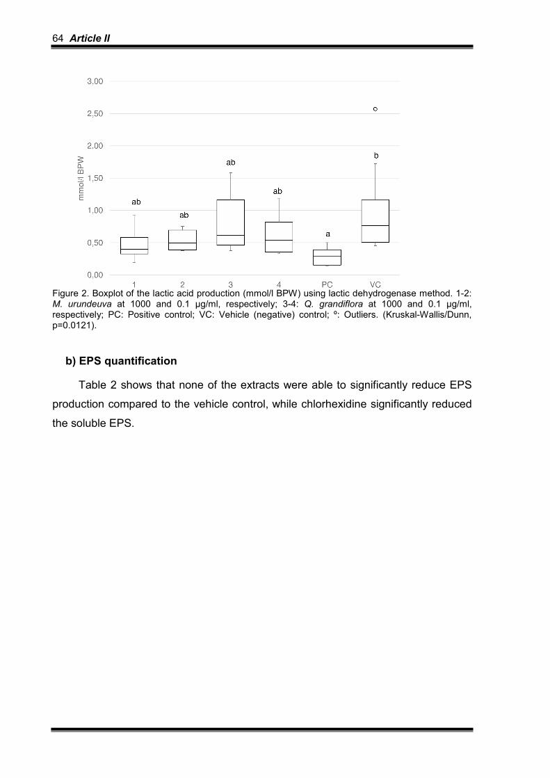
64 Article II
Figure 2. Boxplot of the lactic acid production (mmol/l BPW) using lactic dehydrogenase method. 1-2: M. urundeuva at 1000 and 0.1 µg/ml, respectively; 3-4: Q. grandiflora at 1000 and 0.1 µg/ml, respectively; PC: Positive control; VC: Vehicle (negative) control; º: Outliers. (Kruskal-Wallis/Dunn, p=0.0121).
b) EPS quantification
Table 2 shows that none of the extracts were able to significantly reduce EPS
production compared to the vehicle control, while chlorhexidine significantly reduced
the soluble EPS.

Article II 65
Treatments Soluble EPS (µg/mg) Insoluble EPS (µg/mg)
35% Alcohol (vehicle/negative control) 0.22(0.06)b 0.32(0.17)b
Chlorhexidine (positive control) 0.07(0.05)a 0.35(0.19)ab
M. urundeuva 1000 µg/ml 0.15(0.10)ab 0.45(0.28)ab
M. urundeuva 100 µg/ml 0.22(0.16)ab 0.79(0.65)ab
M. urundeuva 10 µg/ml 0.12(0.04)ab 0.38(0.24)ab
M. urundeuva 1.0 µg/ml 0.14(0.07)ab 0.59(0.28)ab
M. urundeuva 0.1 µg/ml 0.25(0.15)b 0.50(0.23)ab
Q. grandiflora 1000 µg/ml 0.28(0.13)b 0.87(0.44)a
Q. grandiflora 100 µg/ml 0.16(0.06)ab 0.52(0.19)ab
Q. grandiflora 10 µg/ml 0.17(0.07)ab 0.44(0.33)ab
Q. grandiflora 1.0 µg/ml 0.14(0.08)ab 0.52(0.21)ab
Q. grandiflora 0.1 µg/ml 0.17(0.09)ab 0.74(0.42)ab
Table 2. Median (interquartile interval) of soluble and insoluble EPS (µg/mg biofilm). Different letters at the same column show significant differences among the treatments. Soluble EPS (Kruskal-Wallis/Dunn, p<0.0001) and Insoluble EPS (Kruskal-Wallis/Dunn, p = 0.0082).
3.4 TMR
No one of the extracts was able to reduce the integrated mineral loss and the
lesion depth compared to negative control, while chlorhexidine significantly reduced
the enamel caries lesions development (Table 3 and Figure 3). Figure 4 shows TMR
pictures of a representative enamel sample from each group.
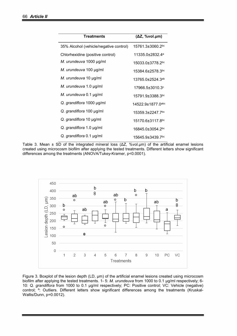
66 Article II
Treatments (∆Z, %vol.µm)
35% Alcohol (vehicle/negative control) 15761.3±3060.2bc
Chlorhexidine (positive control) 11335.0±2832.4a
M. urundeuva 1000 µg/ml 15033.0±3778.2bc
M. urundeuva 100 µg/ml 15384.6±2578.3bc
M. urundeuva 10 µg/ml 13765.0±2524.3ab
M. urundeuva 1.0 µg/ml 17966.5±3010.3c
M. urundeuva 0.1 µg/ml 15791.9±3388.3bc
Q. grandiflora 1000 µg/ml 14522.9±1877.0abc
Q. grandiflora 100 µg/ml 15359.3±2247.7bc
Q. grandiflora 10 µg/ml 15170.6±3117.8bc
Q. grandiflora 1.0 µg/ml 16845.0±3054.2bc
Q. grandiflora 0.1 µg/ml 15645.9±3439.7bc
Table 3. Mean ± SD of the integrated mineral loss (∆Z, %vol.µm) of the artificial enamel lesions created using microcosm biofilm after applying the tested treatments. Different letters show significant differences among the treatments (ANOVA/Tukey-Kramer, p<0.0001).
Figure 3. Boxplot of the lesion depth (LD, µm) of the artificial enamel lesions created using microcosm biofilm after applying the tested treatments. 1- 5: M. urundeuva from 1000 to 0.1 µg/ml respectively; 6-10: Q. grandiflora from 1000 to 0.1 µg/ml respectively; PC: Positive control; VC: Vehicle (negative) control; º: Outliers. Different letters show significant differences among the treatments (Kruskal-Wallis/Dunn, p=0.0012).

Article II 67
Figure 4. Representative TMR pictures (20x) of the artificial enamel lesions created using microcosm biofilm after applying the tested treatments. A) Positive control (Chlorhexidine, PerioGard®), B) Vehicle (negative) control, C-G) M. urundeuva from 1000 to 0.1 µg/ml, respectively; H-L) Q. grandiflora from 1000 to 0.1 µg/ml, respectively.
A B C
D E F
G H I
J K L

68 Article II
4. Discussion
The use of plant extracts is a common practice for the prevention and treatment
of various diseases worldwide. Phytotherapy is considered of low cost and widely
accessible [6]. Brazil is one of countries with the greatest biodiversity [7], which in
turn has stimulated the use of different type of plants for prevention and treatment of
diverse diseases. The most common benefits of the plants in health is their
antimicrobial effect, which may be potentially able to control oral diseases as dental
caries and periodontitis [26-28].
The experimental model for studying the effect of plants on dental caries must
include assays that are capable to show the antimicrobial properties (such as
viability, EPS and lactic acid assays) as well as the anti-caries effect (TMR), since
one is not synonym of the other. Some antimicrobial agents have no anti-caries
potential [4], which unable their use for the prevention of this disease [29].
Accordingly, we have chosen to apply a microcosm biofilm model on enamel, since it
is able to simulate the heterogeneity and the variability of in vivo biofilm, allowing the
analysis of both biofilm and tooth [30].
Myracrodruon urundeuva and Q. grandiflora were chosen as they are easily
found in Brazilian Cerrado. The ethanolic extracts of M. urundeuva leaves and bark
have as active components gallic acid, methyl gallate, ethyl gallate, chlorogenic and
protocatechuic acid, saponins, flavonoids, tannins and polyphenols [31]. The
ethanolic extract of Q. grandiflora leaves present gallic and ellagic acids derivatives,
galotannins, ellagitannins, triterpenes, flavonoids, benzoquinones and
anthraquinones [32]. A previous study showed that the main components of M.
unrundeuva extract are flavonoids and tannins [17].
Generally, our study showed that M. urundeuva has superior antimicrobial effect
compared to Q. grandiflora. Alves et al. [33] observed antimicrobial effect of M.
urundeuva (stem bark) against oral bacteria by quantifying inhibition zone (Minimum
Inhibitory Concentration-MIC) and Minimum Inhibitory Adhesion Concentration
(MICA). They showed MIC and MICA values of 0.125 mg/ml and 0.0625 mg/ml
against mutans streptococci, respectively. Their MIC value is in agreement with our
results of viability, since we have seen antimicrobial effect with 0.1 mg/ml M.
urundeuva. In addition, M. urundeuva, as 5% gel or as aqueous solution (7.5 mg/ml),

Article II 69
is also effective against total microorganisms [34] and mutans streptococci [35]
presented in biofilm of male Wistar rats, respectively.
With respect to Q. grandiflora, there is no study about its effect on cariogenic
bacteria, but only on Staphylococcus aureus, Escherichia coli, Bacillus cereus,
Pseudomonas aeruginosa, Streptococcus pyogenes and Helicobacter pylori in
planktonic phase [13,36,37].
Despite the extracts were able to reduce bacteria viability and the number of
lactobacilli and mutans streptococci, they did not interfere in the bacteria metabolism,
and, therefore, they were unable to reduce caries lesions development. Despite there
were lower number of viable bacteria due to the treatments, they were still able to
produce acid and EPS, which in turn induced enamel demineralization. Our work
provided support for the statement that no all antimicrobial agents have anti-caries
effect [4, 29].
On the other hand, we should take in mind that other bacteria, not analyzed in
the present study, could have contributed to enamel caries development (Scardovia
wiggsiae, Bifidobacterium spp. and Actinomyces spp.) [38], which shall be further
confirmed under this model.
In disagreement with our results, previous work has shown that M. urundeuva
All. (7.5 mg/ml) protected against enamel surface hardness loss in Wistars rats
inoculated with mutans streptococci for 7 days [35]. This difference might be due to
the greater concentration of the extract tested in the former study and to the low
velocity of caries development in vivo.
Chlorhexidine was applied as positive control in agreement with Maske et al.
[30], who observed that chlorhexidine solutions at concentrations of 0.06% and
0.12%, applied 2x/day, significantly reduced the counts of mutans streptococci (65%)
and the percentage of surface hardness loss compared to control (saline solution).
On the other hand, Alcohol (35%) was our negative control, since it was applied as
solvent for the leaves extracts. Previous works have shown that 50% or 80% ethanol
had no antimicrobial effect against mutans streptococci [39,40].
Further studies shall give attention to test the antimicrobial effect of M.
urundeuva extracts, varying concentrations, solvents and vehicles, under microcosm
biofilm, in situ or/and in vivo models, to confirm if they really do not have any anti-
caries effect. Other important point to consider in future studies is the analysis of the
cytotoxic effect of the extracts as well. Some previous studies have shown cytotoxic

70 Article II
effect of M. urundeuva and Q. grandiflora on fibroblasts, but the extracts were diluted
in 80% methanol or 70% ethanol [17,41]. Therefore, this issue should be better
addressed using lower alcohol concentrations as solvent.
5. Conclusions
The extracts showed antimicrobial effects (especially M. urundeuva) on the
microcosm biofilm; however, they had no anti-caries effect under this biofilm model.
Conflict of interest: None
Acknowledgements: The authors thank CAPES for the concession of scholarship to
the first author. The funders had no role in study design, data collection and analysis,
decision to publish, or preparation of the manuscript.
Ethical approval: CEEA 43948115.2.0000.5417.
References
[1] N. Takahashi, B. Nyvad, The role of bacteria in the caries process: ecological
perspectives, J Dent Res. 90 (2011) 294-303. [2] PD. Marsh, A. Moter, DA. Devine, Dental plaque biofilms: communities, conflict
and control, Periodontol 2000 55 (2011) 16-35. [3] A. Rugg-Gunn, Dental caries: Strategies to control this preventable disease
Strategies for caries prevention, Acta Med. Acad. 42 (2013) 117–130. doi:10.5644/ama2006-124.80.
[4] AS. Braga, JG. Pires, AC. Magalhães, Commercial antimicrobials mouthrinses on caries and periodontitis-related biofilm control - a review of literature, Braz Dent Sci. 20 (2017) 13-23.
[5] C.J. Henley-Smith, F.S. Botha, N. Lall, The use of plants against oral pathogens, Microb. Pathog. Strateg. Combat. Them Sci. Technol. Educ. (2013) 1375–1384.
[6] R.C. Forzza, J.F.A. Baumgratz, C.E.M. Bicudo, D.A.L. Canhos, A.A. Carvalho, M.A.N. Coelho, A.F. Costa, D.P. Costa, M.G. Hopkins, P.M. Leitman, L.G. Lohmann, E.N. Lughadha, L.C. Maia, G. Martinelli, M. Menezes, M.P. Morim, A.L. Peixoto, J.R. Pirani, J. Prado, L.P. Queiroz, S. Souza, V.C. Souza, J.R. Stehmann, L.S. Sylvestre, B.M.T. Walter, D.C. Zappi, New Brazilian Floristic List Highlights Conservation Challenges, Bioscience. 62 (2012) 39–45. doi:10.1525/bio.2012.62.1.8.
[7] D.C. Zappi, F.L. Ranzato Filardi, P. Leitman, V.C. Souza, B.M.T. Walter, J.R. Pirani, M.P. Morim, L.P. Queiroz, T.B. Cavalcanti, V.F. Mansano, R.C. Forzza, Growing knowledge: An overview of Seed Plant diversity in Brazil, Rodriguesia. 66 (2015) 1085–1113. doi:10.1590/2175-7860201566411.

Article II 71
[8] R.M. Montanari, L.C.A. Barbosa, A.J. Demuner, C.J. Silva, N.J. Andrade, F.M.D. Ismail, M.C.A. Barbosa, Exposure to anacardiaceae volatile oils and their constituents induces lipid peroxidation within food-borne bacteria cells, Molecules. 17 (2012) 9728–9740. doi:10.3390/molecules17089728.
[9] E.A. Carlini, J.M. Duarte-Almeida, E. Rodrigues, R. Tabach, Antiulcer effect of the pepper trees Schinus terebinthifolius Raddi (aroeira-da-praia) and Myracrodruon urundeuva Allemão, Anacardiaceae (aroeira-do-sertão), Rev. Bras. Farmacogn. 20 (2010) 140–146. doi:10.1590/S0102-695X2010000200001.
[10] C.A. Hiruma-Lima, L.C. Santos, H. Kushima, C.H. Pellizzon, G.G. Silveira, P.C.P. Vasconcelos, W. Vilegas, A.R.M.S. Brito, Qualea grandiflora, a Brazilian “Cerrado” medicinal plant presents an important antiulcer activity, J. Ethnopharmacol. 104 (2006) 207–214. doi:10.1016/j.jep.2005.09.002.
[11] C.M. de M. Sousa, H.R. e Silva, G.M. Vieira-Jr., M.C.C. Ayres, C.L.S. da Costa, D.S. Araújo, L.C.D. Cavalcante, E.D.S. Barros, P.B. de M. Araújo, M.S. Brandão, M.H. Chaves, Fenóis totais e atividade antioxidante de cinco plantas medicinais, Quim. Nova. 30 (2007) 351–355. doi:10.1590/S0100-40422007000200021.
[12] F.O.G. Gaspi, M.A. Foglio, J.E. Carvalho, R.A. Moreno, Pharmacological activities investigation of crude extracts and fractions from Qualea grandiflora Mart., J. Ethnopharmacol. 107 (2006) 19–24. doi:10.1016/j.jep.2006.01.023.
[13] C.O. Moura, G.P.V. Nascimento, C.R.G. Pinto, Atividade antibacteriana de Qualea grandiflora Mart. (Vochysiaceae), BioFar. 8 (2012) 34–42.
[14] W. Marcenes, N.J. Kassebaum, E. Bernabé, A. Flaxman, M. Naghavi, A. Lopez, C.J.L. Murray, Global Burden of Oral Conditions in 1990-2010, J. Dent. Res. 92 (2013) 592–597. doi:10.1177/0022034513490168.
[15] B.M. Souza, C. Fernandes Neto, P.M.A. Salomao, L.R.M.S. Vasconcelos, F.B. Andrade, A.C. Magalhães, Analysis of the antimicrobial and anticariogenic effect of TiF4 varnish under a microcosm biofilm formed on enamel, J. Appl. Oral Sci. (2018). In Press.
[16] J. Pratten, M. Wilson, D. A. Spratt, Characterization of in vitro oral bacterial biofilms by traditional and molecular methods, Oral Microbiol. Immunol. 18 (2003) 45–49. doi:10.1034/j.1399-302X.2003.180107.x.
[17] A.C. Machado, L. P. Souza, L. L. Saldanha, L. G. Pieroni, A. A. Matos, F. A. Oliveira, W. Vilegas, C. A. Damante, A. L. Dokkedal, R. C. Oliveira. “Aroeira” (Myracrodruon urundeuva) methanol extract: the relationship between chemical compounds and cellular effects, Pharm Biol 54 (2016) 2737–2341. doi:10.1080/13880209.2016.1182555.
[18] A.J. McBain, Chapter 4 In Vitro Biofilm Models, in: Adv. Appl. Microbiol., 2009: pp. 99–132. doi:10.1016/S0065-2164(09)69004-3.
[19] K. Zhang, L. Cheng, E. J. Wu, M. D. Weir, Y. Bai,H. H. K. Xu, Effect of water-ageing on dentine bond strength and anti-biofilm activity of bonding agent containing new monomer dimethylaminododecyl methacrylate, J Dent. 41 (2013) 504–513. doi:10.1016/j.jdent.2013.03.011.
[20] C. Hannig, A. Gaeding, S. Basche, G. Richter, R. Helbig, M. Hannig, Effect of Conventional Mouthrinses on Initial Bioadhesion to Enamel and Dentin in situ, Caries Res. 47 (2013) 150–161. doi:10.1159/000345083.
[21] M.E. Saravia, P. Nelson-Filho, I.Y. Ito, L.A. da Silva, R.A. da Silva, C.G. Emilson, Morphological differentiation between S. mutans and S. sobrinus on modified SB-20 culture medium, Microbiol. Res. 166 (2011) 63–67.

72 Article II
doi:10.1016/j.micres.2010.01.001. [22] Z.I. Lima, M. A. Sampaio de Melo, F. M. Borges, A. H. Teixeira, C. Steiner-
Oliveira, M. N. Santos, L. K. Rodrigues, Evaluation of the antimicrobial effect of photodynamic antimicrobial therapy in an in situ model of dentine caries, Eur. J. Oral Sci. 117 (2009) 568–574. doi:10.1111/j.1600-0722.2009.00662.x.
[23] L. Cheng, K. Zhang, M.A.S. Melo, M.D. Weir, X. Zhou, H.H.K. Xu, Anti-biofilm Dentin Primer with Quaternary Ammonium and Silver Nanoparticles, J. Dent. Res. 91 (2012) 598–604. doi:10.1177/0022034512444128.
[24] C.J. Aires CP, Del Bel Cury AA, Tenuta LM, Klein MI, Koo H, Duarte S, J.A. Cury, Effect of Starch and Sucrose on Dental Biofilm Formation and on Root Dentine Demineralization, Caries Res. 42 (2008) 380–386. doi:10.1159/000154783.
[25] B. Angmar, D. Carlstrom, J.E. Glas, Studies on the ultrastructure of dental enamel. IV. The mineralization of normal human enamel, J. Ultrastruct. Res. 8 (1963) 12–23.
[26] E. Pereira, R. Gomes, N. Freire, E. Aguiar, M. Brandão, V. Santos, In vitro Antimicrobial Activity of Brazilian Medicinal Plant Extracts against Pathogenic Microorganisms of Interest to Dentistry, Planta Med. 77 (2011) 401–404. doi:10.1055/s-0030-1250354.
[27] J.P.C. Silva, A.L. Castilho, C.H.C. Saraceni, I.E.C. Diaz, M.L.B. Paciencia, I.B. Suffredini, Anti-Streptococcal activity of Brazilian Amazon Rain Forest plant extracts presents potential for preventive strategies against dental caries, J. Appl. Oral Sci. 22 (2014) 91–97. doi:10.1590/1678-775720130366.
[28] S.M. Bersan, L.C. Galvão, V.F. Goes, A. Sartoratto, G.M. Figueira, V.L. Rehder, S.M. Alencar, R.M. Duarte, P.L. Rosalen, M.C. Duarte, Action of essential oils from Brazilian native and exotic medicinal species on oral biofilms, BMC Complement. Altern. Med. 14 (2014) 451. doi:10.1186/1472-6882-14-451.
[29] P. Boyle, A. Koechlin, P. Autier, Mouthwash Use and the Prevention of Plaque, Gingivitis and Caries, Oral Dis. 20 (2014) 1–68. doi:10.1111/odi.12187.
[30] T.T. Maske, K. V. Brauner, L. Nakanishi, R.A. Arthur, F.H. van de Sande, M.S. Cenci, An in vitro dynamic microcosm biofilm model for caries lesion development and antimicrobial dose-response studies, Biofouling. 32 (2016) 339–348. doi:10.1080/08927014.2015.1130824.
[31] B.C.F. Mota, V. de A. Royo, J.M.S. Fonseca, A.C. Dos Santos, A.F. Melo Junior, E.V. Menezes, V.R. Esperandim, R. da S. Laurentiz, Comparative studies between the chemical constituents and biological properties of the extracts from the leaves and barks of Myracrodruon urundeuva Fr. All., J. Med. Plants Res. 9 (2015) 159–168. doi:10.5897/JMPR2014.5513.
[32] M.C.C. Ayres, S.P. Escórcio, D.A. Costa, M.H. Chaves, Constituintes químicos das folhas de Qualea grandiflora: Atribuição dos dados de RMN de dois flavonóides glicosilados acilados diastereoisoméricos, Quim. Nova. 31 (2008) 1481–1484.
[33] P.M. Alves, L.M.G. Queiroz, J.V. Pereira, M. do S.V.P. Pereira, Atividade antimicrobiana, antiaderente e antifúngica in vitro de plantas medicinais brasileiras sobre microrganismos do biofilme dental e cepas do gênero Candida, Rev. Soc. Bras. Med. Trop. 42 (2009) 222–224.
[34] M.A. Botelho, V.S. Rao, C.B.M. Carvalho, J.G. Bezerra-Filho, S.G.C. Fonseca, M.L. Vale, D. Montenegro, F. Cunha, R.A. Ribeiro, G.A. Brito, Lippia sidoides and Myracrodruon urundeuva gel prevents alveolar bone resorption in

Article II 73
experimental periodontitis in rats, J. Ethnopharmacol. 113 (2007) 471–478. doi:10.1016/j.jep.2007.07.010.
[35] T.E.C. de Menezes, A.C.B. Delbem, F.L. Brighenti, A.C. Okamoto, E. Gaetti-Jardim Jr, Protective efficacy of Psidium cattleianum and Myracrodruon urundeuva aqueous extracts against caries development in rats, Pharm. Biol. 48 (2010) 300–305. doi:10.3109/13880200903122202.
[36] T.M. de A. Alves, A.F. Silva, M. Brandão, T.S.M. Grandi, E. de F.A. Smânia, A. Smânia Júnior, C.L. Zani, Biological screening of Brazilian medicinal plants, Mem. Inst. Oswaldo Cruz. 95 (2000) 367–373. doi:10.1590/S0074-02762000000300012.
[37] C. Bonacorsi, L.M. Da Fonseca, M.S.G. Raddi, R.R. Kitagawa, W. Vilegas, Comparison of Brazilian plants used to treat gastritis on the oxidative burst of helicobacter pylori -stimulated neutrophil, Evidence-Based Complement. Altern. Med. 2013 (2013) 1–8. doi:10.1155/2013/851621.
[38] K. Henne, A.P. Gunesch, C. Walther, H. Meyer-Lueckel, G. Conrads, M. Esteves-Oliveira, Analysis of bacterial activity in sound and cariogenic biofilm: a pilot in vivo study, Caries Res 50 (2016) 480-488.
[39] D.S.V. Barbieri, F. Tonial, P.V.A. Lopez, B.H.L.N. Sales Maia, G.D. Santos, M.O. Ribas, C. Glienke, V.A. Vicente, Antiadherent activity of Schinus terebinthifolius and Croton urucurana extracts on in vitro biofilm formation of Candida albicans and Streptococcus mutans, Arch. Oral Biol. 59 (2014) 887–896. doi:10.1016/j.archoralbio.2014.05.006.
[40] J.G. Cardoso, N.L.P. Iorio, L.F. Rodrigues, M.L.B. Couri, A. Farah, L.C. Maia, A.G. Antonio, Influence of a Brazilian wild green propolis on the enamel mineral loss and Streptococcus mutans’ count in dental biofilm, Arch. Oral Biol. 65 (2016) 77–81. doi:10.1016/j.archoralbio.2016.02.001.
[41] C. Tokuhara, F. Oliveira, A. Matos, G. Oliveira, T. Dionísio, L. Saldanha, A. Leite, P. Pupolin, A. Dokkedal, R. Oliveira, Qualea grandiflora Mart. effect on pre-osteoblast behavior and matrix metalloproteinase, J. Ethnopharmacol. (2018). In press.

74 Article II
4-Article III

Article III 77
4 ARTICLE III
Article formatted according to the guideline of Archives of Biology (ANNEX 3).
Hydroalcoholic extracts of Myracrodruon urundeuva All. and Qualea
grandiflora Mart. leaves on Streptococcus mutans biofilm and tooth
demineralization
Juliana Gonçalves Piresa
aDepartment of Biological Sciences, Bauru School of Dentistry, University of São
Paulo, Al. Octávio Pinheiro Brisolla 9-75, Bauru, São Paulo, Brazil. E-mail:
Sara Salustiano Zabini
aDepartment of Biological Sciences, Bauru School of Dentistry, University of São
Paulo, Al. Octávio Pinheiro Brisolla 9-75, Bauru, São Paulo, Brazil. E-mail:
Aline Silva Bragaa
aDepartment of Biological Sciences, Bauru School of Dentistry, University of São
Paulo, Al. Octávio Pinheiro Brisolla 9-75, Bauru, São Paulo, Brazil. E-mail:
Rita de Cássia Fabris
aDepartment of Biological Sciences, Bauru School of Dentistry, University of São
Paulo, Al. Octávio Pinheiro Brisolla 9-75, Bauru, São Paulo, Brazil. E-mail:
Flaviana Bombarda de Andradeb
bDepartment of Operative Dentistry, Endodontics and Dental Materials, Bauru School
of Dentistry, University of São Paulo, Al. Octávio Pinheiro Brisolla 9-75, Bauru, São
Paulo, Brazil. E-mail: [email protected]
Rodrigo Cardoso de Oliveiraa

78 Article III
aDepartment of Biological Sciences, Bauru School of Dentistry, University of São
Paulo, Al. Octávio Pinheiro Brisolla 9-75, Bauru, São Paulo, Brazil. E-mail:
Ana Carolina Magalhães*a
aDepartment of Biological Sciences, Bauru School of Dentistry, University of São
Paulo, Al. Octávio Pinheiro Brisolla 9-75, Bauru, São Paulo, Brazil. E-mail:
Correspondent author: Ana Carolina Magalhães, Department of Biological Sciences,
Bauru School of Dentistry, University of São Paulo, Al. Octávio Pinheiro Brisolla 9-75,
Bauru, São Paulo, Brazil. Phone:+55 14 32358497. E-mail: [email protected]
Abstract
Objectives: This study evaluated the effect of the hydroalcoholic extracts of
Myracrodruon urundeuva All. and Qualea grandiflora Mart. leaves (alone or
combined) on the viability of Streptococcus mutans biofilm and on the prevention of
enamel demineralization.
Methods: Strain of S. mutans (ATCC 21175) was reactivated in BHI broth. Minimum
inhibitory concentration-MIC, minimum bactericidal concentration-MBC, minimum
biofilm inhibitory concentration-MBIC and minimum biofilm eradication concentration-
MBEC were firstly determined in order to choose the concentrations to be tested
under biofilm model. S. mutans biofilm (5x10⁵ CFU/ml) was produced on bovine
enamel, using McBain saliva under 0.2% sucrose exposure, for 3 days. The biofilm
was daily treated with the extracts for 1 min. The biofilm viability was tested by
fluorescence and the enamel demineralization was measured using TMR.
Results: Myracrodruon urundeuva (isolated or combined), at the concentrations
equal and higher than 0.625 mg/ml, was able to reduce bacteria viability, while
Qualea grandiflora alone had antimicrobial effect at 5 mg/ml only (p<0.05). On the
other hand, none of the extracts were able to reduce enamel caries lesion
development.

Article III 79
Conclusions: The tested hydroalcoholic extracts had antimicrobial action (especially
Myracrodruon urundeuva All.); however, they did not have anti-caries effect under S.
mutans biofilm model.
Keywords: Antimicrobial agents; Biofilm; Dental caries; Plants extracts;
Streptococcus mutans.

80 Article III
1. Introduction
Streptococcus mutans is considered the most important microorganism involved
in the etiology of dental caries, a tooth disease related to the presence of biofilm,
whose metabolism is dependent on the frequent exposure to sugar (Eriksson, Lif
Holgerson, Esberg, & Johansson, 2017) S. mutans are capable of producing acid
from sugar metabolism, surviving in an acidic environment and using sucrose to
synthesize extracellular polysaccharide (EPS), which are mostly glycan responsible
for the development and protection of dental biofilm (Bowen & Koo, 2011; Koo,
Falsetta, & Klein, 2013; Marsh, Martin, Lewis, & Williams, 2009; Takahashi & Nyvad,
2011; Zhao, Li, Lin, Chen, & Yu, 2014).
In order to control dental biofilm and avoid the disease, mechanical practice of
brushing and reduction of sugar consume are advised (Rugg-Gunn, 2013). However,
for patients under high risk of caries, chemical agents may be needed (James et al.,
2017). On the other hand, the conventional chemical agents usually cause a series of
undesired side events (James et al., 2017; Zheng & Wang, 2011) such as tooth
staining, supragingival calculus formation, changes in taste perception, parotid gland
swelling and irritation of the oral mucosa, when applied for periods higher than 2
weeks (James et al., 2017).
Phytotherapy has been developed to overcome this issue (Palombo, 2011;
Vieira et al., 2014). Brazil is the home of the largest number of plant species (32,086
native Angiosperms and 23 native Gymnosperms) worldwide (Forzza et al., 2012;
Mittermeier, van Dijk, Rhodin, & Nash, 2004; Zappi et al., 2015). Myracrodruon
urundeuva All. and Qualea grandiflora Mart. are examples of Brazilian plants with
anti-inflammatory, antiulcerogenic activity, gastric mucosal protection, analgesic
(Carlini, Duarte-Almeida, Rodrigues, & Tabach, 2010; Hiruma et al., 2006; Souza et
al., 2007; Viana et al., 1997; Viana, Bandeira, & Matos, 2003), anticonvulsant (Gaspi,
Foglio, Carvalho, & Moreno, 2006), antiseptic and antibacterial activities (Alves et al.,
2009; Costa et al., 2008).
However, there is no information about their anti-caries potential. Only a study
was performed to check the effect of Myracrodruon urundeuva All. against S. mutans
and mineral loss using an animal model (Menezes, Delbem, Brighenti, Okamoto, &
Gaetti-Jardim Jr., 2010). Therefore, the objective of this study was to test the effect of
the hydroalcoholic extracts of Myracrodruon urundeuva All. and Qualea grandiflora
Article III 81
Mart. leaves, isolated or combined, on S. mutans viability (under planktonic and
biofilm form) and on the prevention of enamel caries lesion formation.
2. Methods
2.1 Plant’s extracts preparation
Leaf samples of Myracrodruon urundeuva All. and Qualea grandiflora Mart.
were collected in October 2013 at Jardim Botânico Municipal de Bauru (Bauru,
Brazil), (22°20'41.4"S - 49°01'45.1"W). Exsiccates were deposited in the Herbarium
of UNESP under code numbers HRCB59831 and UNBA6034. The collections have
authorization issued by SISBIO under code number 39825-1. The hydroalcoholic
extracts were prepared as described by Machado et al. (2016) in the Natural
Products Chemistry Laboratory, at the Faculty of Sciences/UNESP-Bauru-SP. Briefly,
fresh leaves were hot air dried at 45°C and grounded in a knife mill. The extract was
obtained with 70% ethanol via percolation at room temperature. The filtrates were
concentrated under dryness and reduced pressure at 40°C. They were finally
lyophilized, weighed and stored in an amber bottle at room temperature. The
hydroalcoholic extracts of Myracrodruon urundeuva All. and Qualea grandiflora Mart.
were produced using ethanol at a final concentration of 5%. They were tested
isolated and combined.
2.2 Bacterial strain
Strain of S. mutans (ATCC 25175) was defrosted and 100 µl were
resuspended in tubes containing 3 ml of Brain Heart Infusion broth (BHI, Difco,
Detroit, USA). The tubes were then incubated for 24 h at 37º C and 5% CO2 (Hasan,
Danishuddin, & Khan, 2015).
2.3 Minimal inhibitory and minimal bactericidal concentrations
The minimum inhibitory concentration (MIC) was determined using microplate
serial dilution technique according to the M07-A9 of the manual of Clinical and
Laboratory Standards Institute with modifications. Briefly, 80 µl of BHI broth and 100
µl of the hydroalcoholic extracts of M. urundeuva All. and/or Q. grandiflora Mart., at
concentrations ranging from 20 mg/ml to 0.00244 mg/ml, were distributed in 96-well
plates. Twenty microliters of bacterial suspension (5x10⁵ CFU/ml) were added in
each well (CLSI, 2012). BHI and BHI with 5% ethanol were applied as negative
82 Article III
controls, while Periogard® was used as positive control (0.12% chlorhexidine,
Colgate, Brazil, at the dilutions from 0.6 mg/ml to 0.00234 mg/ml).
The plates were incubated for 24 h at 37º C and 5% CO2. Thereafter, the
absorbance was determined using a microplate reader at 540 nm (Fluorstar Optima-
BMG Labtech, Germany). MIC was defined as the lowest concentration of the extract
capable of inhibiting 100% of bacterial growth compared to the negative control.
For the MBC determination, aliquots (20 µl) from the wells, where no
absorbance was detected (values equal or higher than MIC), were spread using the
gout technique (Westergren & Krasse, 1978) on petri dishes containing BHI agar
(Difco, Detroit, USA). The plates were incubated for 24 h at 37º C and 5% CO2. MBC
was defined as the lowest concentration of the extract capable of inhibiting any
visible bacterial growth. All tests were done in biological triplicate (n=3 for each
replicate).
2.4 Minimum biofilm inhibition and eradication concentrations
The minimum biofilm inhibition concentration (MBIC) was determined as done
for MIC, however, after 24-h incubation, the supernatant were discarded and washed
once with phosphate buffered saline (PBS). The viability was quantified using 3-[4, 5-
dimethyl-2-thiazolyl]-2,5-diphenyl-2H-tetrazolium-bromide (MTT, Sigma-Aldrich, St
Louis, MO, USA). Viable bacteria reduce MTT in purple formazan. The absorbance
was measured using a microplate reader (Fluorstar Optima- BMG Labtech,
Ortenberg, Germany) at 540 nm. The MBIC was defined as the lowest concentration
able to reduce in 100% bacterial viability compared to the negative control (Tang et
al., 2011; Teanpaisan, Senapong, & Puripattanavong, 2014).
For the minimum biofilm eradication concentration (MBEC) determination, the
bacteria was cultivated similarly to MIC assay but without any treatment during the
first 24h. The BHI was then removed and the wells washed once with PBS, to
remove any bacteria in suspension. The hydroalcoholic extracts were directly added
to the wells (200 µl/well), and the plates were incubated again for 24 h at 37o C and
5% CO2. The viability of the attached bacterial to the wells was measured using MTT
assay as described above. The MBEC was defined as the lowest concentration able
to reduce in 100% bacterial viability compared to the negative control (Tang et al.,
2011; Teanpaisan, Senapong, & Puripattanavong, 2014). All tests were done in
biological triplicate (n=3 for each replicate).

Article III 83
2.5 Enamel samples preparation
A one hundred and thirty-five enamel samples (4 mm x 4 mm) were prepared
from bovine teeth, using a semi-precision cutting machine (Buehler, Enfield, USA).
The samples were fixed in acrylic discs with wax and polished in a metallographic
polishing machine (Arotec, Cotia, Brazil) using water-cooled silicon-carbide discs
(600-grade papers ANSI grit; Buehler, Enfield, USA) to remove grooves and to
standardize the surface roughness of approximately 0.153 ± 0.037 µm. The average
surface roughness (Ra) was assessed using contact profilometer and Mahr Surf XCR
20 software (Mahr, Göttingen, Germany). Enamel samples were randomly distributed
in the groups according to the Ra mean values, allowing similar enamel surface
roughness means among them.
Two thirds of the enamel surfaces were protected with nail polish to obtain
control areas for the TMR analysis. The samples were sterilized with ethylene oxide
[Gas exposure time (30% ETO/70%CO2) for 4 h under a pressure of 0.5 ± 0.1
kgF/cm2].
2.6 3-days S. mutans biofilm formation and treatment
For the formation of the S. mutans biofilm on bovine enamel, the strain was
reactivated in BHI broth and diluted (concentration of 5x10⁵ CFU/ml) in artificial
saliva. The artificial saliva contained 2.5 g/l mucin from porcine stomach (type II), 2.0
g/l bacteriological peptone, 2.0 g/l tryptone, 1.0 g/l yeast extract, 0.35 g/l NaCl, 0.2 g/l
KCl, 0.2 g/l CaCl2, 0.1 g/l cysteine hydrochloride, 0.001 g/l hemin, 0.0002 g/l vitamin
K1, at pH 7.0 (McBain, 2009).
Twenty-four well plates containing enamel samples were filled with inoculum
(5x10⁵ CFU/ml artificial saliva, 1.5 ml/well), which was incubated at 5% CO2 and
37˚C. After 8h, the inoculum was removed, the samples were washed using PBS and
fresh McBain saliva containing now 0.2% sucrose was added into the wells (1.5
ml/well). The microplates were incubated at 5% CO2 and 37˚C for further 16h,
completing the first day of the experiment (24 h).
During the next 2 days, the biofilm was daily treated with the sub-MIC levels of
Myracrodruon urundeuva All. and Qualea grandiflora Mart. extracts, isolated or
combined (1x60s/day). After treatment, the samples were washed using PBS and
fresh McBain saliva with 0.2% sucrose was added. Periogard® was applied as

84 Article III
positive control (0.12% chlorhexidine, Colgate, Brazil), PBS and PBS with 5%
ethanol were used as negative controls. The microplates were incubated at the same
conditions described above (Zhang et al., 2013). The experiment was done in
biological triplicate (n=3 for each replicate).
2.7 Biofilm viability analysis
The biofilm was stained using the nucleic acid markers diluted in PBS (1ml PBS
+ 1 µl SYTO9 + 1 µl propidium iodide, 10 µl/well) (Kit Live & Dead® cells viability
assay, Thermo Fisher Scientific, Waltham, USA) for 15 min in a dark environment.
Live bacteria were stained with SYTO9 producing a green fluorescence and dead
lysed bacteria were stained with propidium iodide and SYTO9 producing a red
fluorescence (Hannig et al., 2013). The biofilm was examined using confocal laser
scanning microscope-CLSM (Leica TCS SPE, Mannheim, Germany) and Leica
Application Suite-Advanced Fluorescence software (LAS AF, Mannheim, Germany).
Three images (275 µm2) were captured from each sample surface and analyzed
using BioImage L 2.0 software, to quantify the live and dead bacteria (%).
2.8 Analysis of demineralization by transverse microradiography - TMR
After cleaning, all enamel samples were transversally sectioned and polished to
obtain slices with 80-100 µm of thickness. The enamel slices were fixed in a sample-
holder together with an aluminum calibration step wedge with 14 steps. A
microradiograph was taken using an x-ray generator (Softex, Tokyo, Japan) on the
glass plate at 20 kV and 20 mA (at a distance of 42 cm) for 13 min. The glass plates
were developed for 5 min, rinsed in deionized water, fixed for 8 min in a dark
environment, and then rinsed in running water for 10 min and air-dried (all
procedures were done at 20°C). The developed plate was analyzed using a
transmitted light microscope fitted with a 20x objective (Zeiss, Oberkochen,
Germany), a CCD camera (Canon, Tokyo, Japan), and a computer. Two images per
sample were taken using data-acquisition (version 2012) and interpreted using
calculation (version 2006) softwares from Inspektor Research System bv
(Amsterdam, The Netherlands). The mineral content was calculated based on the
work of Angmar, Carlstrom & Glas (1963), assuming the density of the mineral to be

Article III 85
3.15 kg l-1 and 87 vol% of mineral content for the sound enamel. The lesion depth
(LD, µm) and the integrated mineral loss (∆Z, %vol. µm) were calculated.
2.9 Statistical Analysis
Data were statistically analyzed using software Graph Pad Instat for Windows
(GraphPad Software, San Diego, USA). The normal distribution and homogeneity
were checked using Kolmogorov & Smirnov and Bartlett’s tests, respectively. The %
live and dead microorganisms were compared using Kruskal-Wallis followed by Dunn
test and the TMR data were analyzed using ANOVA followed by Tukey test. The
level of significance was set at 5%.
3. Results
Table 1 shows the antimicrobial concentrations values found for the extracts
against S. mutans (planktonic phase). The lowest MIC, MBC, MBIC and MBEC
values were seen for M. urundeuva; effect that was not improved by the combination
with Q. grandiflora. However, no extract reached the lowest antimicrobial
concentration found for chlorhexidine.
With respect to the 3-days S. mutans biofilm, similar antimicrobial effect was
found. M. urundeuva at the concentrations equal and higher than 0.625 mg/ml were
able to reduce bacteria viability, while Q. Grandflora had antimicrobial effect only at 5
mg/ml. The combination of the extracts was effective in reducing bacterial viability at
concentrations equal and higher than 0.625 mg/ml, similarly to M. urundeuva alone
(Table 2). Figure 1 shows representative CLSM pictures of the biofilms treated with
M. urundeuva (1.25 mg/ml), Q. grandiflora (5 mg/ml), chlorhexidine (0.12%) and
PBS.
None of the plant’s extracts were able to reduce the enamel caries lesion
development. Only chlorhexidine significantly reduced mineral loss, but not lesion
depth. Table 3 shows the TMR data and Figure 2 shows representative TMR pictures
of enamel samples from the same groups displayed in Figure 1.
4. Discussion
Considering the important role of S. mutans in dental caries etiology (Eriksson,
Lif Holgerson, Esberg, & Johansson, 2017), we chose to apply this bacteria specie
under planktonic and biofilm models. Myracrodruon urundeuva All. and Qualea

86 Article III
grandiflora Mart. were selected as representative of plants typical from Brazilian
“cerrado”. Our results showed that M. urundeuva had a better antimicrobial effect
against S. mutans than Q. grandiflora; however, it did not reach the antimicrobial
effect of chlorhexidine. The combination of the extracts had no further antimicrobial
effect, differently from the findings of Shafiei, Haji Abdul Rahim, Philip, & Thurairajah
(2016), who observed a synergic effect of three natural extracts (Psidium sp.,
Magifera sp. and Mentha sp.) on S. mutans .
M. urundeuva leaves and barks extracts have as active components: gallic acid,
methyl gallate, ethyl gallate, chlorogenic and protocatechuic acid, saponins,
flavonoids, tannins and polyphenols (Mota et al., 2015), while Q. grandiflora Mart.
leaves’ extract presents gallic and ellagic acids derivatives, galotannins,
ellagitannins, triterpenes, flavonoids, benzoquinones and anthraquinones in its
composition (Ayres, Escórcio, Costa, & Chaves, 2008). Therefore, the best
antimicrobial performance of M. urundeuva All. may be due to the presence of
polyphenols.
Alves, Queiroz, Pereira, & Pereira (2009) determined the MIC and MIAC
(Minimum Inhibitory Adherence Concentration) of hydroalcoholic extracts of M.
urundeuva (stem bark) against S. mutans. The MIC and MIAC values were 0.125
mg/ml and 0.0625 mg/ml, respectively. Their MIC value was lower than our MIC
value, which might be justified by the process of extraction and the part of the plant
tested as well as due to the method applied to determine MIC and the bacteria strain
used for that.
With respect to Q. grandiflora extract, the studies have been done using
Staphylococcus aureus, Escherichia coli, Bacillus cereus, Pseudomonas aeruginosa,
Streptococcus pyogenes and Helycobacter pylori strains (Alves et al., 2000;
Bonacorsi, Da Fonseca, Raddi, Kitagawa, & Vilegas, 2013; Moura, Nascimento, &
Pinto, 2012). This is the first study about its effect against cariogenic bacteria.
In 2010, Menezes, Delbem, Brighenti, Okamoto, & Gaetti-Jardim Jr evaluated
the action of aqueous extracts of Psidium cattleianum and M. urundeuva (7.5 mg/ml)
in Wistar rats under cariogenic diet for 7 weeks. They observed that both extracts
reduced the cariogenic biofilm (level of S. mutans by RT-PCR) and had a protective
effect on enamel surface hardness loss. Oppositely, our study did not show any
protective effect of the extracts against enamel demineralization. The reason to

Article III 87
explain the different findings might be related to the fact that the tested
concentrations in the present study were lower than those applied in the former work.
On the other hand, we should take in mind that high extracts concentrations
could induce some side effects. Cytotoxic potential is an important aspect that should
be considered before testing the effect of a new antimicrobial agent in clinical trials. A
recent study has shown that M. urundeuva leaves’ extracts at concentrations higher
than 100 µg/ml could reduce the viability of fibroblasts; however, the authors applied
80% methanol as solvent (Machado et al., 2016). Therefore, further studies on this
issue are needed to better establish the cytotoxic potential of the hydroalcoholic
plants extracts containing 5% ethanol. Furthermore, the lack of anti-caries effect shall
be confirmed under more complex biofilm models in the future.
5. Conclusion
The hydroalcoholic extracts of Myracrodruon urundeuva All. and Qualea
grandiflora Mart. leaves (isolated or combined, especially M. urundeuva) had
antimicrobial action against S. mutans; however, they did not have anti-caries effect
under 3-days S. mutans biofilm model.
Acknowledgments
The authors thank FAPESP (2016/20212-0) for the concession of financial support to
the second author and Prof. Dr. Anne L. Dokkedal (Department of Biological
Sciences Unesp/Bauru-SP) for the donation of the plants’ extract. The funders had
no role in study design, data collection and analysis, decision to publish, or
preparation of the manuscript.
Ethical approval: Not applicable.
Declarations of interest: none
References
Alves, P., Queiroz, L. M. G., Pereira, J. V., & Pereira, M. S. V. (2009). Atividade antimicrobiana, antiaderente e antifúngica in vitro de plantas medicinais brasileiras sobre microrganismos do biofilme dental e cepas do gênero Candida. Revista da Sociedade Brasileira de Medicina Tropical, 42(2), 222–224.
Alves, T. M. d. A., Silva, A. F., Brandão, M., Grandi, T. S. M., Smânia, E. de F. A., Smânia Júnior, A., & Zani, C. L. (2000). Biological screening of Brazilian medicinal plants. Memórias Do Instituto Oswaldo Cruz, 95(3), 367–373.

88 Article III
https://doi.org/10.1590/S0074-02762000000300012 Angmar, B., Carlstrom, D., & Glas, J. E. (1963). Studies on the ultrastructure of
dental enamel. IV. The mineralization of normal human enamel. Journal of Ultrastructure Research, 8, 12–23.
Ayres, M. C. C., Escórcio, S. P., Costa, D. A., & Chaves, M. H. (2008). Constituintes químicos das folhas de Qualea grandiflora: Atribuição dos dados de RMN de dois flavonóides glicosilados acilados diastereoisoméricos. Quimica Nova, 31(6), 1481–1484.
Bonacorsi, C., Da Fonseca, L. M., Raddi, M. S. G., Kitagawa, R. R., & Vilegas, W. (2013). Comparison of Brazilian plants used to treat gastritis on the oxidative burst of helicobacter pylori -stimulated neutrophil. Evidence-Based Complementary and Alternative Medicine, 2013, 1–8. https://doi.org/10.1155/2013/851621
Bowen, W. H., & Koo, H. (2011). Biology of streptococcus mutans-derived glucosyltransferases: Role in extracellular matrix formation of cariogenic biofilms. Caries Research, 45(1), 69–86. https://doi.org/10.1159/000324598
Carlini, E. A., Duarte-Almeida, J. M., Rodrigues, E., & Tabach, R. (2010). Antiulcer effect of the pepper trees Schinus terebinthifolius Raddi (aroeira-da-praia) and Myracrodruon urundeuva Allemão, Anacardiaceae (aroeira-do-sertão). Revista Brasileira de Farmacognosia, 20(2), 140–146. https://doi.org/10.1590/S0102-695X2010000200001
CLSI. (2012). Methods for Dilution Antimicrobial Susceptibility Tests for Bacteria That Grow Aerobically ; Approved Standard — Ninth Edition. CLSI document M07-A9 (Vol. 32). Wayne, PA: Clinical and Laboratory Standards Institute. https://doi.org/10.4103/0976-237X.91790
Costa, E. S., Hiruma-Lima, C. A., Lima, E. O., Sucupira, C. G., Bertolin, A. O., Lolis, S. F., & Souza-Brito, A. R. M. (2008). Antimicrobial Activity of Some Medicinal Plants of the Cerrado, Brazil. Phytotherapy Research, 22, 705–707. https://doi.org/10.1002/ptr.2397
Eriksson, L., Lif Holgerson, P., Esberg, A., & Johansson, I. (2017). Microbial Complexes and Caries in 17-Year-Olds with and without Streptococcus mutans. Journal of Dental Research, 2203451773175. https://doi.org/10.1177/0022034517731758
Forzza, R. C., Baumgratz, J. F. A., Bicudo, C. E. M., Canhos, D. A. L., Carvalho, A. A., Coelho, M. A. N., & Zappi, D. C. (2012). New Brazilian Floristic List Highlights Conservation Challenges. BioScience, 62(1), 39–45. https://doi.org/10.1525/bio.2012.62.1.8
Gaspi, F. O. G., Foglio, M. A., Carvalho, J. E., & Moreno, R. A. (2006). Pharmacological activities investigation of crude extracts and fractions from Qualea grandiflora Mart. Journal of Ethnopharmacology, 107(1), 19–24. https://doi.org/10.1016/j.jep.2006.01.023
Hannig, C., Gaeding, A., Basche, S., Richter, G., Helbig, R., & Hannig, M. (2013). Effect of Conventional Mouthrinses on Initial Bioadhesion to Enamel and Dentin in situ. Caries Research, 47(2), 150–161. https://doi.org/10.1159/000345083
Hasan, S., Danishuddin, M., & Khan, A. U. (2015). Inhibitory effect of zingiber officinale towards Streptococcus mutans virulence and caries development: in vitro and in vivo studies. BMC Microbiology, 15(1), 1. https://doi.org/10.1186/s12866-014-0320-5
Hiruma-Lima, C. A., Santos, L. C., Kushima, H., Pellizzon, C. H., Silveira, G. G., Vasconcelos, P. C. P., & Brito, A. R. M. S. (2006). Qualea grandiflora, a Brazilian

Article III 89
“Cerrado” medicinal plant presents an important antiulcer activity. Journal of Ethnopharmacology, 104(1–2), 207–214. https://doi.org/10.1016/j.jep.2005.09.002
James, P., Parnell, C., Harding, M., Whelton, H., Worthington, H. V, & Beirne, P. V. (2017). Chlorhexidine mouthrinse as an adjunctive treatment for gingival health. Cochrane Database of Systematic Reviews, 31(3), CD008676. https://doi.org/10.1002/14651858.CD008676
Koo, H., Falsetta, M. L., & Klein, M. I. (2013). The Exopolysaccharide Matrix. Journal of Dental Research, 92(12), 1065–1073. https://doi.org/10.1177/0022034513504218
Machado, A.C., Souza, L.P., Saldanha, L.L., Pieroni, L.G., Matos, A.A., de Oliveira, F.A., Vilegas, W., Damante, C.A., Dokkedal, A.L., & de Oliveira, R.C. (2016). “Aroeira” (Myracrodruon urundeuva) methanol extract: the relationship between chemical compounds and cellular effects. Pharmaceutical Biology, 54, 2737–2741. doi:10.1080/13880209.2016.1182555.
Marsh, P. D., Martin, M. V., Lewis, M. A. O., & Williams, D. (2009). Oral Microbiology. Elsevier Health Sciences UK.
McBain, A. J. (2009). Chapter 4 In Vitro Biofilm Models. In Advances in applied microbiology, 69, 99-132. https://doi.org/10.1016/S0065-2164(09)69004-3
Menezes, T. E. C. de, Delbem, A. C. B., Brighenti, F. L., Okamoto, A. C., & Gaetti-Jardim Jr, E. (2010). Protective efficacy of Psidium cattleianum and Myracrodruon urundeuva aqueous extracts against caries development in rats. Pharmaceutical Biology, 48(3), 300–305.
Mittermeier, R. A., van Dijk, P. P., Rhodin, A. G. J., & Nash, S. D. (2004). Hotspots Revisited: Earth’s Biologically Richest and Most Endangered Ecoregions. Chelonian Conservation and Biology, 14(1), 200. https://doi.org/10.2744/ccab-14-01-2-10.1
Mota, B. C. F., Royo, V. de A., Fonseca, J. M. S., Dos Santos, A. C., Melo Junior, A. F., Menezes, E. V., & Laurentiz, R. da S. (2015). Comparative studies between the chemical constituents and biological properties of the extracts from the leaves and barks of Myracrodruon urundeuva Fr. All. Journal of Medicinal Plants Research, 9(6), 159–168. https://doi.org/10.5897/JMPR2014.5513
Moura, C. O., Nascimento, G. P. V., & Pinto, C. R. G. (2012). Atividade antibacteriana de Qualea grandiflora Mart. (Vochysiaceae). BioFar, 8(2), 34–42.
Palombo, E. A. (2011). Traditional medicinal plant extracts and natural products with activity against oral bacteria: Potential application in the prevention and treatment of oral diseases. Evidence-Based Complementary and Alternative Medicine, 2011, 68035. https://doi.org/10.1093/ecam/nep067
Rugg-Gunn, A. (2013). Dental caries: Strategies to control this preventable disease Strategies for caries prevention. Acta Medica Academica, 42(2), 117–130. https://doi.org/10.5644/ama2006-124.80
Shafiei, Z., Haji Abdul Rahim, Z., Philip, K., & Thurairajah, N. (2016). Antibacterial and anti-adherence effects of a plant extract mixture (PEM) and its individual constituent extracts (Psidium sp., Mangifera sp., and Mentha sp.) on single- and dual-species biofilms. PeerJ, 4, e2519. https://doi.org/10.7717/peerj.2519
Souza, S. M. C., Aquino, L. C. M., Jr, A. C. M., Bandeira, M. A. M., Nobre, M. E. P., & Viana, G. S. B. (2007). Antiinflammatory and antiulcer properties of tannins from Myracrodruon urundeuva Allemão (Anacardiaceae) in Rodents. Phytotherapy Research, 21(3), 220–225. https://doi.org/10.1002/ptr.2011
Takahashi, N., & Nyvad, B. (2011). The Role of Bacteria in the Caries Process.

90 Article III
Journal of Dental Research, 90(3), 294–303. https://doi.org/10.1177/0022034510379602
Tang, H.-J., Chen, C.-C., Ko, W.-C., Yu, W.-L., Chiang, S.-R., & Chuang, Y.-C. (2011). In vitro efficacy of antimicrobial agents against high-inoculum or biofilm-embedded meticillin-resistant Staphylococcus aureus with vancomycin minimal inhibitory concentrations equal to 2 µg/mL (VA2-MRSA). International Journal of Antimicrobial Agents, 38(1), 46–51. https://doi.org/10.1016/j.ijantimicag.2011.02.013
Teanpaisan, R., Senapong, S., & Puripattanavong, J. (2014). In vitro antimicrobial and antibiofilm activity of artocarpus Lakoocha (moraceae) extract against some oral pathogens. Tropical Journal of Pharmaceutical Research, 13(7), 1149–1155. https://doi.org/10.4314/tjpr.v13i7.20
Viana, G. S. B., Bandeira, M. A. M., Moura, L. C., Souza-Filho, M. V. P., Matos, F. J. A., & Ribeiro, R. A. (1997). Analgesic and Antiinflammatory Effects of the Tannin Fraction from Myracrodruon urundeuva Fr. All. Phytotherapy Research, 11(2), 118–122.
Viana, G. S. B., Bandeira, M. A. M., & Matos, F. J. A. (2003). Analgesic and antiinflammatory effects of chalcones isolated from Myracrodruon urundeuva allemão. Phytomedicine : International Journal of Phytotherapy and Phytopharmacology, 10(2–3), 189–95. https://doi.org/10.1078/094471103321659924
Vieira, D. R. P., Amaral, F. M., Maciel, M. C. G., Nascimento, F. R. F., Libério, S. A., & Rodrigues, V. P. (2014). Plant species used in dental diseases: Ethnopharmacology aspects and antimicrobial activity evaluation. Journal of Ethnopharmacology, 155(3), 1441–1449. https://doi.org/10.1016/j.jep.2014.07.021
Westergren, G., & Krasse, B. (1978). Evaluation of a Micromethod for Determination of Streptococcus mutans and Lactobacillus Infection. Journal of Clinical Microbiology, 7(1), 82–83.
Zappi, D. C., Ranzato Filardi, F. L., Leitman, P., Souza, V. C., Walter, B. M. T., Pirani, J. R., & Forzza, R. C. (2015). Growing knowledge: An overview of Seed Plant diversity in Brazil. Rodriguesia, 66(4), 1085–1113. https://doi.org/10.1590/2175-7860201566411
Zhang, K., Cheng, L., Imazato, S., Antonucci, J. M., Lin, N. J., Lin-Gibson, S., & Xu, H. H. K. (2013). Effects of dual antibacterial agents MDPB and nano-silver in primer on microcosm biofilm, cytotoxicity and dentine bond properties. Journal of Dentistry, 41(5), 464–74. https://doi.org/10.1016/j.jdent.2013.02.001
Zhao, W., Li, W., Lin, J., Chen, Z., & Yu, D. (2014). Effect of sucrose concentration on sucrose-dependent adhesion and glucosyltransferase expression of S. mutans in children with severe early-childhood caries (S-ECC). Nutrients, 6(9), 3572–3586. https://doi.org/10.3390/nu6093572
Zheng, C. Y., & Wang, Z. H. (2011). Effects of chlorhexidine, listerine and fluoride listerine mouthrinses on four putative root-caries pathogens in the biofilm. Chinese Journal of Dental Research, 14(2), 135–140.

Article III 91
Figure 1. Representative CLSM pictures of the biofilms treated with: A) PBS
(Negative control). B) M. urundeuva All. extract at the concentration of 1.25 mg/ml. C)
Q. grandiflora Mart. extract at the concentration of 5.0 mg/ml. D) 0.12%
Chlorhexidine (Positive control)

92 Article III
Figure 2. Representative TMR pictures of the enamel samples treated with: A) PBS
(Negative control). B) M. urundeuva All. extract at the concentration of 1.25 mg/ml. C)
Q. grandiflora Mart. extract at the concentration of 5.0 mg/ml. D) 0.12%
Chlorhexidine (Positive control)

Article III 93
Table 1. Antimicrobial concentrations of Myracrodruon urundeuva All. and Qualea
grandiflora Mart., isolated or combined (mg/ml), on S. mutans
Hydroalcoholic extracts S. mutans (ATCC 25175)
MIC MBC MBIC MBEC
Myracrodruon urundeuva All. 2.5 2.5 1.25 2.5
Qualea grandiflora Mart. 5.0 - 5.0 10
Myracrodruon urundeuva All. +
Qualea grandiflora Mart. 2.5 2.5 2.5 5.0
Chlorhexidine 0.004 0.009 0.004 0.0187
Table 2. Median (interquartile interval) of the percentage (%) of live and dead microorganisms (viability assay using CLSM)
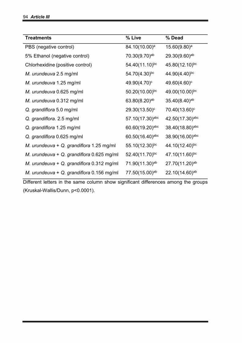
94 Article III
Treatments % Live % Dead
PBS (negative control) 84.10(10.00)a 15.60(9.80)a
5% Ethanol (negative control) 70.30(9.70)ab 29.30(9.60)ab
Chlorhexidine (positive control) 54.40(11.10)bc 45.80(12.10)bc
M. urundeuva 2.5 mg/ml 54.70(4.30)bc 44.90(4.40)bc
M. urundeuva 1.25 mg/ml 49.90(4.70)c 49.60(4.60)c
M. urundeuva 0.625 mg/ml 50.20(10.00)bc 49.00(10.00)bc
M. urundeuva 0.312 mg/ml 63.80(8.20)ab 35.40(8.40)ab
Q. grandiflora 5.0 mg/ml 29.30(13.50)c 70.40(13.60)c
Q. grandiflora. 2.5 mg/ml 57.10(17.30)abc 42.50(17.30)abc
Q. grandiflora 1.25 mg/ml 60.60(19.20)abc 38.40(18.80)abc
Q. grandiflora 0.625 mg/ml 60.50(16.40)abc 38.90(16.00)abc
M. urundeuva + Q. grandiflora 1.25 mg/ml 55.10(12.30)bc 44.10(12.40)bc
M. urundeuva + Q. grandiflora 0.625 mg/ml 52.40(11.70)bc 47.10(11.60)bc
M. urundeuva + Q. grandiflora 0.312 mg/ml 71.90(11.30)ab 27.70(11.20)ab
M. urundeuva + Q. grandiflora 0.156 mg/ml 77.50(15.00)ab 22.10(14.60)ab
Different letters in the same column show significant differences among the groups
(Kruskal-Wallis/Dunn, p<0.0001).

Article III 95
Table 3. Mean ± SD of the integrated mineral loss (∆Z, vol% µm) and the lesion
depth (LD, µm) of enamel lesions provoked by 3-days S. mutans biofilm
Treatments ∆Z LD
PBS (negative control)
5% Ethanol (negative control)
1745±706bc
1536±354abc
55±5ab
57±18ab
Chlorhexidine (positive control) 812±438a 37±15b
M. urundeuva 2.5 mg/ml 1994±540c 77±26a
M. urundeuva 1.25 mg/ml 1113±661abc 46±22ab
M. urundeuva 0.625 mg/ml 954±373ab 43±10ab
M. urundeuva 0.312 mg/ml 912±281ab 35±16b
Q. grandiflora 5.0 mg/ml 1401±511abc 54±16ab
Q. grandiflora. 2.5 mg/ml 1193±469abc 55±17ab
Q. grandiflora 1.25 mg/ml 1116±675abc 49±20ab
Q. grandiflora 0.625 mg/ml 1498±478abc 63±23ab
M. urundeuva + Q. grandiflora 1.25 mg/ml 1246±556abc 54±22ab
M. urundeuva + Q. grandiflora 0.625 mg/ml 1673±334abc 63±19ab
M. urundeuva + Q. grandiflora 0.312 mg/ml 1240±575abc 60±22ab
M. urundeuva + Q. grandiflora 0.156 mg/ml 1633±414abc 56±9ab
Different letters in the same column show significant differences among the groups
(ANOVA/Tukey test, p = 0.001 for ∆Z, and p = 0.008 for LD).


5- Discussion

Discussion 99
5 DISCUSSION
Brazil is one of countries with the greatest biodiversity wordwile (SILVA et al.,
2014), which in turn has stimulated the use of different type of plants for prevention
and treatment of diverse diseases. The most common benefit of the plants in health
is related to their antimicrobial effect, which may have potential to control oral
diseases as dental caries and periodontitis (BERSAN et al., 2014).
Therefore, the present thesis started with a review of literature about this topic
and, thereafter, tested the effect of two plants extracts (Myracrodruon urundeuva All.
and Qualea grandiflora Mart.) against enamel caries under a microcosm biofilm
model and S. mutans biofilm, respectively. Microcosm biofilm is a model able to
induce biofilm formation using inoculum from pooled human saliva or biofilm mixed
with nutrients, being able to mimic the heterogeneity and variability of the
supragingival biofilms (RUDNEY et al., 2012; AZEVEDO et al., 2014; MASKE et al.,
2016). We also included a monospecie biofilm test, allowing the detection of the
effect of the extracts on specie; in this case S. mutans, which is one of the most
important bacteria involved in the etiology of dental caries (ERIKSSON et al., 2017).
We have chosen two plants extracts available in the Brazilian Cerrado. No
previous study has compared these extracts against dental caries using a biofilm
model. According to the literature review, there are a lot of studies on the use of
Brazilian natural agents in dentistry, however, most of them are focus on
periodontitis/gingivitis, while few clinical trials have been done about caries
prevention. Unfortunately, the in vitro studies have still been done using bacteria in
planktonic phase and not under more complex environment, as biofilm. It is also
important to keep in mind that there is a wide variety of ways of acquiring the natural
agents (essential oils, propolis, crude extracts) varying according to season and the
collected parts of the plants (root or leaves) that may contain different fractions of
active components (polyphenols, terpene, monoterpene, dimeric chalcones and/or
carvacrol) with antimicrobial potential. In the present study, we have applied
hydroalcoholic extracts of the leaves, since these extracts have been produced by
collegues from FC-UNESP/Bauru and tested with respect to biological properties by
a co-partner research group (MACHADO et al., 2016).

100 Discussion
Other important point is that the experimental model for studying the effect of
plants on dental caries must include assays that are capable to show the
antimicrobial properties (such as viability, EPS and lactic acid assays) as well as the
anti-caries effect (TMR), since one is not synonym of the other. Some antimicrobial
agents have no anti-caries potential (BRAGA; PIRES; MAGALHÃES, 2017), which
unfeasible their inclusion in oral products and their use for the prevention of this
disease.
Generally, our study showed that M. urundeuva has a superior antimicrobial
effect compared with Q. grandiflora under both microcosm biofilm and S. mutans
biofilm; however, it did not reach chlorhexidine action. With respect to microcosm
biofilm, we observed that despite the extracts were able to reduce bacteria viability
and the number of lactobacilli and mutans streptococci, they did not interfere in the
bacteria metabolism, and, therefore, they were unable to reduce caries lesions
development. On the other hand, we must consider that other bacteria present in
microcosm biofilm, but not analyzed in the present study, could have contributed to
enamel caries development (Scardovia wiggsiae, Bifidobacterium spp. and
Actinomyces spp.) (HENNE et al., 2016), which shall be further confirmed in the
future.
Considering the lack of anti-caries effect of the extracts under microcosm
biofilm, we decided to test higher concentrations of the extracts and combined both
plants on S. mutans biofilm viability and the enamel caries lesions development.
Considering the agressivity of this specie, the biofilm was produced only for 3 days,
as tested in a pilot study, to produce non-cavitated enamel lesions. Similar results, as
reported in the previous article (2nd chapter), were found under S. mutans biofilm
model. M. urundeuva had a better antimicrobial effect than Q. grandiflora, but it did
not reach the action of chlorhexidine. The combination of the extracts did not
enhance their antimicrobial effect, at least not compared to M. urundeuva alone.
None of the extracts were able to reduce the development of enamel caries lesions.
In this 3rd chapter, we only applied two response variables, since it involved an
undergraduate student research. Besides, the lack of anti-caries effect
(demineralization prevention) justifies not including other response variables.
M. urundeuva leaves extract has as active components gallic acid, methyl
gallate, methyl gallate, ethyl gallate, chlorogenic and protocatechuic acid, saponins,
flavonoids, tannins and polyphenols (MOTA et al., 2015), while Q. grandiflora leaves

Discussion 101
extract presents gallic and ellagic acids derivatives, galotannins, ellatannins,
triterpenes, flavonoids, benzoquinones and anthraquinones in its composition
(AYRES et al., 2008). Therefore, the best antimicrobial performance of M. urundeuva
might be due to the presence of polyphenols.
With respect to the antimicrobial effect of Q. grandiflora extract, previous
studies have been done using S. aureus, E. coli, B. cereus, P. aeruginosa, S.
pyogenes and H. pylori strains (ALVES et al., 2000; MOURA; NASCIMENTO;
PINTO, 2012; BONACORSI et al., 2013), but not species involved in caries etiology.
On the other hand, M. urundeuva has been tested against cariogenic species.
Alves et al. (2009) observed antimicrobial effect of M. urundeuva (stem bark) against
oral bacteria by quantifying inhibition zone (MIC) and Minimum Inhibitory Adhesion
Concentration (MICA). They showed MIC and MICA values of 0.125 mg/ml and
0.0625 mg/ml against S. mutans, respectively. Their MIC value is in agreement with
our viability results under microcosm biofilm but not under planktonic phase (MIC for
S. mutans), which might be due to the bacteria strain used, the method applied to
determine MIC and the preparation of the extract.
M. urundeuva, as 5% gel or as aqueous solution (7.5 mg/ml), has been also
tested against total microorganisms (BOTELHO et al., 2007) and S. mutans
(MENEZES et al., 2010) presented in biofilm of male Wistar rats, respectively, with
positive results compared to saline-based gel and water, respectively. Menezes et al.
(2010) evaluated the action of aqueous extracts of Psidium cattleianum and M.
urundeuva (7.5 mg/ml) in Wistar rats under cariogenic diet for 7 weeks. They
observed that both extracts reduced the cariogenic biofilm (level of S. mutans by RT-
PCR) and had a protective effect on enamel surface hardness loss. Oppositely, our
study did not show any protective effect of the extracts against enamel
demineralization. The reason to explain the different findings might be related to the
fact that the tested concentrations in the present study were lower than those applied
in the former work.
Chlorhexidine was applied as positive control in both articles in agreement
with Maske et al. (2016), who observed that chlorhexidine solutions at concentrations
of 0.06% and 0.12%, applied 2x/day, significantly reduced the counting of S. mutans
(65%) and the percentage of surface hardness loss compared to control (saline
solution). On the other hand, alcohol was used as negative control, since it was
applied as solvent for the extracts leaves. In the 2nd article, we applied 35% ethanol

102 Discussion
as solvent, while we decided to reduce its concentration in the 3rd article, to avoid
questioning about a possible antimicrobial effect of a high ethanol concentration.
Previous works have shown that 50% or 80% ethanol had no antimicrobial effect
against S. mutans (BARBIERI et al., 2014; CARDOSO et al., 2016).
Further studies shall give attention to test the antimicrobial effect of M.
urundeuva extracts, varying concentrations, solvents and vehicles, under microcosm
biofilm, in situ or in vivo models, to confirm if they really do not have any anti-caries
effect. Other important point to consider in the future is the analysis of its cytotoxic
effect. Cytotoxic potential is an important aspect that should be considered before
testing the effect of a new antimicrobial agent in clinical trials. A recent study has
shown that M. urundeuva leaves’ extracts at concentrations higher than 100 µg/ml
could reduce the viability of fibroblasts; however, the authors applied 80% methanol
as solvent (MACHADO et al., 2016). Therefore, further studies on this issue are
needed to better establish the cytotoxic potential of the hydroalcoholic plants’
extracts containing 5% or 35% ethanol.
In conclusion, the extracts showed antimicrobial effects (especially M.
urundeuva) on microcosm and S. mutans biofilms, which was not improved by the
increasing concentration or combining the extracts. Unfornately, none of the extracts,
under both tested conditions, was able to reduce the development of enamel caries
lesions, unfeasible their use in the present form. It would be interesting to isolate their
active components in fractions and test their anti-caries potential.

6-Conclusions


Conclusions 105
6 CONCLUSIONS
According to chapter 1, more studies involving protocols closer to the clinical
condition and using response variables that allow understanding the mechanism of
action of the natural agents are needed, to thus allow the incorporation of these
natural agents in dental products.
Under microcosm biofilm model (2nd chapter), the tested extracts showed
antimicrobial effects (especially M. urundeuva); however, they had no anti-caries
effect on enamel.
Under S. mutans biofilm model (3rd chapter), the tested extracts showed
antimicrobial effects (especially M. urundeuva); however, they had no anti-caries
effect on enamel.


References


References 109
REFERENCES
ABANTO, J. et al. Impact of oral diseases and disorders on oral health-related quality of life of preschool children. Community Dent Oral Epidemiol, v. 39, n. 2, p. 105-114, 2011.
ABUSLEME, L. et al. The subgingival microbiome in health and periodontitis and its relationship with community biomass and inflammation. ISME J, v.7, p. 1016–1025, 2013.
ALVES, P. M. et al. Atividade antimicrobiana, antiaderente, antifúngica in vitro de plantas medicinais brasileiras sobre microrganismos do biofilme dental e cepas do gênero Cândida. Rev Soc Bras Med Trop, v. 42, n. 2, p. 222-224, 2009.
ALVES, T. M. d. A. et al. Biological screening of Brazilian medicinal plants. Memórias Do Instituto Oswaldo Cruz, v. 95, n. 3, p. 367–373, 2000.
AYRES, M. C. C. et al. Atividade antibacteriana de plantas úteis e constituintes químicos da raiz de Copernica prunifera. Rev Bras Farmag, v. 18, n. 1, p. 1-10, 2008.
AZEVEDO, M. S. et al. Correlation between the cariogenic response in biofilms generated from saliva of mother/child pairs. Biofouling, v. 30, n. 8, p. 903-909, Sep 2014.
BAKRI, I.M.; DOUGLAS, C. W. Inhibitory effect of garlic extract on oral bacteria. Arch Oral Biol, v. 50, n. 7, p. 645-651, 2005.
BARBIERI, D. S. et al. Antiadherent activity of Schinus terebinthifolius and Croton urucurana extracts on in vitro biofilm formation of Candida albicans and Streptococcus mutans. Arch. Oral Biol, v. 59, p. 887–896, 2014.
BERSAN, S. M. et al. Action of essential oils from Brazilian native and exotic medicinal species on oral biofilms. BMC Complement. Altern. Med, v. 14, p. 451, 2014.
BETTEGA, P. V. C. et al. Phytotherapy: from greenhouses to drugstore’s bench. Arch Oral Res, v. 7, n. 1, p. 89-97, 2011.

110 References
BONACORSI, C. et al. Comparison of Brazilian Plants Used to Treat Gastritis on the Oxidative Burst of Helicobacter pylori-Stimulated Neutrophil. Evid Based Complement Alternat Med, v. 2013, p. 1-8, 2013.
BOTELHO, M. A. et al. Lippia sidoides and Myracrodruon urundeuva gel prevents alveolar bone resorption in experimental periodontitis in rats, J. Ethnopharmacol, v. 113, p. 471–478, 2007.
BRAGA, A. S.; PIRES, J. G.; MAGALHÃES, A. C. Commercial antimicrobials mouthrinses on caries and periodontitis-related biofilm control - a review of literature. Braz. Dent. Sci, v. 20, p. 13-23, 2017.
CARDOSO, J. G. et al. Influence of a Brazilian wild green propolis on the enamel mineral loss and Streptococcus mutans’ count in dental biofilm. Arch. Oral Biol, v. 65, p. 77–81, 2016.
CARLINI, E. A. et al. Antiulcer effect of the pepper trees Schinus terebinthifolius Raddi (aroeira-da-praia) and Myracrodruon urundeuva Allemão, Anacardiaceae (aroeira-do-sertão), Rev. Bras. Farmacogn, v. 20, p. 140–146, 2010.
CAVALCANTI, Y. W. et al. A three-species biofilm model for the evaluation of enamel and dentin demineralization. Biofouling, v. 30, n. 5, p. 579-588, 2014.
ERIKSSON, L. et al. Microbial Complexes and Caries in 17-Year-Olds with and without Streptococcus mutans. J Dent Res, p. 2203451773175, 2017.
FANI, M. M.; KOHANTEB, J.; DAYAGHI, M. Inhibitory activity of garlic (Allium sativum) extract on multidrug-resistant Streptococcus mutans. J Indian Soc Pedod Prev Dent, v. 25, n. 4, p. 164-168, 2007.
FERNANDEZ, C. E.; TENUTA, L. M.; CURY, J. A. Validation of a Cariogenic Biofilm Model to Evaluate the Effect of Fluoride on Enamel and Root Dentine Demineralization. PLoS One, v. 11, n. 1, p. e0146478, 2016.
FORZZA, R.C. et al. New brazilian floristic list highlights conservation challenges. BioScience, v. 62, n. 1, p. 7, 2012.
GASPI, F. O. G. et al. Pharmacological activities investigation of crude extracts and fractions from Qualea grandiflora Mart., J. Ethnopharmacol, v. 107, p. 19–24, 2006.
HENLEY-SMITH, C.; BOTHA, F.; LALL, N. The use of plants against oral pathogens. Microb Pathog Strateg Combat Them Sci Technol Educ, v. 10, p?, 2013.

References 111
HENNE, K. et al. Analysis of bacterial activity in sound and cariogenic biofilm: a pilot in vivo study. Caries Res, v. 50, p. 480-488, 2016.
HIRUMA-LIMA, C. A. et al. Qualea grandiflora, a Brazilian “Cerrado” medicinal plant presents an important antiulcer activity. J. Ethnopharmacol, v. 104, p. 207–214, 2006.
JAMES, P. et al. Chlorhexidine mouthrinse as an adjunctive treatment for gingival health. Cochrane Database Syst Rev, v. 31, n. 3, p. CD008676, 2017.
JONES, C. G. Chlorhexidine: is it still the gold standart? Periodontol, v. 15, p. 55-62, 1997.
KEYES, P. H. The infectious and transmissible nature of experimental dental caries. Findings and implications. Arch Oral Biol, v. 1, p. 304-320, 1960.
KOO, H.; FALSETTA, M.L.; KLEIN, M.I. The Exopolysaccharide Matrix. J. Dent. Res, v. 92, p. 1065–1073, 2013.
MACHADO, A. C. et al. “Aroeira” (Myracrodruon urundeuva) metanol extract: the relationship between chemical compounds and cellular effects. Pharm Biol, v. 54, n. 11, p.2737-2741, 2016.
MARCENES, W. et al. Global Burden of Oral Conditions in 1990-2010, J. Dent. Res, v. 92, p. 592–597, 2013.
MARSH, P. D. Are dental diseases examples of ecological catastrophes? Microbiology, v. 149, p. 279–294, 2003.
MARSH, P. D.; MOTER, A.; DEVINE, D. A. Dental plaque biofilms: communities, conflict and control. Periodontol 2000, v. 55, n. 1, p. 16-35, 2011.
MASKE, T. T. et al. An in vitro dynamic microcosm biofilm model for caries lesion development and antimicrobial dose-response studies. Biofouling v. 32, p. 339–348, 2016.
MENEZES, T. E. C. et al. Protective efficacy of Psidium cattleianum and Myracrodruon urundeuva aqueous extracts against caries development in rats. Pharm. Biol, v. 48, p. 300–305, 2010.

112 References
MITTERMEIER, R. A. et al. Global biodiversity conservation: the critical role of hotspots. In: Biodiversity hotspots. Springer Berlin Heidelberg, 2011. p. 3-22.
MONTANARI, R. M. et al. Exposure to anacardiaceae volatile oils and their constituents induces lipid peroxidation within food-borne bacteria cells. Molecules, v. 17, p. 9728–9740, 2012.
MOTA, B. C. F. et al. Comparative studies between the chemical constituents and biological properties of the extracts from the leaves and barks of Myracrodruon urundeuva Fr. All. J Med Plants Res, v. 9, n. 6, p. 159–168, 2015.
MOURA, C. O.; NASCIMENTO, G. P. V.; PINTO, C. R. G. Atividade antibacteriana de Qualea grandiflora Mart. (Vochysiaceae), BioFar, v. 8, p. 34–42, 2012.
PALMER R.J. JR. Supragingival and subgingival plaque: paradigm of biofilms. Compend Contin Educ Dent, v.31, n.2, p.104-106, 2010.
PALOMBO EA. Traditional Medicinal Plant Extracts and Natural Products with Activity against Oral Bacteria: Potential Application in the Prevention and Treatment of Oral Diseases. Evid Based Complement Alternat Med, v. 2011, p. 68035, 2011.
PARWANI, S. R. et al. Comparative evalution of anti-plaque efficacy of herbal and 0,2% chlorhexidine gluconate mouthwash in a 4-day plaque re-growth study. J Indian Soc Periodontol, v. 17, p. 72-77, 2013.
PATRA, J. K. et al. Antibacterial effect of crude extract and metabolites of Phytolacca americana on pathogens responsible for periodontal inflammatory diseases and dental caries. BMC Complement Altern Med, v. 14, p. 343-349, 2014.
PETERSEN, P.E. et al. The global burden of oral diseases and risks to oral health. Bull World Health Organ, v. 83, n. 9, p. 661-669, 2005.
PETERSEN, P.E. The World Oral Health Report 2003: continuous improvement of oral health in the 21st century--the approach of the WHO Global Oral Health Programme. Community Dent Oral Epidemiol, v. 31, n. 1, p. 3-23, 2003.
PITTS, N. B. et al. Dental caries. Nat Rev Dis Primers, v. 3, p. 17030, 2017.
RAMOS-JORGE, J. et al. Impact of dental caries on quality of life among preschool children: emphasis on the type of tooth and stages of progression. Eur J Oral Sci, v. 123, p. 88-95, 2015.

References 113
RUDNEY, J. D. et al. A reproducible oral microcosm biofilm model for testing dental materials. J Appl Microbiol, v. 113, n. 6, p. 1540-1553, Dec 2012.
RUGG-GUNN, A. Dental caries: Strategies to control this preventable disease Strategies for caries prevention. Acta Medica Academica, v. 42, n. 2, p. 117–130, 2013.
SILVA, J. P. C. et al. Anti-Streptococcal activity of Brazilian Amazon Rain Forest plant extracts presents potential for preventive strategies against dental caries. J. Appl. Oral Sci, v. 22, p. 91–97, 2014.
SOUSA, C. M. de M. et al. Fenóis totais e atividade antioxidante de cinco plantas medicinais, Quim. Nova, v. 30, p. 351–355, 2007.
TAKAHASHI, N.; NYVAD, B. The Role of Bacteria in the Caries Process. J Dent Res, v. 90, n. (3), p. 294–303, 2011.
TAKAHASHI, N.; WASHIO, J.; MAYANAGI, G. Metabolomics of supragingival plaque and oral bacteria. J Dent Res, v. 89, p. 1383–1388, 2010.
WHO - World Health Organization. Regulatory situation of herbal medicines: a worldwide rewiew. Geneva, 1998.
ZAPPI, D.C. et al. Growing knowledge: an overview of Seed Plant diversity in Brazil. Rodriguésia, v. 66, n. 4 p. 30, 2015.
ZHAO, W. et al. Effect of sucrose concentration on sucrose-dependent adhesion and glucosyltransferase expression of S. mutans in children with severe early-childhood caries (S-ECC). Nutrients, v. 6, n. 9, p. 3572–3586, 2014.
ZHENG, C. Y.; WANG, Z. H. Effects of chlorhexidine, listerine and fluoride listerine mouthrinses on four putative root-caries pathogens in the biofilm. Chin J Dent Res, v. 14, n. 2, p. 135–140, 2011.


APPENDIX


Appendix 117
APPENDIX 1
DECLARATION OF EXCLUSIVE USE OF THE ARTICLE IN DISSERTATION/THESIS
We hereby declare that we are aware of the article “Brazilian natural antimicrobial agents on
caries and periodontitis-related biofilm: a literature review” will be included in Thesis of the
student Juliana Gonçalves Pires was not used and may not be used in other works of
Graduate Programs at the Bauru School of Dentistry, University of São Paulo.
Bauru, January 15, 2018.
Juliana Gonçalves Pires ________________________
Author Signature
Aline Silva Braga ________________________
Author Signature
Ana Carolina Magalhães _______ ________________________
Author Signature

118 Appendix
APPENDIX 2
DECLARATION OF EXCLUSIVE USE OF THE ARTICLE IN DISSERTATION/THESIS
We hereby declare that we are aware of the article “Effect of Myracrodruon urundeuva and
Qualea grandiflora extracts on viability and activity of microcosm biofilm and prevention of
enamel demineralization in vitro” will be included in Thesis of the student Juliana Gonçalves
Pires was not used and may not be used in other works of Graduate Programs at the Bauru
School of Dentistry, University of São Paulo.
Bauru, January 15, 2018.
Juliana Gonçalves Pires ________________________
Author Signature
Aline Silva Braga ____ ________________________
Author Signature
Flaviana Bombarda de Andrade_____ ________________________
Author Signature
Rodrigo Cardoso de Oliveira ______ ________________________
Author Signature
Ana Carolina Magalhães _______ ________________________
Author Signature

Appendix 119
APPENDIX 3
DECLARATION OF EXCLUSIVE USE OF THE ARTICLE IN DISSERTATION/THESIS
We hereby declare that we are aware of the article “Hydroalcoholic extracts of
Myracrodruon urundeuva All. and Qualea grandiflora Mart. leaves on S. mutans biofilm and
tooth demineralization” will be included in Thesis of the student Juliana Gonçalves Pires was
not used and may not be used in other works of Graduate Programs at the Bauru School of
Dentistry, University of São Paulo.
Bauru, January 15, 2018.
Juliana Gonçalves Pires ________________________
Author Signature
Sara Salustiano Zabini ____ ________________________
Author Signature
Aline Silva Braga ____ ________________________
Author Signature
Rita de Cássia Fabris __________ ________________________
Author Signature
Flaviana Bombarda de Andrade_____ ________________________
Author Signature
Rodrigo Cardoso de Oliveira ______ ________________________
Author Signature
Ana Carolina Magalhães _______ ________________________
Author Signature


Annex

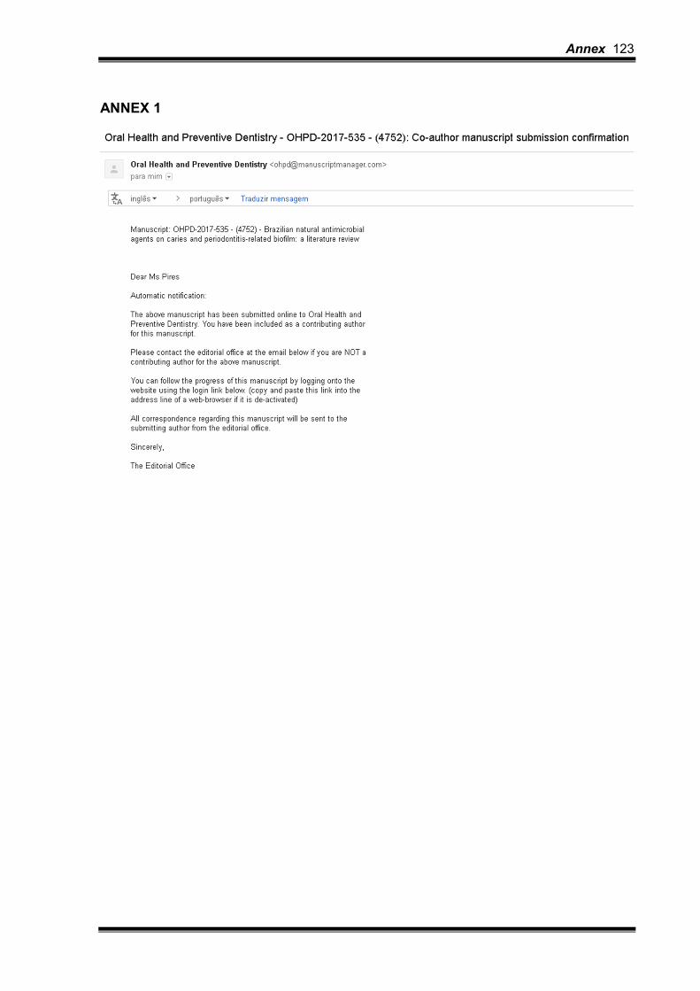
Annex 123
ANNEX 1

124 Annex
ANNEX 2

Annex 125
ANNEX 3

126 Annex
ANNEX 4

Annex 127

128 Annex
ANNEX 5

Annex 129
ANNEX 6

130 Annex
ANNEX 7
Annex 131

132 Annex