
UNIVERSIDAD DE CHILE · Grado de Doctor en Farmacología en Examen de Defensa de Tesis rendido el...
74
UNIVERSIDAD DE CHILE FACULTAD DE CIENCIAS QUÍMICAS Y FARMACÉUTICAS THESIS TO OBTAIN THE DEGREE OF Ph.D. IN PHARMACOLOGY Pharmacological evaluation of new meta- and para-hydroxyl substituted C-4-aryl-1,4-dihydropyridines in cardiomyocytes THESIS ADVISORS Luis Núñez-Vergara Ph.D. Sergio Lavandero Ph.D. DAVID ANDRÉS GALVIS PAREJA Santiago, Chile 2011
Transcript of UNIVERSIDAD DE CHILE · Grado de Doctor en Farmacología en Examen de Defensa de Tesis rendido el...

1
UNIVERSIDAD DE CHILE FACULTAD DE CIENCIAS QUÍMICAS Y FARMACÉUTICAS
THESIS
TO OBTAIN THE DEGREE OF
Ph.D. IN PHARMACOLOGY
Pharmacological evaluation of new meta- and para-hydroxyl substituted C-4-aryl-1,4-dihydropyridines
in cardiomyocytes
THESIS ADVISORS
Luis Núñez-Vergara Ph.D. Sergio Lavandero Ph.D.
DAVID ANDRÉS GALVIS PAREJA
Santiago, Chile
2011

2
UNIVERSIDAD DE CHILE
FACULTAD DE CIENCIAS QUÍMICAS Y FAMACÉUTICAS
ESCUELA DE POSTGRADO Se informa a la Dirección de Postgrado de la Facultad de Ciencias Químicas y Farmacéuticas que la Tesis de Doctorado presentada por el candidato:
DAVID ANDRÉS GALVIS PAREJA
ha sido aprobada por la Comisión Informante de Tesis como requisito para optar al Grado de Doctor en Farmacología en Examen de Defensa de Tesis rendido el día _____ de _____ de 2011.
Dr. Sergio Lavandero González Dr. Luis J Núñez Vergara Facultad de Ciencias Químicas y Farmacéuticas
Universidad de Chile
COMISION INFORMANTE DE TESIS
PROF. DR. ANTONIO MORELLO PROF. DR. LUIS AGUAYO PROF. DR. MARCO TULIO NÚÑEZ PROF. DR. RAMÓN RODRIGO Presidente Comisión de examen

3

4
To my beloved mother

5
“…if you are going through hell, keep going.” Sir Winston Churchill

6
ACKNOWLEDGEMENTS
In first place a most sincere acknowledgement to my thesis advisors: Dr Sergio Lavandero and Dr Luis Núñez Vergara; for allowing me to pursue and build this PhD, and also because in my worst hours. They were such a pulling force, and helped me to get through the darkest times.
I am so much obliged to Dr Jorge Hidalgo and Dr Gerald Zapata for their inspiring words and their immense support in experiments.
To Dr. Mario Chiong for being always a helping hand and a tremendous sparring.
I want to thank from the bottom of my heart, to my gang, the merry men, whom were always smiling and were always supporting, always coming to my aid and boosting my psyche, so I could get a grip: Dr. Miguel Copaja, Dr. José Miguel Vicencio, Dra. Carla Ortiz, Dr. Juan Pablo Muñoz, Dr. Christian Ibarra, Fidel Albornoz, Dra. Zully Pedrozo, Magdalena Cuevas, Gustavo Moraga, Carolina Muñoz, Braulio Muñoz, Pedro Ayala, Deisy Pivet, Leticia González, Dr. Rodrigo Troncoso, Dr. Ariel Contreras, Dra. Clara Quiroga, Christian Pennanen, Carolina Fernández and Ramón Pérez, you people are the greatest!!!.
Thanks so much to my dioskuroi brother, Castor: Alejandro Leyton, those long conversations about philosophy and life…no matter how good or self explanatory philosophy was, we always chose life!!!
A debt of gratitude to Cecilia Zuñiga, Miriam Salazar, Hector Bravo and Helen Gallegos for their good mood and support.
To Sir Winston Churchill, Leonidas, Caius Julius Caesar and Blas de Lezo, whose lesson
was always: never surrender, never give in no matter how dark the future might seem…you were always such an encouraging example to keep marching on.

1
TABLE OF CONTENTS
TABLE OF CONTENTS ...................................................................................................................... 1TABLE OF FIGURES .......................................................................................................................... 3LIST OF ABBREVIATIONS ................................................................................................................. 4 SUMMARY .......................................................................................................................................... 6 RESUMEN ........................................................................................................................................... 8 1. INTRODUCTION ........................................................................................................................ 10
1.1. Ion channels ........................................................................................................................... 101.2. Voltage dependent Ca2+ channels .......................................................................................... 12
1.2.1. L-type calcium channel .................................................................................................... 131.3. Calcium ................................................................................................................................... 171.4. Calcium channel blockers ....................................................................................................... 18
1.4.1. Dihydropyridines .............................................................................................................. 201.5. Reactive oxygen species (ROS) ............................................................................................. 23
2. WORKING HYPOTHESIS ......................................................................................................... 25 3. AIMS ........................................................................................................................................... 25 4. MATERIAL AND METHODS ......................................................................................................... 26
4.1. Materials ................................................................................................................................. 264.2. Animals ................................................................................................................................... 264.3. Culture .................................................................................................................................... 264.4. Confocal microscopy .............................................................................................................. 27
4.4.1. Dynamic in vivo Ca2+ measurements ............................................................................... 274.4.2. ROS measurements ......................................................................................................... 28
4.5. Docking studies ...................................................................................................................... 294.5.1 Homology model ............................................................................................................... 294.5.2. Docking analysis .............................................................................................................. 29
4.6 Patch clamp ............................................................................................................................. 304.7. Flow cytometry ........................................................................................................................ 31
4.7.1. Cell toxicity ....................................................................................................................... 314.7.2. ROS measurements ......................................................................................................... 31
5. RESULTS ...................................................................................................................................... 32
5.1. AIM 1. ...................................................................................................................................... 335.2. AIM 2 ....................................................................................................................................... 475.3. AIM 3. ...................................................................................................................................... 52

2
6. DISCUSSION ................................................................................................................................ 54 LTCC blocking activity ................................................................................................................ 55Patch clamp ............................................................................................................................... 57Docking studies .......................................................................................................................... 58Antioxidant activity ..................................................................................................................... 59Cell toxicity ................................................................................................................................. 60
7. FINAL REMARKS ......................................................................................................................... 61 8. REFERENCES .............................................................................................................................. 62

3
TABLE OF FIGURES
Figure 1. Voltage dependent calcium channels classification. ....................................................... 14Figure 2. Voltage dependent Ca2+ channels topology. ................................................................... 16Figure 3. Chemical formulae of three main calcium channel antagonists. ..................................... 19Figure 4. Structure activity relationship aspects of DHPs. ............................................................. 22Figure 5. Chemical structures of 1,4-dihydropyridines studied in this thesis. ................................ 24Figure 6. Effect of DMSO on basal calcium oscillations in isolated adult cardiomyocytes. ........... 37Figure 7. Effect of NIT on basal calcium oscillations in isolated adult cardiomyocytes. ................ 38Figure 8. Effect of PDHP on basal calcium oscillations in isolated adult cardiomyocytes. ............ 39Figure 9. Effect of 3-OH on basal calcium oscillations in isolated adult cardiomyocytes. .............. 40Figure 10. Effect of 3,5-OH on basal calcium oscillations in isolated adult cardiomyocytes. ........... 41Figure 11. Effect of 4-OH on basal calcium oscillations in isolated adult cardiomyocytes. .............. 42Figure 12. Effect of VDHP on basal calcium oscillations in isolated adult cardiomyocytes. ............. 43Figure 13. Effect of IDHP on basal calcium oscillations in isolated adult cardiomyocytes. .............. 44Figure 14. Patch clamp experiment for 3-OH. ................................................................................... 45Figure 15. Orientation of DHP ligands in the homology model of LTCC. ......................................... 46Figure 16: Antioxidant activity of some DHPs. .................................................................................. 49Figure 17. Assessment of antioxidant activity of DHPs in cultured cardiomyocytes. ........................ 50Figure 18. Measurement of antioxidant activity for all DHPs by H2O2. ............................................. 51Figure 19. Effect of new synthetic DHPs on neonatal cardiomyocytes viability. ............................... 53

4
LIST OF ABBREVIATIONS
[Ca2+]i Intracellular calcium concentration AP Action potential ATP Adenosin triphosphate BDM 2,3-butanedione monoxime BTZ Benzothiazepine C Carbon cation C4(3,5-diOH-phenyl)-1,4-dihydropyridine 3,5-OH-DHP C4(3-OH-phenyl)-1,4-dihydropyridine 3-OH-DHP C4(4-isovanillin)-1,4-dihydropyridine IDHP C4(4-OH-phenyl)-1,4-dihydropyridine 4-OH—DHP C4(4-phenyl)-1,4-dihydropyridine PDHP C4(4-vanillin)-1,4-dihydropyridine VDHP Ca2+ Calcium ion CaCl2 Calcium chloride cDNA Deoxyribonucleic acid CICR Calcium induced calcium release Cs-OH Cesium hydroxide DHP Dihydropyridines DMEM Dulbecco's modified Eagle´s medium DMSO Dimethyl sulfoxide ECC Excitation contraction Coupling EDTA Ethylenediamine tetra-acetic acid EGTA Ethylene-glycol tetra-acetic acid EP Electrophysiology Fe2SO4 Ferric sulfate HEPES 4-(2-hydroxyethyl)-1-piperazine-ethanesulfonic acid HVA High voltage activated I Current IUPHAR International Union of Pharmacology jSR Junctional portion of sarcoplasmic reticulum KCl Potassium chloride kDa kilo Dalton L Long lasting lSR Longitudinal portion of sarcoplasmic reticulum LTCC L-type calcium channel LVA Low voltage activated M199 Medium 199 MgCl2 Magnesium chloride MgSO4 Magnesium sulfate N Neuronal Na+ Sodium ion NaCl Sodium chloride NIT Nitrendipine P Purkinje P/Q Purkinje PAA Phenylalkylamines
5
PHAR Pharmacology PI Propidium iodide QSAR Quantitative structure activity relationship R Resistant ROS Reactive oxygen species RyR Ryanodine receptor SAR Structure activity relationship SR Sarcoplasmic reticulum T Transient TEA-Cl Tetraethylammonium chloride TEA-OH Tetraethylammonium hydroxyde V Voltage VDC Voltage dependent channel VDCC Voltage dependent calcium channel ω-Aga IVA ω-Agatoxin IVA ω-CTX GVIA ω-conotoxin GVIA

6
SUMMARY
Voltage-dependent calcium channels are widely expressed throughout the different tissues,
including those from the cardiovascular system. They constitute the main route for calcium entry.
Voltage gated calcium channels are multimeric proteins that consist of a principal
pore forming α1 subunit in association with auxiliary subunits. There are various types of voltage
dependent calcium channels but in cardiomyocytes predominantly the L-type calcium channels is
found. Due to the relevance of the L-type calcium channel in cardiac diseases; these channels are
the primary target of the calcium channel blockers.
Commonly used calcium channels blockers (CCBs) are classified onto three major classes;
they vary chemically, and exhibit different functional effects depending on their biophysical,
conformation-dependent interactions with the L-type channel in the α1. Even though at the
beginning of their pharmacological use the phenylalkylamines (diltiazem), the benzothiazepines
(verapamil) and the dihydropyridines (nitrendipine), exhibited a somehow similar cardiodepressant
activity, the development of new calcium channel blockers seems to have centered in the DHPs
which have evolved from a first until a third generation, being the members of this last generation
equally potent blockers, and some of them with a reported antioxidant activity, which offers
therapeutic benefits based upon the acquiring relevance of oxidative stress in cardiovascular
diseases such as hypertension; where reactive oxygen species have deleterious effects due to their
interaction with ion transport systems in the plasma membrane.
The aim of this thesis was to explore the calcium channel blocking as well as antioxidant
properties, and cell toxicity of novel DHPs. These presented unreported substitutions in the phenyl
ring, such as a hydroxyl group, mono substituted, in meta and para, respectively; then disubstituted
in meta; a vanillin with and hydroxyl group in meta and a carboxy group in para and its isomer;
besides an unsubstituted aryl ring. These unreported molecules were all compared to nitrendipine.
We used confocal microscopy in the line scan and patch clamp in the whole cell mode to
test Ca2+ antagonism. Antioxidant activity was also measured through confocal microscopy; and
flow cytometry, the latter was also applied to assess cell toxicity and antioxidant properties. None of
the compounds exhibited significant toxic effects after a 24 hour incubation period, tested on
neonatal ventricular myocytes. Our results indicated that these effects were not significantly
different from the drug control, nitrendipine (NIT). In consequence, the pharmacological effects
here found are the outcome of the DHPs themselves and not because of a toxic side effect.

7
Doing a quick recap, only one of the novel DHPs exhibited a blocking effect on LTCCs on
adult rat cardiomyocytes, the hydroxyl meta substituted (3-OH-DHP), at all three concentrations
tested whilst the one with the unsubstituted aryl had a blocking effect only at its highest
concentration. Patch clamp results for 3-OH-DHP at concentrations lower than 0.1 µM also confirm
this antagonizing effect.
None of the compounds seemed to possess an antioxidant activity in the tests performed
on cultured rat cardiomyocytes. Concerning the studies on ROS, it can be inferred, that a bulky
substituent in C2 or C3 on the dihydropyridine ring, is relevant to achieve an antioxidant effect.
Our research led to several accomplishments, first, and in agreement with the literature,
that any para substitution in the aryl ring abolishes or nullifies the LTCC blocking effect of DHPs.
Second, the unsubsituted aryl ring it is a pharmacophore, but it requires an adequate substitution to
expect calcium channel blocking effect. Third, despite the structural similarity between a meta-
hydroxy-substituted and a meta-dihydroxy- substituted aryl ring, only the former had an effect on the
channels; probably this difference can be attributed to a steric effects.

8
RESUMEN
Los canales de calcio dependientes de voltaje son expresados ampliamente en todos los
tejidos, incluyendo los del tejido cardiovascular. Ellos son la principal ruta para la entrada de calcio
dentro de la célula. Estos canales son proteínas multiméricas que son compuestos por una
subunidad principal, α1, que es la que forma el poro, y se asocia con otras subunidades
denominadas auxiliares: α2δ, β and γ. Hay varios tipos de canales de calcio pero en las células
ventriculares cardíacas se encuentran primordialmente los canales de calcio tipo L. Debido a su
relevancia en enfermedades cardíacas; estos canales son el blanco de los bloqueadores de los
canales de calcio.
Los bloqueadores de los canales de calcio (CCBs) más utilizados, se clasifican en tres
grandes clases; varían químicamente y exhiben diferentes efectos funcionales que dependen de
sus interacciones tanto conformacionales como biofísicas con la subunidad α1 del canal. Aunque al
inicio de su uso farmacológico fenilalquilaminas (verapamilo); benzotiazepinas (diltiazem) y
dihidropiridinas (nitrendipino), mostraban una actividad cardiodepresora parecida, el desarrollo de
nuevos bloqueadores parece haberse centrado en las dihidropiridinas (DHPs), las cuales han
evolucionado desde una primera hasta una tercera generación, siendo los miembros de ésta última
bloqueadores igualmente potentes además de que tienen reportada actividad antioxidante, lo cual
ofrece un mayor beneficio terapéutico, máxime con la relevancia que está adquiriendo el estrés
oxidativo en enfermedades cardiovasculares como la hipertensión; donde las especies reactivas de
oxígeno tienen efectos deletéreos debido a su interacción con sistemas de transporte iónico en la
membrana plasmática.
El objetivo de esta tesis fue investigar la potencia bloqueadora del canal de calcio como
también propiedades antioxidantes de nuevas dihidropiridinas. Estas presentaban sustituciones
que no han sido reportadas en el anillo fenil, como un grupo hidroxilo; primero en una mono-
sustitución en meta y en para, respectivamente, y una di-sustituida en posición meta; segundo, una
vainillina con un grupo hidroxilo en posición meta y un grupo carboxi en posición para, junto con su
isómero; además de arilo sin sustituciones. Estas moléculas no reportadas se compararon frente a
nitrendipino.
Se empleó microscopía confocal en su modo line scan y patch clamp, también en este
mismo modo para investigar su antagonismo de Ca2+; para medir su capacidad antioxidante se
usaron además de la microscopía confocal, la citometría de flujo, ésta última también fue utilizada
para evaluar tanto la toxicidad celular como sus propiedades antioxidantes.
9
Ninguno de los compuestos demostró tener efectos tóxicos relevantes luego de un periodo
de incubación de 24 horas en cardiomiocitos. Nuestros resultados indican que estos efectos no
fueron significativamente diferentes de aquellos del compuesto control, nitrendipino (NIT).
Entonces los efectos farmacológicos observados son el resultado de los mismos compuestos y no
de un efecto tóxico.
Sólo una de las nuevas DHPs exhibió un efecto bloqueador sobre los canales de calcio
tipo L en cardiomiocitos, meta sustituido con un hidroxilo (3-OH-DHP), en todas las
concentraciones en que fue probada, mientras que la que presentaba el arilo sin sustituciones, solo
tuvo este efecto cuando se ensayó en su concentración más alta (10 µM). Los resultados obtenidos
con patch clamp para 3-OH-DHP a concentraciones inferiores a 0.1 µM, confirman este efecto
antagonista.
Ninguno de los compuestos pareció tener actividad antioxidante en los experimentos
realizados en cardiomiocitos de rata neonata.
Nuestra investigación llevó a varios logros, primero, y de acuerdo con lo que establece la
literatura cualquier sustitución en para del arilo disminuye ostensiblemente o anula el efecto
bloqueador de las DHPs sobre los CCTL. Segundo, el anillo arilo sin sustituciones es farmacóforo
pero requiere de un sustituyente adecuado para esperar un efecto bloqueador. Tercero, a pesar de
las similitudes estructurales entre el compuesto sustituido con un hidroxilo en meta y el bisustituido
en la misma posición, solo el primero tuvo un efecto sobre los canales; probablemente esta
diferencia se deba a un impedimento estérico. En lo referente a los estudios sobre ROS, se puede
inferir que se requiere un sustituyente voluminoso en C2 ó C3 del anillo dihidropiridinico para lograr
actividad antioxidante.

10
1. INTRODUCTION
1.1. Ion channels
They are ubiquitously expressed structures throughout cellular life. Ion channels constitute
integral membrane proteins that both produce and transduce electrical signals which are crucial to
the maintenance of vital functions in a plethora of cellular types. Ion channels gate or regulate the
ion flow through biological membranes as it occurs between the cytoplasmic compartment and the
extracellular space as well as between subcellular compartments. Ion channels are water-filled,
biological “sub-nanotubes” formed by large peptidic structures and they constitute a class of
membrane proteins that serve as conduits for rapid and regulated ion movement across cellular
membranes. Thereby, ion channels provide the molecular substrate for fast, electrical signaling in
excitable tissues.1 In addition to playing this important role, ion channels regulate the release of
hormones and neurotransmitters and they control cell and body electrolyte and volume
homeostasis. They are also involved in the signaling transduction of external stimuli to sensory
signals.1,3
Opening and closing of ion channels occurs in response to changes in membrane potential,
changes in ion concentrations on either side of the membrane, and agonist binding to the channel
or closely associated regulatory proteins. Under pathological conditions, ion channels contribute to
a variety of diseases among which are different cardiovascular pathologies.
Historically ion channels have been classified in several ways, according to the state of the
art knowledge that characterized each of the last decades. For instance, as early as in the 1940s,
their first classification refers to the ions for which they are selective. Later on the 50’s they were
classified according to their primary mode of stimulus namely ligand-gated and voltage-gated
channels a classification that is maintained nowadays. With the development of the patch clamp
technique in the 70’s, ion channels were alternatively classified by their electrophysiological
properties. From 50’s to 80’s, a combination of electrophysiological and biochemical studies led to a
classification based on their pharmacological sensitivity to toxins and synthetic drugs. The 21st
century was heralded the molecular era in ion channel research that begun at the end of the 70’s
and early beginning of the 80’s. The identification of their genetic sequences demonstrates that ion
channels exist as super-families with considerable structural homology between members, despite
very different electrophysiological and pharmacological properties.
The first naming of ion channels is typically not systematic,3 early biophysical work
attempted to distinguish different components of membrane permeability by their kinetics,
pharmacology, and response to ion substitution. A kinetic model is often made expressing each of

11
the apparent components mathematically. Finally it is implicitly assumed that each model of the
component corresponds to a type of channel, and the putative channels are given the same names
as the permeability components in the original analysis. Thus in the cornerstone analysis of ionic
currents performed by Hodgkin & Huxley 2 three different components of currents were identified,
which they called sodium, potassium and leakage. Today the names of sodium channel and
potassium channel are accepted by the scientific community for the corresponding classes of ion
channels.
Long after those initial experiments and subsequent naming, there was a new beginning in
the identification of ion channels. Molecular genetics tools made possible the cloning of individual
channels with this leading to sequencing entire genomes. Nowadays, a large number of channel
genes have been discovered and there are more findings to come. There are more channels than
the electrophysiological tools have been able to detect. Just for the most extensively studied
channels, Ca2+, Na+ and K+, more than a hundred genes have been identified in mammals such as
rats. The problem then lies in how to name them in order to classify them. As discussing channels
nomenclature and names is a very complex and extensive topic and go beyond the scope of this
thesis, for the sake of clarity, the classification of ion channels that will be referred in this project
shall be the one that divides them into two main families, which will be addressed and discussed
briefly.3
According to International Union of Pharmacology (IUPHAR), there are two main ion
channel families: Voltage gated and Ligand gated ion channels (also called voltage dependent or
ligand gated channels, the name we will use herein after is voltage dependent channel).4 This
classification is based on the early work of biophysists, who remarked that voltage gated Ca2+, Na+
and K+ channels have many functional similarities. This was also the case for channels gated by
acetylcholine, glycine and γ-aminobutyric acid. The predicted sequences of amino acids for
channels reveals strong structural similarities among these groups of channels, currently
considered as the result of successive gene duplication, mutation and selection from common
ancestral channels.3 A functionally defined type of channel is not a single structure entity. All
channels can be expressed in various isoforms coded by different genes or as different splice
variants that may be selectively expressed in certain cell types and in specific time windows during
development and growth. Current evidences indicate that in the never-ending process of evolution
new classes of channels appear when parts of old ones are recombined with functional domains of
enzymes and signaling proteins to create new functions.
Ligand-dependent ion channels recognize and react to specific molecules in their
surrounding environment. Upon ligand binding, the ion channel changes its conformation and starts

12
(or stops) conducting ions. Examples of ligand-gated ion channels include receptors for taste and
odors, receptors for different hormones and neurotransmitters such as dopamine and acetylcholine.
Voltage dependent channels (VDCs) switch their conductivity in response to a change of
the voltage across the membrane. These channels make it possible for impulses to travel along
nerve and muscle fibers; it is on these voltage-sensitive ion channels or voltage dependent
channels or voltage gated channels that we focused our interest here, and constitute the
cornerstones for this thesis.4
In turn, VDCs are divided into the following subfamilies:
• Calcium activated Potassium channels
• CatSper and two-pore channels
• Cyclic-nucleotide channels
• Inwardly rectifying potassium channels
• Transient receptor channels
• Two-pore Potassium channels
• Voltage dependent Potassium channels
• Voltage dependent Sodium channels
• Voltage dependent Calcium channels
In the work presented here, is the last subfamily: Voltage dependent Calcium channels,
to which from now on we will refer as VDCC. In the paragraph below: VDCC will be presented and
discussed generally, first describing the subfamily as a whole, then emphasizing in a
subclassification, highlighting L-type calcium channels, and finally describing their structure, in
which the binding sites for calcium channel blockers will be pointed out.
1.2. Voltage dependent Ca2+ channels
VDCCs were first identified in crustacean muscle by Fatt & Katz 5 and subsequently
studied. These muscles showed action potentials in the absence of external Na+, which were
dependent Ca2+ entry. Ca2+ is generally present at a concentration of a few mM in the extracellular
space, but inside the cell, the cytoplasmic concentration is near 0.1μM, kept low by a number of
different pumps and buffering systems, as well as the general impermeability of the plasma
membrane to the entry of Ca2+. VDCCs have subsequently been found in all types of excitable cell
in vertebrates and invertebrates, even plants. They fulfill numerous functions depending on the

13
tissue, and it is thus not surprising that a number of subclasses of VDCC have been identified, as
well as their role in different diseases.6
VDCCs are normally closed at resting membrane potentials, and open upon depolarization,
because part of the channel structure senses the change in transmembrane voltage. The resultant
current through the cell membrane can be characterized by a number of properties, including the
membrane potential range over which the channel reaches its maximum open probability, and as
well as the kinetics or time-dependent properties of the current. Different single channel currents
can also be identified, with varying properties. The task of matching these single channel types with
the currents observed in entire cells is a difficult one, but has been made easier by the cloning of
the cDNAs for a number of VDCCs and the use of selective drugs and toxins to identify specific
current components, that correspond to particular channel types.3,6
According to the membrane potential range at which voltage dependent calcium channels
open, they can then be divided into two main groups, Low threshold and high threshold voltage
gated. In a number of tissues, including cardiac muscle cells, neurons and other excitable cells, it
became apparent that there are two types of calcium currents. One is activated by small
depolarizations and shows rapid voltage-dependent inactivation; this is termed low voltage-
activated (LVA), or T for transient. The second is activated by large depolarizations and is termed
high voltage-activated (HVA). The single calcium channels underlying these currents are clearly
distinct, T type channels being of small conductance (5-9 pS) and showing rapid inactivation during
a voltage step, whereas HVA channels are of larger conductance (13-24 pS).7
High threshold voltage gated currents have been further divided into smaller subgroups.
This has been done in great part to pharmacological tools that have allowed distinguishing these
subgroups.
1.2.1. L-type calcium channel
One of this HVA groups was defined as L current; for long lasting (ergo the name L-type
calcium channels). It was found that this current seemed to be blocked by certain type of groups
known as calcium channel blockers amongst which are dihydropyridines (DHPs). Furthermore
other drugs had the opposite effect on this current and did not block it but enhance it, they were
also in the dihydropyridine group, such as BAYK 8644. Dr Roger Tsien has contributed most to the
characterization of these currents (and the channel). His group further found that in sensory
neurons, besides exhibiting the L current, another component termed N, for neuronal, was present,8
of intermediate conductance, was insensitive to DHPs but was selectively and irreversibly inhibited

14
by ω-conotoxin GVIA (ω-CTX GVIA), a peptide toxin from the cone shell mollusk called Conus
geographus. Another sub-group of calcium currents, insensitive to both ω-CTX GVIA and DHPs,
has now been reported in many tissues, indicating the presence of further current components. For
instance in the cerebellar Purkinje cells in which a small proportion of the calcium current
corresponds to N and L current, the major calcium current has been termed P type. A selective
blocker for the Purkinje cell Ca2+ current has been found in a peptide toxin from the venom of the
american funnel web spider Agelenopsis aperta, called ω-Agatoxin IVA (ω-Aga IVA). At higher
concentrations this toxin also blocks a current component that has been termed Q (the letter after
P), although the distinction between P and Q current is not always clear6. In many neurons, despite
the application of all three blockers, there often remains a substantial proportion that cannot be
classified as L, N or P/Q. This residual current has been termed R (for resistant). See Figure1.
Figure 1. Voltage dependent calcium channels classification. This figure describes briefly, the nomenclature that calcium channels have undertaken along the years according to the discipline that has studied them. In the case of the clon names, the name comes from the most important subunit the channel has, that is the one that forms the ion pore. As it can be seen the role of pharmacology in identifying and classifying calcium channels and currents has proven to be critical and determinant. EP: electrophysiology; Phar: pharmacology.

15
Understanding the molecular basis for the physiological subtypes of calcium channel first required
the identification of voltage-gated calcium channels as large heteromeric proteins. This era started
with purification of the skeletal muscle calcium channel complex, also termed the DHP receptor,
which is highly abundant in T-tubules. The purified DHP receptor complex was found to contain five
components, which were termed α1 (170 kDa), α2 (150 kDa), β (52 kDa), δ (17–25 kDa) and σ
(32 kDa), in an approximate stoichiometry.9-11 The α1 protein was identified as the component that
bound DHPs, and was therefore provisionally established as the pore-forming subunit. After
individual subunits were identified, the cDNA for the DHP receptor from skeletal muscle was
cloned12, and subsequently from heart.13 Analysis indicated that the α1 subunits have about 2,000
amino acid residues with an amino acid sequence and predicted transmembrane structure like the
previously characterized, pore-forming α1 subunit of Na+ channels.12 The amino acid sequence is
organized in four repeated domains (I to IV), each of which contains six transmembrane segments
(S1 to S6), and a membrane-associated loop between transmembrane segments S5 and S6, with
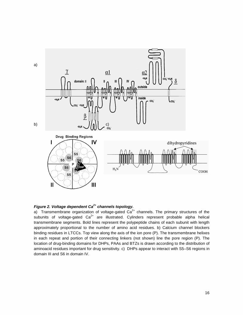
intracellular linkers and N- and C-termini (figure 2). In each domain S4 constitutes the voltage
sensor, besides this voltage sensor, domains III and IV of this pore forming subunit, contain the
specific binding sites for calcium channel blockers found so far; these are located in III S5 and S6,
and IV S6 for DHP and III S6 and IV S6 for the other members of the calcium channel blockers
family. In all, 10 α1 subunits have now been cloned, and they all have specialized functions and
distributions.14 The four members of the CaV1 family are all L-type channels, with CaV1.1 being the
skeletal muscle isoform and CaV1.2 being particularly prevalent in cardiac muscle;15 whereas the
more recently cloned CaV1.3 and 1.4 are activated at lower voltage thresholds, and have a more
restricted distribution. CaV2.1 is the molecular counterpart of P/Q-type calcium channels,16 CaV2.2
or a1B, is the molecular counterpart of the neuronal N-type calcium channels. CaV2.3 or a1E was
initially thought to be a low voltage-activated channel, and it is certainly more inactivating than the
other HVA channels cloned.17 However, it is now thought to contribute to the molecular counterpart
of the R-type calcium current. The CaV3 group of channels was the last to be cloned.18
16
a) b) c) Figure 2. Voltage dependent Ca2+ channels topology. a) Transmembrane organization of voltage-gated Ca2+ channels. The primary structures of the subunits of voltage-gated Ca2+ are illustrated. Cylinders represent probable alpha helical transmembrane segments. Bold lines represent the polypeptide chains of each subunit with length approximately proportional to the number of amino acid residues. b) Calcium channel blockers binding residues in LTCCs. Top view along the axis of the ion pore (P). The transmembrane helixes in each repeat and portion of their connecting linkers (not shown) line the pore region (P). The location of drug-binding domains for DHPs, PAAs and BTZs is drawn according to the distribution of aminoacid residues important for drug sensitivity. c) DHPs appear to interact with S5–S6 regions in domain III and S6 in domain IV.

17
1.3. Calcium
Ca2+ is a very versatile second messenger. This ion, is involved in the regulation of diverse
functions such as fertilization, electrical signaling, contraction, secretion, memory, gene
transcription, and cell death. It has a broad time of action, from microseconds (e.g. exocytosis) to
months or even years (e.g. memory processes). At the same time, the actual molecular systems
responsible for producing Ca2+ signaling events are limited to very few protein families (Ca2+
channels and transporters), and these systems appear to be very well conserved and ubiquitously
expressed within all kinds of cells. Most importantly, all of these systems are regulated by Ca2+
itself, thus making it a very robust, albeit versatile and adaptable messenger.19
In the cardiovascular system, most specifically in cardiomyocytes, Ca2+ is essential in a
variety of processes, of which the most relevant to this thesis are: a) electrophysiology [via ion
currents and the mediation of action potential (AP) shape, arrhythmogenic mechanisms; b)
excitation-contraction coupling (ECC), which governs the Ca2+ transient that drives contraction; c)
contraction itself, in which Ca2+ is the activating switch of the myofilaments and a modulator of key
contractile properties (e.g., cooperativity, length-dependent activation, and frequency dependent
acceleration of relaxation) and d) energy consumption (by contraction and Ca2+ transport) and
production (via the regulation of mitochondrial ATP production).20
Excitation-contraction coupling (ECC) in cardiomyocytes is the process by which
depolarization of the cell (action potentials) leads to contraction. LTCC localized in transverse-
tubule (T-tubule) membranes are opened in response to depolarization, allowing the influx of
extracellular Ca2+. This incoming Ca2+ ("trigger Ca2+") binds to ryanodine receptors (RyRs) of the
SR and causes Ca2+ release, giving rise to calcium induced calcium release (CICR).21,22 In more
detail, CICR starts following Ca2+ diffusion across a small sub-membrane dyadic space, this influx
activates clusters of RyRs controlling ryanodine-sensitive Ca2+ release channels in the junctional
portion of the sarcoplasmic reticulum (jSR).23,24 Ca2+ ions released from RyR s diffuse through the
cytosolic space to the contractile proteins, constitute the peak of Ca2+ transients, and bind to
troponin C. Ca2+ binding to troponin C releases the troponin I-induced inhibition of thin
(actin-troponin C-tropomyosin) and thick (myosin) filament interactions,25 leading to sliding of the
filaments (ie,, cardiac contraction). Ultimately, intracellular Ca2+ concentration returned to resting
levels by combination of: (a) Ca2+ buffering in the dyadic space and cytosol; (b) sequestration of
Ca2+ by sarcoplasmic/endoplasmic reticulum Ca2+-ATPase (SERCA)-type calcium pumps lining the
longitudinal portion of the sarcoplasmic reticulum (LSR); and (c) Ca2+ extrusion from the cytosol by
Na+/ Ca2+ exchangers and Ca2+-ATPase pumps on the sarcolemmal membrane. CICR in cardiac
muscle exhibits both graded behavior and a high gain. Graded behavior refers to the observation

18
that SR Ca2+ release is proportional to the influx of trigger Ca2+, whereas high gain indicates that the
SL trigger current elicits a high SR Ca2+ release flux.26
1.4. Calcium channel blockers
Calcium channel blockers (CCBs) are a class of drugs that disrupt calcium (Ca2+) entry
through calcium channels. They have effects on many excitable cells, such as cardiac muscle,
skeletal muscle and vascular smooth muscle among others.27 The most widespread clinical usage
of calcium channel blockers is to decrease blood pressure in patients with hypertension, with
particular efficacy in treating elderly patients. Also, CCBs frequently are used to control heart rate
and reduce chest pain due to angina pectoris.28
The majority of CCBs decrease the force of contraction of the myocardium. This is known
as the negative inotropic effect29 plus they also slow down the conduction of electrical activity
within the heart, by blocking the calcium channel during the plateau phase of the action potential of
the heart. This results in a negative chronotropic30 effect ending up in a lowering of the heart rate
and the potential for heart block. The negative chronotropic effects of CCBs make them
commonly used drugs in patients with atrial fibrillation or flutter. Negative chronotropy can be
beneficial in that elevated heart rate can result in significantly higher 'cardiac work', which can result
in anginal symptoms: lower heart rates represent lower cardiac oxygen requirements.31
The concept of Ca2+ antagonism was first developed by Fleckenstein who, demonstrated
that verapamil and prenylamine produced effective electromechanical uncoupling in the heart and
this effect was mimicked by Ca2+ removal and this uncoupling could be overcome by increasing
extracellular Ca2 concentrations. Almost simultaneously, Godfraind & Polsteri noted the effects of
two others CCBs on excitation– contraction coupling. Fleckenstein continued using verapamil as a
tool and these initial pharmacologic studies led to the discovery of the “calcium antagonists” in
1968. Fleckenstein confirmed that nifedipine was indeed not only a Ca2+ antagonist and led to the
introduction of dihydropyridines.32 The novel CCBs were not only able to block LTCCs but also
possess some other pharmacological properties such as antioxidant.
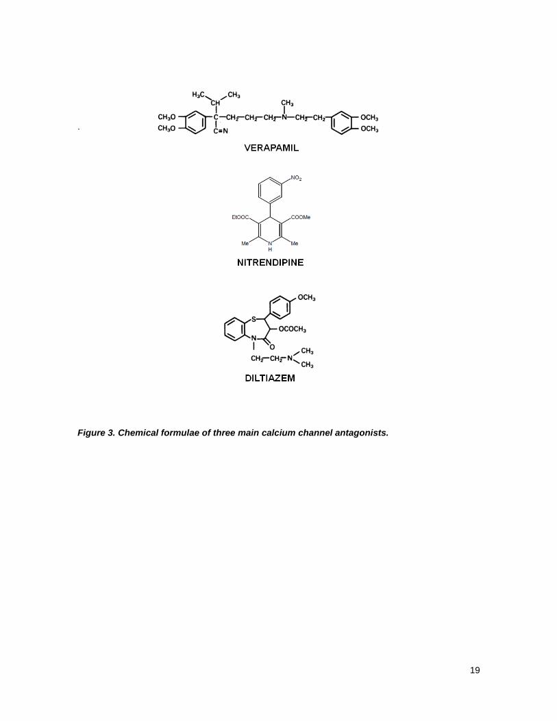
One of the most puzzling characteristics of the Ca2+ antagonists is their chemical
heterogeneity.33,35 The currently available CCB drugs are subdivided into three groups, on the basis
of their chemistry. These are the phenylalkylamines (PAA) where verapamil is the prototype,
containing two benzene cycles connected by long carbohydrate chain with one nitrogen atom; the
dihydropyridines (DHP), including nitrendipine, which contains one DHP and one benzene and the
benzothiazepines (BTZ), including diltiazem, that contains two benzenes linked by nitrogen and
sulfur atoms in thiazepine structure.34, 35 See Fig 3 for details
19
.
Figure 3. Chemical formulae of three main calcium channel antagonists.

20
1.4.1. Dihydropyridines
DHPs are still the most potent group of calcium channel blocker and many efforts have
been done to prepare additional cardioselective compounds. DHPs have a broad range of
pharmacological actions such as vasodilation, bronchodilation, antiatherosclerosis, antioxidant and
anticonvulsant effects. DHP analogs have been synthesized and several second generation
commercial products have appeared on the market with superior bioavailability and a slower onset
and longer duration of action. Such compounds include nimodipine, nisoldipine and a third
generation which not only encompasses calcium channel blocking activity but also antioxidant,
among third generation DHPs are amlodipine, felodipine, isradipine, manidipine, nicardipine and
nilvadipine.36 DHPs of the nifedipine type are flexible molecules, in which the C-4-aryl moiety and
the C-3 and C-5 ester substituents can rotate and the conformation of the 1,4-DHP ring can
change.37-41 SAR studies of DHP derivatives show that the following structural features are
important for their Ca2+-channel-blocking activity:
• The nature and position of C-4-aryl ring substituents optimizes activity. Phenyl is preferred,
owing to animal toxicity observed with heteroaromatic rings. The pseudoaxial conformation
of C-4 aryl ring is also important.42,43
• The 1,4-DHP ring is essential for activity because it is necessary for hydrogen bonding.
Substitution at the N-1 position or the use of oxidized (piperidine) or reduced (pyridine) ring
systems greatly decrease or abolish activity. Nifedipine and related analogs exist in a boat
conformation.
• C-3 and C-5 substituents modulate activity and tissue selectivity.44,45,46 Asymmetrical
substituents in C-3 and C-5 alter the activity.47,48 In fact, the electronic features of the
oxygen of the carboxyl ester group influenced biological activity. Carbonyl oxygen
participates in hydrogen bonding with the receptor.49 Molecular orbital conformational
calculations suggest that in DHPs, both carboxy groups are preferentially oriented in a
plane that intersects the plane of the DHP ring with an angle of between 30º and 60º.49,50
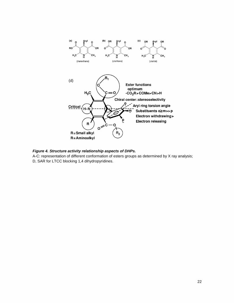
In addition, it has been proposed that based on the orientation of the individual carbonyl
groups of the C-3 and C-5 ester substituents with respect to the DHP ring double bond,
three different conformations are, in principle, possible for the ester groups: trans/trans, in
which the carbonyl groups of the both esters are trans to the double bond of DHP (Fig. 4a);
cis/cis, with a cis arrangement of both carbonyl groups (Fig 4c); and enantiomeric cis/trans
and trans/cis arrangements (Fig. 4c); and enantiomeric cis/trans and trans/cis
arrangements (Fig. 4b).40,47 X-ray structural investigations, theoretical calculations and in

21
vitro analyses of fused 1,4-DHPs (compounds with an immobilized ester group) indicate
that at least one ester must be in the cis arrangement, which is necessary for hydrogen
bonding to the receptor.40,47 It is also suggested that synperiplanar carbonyl groups might
be a common feature of DHP calcium-channel antagonists and that an antiperiplanar
carbonyl group, such as the lactone group in the rigid compounds, might be a requirement
for calcium-channel agonist activity.43,47
• When the esters at C-3 and C-5 are nonidentical, C-4 carbon becomes chiral and
stereoselectivity between enantiomers is observed.49,50 The DHP-class of compounds has
been the aim of many quantitative structure–activity relationship (QSAR) studies to find the
most important quantitative parameter for optimal activity of these compounds. Hansch
analysis method has been applied to a series of 4-phenyl-substituted DHPs and, according
to this method, the biological activity of DHPs is dependent on the lipophilic, as well as the
electronic and steric properties, of the substituents on 4-phenyl DHP analogs of nifedipine.51
The same group also found that aryl ring substituents exert significant effects both on
binding and on pharmacological activity. Para substitutions in the 4-phenyl ring lead to
activity loss regardless of substituent type. 3D QSAR study (comparative molecular field
analysis and comparative molecular similarity studies) of 4-phenyl-substituted DHPs
indicates unfavorable steric interactions for any moiety in the para-position of the phenyl
ring and that electron withdrawing substituents are favorable in ortho- and meta-
positions.51,52,53
22
(d) Figure 4. Structure activity relationship aspects of DHPs. A-C: representation of different conformation of esters groups as determined by X ray analysis; D, SAR for LTCC blocking 1,4 dihydropyridines.

23
1.5. Reactive oxygen species (ROS)
Changes in the redox state of proteins play an important role in many cell functions
including immunological host defense, gene transcription, cell metabolism, ionic homeostasis and
cell signaling. Intracellular concentrations of redox-active molecules can significantly increase in the
heart as a result of activation of specific signaling pathways or the development of certain
pathophysiological conditions. Changes in the intracellular redox environment can affect many
cellular processes, including the gating properties of ion channels and the activity of ion
transporters. Because cardiac contraction is highly dependent on intracellular Ca2+ levels ([Ca2+]i)
and [Ca2+]i regulation, redox modification of Ca2+ channels and transporters has a profound effect
on cardiac function.54,55 Key components of cardiac EC coupling such as the SR Ca2+ ATPase and
LTCC are subject to redox modulation. Redox-dependent activity of ion channels and pumps is
directly involved in cardiac pathologies. Significant bursts of reactive oxygen species (ROS)
generation occur during reperfusion of the ischemic heart, and changes in the activity of the major
components of [Ca2+]i regulation, such as RyR, Na+–Ca2+ exchange and Ca2+ ATPases, are likely to
play an important role in ischemia-related Ca2+ overload.54,55 Oxidative stress refers to an imbalance
between the production of ROS (including free radicals such as superoxide and non-radicals such
as hydrogen peroxide) and endogenous antioxidant defense mechanisms.
Oxygen is the substrate for the generation of reactive species. ROS are produced as a
result of the reduction of oxygen or compounds of oxygen with hydrogen or nitrogen. The term ROS
is a generalized description for a number of reactive oxygen molecules (e.g. superoxide, hydroxyl
radical, hydrogen peroxide) of biological significance.56,57 Superoxide is produced as a result of a
single electron donation to oxygen. Since superoxide does not easily cross lipid membranes, its role
may be limited to oxidation of proteins in the organelle in which it is produced. Hydrogen peroxide is
also able to oxidise the thiol groups of cysteine residues to form disulphide bonds with either
glutathione, nearby cysteine residues or small protein thiols such as thioredoxin.57
Cysteine residues are generally thought to be the most likely target of redox or nitrosylation
modification in proteins, as free thiols can easily react with oxygen or reactive nitrogen species and
can be assisted in forming intramolecular disulfide bonds.58 For this reason, the location of the
cysteine residues is important when considering the mechanism of redox regulation of calcium
channels. Voltage gated calcium channels contain many cysteines. LTCCs’ α1 subunit for example,
has 48, not all of these will be susceptible to oxidation or reactions, however, as many will already
be involved in disulfide bonds. Oxygen could influence ion channels through many pathways, but it
has been shown that Ca2+ currents are altered by thiol reducing and oxidizing agents 59-62

24
demonstrating that free thiols in the protein are likely to be sensitive to the oxidation, and when
oxidation does occur, it augments the probability of an increase in LTCC current.
Figure 5. Chemical structures of 1,4-dihydropyridines studied in this thesis. a) DHP control, with docking studies orientations; b) new synthesized DHPs, product of research done in cell free assays at the Facultad Ciencias Químicas y Farmacéuticas, Universidad de Chile by Dr Núñez-Vergara’s group.
Considering the above-discussed antecedents, the following questions appear as
fundamental:
• How do these novel substituents (see Fig. 5) affect the impingement on LTCCs?
• Would they allow better interaction with α1C?
• Would they cause these new DHP molecules to be toxic?
• Would they confer antioxidant activity?
• Could they grant the pleiotropic action so much needed for drugs used in
cardiovascular pharmacotherapy?

25
2. WORKING HYPOTHESIS
In order to assess the questions raised above, we propose the following hypothesis:
“The new synthetic 1,4-dihydropyridine molecules are more potent in antioxidant activity than first generation nitrendipine and have similar effects in their calcium channel blocking activity”.
3. AIMS
• To investigate the L-type calcium channel blocking activity of PDHP; VDHP; IDHP,
3,5-OH-DHP; 3-OH-DHP; 4-OH-DHP
• To study in vitro the antioxidant activity of PDHP; VDHP; IDHP, 3,5-OH-DHP; 4-OH-DHP,
3-OH-DHP.
• To evaluate the toxicity of PDHP; VDHP; IDHP, 3,5-OH-DHP; 4-OH-DHP, 3-OH-DHP
26
4. MATERIAL AND METHODS
4.1. Materials
From Sigma Chemical Co (St Louis, MO, USA): EDTA; EGTA; butane 2,3 monoxime
(BDM), M199 medium, Dulbecco’s modified Eagle’s medium (DMEM); dihydrorhodamine 1,2,3
(DHR 1,2,3); besides some other biochemical reagents.
From Biological Industries (Kibbutz, Beit Haemek, Israel): fetal bovine serum (FBS), fetal
calf serum (FCS), penicillin and streptomycin.
From Invitrogen (Eugene, Oregon, USA): 5-(and-6)-chloromethyl-2′,7′
dichlorodihydrofluorescein diacetate, acetyl ester (CM-H2DCFDA)¸ laminin, fluo3-acetoxymethyl
ester (Fluo3-AM).
4.2. Animals
Both neonatal and adult rats were bred in the Animal Breeding Facility from the Faculty of
Chemical and Pharmaceutical Sciences, University of Chile (Santiago, Chile). We performed all
studies with the approval of the Institutional Bioethical Committee at the Faculty of Chemical and
Pharmaceutical Sciences, University of Chile, Santiago. This investigation conforms to the “Guide
for the Care and Use of Laboratory Animals” published by the United States National Institutes of
Health
4.3. Cell culture
a) Neonatal cardiomyocytes were prepared from hearts of 1–3-day-old Sprague Dawley rats.
Cardiomyocytes were isolated and cultured according Foncea et al. Cardiomyocytes were
dissociated from the ventricles of 1-2-day-old rat hearts using 0.4 mg/mL collagenase and
0.6 mg/mL pancreatin in 116 mM NaCl, 20 mM HEPES, 0.8 mM Na,HPO, 5.6 mM glucose,
5.4 mM KCl, 0.8 mM MgSO4, (pH 7.35). The cells were resuspended in Dulbecco’s
modified Eagle’s medium 199 (4:1 v/v) supplemented with 10% horse serum, 5% fetal calf
serum, and 100 units/mL of both penicillin and streptomycin. Cells were preplated for 2 h on

27
100-mm culture dishes to deplete fibroblasts. After preplating, cardiomyocytes were
centrifuged, and were resuspended in the previously mentioned medium. Cell viability was
assessed by the Trypan Blue method. Then cardiomyocytes were plated at the final density
required; for the experiments done for antioxidant activity with dihydrorhodamine, they were
used in suspension. After 18 h, cardiomyocytes were confluent and beating spontaneously.
b) Adult cardiomyocytes were prepared from hearts of male Sprague Dawley rats (2-3 months,
250-300 g). Animals were anaesthetised by an intraperitoneal injection (i.p.)
ketamine:xilazine 2:1. The heart was removed via thoracotomy and transferred to a Gerard
ice cold solution. The aorta was cannulated and the heart mounted into a Laggendorf
apparatus then successively perfused with the following oxygenated solutions: Gerard
buffer (Ca2+ 2.6 mM), to allow recovery of spontaneous activity; nominally EGTA (2,5 mM)
until contraction ceased and then digestion solution supplemented with collagenase type A
and 2,3-butanedione monoxime (BDM), for 30 min. Once flaccid, the heart was rinsed for 2
min without collagenase. Ventricles were removed and finely minced and gently triturated.
Then placed in 15 mL of digestion solution, at 37ºC under constant and soft agitation for 10
min; after which the supernatant was transferred to a Falcon tube and centrifuged at 500
rpm for 2 min. The pellet was gently resuspended in a Gerard buffer supplemented with
BDM. The cells were then seeded in plates pre-treated with laminin (5 µg/mL). After 20-30
min the buffer was changed and replaced with M199/HEPES/Ca2+ 2 mM, supplemented or
not, according to the need, with BDM.
4.4. Confocal microscopy
All tests were performed on an inverted confocal microscope (Carl Zeiss Axiovert 200 M-
LSM Microsystems).Two parameters were evaluated through this technique, and in accordance to
previously reported protocols, cells were labeled with the following fluorochromes: For Ca2+: Fluo 3-
AM and for ROS: CM-H2DCFDA
4.4.1. Dynamic in vivo Ca2+ measurements
Adult rat cardiomyocytes were cultured as described earlier. The cells were used on the
same day that the culture was done and only spontaneously beating adult cardiomyocytes were
analyzed. Cardiomyocytes were washed three times with Ca2+-containing resting media (Krebs
buffer: 145 mM NaCl, 5 mM KCl, 2.6 mM CaCl2, 1 mM MgCl2, 10 mM HEPES-Na, 5.6 mM glucose,

28
pH 7.4) to remove M199 culture medium, and loaded with 5.4 µM Fluo 3-AM (coming from a stock
in 20% pluronic acid, DMSO) for 30 min at room temperature. After loading, cardiomyocytes were
washed with the same buffer and used immediately thereafter. The cell-containing coverslips were
mounted in a 1 mL capacity plastic chamber and placed in the microscope for fluorescence
measurements after excitation with a 488-nm wavelength argon laser beam or filter system.
Fluorescence measurements were performed on individual cardiomyocytes. Line-scan images were
acquired at a sampling rate of 15.8 ms per line, along the longitudinal axis of the cell, always taking
care of not crossing the nucleus. An objective lens Plan Apo 63x (numerical aperture 1.4) and a
pinhole of 1 Airy unit disc were used. In all acquisitions, the image dimension was 512 x 120 pixels.
Intracellular calcium was expressed as a percentage of fluorescence intensity. All line scan images
of Ca2+ transients were background subtracted. Compounds were added after 30 sec of initiated the
recordings. Time zero (0 s) was defined at the point in which the compound was added and the
action peak at 180 s.
4.4.2. ROS measurements
Cultured neonatal cardiomyocytes were loaded with ROS-sensitive fluorescent probe
5-(and-6)-chloromethyl-2',7'-dichlorodihydrofluorescein diacetate, acetyl ester (CM-H2DCFDA) was
used to monitor ROS formation. Cells were washed three times with Ca2+-containing resting media
(Krebs buffer:145 mM NaCl, 5 mM KCl, 2.6 mM CaCl2, 1 mM MgCl2, 10 mM HEPES-Na, 5.6 mM
glucose, pH 7.4), to remove culture medium, cells were then loaded with 10 μM CM-H2DCFDA
(coming from a stock in 20% pluronic acid, DMSO), for 10 min at 37°C together with the compounds
being assayed, all the compounds were tested at a 10 µM concentration. After this incubation
period, cells were washed three times with the same buffer, then placed on a chamber, upon which
we took a picture, then added H2O2 50 µM and 3 min later a picture was taken again. Fluorescence
images were recorded with a 63x oil immersion lens (numerical aperture 1.4). Frame imaging
modes were used for measuring ROS; the image dimension was 512 x 120 pixels. CM-H2DCFDA
was excited by 488 nm laser and the fluorescence was collected at 515 nm.
29
4.5. Docking studies
4.5.1. Homology model
We used for this study the LTCC channel model, a kind gift of Dr. Zhorov. This model
contains transmembrane segments S5 and S6 and P-loops from the four repeats. All segments far
from the DHP binding site, were not included in the model. Among available x-ray structures that
could serve as templates for modeling the open state CaV1.2, we have chosen KvAP because both
channels are voltage-gated. The closed state of CaV1.2 was modeled using the KcsA structure as a
template. The selectivity filter region was built using the NaV1.4 P-loop domain model as a template.
We designate residues using labels, which are universal for P-loop channels. A residue label
includes the domain (repeat) number (1 to 4), segment type (p, P-loop; i, the inner helix; o, the outer
helix), and relative number of the residue in the segment. The energy expression included van der
Waals, electrostatic, H-bonding, hydration, and torsion components, as well as the energy of
deformation of bond angles in DHPs. The bond angles of the protein were kept rigid. The hydration
energy was calculated by the implicit solvent method. Nonbonded interactions were calculated
using the AMBER force field version, which is consistent with the implicit solvent approach. The
energy was minimized in the space of generalized coordinates, which include all torsion angles,
bond angles of the ligand, positions of ions, and positions and orientations of root atoms of ligands.
The channel models and their complexes with ligands were optimized by the Monte Carlo
minimization (MCM) method. To ensure similar backbone geometry with the x-ray template, Cα
atoms of the model were pinned. The proposed specific interactions between ligands and DHP-
sensing residues were imposed by distance constraints. Thus, DHP-LTCC H-bonds were imposed
with distance constraints that allow penalty-free separation of the H-bonding atoms up to 2.2 Å. The
complexes MC minimized with the distance constraints were refined in unconstrained MCM
trajectories to ensure that the proposed models are energetically stable. All calculations were
performed with the ZMM program.
4.5.2. Docking analysis
Molecular docking of DHPs at the LTCC homology model was investigated using the
Lamarckian genetic algorithm search method as implemented in AutoDock. The receptor was kept
rigid, while full flexibility was allowed for the ligands so as to translate/rotate. Polar hydrogens were
added to the receptors and Kollman-united atom partial charges along with atomic solvation
parameters were assigned to the individual protein atoms. The three-dimensional structures of each
ligand were generated using the GaussView 98 program. Ligands were then energy minimized

30
using Gaussian98 software. For each ligand, a rigid root and rotatable bonds were assigned
automatically. The non-polar hydrogens were removed and the partial charges from these were
added to the carbon (Gasteiger charges). The atom type for aromatic carbons was reassigned in
order to use the AutoDock 4.0 aromatic carbon grid map. Docking was carried out using 60 × 60 ×
60 grid points with a default spacing of 0.375 Å. The grid was positioned to include the full ligand
binding site in the central part of the α1 subunit so as to allow extensive sampling around
residue Y 216. Within this grid, the Lamarckian genetic search algorithm was used with a population
size of 150 individuals, calculated using 200 different runs (i.e. 200 dockings). Each run had two
stop criteria, a maximum of 1.5 × 10⁶ energy evaluations or a maximum of 50,000 generations,
starting from a random position and conformation; default parameters were used for the Lamarckian
genetic algorithm search.
4.6. Patch clamp Patch clamp experiments were performed on the whole cell mode and on isolated adult rat
cardiomyocytes. Holding potential was set at -80 mV. Single cell voltage clamp recordings were
obtained with an Axopatch 1D patch clamp amplifier. Glass micropipettes (G85150T-3 Warner
Instruments) with a tip resistance in the range of 1 to 3 MOhms were obtained with a pipette puller
and fire polished with a microforge. Once a seal was formed between the pipette tip and the cell
membrane, the whole-cell patch configuration was obtained by gentle suction applied to the inside
of the pipette. The rupture of the section of membrane sealed against the tip was monitored by a
2 mV test pulse and verified by the increase in the transient capacitive current trace. The membrane
capacity was obtained from the integration of the current transient obtained for a -10 mV pulse. The
pipette ionic composition was (in mM): Cs-methane sulphonate 150, EGTA 5, MgCl2 5, Hepes 10
adjusted to pH 7.3 with CsOH. The external saline composition was (in mM): TEA-Cl 140,
MgCl2 2, CaCl2 1.5, glucose 10 and HEPES 10. Intracellular solution (in mM): Cs-methanesulfonate
150, EGTA 5, HEPES 10 and MgCl2 5 adjusted to pH 7.3 with TEA-OH.
Voltage clamp stimulus protocol: The pClamp suite of programs was used. Clampex 5.5
was used to generate the command voltage protocol as family of 16 pulses of 50 ms duration and
stepped from 10 mV above the holding potential (usually -80 mV) with an 8 mV increment between
them. In this manner, the protocol voltage range covered from -70 to +58 mV. The net current at
each voltage step was obtained after the cancellation of the linear components of the capacity and
conductance by subtraction of 6 pulses of the inverse polarity and 1/6 of the amplitude (P/6
protocol).

31
Data acquisition: the family of the net current signals for a protocol run was filtered at 5 KHz
and digitized at 15 KHz. Data generated was stored for off-line analysis.
Analysis of membrane current from voltage clamp recordings: The net inward current
obtained during the pulse protocol was considered to be carried mostly by calcium ions due to ionic
composition of the extra and intracellular solutions. With the Clampfit program, the peak magnitude
of this current was tabulated to generate I-V curves after being normalized for membrane capacity
(nA/nF). The value of the peak current obtained at 20 mV showed throughout the records to be the
largest. Because each cell studied has control and test values, all the data can be normalized and
grouped. The grouped data was adjusted to a single site dose-response curve.
4.7. Flow cytometry
All tests were performed on a flow cytometer: FACSCanto 2 Laser, 488nm y 633nm; six
colors. Two parameters were evaluated through this technique, and in accordance to previously
reported protocols, cells were labeled with the following fluorochromes: for cell viability: PI and for
ROS: DHR 1,2,3
4.7.1. Cell toxicity
To study toxic effects of the compounds, the number of viable cells was assessed by the PI
test. Neonatal rat cardiomyocytes were exposed to 0.1; 1 and 10 µM of each one of the compounds
for 24 h; this assay also included hydrogen peroxide (H2O2) as a positive control and the vehicle
(DMSO). After that time, cells were trypsinized and resuspended in PBS, 10-20 sec before data
acquisition PI was added. Levels of PI incorporation were quantified. Cell size was evaluated by
forward-angle light scattering (FSC). PI negative cells of normal size were considered alive.
4.7.2. ROS measurements
To study the antioxidant capacity of our different DHPs, we used DHR 1,2,3, (25 µM); a
probe to study various species of ROS. Neonatal rat cardiomyocytes were used in suspension, in a
density of 3 x 10⁵/ mL, then they were centrifuged at 1000 rpm for 5 min, the cultured medium was
withdrawn and replaced with either a control (Krebs-Ringer:145 mM NaCl, 5 mM KCl, 2.6 mM
CaCl2, 1 mM MgCl2, 10 mM HEPES-Na, 5.6 mM glucose, pH 7.4) or Krebs plus the compounds.
After 30 min, the cells were centrifuged again and the supernatant medium replaced with Krebs

32
containing H2O2 (100 µM) for 1 h, 30 min after these stimuli, the probe was added and the whole
mixture was incubated for the remaining time.

33
5. RESULTS
5.1. AIM 1. To investigate the L-type calcium channel blocking activity of PDHP; VDHP; IDHP, 3,5-OH-DHP; 3-OH-DHP; 4-OH-DHP
We had six new DHP compounds available to be tested. Their novelty was that new
substitutions never described before were introduced in the aryl ring in C4 (see Fig. 5). In our
Faculty, Dr. Luis Núñez Vergara’s group has been working extensively in the last decade in
synthesizing new DHPs that would have two pharmacological activities: Ca2+ channel blocking and
antioxidant, but the main goal of this research group was their antioxidant capacity.70-72
Some of these new DHPs (PDHP, VDHP, IDHP and 3,5-OH-DHP) have shown in vitro an
antioxidant capacity in chemical assays.70.90 However both their antioxidant and LTCC blocking
activities in vivo have not already been tested. So the next step of this research was to study these
DHPs using cultured cardiomyocytes. This model was chosen because of its critical involvement in
the development of cardiovascular diseases and cardiomyocytes, especially ventricles, have a rich
population of LTCC channels; which are the target of DHPs. This and the fact that DHPs also have
a high affinity for cardiac tissue89 made our model a reasonable choice for the proposed research.
The first step was to test the new DHP’s Ca2+ antagonism, the activity DHP are recognized for in
therapeutics.
We approached this aim in three different ways: two practical and another theoretical. First
we assayed the new compounds and NIT on adult rat cardiomyocytes through a calcium-
fluorescent dye sensor and confocal microscopy, second through patch clamp and the theoretical
approach was docking studies to help understand our results in vivo.
The LTCC blocking efficacy was evaluated through visualization of Ca2+ dynamics by
confocal microscopy and patch clamp. Pharmacology parameters of new DHPs were measured
through two different actions, first the effect on inotropy (fluorescence intensity) and second,
chronotropy (frequency of contraction) in microscopy and for patch clamp through current peak
inhibition.
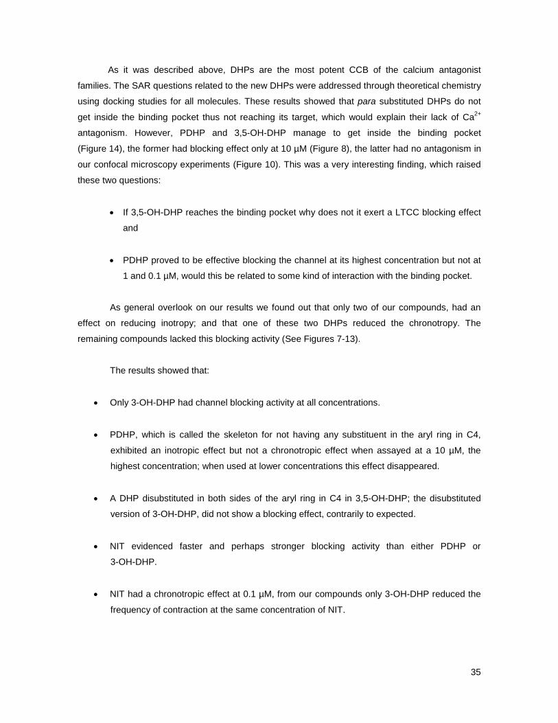
The vehicle (DMSO) was assayed to rule out any possible interference with analysis of the
Ca2+ blocking activity. We found no interference at all with the outcome of the evaluation performed
on the compounds (Figure 6). All compounds were evaluated at three different concentrations, 10, 1
and 0.1 µM.
34
3-OH-DHP was the only new DHP that shows an inotropic effect at all concentrations (0.1; 1
and 10 µM) as well as being the only to reduce the inotropy and chronotropy at 0.1 µM, the lower
concentration; this all comes as a consequence of LTCC blocking. 3-OH-DHP mimicked NIT in all
aspects, they share that they did not have a negative chronotropic effect at 1 and 10 µM and the
difference between these two compounds was that 3-OH-DHP took a longer time to achieve the
inotropic effect, Fig 7 and Fig 9. PDHP, when was tested at a higher concentration blocked the
channel, which proved to be effective at 10 µM, but this was only a negative inotropic effect, at this
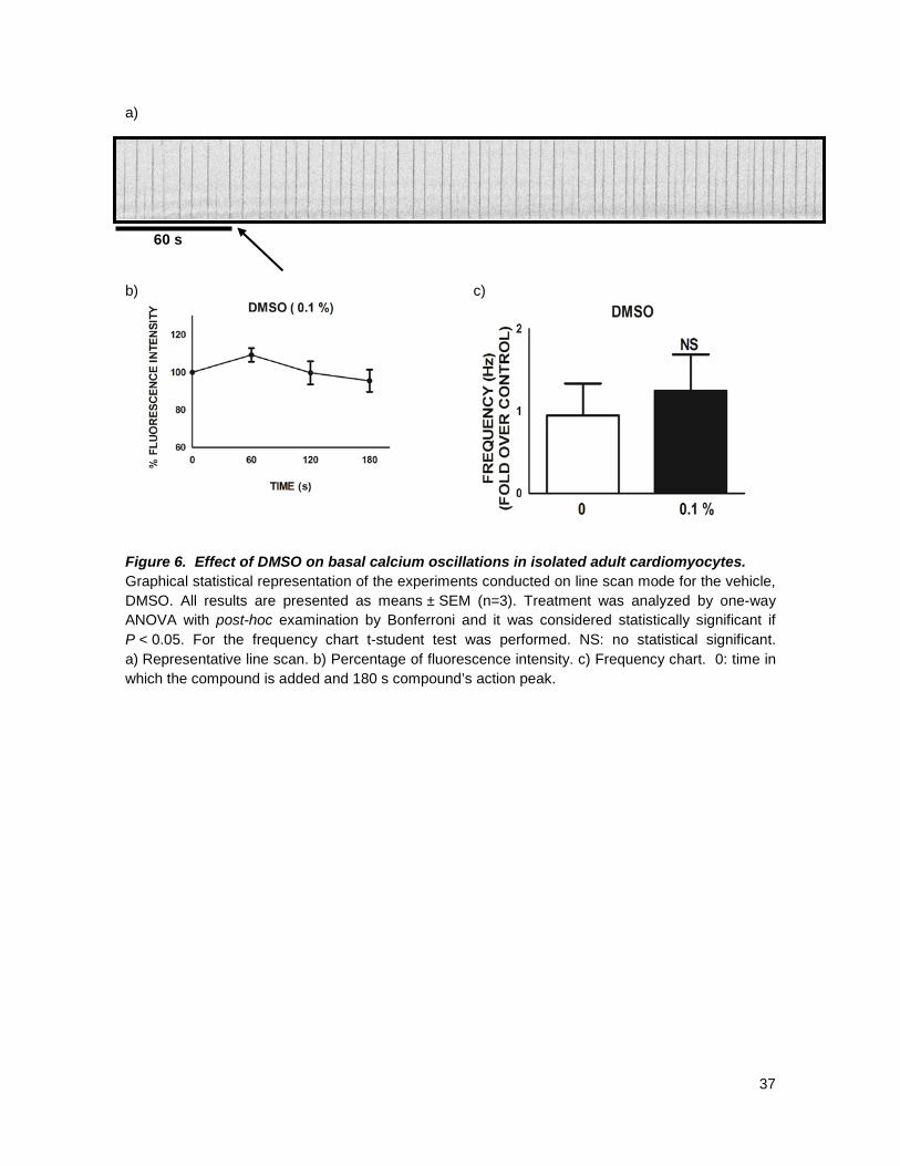
same concentration, the chronotropy remained unaffected, being equal to the control, Fig. 6. Aryl
para substituted compounds were not Ca2+ antagonists at all, they had no inotropic or chronotropic
effect, Figs 11-13, at any concentration tested; these findings are all in agreement with what has
already been stated in literature, that, any para-substitution the in aryl ring in C4, regardless of its
nature, either abolishes or diminishes to the minimum the Ca2+ blocking activity. This also further
substantiates our docking studies in which they were not able to reach the binding pocket of the
channel, thus having no Ca2+ antagonism.
However the most interesting finding was that for the disubstituted homologue of
3-OH-DHP, the 3,5–OH-DHP compound. This molecule was not effective blocking LTCC; not even
at the highest concentration there was the slightest hint that this new DHP affected any of the
parameters being evaluated. As for the lower concentrations, there was no effect either (Fig 10).
In patch clamp, only 3-OH-DHP was managed to be tested and at concentrations lower
than 0.1 µM: This preliminary result shows LTCC blockage below the mentioned concentration,
which can be associated with a powerful Ca2+ antagonist.
To clarify matters further, docking studies were performed in collaboration with Dr. Gerald
Zapata from the Departamento de Química Inorganica y Analítica, Facultad de Ciencias Químicas y
Farmacéuticas, Universidad de Chile. These studies were also supported by Dr. Boris Zhorov,
Department of Biochemistry and Biomedical Sciences, Mc Master University, Hamilton, Ontario,
Canada, whom kindly shared his LTCC model with us. This model includes only the segments of
DHPs binding site and those far from it are all excluded. We first had to set our system with that
one of Dr. Zhorov, who proposed an orientation for DHPs in his LTCC model. This was done with
nimodipine. We set our system with this molecule then we ran NIT and they followed the same
orientation inside the binding pocket and having the same critical aminoacid interactions (M4i12,
Y4i11 and Y3i10); meaning that our system was fine and ready to use for the rest of the
compounds.
35
As it was described above, DHPs are the most potent CCB of the calcium antagonist
families. The SAR questions related to the new DHPs were addressed through theoretical chemistry
using docking studies for all molecules. These results showed that para substituted DHPs do not
get inside the binding pocket thus not reaching its target, which would explain their lack of Ca2+
antagonism. However, PDHP and 3,5-OH-DHP manage to get inside the binding pocket
(Figure 14), the former had blocking effect only at 10 µM (Figure 8), the latter had no antagonism in
our confocal microscopy experiments (Figure 10). This was a very interesting finding, which raised
these two questions:
• If 3,5-OH-DHP reaches the binding pocket why does not it exert a LTCC blocking effect
and
• PDHP proved to be effective blocking the channel at its highest concentration but not at
1 and 0.1 µM, would this be related to some kind of interaction with the binding pocket.
As general overlook on our results we found out that only two of our compounds, had an
effect on reducing inotropy; and that one of these two DHPs reduced the chronotropy. The
remaining compounds lacked this blocking activity (See Figures 7-13).
The results showed that:
• Only 3-OH-DHP had channel blocking activity at all concentrations.
• PDHP, which is called the skeleton for not having any substituent in the aryl ring in C4,
exhibited an inotropic effect but not a chronotropic effect when assayed at a 10 µM, the
highest concentration; when used at lower concentrations this effect disappeared.
• A DHP disubstituted in both sides of the aryl ring in C4 in 3,5-OH-DHP; the disubstituted
version of 3-OH-DHP, did not show a blocking effect, contrarily to expected.
• NIT evidenced faster and perhaps stronger blocking activity than either PDHP or
3-OH-DHP.
• NIT had a chronotropic effect at 0.1 µM, from our compounds only 3-OH-DHP reduced the
frequency of contraction at the same concentration of NIT.

36
• NIT and 3-OH-DHP did not exhibit this negative chronotropic effect at concentrations above
the one described earlier.
• All DHPs with a substituent on para position of the aryl ring in C4 had no blocking activity at
any of the concentrations tested, as it was predicted by our docking studies.
37
a)
60 s b) c) Figure 6. Effect of DMSO on basal calcium oscillations in isolated adult cardiomyocytes. Graphical statistical representation of the experiments conducted on line scan mode for the vehicle, DMSO. All results are presented as means ± SEM (n=3). Treatment was analyzed by one-way ANOVA with post-hoc examination by Bonferroni and it was considered statistically significant if P < 0.05. For the frequency chart t-student test was performed. NS: no statistical significant. a) Representative line scan. b) Percentage of fluorescence intensity. c) Frequency chart. 0: time in which the compound is added and 180 s compound’s action peak.

38
A. Representative line scan images 0.1 µM NIT 1 µM NIT 10 µM NIT 60 s B. Fluorescence intensity C. Frequency Figure 7. Effect of NIT on basal calcium oscillations in isolated adult cardiomyocytes. Graphical statistical representation of the experiments conducted on line scan mode for NIT. All results are presented as means ± SEM. (0.1 µM, n=5, 1 µM, n=3; 10 µM, n=5). Treatment was analyzed by one-way ANOVA with post-hoc examination by Bonferroni and it was considered statistically significant if P < 0.05. A) Representative line scan. B. Percentage of fluorescence intensity. C. Frequency chart. 0: time in which the compound is added and 180 s compound’s action peak. Arrow points the moment when the stimulus was added.

39
A. Representative line scan images 0.1 µM PDHP 1µM PDHP 10 µM PDHP 60 s
B. Fluorescence intensity C. Frequency Figure 8. Effect of PDHP on basal calcium oscillations in isolated adult cardiomyocytes. Graphical statistical representation of the experiments conducted on line scan mode for PDHP. All results are presented as means ± SEM. (n=3). Treatment was analyzed by one-way ANOVA with post-hoc examination by Bonferroni and it was considered statistically significant if P < 0.05. A) Representative line scan. B. Percentage of fluorescence intensity. C. Frequency chart. 0: time in which the compound is added and 180 s compound’s action peak. Arrow points the moment when the stimulus was added.

40
A. Representative line scan images 0.1 µM 3-OH-DHP 1 µM 3-OH-DHP 10 µM 3-OH-DHP 60 s
B. Fluorescence intensity C. Frequency Figure 9. Effect of 3-OH-DHP on basal calcium oscillations in isolated adult cardiomyocytes. Graphical statistical representation of the experiments conducted on line scan mode for 3-OH. All results are presented as means ± SEM. (n=4). Treatment was analyzed by one-way ANOVA with post-hoc examination by Bonferroni and it was considered statistically significant if P < 0.05. A) Representative line scan. B. Percentage of fluorescence intensity. C. Frequency chart. 0: time in which the compound is added and 180 s compound’s action peak. Arrow points the moment when the stimulus was added.

41
A. Representative line scan images 0.1 µM 3,5-OH-DHP 1 µM 3,5-OH-DHP 10 µM 3,5-OH-DHP 60 s B. Fluorescence intensity C. Frequency Figure 10. Effect of 3,5-OH-DHP on basal calcium oscillations in isolated adult cardiomyocytes. Graphical statistical representation of the experiments conducted on line scan mode for 3,5-OH-DHP. All results are presented as means ± SEM. (0.1 µM, n=4; 1-10 µM, n=3). Treatment was analyzed by one-way ANOVA with post-hoc examination by Bonferroni and it was considered statistically significant if P < 0.05. A) Representative line scan. B. Percentage of fluorescence intensity. C. Frequency chart. 0: time in which the compound is added and 180 s compound’s action peak. Arrow points the moment when the stimulus was added.
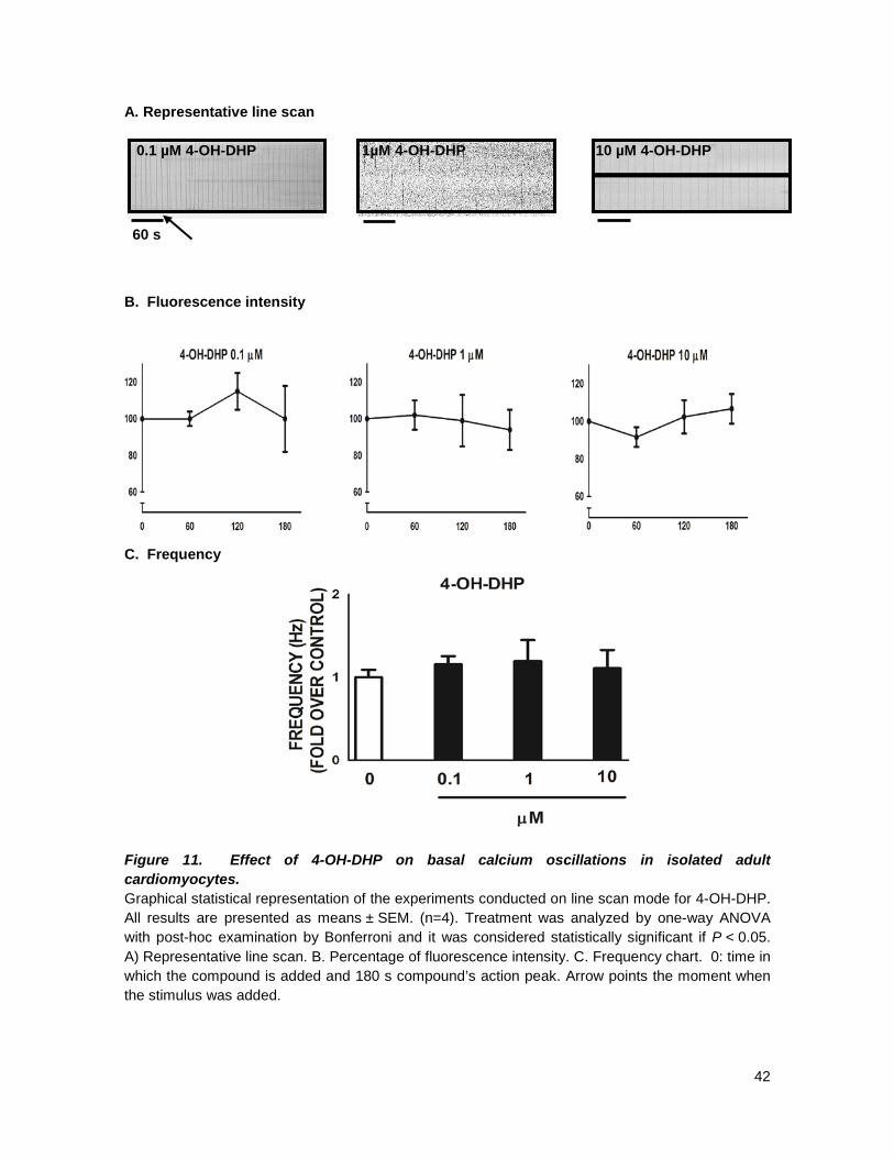
42
A. Representative line scan 0.1 µM 4-OH-DHP 1µM 4-OH-DHP 10 µM 4-OH-DHP 60 s B. Fluorescence intensity C. Frequency Figure 11. Effect of 4-OH-DHP on basal calcium oscillations in isolated adult cardiomyocytes. Graphical statistical representation of the experiments conducted on line scan mode for 4-OH-DHP. All results are presented as means ± SEM. (n=4). Treatment was analyzed by one-way ANOVA with post-hoc examination by Bonferroni and it was considered statistically significant if P < 0.05. A) Representative line scan. B. Percentage of fluorescence intensity. C. Frequency chart. 0: time in which the compound is added and 180 s compound’s action peak. Arrow points the moment when the stimulus was added.

43
A. Representative line scan images 0.1 µM VDHP 1µM VDHP 10 µM VDHP 60 s B. Fluorescence C. Frequency Figure 12. Effect of VDHP on basal calcium oscillations in isolated adult cardiomyocytes. Graphical statistical representation of the experiments conducted on line scan mode for VDHP. All results are presented as means ± SEM. (n=3). Treatment was analyzed by one-way ANOVA with post-hoc examination by Bonferroni and it was considered statistically significant if P < 0.05. A) Representative line scan. B. Percentage of fluorescence intensity. C. Frequency chart. 0: time in which the compound is added and 180 s compound’s action peak. Arrow points the moment when the stimulus was added.
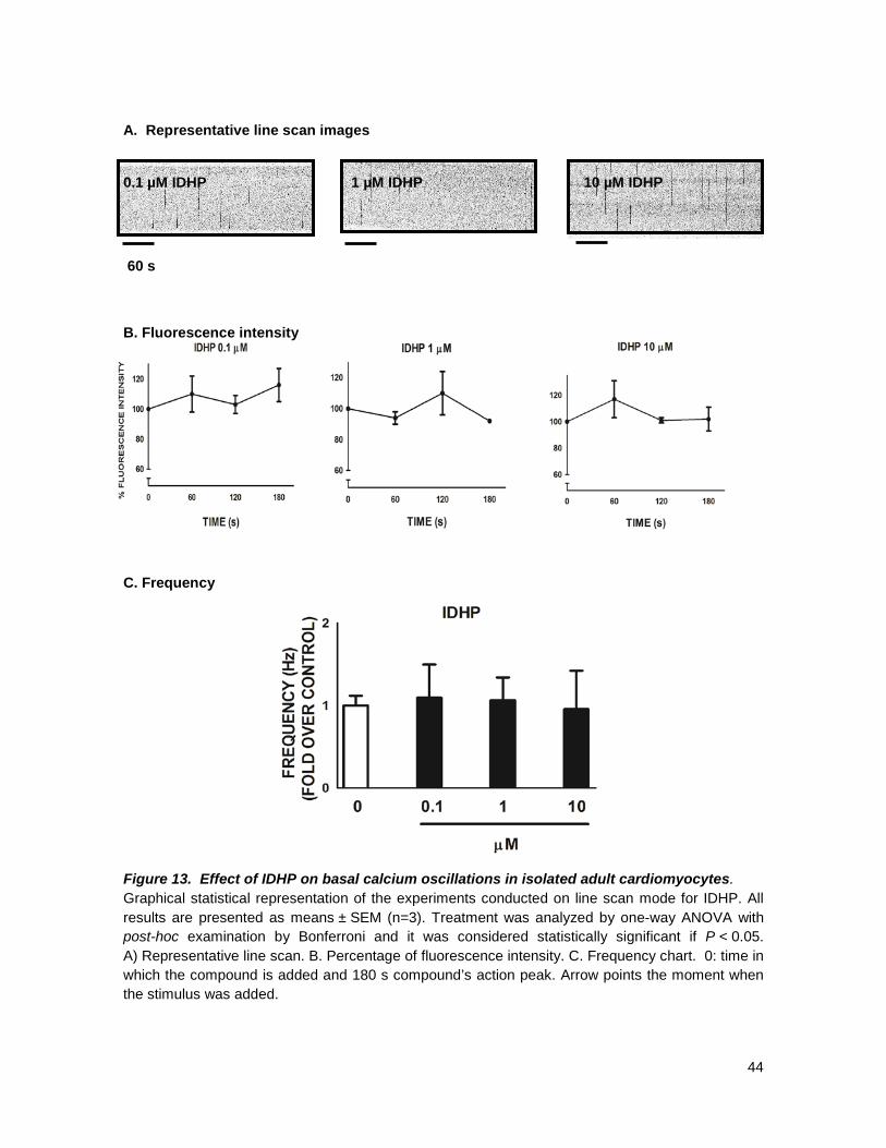
44
A. Representative line scan images 0.1 µM IDHP 1 µM IDHP 10 µM IDHP 60 s B. Fluorescence intensity C. Frequency Figure 13. Effect of IDHP on basal calcium oscillations in isolated adult cardiomyocytes. Graphical statistical representation of the experiments conducted on line scan mode for IDHP. All results are presented as means ± SEM (n=3). Treatment was analyzed by one-way ANOVA with post-hoc examination by Bonferroni and it was considered statistically significant if P < 0.05. A) Representative line scan. B. Percentage of fluorescence intensity. C. Frequency chart. 0: time in which the compound is added and 180 s compound’s action peak. Arrow points the moment when the stimulus was added.

45
A. B. C. D.
Figure 14. Patch clamp experiment for 3-OH-DHP in cultured cardiac myocytes. Whole cell patch clamp experiment. A. Quantification of B-D expressed as peak current inhibition. B-D; IV graphs for 0.001; 0.01 and 0.1 µM, respectively.

46
Nimodipine NIT 3-OH-DHP 3,5-OH-DHP PDHP Figure 15. Orientation of DHP ligands in the homology model of LTCC. S6, P, and S5 helices are shown, respectively, as by smooth ribbons, sharp ribbons, and strands. Repeat III is green, repeat IV is purple, and repeats I and II are cyan. Ca2+ ions are shown as yellow spheres. Cytoplasmatic and side views of the MC minimized complex. DHP-sensing residues are shown by thin sticks and ligands by thick sticks. Cytoplasmic views with nitrendipine, the stern of nimodipine interacts with Y4i11 so do NIT and 3-OH-DHP and 3,5-OH-DHP and this is not for PDHP. The portside group does not approach the Ca2+ ion chelated by the selectivity filter glutamates in repeats III and IV.
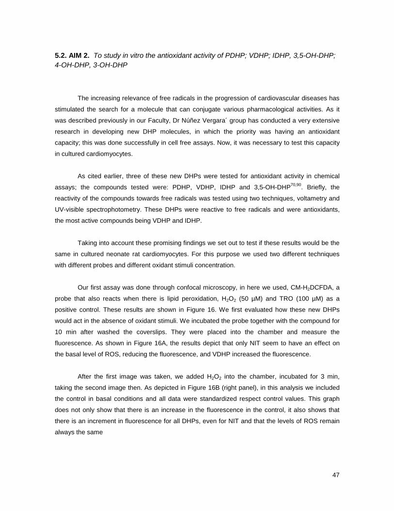
47
5.2. AIM 2. To study in vitro the antioxidant activity of PDHP; VDHP; IDHP, 3,5-OH-DHP; 4-OH-DHP, 3-OH-DHP
The increasing relevance of free radicals in the progression of cardiovascular diseases has
stimulated the search for a molecule that can conjugate various pharmacological activities. As it
was described previously in our Faculty, Dr Núñez Vergara´ group has conducted a very extensive
research in developing new DHP molecules, in which the priority was having an antioxidant
capacity; this was done successfully in cell free assays. Now, it was necessary to test this capacity
in cultured cardiomyocytes.
As cited earlier, three of these new DHPs were tested for antioxidant activity in chemical
assays; the compounds tested were: PDHP, VDHP, IDHP and 3,5-OH-DHP70,90. Briefly, the
reactivity of the compounds towards free radicals was tested using two techniques, voltametry and
UV-visible spectrophotometry. These DHPs were reactive to free radicals and were antioxidants,
the most active compounds being VDHP and IDHP.
Taking into account these promising findings we set out to test if these results would be the
same in cultured neonate rat cardiomyocytes. For this purpose we used two different techniques
with different probes and different oxidant stimuli concentration.
Our first assay was done through confocal microscopy, in here we used, CM-H2DCFDA, a
probe that also reacts when there is lipid peroxidation, H2O2 (50 µM) and TRO (100 µM) as a
positive control. These results are shown in Figure 16. We first evaluated how these new DHPs
would act in the absence of oxidant stimuli. We incubated the probe together with the compound for
10 min after washed the coverslips. They were placed into the chamber and measure the
fluorescence. As shown in Figure 16A, the results depict that only NIT seem to have an effect on
the basal level of ROS, reducing the fluorescence, and VDHP increased the fluorescence.
After the first image was taken, we added H2O2 into the chamber, incubated for 3 min,
taking the second image then. As depicted in Figure 16B (right panel), in this analysis we included
the control in basal conditions and all data were standardized respect control values. This graph
does not only show that there is an increase in the fluorescence in the control, it also shows that
there is an increment in fluorescence for all DHPs, even for NIT and that the levels of ROS remain
always the same
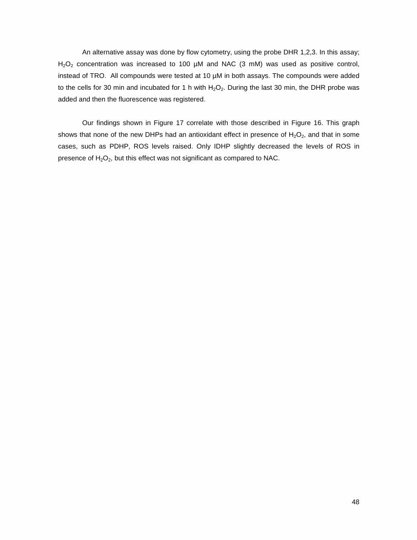
48
An alternative assay was done by flow cytometry, using the probe DHR 1,2,3. In this assay;
H2O2 concentration was increased to 100 µM and NAC (3 mM) was used as positive control,
instead of TRO. All compounds were tested at 10 µM in both assays. The compounds were added
to the cells for 30 min and incubated for 1 h with H2O2. During the last 30 min, the DHR probe was
added and then the fluorescence was registered.
Our findings shown in Figure 17 correlate with those described in Figure 16. This graph
shows that none of the new DHPs had an antioxidant effect in presence of H2O2, and that in some
cases, such as PDHP, ROS levels raised. Only IDHP slightly decreased the levels of ROS in
presence of H2O2, but this effect was not significant as compared to NAC.
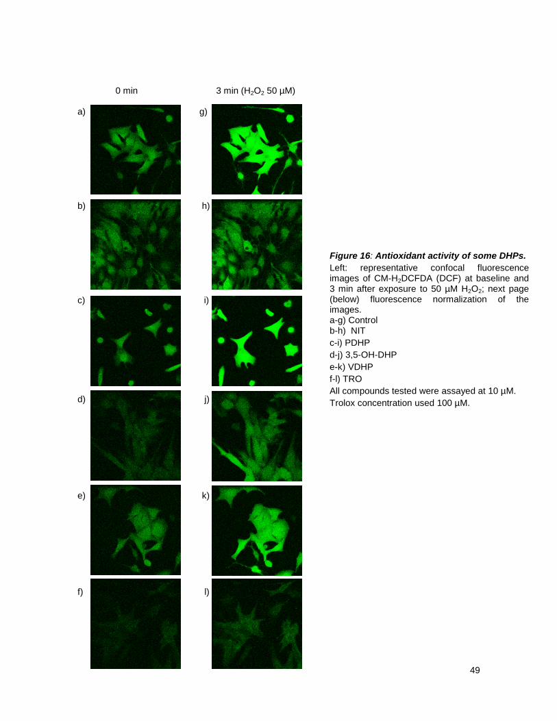
49
0 min 3 min (H2O2 50 µM) a) g) b) h) c) i) d) j) e) k) f) l)
Figure 16: Antioxidant activity of some DHPs. Left: representative confocal fluorescence images of CM-H2DCFDA (DCF) at baseline and 3 min after exposure to 50 µM H2O2; next page (below) fluorescence normalization of the images. a-g) Control b-h) NIT c-i) PDHP d-j) 3,5-OH-DHP e-k) VDHP f-l) TRO All compounds tested were assayed at 10 µM. Trolox concentration used 100 µM.

50
a) b) Figure 17. Assessment of antioxidant activity of DHPs in cultured cardiomyocytes. a) ROS basal levels. b) The graph was standardized against the white bar, basal control. Red bars represent same samples in the left panel but H2O2 treated. Comparison among different treatment compounds vs control (vehicle) was analyzed by one-way ANOVA with post-hoc examination by Dunnet test. Difference was considered statistically significant if P < 0.05. Test was performed separately for baseline and H2O2 three min incubation.

51
a) b) c) d) e) f) g) h) Figure 18. Measurement of antioxidant activity for all DHPs by flow cytometry in cultured cardiomyocytes. NAC was used as a positive control, at 3 mM. Comparison among different treatment compounds was analyzed by one-way ANOVA with post-hoc examination by Bonferroni test, comparing selected pairs of columns, first all points were compared vs. control, but priority was given to the comparison between the oxidant stimulus H2O2 vs. the compound being assayed the compound being assayed plus the oxidant stimuli. Difference was considered statistically significant if P < 0.05. * vs control; # vs H2O2

52
5.3. AIM 3. To evaluate the toxicity of PDHP; VDHP; IDHP, 3,5-OH-DHP; 4-OH-DHP, 3-OH-DHP
Our last aim was to investigate the toxicities of these new DHP compounds in cultured
neonate rat cardiomyocytes. Cell viability was measured as end point. To this end, cells were
treated with the compounds (0.1, 1 and 10 µM) for 24 h. These concentrations were the same to
those assayed when we tested their channel blocking activity. H2O2 (300 mM) was used as a
positive cell death control. The cells were incubated with the compounds for the time described
above and cell viability was assayed using PI and flow cytometry.
This technique and this probe are widely used and accepted as determinants of cell
viability; based on PI exclusion staining for drug toxicity determination: PI is a DNA intercalating dye
that is excluded by cells that have their plasma membrane integrity preserved but it enters and
stains DNA in cells that have damaged membranes and these are considered dead cells.
Figure 18 shows percentage of live cells, as ones that excluded PI.
Our results show that none of the compounds exhibited relevant toxic effects during the
time period tested (24 h). Among these new DHPs tested for cell toxicity, 4-OH-DHP proved to be
the safest, because the percentage of death cells in all concentrations was similar to the control,
DMSO. NIT and most of the new DHPs have slightly toxic effect at higher concentrations (1 and
10 µM). At 0.1 µM, none of the new DHPs differ from the control except for 3,5-OH--DHP.

53
Figure 19. Effect of new synthetic DHPs on cardiomyocyte viability. A. viability tested for µM range concentration, 24 h incubation; flow cytometry, PI. C: DMSO. Data are mean ± SEM. Comparison among different treatment compounds vs control (vehicle) was analyzed by one-way ANOVA with post-hoc examination by Dunnet test. Difference was considered statistically significant if P < 0.05.

54
6. DISCUSSION
As cited previously in this work DHPs are known for their powerful effect blocking LTCCs; given this
effect they are used in the treatment of a wide range of cardiovascular diseases. The research for
more DHPs is continuously evolving making them one of the most investigated cardiovascular
drugs. In this thesis the Ca2+ blocking and antioxidant activities of new synthesized DHPs were
evaluated. The main results are described in the following tables:
Table 1. DHPs in the binding pocket of LTCC and interaction with selected aminoacid residues
Table 2. Inotropic and chronotropic effects of new tested DHPs
COMPOUND
BINDING POCKET
AMINO ACID RESIDUES
M4i12 Y4i11 Y3i10 NIT Yes Yes Yes Yes
PDHP Yes No No Yes 3-OH-DHP Yes No Yes Yes
3,5-OH-DHP Yes Yes Yes Yes 4-OH-DH No - - -
VDHP No - - - IDHP No - - -
COMPOUND INOTROPY CHRONOTROPY
0.1 µM 1µM 10 µM 0.1 µM 1µM 10 µM DMSO - - - - - -
NIT + + + + - - PDHP - - + - - -
3-OH-DHP + + + + - - 3,5-OH-DHP - - - - - - 4-OH-DHP - - - - - -
VDHP - - - - - - IDHP - - - - - -

55
Table 3. Cell free assays of antioxidant activity of new tested DHPs
LTCC blocking activity
These DHPs had new substituents in the aryl ring in C4, which had never been previously
tested for their Ca2+ antagonism, it was very appealing and interesting to find out how these new
substituents would affect the LTCC blocking activity.
The first step in doing these experiments was to rule out a possible interference of DMSO,
as there is controversy in reports that state that it increases LTCCs Ca2+ current. Our results proved
otherwise, and in Fig. 6 it can be seen that DMSO did not increase or decrease this current, and
this result is in agreement with what can be read in the literature.95-98
Moving on to our experimental findings about these new DHPs, the results showed that only
one of all compounds, namely 3-OH-DHP, demonstrated a blocking activity on the channel at all
concentrations used; when looking at all the chemical structures of the DHPs tested, it can be
observed that 3-OH resembles the most the structure of the DHP control, NIT. The main difference
being the 3-OH substituent in the aryl ring in C4 at meta- position. The nature of the substituent is
different in each compound, for instance NIT bears a nitro group, which is an electron withdrawing
substituent and 3-OH having a hydroxyl, which is an electron releaser group. Both have the
capacity to form hydrogen bonds, but as the literature states, the one with an electron withdrawing
substitution would appear to be most effective in blocking LTCCs41, which is absolutely in
agreement with our results, where at all concentrations requires less time to start blocking the
calcium channels.
PDHP, which is called the skeleton for not having any substituent in the aryl ring in C4,
proved to have an inotropic effect but not a chronotropic effect when assayed at its highest
concentration (10 µM); when used at lower concentrations this effect disappeared completely;
COMPOUND In vitro reported antioxidant activity In vivo
In vivo Basal Stimulated
NIT ? Yes? No PDHP Yes70,90 No No
3-OH-DHP ? No No 3,5-OH-DHP Yes90 No No 4-OH-DHP ? No No
VDHP Yes70 No No IDHP Yes70 No Yes

56
raising two ideas, first, the necessity of an ortho- or meta- substituent in the C4 aryl ring to achieve
this chronotropic effect, and that in order to maintain either the inotropic or chronotropic effect at
concentrations lower than 10 µM, the need for this substituent becomes imperative, and secondly
this or these substitutions should bear an electron withdrawing or an electron releasing group.41
As expected 3,5-OH-DHP; the disubstituted version of 3-OH-DHP, would exhibit a blocking
effect; however, this blocking effect did not come through. If the literature is consulted, felodipine is
the only one commercially available DHP, dichloro-substituted in the aryl ring contiguously on same
side of the ring at ortho- and meta- positions. It can be inferred that substituents in different sides of
the aryl ring may affect the conformation of the molecule, disabling it to achieve its active one,
which is boat.44,85,86 It is possible as well, that hydroxyl groups, being such good hydrogen bond
donors, might steer 3,5-OH outside the binding pocket where it gets clogged forming hydrogen
bonds with other aminoacid than the ones in the binding pocket.
NIT began its blocking activity faster and perhaps stronger, statiscally at least; than either
PDHP, or 3-OH-DHP; in both cases, the explanation for this lies in the structure of the DHPs,
assimetry40 in the dihydropyridine ring as in NIT and the nitro group, meta-position, in the aryl ring in
C4, may account for a faster insertion into the lipid bilayer,40 as it was mentioned before, being a
lipophilic molecule, the nitro group should confer a more perdurable interaction with the binding
site.44
There is concern why NIT as well as 3-OH-DHP only reduced the frequency of contraction,
at the lowest concentration, 0.1 µM. We postulated three possibilities, first that the compounds
precipitated, but if this was the case, then, the minimum concentration would still be there and thus
achieving the reduction of frequency would have been the same for all three concentrations used,
so this theory was discarded. Second, the idea of an allosteric site to which DHPs could bind and in
doing so modifying the canonical binding pocket, rendering it inaccessible to exert the blocking
effect. No allosteric binding site has been described so far for DHPs in literature. The only allosteric
site described is for the other members of the CCBs, such as diltiazem and verapamil. If two
calcium antagonists were to be added together in a solution to cultured cells, a positive cooperative
effect would happen, making a stronger block of LTCC.91 Third, a compensatory mechanism. If
there are strong negative inotropic and chronotropic effects the cardiomyocytes would die. In a
series of epifluorescence experiments not shown, in which cells were treated first with ryanodyne,
10 µM, and thapsigargin 0.1 µM, for thirty minutes, and then the DHP was added as the experiment
was going along, the cells, did not respond well. Although no viability test was performed, when
cells were checked, morphologically they did not exhibit their classic rod shape, they were
shrinking, vacuolising, going into a death road. When they were treated with ryanodyne alone, they
looked better but not as a control. Depletion of SR is therefore a key issue in the integrity of muscle

57
cells, cardiac muscle cells. It has been suggested by some authors that SR, and its Ca2+ release,
are responsible for the frequency of contraction, in isolated rat cardiomyocytes.92,93 Gathering this
information gives the notion that a strong blockage of LTCC leading to a powerful inotropic and
chronotropic effect, from which the cardiomyocyte might not recover, could cause its death.
However if treated with a DHP, at a concentration in which this phenomena is present, grants the
cell the need to look for a compensatory mechanism through which it loses either the force of
contraction or contraction but not both because it would mean its death. In our case, the ryanodyne
receptor releases Ca2+, in absence of CICR, and this Ca2+ is able to activate sensitized contractile
machinery. Maintaining one of their functions then staying alive and this argument is supported by
the literature.94
It was proven, as it is also stated in the literature, that any substituent on para-position of
the aryl ring in C4 has nullifying or abrogating effect on the Ca2+ antagonist activity of the
molecules.51-53 Two of our compounds, VDHP and its isomer: IDHP, had two substituents, one
adjacent to the other, in meta- and in para-position of the aryl ring in C4 and one had only one
substituent in para: 4-OH-DHP. All of them failed to block the LTCC, all the same, compounds
similar to VDHP or IDHP, had never been tested, or at least we did not find proof of it
bibliographically, being that our main reason to test them. As mentioned previously, in all channel
blocking experiments, none of them exerted any effect. This can be associated to a steric effect,
predictably one associated to size87, since the literature states that regardless of the nature of this
para-substituent51, it is then not associated to an attraction or repulsion, the only option left is size,
upon acquiring its active boat shape, this substituent would make the DHP too big to enter the
binding pocket. This is correlated with our docking studies, which point out that 4-OH-DHP, VDHP
and IDHP do not get inside the action site.
Patch clamp
Even though there is not a big population for 3-OH-DHP in this set of experiments and we
do have results for NIT yet, this supports the idea of a novel LTCC blocker, because even at
concentrations below the minimal tested in confocal microscopy (0.1 µM), there was a remarkable
reduction in the Ca2+ currents. So summing up, all data obtained in experiments regarding Ca2+
antagonizing activity relating to 3-OH-DHP, we have a compound that effectively blocks LTCCs,
proven through two different techniques and is confirmed by docking studies, which reveal that the
molecule inserts into the binding pocket and even in the absence of one critical interaction
maintains the blocking effect on the channel.

58
Docking studies
Using Zhorov’s model of LTCC45, which was docked with nimodipine, a molecule very
similar to our NIT, the only difference is a larger alkyl chain in C3; they found a favorable stern-to-
Y3i10 orientation of nimodipine in the LTCC pore and verified key DHP-sensing residues identified
in previous mutational studies that confirmed favorable contacts with the nimodipine in their model.
To make sure our docking calculations were correct, we validated the nimodipine docking model,
compared it to NIT, and NIT docked the same as nimodipine. In all dockings the ligand was in a
Ca2+ deficient state in which the selectivity filter region was loaded with a single Ca2+ ion. This
configuration was selected because the Ca2+ deficient state was proposed to favor binding of DHP
antagonists88. Summary of the modeling predictions of favorable DHP contacts with the channel are
shown in Figure 15. In 3-OH-DHP, as well as in NIT, the NH group at the stern forms an H-bond
with Y3i10. The starboard methyl group fits into a hydrophobic pocket formed by I3i14 and M3i18.
The portside group is bound in a hydrophobic pocket formed by M3i19, F3p49, and I4i19.
For 3-OH-DHP, the polar OH group at the bowsprit does not interact with the side chain of Y4i11,
this would suggest that this hydroxyl it’s interacting with another amino acidic residue and that this
interaction may be weaker explaining therefore why NIT may be more potent than 3-OH-DHP. The
carbonyl oxygen at the starboard accepts an H-bond from Q3o18. The side chain of T3o14 does
not interact with the DHP molecule directly, but it is exposed toward the starboard group in such a
way that its enlargement would sterically prevent DHP binding. For, PDHP, the interactions were all
the same but in the aryl ring in C4 it was different, as there was not substituent, the interaction with
M4i12 is absent. The lack of this interaction might explain why PDHP did not block at 1 and 0.1 µM
concentrations, as it would render its interaction with the binding site weak, ergo being only present
at very high concentrations (10 µM). The most interesting case of all, was the one regarding
3,5-OH-DHP, this molecule inserted into the binding site exactly the same way, as NIT and
3-OH-DHP, this would therefore suggest that a Ca2+ channel blocking activity ought to be present,
but in practice, this was not true at all. This LTCC model for docking studies, represents the
molecule in an ideal state, it does not account possible interactions of the ligand outside its binding
site; the odyssey that DHP must endure to get inside the binding pocket, it is discarded; having two
hydroxyls, polar substituents, makes 3,5-OH-DHP an easy target to form hydrogen bonds, and get
stuck at some point before reaching out its final destination.
Concerning all para-substituted DHPs used in our studies: 4-OH-DHP, VDHP and IDHP, all of
them were anywhere in the LTCC but in the binding pocket; confirming our hypothesis about a
sterical hindrance, that disables them to bind to the action site.

59
Antioxidant activity
Our results show that any of these new DHPs have that antioxidant effect. There are various
things to consider, let us first recall the results in figure 16, in basal conditions for NIT, the graph
suggests that NIT may have an antioxidant effect but not in presence of H2O2 (50 µM). The results
in Figure 17, point out a very little antioxidant effect for IDHP when incubated with H2O2 (100 µM).
The results for NIT and IDHP prompted us to say that these molecules have an antioxidant effect
but that this effect may be inappreciable in the experimental approach we used. It is certain that
these new DHPs exhibited a very promising antioxidant effect in cell free assays; and that they
included in their structure as substituents in C4 of the DHP ring, molecules such as vanillin,70 that
have been reported to be very effective antioxidants as well,71-73 but they lack what is reported as
an absolute requirement for having such activity in cells in-vitro assays, and that is a very large or
simply large substituent in C2 or C3 of the dihydropyridine ring.74 This is a characteristic feature of
third generation DHPs, such as amlodipine or felodipine.75,76 This feature of a long substituent in C3
or C5 of the dihydropyridine ring, is what allows compounds such as amlodipine (third generation
prototype), not only to easily penetrate the plasma membrane to interact with the LTCC but also
have additional biologic or pleiotropic actions that are independent of interaction with the calcium
channel,77 that is antioxidant, in which they seem to insert to a location in the membrane78-80 near
conjugated double bonds, highly lipophilic DHPs are capable of donating protons to lipid peroxide
molecules, thereby blocking the peroxidation process.78-80 So, basically two things can be said
about this and our molecules, the first one is that there might not be technique sensitive enough to
detect their antioxidant activity or, second, that to improve the antioxidant action of our DHPs, they
must be synthesized in a way that the dihydropyridine ring has a long substituent in either C3 or C5.
The matter with these series of experiments for proving a biological antioxidant activity was
that none of the techniques was specific for lipid peroxidation. Another important topic in this
antioxidant discussion is the concentration of DHPs used, all molecules used in many and different
ROS experiments and through various techniques were above or at 100 µM,81,84 we could not get
beyond that concentration due to solubility problems. Besides, all the papers that reported this
activity failed to include a toxicity assay at those concentrations in which DHPs were tested. As
mentioned above, before finding the antioxidant effect of amlodipine and later on some other third
generation DHPs, they were already known for their calcium channel blocking properties. The main
aim of this thesis was to test the calcium channel blocking properties of these new DHP agents.

60
Cell toxicity
Among our aims were also to investigate the effect of these new susbstituents on cell
toxicity and their antioxidant properties. When any new drug candidate is synthesized, it must first
undergo a series of trials to measure its overall toxicity. This testing allows highly toxic molecules to
be identified before proceeding on to more advanced studies to measure therapeutic efficacy.63,64
Our approach to test a possible toxicity of our new DHPs was to determine cardiomyocyte
viability.65,66 We used three different concentrations (0.1-10 µM); all which are within the range of
previously known effective for Ca2+ channel blocking. First, we compared all DHPs vs. control, the
control compound, NIT, exhibited a slight toxic effect for its highest concentration, the same was
observed for all compounds, accordingly in between 1 and 10 µM. A concentration of 0.1 µM
seemed to be the most innocuous of all concentrations tested (See Figure 6 for details). It is
important to bear in mind that all drugs appear to have a toxic effect,67 it is sought however that this
effect be kept to a minimum,68,69 which is the case of all our DHPs. It is important to recall that all
our assays, be it for LTCC blocking activity or antioxidant activity were short time, being the longest
the antioxidants the prolonged ones lasting up to one hour and a half. The conclusion in this aim tell
us that the results we would see for the rest of the aims will be the product of a known mechanism
for the compounds and not as a by-product of an unlikely toxic effect. One might expect that DHPs
would be anything but deleterious; considering that they are reported to be antioxidant, which
means a protective outcome, is reasonably expected.

61
7. FINAL REMARKS
• None of these new synthesized DHPs display toxic effects.
• It is a requisite for any DHP to have a substituent in the aryl in C4 of the dihydropyridine
ring in order to exert a LTCC blocking activity.
• This substituent must be either in ortho or meta position.
• For a faster blocking effect substituents in C3 or C5 of the dihydropyridine ring must be
asymmetric.
• There might be also a correlation between the nitro group in the aryl in C4 and a faster
course of action in time.
• All para substitutions in the aryl in C4 of the dihydropyridine ring abrogate the calcium
channel blocking activity as a consequence of steric hindrance.
• Disubstituted DHPs in the aryl ring, in order to block the calcium channel must have these
substitutions contiguous and in the same side of ring.
• Electron releasing substituents in aryl ring also exhibit an antagonizing effect on LTCC.
• These new DHPs do have an antioxidant activity, but it is too little to be determined.
• For any DHP to have an assessable antioxidant effect, a long chain substituent in C3 or C5
of the dihydropyridine ring it is compulsory.
• If phenols are inserted in C4 of the dihydropyridine ring and also this ring presents a long
chain either in C3 or C5, a powerful antioxidant can be expected, but not a calcium channel
blocker.

62
8. REFERENCES
1 Triggle DJ, Gopalakrishnan M, Rampe Zheng W (2006) Voltage-Gated Ion Channels as Drug
Targets, Volume 29. Wiley-VCH Verlag GmbH & Co
2 Hodgkin AL, Huxley AF, Katz B. 1952. Measurement of current-voltage relations in the membrane of the giant axon of Loligo. J Physiol.16:424-48.
3 Hille B. 2001. Ion Channels of Excitable Membranes, 3rd Ed. Sinauer Associates,
Sunderland, Mass. 4 www.iuphar-db.org/DATABASE/ReceptorFamiliesForward?type=IC 5 Fatt P, Katz B. 1953. The electrical properties of crustacean muscle fibres. J Physiol.
120:171-204. 6 Dolphin AC. 2006. A short history of voltage-gated calcium channels. Br J
Pharmacol.147:S56-62. 7 Carbone E, Lux HD. 1984. A low voltage-activated, fully inactivating Ca channel in vertebrate
sensory neurones. Nature. 310:501-2 8 Nowycky MC, Fox AP, Tsien RW. 1985. Three types of neuronal calcium channel with
different calcium agonist sensitivity. Nature. 316:440-3 9 Colecraft HM, Alseikhan B, Takahashi SX, Chaudhuri D, Mittman S, Yegnasubramanian V,
Alvania RS, Johns DC, Marbán E, Yue DT. 2002. Novel functional properties of Ca(2+) channel beta subunits revealed by their expression in adult rat heart cells. J Physiol. 541:435-52.
10 Yang L, Katchman A, Morrow JP, Doshi D, Marx SO. 2011. Cardiac L-type calcium channel
(Cav1.2) associates with {gamma} subunits. FASEB J. 25:928-936. 11 Dolphin AC. 2003. Beta subunits of voltage-gated calcium channels. J Bioenerg
Biomembr.;35:599-620. 12 Tanabe T, Takeshima H, Mikami A, Flockerzi V, Takahashi H, Kangawa K, Kojima M, Matsuo
H, Hirose T, Numa S. 1987. Primary structure of the receptor for calcium channel blockers from skeletal muscle. Nature. 328:313-318.
13 Mikami A, Imoto K, Tanabe T, Niidome T, Mori Y, Takeshima H, Narumiya S, Numa S.
1989. Primary structure and functional expression of the cardiac dihydropyridine-sensitive calcium channel. Nature. 340:230-233
14 Ertel EA, Campbell KP, Harpold MM, Hofmann F, Mori Y, Perez-Reyes E, Schwartz A,
Snutch TP, Tanabe T, Birnbaumer L, Tsien RW, Catterall WA. 2000. Nomenclature of voltage-gated calcium channels. Neuron 3:533-535.
15 Walsh CP, Davies A, Nieto-Rostro M, Dolphin AC, Kitmitto A. 2009. Labelling of the 3D
structure of the cardiac L-type voltage-gated calcium channel. Channels 3: 387-392.

63
16 Mori Y, Friedrich T, Kim MS, Mikami A, Nakai J, Ruth P, Bosse E, Hofmann F, Flockerzi V, Furuichi T, et al. 1991. Primary structure and functional expression from complementary DNA of a brain calcium channel. Nature. 350:398-402
17 Soong TW, Stea A, Hodson CD, Dubel SJ, Vincent SR, Snutch TP. 1993. Structure and
functional expression of a member of the low voltage-activated calcium channel family. Science. 260:1133-1136.
18 Cataldi M, Perez-Reyes E, Tsien RW. 2002. Differences in apparent pore sizes of low and
high voltage-activated Ca2+ channels. J Biol Chem. 277:45969-45976. 19 Petersen OH, Michalak M, Verkhratsky A. 2005. Calcium signalling: past, present and future.
Review. Cell Calcium 38:161-169. 20 Bers DM. 2008. Calcium cycling and signaling in cardiac myocytes. Annu Rev Physiol. 70:23-
49. 21 Greenstein JL, Hinch R, Winslow RL. 2006 Mechanisms of excitation-contraction coupling in
an integrative model of the cardiac ventricular myocyte. Biophys J. 1:77-91. 22 Fabiato A, Fabiato F. 1979. Review. Calcium and cardiac excitation-contraction coupling.
Annu Rev Physiol. 41:473-84. 23 Krishna A, Sun L, Valderrábano M, Palade PT, Clark JW Jr. 2010..Modeling CICR in rat
ventricular myocytes: voltage clamp studies. Theor Biol Med Model. 7:43. 24 Fabiato A, Fabiato F. 1975. Contractions induced by a calcium-triggered release of calcium
from the sarcoplasmic reticulum of single skinned cardiac cells. J Physiol. 249:469-95. 25 Fabiato A. 1981. Myoplasmic free calcium concentration reached during the twitch of an
intact isolated cardiac cell and during calcium-induced release of calcium from the sarcoplasmic reticulum of a skinned cardiac cell from the adult rat or rabbit ventricle. J Gen Physiol. 78:457-97.
26 Beuckelmann DJ, Wier WG. 1988. Mechanism of release of calcium from sarcoplasmic
reticulum of guinea-pig cardiac cells. J Physiol. 405:233-55. 27 Triggle DJ. 2007. Calcium channel antagonists: clinical uses--past, present and future.
Biochem Pharmacol. 74:1-9. 28 Krzemiński TF, Mitręga K, Varghese B, Hudziak D, Porc M, Kędzia A, Sielańczyk AW,
Jabłecka A, Jasiński M. 2010. Cardio-protective effects of an active metabolite of furnidipine in two models of isolated heart and on in vivo ischemia- and re-perfusion- induced arrhythmias in rats. J Cardiovasc Pharmacol. Nov 3. [Epub ahead of print].
29 Schramm M, Thomas G, Towart R, Franckowiak G. 1983. Novel dihydropyridines with
positive inotropic action through activation of Ca2+ channels. Nature. 303:535-537. 30 Millard RW, Grupp G, Grupp IL, DiSalvo J, DePover A, Schwartz A. 1983. Chronotropic,
inotropic, and vasodilator actions of diltiazem, nifedipine, and verapamil. A comparative study of physiological responses and membrane receptor activity. Circ Res. 52:I29-39.
31 Scholz H. 1997. Pharmacological aspects of calcium channel blockers. Cardiovasc Drugs
Ther. Suppl 3:869-72.

64
32 Fleckenstein A. 1977. Specific pharmacology of calcium in myocardium, cardiac pacemakers, and vascular smooth muscle. Annu Rev Pharmacol Toxicol. 17:149-66.
33 Daly MJ, Perry S, Nayler WG. 1983. Calcium antagonists and calmodulin: effect of
verapamil, nifedipine and diltiazem. Eur J Pharmacol. 90:103-8. 34 Nayler WG. 1983. The heterogeneity of the slow channel blockers (calcium antagonists). Int J
Cardiol. 3:391-400. 35 Nayler WG, Dillon JS. 1986. Calcium antagonists and their mode of action: an historical
overview. Br J Clin Pharmacol 21:97S-107S. 36 Aouam K, Berdeaux A. 2003. Dihydropyridines from the first to the fourth generation: better
effects and safety. Therapie. 58:333-9.
37 Bechem M, Goldmann S, Gross R, Hallermann S, Hebisch S, Hütter J, Rounding HP, Schramm M, Stoltefuss J, Straub A. 1997. A new type of Ca-channel modulation by a novel class of 1,4-dihydropyridines. Life Sci. 60:107-18.
38 Zheng W, Stoltefuss J, Goldmann S, Triggle DJ. 1992. Pharmacologic and radioligand
binding studies of 1,4-dihydropyridines in rat cardiac and vascular preparations: stereoselectivity and voltage dependence of antagonist and activator interactions. Mol Pharmacol. 41:535-41.
39 Goldmann S, Stoltefuss J, Born L. 1992. Determination of the absolute configuration of the
active amlodipine enantiomer as (-)-S: a correction. J Med Chem. 35:3341-4. 40 Goldmann, S, Stoltefuss, J. 1991. 1,4-Dihydropyridines: effects of chirality and conformation
on the calcium antagonist and agonist activities. Angew. Chem. Int. Ed. 30:1559–1578. 41 Triggle DJ. 2003. 1,4-Dihydropyridines as calcium channel ligands and privileged structures.
Cell Mol Neurobiol. 23:293-303. 42 Mohajeri A, Hemmateenejad B, Mehdipour A, Miri R. 2008. Modeling calcium channel
antagonistic activity of dihydropyridine derivatives using QTMS indices analyzed by GA-PLS and PC-GA-PLS. J Mol Graph Model. 26:1057-65.
43 Mojarrad JS, Miri R, Knaus EE. 2004. Design and synthesis of methyl 2-methyl-7,7-dihalo-5-
phenyl-2-azabicyclo[4.1.0]hept-3-ene-4-carboxylates with calcium channel antagonist activity. Bioorg Med Chem. 12:3215-20.
44 Hofmann HJ, Cimiraglia R. 1988. Conformation of 1,4-dihydropyridine--planar or boat-like?
FEBS Lett. 241:38-40. 45 Tikhonov DB, Zhorov BS 2009. Structural model for dihydropyridine binding to L-type calcium
channels. . J Biol Chem. 284:19006-17. 46 Fossheim R, Svarteng K, Mostad A, Rømming C, Shefter E, Triggle DJ. 1982. Crystal
structures and pharmacological activity of calcium channel antagonists: 2,6-dimethyl-3,5-dicarbomethoxy-4-(unsubstituted, 2-methyl-, 4-methyl-, 3-nitro-, 4-nitro-, and 2,4-dinitrophenyl)-1,4-dihydropyridine. J Med Chem. 25:126-31.
47 Miri R, Javidnia K, Hemmateenejad B, Tabarzad M, Jafarpour M. 2009. Synthesis, evaluation
of pharmacological activities and quantitative structure-activity relationship studies of a novel group of bis(4-nitroaryl-1,4-dihyropyridine). 73:225-35.

65
48 Miri R, Javidnia K, Sarkarzadeh H, Hemmateenejad B. 2006. Synthesis, study of 3D structures, and pharmacological activities of lipophilic nitroimidazolyl-1,4-dihydropyridines as calcium channel antagonist. Bioorg Med Chem. 14:4842-9.
49 Mahmoudian, M. and Richards, W. 1986. A conformational distinction between
dihydropyridine calcium agonists and antagonists. J. Chem. Soc. Chem. Commun. 10:739–741
50 Handrock, R. and Herzig, S. 1996. Stereoselectivity of Ca2+ channel block by dihydropyridines: no modulation by the voltage protocol. Eur. J. Pharmacol. 309:317–321.
51 Coburn RA, Wierzba M, Suto MJ, Solo AJ, Triggle AM, Triggle DJ. 1988. 1,4-Dihydropyridine
antagonist activities at the calcium channel: a quantitative structure-activity relationship approach. J Med Chem. 31:2103-7.
52 Triggle A.M, Shefter E, Triggle DJ. 1980. Crystal Structures of Calcium Channel Antagonists:
2,6-Dimethyl-3,5-dicarbomethoxy-4-2[-nitro-,3-cyano-,4-(dimethylamino)-,and 2,3,4,5,6-pentafluorophenyll-1,4-dihydropyridine. J. Med. Chem. 23:1442-1445.
53 Fossheim R, Svarteng K, Mostad A, Ramming C, Shefter E. Triggle D.J. 1982. Crystal
structures and pharmacological activity of calcium channel antagonists: 2,6-dimethyl-3,5-dicarbomethoxy-4u-n( substituted, 3-methyl-, 4-methyl-, 3-nitro-, 4-nitro-, and 2,4-dinitrophenyl)-l,4-dihydropyridine. J Med Chem 25:126-13.
54 Kourie JI. 1998. Interaction of reactive oxygen species with ion transport mechanisms. Am J
Physiol. 275:C1-24. 55 Waring P. 2005. Redox active calcium ion channels and cell death. Arch Biochem Biophys.
434:33-42. 56 Halliwell B, Whiteman M. 2004. Measuring reactive species and oxidative damage in vivo
and in cell culture: how should you do it and what do the results mean? Br J Pharmacol.142:231-55.
57 Antunes F, Cadenas E. 2000. Estimation of H2O2 gradients across biomembranes. FEBS
Lett. 475:121-6. 58 Campbell DL, Stamler JS, Strauss HC. 1996. Redox modulation of L-type calcium channels
in ferret ventricular myocytes. Dual mechanism regulation by nitric oxide and S-nitrosothiols. J Gen Physiol. 108:277-93.
59 Fearon IM, Palmer AC, Balmforth AJ, Ball SG, Varadi G, Peers C. 1999. Modulation of
recombinant human cardiac L-type Ca2+ channel alpha1C subunits by redox agents and hypoxia. J Physiol. 514:629-37.
60 Hool LC. 2001. Hypoxia alters the sensitivity of the L-type Ca(2+) channel to alpha-
adrenergic receptor stimulation in the presence of beta-adrenergic receptor stimulation. Circ Res. 88:1036-43.
61 Hool LC. 2000. Hypoxia increases the sensitivity of the L-type Ca(2+) current to beta-
adrenergic receptor stimulation via a C2 region-containing protein kinase C isoform. Circ Res. 87:1164-71.
66
62 Lacampagne A, Duittoz A, Bolaños P, Peineau N, Argibay JA. 1995. Effect of sulfhydryl oxidation on ionic and gating currents associated with L-type calcium channels in isolated guinea-pig ventricular myocytes. Cardiovasc Res. 30:799-806.
63 Fletcher JT; Boriraj G. 2010. Benzodiazepine synthesis and rapid toxicity assay. J Chem
Educ. 87:631-633. 64 Riss TL, Moravec RA. 2004. Use of multiple assay endpoints to investigate the effects of
incubation time, dose of toxin, and plating density in cell-based cytotoxicity assays. Assay Drug Dev Technol 2:51-62.
65 Niles AL, Moravec RA, Riss TL. 2008. Update on in vitro cytotoxicity assays for drug
development. Expert Opin Drug Deliv Disc 3: 655-669. 66 Xiong S, Pang HD, Fan J, Ge F, Yang XX, Liu QY, Liao XJ, Xu SH. 2010. In vitro and in vivo
antineoplastic activity of a novel bromopyrrole and its potential mechanism of action. Br J Pharmacol. 159:909-18.
67 Chatelut E, Delord JP, Canal P. 2003. Toxicity patterns of cytotoxic drugs. Invest New Drugs.
21:141-8. 68 Bosanquet AG, Bell PB. 2004. Ex vivo therapeutic index by drug sensitivity assay using
fresh human normal and tumor cells. J Exp Ther Oncol. 4:145-54. 69 Pabla D, Akhlaghi F, Zia H. 2010. Intestinal permeability enhancement of levothyroxine
sodium by straight chain fatty acids studied in MDCK epithelial cell line. Eur J Pharm Sci. 40:466-72.
70 Salazar R, Navarrete-Encina P.A., Camargo C., Squella J.A., Núñez-Vergara LJ. 2008.
Electrochemical oxidation of C4-vanillin- and C4-isovanillin-1,4-dihydropyridines in aprotic medium: Reactivity towards free radicals. J Electroanal Chem. 622: 29-36.
71 Salazar R, Navarrete-Encina P.A., Camargo C., Squella J.A., Núñez-Vergara LJ. 2008.
Oxidation of 4-(3-Indolyl)- and 4-(5-Indolyl)-1,4-dihydropyridines in aprotic and protic media: reactivity toward alkylperoxyl radicals. J. Electrochem. Soc. 155: P103-P108.
72 Salazar R, Navarrete-Encina P.A., Camargo C., Squella J.A., Núñez-Vergara LJ. 2009.
Reactivity of C4-indolyl substituted 1,4-dihydropyridines toward superoxide anion (O2•) in
dimethylsulfoxide. J. Phys. Org. Chem. 22:569–577. 73 López-Alarcón C, Speisky H, Squella JA, Olea-Azar C, Camargo C, Núñez-Vergara LJ.
2004. Reactivity of1,4-dihydropyridines toward SIN-1-derived peroxynitrite. Pharm Res. 21:1750-7.
74 Mason RP, Walter MF, Trumbore MW, Olmstead EG Jr, Mason PE. 1999. Membrane
antioxidant effects of the charged dihydropyridine calcium antagonist amlodipine. J Mol Cell Cardiol. 31:275–281.
75 Hishikawa K, Lüscher TF. 1998. Felodipine inhibits free-radical production by cytokines and
glucose in human smooth muscle cells. Hypertension. 32:1011-5. 76 Sugawara H, Tobise K, Kikuchi K. 1996. Antioxidant effects of calcium antagonists on rat
myocardial membrane lipid peroxidation. Hypertens Res. 19:223-8.
67
77 Mason RP, Marche P, Hintze TH. 2003. Novel vascular biology of third-generation L-type calcium channel antagonists: ancillary actions of amlodipine. Arterioscler Thromb Vasc Biol. 23:2155-63.
78 Bäuerle HD, Seelig J. 1991. Interaction of charged and uncharged calcium channel
antagonists with phospholipid membranes. Binding equilibrium, binding enthalpy, and membrane location. Biochemistry. 30:7203-11.
79 Mason RP, Mak IT, Trumbore MW, Mason PE. 1999. Antioxidant properties of calcium
antagonists related to membrane biophysical interactions. Am J Cardiol. 84:16L-22L. 80 Mason RP, Trumbore MW. 1996. Differential membrane interactions of calcium channel
blockers. Implications for antioxidant activity. Biochem Pharmacol. 51:653-60. 81 Janero DR, Burghardt B. 1989. Antiperoxidant effects of dihydropyridine calcium antagonists.
Biochem Pharmacol. 38:4344-8. 82 Chen L, Haught WH, Yang B, Saldeen TG, Parathasarathy S, Mehta JL. 1997. Preservation
of endogenous antioxidant activity and inhibition of lipid peroxidation as common mechanisms of antiatherosclerotic effects of vitamin E, lovastatin and amlodipine. J Am Coll Cardiol. 30:569-75.
83 Hirooka Y, Kimura Y, Nozoe M, Sagara Y, Ito K, Sunagawa K. 2006. Amlodipine-induced
reduction of oxidative stress in the brain is associated with sympatho-inhibitory effects in stroke-prone spontaneously hypertensive rats. Hypertens Res. 29:49-56.
84 Aruoma OI, Smith C, Cecchini R, Evans PJ, Halliwell B. 1991. Free radical scavenging and
inhibition of lipid peroxidation by beta-blockers and by agents that interfere with calcium metabolism. A physiologically-significant process?. Biochem Pharmacol. 42:735-43.
85 Zhorov BS, Folkman EV, Ananthanarayanan VS. 2001. Homology model of dihydropyridine
receptor: implications for L-type Ca(2+) channel modulation by agonists and antagonists. Arch Biochem Biophys. 393:22-41.
86 Wang SD, Herbette LG, Rhodes DG. 1989. Structure of the calcium channel antagonist,
nimodipine. Acta Crystallogr C. 45:1748-51. 87 Torrent-Sucarrat M, Liu S, De Proft F. 2009 Steric effect: partitioning in atomic and functional
group contributions. J Phys Chem A. 113:3698-702. 88 Berjukow S, Marksteiner R, Gapp F, Sinnegger MJ, Hering S. 2000. Molecular mechanism of
calcium channel block by isradipine. Role of a drug-induced inactivated channel conformation. J Biol Chem. 275:22114-20.
89 Uneyama H, Uchida H, Konda T, Yoshimoto R, Akaike N. 1999. Selectivity of
dihydropyridines for cardiac L-type and sympathetic N-type Ca2+ channels. Eur J Pharmacol. 373:93-100.
90 Salazar R, Navarrete-Encina P.A., Squella J.A., Barrientos C., Pardo-Jiménez V., Núñez-
Vergara LJ. 2010. Study on the oxidation of C4-phenolic-1,4-dihydropyridines and its reactivity towards superoxide radical anion in dimethylsulfoxide. Electrochim Acta. 56: 841-852.
91 Materson BJ. 1995. Calcium channel blockers. Is it time to split the lump?. Am J Hypertens.
3: 325-329

68
92 Fauconnier J, Bedut S, Le Guennec JY, Babuty D, Richard S. 2003. Ca2+ current-mediated regulation of action potential by pacing rate in rat ventricular myocytes. Cardiovasc Res. 3:670-80.
93 Parilak LD, Taylor DG, Song Y, Burkart T, Shryock JC, Curtis AB, Knot HJ. 2008.
Contribution of frequency-augmented inward Ca2+ current to myocardial contractility. Can J Physiol Pharmacol. 1:69-75.
94 Cazorla O, Lacampagne A, Fauconnier J, Vassort G. 2003. SR33805, a Ca2+ antagonist with
length-dependent Ca2+-sensitizing properties in cardiac myocytes. Br J Pharmacol. 1:99-108.
95 Grover LM, Kim E, Cooke JD, Holmes WR. 2009. LTP in hippocampal area CA1 is induced by burst stimulation over a broad frequency range centered around delta. Learn Mem.1:69-81.
96 Urbano FJ, Depetris RS, Uchitel OD. 2001. Coupling of L-type calcium channels to neurotransmitter release at mouse motor nerve terminals. Pflugers Arch. 6:824-31.
97 Chen B, Cai J, Song LS, Wang X, Chen Z. 2005. Effects of ginkgo biloba extract on cation currents in rat ventricular myocytes. Life Sci.10:1111-21.
98 Hopf FW, Reddy P, Hong J, Steinhardt RA. 1996. A capacitative calcium current in cultured skeletal muscle cells is mediated by the calcium-specific leak channel and inhibited by dihydropyridine compounds. J Biol Chem. 37:22358-67.