UNIVERSIDAD BRIGHAM & WOMENʼS HOSPITAL DE ...hera.ugr.es/tesisugr/20229781.pdfDepartamento de...
182
Departamento de Bioquímica y Biología Molecular 3 e Inmunología UNIVERSIDAD DE GRANADA Department of Rheumatology, Immunology and Allergy Division of Medicine BRIGHAM & WOMENʼS HOSPITAL HARVARD UNIVERSITY TESIS DOCTORAL ESTABLISHMENT OF AN IN VITRO MAST CELL RAPID DESENSITIZATION PROTOCOL AND ELUCIDATION OF ITS MECHANISM FOR IgE-DEPENDENT ANTIGENS Presentada por María del Carmen Sancho Serra para optar al título de Doctora por la Universidad de Granada Granada, Octubre 2011
Transcript of UNIVERSIDAD BRIGHAM & WOMENʼS HOSPITAL DE ...hera.ugr.es/tesisugr/20229781.pdfDepartamento de...

Departamento de Bioquímica y Biología Molecular 3 e
Inmunología
UNIVERSIDAD DE GRANADA
Department of Rheumatology,
Immunology and Allergy Division of Medicine
BRIGHAM & WOMENʼS HOSPITAL
HARVARD UNIVERSITY
TESIS DOCTORAL
ESTABLISHMENT OF AN IN VITRO MAST CELL RAPID DESENSITIZATION PROTOCOL
AND ELUCIDATION OF ITS MECHANISM FOR IgE-DEPENDENT ANTIGENS
Presentada por
María del Carmen Sancho Serra para optar al título de Doctora por la Universidad de Granada
Granada, Octubre 2011

Editor: Editorial de la Universidad de GranadaAutor: María del Carmen Sancho SerraD.L.: GR 1069-2012ISBN: 978-84-695-1084-1


2

3
a Lola i a Xavi… …els dos meus amors

4

5
ACKNOWLEDGEMENTS A María Simarro, porque sin su ayuda mi “carrera científica” nunca hubiera existido. A Tito, por los dos años de aventuras en el labo. A Alicia, Inma, Jaime, Lucía, María y Sara, por los buenos ratos entre incubaciones. To all scientific and non-scientific personnel from the “Rheumatology, Immunology and Allergy Division” at Brigham & Womenʼs Hospital, for their kindness and help. To Silvia, Emma & Anders, Giorgio & Monica, Laura, Scott & Elisa, Ritchie, Irene Zaderenko, Rebecca Breslow, Nora Barrett, Miguel Angel De la Fuente, Akiko and Tony, for their friendship and support all these years in Boston. A mi directora de tesis Ana, por brindarme esta oportunidad y por su increíble interés y ayuda todos estos años. A la meva directora de tesi Mariana, per acollirme al seu laboratori i per creure cegament en les meves possibilitats com a aprenent de científica i de mare, per fer-me sentir part la seva família tots aquests anys i per estar sempre de part meva. A la Joana Daradoumis, al Ferran, al Joan, a la Joana Beltran, a la Duvi petita i a tota la família del Papiol, per fer-nos sentir prop tant lluny a Boston i perquè sense la seva ajuda aquesta tesi no sʼhagués escrit. A Imma Boluña, pel seu recoltzament durant més de mitja vida i perquè el “jo i les meves circumstàncies” mai haguessin estat les mateixes sense ella. A les meves germanetes bessones Duvi i Txus, per tota una vida de convivència i recoltzament. Als meus pares, pel seu amor immens i inesgotable i en reconeixement per tot el que mʼhan donat. A Xavi, per tots els anys viscuts al meu costat, per oferir-me la seva tendresa i el seu bon humor, per donar-me suport i soportar-me, i en definitiva, per ser lʼamor de la meva vida. A Lola, perquè la seva vida ha canviat la meva.

6

7
The conception that antibodies, which should protect against
disease, are also responsible for disease, sounds at first absurd
Clemens von Pirquet (1906)
50 Euro gold coin
“Begründer der Allergielehre” (Founder of the science of allergies)

8

9
INDEX ABBREVIATIONS ...................................................................................................... SUMMARY ................................................................................................................... RESUMEN .................................................................................................................... INTRODUCTION .........................................................................................................
13 17 21 25
1. Mast cell biology and relevance ................................................................. 2. Mast cell receptors .........................................................................................
2.1. Activating receptors ............................................................................ 2.2. Inhibitory receptors .............................................................................
3. Mast cell activation via the FcεRI ............................................................... 3.1. FcεRI structure ..................................................................................... 3.2. FcεRI signaling ....................................................................................
4. Mast cell mediators ........................................................................................ 5. Tissue targets of mast cell mediators and related symptoms ........... 6. Hypersensitivity reactions involving mast cells .....................................
6.1. Immediate hypersensitivity Type I: a two-step process .......... 6.2. Drug hypersensitivity: IgE and non-IgE mediated ....................
7. Mast cell rapid IgE desensitization .......................................................... 7.1. Protocols for human rapid desensitizations ................................ 7.2. Protocols for in-vitro rapid desensitizations ................................
27 29 29 29 30 30 31 33 34 35 35 37 39 39 43
OBJECTIVES ............................................................................................................... MATERIAL AND METHODS ..................................................................................
47 51
CHAPTER 1: General techniques ................................................................... 1. Cell cultures .....................................................................................................
1.1 Bone Marrow Mast Cells .................................................................... 1.2 293T cell line .......................................................................................... 1.3 RBL-2H3 cell line .................................................................................
2. Recombinant IL-3 production ..................................................................... 3. OVA IgE production ...................................................................................... 4. β-hexosaminidase release assay .............................................................
4.1 Protocol for mBMMCs ......................................................................... 4.2 Protocol for RBL-2H3 cell line ...........................................................
5. Measurement of calcium flux ......................................................................
53 53 53 53 53 54 54 54 55 56 56
6. ELISAs: IL-3, IL-6 and TNFα ....................................................................... 7. Immunoblot analysis ......................................................................................
57 57

10
8. Flow cytometry analysis ................................................................................ 9. RP-HPLC analysis ..........................................................................................
10. Confocal microscopy .....................................................................................
58 58 58
CHAPTER 2: In-vitro protocol for rapid IgE desensitization ..................... 1. Design of the rapid desensitization protocol for BMMCs
to DNP and OVA antigens ........................................................................... 2. Activation and rapid desensitization of BMMCs to DNP and OVA ... 3. Activation and rapid desensitization of RBL-2H3 cells to DNP ......... 4. Specificity experiments ................................................................................. 5. Challenge with anti-IgE ................................................................................. 6. Duration of desensitization ........................................................................... 7. Statistical analysis ..........................................................................................
59 59 61 61 62 62 62 62
RESULTS ......................................................................................................................
1. Protocol for DNP-HSA (1 ng) and OVA (10 ng) antigens ............. 2. Establishment of controls ........................................................................
2.1 Step-by-step control with media without DNP-HSA .................... 2.2 Control with HSA (DNP carrier) added to sensitized cells ......... 2.3 Control with DNP-HSA or OVA added to non-sensitized cells .. 2.4 Comparison of controls ........................................................................
3. Sensitization assays ..................................................................................... 3.1 Amount of anti-DNP IgE ...................................................................... 3.2 Time of incubation with IgE anti-DNP IgE ......................................
4. Dose-response curve to DNP-HSA and OVA ..................................... 5. Antigen doses added sequentially induce
hypo-responsiveness................................................................................... 6. Achievement of hypo-responsiveness ................................................ 7. Hypo-responsiveness after rapid desensitization .......................... 8. Validation of the rapid desensitization protocol .............................
8.1 With different target dose: 1, 5, 10 ng DNP-HSA ......................... 8.2 With different cell type: RBL-2H3 ......................................................
9. Rapid desensitization impairs early activation responses in BMMCs .......................................................................................................... 9.1 Degranulation ............... .........................................................................
9.1.1 β-hexosaminidase release assay ....................................... 9.1.2 Analysis of pre-formed TNF-α .............................................
9.2 Calcium mobilization assay ............................................................... 9.3 Analysis of arachidonic acid metabolites: LTC4, LTB4
and 12-HHT .............................................................................................
63 65 65 65 66 66 67 67 68 68 69 70 72 74 75 75 76 77 77 77 78 79 81

11
9.4 Phosphorylation of several signal molecules ................................. 9.4.1 Analysis of signal transducer and activator
of transcription 6 (STAT6) ...................................................... 9.4.2 Analysis of linker for the activation of T cells (LAT) ........ 9.4.3 Analysis of p38 mitogen-activated protein
kinase (p38-MAPK) ................................................................
82 82 83 84
10. Rapid desensitization impairs late activation responses in BMMCs: IL-6 and newly generated TNF-α ....................................
11. Duration of hypo-responsiveness after desensitization ............. 12. Inhibition of FcεRI internalization .........................................................
12.1 Surface expression of FcεRI and anti-DNP IgE ......................... 12.2 Confocal images of OVA antigen internalization .......................
13. Availability of free IgE receptors after desensitization ................
84 86 88 88 90 91
14. Specificity of rapid desensitization ...................................................... 14.1 β-hexosaminidase release assay ................................................... 14.2 Calcium mobilization assay .............................................................. 14.3 Confocal images of OVA and DNP antigens internalization ...
15. Actin polimerization during desensitization ......................................
92 92 93 94 95
DISCUSSION ............................................................................................................... CONCLUSIONS .......................................................................................................... CONCLUSIONES .......................................................................................................
97 105 109
PERSPECTIVES ......................................................................................................... PUBLICATIONS ..........................................................................................................
113 117
REFERENCES ............................................................................................................. ANNEX (published article, review and book chapter) ................................
121 133

12

13
ABBREVIATIONS

14

15
12-HHT: 12(S)-hydroxyheptadeca-5Z, 8E, 10E-trienoic acid BMMC: bone marrow derived mast cell BSA: bovine serum albumin CO2: carbon dioxide COX: cyclooxygenase CRAC: Ca2+ release-activated Ca2+ (CRAC) channels DAPI: 40,6-diamidino-2-phenylindole dihydrochloride DME: Dulbeccoʼs modified Eagleʼs medium DNA: deoxyribonucleic acid DNP-HSA: 2,4-Dinitrophenyl HSA-conjugated DTT: dithiothreitol EGTA: ethyleneglycol-bis(b-aminoethyl ether)-N,N,Nʼ,Nʼ-tetraacetic acid ELISA: enzyme-linked immunosorbent assay ER: endoplasmic reticulum ERK: extracellular signal-regulated kinase FACS: fluorescence-activated cell sorter FBS: fetal bovine serum FCS: fetal calf serum FITC: Fluorescein isothiocyanate Fura: 2-AM fura 2-acetoxymethyl ester HBSS: Hanksʼ balanced salt solution HSA: globulin free human serum albumin Ig: immunoglobulin IL: Interleukin IL: interleukin (e.g., IL-3) i.p.: intra peritoneal IP3: inositol-1,4,5-trisphosphate ITAM: Immunoreceptor Tyrosine-based Activation Motif ITIM: Immunoreceptor Tyrosine-based Inhibitory Motif kD: kilo Dalton KO: Knockout LAT: linker for the activation of T cells LTB4: leukotriene B4 LTC4: cysteinyl leukotriene C4 MAPK: mitogen-activated protein kinase MFI: mean fluorescence intensity MHC: major histocompatibility complex Na3VO4: sodium orthovanadate NaCL: sodium chloride NaF: sodium fluoride NFAT: nuclear factor of activated T cells NP-40: Nonidet P-40 OVA: ovalbumin PBS: phosphate-buffered saline

16
PE: phycoerythrin PG: prostaglandin PIP2: phosphatidylinositol-4,5-bisphosphate PI3K: phosphatidylinositol 3-kinase PMSF: phenylmethylsulfonyl fluoride R: receptor (e.g., IL-4R) RBL-2H3: rat basophilic leukemia cell line RP-HPLC: reverse-phase high-performance liquid chromatography RPMI: Roswell Park Memorial Institute media SDS-PAGE: sodium dodecyl sulfate polyacrylamide gel electrophoresis SEM: standard error of the mean SHIP: Src homology 2 domain-containing inositol 5ʼ phosphatase SHP: Src homology 2 domain-containing tyrosine phosphatase STAT: signal transducer and activator of transcription (e.g., STAT6) TBST: Tris Buffered Saline (TBS) with Tween 20 TNF: tumor necrosis factor TNP: trinitrophenyl Tris: tris (hydroxymethyl) aminomethane UV: ultraviolet WT: wild-type

17
SUMMARY

18

19
Mast cells are immune effector cells that can store a wide range of inflammatory mediators in their granules, with important roles in innate and adaptive immunity. Mast cell activation can be triggered by the crosslinking of two or more high affinity receptors for IgE (FcεRI) by IgE and antigen/allergen binding to the alpha subunit, which induces the aggregation of the receptors and transphosphorylation of the beta and gamma subunits. This activation triggers the release of powerful mediators such as histamine, proteases, proteoglycans, prostaglandins and leukotrienes among others. These mediators act upon local and systemic tissue receptors and induce symptoms such as, flushing, puritus, hives, angioedema, bronchoconstriccion, diarrhea, vomiting, hypotension and cardiovascular collapse, which can lead to death in a few minutes (anaphylaxis) in patients sensitized to drugs, foods or environmental allergens (Schwartz LB et al. / Vadas P et al.).
IgE-mediated mast cell activation through the FcεRI, during type I hypersensivity reactions, has been implicated in diseases such as asthma, rhinitis and drug and food allergies and anaphylaxis. The prevalence of allergic diseases has been increasing the last 30 years in developed countries with poor exposure to the sun (Raby BA et al.) and parasites (hygiene hypothesis) (von Mutius E). Reactions to essential drugs are also increasing because patients live longer and better due to better treatment options and targeted therapies. Thus prolonged exposure to the drugs induces sensitization in a significant proportion of this patientʼs population (Brennan PJ et al.). For patients sensitized to a first line medication for the treatment of cancer or a serious infection, the allergy to such drug may increase the morbidity and reduce their life span (Navo M et al.). For patients sensitized to certain foods, allergic reactions can occur when eating cross-contaminated or cross-reactive foods even in patients who do consistent avoidance of the offending foods. To overcome avoidance for the patients in need of first line therapy, rapid drug desensitization protocols have been generated for type I hypersensitivity reactions. These protocols have become an essential tool which allows the delivery of therapeutic doses of the offending drug in a relatively short time and in an effective and safe way with minimal risk for the patients (Lee CW et al. (1) / Lee CW et al. (2) / Castells MC et al. (3) / Legere HJ 3rd et al.). IgE sensitized patients present positive skin test to the specific drug implicated in the reaction, which indicates that mast cells and IgE are the main cellular and molecular targets implicated in allergic reactions. After rapid drug desensitization, the specific skin test becomes negative, which demonstrates a profound inhibition of mast cell activation (Lee CW et al. (1)). Because mechanisms underlying the inhibition of mast cell responses are not completely understood and due to its clinical importance and relevance, it is critical to identify the cellular and molecular mechanisms behind the temporary tolerance induced by rapid desensitization.
Initial studies of in vitro mouse mast cell desensitizations showed that incubation of mast cells with non-activating antigen doses and in the absence of

20
calcium, induces the inhibition of the activation with optimal doses once the calcium is re-introduced (Ishizaka T et al.). Similar results were obtained in human basophils, inducing inhibition of cellular activation with repetitive suboptimal doses of antigen or anti-IgE added at regular intervals or for a prolonged period of time (Mendoza GR et al. (1) / Mendoza GR et al. (2)). But calcium-free conditions cannot possibly be applied to human desensitizations. Few studies have undertaken physiological desensitizations of mast cells (Shalit M et al.) and basophils examining their releasability following hours to days of culture with low levels of antigen or anti-IgE antibody in calcium-containing medium, resulting in full desensitization (Kepley CL (2) / Komiya A et al.).
In this PhD thesis, we propose a mouse mast cell rapid desensitization protocol in physiological calcium conditions. We study and analize the desensitization process, characterizing its kinetics and proving the reproducibility, the effectiveness and the versatility of the protocol. This protocol uses two important allergens: dinitrophenyl (DNP), which has been validated in a previous study (Morales AR et al.) and ovalbumin (OVA), moreover, the protocol can be used for murine bone marrow mast cells (m-BMMCs) and a rat basophilic leukemia cell line (RBL-2H3) and it can be adapted to different target doses (1, 5 and 10 ng for DNP and 10 and 50 ng for OVA). Desensitized mast cells show an almost complete inhibition of degranulation, calcium flux, arachidonic acid metabolism for prostaglandin and leukotriene generation, IL-6 and TNF-α synthesis and STAT6, LAT and p38 MAPK phosphorylation. Thus, rapid desensitization inhibits all hallmarks of mast cell activation. The model provides insight into the specificity of the rapid desensitization process since DNP desensitized mast cells fully respond to OVA and vice versa. Even more importantly, we show that the mechanism of impaired activation is due to the luck of internalization of the antigen/IgE/FcεRI complex. This model for the first time offers an initial mechanistic approach to rapid desensitizations and validates the safety of human desensitizations. Furthermore, this study may constitute an important tool for the development of improved and safer protocols for drug and food desensitizations.

21
RESUMEN

22

23
Los mastocitos son células efectoras del sistema inmunitario que poseen la habilidad de almacenar un amplio rango de mediadores inflamatorios en los gránulos, con un papel relevante en la inmunidad innata y en la adquirida. La activación del mastocito se produce cuando la inmunoglobulina E (IgE) y el antígeno/alérgeno se unen a la subunidad alfa de dos o más receptores de elevada afinidad por la IgE (FcεRI) provocando el entrecruzamiento y la agregación de dichos receptores y la transfosforilación de sus subunidades beta y gamma. Esta activación provoca la liberación de potentes mediadores tales como la histamina, proteasas, proteoglicanos, prostaglandinas y leucotrienos entre otros. Estos mediadores actúan en receptores de tejidos de forma local y sistémica induciendo síntomas como enrojecimiento, prurito, urticaria, angioedema, broncoconstricción, diarrea, vómito, hipotensión y colapso cardiovascular, lo que puede conllevar la muerte en pocos minutos (anafilaxia) en pacientes sensibilizados a medicamentos, alimentos o alérgenos ambientales (Schwartz LB et al. / Vadas P et al.).
La activación del mastocito mediada por IgE a través del FcεRI, en las reacciones de hipersensibilidad de tipo I, ha sido implicada en enfermedades tales como asma, rinitis, alergia a medicamentos y alimentos y anafilaxia. La prevalencia de enfermedades alérgicas ha ido creciendo en los últimos 30 años en países desarrollados con poca exposición al sol (Raby BA et al.) y a parásitos (hipótesis de la higiene) (von Mutius E). Reacciones a medicamentos esenciales van también en aumento porque los pacientes viven más y mejor debido a mejores opciones de tratamiento y a terapias diana. Es por ello que la exposición prolongada a medicamentos induce sensibilización en una proporción significativa en dicha población de pacientes (Brennan PJ et al.). Para pacientes sensibilizados a un medicamento de primera necesidad para el tratamiento de cáncer o de una infección grave, la alergia a dicho medicamento puede incrementar la morbosidad y reducir su tiempo de vida (Navo M et al.). Para pacientes sensibilizados a ciertos alimentos, las reacciones alérgicas pueden ocurrir al ingerir un alimento contaminado con otro de reactividad cruzada incluso en pacientes que evitan de forma consistente los alimentos alergénicos. En el caso de pacientes alérgicos a un medicamento de primera necesidad y para superar el rechazo, se han generado protocolos de desensibilización rápida a medicamentos para reacciones de hipersensibilidad de tipo I. Dichos protocolos se han convertido en una herramienta esencial que permite el suministro de dosis terapéuticas del medicamento alergénico en un tiempo relativamente corto y de manera efectiva y segura con un riesgo mínimo para los pacientes (Lee CW et al. (1) / Lee CW et al. (2) / Castells MC et al. (3) / Legere HJ 3rd et al.). Pacientes con sensibilización mediada por IgE presentan una prueba cutánea positiva al medicamento específico implicado en la reacción, lo que indica que los mastocitos y la IgE son las principales células y moléculas clave implicadas en las reacciones alérgicas. Una vez finalizada la desensibilización rápida al medicamento, la prueba cutánea aparece negativa, lo que demuestra

24
una enorme inhibición de la activación del mastocito (Lee CW et al. (1)). Debido a que los mecanismos subyacentes en la inhibición de la respuesta del mastocito no están completamente entendidos y debido a su relevancia e importancia clínica, es fundamental identificar los mecanismos celulares y moleculares que hay detrás de la tolerancia temporal inducida por la desensibilización rápida. Estudios iniciales de desensibilizaciones de mastocitos de ratón in vitro han mostrado que la incubación de mastocitos con dosis no activadoras de antígeno y en ausencia de calcio, inducen la inhibición de la activación con dosis óptimas una vez el calcio es reintroducido (Ishizaka T et al.). Resultados similares fueron obtenidos en basófilos humanos, induciendo la inhibición de la activación celular con dosis subóptimas repetitivas de antígeno o de anti-IgE adicionadas a intervalos regulares o durante un largo periodo de tiempo (Mendoza GR et al. (1) / Mendoza GR et al. (2)). Sin embargo, condiciones sin calcio no pueden ser aplicadas a desensibilizaciones humanas. Algunos estudios de desensibilizaciones fisiológicas se han llevado a cabo con mastocitos (Shalit M et al.) y con basófilos examinando su capacidad de degranulación después de horas y días de cultivo con bajos niveles de antígeno o de anticuerpos anti-IgE en un medio con calcio, dando como resultado una desensibilización completa (Kepley CL( 2) / Komiya A et al.). En esta tesis, proponemos un protocolo de desensibilización rápida de mastocitos de ratón en condiciones de calcio fisiológicas. Estudiamos y analizamos el proceso de desensibilización, caracterizando su cinética y probando la reproducibilidad, efectividad y versatilidad del protocolo. Dicho protocolo utiliza dos alérgenos importantes: dinitrofenol (DNP), que ya ha sido validado en un estudio previo (Morales AR et al.) y ovalbúmina (OVA), además, el protocolo puede ser utilizado con mastocitos derivados de médula osea de ratón (m-BMMCs, mouse Bone Marrow Mast Cells) y con la línea celular mastocitaria RBL-2H3 (Rat Basophilic Leukemia-Leucemia Basofílica de Rata) y puede ser adaptado a diversas dosis objetivo (1, 5 y 10 ng para DNP y 10 y 50 ng para OVA). Los mastocitos desensibilizados muestran una inhibición casi completa de la degranulación, del flujo de calcio, del metabolismo del ácido araquidónico para la generación de prostaglandinas y leucotrienos, de la síntesis de IL-6 y de TNF-α y de la fosforilación de STAT6, LAT y p38 MAPK. Así pues, la desensibilización rápida inhibe todas las funciones características de la activación mastocitaria. El modelo proporciona una percepción de la especificidad del proceso de desensibilización rápida ya que mastocitos desensibilizados a DNP responden completamente a OVA y viceversa. Aún más importante, mostramos que el mecanismo de la anulación de la activación se debe a la ausencia de internalización del complejo antígeno/IgE/FcεRI. Este modelo, por primera vez ofrece una aproximación inicial al mecanismo de la desensibilización rápida y valida la seguridad de las desensibilizaciones humanas. Además, este estudio puede proporcionar una herramienta para la mejora y mayor seguridad de las desensibilizaciones a medicamentos y a alimentos.

25
INTRODUCTION

26

27
1. Mast cell biology and relevance
Mast cells were first described by Paul Ehrlich in his 1878 doctoral thesis and he named them "Mastzellen" (fattened cells) because they contain granules rich in acidic proteoglycans that confers them a unique staining characteristics with basic dyes such as Toluidine blue (Figure 1).
Heavy Toluidine Blue Light Toluidine Blue
Figure 1. Mast cells stained with Toluidine blue, one of the most common stains for acid mucopolysaccarides and glycoaminoglycans, components of mast cells granules. Adapted from Gurish MF et al.
The origin of these cells, however, remained obscure for many years. It is
now accepted that mast cells arise from pluripotential hematopoietic cells in the bone marrow (Sonoda T et al.) and then enter the circulation in an immature form. Once settled into a tissue site, they mature, taking on characteristics specific for that tissue.
Their heterogeneity has been described in mice and humans, based on the protease content of the granules. Thus, mast cells found in connective tissue differ than those found in mucosal tissues. In most cases, mast cells are strategically located around blood vessels, in the lining of all mucosal tissues and in the connective tissues. They are also present in places exposed to the external environment such as the skin (Figure 2) but especially the mucosa of the lungs, digestive tract, mouth, nose, and eyes (Marshall JS).

28
Figure 2. Transmission electron microscopy image of a skin mast cell. Mature mast cell with a cytoplasm full of secretory granules and few cytoplasmic organelles, with the nucleus in the center. Courtesy of Mariana Castells
Mast cells participate in the innate and acquired immune response and
play a key role in many immunological and inflammatory reactions such as asthma, rhinitis, food allergies, anaphylaxis, rheumatoid arthritis, and other autoimmune diseases. A beneficial role for these cells has been described in defense against bacteria, virus and parasites (Galli SJ et al. (1)). They are, however, best known as the critical effector cells in immunoglobulin E-associated allergic disorders.
The manifestations of mast cell-driven allergic reactions are the consequence of the release of an array of pro-inflammatory mediators. These mediators include pre-formed granule-associated components, newly generated membrane-derived lipid mediators, and newly synthesized cytokines that are generated and released over several hours (Blank U et al.).
2. Mast cell receptors
The ultimate response of a cell to its environment is determined by the balance of stimulatory and inhibitory factors present at a given moment and acting on different receptors. Mast cells appear to be highly regulated cells with multiple critical biological functions, displaying a host of stimulatory and inhibitory surface receptors that allow them to respond to a variety of stimuli.

29
2.1. Activating receptors
The most important stimulatory receptors on the surface of mast cells include the high affinity IgE receptor (FcεRI), receptors for stem cell factor (c-Kit), IgG receptors (FcγRI or CD64 and FcγRIII or CD16), toll-like receptors and complement proteins. Many of the activating receptors contain immuno-receptor tyrosine-based activation motifs (ITAMs) that are crucial in the generation of the activating signal.
Receptor c-Kit is a single chain receptor with intrinsic tyrosine kinase activity. The extracellular domain possesses five immunoglobulin-like regions that contain the binding site for its ligand, stem cell factor (SCF). Within the cytosolic tail, there is a split tyrosine kinase catalytic domain and multiple tyrosine residues that serve as auto-phosphorylation sites after kit activation. These phosphorylated sites subsequently recruit specific signaling molecules that are crucial for kit-mediated responses (Roskoski R Jr).
The high affinity FcεRI is a multi-chain receptor complex consisting of an α chain, a β chain and a γ chain homodimer. FcεRI, a most important receptor for IgE-mediated mast cell activation, is described below in point 3. 2.2. Inhibitory receptors
Immune responses are crutial to fight pathogensMast cells express several inhibitory receptors that have been shown to regulate mast cell-mediated events and mast cell-dependent inflammation. Many of the inhibitory receptors contain immuno-regulatory tyrosine inhibition motifs (ITIMs).
Examples of ITIM-associated receptors capable of suppressing mast cell activation are FcγRII-B1 or CD32, CD300a, platelet-endothelial cell adhesion molecule 1 (PECAM-1), paired immunoglobulin-like receptor B (PIR-B), the c-lectin mast cell function-associated antigen (MAFA) and Leukocyte Immunoglobulin-like receptor B4 (LILRB4) also know as glycoprotein 49B1 (gp49B1).
Antibody-mediated colligation of gp49B1 with FcεRI on mast cells in vitro inhibits the release of secretory granule mediators such as histamine and β-hexosaminidase, as well as suppressing the generation and secretion of the lipid mediator, leukotriene C4 (Katz HR. (3) / Castells MC et al. (2) / Lu-Kuo JM et al. / Katz HR. et al. (1) / Katz HR. et al. (2)).

30
3. Mast cell activation via the FcεRI
Mast cell activation requires coordinated events in order to respond to an allergen and depends on IgE and its high affinity receptor for IgE (FcεRI) present on the surface of these cells.
3.1 FcεRI structure
The FcεRI is a multimeric cell-surface receptor that binds the Fc fragment
of the IgE with high affinity. In humans, it exists as a tetrameric form on mast cells and basophils and as a trimeric form on antigen-presenting cells. Murine FcεRI exists just as a tetramer and it is only present in mast cells and basophils (Kraft S et al. / Abramson J et al.).
Figure 3. High affinity receptor for IgE. An IgE-binding α chain, a membrane tetraspanning β chain with ITAMs and a disulfide-linked homodimer of γ -chains also with ITAMS. Adapted from Galli SJ et al. (2)
As a tetrameric molecule FcεRI is composed of three polypeptide
subunits, an IgE-binding α chain, a membrane tetraspanning β chain and a disulfide-linked homodimer of γ-chains (Figure 3). This receptor lacks any intrinsic enzymatic activity but both β and γ chains contain immunoreceptor tyrosine-based activation motifs (ITAMs), which are essential for its signaling competence once phosphorylated.
The extracellular part of the α-subunit contains two extracellular immunoglobulin (Ig)-like domains (Metzger H / Garman SC et al.), which bind with a high affinity to the Fc part of monomeric IgE class antibodies. The γ-subunit is essential for the FcεRI-induced signal transduction and the β subunit has been proposed to function as an amplifier (Lin S et al.) and / or as a suppressor of the γ-chain-mediated signaling events (Furumoto Y et al.).

31
3.2 FcεRI signaling The activation of mast cells, via its high affinity receptor FcεRI present on
their surface, starts with the binding of the multimeric antigen to the receptor-bound IgE, thus initiating the cross-linking and aggregation of at least two receptors. Clustering of the receptors initiates multiple signaling pathways and internalization of the receptor complex and triggers a network of processes that are propagated inside the cell through a sophisticated network of signaling molecules that controls the cell response (Rivera J et al. / Gilfillan et al. (2)). A general outline of FcεRI signaling is represented in Figure 4.
Figure 4. Schematic representation of FcεRI signaling in mast cells. Adapted from Galli SJ et al. (2)
Since FcεRI lacks intrinsic tyrosine kinase activity, it associates with the
nonreceptor Src family tyrosine kinase Lyn kinase, whose activity is key for phosphorylation of the tyrosine residues in the immunoreceptor tyrosine-based activation motifs (ITAM) of the β and γ subunits of the receptor through transphosphorylation (Pribluda VS et al. / Jouvin MH et al.). Efficient phosphorylation of the receptor also requires plasma membrane liquid-ordered phase domains or lipid rafts that are enriched in cholesterol, sphingolipids and other saturated

32
phospholipids, as well as with a variety of signaling proteins including Lyn kinase (Young RM et al.). Coalescence of these dynamic cholesterol-rich domains (some of which might include Lyn) would stabilize or increase this transphosphorylation and cause assembly of a stable signaling complex. Another Src family member, Fyn kinase, also appears to associate with the ITAM motif of FcεRIβ and, although it does not appear to participate in the phosphorylation of FcεRI, it is required for extracellular calcium entry, responsible for full degranulation and IL-2 cytokine production (Sanchez-Miranda E et al. / Rivera J et al.). Tyrosine phosphorylated receptor ITAMs recruit a variety of proteins that are key for signal amplification such as the protein spleen tyrosine kinase (syk) and tyrosine phosphatases SHP-1 and SHP-2. Once activated, Fyn, Lyn, and Syk contribute to the formation of multi-molecular signaling complexes that are coordinated by adaptors, like LAT 1 and 2, Gab2, Grb2, Gads, among others (Alvarez-Errico D et al. / Saitoh S et al., (1) / Saitoh S et al. (2) / Kambayashi T et al. (2)). These signaling complexes provide docking sites for other signaling proteins including PLCγ1 and PLCγ2, SLP76, Vav1, Sos and others. PLCγ1 and PLCγ2 produce diacylglycerol (DAG) and inositol 1,4,5-trisphosphate (IP3) from phosphatidylinositol 4,5-bisphosphate (PIP2). IP3 releases Ca2+ from intracellular stores as it binds to receptors in the endoplasmic reticulum (ER) and activates store-operated calcium channels (SOCs) for the influx of Ca2+ (Wang Z et al.). This increase in intracellular calcium concentration not only leads to PKC activation and degranulation but also plays a critical role in signals for de novo synthesis and secretion of eicosanoids and cytokines (Siraganian RP et al.), responsible for many signs and symptoms of allergic reactions.

33
4. Mast cell mediators
The result of mast cell activation is the appearance into the extracellular space of an impressive array of vasoactive mediators, proteases, chemokines, and cytokines that enhance vascular permeability, recruitment and function of leukocytes, and cause local inflammation (Table 1).
The granule-associated mediators, together with the newly synthesized lipid mediators, are responsible for many signs and symptoms of acute allergic reactions and anaphylaxis. In some cases, those mediators can recruit other inflammatory cells, and a late-phase reaction (sometimes more severe reaction) can develop a few hours later (Olivera A). Cytokines and chemokines can further amplify the allergic reaction by recruiting more inflammatory cells to the site.
Mediators released by mouse mast cells
Biogenic Amines Histamine Serotonin
Proteases mMCP-1, mMCP-2, mMCP-4, mMCP-5, mMCP-6, mMCP-7 mMCP-9, mMCP-10 and mMCP-11 Carboxypeptidase A3
Lysosomal enzymes β-hexosaminidase Cathepsins
Proteoglycans Heparin & chondroitin sulfate
Others Peroxidase MBP
Chemokines CCL1 CCL2 CCL3, CXCL1 CXCL2
Cytokines TNF-α (preformed and newly generated) IL-1, 3, 4, 5, 6, 9,10,13 MIP-1 family TGF-β IFN-γ
Lipid mediators prostaglandins cysteinyl leukotrienes PAF
Table 1. Mediators released by mouse mast cells

34
Upon activation of mast cells, there are two possible outcomes: release of preformed mediators stored in the granules or de novo synthesis and secretion of mediators. The former occurs through a process known as “degranulation”. When mast cells degranulate, preformed compounds are released. Preformed mast cell mediators include well-known substances such as biogenic amines, lysosomal enzymes, proteoglycans, proteases and the preformed cytokine TNF-α (Wong GW et al. / Lundequist A. et al. / Humphries DE et al.). De novo synthesized mediators include lipid mediators and many cytokines and chemokines products of gene transcription and translation. 5. Tissue targets of mast cell mediators and related symptoms Activated mast cells release large amounts of inflammatory mediators from their granules into the surrounding tissues. The effects of these mediators induce related organ symptoms (Figure 5). Reactions to drugs range from a mild localized rash to serious effects on vital systems. Severe allergic reactions affect primarily the skin, the upper and lower respiratory systems, the gastrointestinal system and the cardiovascular system.
Figure 5. Mediators released from mast cells and related organ symptoms

35
Some systemic effects including vasodilation, mucous secretion, nerve stimulation, and smooth muscle contraction result in rhinorrhea, itchiness, dyspnea, laringeal edema, diarrhea, vomiting, hypotension and life-threatening circulatory collapse that can lead to death during anaphylaxis. 6. Hypersensitivity reactions involving mast cells
Multiple mechanisms of the innate and acquired immune system are required for defending the body against infections but immune responses are also capable of causing tissue injury and disease such as in hypersensitivity reactions. These reactions are classified following the principal immunologic mechanism responsible for tissue injury and disease such as:
- Immediate hypersensitivity (Type I), in which mast cells are involved - Antibody-mediated (Type II) - Immune complex-mediated (Type III) - Tcell-mediated or delayed hypersensitivity (Type IV)
Immediate hypersensitivity (Type I), also called allergic reaction, is a rapid
IgE antibody-and mast cell/basophil-mediated vascular and smooth muscle reaction, often followed by inflammation, that occurs in some individuals on encounter with certain foreign antigens to which they have been exposed and previously sensitized. Such reactions may affect various tissues and may be of varying severity in different individuals. Common types of immediate hypersensitivity reactions include hay fever, food allergies, bronchial asthma, and anaphylaxis.
Hypersensitivity reactions Type III are associated with IgG immune complexes and can be localized (Arthus reactions) or systemic (serum-sickness like reactions) or T cell-mediated (contact dermatitis).
We will focus in the immediate hypersensitivity Type I reactions.
6.1 Immediate hypersensitivity Type I: a two-step process The development of an immediate hypersensitivity reaction, or allergic
reaction, is a two-step process:
- Step 1 (Figure 6): Immediate hypersensitivity diseases are initiated by the introduction of an allergen, which stimulates Th2 reactions and IgE production. Dendritic cells internalize allergens and after processing them, display a recognizable portion of those molecules as an antigen to the naïve Tcells, which induce a Th2 response.

36
Activated Th2 cells secrete IL-4 and IL-13 cytokines and express CD40 ligand. B lymphocytes specific for an antigen express CD40 receptor. CD40 ligand-CD40 receptor signalling is critical for T cell dependent B cell activation, stimulating antigen specific B lymphocytes to switch to IgE-producing plasma cells (Elgueta R et al.). IgEs attach themselves to the surface FcεRI receptors of mast cells and remain on the lookout of specific antigens that triggers the mast cell response. Atopic individuals produce large amounts of IgE antibody in response to antigens that do not elicit IgE responses in the general population.
STEP 1
Figure 6. Step 1 in an Immediate hypersensitivity Type I reaction. After a first allergen exposure, there is specific IgE antibodies production
- Step 2 (Figure 7): The process of coating mast cells with IgE is called “sensitization”. When mast cells sensitized by IgE are exposed to the allergen, the cells are activated by the binding of the allergen to two or more IgE antibodies on the surface of the cell (cross-linking of the bound IgE by the antigen), triggering biochemical signals leading to the release of mast cell mediators (Galli SJ et al. (2)).

37
Some mast cell mediators cause a rapid increase in vascular permeability and smooth muscle contraction that may occur within minutes, thus the name immediate hypersensitivity. Other mast cell mediators are cytokines that recruit neutrophils and eosinophils to the site of the reaction over several hours. This inflammatory component is called the late phase reaction, and it is mainly responsible for the tissue injury.
STEP 2
Figure 7. Step 2 in an Immediate hypersensitivity Type I reaction. After a second allergen exposure, IgE antibodies recognize the allergen and trigger an allergic response
6.2 Drug hypersensitivity: IgE and non-IgE mediated
Most people experience some type of adverse reaction to a medication at
some point in their lives. Some of these adverse drugs reactions are mast cell-mediated hypersensitivity reactions, a subset of which occurs through an IgE-dependent mechanism.
Although any drug can potentially trigger an allergic response or some other type of hypersensitivity, some drugs have been recognized as more problematic than others (Table 2).

38
Drug-induced hypersensitivity reactions type I result from the release of mediators from IgE-sensitized mast cells or basophils and can affect all organ systems, leading to anaphylaxis and death. Cross-linking of IgE by drug antigens can lead to limited skin reactions or multiorgan system involvement, with decreased blood pressure and cardiovascular collapse during anaphylaxis. Reactions can occur within minutes of exposure and minimal amounts of the drug can induce severe reactions in highly sensitized individuals, such as laryngeal edema with asphyxiation.
Drug induced hypersensitivity reactions Antibiotics • cephalosporins • penicillin • sulfonamides • vancomycin
Monoclonal Antibodies • anti-TNF-α (Infliximab) • anti-CD-20 (Rituximab) • anti-CD-340 (Trastuzumab)
Aspirin and nonsteroidal anti-inflamatory drugs • acetylsalicylic acid • ibuprofen • naproxen
Insulin • porcine insuline • recombinant
human insulin
Chemotherapeutic agents • platins (carboplatin, oxiplatin,
cisplatin) • taxanes (paclitaxel and
docetaxel)
Others • anticonvulsants • anesthesic agents • blood products • morphine derivatives • antiretrovirals
Table 2. Some of the drug antigens that can induce hypersensitivity reactions.
Drug antigens can sensitize patients after multiple courses, and repeated exposures are needed for the development of specific IgE. Sensitizing drugs can act as complete antigens, such as insulin, or haptens, which are coupled to a carrier protein, such as penicillin. Among chemotherapy drugs, patients sensitive to carboplatin typically presented on their seventh to tenth drug exposure with predominantly cutaneous, cardiovascular, respiratory, and gastrointestinal symptoms, a pattern consistent with anaphylaxis. These reactions are caused by the rapid release of preformed and newly synthesized mediators from sensitized mast cells through the cross-linking of FcεRI by drug antigens. Patients reacting to paclitaxel, however, experienced chest pain, back pain, oxygen desaturation, hypertension, and presyncope on their first or second exposure, which are symptoms presumed to be due to IgE-independent mechanisms (Castells MC et al. (3) / Sheffer AL et al.).

39
7. Mast cell rapid IgE desensitization
Drug hypersensitivity reactions can occur with most drugs. In most cases, the suspected drug is avoided in the future. However, first line therapy may prolongue the patientʼs life in the case of cancer patients or provide a better quality life for patients with chronic inflammatory conditions. Clinicians must decide which agent is the best for a particular patient with a given disease. Adverse drug reactions are frequently encountered and threaten to relegate patients to a secondary therapy.
Adverse drug reactions inducing a type I hypersensitivity reaction, whether IgE or non-IgE mediated, are eligible for rapid desensitizations. The symptoms of these reactions include cutaneous, respiratory, gastrointestinal, cardiovascular, neuromuscular, and/or throat tightness during the infusion or shortly after the administration of these medications. Under these circumstances, desensitization may be performed to induce a temporary state of tolerance to a drug, allowing patients with such a drug hypersensitivity to receive the optimal agent for their disease.
Rapid desensitization is an effective technique for safely administering important medications while minimizing or entirely circumventing such adverse reactions in sensitized patients.
7.1 Protocols for human rapid desensitizations
Rapid desensitization protocols have been developed and are used in
patients with allergic reactions to antibiotics (mainly penicillin), insulins, sulfonamides, chemotherapeutic and biologic agents, and many other drugs.
Desensitization protocols are performed by administering increasing doses of the medication concerned over a short period of time (from several hours to a few days) until the total cumulative therapeutic dose is achieved and tolerated. It is a high-risk procedure used only in patients in whom alternatives are less effective or not available after a positive risk/benefit analysis. The Brigham and Womenʼs Hospital (BWH) Desensitization Program, under Dr. Castells direction, devised a 12-step standard protocol that is safe and effective and has been used for desensitization to three different drugs: chemotherapic agents (carboplatin), monoclonals (infliximab) and antibiotics (Ceftazidime). An example of each protocol is shown in Figure 8.

40
Rapid Drug Desensitization protocols
A. Carboplatin (Chemotherapic agent)
B. Infliximab (Monoclonal)
B. Ceftazidime (Antibiotic)
Figure 8. A. All reported results have been generated at the Desensitization Unit of the Brigham and Womenʼs Hospital. Desensitization protocol for a total carboplatin dose of 500 mg Adapted from Lee CW et al. (2) B. Desensitization protocol for intravenous infliximab (600 mg). Adapted from Brennan PJ et al. C. Desensitization protocol for a total ceftazidime dose of 2 g, using solution volumes of 100mL. Adapted from Legere HJ 3rd et al.

41
The most commonly used protocol has 12 steps, using three 10-fold diluted solution bags, at escalating rates. Patients who have had severe anaphylactic reactions to the agent of choice or who have reacted early in the standard 12-step desensitization may experience fewer symptoms if desensitized using a 16-step protocol, which adds another bag containing 1/1000th of the full dose. The use of a 16-step (four solution bags) or a 20-step (five solution bags) protocol is reserved for high-risk patients. Common side effects include flushing, warmth, pruritus, erythema, and urticaria, and patients are cautioned about the low but real risk of anaphylaxis. No life-threatening HSRs or deaths occurred during the procedure, and all patients received their full target dose. Most reactions occurred during the first desensitization. Reactions were most commonly reported at the last step of the protocol (Figure 9).
The BWH standardized desensitization protocol is a dynamic and a flexible protocol that begins with an analysis of the patientʼs hypersensitivity reaction, design and testing of an initial desensitization protocol, and adjustment of this protocol in an iterative fashion based on the patientʼs response.
The steps followed are: 1. Evaluate the patient, attempting to characterize the nature of a
patientʼs adverse reaction. 2. Determine the likelihood that rapid drug desensitization will be
effective and safe. 3. Apply or design a reasonable desensitization protocol (often using our
standard 12-step protocol as a starting place) 4. Collect information about how the patient responds to each
desensitization and modify the protocol as needed: (a) adding, subtracting or changing premedications (b) changing the number of steps in the protocol (c) altering the rate or time of one or more steps (d) some combination of these
Over the past 10 years, more than 99.9% of nearly 800 patients have
received the full dose of their first-line medication in thousands of desensitizations at BWH, and there have been no deaths from hypersensitivity reactions. Rapid drug desensitization protocols have been remarkably successful and hundreds of patients with infections, cancer, and inflammatory conditions have been treated, providing improved quality of life and increased survival rates. However, the molecular basis of rapid desensitization is not completely understood and in-vitro studies are needed to do so. (Liu et al.)

42
A. Number and severity of reactions during desensitization
B. Desensitization step at which reactions occurred
C. Desensitization course at which reactions recurred
Figure 9. A. Number and severity of reactions during the desensitization process. A mild reaction was defined as absence of chest pain, changes in blood pressure, dyspnea, oxygen, desaturation, or throat tightness. A severe reaction included 1 of these. B. Desensitization step at which reactions occurred (total number of reactions 5 180). C. Desensitization course at which reactions recurred. Total number of reactions 135 (111 mild and 24 severe). Adapted from Castells MC et al. (3)

43
7.2 Protocols for in-vitro rapid desensitizations
Since all clinical desensitization protocols are empiric and based on error and trial clinical experiences, basic research have been done to uncover the cellular and molecular mechanisms underlying the temporary toleration induced by rapid desensitization, thus improving its safety and efficacy (Castells MC et al. (3)).
Mast cells and basophils seem to be targets in the process since mediators from these cells are released during hypersensitivity reactions to drugs, as well as during desensitization procedures. After rapid desensitization, specific skin test reactivity is abolished, indicating that the allergen is no longer able to trigger skin mast cell activation and that systemically distributed mast cells have lost the ability to release mediators (Lee CW et al. (1)).
In vitro desensitization protocols performed with mast cells and basophils have been developed to unrable the mechanisms underlying successful in vivo desensitizations.
First in vitro desensitization data were reported in 1979 by Lichtensteinʼs group using human basophils passively sensitized with serum from penicillin and ragweed allergic patients (Sobotka AK et al. / Dembo M et al.). Later on, in vitro desensitizations of human basophils were done in the presence of calcium, with increasing doses of IgG anti-IgE (Mendoza GR et al.) or incubating them with suboptimal concentrations of antigen for 45 minutes (Pruzansky JJ et al.). More recently, prolonged antigen incubation has been used to desensitize human mast cells and basophls (MacGlashan DJr (5) / Macglashan D et al. (2) / Kepley CL (2)/ Komiya A et al.). Same kind of in vitro desensitization approaches were done to render mast cells unresponsive to antigens, by exposure to low antigen doses in calcium depleted conditions (Ishizaka T et al.) or, in the presence of calcium, using repeated doses of antigen (Rubinchik E et al.) or incremental doses of antigen with long time periods between steps (Shalit M et al. / Rubinchik E et al.).
The very first publication from Dr. Castellsʼ group related to mouse mast cell rapid desensitization was done in 2005 (Morales AR et al.). In this study, two different antigens (DNP-HSA or TNP-HSA) were used for the evaluation of activation and rapid desensitization of mouse BMMCs. Two protocols were developed, a first one, using repetitive suboptimal doses of antigen (18 doses of 1pg) at 5 min intervals and a second one, using doubling doses of antigen (1, 2, 4, 8, 16, 32, 64, 128, 256 and 500 pg) at 10 min intervals.
They showed (Figure10) that rapid desensitization was dose dependent (cells were successfully desensitized with 16 and 18 doses of 1 pg DNP-HSA at 5-minute intervals) and time dependent (delivering doses too fast delayed unresponsiveness).

44
A. Dose dependency
B. Time dependency
Figure 10. A. β-hexosaminidase release obtained with rapid desensitization of mBMMCs by several number of single repetitive 1-pg doses of dinitrophenyl (DNP) human serum albumin (HSA). B. β-hexosaminidase release obtained when DNP-HSA, TNPHSA, or HSA (1 ng) was added to mBMMCs sensitized with DNP-IgE or TNP-IgE. For desensitization, doses were delivered at 1-, 2-, 3-, 4-, and 5-minute intervals (columns 5–9). Adapted from Morales AR et al.

45
Furthermore, they showed that mBMMCs from BALB/c STAT6-null mice had no statistically significant difference with activated cells, indicating a failure of desensitization, thus pointing the transcription factor STAT6 as a molecular target for desensitization (Figure 11).
Figure 11. Signal transducer and activator of transcription 6 (STAT6)–null mouse BMMCs from BALB/c cannot be rapidly desensitized to dinitrophenyl (DNP) human serum albumin (HSA). The mBMMCs from BALBc STAT6-null mice were activated with DNP-HSA (column 1), HSA (column 2), or desensitized (DESENS) (column 3) Adapted from Morales AR et al.
Five hypotheses (Figure 12) explaining how rapid desensitization could
impair mast cell activation have been articulated:
1. Depletion of activating signal transduction components such as syk kinase (Gilfillan AM et al. (1) / Kepley CL (1) / Gomez G et al. / Kepley CL et al. (2))
2. Blockade of the components for signal transduction, with low Ag dosis or monomeric antigens, that avoid the aggregation and the crosslinking of the FceRI receptors engaged to the IgE (Paolini R et al.)
3. Sub-threshold depletion of mediators (Shalit M et al. / Rubinchik E et al.) 4. Internalization of FcεRI through progressive cross-linking at a low antigen
concentration (Shalit M et al.) 5. Activation of inhinitory receptors capable of bloking the signal transduction
for mast cell activation (Castells MC et al. (2))

46
Figure 12. Schematic representation of five suggested hypotheses of mast cell desensitization.
The present PhD thesis describes an improved physiologic model of rapid
mouse BMMCs desensitization to IgE antigens, studies its kinetics, reproducibility, effectiveness and versatility and provides the first mechanistic basis for the profound mast cell inhibition resulting from the process.

47
OBJECTIVES

48

49
Our main goal is to generate a reproducible, effective and versatile in vitro
rapid desensitization protocol for murine bone marrow derived mast cells (mBMMC), under physiological calcium conditions, and elucidate:
• the kinetics, the duration and the specificity of the desensitization
• the molecules involved in the signal transduction with desensitization
• the acute and late phase mast cell responses with desensitization
• the fate of the FcεRI/IgE/antigen complex with desensitization

50

51
MATERIAL & METHODS

52

53
CHAPTER 1: General techniques 1. Cell cultures 1.1 Bone Marrow Mast Cells BMMCs obtention Male BALB/c mice 8–12 wk old (Jackson Laboratory) were euthanized by CO2 asphyxiation. Mouse fur was wet with 70% ethanol and the skin was cut off legs. Foot was removed by cutting below the ankle joint and the leg was cut off above hip. Under a biological hood, the muscle was trimmed away and the bone was cut off at the ends. Then, 3 ml of culture medium was passeed through each end of the bone using a 3-cc syringe with a 30-G needle. The medium was collected in a 50-ml conical centrifuge tube. Cells were resuspended in 10 ml of BMMC medium with IL-3 and transfered to a 75-cm2 tissue culture flask already with 10 ml. Flasks was incubated upright at a 37°C in a 5% CO2 incubator. Cells were maintained at 1x10⁶ cells/ml passing them every 5-6 days. Before BMMCs were used for assays, cells were cultured at least for 4-5 weeks or until c-Kit FACS is positive. Culture medium for BMMCs: 500 ml RPMI 1640 medium without glutamine (GIBCO) supplemented with 6 ml 100Å~ Penicillin-Streptomycin-Glutamine (Invitrogen), 12.5 ml 1 M HEPES (Invitrogen), 5 ml 100Å~ nonessential amino acids (Sigma-Aldrich), 60 ml fetal bovine serum (FBS) and 10 ng/ml IL-3 (obtained from 293T as described below). 1.2 293-T cell line Thaw 293-T cells and place in DMEM Media. Pass cells first time at 24h with 25 ml DMEM media in 175 cm2 flasks. Culture medium for 293-T: 500 ml Earleʼs Minimal Essential Medium (Cellgro) supplemented with 100 ml fetal bovine serum (FBS) and 6 ml 100Å~ Penicillin-Streptomycin-Glutamine (Invitrogen). 1.3 RBL-2H3 cell line Thaw RBL-2H3 cells and place in DMEM Media. Pass cells first time at 24h, with 25 ml DMEM media in 175 cm2 flasks. Culture medium for RBL-2H3: 500 ml Earleʼs Minimal Essential Medium (Cellgro) supplemented with 100 ml fetal bovine serum (FBS) and 6 ml 100Å~ Penicillin-Streptomycin-Glutamine (Invitrogen).

54
2. Recombinant IL-3 production DNA production E.coli transformed with mouse IL-3 vector (Yokota T et al. / Niwa H et al.) in LB (Luria Bertani) media with 1µg/mL of Ampicilin (Sigma) in a shaker overnight at 37°C. Bacteria was collected by centrifugation at 6000rpm 4ºC for 15 minutes and pellets were kept at -20ºC. Plasmid was purified with a Quiafilter Plasmid Midi Kit (Quiagen) following manufacturer protocol. DNA was measured by optical density and adjusted to 1µg/µL. Vector was kept at -80°C. DNA transfection 293-T cells at 70% of confluence in 5 plates (13 mm Molded-In Grid, BDBiosciences) were transfected with 22 μg of mouse IL-3 vector with Fugene following manufacturer protocol. Supernatants were collected 48 h. later, mixed and filtered in a 0.2 μm Millipore, aliquoted in 10 ml tubes and kept at -70º C. IL-3 concentration was determined with Peprotech kit for IL-3 determination. 3. OVA IgE production
BALB/c mice were injected via i.p. with 200 µL/mouse of sensitization solution on days 1,7 and 14. 30 days later, challenged mice with 200 µL/mouse of OVA shot. After 5 days blood were harvested from anesthetized mice via cardiac puncture. Blood were placed in eppendorfs at RTa for 30 min, centrifuged at 10,000 rpm for 15 min. Serum were aliquoted and kept at -80°C. Sensitization solution (1.5 mL): - OVA solution: 50 mg OVA powder were diluted in 50 mL with HBSS and filtered - Sensitization solution: 750 µL OVA solution were mixed with 750 µL Alum suspension in a 50 mL tub. Tube was shaked more than 2 hours. - OVA shot (2mL): 50 mg OVA powder diluted in 50 mL HBSS- - and filtered 4. β-hexosaminidase release assay β-hexosaminidase is a lysosomal enzyme that hydrolyzes terminal nonreducing N-acetyl-D-hexosamine residues in N-acetyl-β-D-hexosaminides (Aronson NN Jr et al.). It acts on glucosides, galactosides, and several oligosaccharides. Mast cell granules contain large quantities of this enzyme due to the lysosomal nature of their granules, which are also called secretory lysosomes.

55
The assay exploits the hydrolytic property of β-hexosaminidase and measures catalysis of the following reaction:
The amount of p-nitrophenol formed is directly proportional to the amount of β-hexosaminidase in the supernatant or in the cell lysates and absorbs light at 410 nm, thus providing a good quantitative assay for measuring mast cell degranulation (Schwartz LB et al. (1) / Schwartz LB et al. (2)). 4.1 Protocol for mBMMCs After activation, control or desensitization, cells were centrifuged to separate supernatants and were resuspended in 100 µL of fresh media each. Cells were disrupted by three 5-minute cycles of freeze (dry ice slurry) and thaw (37°C bath), and cellular debris was removed by centrifugation. 20 µL from each supernatant and pellet in duplicate were added to 80 µL of β-hexosaminidase cocktail in a 96-well microtiter plate (F-Immuno Module; Costar Corning Inc, Corning, NY). Medium alone was added as control. After incubation at 37°C for 30 minutes in the dark, 200 µL of stop solution was added, and fluorescence was read at 405 nm on a kinetic microplat enzyme-linked immunosorbent assay reader (Molecular Devices, Sunnyvale, CA).
β-hexosaminidase cocktail for BMMCs (pNAG substrate): 70 mL of 0.2M Na2HPO4 were mixed with 20 mL of 0.4 M citric acid monohydrated and pH adjusted to 4.5. The former solution was used to prepare 4 mM p-nitrophenyl acetyl-D-glucosamine (Sigma), vortexing and warming to 37°C to dissolve. Aliquots were stored at −20°C. Stop solution for BMMCs: 0.2 M glycine solution and adjust pH to 10.7.

56
4.2 Protocol for RBL-2H3 cell line Cell supernatants and cell lysates from desensitization, activation, control or spontaneous release were used for β-hexosaminidase release assay. 25 µL of each supernatant and cell lysate in duplicate were added to 50 µL of β-hexosaminidase cocktail in a well of a flat-bottom 96-well plate. After incubation at 37°C for 90 minutes in the dark, 150 µL of stop solution was added, and absorbance was read at 405 nm on a kinetic microplat enzyme-linked immunosorbent assay reader (Molecular Devices, Sunnyvale, CA).
β-hexosaminidase cocktail for RBLs (pNAG substrate): 1 mM p-nitrophenyl acetyl-D-glucosamine (Sigma) was prepared in citrate buffer by vortexing and warming to 37°C to dissolve. Aliquots were stored at −20°C. Citrate buffer 0.1 M, pH 4.5: 26.5 ml of 0.1 M citric acid were mixed with 23.25 ml of 0.1 M sodium citrate. The solution was filtered to sterilize. Stop solution for RBLs (0.1M Carbonate buffer): 1.06 g Na2CO3 and 0.840 g NaHCO3 were dissolved in 100 ml of distilled H2O, filtered and pH adjusted to 9. Tyrodeʼs buffer for all RBL experiments: 135 mM NaCl, 5 mM KCl, 5.6 mM glucose, 1.8 mM CaCl2, 1 mM MgCl2, 0.5 mg/ml BSA, 20 mM HEPES. Adjust pH to 7.3 with NaOH. 5. Measurement of calcium flux
Desensitized, non-desensitized and non-IgE treated cells were washed
and resuspended in HBSS containing 1mM CaCl2, 1mM MgCl2 and 0.1% BSA (Buffer A). Cells were then loaded with 2.5 mM Fura-2AM (from Molecular Probes) in the presence of 2.5mM probenecid for 30 min at 37°C following a published protocol (Maekawa A et al.). After being labeled, cells were washed and resuspended in cold Buffer A (0.5 x10⁶ cells/mL). Fluorescence output was measured with excitation at 340 and 380nm in the F-4500 Fluorescence Spectrophotometer (Hitachi), and the relative ratio (R) of fluorescence emitted at 510nm was recorded. For all fluorescence ratios to start at zero, the first fluorescence value of each sample was subtracted from all its subsequent fluorescence values.

57
6. ELISAs: IL-3, IL-6 and TNF-α IL-3 Supernatants containing IL-3 obtained from 293T transfected cells were analized according to manufacturerʼs protocol (Peprotech). IL-6 and TNF-α
After desensitization or challenge, TNF-α and IL-6 contents in cell-free
supernatants were estimated using a mouse TNF-α or IL-6 ELISA kits (eBioscience), either 30 min or 4 h after activation or desensitization, according to the manufacturerʼs protocol. 7. Immunoblot analysis
After desensitization or challenge, cells were collected and washed with
cold PBS. Pellets were lysed in RIPA buffer supplemented with protease and phosphatase inhibitor cocktails (Roche). Total protein lysates were subjected to SDS-PAGE on a 4–12% polyacrylamide gel and transferred to a nitrocellulose membrane (both from Invitrogen). Membranes were blotted with anti-Phospho-STAT6 (phosphotyrosine 641) and anti-STAT6 from Sigma-Aldrich or with anti-LAT and anti-Phospho-LAT (Tyr 191) or with anti-Phospho-p38MAP kinase and anti-p38MAP kinase from Cell Signaling. Signal detection was performed with SuperSignal West Pico Chemiluminescent Substrate (Pierce). RIPA buffer: Tris-HCl 50mM, pH 7.4 NP-40 1% Na-deoxycholate 0.25% NaCL 150mM EDTA 1mM NaF 1mM PMSF 1mM Aprotitin, Leupeptin, Pepstatin: 1ug/ml each Na3VO4 1mM RIPA buffer was kept at -20°C until ready to use. Protease, phosphatase inhibitors (Roche) and 100µl of 100mM PMSF and 100µl of Na3VO4 100mM (per 10ml of lysis buffer) were added to the solution thesame day the assay was run.

58
8. Flow cytometry analysis After desensitization or challenge, cells were placed at 4°C, then washed
and resuspended in PBS containing 0.5% BSA and 0.05% sodium azide at 4°C and incubated with anti-FcγRI/II mAb (eBioscience) for 20 min on ice to block Fcγ receptors. Cells were then incubated with 5 mg/mL of FITC rat anti-mouse IgE (BD Biosciences) or 2 mg/mL of PE Armenian hamster anti-mouse FcεRIα (eBioscience) or with the recommended isotype controls. Cells were analyzed on a BD Biosciences FACS Canto flow cytometer, using FACSDiva acquisition software and FlowJo analysis software. 9. RP-HPLC analysis
After desensitization or challenge, cell supernatants were collected and
LTB4, LTC4 and 12-HHT were measured by RP-HPLC following a published protocol (Hsieh FH et al.). Briefly, samples were applied to a C18 Ultrasphere RP column (Beckman Instruments) equilibrated with a solvent consisting of methanol/ACN/water/acetic acid (10:15:100:0.2, v/v), pH 6.0 (Solvent A). After injection of the sample, the column was eluted at a flow rate of 1 mL/min with a programmed concave gradient to 55% of the equilibrated Solvent A and 45% of Solvent B (100% methanol) over 2.5 min. After 5 min, Solvent B was increased linearly to 75% over 15 min and maintained at this level for an additional 15 min. The UV absorbance at 280 and 235nm and the UV spectra were recorded simultaneously. PGB2 was used as an internal standard. 10. Confocal microscopy
Antigens used were Alexa Fluor 488-conjugated OVA (from Molecular
Probes) and DyLight Fluor 649-conjugated DNP, labeled with DyLight 649 NHS Ester (Thermo Scientific). Due to detection limitations, OVA activation dose was 50 ng, DNP activation dose was 5 ng and the rapid OVA desensitization protocol was consequently adjusted based on the volumes used in the protocol in Table 1 but at higher concentrations. After desensitization or challenge, cells were washed and resuspended in cold PBS. Cells were transferred onto poly-L-lysine-coated round cover slips for 20 min at 4°C and then fixed with 4% paraformaldehyde in PBS for 10 min at 4°C. After three washes with PBS, cells were incubated with cholera toxin subunit B-Alexa Fluor 555 conjugate (Molecular Probes) 1:500 in PBS for 10 min at 4°C, washed three times with PBS and mounted using an aqueous mounting medium (15% wt/v polyvinyl alcohol, 33% v/v glycerol, 0.1% azide). Images were collected sequentially using a 63x plan Apo NA 1.4 objective on Leica SP5X laser scanning confocal system attached to an inverted Leica DMI6000 microscope.

59
CHAPTER 2: In-vitro protocol for rapid desensitization 1. Design of the rapid desensitization protocol for BMMCs to DNP and OVA antigens
The initial human protocols for penicillin desensitization that successfully
reintroduced penicillin in allergic patients started with 1/100 to 1/1,000 the target dose, and doubling or 10-fold increments of that dose were administered at fixed intervals until the target dose was reached (Wendel G et al. / Sullivan T et al.). More recent protocols (Lee CW et al. Gynecol Oncol. (1) / Castells MC et al. (3) / Legere HJ 3rd / Brennan PJ et al.) have provided support for these early empirical starting doses and for the need for interval times between doses.
Based on human data and on previous data of Dr. Castellsʼ group (Morales AR et al.), an in vitro desensitization protocol using BALBc mouse BMMCs was generated. In our protocol, eleven doses of antigen DNP-HSA are delivered to mouse BMMC at fixed 10-minutes-time intervals until the target dose, 1 ng of DNP-HSA, is reached. The antigen solutions used are of the following concentrations: 1 pg/µL, 5 pg/µL, 10 pg/µL and 20 pg/µL. As described in Table 3, the first dose is a non-triggering dose of 1 pg in the DNP system and 10 pg in the OVA system which is 1/1000 of the target dose.
Table 3. Rapid desensitization protocol in which eleven incremental doses of antigen DNP-HSA were delivered to mBMMCs at fixed time intervals until the target dose (1 ng DNP) was reached.

60
In the four following steps doses are doubled and repeated (5, 5, 10 and 10 pg) and from the 5th dose on, doses are doubled (20, 40, 80, 160 and 320 pg). The last dose is 350 pg which is the amount of picograms left in order to accomplish the target dose (1 ng DNP-HSA).
In our protocol for OVA rapid desensitization, eleven doses of antigen OVA are delivered to mouse BMMC at fixed 10-minutes-time intervals until the target dose, 10 ng of OVA, is reached. The antigen solutions used are of the following concentrations: 10 pg/µL, 50 pg/µL, 100 pg/µL and 200 pg/µL.
As described in Table 4, the first dose is 10 pg which is 1/1000 of the target dose. In the four following steps doses are doubled and repeated (50, 50, 100 and 100 pg) and from the 5th dose on, doses are doubled (200, 400, 800, 1600 and 3200 pg). The last dose is 3500 pg which is the amount of picograms left in order to accomplish the target dose (10 ng OVA).
Table 4. Rapid desensitization protocol in which eleven incremental doses of antigen OVA were delivered to mBMMCs at fixed time intervals until the target dose (10 ng OVA) was reached.
The release of the lysosomal enzyme β-hexosaminidase after stimulation correlates well with the release of histamine and it is used as a measure of mast cell degranulation. β-hexosaminidase assay is well described in CHAPTER 1, section 4.

61
2. Activation and rapid desensitization of BMMCs to DNP and OVA DNP antigen: Cells were sensitized overnight with anti-DNP IgE (0.25 µg/106 cells/mL). The next day, cells were washed to eliminate possible excess of unbound IgE, resuspended in 50 µL of fresh medium without IL-3 and placed at 37°C. For desensitization, cells were treated as per Table 1 (rapid desensitization protocol), and 10 min after the last DNP-HSA addition, placed on ice for β-hexosaminidase release assay. For activation, cells were challenged with 50 µL of DNP-HSA at 20 pg/µL (1 ng DNP) and for control, with 50 µL of HSA at 20 pg/µL (1 ng HSA), and after 10 min, placed on ice for β-hexosaminidase release assay. OVA antigen: Same described method used for DNP antigen, but with overnight sensitization performed with murine post-immunization serum with OVA-specific IgE (0.25 µg/106 cells/mL) (anti-OVA IgE). For activation, 50 mL of OVA at 200 pg/µL (10 ng OVA) was used. For control, 50 µL of OVA at 200 pg/µL was added to cells without anti-OVA IgE overnight incubation. 3. Activation and rapid desensitization of RBL-2H3 cells to DNP 105 RBL-2H3 cells were seeded in 200 µL culture medium in flat-bottom 96-well plates and incubate for 3 hr to allow adhession. 0.005 µg of IgE-anti-DNP were added for a final concentration of 0.05 µg/mL. Cells were incubated overnight. Then cells were centrifuged for 5 min at 1000 rpm, in a centrifuge with a microtiter plate carrier. Medium was removed and 200 µL/well of Tyrodeʼs buffer was added. Plates were centrifuged 5 min at 1000 rpm. Then, the supernatants were removed and plates were whased a second time to remove unbound IgE. Finally, 50 µL/well of Tyrodeʼs buffer prewarmed to 37°C was added. Desensitization protocol was started as per Table 3 in one of the wells. At step 9, other wells were challenged with DNP-HSA or with HSA for control, or with Tyrodeʼs buffer alone for determination of spontaneous release. Ten minutes after the last desensitization step the plate was placed on ice. 70 µL of each supernatant was collected for further β-hexosaminidase determination. Then, 95 µL/well of cold Tyrodeʼs buffer was added for pellet content determination. To determine pellet content, 5 µL of 10% Triton X-100 were added to each welll and incubated 5 min at RTa, occasionally pipetting up and down to lyse cells. Then the plate was centrifuged 10 min at 1000 rpm, 4°C. 70 µL of supernatant was collected for further β-hexosaminidase determination. β-hexosaminidase assay was performed as described in CHAPTER 1, section 4.

62
4. Specificity experiments Cells were sensitized overnight with 0.25 µg/106 cells/mL of both anti-DNP IgE and anti-OVA IgE and then desensitized to DNP and challenged with DNP or OVA or desensitized to OVA and challenged with OVA or DNP. 5. Challenge with anti-IgE After cells were desensitized or challenged with DNP or HSA, they were treated with 100 µg of rat anti-mouse IgE (clone R35-72 from BD Pharmingen). For control, cells incubated overnight with or without anti-DNP IgE were also treated with 100 µg of rat anti-mouse IgE. 6. Duration of desensitization Cells were rapid desensitized as per Table 3. After desensitization (nearly 2 h) cells were maintained for 10 min, 2 hours, or 4 hours at 37°C. After each time period, 1 ng of DNP-HSA or 25 mL of calcium ionophore A23187 (Sigma-Aldrich) 10 mM was added. Non-desensitized cells were kept at 37°C and challenged with 1 ng of DNP-HSA or 1 ng HSA at the same time points as for desensitized cells. The total time for all cells at 37°C, since rapid desensitization protocol lasts nearly 2 h, was 6 h. Cell viability was assessed by trypan blue dye exclusion. 7. Statistical analysis Data are expressed as mean ± SEM using Prism4. Statistical significance was determined using Student unpaired two-tailed t test. P < 0.05 was considered to be significant.

63
RESULTS

64

65
1. Protocol for DNP-HSA (1 ng) and OVA (10 ng) antigens
To induce hypo-responsiveness to IgE antigens, we sensitized mouse bone marrow-derived mast cells (mBMMC) with anti-DNP IgE and anti-OVA IgE and we delivered 11 incremental doses at 10-min intervals, for a total time of 100 min and for a total dose of 1 ng of DNP-HSA or 10 ng OVA. A protocol based on our previous publication (Morales AR et al.) was designed and used throughout the study (Table 5). The rapid desensitization protocol we present here is an in vitro protocol for mouse bone marrow-derived mast cells (mBMMC), consisting of delivering 11 incremental doses of antigen, DNP or OVA, at 10-min intervals in a cumulative fashion until the target dose (1 ng DNP or 10 ng OVA) is reached. This protocol is well described in Material and Methods Chapter 2, section 1.
Table 5. Rapid desensitization protocol in which eleven incremental doses of antigen DNP-HSA or OVA were delivered to mBMMCs at fixed time intervals until the target dose (1 ng DNP or 10 ng OVA) was reached.
2. Establishment of controls 2.1. Step-by-step control with media without DNP-HSA
The first control named “Control Desens” consists of doing the
desensitization protocol using media alone instead of antigen solutions and was done in parallel with the desensitization itself, adding the same volumes at each step. The only difference is the last step in which 17.5 µL of a 57.5 pg/µL solution is added to the tub, as explained in Table 6.

66
Table 6. “Control desens” in which the first ten steps are done in parallel with the regular desensitization protocol and using same volumes but with media alone. In the last step 17.5 µL of solution of 57.5 pg/µL is added to reach the target dose (1 ng DNP-HSA).
This control was just used at the beginning of the study in order to check
the desensitization protocol. Once the protocol was established and replicated, there was no need for it. 2.2 Control with HSA (DNP carrier) added to sensitized cells
This control named “HSA” consists in adding 50 µL of HSA solution (20
pg/µL) to an eppendorf containing 50 µL of IgE-anti-DNP-sensitized 106 cells, The final amount received by the cells is 1 ng HSA, compared to 1 ng DNP-HSA used for activation. 2.3 Control with DNP-HSA or OVA added to non-sensitized cells This control named “No IgE+1ng DNP” consist in adding 50 µL of DNP-HSA solution (20 pg/µL) to an eppendorf containing 50 µL of non-sensitized 106 cells, for a total amount of 1 ng DNP-HSA.
This control was also used with the OVA system “No IgE+10ng OVA”, adding 50 µL of OVA solution (200 pg/µL) to an eppendorf containing 50 µL of non-sensitized 106 cells, for a total amount of 10 ng OVA.

67
2.4 Comparison of controls All controls used in this study are summarized in (Figure 13).
DNP controls
OVA controls
Figure 13. β-hexosaminidase released from of mBMMCs with all controls used in this study. Rapid desensitization to 1ng DNP-HSA or 10ng OVA (DNP Des and OVA Des). Activation with 1ng DNP-HSA or 10 ng OVA (1ng DNP-HSA and 10ng OVA). Step-by-step control with media without DNP-HSA until the last step of the protocol where 1ng DNP-HSA was added (Control Des). Control with DNP carrier added to sensitized cells (1ng HSA). Control with DNP-HSA or OVA added to non-sensitized cells (No IgE+1ng DNP and No IgE+10ng OVA).
3. Sensitization assays Early studies of IgE antibody binding to the FcεRI receptor (MacGlashan DJr et al. (1)), showed that unoccupied receptors on human basophils should be fully sensitized following a 20 min incubation with 10 pg/ml of anti-DNP IgE at 37°C. But posterior studies showed that DNP-HSA-induced histamine release following this previous sensitization protocol was rather poor and that sensitizing mast cells for 2 days with 10 pg/ml IgE antibody induced much better histamine release (Hsu C et al.). Thus, BMMC response to antigenic challenge does improve if sensitization is prolonged for several hours. In order to test our cells (mBMMCs cultured in IL-3 enriched media) and our system (activation and desensitization of 106 BMMCs), and taking on account a previous study done by our group (Morales AR et al.), two different experiments were done. A first one, to establish the amount of anti-DNP IgE used for sensitization, and a second one, to show how anti-DNP IgE incubation time affects our system.

68
3.1 Amount of anti-DNP IgE In the first publication of our group (Morales AR et al.), 0.25 µg of anti-DNP IgE were used for cells sensitization. We were concerned about the optimal amount of IgE to achieve an optimal receptor occupancy, therefore we doubled this amount to check if better results could be obtained. 106 BMMCs were sensitized overnight with 0.25 µg or 0.50 µg of anti-DNP IgE and DNP-HSA desensitized (DNP Des), DNP-HSA activated (1 ng DNP) and HSA challenged (1 ng HSA) as a control (Figure 14).
Amount of anti-DNP IgE
Figure 14. β-hexosaminidase released from 106 mBMMCs overnight incubated with 0.25 µg or 0.50 µg of anti-DNP IgE per ml and then rapidly desensitized (DNP Des), challenged with 1 ng DNP-HSA (1 ng DNP) or challenged with 1 ng HSA (1 ng HSA).
Based on the results, our best window between desensitization and activation was accomplished using 0.25 µg of anti-DNP IgE as sensitization amount and this is the one used over the course of this study, unless specifically pointed. 3.2 Time of incubation with anti-DNP IgE In order to know the best sensitization time for our system, 106 BMMCs were sensitized for three different periods of time: 24, 16 and 3 hours with 0.25 µg of anti-DNP IgE. Cells were DNP-HSA desensitized (DNP Des), DNP-HSA activated (1 ng DNP) and HSA challenged (1 ng HSA) as a control.

69
Incubation time with anti-DNP IgE
Figure 15. β-hexosaminidase released from mBMMCs incubated with 0.25 µg of anti-DNP IgE for 3h, 16h or 24h and then rapidly desensitized (DNP Des), challenged with 1 ng DNP-HSA (1 ng DNP) or challenged with 1 ng HSA (1 ng HSA).
Based on the results (Figure 15), our best window between desensitization and activation was accomplished with 16 hours of incubation with 0.25 µg of anti-DNP IgE. Thus, overnight sensitization is used all over the course of this research study. 4. Dose-response curve to DNP-HSA
In order to compare single dose antigen delivery (activation) with sequential cumulative doses (rapid desensitization), we first assessed the dose response curve to DNP-HSA antigen, by β-hexosaminidase release, with cells sensitized with anti-DNP IgE (Figure 16).
DNP-HSA doses of 1, 5 and 10 pg were non-activating as single doses, since the percentage of β-hexosaminidase release was comparable to that of the control (1 ng HSA, lower white bar in Figure 16). Cells challenged with higher doses of antigen (>10 pg DNP-HSA) delivered as single doses achieved significant β-hexosaminidase release.

70
Figure 16. Dose response curve (single dose additions). β-hexosaminidase released from 106 mBMMCs, overnight incubated with 0.25 µg of anti-DNP, challenged with increasing single doses of DNP-HSA. 1 ng DNP and 1 ng HSA are used as controls.
Values of β-hexosaminidase release were dose-dependent, achieving the highest value at 1 ng DNP-HSA (1 ng DNP, higher white bar in Figure 16), which represents the optimal triggering dose used as target dose for rapid desensitization. 5. Antigen doses added sequentially induce hypo-responsiveness The release obtained with single-dose additions (Figure 16) was compared to that obtained with doses added sequentially, following every step of the desensitization protocol (Figure 17 white bars). White bars represent β -hexosaminidase released at each particular point in the cumulative sequence of antigen additions. A maximum of 10% β-hexosaminidase release was achieved at all points in the sequence, showing that the desensitization process did not induce a slow release of mediators.

71
1 2 3 4 5 6 7 8 9 10 11 1ngDNP1ngHSA0
10
20
30
40
Time (min) 1 2 3 4 5 6 7 8 9 10 11
0 1pg 1pg 1pg 1pg 1pg 1pg 1pg 1pg 1pg 1pg 1pg
10 … 5pg 5pg 5pg 5pg 5pg 5pg 5pg 5pg 5pg 5pg
20 … … 5pg 5pg 5pg 5pg 5pg 5pg 5pg 5pg 5pg
30 … … … 10pg 10pg 10pg 10pg 10pg 10pg 10pg 10pg
40 … … … … 10pg 10pg 10pg 10pg 10pg 10pg 10pg
50 … … … … … 20pg 20pg 20pg 20pg 20pg 20pg
60 … … … … … … 40pg 40pg 40pg 40pg 40pg
70 … … … … … … … 80pg 80pg 80pg 80pg
80 … … … … … … … … 160pg 160pg 160pg
90 … … … … … … … … … 320pg 320pg
100 … … … … … … … … … … 349pg
Total dose 1pg 6pg 11pg 21pg 31pg 51pg 91pg 171pg 331pg 651pg 1ng
Figure 17. Sequentially added doses. Accumulation of β-hexosaminidase at each particular point in the sequence of DNP-HSA additions (DNP in the graph) as per protocol in Table 5. Experiments were done with 106 mBMMCs overnight incubated with 0.25 µg of anti-DNP IgE. Data are expressed as mean ± SEM of three independent experiments.
The release obtained with single-dose additions in Figure 16 and that obtained with doses added sequentially in Figure 17, is compared in Figure 18. Sequentially added doses in a cumulative fashion induce mast cell hyporesponsiveness as shown by lack of degranulation.

72
Total dose
(single doses)
1pg 5pg 5pg 10pg 10pg 20pg 40pg 80pg 160pg 320pg 349pg
Total dose*
(sequential doses)
1pg* 6pg* 11pg* 21pg* 31pg* 51pg* 91pg* 171pg* 331pg* 651pg* 1ng*
Figure 18. β-hexosaminidase release obtained with single dose challenges (grey bars) versus sequentially added doses (white bars). Experiments were done with 106 mBMMCs overnight incubated with 0.25 µg of anti-DNP IgE. Data are expressed as mean ± SEM of three independent experiments.
6. Achievement of hypo-responsiveness
It is not known whether human desensitizations are achieved through a threshold dose or if the process required all doses. To determine whether there was a threshold dose that initiated hypo-responsiveness, cells that were treated with sequentially added doses in a cumulative fashion, were followed by a triggering dose of 1 ng of DNP-HSA (Figure 19).
At each cumulative dose followed by a triggering dose of 1 ng DNP-HSA, inhibition of activation was dose-dependent, with the greatest inhibition at the highest cumulative dose.

73
1 2 3 4 5 6 7 8 9 10 11 1ngDNP2ngDNP1ngHSA2ngHSA0
10
20
30
40
Time (min) 1 2 3 4 5 6 7 8 9 10 11
0 1pg 1pg 1pg 1pg 1pg 1pg 1pg 1pg 1pg 1pg 1pg
10 1ng 5pg 5pg 5pg 5pg 5pg 5pg 5pg 5pg 5pg 5pg
20 … 1ng 5pg 5pg 5pg 5pg 5pg 5pg 5pg 5pg 5pg
30 … … 1ng 10pg 10pg 10pg 10pg 10pg 10pg 10pg 10pg
40 … … … 1ng 10pg 10pg 10pg 10pg 10pg 10pg 10pg
50 … … … … 1ng 20pg 20pg 20pg 20pg 20pg 20pg
60 … … … … … 1ng 40pg 40pg 40pg 40pg 40pg
70 … … … … … … 1ng 80pg 80pg 80pg 80pg
80 … … … … … … … 1ng 160pg 160pg 160pg
90 … … … … … … … … 1ng 320pg 320pg
100 … … … … … … … … … 1ng 349pg
110 … … … … … … … … … … 1ng
Total dose 1.001pg 1.006pg 1.011pg 1.021pg 1.031pg 1.051pg 1.091pg 1.171pg 1.331pg 1.651pg 2ng
Figure 19. Responsiveness of different DNP-HSA challenged BMMC to 1 ng DNP-HSA that was added 10 min after the last DNP-HSA addition in the sequence of DNP-HSA additions as per protocol in Table 5. Experiments were done with 106 mBMMCs overnight incubated with 0.25 µg of anti-DNP IgE. Data are expressed as mean ± SEM of three independent experiments.
In Figure 20 there is a comparation between the β -hexosaminidase release from cells with sequentially added doses (white bars) and the release obtained in a replicated tub with a 1ng DNP-HSA challenge 10 min after the last DNP-HSA addition in the sequence of DNP-HSA additions as per the rapid desensitization protocol (grey bars).

74
Figure 20. White bars show accumulation of β-hexosaminidase at each particular point in the sequence of DNP-HSA additions (DNP in the graph) as per protocol in Table 5. Grey bars show replicate samples in which 1 ng DNP-HSA was added 10 min after the last DNP-HSA addition in the sequence of DNP-HSA additions as per protocol in Table 5.
Response to the triggering dose declined with increasing number of
sequential doses and the greatest hypo-responsiveness was achieved with the highest number of sequential additions (11, in Figure 20) indicating that at each additional dose hypo-responsiveness increases but is not accomplished until the end of the desensitization protocol. Thus, all doses of the protocol are needed in order to achieve hypo-responsiveness. 7. Hypo-responsiveness with increase activating doses is maintained after rapid desensitization To test whether cells hypo-responsiveness achieved with rapid desensitization could be overcome with higher challenging doses, we analyzed the response of desensitized cells to 1ng DNP-HSA (DNPDes in Figure 21) to activating doses of 1, 2, 3, 4 and 5 ng of DNP-HSA. Up to 5-fold increase in challenging dose did not reverse desensitization.

75
Figure 21 Responsiveness of desensitized BMMC to 1ng DNP-HSA (DNPDes) to different DNP-HSA challenge doses (1, 2, 3, 4 and 5 ng of DNP-HSA). Experiments were done with 106 mBMMCs overnight incubated with 0.25 µg of anti-DNP IgE. Data are expressed as mean ± SEM of three independent experiments.
8. Validation of the rapid desensitization protocol Our understanding of IgE desensitizations has been hampered by the lack of valid in vitro mast cell models to provide quantitative and qualitative insight into the process. We wanted to validate our protocol from two different perspectives: first, showing that our protocol for rapid desensitization can be used with different target doses and second, using the protocol with a different cell type, a rat basophilic leukemia (RBL-2H3) cell line. 8.1 Desensitization with different target dose: 1, 5, 10 ng DNP-HSA
Our protocol was effective when increasing the target dose to 5 and 10 ng (Figure 22), with the same number of steps, time between steps and starting dose (1/1000 the target dose).

76
Figure 22. Rapid desensitizations to 1, 5 and 10 ng of DNP-HSA with their correspondent controls for DNP-HSA and HSA challenges of 1, 5 and 10 ng. Experiments were done with 106 mBMMCs overnight incubated with 0.25 µg of anti-DNP IgE. Data are expressed as mean ± SEM of three independent experiments.
Cells desensitized to 1 ng DNP-HSA showed a 75% inhibition whereas
cells desensitized to 5 and 10 ng DNP-HSA had a 65% and 41% inhibition of β-hexosaminidase release, respectively. Thus the higher the target dose, the lower the level of desensitization shown by a higher amount of β-hexosaminidase release.
8.2 Desensitization with different cell type: RBL-2H3
RBL 2H3 cell line has been used to examine signal transduction events in FcεRI mediated mast cell activation. Thus, they are a good cell line to validate our protocol. RBL 2H3 cells were senswitized overnight with 0.005 μg of anti-DNP IgE and rapidly desensitized to 1ng DNP-HSA following the protocol in Table 5. Cells were also challenged with 1ng DNP-HSA or 1ng HSA as detailed in CHAPTER 2 Section 3. Rapidly desensitized cells showed a 55% inhibition of β-hexosaminidase release compared to cells activated with the same amount of DNP-HSA (Figure 23).

77
Figure 23. β-hexosaminidase released from RBL-2H3 cells overnight incubated with 0.005 μg of anti-DNP IgE and rapidly desensitized (DNP Des), challenged with 1 ng DNP-HSA (1 ng DNP) or challenged with 1 ng HSA (1 ng HSA). Experiments were done with 105 cells per sample. Data are expressed as mean ± SEM of three independent experiments.
This result shows that even with an adhesive cell line and adapting the methodology in accordance, our rapid desensitization protocol is valid for two different cell types. 9. Rapid desensitization impairs early-phase activation responses in BMMCs
9.1 Degranulation
Mast cells release multiple classes of enzymes such as β-hexosaminidase and TNFα, a preformed cytokine, as a part of the early-phase response that occurs within minutes after the antigen cross-linking of the IgE/FcεRI complex, in the process of degranulation.
9.1.1 β-hexosaminidase release assay BMMC sensitized with anti-DNP IgE or anti-OVA IgE, were rapidly desensitized as per the protocol presented in Table 5. In both DNP and OVA systems, we measured the release of β-hexosaminidase when antigen was delivered as a single dose (1 ng DNP-HSA / 10 ng OVA, black bars in Figure 24) or when antigen was delivered following the rapid desensitization protocol (white bars in Figure 24). Negative controls were 1ng HSA or No IgE+10 ng OVA (dashed bars in Figure 24).

78
DNP-HSA
rapid desensitization
OVA
rapid desensitization
Figure 24. β-hexosaminidase release from 106 mBMMCs overnight incubated with 0.25 µg of anti-DNP IgE, after DNP or OVA desensitization (DNPDes or OVADes) or DNP-HSA or OVA challenge (1 ng DNP or 10 ng OVA). Negative controls were 1ng HSA or No IgE+10 ng OVA. Data are expressed as mean ± SEM of three independent experiments.
Cells desensitized to 1 ng DNP-HSA showed a 78% inhibition of β-hexosaminidase compared to cells activated with the same amount of DNP-HSA (1 ng) and cells desensitized to 10 ng OVA showed a 71% reduction in β-hexosaminidase release compared to cells activated with the same amount of OVA (10 ng).
These results demonstrate that rapid desensitization inhibits degranulation as seen by β-hexosaminidase release assay.
9.1.2 Analysis of pre-formed TNF-α
TNF-α is a preformed proinfammatory cytokine released from mast cells
upon degranulation. Pre-formed TNF-α is released upon IgE stimulation in the early-phase response, while secretion of de novo synthesized TNF-α and IL-6 production occurs several hours post-stimulation, in the late-phase response. Pre-formed TNF-α released after 1 ng DNP-HSA challenge was 490 ± 52 pg, while in rapidly desensitized cells the release was 185 ± 35 pg, as shown in Figure 25.

79
Figure 25. Amount of pre-formed TNF-α released from 106 mBMMCs overnight incubated with 0.25 µg of anti-DNP IgE, 30 min after being rapidly desensitized, challenged with 1 ng DNP-HSA or challenged with 1 ng HSA as control. Data are expressed as mean ± SEM of three independent experiments.
There is a significant 62% reduction in pre-formed TNF-α released by
rapidly desensitized cells compared to 1 ng DNP-HSA challenge. This result demonstrates that desensitization impairs degranulation, as seen by Elisa quantification of the preformed TNF-α store in granules.
9.2 Calcium mobilization assay
Exocytosis of pre-formed mediators from granules cannot occur without external calcium entry. During mast cells activation, there is a Ca2+ influx across the plasma membrane, but this calcium entry requires the depletion of intracellular calcium stores in the endoplasmic reticulum (ER) (Figure 26). Depletion of the ER stores leads to activation of plasma membrane calcium-release activated calcium (CRAC) channels that allow Ca2+ entry, through the interaction of the ER Ca2+ sensor, stromal interacting molecule-1 (STIM1), with a unique Ca2+ channel protein, Orai1/CRACM1, acting as a modulator with a channel pore highly selective for Ca2+ (Vig M et al. / Ma HT et al.).
Figure 26. Schematic representation of calcium mobilization after antigen crosslinking of FcεRI receptors. Adapted from Rivera J et al.

80
We compared changes in fluorescence ratios when a triggering dose of 1 ng DNP-HSA was added to five different cell samples. Calcium flux was observed in non-desensitized cells after antigen addition, (1) in Figure 27, but a 90 % inhibition of extracellular calcium influx was observed when the antigen was added to desensitized cells (2) and, as expected, to cells that were not sensitized with anti-DNP IgE (3). The last two samples represent how an addition of EGTA, a calcium chelator, before DNP-HSA challenge of non-desensitized cells and of rapidly desensitized cells, can affect calcium mobilization. Chelation of extracellular calcium with EGTA provided a lower intracellular calcium response in desensitized cells (light grey in Figure 27) as compared to activated cells (dark grey in Figure 27). The inhibition of calcium influx was paralleled by the inhibition of degranulation.
Figure 27. Rapid desensitization to DNP-HSA prevents calcium influx. Graph shows calcium mobilization with and without EGTA addition (at 25 seconds) before antigen addition (at 50 seconds). Columns show β -hexosaminidase release in a parallel experiment. The numbers on the right of the graph correspond to numbers in the bar graph and in the legend. Data are representative of four independent experiments.

81
DNP-desensitized cells showed 90% inhibition of calcium mobilization, indicating that calcium-dependent events are possibly impaired during desensitization, and because desensitized cells had a lower EGTA calcium response than activated cells, it is possible that a threshold calcium signal cannot be reached during desensitization, which may disable CRAC channels preventing degranulation.
9.3 Analysis of arachidonic acid metabolites: LTC4, LTB4 and 12-HHT
In addition to degranulation, rapidly synthesized lipid mediators such as
leukotrienes (LTs) and prostaglandins are released within a short time after activation. Because calcium mobilization is key to arachidonic acid metabolization and generation of prostaglandins and leukotrienes, we studied some of the products of the arachidonic acid metabolization (Figure 28).
Figure 28. Simplified flowchart of the arachidonic acid metabolism. Red arrows point out the products of the arachidonic acid metabolization measured in this study.
Thirty minutes after 1 ng DNP-HSA challenge, cell supernatants was
analysed by reverse-phase high-performance liquid chromatography (RP-HPLC); cysteinyl leukotriene C4 (LTC4), leucotriene B4 (LTB4), and 12(S)-hydroxyheptadeca-5Z, 8E, 10E-trienoic acid (12-HHT) were identified with retention times of 21.4, 23.7, and 24.4 min, respectively, with prostaglandin B2 (PGB2) as an internal standard.

82
LTB4, LTC4, and 12-HHT were not detected in rapidly desensitized cell supernatants or in cells treated with 1 ng HSA as seen in Figure 29. This result indicates a lack of arachidonic acid metabolization with desensitization.
Figure 29. RP-HPLC analysis of arachidonic acid products in supernatants of cells treated as indicated, with retention times for PGB2 (internal standard), LTC4, LTB4, and 12-HHT of 20.6, 21.4, 23.7, and 24.4 min, respectively. Data are representative of four independent experiments.
9.4 Phosphorylation of several signal molecules
9.4.1 Analysis of signal transducer and activator of transcription 6 (STAT6) It has been reported that STAT6 plays a pivotal role in antigen/IgE/FcεRI-mediated cytokine release from mast cells and that STAT6 phosphorylation occurs not only through the JAK-STAT pathway after IL-4 receptor activation but also after antigen cross-linking of FcεRI/IgE (Malaviya R et al.). Since our previous studies showed that STAT6-null BMMCs from BALB/c and C57BL/6 mice could not be desensitized (Morales AR et al.), we explored how rapid desensitization targeted STAT6. We evaluated STAT6 phosphorylation in

83
DNP-HSA-activated and desensitized cells and observed that STAT6 was not phosphorylated with rapid desensitization, as seen in Figure 30.
Figure 30. Rapid desensitization prevents STAT6 phosphorylation. Cell lysates from anti-DNP IgE-sensitized BMMCs treated as indicated were analyzed with anti-phospho-STAT6 Ab and reblotted with anti-STAT6 Ab. Controls: 1 ng HSA challenge and non IgE treated cells (No IgE). Data are representative of three independent experiments.
9.4.2 Analysis of linker for the activation of T cells (LAT) Following FcεRI/IgE aggregation and cross-linking by antigen, Lyn phosphorylation of β and γ chains is followed by Syk and Fyn phosphorylation. LAT, which is phosphorylated by Syk, serves as a docking site for further events leading to degranulation (Kraft S et al.) and LAT-deficient BMMC have impaired calcium mobilization and granule mediator release (Saitoh S et al. (1)). We detected LAT phosphorylation during activation but not during rapid desensitization (Figure 31).
Figure 31. Rapid desensitization prevents LAT phosphorylation. Cell lysates from anti-DNP IgE-sensitized BMMCs treated as indicated were analyzed with anti-phospho-LAT Ab and reblotted with anti-LAT Ab. Controls: 1 ng HSA challenge and non IgE treated cells (No IgE). Data are representative of three independent experiments.

84
9.4.3 Analysis of p38 mitogen-activated protein kinase (p38 MAPK) Since FcεRI-mediated mitogen-activated protein kinases (MAPKs) activation leads to gene transcription of several cytokines (Kalesnikoff J et al. / Ishizuka T et al.), we next examined the levels of phosphorylation of p38 MAPK in DNP-HSA-activated and desensitized cells (Figure 32).
Figure 32. Rapid desensitization prevents p38-MAPK phosphorylation. Cell lysates from anti-DNP IgE-sensitized BMMCs treated as indicated were analyzed with anti-phospho-p38 Ab and reblotted with anti-p38 Ab. Controls: 1 ng HSA challenge and non IgE treated cells (No IgE). Data are representative of three independent experiments.
As expected by the low levels of TNF-α and IL-6 production (see following section 10), p38 MAPK phosphorylation was inhibited by rapid desensitization. Overall, this lack of phosphorylation of signal molecules in the activation cascade suggest that mast cell signal transduction is blocked at an early step preventing phosphorylation of STAT6 and LAT and that molecular events such as p38 MAP kinase activation leading to cytokine gene transcription, were also inhibited during rapid desensitization.
10. Rapid desensitization impairs late activation responses in BMMC: IL-6 and newly generated TNF-α
Within hours after activation and as a part of the late-phase response,
mast cells produce large amounts of various cytokines and chemokines. As seen in section 9.1.2, pre-formed TNF-α is released upon IgE stimulation in the early-phase response, but there is secretion of de novo synthesized TNF-α in the late-phase response, as well as IL-6 production that occur several hours post-stimulation.

85
Because early-phase activation events may influence late-phase responses, and because desensitization may affect early and late-phase responses differently, we studied TNF-α, a product of mast cell responses in both phases, and IL-6, a cytokine not typically stored but produced in the late phase.
Newly generated TNFα
IL-6
Figure 33. Release of IL-6 and newly generated TNFα. Levels were estimated in cell-free supernatants of mBMMC by ELISA. Cell supernatants were collected 4 h after rapid desensitization, after challenge with 1 ng DNP-HSA and after challenge with 1 ng HSA as control. During the late phase response, 4 hours after activation or desensitization,
the release of newly generated TNF-α from DNP-activated cells was 978 ± 178 pg/ml, while rapidly desensitized cells released 272 ± 47 pg/ml, which consists in a significant 72% reduction (Figure 33). The production of IL-6 assessed 4 hours after activation or desensitization was 14,362 ± 3,590 pg/ml and 3,665 ± 843 pg/ml, respectively, showing a 75% reduction (Figure 33).
These results, together with results from section 9.1.2, indicate that desensitization impairs the release of pre-formed (30 minutes) and newly generated TNFα (4 hours) and this can be compared in Figure 34.

86
Pre-formed and newly generated TNFα
Figure 34. Release of pre-formed (30 min) and newly generated (4 h) TNFα. Cell-free supernatants from 106 mBMMCs were collected 30 min and 4 h after rapid desensitization, after challenge with 1 ng DNP-HSA and after challenge with 1 ng HSA as control. TNFα levels were estimated by ELISA. Data are expressed as mean ± SEM of three independent experiments
11. Duration of hypo-responsiveness after desensitization Because the duration of desensitization may depend on the presence of bound and soluble antigen we determined the duration and antigen requirements for maintaining hypo-responsiveness after desensitization. Cells challenged with 1 ng DNP-HSA at 10 min, 2 h, and 4 h after desensitization, remained hypo-responsive with a 20% β-hexosaminidase release (Figure 35, first bar of each time group of bars). Treatment of desensitized cells with ionomycin (A23187, Sigma-Aldrich) at 10 min, 2 h, or 4 h after desensitization, resulted in high levels of β-hexosaminidase release (Figure 35, second bar of each time group of bars), indicating that desensitized cells were not mediator-depleted. Further time points were not pursued due to diminishing cell viability (from 91% to 83%) after nearly 6 h (100 min during desensitization and then 4 h). This decrease in cell viability was attributed to low volume (106 cells in 50 to 100 μL) and IL-3 and CO2 depletion.

87
Figure 35. Duration of hypo-responsiveness after rapid desensitization. % β -hexosaminidase release from cells sensitized overnight with anti-DNP IgE in response to the indicated treatments at various time points (10 min, 2 h and 4 h) after desensitization. Data are expressed as mean ± SEM of three independent experiments.
We then considered the possibility that desensitized BMMCs could remain hypo-responsive to further stimulation due to the excess of soluble antigen. Washed and non-washed desensitized cells responded similarly to antigen challenge (Figure 36), indicating that once hypo-responsiveness was achieved, the presence of soluble antigen was not required for maintaining desensitization.
Figure 36. Antigen requirements for hypo-responsiveness after rapid desensitization. Maintenance of desensitization with or without washing before challenge with 1 ng DNP-HSA. Data are expressed as mean ± SEM of three independent experiments

88
12. lnhibition of FcεRI internalization
12.1 Surface expression of FcεRI and IgE anti-DNP Internalization of antigen/IgE/FcεRI complexes has been demonstrated after cell activation (Fattakhova GV et al. / Mao SY et al.), and it has been suggested that mast cell hyporesponsiveness to low antigen doses is due to internalization of antigen-bound receptors (Shalit M et al.). We wanted to determine the fate of the antigen/IgE/FcεRI complex with desensitization. Thus we analyzed surface expression of FcεRIα and IgE anti-DNP in rapidly desensitized cells, in cells challenged with 1 ng DNP-HSA or with 1ng HSA, and in non-sensitized cells (Figure 37 and Figure 38).
IgE anti-DNP
Figure 37. Cells sensitized overnight with anti-DNP IgE or non-sensitized cells used as a control (No IgE), were treated as indicated. Representative histograms with IgE surface expression and mean fluorescence intensities are shown. Data are representative of four independent experiments.

89
Surface expression levels of FcεRIα and IgE in desensitized cells were similar to those of cells challenged with 1 ng HSA and significantly higher than in activated cells (Figure 37 and Figure 38), indicating the impairment of internalization of IgE and FcεRIα.
FcεRI
Figure 38. Cells sensitized overnight with anti-DNP IgE or non-sensitized cells used as a control (No IgE), were treated as indicated. Representative histograms with FcεRIα surface expression and mean fluorescence intensities are shown. Data are representative of four independent experiments.

90
12.2 Confocal images for OVA antigen internalization To assess the fate of the desensitizing antigen, we used Alexa Fluor 488-conjugated OVA for activation and for desensitization. Due to the low amount of OVA antigen used in our system and the impossibility of using such amount to obtain successful confocal images, the protocol was adapted to 50 ng OVA in spite of 10 ng OVA. This adapted protocol is specified in Table 7.
Table 7. Adapted protocol for rapid desensitization to 50 ng OVA We followed OVA antigen localization in activated and desensitized cells (Figure 39). A cross-sectional view of the intracellular compartment revealed that cells challenged with 50 ng of fluorescently labeled OVA showed large internalized aggregates, as confirmed by other researchers (Kambayashi T et al. (1)). In contrast, OVA-desensitized cells showed fewer and smaller fluorescent aggregates, and their visual appearance was similar to that of cells challenged at 4°C, in which crosslinked receptors were not internalized and appeared with small aggregates bound to the membrane.

91
OVA Des (50 ng) 50 ng OVA
4°C No IgE
Figure 39. Anti-OVA IgE sensitized cells or non-sensitized cells (No IgE) were treated as indicated and visualized by confocal microscopy. Cholera Toxin subunit B-Alexa Fluor 555 (red), OVA-Alexa Fluor 488 (green). Fields were obtained from one experiment and are representative of four independent experiments. The scale bar is 8.5μm and the original magnification is 630 x.
13. Availability of free IgE receptors after desensitization Since most of the IgE/FcεRI complexes remained on the cell surface, we sought to determine whether anti-IgE could crosslink free IgE on desensitized cells. DNP-desensitized cells released β-hexosaminidase when treated with anti-IgE (Figure 40), indicating that unbound IgE was available for cross-linking and remained accessible.

92
Figure 40. β-hexosaminidase release from sensitized BMMCs desensitized, challenged with DNP-HSA or HSA, with (black bars) or without (grey bars) further crosslinking with anti-IgE Ab. Controls (white bars): sensitized and non sensitized cells crosslinked with anti-IgE Ab. Data are expressed as mean ± SEM of three independent experiments.
14. Specificity of rapid desensitization Since desensitized cells were hypo-responsive to further triggering doses of the same antigen, we studied the response to a second triggering antigen. Cells sensitized with anti-DNP IgE and anti-OVA IgE, were desensitized to OVA or to DNP and then challenged with triggering doses of DNP-HSA or OVA, respectively. We analyzed specificity from thre different points of view: degranulation (β-hexosaminidase release), calcium mobilization and confocal images with fluorescently labeled OVA and DNP antigens.
14.1 β-hexosaminidase release assay
As seen by β-hexosaminidase release assay, cells desensitized to OVA
responded to a triggering dose of 1 ng DNP-HSA, and cells desensitized to DNP responded to a triggering dose of 10 ng OVA (Figure 41).
Figure 41. β-hexosaminidase release from BMMCs in response to the indicated treatments. Cells were sensitized with both anti-OVA IgE and anti-DNP IgE and then desensitized to OVA (OVA Des) or to DNP (DNP Des) and then challenged with OVA and DNP-HSA (light grey) or viceversa (dark grey). OVA and DNP-HSA activation (black bars). Data are expressed as mean ± SEM of three independent experiments.

93
These results indicated that mediators were not depleted after desensitization to one antigen and that desensitization disabled the specific response only to the desensitizing antigen.
14.2 Calcium mobilization assay
We then analyzed the specificity of the calcium responses. Cells desensitized to OVA had impaired calcium influx when triggered with 10 ng OVA, but the influx was restored by a triggering dose of 1 ng DNP-HSA (Figure 42, red line), indicating that the calcium response was compartmentalized by specific antigen.
Figure 42. 106 mBMMCs were sensitized with both anti-DNP IgE and anti-OVA IgE and non-sensitized cells were used as negative control for OVA activation (No IgE). % β-hexosaminidase release and calcium flux of cells in response to the indicated treatments. Data are representative of three independent experiments.

94
14.3 Confocal images of OVA and DNP antigens internalization We then analyzed specificity using confocal microscopy (Figure 43). OVA-desensitized cells showed low internalization of labeled OVA antigen (green) as compared to the larger aggregates seen in OVA-activated cells. When OVA-desensitized cells were challenged with DNP-HSA (purple), the amount of internalization was comparable to that of DNP-HSA activated cells, indicating that desensitization left unaffected the specific mechanisms of cell activation and receptor internalization.
OVA Des (50 ng) OVA Des + 10 ng DNP
50 ng OVA 10 ng DNP
Figure 43. Confocal microscopy of cells treated as indicated. Cholera Toxin subunit B-Alexa Fluor 555 (red), OVA-Alexa Fluor 488 (green) and DNP-DyLight 649 (purple). Fields were obtained from one experiment and are representative of three independent experiments. The scale bar is 3μm and the original magnification is 630 x.
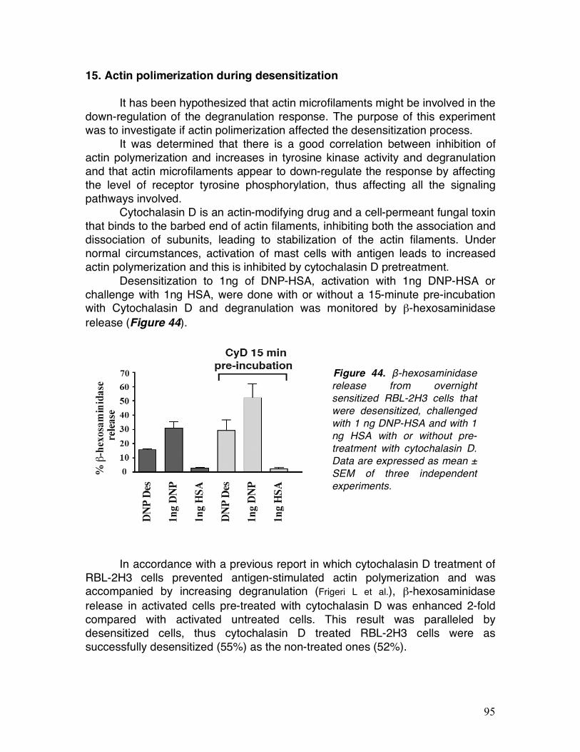
95
15. Actin polimerization during desensitization It has been hypothesized that actin microfilaments might be involved in the down-regulation of the degranulation response. The purpose of this experiment was to investigate if actin polimerization affected the desensitization process. It was determined that there is a good correlation between inhibition of actin polymerization and increases in tyrosine kinase activity and degranulation and that actin microfilaments appear to down-regulate the response by affecting the level of receptor tyrosine phosphorylation, thus affecting all the signaling pathways involved. Cytochalasin D is an actin-modifying drug and a cell-permeant fungal toxin that binds to the barbed end of actin filaments, inhibiting both the association and dissociation of subunits, leading to stabilization of the actin filaments. Under normal circumstances, activation of mast cells with antigen leads to increased actin polymerization and this is inhibited by cytochalasin D pretreatment. Desensitization to 1ng of DNP-HSA, activation with 1ng DNP-HSA or challenge with 1ng HSA, were done with or without a 15-minute pre-incubation with Cytochalasin D and degranulation was monitored by β-hexosaminidase release (Figure 44).
Figure 44. β-hexosaminidase release from overnight sensitized RBL-2H3 cells that were desensitized, challenged with 1 ng DNP-HSA and with 1 ng HSA with or without pre-treatment with cytochalasin D. Data are expressed as mean ± SEM of three independent experiments.
In accordance with a previous report in which cytochalasin D treatment of RBL-2H3 cells prevented antigen-stimulated actin polymerization and was accompanied by increasing degranulation (Frigeri L et al.), β-hexosaminidase release in activated cells pre-treated with cytochalasin D was enhanced 2-fold compared with activated untreated cells. This result was paralleled by desensitized cells, thus cytochalasin D treated RBL-2H3 cells were as successfully desensitized (55%) as the non-treated ones (52%).

96

97
DISCUSSION

98

99
Rapid drug desensitization procedures have emerged as critical components of treatment for allergic patients in need of first line therapy. These protocols have been refined to allow for safe delivery of chemotherapy drugs, antibiotics, monoclonals and other life saving medication in a very short time to sick patients (Castells et al. (3) / Lee CW et al. (1) / Lee CW et al. (2) / Legere HJ 3rd et al. / Brennan PJ et al.). Due to their success and relatively safe outcomes, rapid desensitization protocols have been produced recently to prevent anaphylaxis in food allergic patients when exposed to food antigens (Mansfield L). Although these procedures are elective, they are aimed at improving the quality of life of millions of patients who can be accidentally exposed to foods that can trigger deadly reactions. The lack of severe anaphylactic reactions in clinical reports (Lee CW et al. (2) / Castells et al. (3)), including hundreds of drug desensitizations using a standardized 12-step protocol, illustrates a profound inhibition of acute and delayed in vivo mast cell activation responses. Despite the clinical success, the mechanisms of rapid desensitization are not well understood and most of the molecular players have remained elusive. Our understanding of IgE desensitizations has been hampered by the lack of in vitro mast cell models providing quantitative and qualitative insight into the early and late activation responses. Multiple studies have been done with mast cells and basophils, but most of them in calcium-depleted conditions (non physiologic conditions) that could not be extrapolated to humans. The present study describes a physiologic mouse mast cell model of rapid desensitization to antigens in which increasing doses of antigens added at fixed time intervals renders mast cells hypo-responsive. This in vitro study under physiologic calcium conditions provides proof of concept for the effectiveness, safety and specificity of human desensitizations. The rapid (100 minutes) desensitization protocol presented here is based on previous data from Dr. Mariana Castells laboratory and others (Morales AR et al. / Shalit M et al.) in which it was shown that the dose escalation (2 or 10 fold) and the time between doses (5 or 30 min) were critical parameters in order to achieve desensitization. In the present protocol antigens are delivered at 10-min intervals in a cumulative fashion that provides an optimal dose-time relationship (Table 5). When starting at 1/1000 the target dose, the sequential delivery of increasing doses of antigen induced an almost complete inhibition of the activating responses seen in the dose-response curve. Each additional dose decreased responsiveness, and all 11 steps of the protocol were necessary for mast cells to become desensitized, since the greatest inhibition took place at the highest cumulative dose (Figure 20). This indicates that desensitization is a dynamic process in which each step provides a baseline platform for the next level of response reduction and that once desensitized, mast cells remain hyporesponsive to further antigen challenges (Figure 21). Increasing the target antigen dose for desensitization decreased the hyporesponsiveness (Figure 22), underscoring a critical relationship between IgE sensitization and the target

100
antigen dose for desensitization. Whether additional steps and/or time between steps might help achieve more hyporesponsiveness was not explored. In human desensitizations the level of IgE sensitization varies and is unknown for each patient and the target dose used for desensitization is empirical, which impacts its safety (Castells et al. (3) / Lee CW et al. (1) / Lee CW et al. (2) / Legere HJ 3rd et al. / Brennan PJ et al.). Once mast cells are triggered by antigen, there is depletion of the ER stores leading to activation of plasma membrane calcium-release activated calcium (CRAC) channels that allow Ca2+ entry. Consistent with a lack of degranulation, entry of extracellular calcium was almost completely inhibited (Figure 27) in desensitized mast cells. The sequential delivery of low antigen doses during desensitization may provide continued low levels of calcium entry and induce conformational changes of CRAC and other calcium-related channels locking further calcium entry and blocking signal transduction. Because calcium entry is clearly specifically impaired in our model, since a second non-desensitizing antigen allowed restoration of calcium flux, membrane compartmentalization may be required to exclude signal transduction molecules around desensitized receptors. It is also possible that a threshold calcium signal to activate ER Ca2+ sensors cannot be reached during desensitization, preventing calcium influx and degranulation. It has been shown that in vitro rapid desensitization of human mast cells induces decreased levels of signal-transducing molecules, such as syk, because of ubiquitinilation and degradation (Macglashan D et al. (2) / Odom S et al.) and that naturally occurring syk-deficient basophils are unresponsive to drug antigens (Gomez G et al.), indicating that syk is critical for activation and for desensitization (Kepley CL. (2)). In our hands, detection of Syk phosphorylation with desensitization was unsuccessful, possibly due to rapid phosphorylation-dephosphorylation and/or degradation of Syk during the desensitization process. However, since LAT, that is phosphorylated by Syk, serves as a docking site for further events leading to degranulation (Kraft S et al.) and LAT-deficient BMMC have impaired calcium mobilization and granule mediator release (Saitoh S et al. (1)), we established the fate of LAT and p38 MAP kinase. Both molecules phosphorylation were impaired in desensitized cells and consequently TNF-α and IL-6 production were diminished. STAT6, which is responsible for the transcription of IL-4 and IL-13, has been also involved in rapid desensitizations. STAT-6-deficient mast cells are capable of releasing mediators during the early phase of IgE cell activation but cannot release late cytokines, such as tumor necrosis factor TNF-α and IL-6 (Malaviya R et al.), and cannot be desensitized to DNP antigen (Morales AR et al.). STAT-6 phosphorylation was impaired in desensitized cells and it is possible that STAT6 activity is required for desensitization, via a pathway different from the one leading to the acute and late activating responses.

101
Our system is limited by the fact that BMMC are cultured in IL-3, which may affect cytokine production (Gonzalez-Espinosa C et al.). Nonetheless, this may have an important correlate in human desensitizations since our group has not observed delayed reactions in desensitized patients, confirming that the inhibition of mast cell activation during desensitization prevented later hypersensitivity reactions (Lee CW et al. (2) / Castells MC et al. (3)). In humans, temporary tolerization is achieved in hours and can be maintained if drug antigens are administered at regular intervals, depending on pharmacokinetic parameters (Castells M (1)). Maintenance of hyporesponsiveness in desensitized cells was not sustained by the presence of an excess of soluble antigen since washed cells remained desensitized. It is possible that bound antigen is equilibrated in desensitized cells. Earlier studies (Shalit M et al. / Rubinchik E et al.) suggested that the hypo-responsiveness induced by desensitization was due to internalization of antigen/IgE/FcεRI complexes and that the lack of available IgE renders the cells refractory to further stimulation. In contrast, we show here that, unlike in activation, during desensitization internalization of IgE and FcεRI is impaired (Figure 37 and Figure 38) and that desensitized cells can be triggered by anti-IgE, since unbound IgEs remain accessible and are available for cross-linking (Figure 40). Saturating doses of IgE in a co-culture system and the use of higher antigen doses (Shalit M et al.) promote internalization while low doses may redistribute antigen-bound receptor at the membrane level. Moreover, others have shown that low doses of antigen can crosslink IgE receptors, thus remaining mobile on the cell surface whereas high doses of antigen immobilize IgE-crosslinked receptors and induce degranulation (Andrews NL et al.). In that study mobile receptors bound to low dose of antigen had a threshold for activation responses. It is possible that during desensitization the low incremental additions of antigen promote receptor mobility and lack of internalization. Other studies suggested that activation of FcεRI induced by low antigen concentrations results in nuclear signals in the absence of degranulation (Grodzki AC et al.) but our results show that with desensitization, there is no phosphorylation of p38-MAPK (Figure 32) no IL-6 and no pre-formed or newly generated TNF-α (Figure 25 and Figure 33). The substrate component of specific desensitization remains unsolved, and it is of particular importance in desensitized patients since mast cell reactivity is maintained for non-desensitizing antigens. Previous studies in calcium-depleted conditions have shown that cells desensitized to one antigen could not be triggered with a non-desensitizing second antigen (MacGlashan DJr et al. (3)), possibly through a disabling mechanism involving syk ubiquitination (MacGlashan D et al. (2)). Due to the amount of IgE sensitization and low antigen doses used in our model, we could not detect syk phosphorylation but it is also possible that low amounts of antigen cannot trigger its phosphorylation.

102
Confocal microscopy studies gave us the opportunity to directly look into the antigen localization after desensitization. Our findings indicate that the mast cell activating machinery was intact for a non-desensitizing antigen action, since no mediator depletion occurred with desensitization. Calcium flux was restored in desensitized cells when challenged with a non-desensitizing antigen (Figure 42), and microscopic images confirmed that rapid desensitization is antigen specific (Figure 43) and does not induce anergy (MacGlashan DJr et al. 2008). Mast cell activation requires coordinated events in order to respond to an allergen and may differ depending on the type and strength of a stimulus. These events begin with the allergen-dependent aggregation of the IgE antibody-occupied high affinity receptor for IgE (FcεRI) and are propagated inside the cell through a sophisticated network of signaling molecules. While we do not know the exact mechanism that could explain the inhibition of receptor internalization during desensitization, it is possible that the mobility of antigen/IgE/FcεRI complexes and membrane re-arrangement could prevent their internalization, as shown by others with low doses of multivalent antigen (Andrews NL et al.). In addition, receptors engaged with low doses of antigen could be segregated into different compartments (Simons K et al.), preventing access to phosphorylating molecules. Inhibitory phosphatases such as SHP-1 may not be excluded from those compartments, thus preventing phosphorylation of key molecules required for signal transduction. A time course study of SHP-1 phosphorylation in RBL-2H3 cells (Ozawa T et al.) has shown a peak at 1 min of FcεRI crosslinking and a gradually decline within 10 min. Our initial results indicated a lack of phosphorylation at 100 min. Further studies are planned to look for phosphorylation of SHP-1 and other ITIM-bearing molecules (Lu-Kuo JM et al. / Ozawa T et al. / Castells MC et al. (2)) at each step of the desensitization protocol since it may be transient. Because our study indicates a strong inhibition of early signal transduction, it is possible that inhibitory molecules, such as LILRB4 that become phosphorylated upon IgE crosslinking could dephosphorylate the β and the γ chains of the FcεRI upon antigen engagement. The molecular mechanism by which gp49B1 inhibits the FcεRI-induced mast cell activation seems to involve SHP-1 rather than SHP-2. As shown in our study, inhibition of internalization prevents calcium entry, which would abrogate the acute and late phase activation. Indeed, co-clustering of gp49B1 with FcεRI on mast cells inhibits the induced secretion of stored proinflammatory mediators as well as of newly generated lipid mediators (Lu-Kuo JM et al. / Castells MC et al. (2) / Katz HR et al. (2)). Another molecule that we have not explored is the inositol phosphatase SHIP that has been reported to bind directly to the FcεRI β chain in vitro and regulates FcεRI signal transduction independently from FcγRIIB (Osborne MA et al. / Kimura T et al.). It has also been shown that FcεRI-induced calcium mobilization and MAPK activation are enhanced in BMMC derived from SHIP-deficient mice compared to wild-type mice (Huber M et al.). Thus it is also possible that SHIP could allow for desensitization.

103
Whether SHIP, LILRB4 or other ITIM-containing mast cell inhibitory receptors participate in the gradual re-arrangement of the antigen-crosslinked receptor preventing its internalization needs to be explored. Rat basophilic leukemia (RBL 2H3) cell line has been commonly used as a histamine-releasing cell line and has been proved to be a suitable model to examine signal transduction events in FcεRI mediated mast cell activation. We decided to use this cell line due to their adhesive characteristics for further confocal microscopy experiments but also for actin polimerization studies. It has been hypothesized that actin microfilaments might be involved in the down-regulation of the degranulation response at the level of receptor phosphorylation (Frigeri L et al. / Benhamou M et al.) and that actin assembly and reorganization plays an important role in FcεRI-induced mast cell degranulation and calcium signaling (Oka T et al. / Wu M et al.). This is what drives us to investigate if desensitization could be affected by actin polimerization. Our results, accordingly with those previously cited, determined that there is a good correlation between inhibition of actin polymerization and increases in degranulation for activation as well as for desensitization (Figure 44). Thus, actin microfilaments could down-regulate the response by affecting the level of receptor tyrosine phosphorylation and uncoupling Lyn from the cross-linked receptor, but since our results showed that the antigen/IgE/FcεRI complexes do not internalize with desensitization, further receptor phosphorylation could not be relevant. The data presented here provides a unique in vitro model of rapid IgE desensitization, under physiologic calcium conditions, that is effective and reproducible and sheds some light on the mechanisms underlying downregulation of mast cell responses. This data shows how an optimal antigen dose-time relationship leads to almost complete abrogation of early- and late-phase activation events and establishes the first in vitro model of antigen-specific desensitization, disabling the specific response to one antigen but keeping the cell machinery unaffected, unlike non-specific desensitization. Furthermore, there is a clear demonstration that low doses of antigen sequentially added are inhibitory for mast cell activation in the presence of calcium, and induce no depletion of mediators. Most importantly, this data demonstrates that specific rapid desensitization inhibits the internalization of the antigen/IgE/FcεRI complexes. We speculate that the cellular cytoskeleton plays an important role in the desensitization process, such that antigen/IgE/FcεRI complexes possibly remain mobile on the surface of the cell during rapid desensitization allowing their re-arrangement. The plasma membrane may redistribute selectively with the desensitized receptor clusters allowing compartmentalization and possibly segregation of activating molecules therefore impairing downstream signal transduction. Additionally, inhibitory molecules may become associated with desensitized receptor clusters preventing cellular activation. A simplified cartoon in Figure 45 compares the outcomes of mBMMC activation and rapid

104
desensitization and presents our hypothesis explaining how desensitization affects antigen/IgE/FcεRI complexes re-arrangement at the cell membrane.
Figure 45. Simplified cartoon comparing activation and desensitization outcomes as well as a possible explanation of how rapid desensitization works and the re-arrangement of the FcεRI receptors at the cell membrane.

105
CONCLUSIONS

106

107
In this PhD thesis,
1. we established the first in vitro model of m-BMMC IgE rapid desensitization • that provides an optimal relationship between antigen dose and
time between doses • that is effective and reproducible, leading to almost complete
abrogation (50-80%) of mast cell activation events • that is versatile (can be used for various target doses, with several
antigens and with different cell types)
2. we demonstrated that m-BMMC IgE rapid desensitization • inhibits acute and late phase mast cell responses • is an antigen specific process that disables the specific response to
one antigen but keeps the cell machinery unaffected, unlike non-specific desensitization
• can be maintained, in the presence or absence of antigen, up to 6 h • impairs internalization of the FcεRI /IgE/antigen complexes

108

109
CONCLUSIONES

110

111
En esta tesis,
1. se ha establecido el primer modelo in vitro de desensibilización rápida por IgE de mastocito derivado de médula ósea de ratón (m-BMMC) • que proporciona una relación óptima dosis de antígeno-tiempo
entre dosis • que es efectivo y reproducible, llegando hasta casi la completa
desaparición (50-80%) de la activatión del mastocito • que es versátil (puede ser usado con varias dosis objetivo, con
diversos antígenos y con diferentes tipos de células)
2. se ha demostrado que la desensibilización rápida por IgE de m-BMMC • inhibe la respuesta inmediata y la respuesta tardía del mastocito • es un proceso específico para antígeno que discapacita la
respuesta específica a un antígeno pero mantiene la maquinaria celular intacta, no como en la desensibilización no específica
• se puede mantener hasta 6 h en presencia o ausencia de antígeno • afecta la internalización del complejo antígeno /IgE/ FcεRI

112

113
PERSPECTIVES

114

115
Rapid drug desensitizations are currently emerging as highly effective and safe procedures for the administration of important medications while minimizing or entirely circumventing hypersensitivity reactions in sensitized patients. Strategies aimed at dilucidating the full mechanism underlying rapid desensitization may constitute an important tool for a better understanding of the process and for the development of improved and safer protocols for drug and food desensitizations. We therefore anticipate that future research will also focus on using our in vitro model as the first step for using patient serum and establishing the minimal dose and concentration for the initial desensitization steps and the creation of individualized desensitization protocols.

116

117
PUBLICATIONS

118
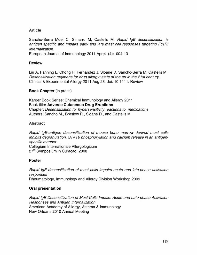
119
Article Sancho-Serra Mdel C, Simarro M, Castells M. Rapid IgE desensitization is antigen specific and impairs early and late mast cell responses targeting FcεRI internalization. European Journal of Immunology 2011 Apr;41(4):1004-13 Review Liu A, Fanning L, Chong H, Fernandez J, Sloane D, Sancho-Serra M, Castells M. Desensitization regimens for drug allergy: state of the art in the 21st century. Clinical & Experimental Allergy 2011 Aug 23. doi: 10.1111. Review Book Chapter (in press) Karger Book Series: Chemical Immunology and Allergy 2011 Book title: Adverse Cutaneous Drug Eruptions Chapter: Desensitization for hypersensitivity reactions to medications Authors: Sancho M., Breslow R., Sloane D., and Castells M. Abstract Rapid IgE-antigen desensitization of mouse bone marrow derived mast cells inhibits degranulation, STAT6 phosphorylation and calcium release in an antigen-specific manner. Collegium Internationale Allergologicum 27th Symposium in Curaçao, 2008 Poster Rapid IgE desensitization of mast cells impairs acute and late-phase activation responses Rheumatology, Immunology and Allergy Division Workshop 2009 Oral presentation Rapid IgE Desensitization of Mast Cells Impairs Acute and Late-phase Activation Responses and Antigen Internalization American Academy of Allergy, Asthma & Immunology New Orleans 2010 Annual Meeting

120

121
REFERENCES

122

123
Abramson J, Pecht I. Regulation of the mast cell response to the type 1 Fc epsilon receptor. Immunol Rev. 2007 Jun;217:231-54
Alvarez-Errico D, Lessmann E, Rivera J. Adapters in the organization of mast cell signaling. Immunol Rev. 2009 Nov;232(1):195-217 Andrews NL, Pfeiffer JR, Martinez AM, Haaland DM, Davis RW, Kawakami T, Oliver JM et al. Small, mobile FcεRI receptor aggregates are signaling competent. Immunity 2009; 31:469–479 Aronson NN Jr, Kuranda MJ. Lysosomal degradation of Asn-linked glycoproteins. FASEB J. 1989 Dec;3(14):2615-22 Benhamou M, Siraganian RP. Protein-tyrosine phosphorylation: an essential component of FceRI signaling. Immunol. Today 1992;13, 195–197 Blank U, Rivera J. The ins and outs of IgE-dependent mast-cell exocytosis. Trends Immunol. 2004;25, 266–273 Brennan PJ, Rodriguez Bouza T, Hsu FI, Sloane DE, Castells MC. Hypersensitivity reactions to mAbs: 105 desensitizations in 23 patients, from evaluation to treatment. J Allergy Clin Immunol 2009 Dec;124(6):1259-66 Castells M (1). Desensitization for drug allergy. Curr Opin Allergy Clin
Immunol, 2006. 6(6): p. 476-81 Castells MC (2), Klickstein LB, Hassani K, Cumplido JA, Lacouture ME,
Austen KF, Katz HR. gp49B1-alpha(v)beta3 interaction inhibits antigen-induced mast cell activation. Nat Immunol. 2001; 2(5):436-42
Castells MC (3), Tennant NM, Sloane DE, Hsu FI, Barrett NA, Hong DI, et al. Hypersensitivity reactions to chemotherapy: outcomes and safety of rapid desensitization in 413 cases. J Allergy Clin Immunol 2008; 122:574-80
Dembo M, Goldstein B, Sobotka AK, Lichtenstein LM. Histamine release due to bivalent penicilloyl haptens the relation of activation and desensitization of basophils to dynamic aspects of ligand binding to cell surface antibody. J Immunol. 1979; 122(2): 518-28 Elgueta R, Benson MJ, de Vries VC, Wasiuk A, Guo Y, Noelle RJ. Molecular mechanism and function of CD40/CD40L engagement in the immune system. Immunol. Rev 2009 May;229(1):152-72

124
Fattakhova GV, Masilamani M, Narayanan S, Borrego F, Gilfillan AM, Metcalfe DD, Coligan JE. Endosomal trafficking of the ligated FceRI receptor. Mol. Immunol. 2009. 46:793-802 Frigeri L, Apgar JR. The role of actin microfilaments in the down-regulation of the degranulation response in RBL-2H3 mast cells. J. Immunol. 1999. 162, 2243–2250 Furumoto Y, Nunomura S, Terada T, Rivera J, Ra C. The Fc epsilon RI beta immunoreceptor tyrosine-based activation motif exerts inhibitory control on MAPK and I kappa B kinase phosphorylation and mast cell cytokine production. J Biol Chem 2004; 279:49177–87 Galli SJ (1), Nakae S, Tsai M. Mast cells in the development of adaptive
immune responses. Nat Immunol. 2005; 6(2):135-42. Review Galli SJ (2), Tsai M, Piliponsky AM. The development of allergic inflammation. Nature. 2008; 454(7203):445-54. Review Garman SC, Kinet JP, Jardetzky TS. Crystal structure of the human high- affinity IgE receptor. Cell 1998; 95:951–61 Gilfillan AM (1), Rivera J. The tyrosine kinase network regulating mast cell activation. Immunol Rev 2009; 228:149–69 Gilfillan AM (2), Tkaczyk C. Integrated signalling pathways for mast-cell activation. Nat. Rev. Immunol. 2006; 6, 218–230 Gomez G, Schwartz L, Kepley C. Syk deficiency in human non-releaser lung mast cells. Clin Immunol. 2007; 125(1): 112-5 Gonzalez-Espinosa C, Odom S, Olivera A, Hobson JP, Cid Martinez ME, Olivera-dos-Santos A, Barra L et al. Preferential Signaling and Induction of Allergy-promoting Lymphokines Upon Weak Stimulation of the high Affinity IgE Receptor on Mast Cells. J. Exp. Med. 2003. 197:1453-1465 Grodzki AC, Moon KD, Berenstein EH, Siraganian RP. FceRI-induced activation by low antigen concentrations results in nuclear signals in the absence of degranulation. Molecular Immunology 2009;46, 2539–2547 Gurish MF, Friend DS, Webster M, Ghildyal N, Nicodemus CF, Stevens RL. Mouse Mast Cells That Possess Segmented/Multi-lobular Nuclei. Blood 1997; 90: 382-390

125
Hsieh FH, Lam BK, Penrose JF, Austen KF, Boyce JA. T helper cell type 2 cytokines coordinately regulate immunoglobulin E-dependent cysteinyl leukotriene production by human cord blood-derived mast cells: profound induction of leukotriene C(4) synthase expression by interleukin 4. J. Exp. Med. 2001; 193:123-133 Hsu C, MacGlashan DJr. IgE antibody up-regulates high affinity IgE binding on murine bone marrow-derived mast cells. Immunology Letters, Volume 52, Issues 2-3, September 1996, Pages 129-134 Huber M, Helgason CD, Damen JE, Liu L, Humphries RK, and Krystal G. The src homology 2-containing inositol phosphatase (SHIP) is the gatekeeper of mast cell degranulation. Proc. Natl. Acad. Sci. USA 1998; 95:11330 Humphries DE, Wong GW, Friend DS, Gurish MF, Qiu W, Huang C, Sharpe AH, and Stevens RL. Heparin is essential for the storage of specific granule proteases in mast cells. Letters to Nature 1999 Ishizaka T, Sterk AR, Daeron M, Becker EL, Ishizaka K. Biochemical analysis
of desensitization of mouse mast cells. J Immunol 1985; 135:492-501 Ishizuka T, Terada N, Gerwins P, Hamelmann E, Oshiba A, Fanger GR, Johnson GL et al. Mast cells tumor necrosis factor alpha production is regulated by MEK kinases. Proc. Natl. Acad. Sci. USA 1997. 94:6358-63 Jouvin MH, Adamczewski M, Numerof R, Letourneur O, Vallé A, Kinet JP. Differential control of the tyrosine kinases Lyn and Syk by the two signaling chains of the high affinity immunoglobulin E receptor. J Biol Chem. 1994 Feb 25; 269(8):5918-25 Kalesnikoff J, Baur N, Leitges M, Hughes MR, Damen JE, Huber M, Krystal G. SHIP negatively regulates IgE+antigen-induced IL-6 production in mast cells by inhibiting NF-kappa B activity. J. Immunol. 2002; 168:4737-46 Kambayashi T (1), Baranski JD, Baker RG, Zou T, Allenspach EJ, Shoag JE, Jones PL et al. Indirect involvement of allergen-captured mast cells in antigen presentation. Blood 2008; 111:1489-1496 Kambayashi T (2), Okumura M, Baker RG, Hsu CJ, Baumgart T, Zhang W, Koretzky GA. Independent and cooperative roles of adaptor molecules in proximal signaling during FcepsilonRI-mediated mast cell activation. Mol Cell Biol. 2010; 30(17):4188-96

126
Katz HR (1), Austen KF. A newly recognized pathway for the negative regulation of mast cell-dependent hypersensitivity and inflammation mediated by an endogenous cell surface receptor of the gp49 family. J Immunol. 1997;158(11):5065-70. Review
Katz HR (2), Vivier E, Castells MC, McCormick MJ, Chambers JM, Austen KF. Mouse mast cell gp49B1 contains two immunoreceptor tyrosine-based inhibition motifs and suppresses mast cell activation when coligated with the high-affinity Fc receptor for IgE. Proc Natl Acad Sci U S A. 1996; 93(20):10809-14 Katz, HR (3) Inhibitory receptors and allergy. Curr Opin Immunol 2002;
14:698-704 Kepley CL (1), Youssef L, Andrews RP, Wilson BS, Oliver JM. Syk deficiency
in nonreleaser basophils. J Allergy Clin Immunol. 1999; 104(2 Pt 1): 279-84
Kepley CL (2), Antigen-induced reduction in mast cell and basophil functional responses due to reduced Syk protein levels. Int Arch Allergy Immunol 2005; 138:29–39
Kimura T, Sakamoto H, Appella E, and Siraganian RP. The negative signaling molecule SH2 domain-containing inositol-polyphosphate 5- phosphatase (SHIP) binds to the tyrosine phosphorylated β subunit of the high affinity IgE receptor. J. Biol. Chem. 1997; 272:13991
Komiya A, Hirai K, Iikura M, Nagase H, Yamada H, Miyamasu M, et al. Induction of basophil desensitization in physiological medium: enhancement after IgE-dependent upregulation of surface IgE binding on basophils. Int Arch Allergy Immunol. 2003 Jan;130(1):40-50 Kraft S, Kinet JP. New developments in FcepsilonRI regulation, function and inhibition. Nat.Rev.Immunol. 2007; 7:365-78 Lee CW (1), Matulonis UA, Castells MC. Carboplatin hypersensitivity: a 6-h 12-step protocol effective in 35 desensitizations in patients with gynecological malignancies and mast cell/IgE-mediated reactions. Gynecol. Oncol. 2004; 95: 370–376 Lee CW (2), Matulonis UA, Castells MC. Rapid inpatient/outpatient desensitization for chemotherapy hypersensitivity: standard protocol effective in 57 patients for 255 courses. Gynecol Oncol 2005; 99:393-9

127
Legere HJ 3rd, Palis RI, Rodriguez Bouza T, Uluer AZ, Castells MC. A safe protocol for rapid desensitization in patients with cystic fibrosis and antibiotic hypersensitivity. J Cyst Fibros 2009; 8:418–24 Lin S, Cicala C, Scharenberg AM, Kinet JP. The Fc(epsilon)RIbeta subunit functions as an amplifier of Fc(epsilon)RIgamma mediated cell activation signals. Cell 1996;85:985–95 Liu A, Fanning L, Chong H, Fernandez J, Sloane D, Sancho-Serra M, Castells M. Desensitization regimens for drug allergy: state of the art in the 21st century. Clin Exp Allergy. 2011 Aug 23. doi: 10.1111. Review Lu-Kuo JM, Joyal DM, Austen KF, Katz HR. gp49B1 inhibits IgE-initiated mast cell activation through both immunoreceptor tyrosine-based inhibitory motifs, recruitment of src homology 2 domain-containing phosphatase-1, and suppression of early and late calcium mobilization. J. Biol. Chem. 1999. 274:5791-6. Lundequist A, Pejler G. Biological implications of preformed mast cell mediators. Cell Mol Life Sci. 2011 Mar; 68(6):965-75 Ma HT, Beaven MA. Regulation of Ca2+ signaling with particular focus on mast cells. Crit. Rev. Immunol. 2009. 129: 155–186 MacGlashan D Jr (1), Lichtenstein LM. Studies of antigen binding on human basophils. I. Antigen binding and functional consequences. J Immunol. 1983; 130(5):2330-6. Macglashan D (2), Miura K. Loss of syk kinase during IgE-mediated
stimulation of human basophils. J Allergy Clin Immunol. 2004; 114(6): 1317-24
MacGlashan DJr (3), Lichtenstein LM. Basic characteristics of human lung
mast cell desensitization. J. Immunol. 1987. 139:501-505 MacGlashan DJr (4), Undem BJ. Inducing an anergic state in mast cells and basophils without secretion. J. Allergy Clin. Immunol. 2008.121:1500-1506 MacGlashan DJr (5) Desensitization of IgE-mediated IL-4 release from human
basophils. J. Leukoc. Biol. 1998. 63: 59–67 Maekawa A, Kanaoka Y, Lam BK, Austen KF. Identification in mice of two
isoforms of the cysteinyl leukotriene 1 receptor that result from alternative splicing. Proc Natl Acad Sci U S A 2001; 98:2256-61.

128
Malaviya R, Uckun FM. Role of STAT6 in IgE receptor/FcepsilonRI-mediated late phase allergic responses of mast cells. J. Immunol. 2002. 168:421-26. Mansfield L. Successful oral desensitization for systemic peanut allergy. Ann Allergy Asthma Immunol 2006; 97:266-7 Mao SY, Pfeiffer JR, Oliver JM, Metzger H. Effects of subunit mutation on the localization to coated pits and internalization of cross-linked IgE-receptor complexes. J. Immunol. 1993. 151:2760-2774 Marshall JS. Mast-cell responses to pathogens. Nat Rev Immunol. 2004 Oct; 4(10):787-99 Mendoza GR (1), Minagawa K. Subthreshold and suboptimal desensitization human basophils. I. Kinetics of decay of releasability. Int Arch Allergy Appl Immunol. 1982; 68(2):101-7
Mendoza GR (2), Minagawa K. Subthreshold and suboptimal desensitization of human basophils. II. Nonspecificity and irreversibility of desensitization. Int Arch Allergy Appl Immunol 1982; 69:282-84
Metzger H. The receptor with high affinity for IgE. Immunol Rev. 1992 Feb;125:37-48 Morales AR, Shah N, Castells M. Antigen-IgE desensitization in signal transducer and activator of transcription 6-deficient mast cells by suboptimal doses of antigen. Ann. Allergy Asthma Immunol. 2005; 94:575-80 Navo M, Kunthur A, Badell ML, Coffer Ii LW, Markman M, Brown J, Smith JA. Evaluation of the incidence of carboplatin hypersensitivity reactions in cancer patients. Gynecol Oncol 2006; 103: 608-613 Niwa H, Yamamura K, Miyazaki J. Efficient selection for high-expression transfectants with a novel eukaryotic vector. Gene 1991; 108:193-199. Odom S, Gomez G, Kovarova M, et al. Negative regulation of immunoglobulin E-dependent allergic responses by Lyn kinase. J Exp Med 2004; 199(11): 1491–1502 Oka T, Sato K, Hori M, Ozaki H, Karaki H. FcepsilonRI cross-linking-induced actin assembly mediates calcium signalling in RBL-2H3 mast cells. Br J Pharmacol. 2002 Jul; 136(6):837-46.

129
Olivera A. Unraveling the complexities of sphingosine-1-phosphate function: the mast cell model. Prostaglandins Other Lipid Mediat. 2008 Jun; 86(1- 4):1-11 Osborne MA, Zenner G, Lubinus M, Zhang X, Songyang Z, Cantley LC, et al. The inositol 5ʼ-phosphatase SHIP binds to immunoreceptor signaling motifs and responds to high affinity IgE receptor aggregation. J. Biol. Chem. 1996; 271:29271 Ozawa T, Nakata K, Mizuno K, Yakura H. Negative autoregulation of Src homology region 2-domain-containing phosphatase-1 in rat basophilic leukemia-2H3 cells. Int. Immunol. 2007; 19:1049-1061 Paolini R, Numerof R, Kinet JP. Phosphorylation/dephosphorylation of high- affinity IgE receptors: a mechanism for coupling/uncoupling a large signaling complex. Proc. Natl. Acad. Sci. USA. 1992 Nov 15; 89(22):10733-37 Pribluda VS, Pribluda C, Metzger H. Transphosphorylation as the mechanism by which the high-affinity receptor for IgE is phosphorylated upon aggregation. Proc Natl Acad Sci USA 1994; 91:11246–50 Pruzansky JJ, Patterson R. Desensitization of human basophils with suboptimal concentrations of agonist. Evidence for reversible and irreversible desensitization. Immunology. 1988; 65(3): 443-7
Raby BA, Lazarus R, Silverman EK, Lake S, Lange C, Wjst M, Weiss ST. Association of vitamin D receptor gene polymorphisms with childhood and adult asthma. Am.J.Respir.Crit Care Med. 2004; 170: 1057-65 Rivera J, Fierro NA, Olivera A, Suzuki R. New Insights on Mast Cell Activation via the High Affinity Receptor for IgE. Adv Immunol. 2008 ; 98: 85–120 Roskoski RJr. Signaling by Kit protein-tyrosine kinase–the stem cell factor receptor. Biochem. Biophys. Res. Commun. 2005; 337, 1–13 Rubinchik E, Shalit M, Levi-Schaffer F. Responsiveness of human skin mast cells to repeated activation: an in vitro study. Allergy. 1998; 53(1):14-9 Saitoh S (1), Arudchandran R, Manetz TS, Zhang W, Sommers CL, Love PE,
et al. LAT is essential for Fc(epsilon)RI-mediated mast cell activation. Immunity. 2000; 12:525-35

130
Saitoh S (2), Odom S, Gomez G, Sommers CL, Young HA, Rivera J, Samelson LE. The four distal tyrosines are required for LAT-dependent signaling in FcepsilonRI-mediated mast cell activation. J. Exp Med. 2003; 198(5):831-43.
Sanchez-Miranda E, Ibarra-Sanchez A, Gonzalez-Espinosa C. Fyn kinase controls FceRI receptor-operated calcium entry necessary for full degranulation in mast cells. Biochem biophys Res Commun 2010 Jan 22;391(4):1714-20 Sancho-Serra Mdel C, Simarro M, Castells M. Rapid IgE desensitization is antigen specific and impairs early and late mast cell responses targeting FcεRI internalization. Eur J Immunol. 2011 Apr;41(4):1004-13 Schwartz LB (1), Austen KF, Wasserman SI. Immunologic release of beta- hexosaminidase and beta-glucuronidase from purified rat serosal mast cells. J Immunol. 1979; 123(4):1445-50 Schwartz LB (2), Austen KF. Enzymes of the mast cell granule. J Invest Dermatol. 1980; 74(5):349-53 Schwartz LB, Metcalfe DD, Miller JS, Earl H, Sullivan T. Tryptase levels as an indicator of mast-cell activation in systemic anaphylaxis and mastocytosis. N Engl J Med 1987; 316:1622-6 Shalit M, Levi-Schaffer F. Challenge of mast cells with increasing amounts of antigen induces desensitization. Clin Exp Allergy 1995; 25:896–902. Sheffer AL, Feldweg A, Castells M. Chapter 11: Anaphylaxis: Holgate ST, Church MK, Lichtenstein LM, eds. Allergy 3rd Edition, Mosby International Ltd. 2006, p. 167-78 Simons K, Toomre D. Lipid rafts and signal transduction. Nat Rev Mol Cell Biol 2000; 1:31-39 Siraganian RP. Mast cell signal transduction fromthe high-affinity IgE receptor. Curr. Opin. Immunol. 2003; 15, 639–646 Sobotka AK, Dembo M, Goldstein B, Lichtenstein LM. Antigen-specific desensitization of human basophils. J Immunol. 1979; 122(2): 511-7 Sonoda T, Kitamura Y, Haku Y, Hara H and Mori KJ. Mast cell precursors in
various hematopoietic colonies of mice produced in vivo and in vitro. Br. J. Haematol. 1983; 53: 611-620.

131
Sullivan T, Yeccies L, Shatz G, Parker C, Wedner HJ. Desensitization of patients allergic to penicillin using orally administered beta-lactam antibiotics. J Allergy Clin Immunol. 1982; 69:275–282 Vadas P, Gold M, Perelman B, Liss GM, Lack G, Blyth T, et al. Platelet- activating factor, PAF acetylhydrolase, and severe anaphylaxis. N Engl J Med 2008; 358:28-35 Vig M, Beck A, Billingsley JM, Lis A, Parvez S, Peinelt C, et al. CRACM1 multimers form the ion-selective pore of the CRAC channel. Curr Biol 2006; 16:2073-9 von Mutius E. 99th Dahlem conference on infection, inflammation and chronic inflammatory disorders: farm lifestyles and the hygiene hypothesis. Clin Exp Immunol. 2010 Apr; 160(1): 130-5 Wang Z, Deng Y, Yu D, Bai G. Nitric oxide inhibits a rise of ATP-introduced cytosolic free Ca2+ concentration and release from intracellular stored Ca2+. Chin Med Sci J. 2000; 15(4):217-21 Wendel G, Stark B, Jamison RB, Molina RD, Sullivan TJ. Penicillin allergy and desensitization in serious infections during pregnancy. N Engl J Med. 1985; 312:1229-32 Wong GW, Yasuda S, Morokawa N, Li L, Stevens RL. Mouse chromosome 17A3.3 contains 13 genes that encode functional tryptic-like serine proteases with distinct tissue and cell expression patterns. Journal of Biological Chemistry 2004; 279: 2438-52 Wu M, Holowka D, Craighead HG, Baird B. Visualization of plasma membrane compartmentalization with patterned lipid bilayers. Proc Natl Acad Sci USA. 2004; 101(38):13798-803 Yokota T, Lee F, Rennick D, Hall C, Arai N, Mosmann T, Nabel G et al. Isolation and characterization of a mouse cDNA clone that expresses mast-cell growth-factor activity in monkey cells. Proc. Natl. Acad. Sci. USA 1984; 81:1070-1074 Young RM, Holowka D, Baird B. A lipid raft environment enhances Lyn kinase activity by protecting the active site tyrosine from dephosphorylation. J Biol Chem. 2003; 278(23):20746-52

132

133
ANNEX

134

Rapid IgE desensitization is antigen specific and impairsearly and late mast cell responses targeting FceRIinternalization
Maria del Carmen Sancho-Serra1, Maria Simarro2 and Mariana Castells1
1 Division of Rheumatology, Immunology and Allergy, Department of Medicine, Brigham
and Women’s Hospital, Harvard Medical School, Boston, MA, USA2 Unidad de Investigacion, Hospital Clinico Universitario de Valladolid, Valladolid, Spain
Rapid IgE desensitization provides temporary tolerization for patients who have
presented severe hypersensitivity reactions to food and drugs, protecting them from
anaphylaxis, but the underlying mechanisms are still incompletely understood.
Thus, here we develop an effective and reproducible in vitro model of rapid IgE desensi-
tization for mouse BM-derived mast cells (BMMCs) under physiologic calcium
conditions, and we characterize its antigen specificity and primary events. BMMCs were
challenged with DNP-human serum albumin conjugated (DNP-HSA) and/or OVA
antigens, delivered either as a single dose (activation) or as increasing sequential doses
(desensitization). Compared to activated cells, desensitized BMMCs had impaired
degranulation, calcium flux, secretion of arachidonic acid products, early and late
TNF-a production, IL-6 production, and phosphorylation of STAT6 and p38 mitogen-acti-
vated protein kinase (p38 MAPK). OVA-desensitized cells responded to DNP and DNP-
desensitized cells responded to OVA, proving specificity. Internalization of specific antigen,
IgE and high-affinity receptor for IgE (FceRI) were impaired in desensitized BMMCs. Our
results demonstrate that rapid IgE desensitization is antigen specific and inhibits early
and late mast cell activation responses and internalization of the antigen/IgE/FceRI
complexes.
Keywords: Desensitization . DNP/OVA . FceRI . IgE . Mast cells
Introduction
Exposure of IgE-sensitized patients to medication or food
allergens can cause the sudden systemic release of inflammatory
mediators from activated mast cells, leading to anaphylaxis
[1, 2]. Avoidance may be difficult for food-sensitized patients due
to cross-reactive food allergens. For medication-sensitized
patients, avoidance may lead to significant morbidity and
mortality if treatment for cancer or severe infection becomes
necessary, and may decrease the quality of life among patients
with chronic inflammatory diseases sensitized to monoclonal
antibodies. Desensitization protocols have been developed to
help deliver full therapeutic doses of drug allergens, in an
incremental, stepwise fashion without eliciting life-threatening
symptoms [3–5]. More recently, food desensitization protocols
have been generated to protect children and adults from
accidental exposures to allergenic foods [6, 7]. Most IgE-
sensitized patients present a positive skin test to the offending
food or medication, indicating that mast cells and IgE are the
main targets of these reactions. After rapid desensitization,
specific skin test reactivity is abolished, implying a complete
inhibition of the mechanisms that induce mast cell activation [8].Correspondence: Dr. Mariana Castellse-mail: [email protected]
& 2011 WILEY-VCH Verlag GmbH & Co. KGaA, Weinheim www.eji-journal.eu
DOI 10.1002/eji.201040810 Eur. J. Immunol. 2011. 41: 1004–1013Maria del Carmen Sancho-Serra et al.1004

Mast cells are activated by antigen crosslinking of IgE-bound
high-affinity receptor for IgE (FceRI) receptors, and aggregation
of these receptors results in rapid phosphorylation of tyrosine
residues in the ITAMs of b and g chains by lyn kinase, which leads
to recruitment and activation of spleen tyrosine kinase (syk) and
fyn. Both fyn and syk phosphorylate downstream targets, leading
to calcium mobilization, degranulation, arachidonic acid meta-
bolization, and cytokine and chemokine gene transcription
[9, 10]. As opposed to activation, desensitization is a process in
which mast cells are rendered hypo-responsive to an activating
challenge, either by exposure to low antigen doses in calcium-
depleted conditions [11] or by exposure to incremental doses of
antigen, in the presence of calcium [12, 13]. Calcium-depleted
conditions cannot be applied to human desensitizations, and few
studies have addressed physiological desensitizations, since
events occurring in the absence of extracellular calcium may not
reflect the same pathways as those occurring in the presence of
calcium [14]. Internalization of FceRI through progressive
crosslinking at low levels of antigen has been postulated as the
likely mechanism for cell-surface depletion of IgE and cellular
unresponsiveness to specific activating doses of allergen [12].
Depletion of molecular targets of activation such as syk has been
shown in prolonged antigen desensitization, indicating a
universal rather than specific desensitization [15].
Based on our previous study [16], we report here a model of
mouse BM-derived mast cell (BMMCs) specific rapid desensiti-
zation to DNP and OVA antigens in the presence of physiologic
levels of calcium. Increasing doses of antigen delivered at fixed
time intervals induced a highly specific and prolonged hypo-
responsiveness to triggering doses of the desensitizing antigen.
Mast cells desensitized to DNP or OVA demonstrated almost
complete inhibition of b-hexosaminidase and pre-formed TNF-arelease, calcium flux and arachidonic acid metabolization. They
did not release significant amounts of newly generated IL-6 or
TNF-a and failed to phosphorylate STAT6 and p38 MAPK. When
sensitized to both DNP and OVA antigens, DNP-desensitized cells
responded fully to OVA and vice versa. Most importantly, specific
rapid desensitization targeted the internalization of antigen/IgE/
FceRI complexes since antigen-specific IgE bound to the a chain
of the FceRI remained at the membrane level. This model may
provide support for the specificity and effectiveness of human
desensitizations.
Results
Inhibition of BMMC responses to sequential versussingle-dose DNP antigen delivery
In order to compare single-dose antigen delivery (activation)
with sequential cumulative doses (rapid desensitization), we first
assessed the dose response curve to DNP-human serum albumin
conjugated (DNP-HSA) antigen, by b-hexosaminidase release,
with cells sensitized with anti-DNP IgE (see Fig. 1A). DNP-HSA
doses of 1, 5 and 10 pg (DNP in figure) were non-activating as
single doses, since the percentage of b-hexosaminidase release
was comparable to that of the control (1 ng HSA). Cells
challenged with higher doses of antigen (410 pg DNP-HSA)
delivered as single doses achieved significant b-hexosaminidase
release. The black bar in Fig. 1A (1 ng DNP) represents the
optimal triggering dose of 1 ng DNP-HSA used as target dose for
rapid desensitization to 1 ng of DNP-HSA (DNP Des). The release
obtained with single-dose additions in Fig. 1A was compared to
that obtained with doses added sequentially, following every step
of the desensitization protocol (see Fig. 1B, white bars). White
bars represent b-hexosaminidase release at each particular point
in the cumulative sequence of antigen additions. A maximum of
10% b-hexosaminidase release was achieved at all points in the
sequence, showing that the desensitization process did not induce
a slow release of mediators.
To determine whether there was a threshold dose that initi-
ated hypo-responsiveness, replicate samples were used, and at
each particular point in the sequence of antigen additions, cells
were also challenged with a triggering dose of 1 ng DNP-HSA (see
Fig. 1B, gray bars). Response to the triggering dose declined with
increasing number of sequential doses. The greatest hypo-
responsiveness was achieved with the highest number of
sequential additions (11, in Fig. 1B), indicating that hypo-
responsiveness was not stabilized until the end of the desensiti-
zation protocol.
To test whether cells’ hypo-responsiveness achieved with
rapid desensitization to 1 ng DNP-HSA could be overcome with
higher challenging doses, we analyzed the response of desensi-
tized cells to activating doses of 1, 2, 3, 4 and 5 ng of DNP-HSA.
Up to five-fold increase in challenging dose did not reverse
desensitization (see Fig. 1C).
The protocol was effective when increasing the target dose to
5 and 10 ng, with the same number of steps, time between steps
and starting dose (1/1000 the target dose), but less inhibition of
b-hexosaminidase release was observed (see Fig. 1D). Cells
desensitized to 1 ng DNP-HSA showed a 75% inhibition whereas
cells desensitized to 5 and 10 ng DNP-HSA had a 65 and 41%
inhibition of b-hexosaminidase release, respectively.
BMMC rapid desensitization impairs early and latephase cell responses
BMMCs sensitized with anti-DNP IgE or anti-OVA IgE were rapid-
desensitized as per the protocol presented in Table 1. In both
DNP and OVA systems, we measured the release of b-hexosami-
nidase when antigen was delivered as a single dose (1 ng DNP-
HSA/10 ng OVA, black bars in Fig. 2A) or when antigen was
delivered following the rapid desensitization protocol (white bars
in Fig. 2A). Cells desensitized to 1 ng DNP-HSA and 10 ng
OVA showed a 78 and 71% inhibition of b-hexosaminidase,
respectively.
Exocytosis of pre-formed mediators from granules cannot
occur without external calcium entry. During mast cell activation,
the release of calcium from the endoplasmic reticulum provides
& 2011 WILEY-VCH Verlag GmbH & Co. KGaA, Weinheim www.eji-journal.eu
Eur. J. Immunol. 2011. 41: 1004–1013 Immunomodulation 1005

the signal for calcium-release-activated calcium (CRAC) channels
to open, allowing extracellular calcium flux [17]. We compared
changes in fluorescence ratios when a triggering dose of 1 ng
DNP-HSA was added to non-desensitized cells, to desensitized
cells and to cells that had not been sensitized with anti-DNP IgE.
DNP-desensitized cells showed 90% inhibition of calcium mobi-
lization (see Fig. 2B), indicating that calcium-dependent events
are impaired during desensitization.
A
0
10
20
30
40
50
DNP (pg)
1 160 34932080 6405 10 20 40
DNP doses added sequentially
DNP doses added sequentially + 1ng DNP-HSA
% β
-hex
osam
inid
ase
rele
ase
0
10
20
30
40
B
1 7 9862 3 4 5 10 11
Number of sequential DNP additions
1ng
DN
P2n
g D
NP
1ng
DN
P
1ng
HSA
1ng
HSA
0
10
20
30
40
50
% β
-hex
osam
inid
ase
rele
ase
% β
-hex
osam
inid
ase
rele
ase
1ng
DN
P
2ng
DN
P
1ng
HSA
5ng
HSA
3ng
DN
P
6ng
DN
P
5ng
DN
P
4ng
DN
P
DN
PD
es+1
ng D
NP
DN
PD
es
DN
PD
es+2
ng D
NP
DN
PD
es+3
ng D
NP
DN
PD
es+4
ng D
NP
DN
PD
es+5
ng D
NP
C D
0
10
20
30
40
50
% β
-hex
osam
inid
ase
rele
ase
60
5ng
DN
P
DN
PD
es (
5ng)
5ng
HSA
1ng
DN
P
DN
PD
es (
1ng)
1ng
HSA
DN
PD
es (
10ng
)
10ng
DN
P
10ng
HSA
Figure 1. Inhibition of BMMC responses to sequential versus single-dose DNP antigen delivery. Percentage of b-hexosaminidase release assay incells sensitized overnight with anti-DNP IgE. (A) Dose response to DNP-HSA. (B) DNP-HSA doses sequentially added. White bars showaccumulation of b-hexosaminidase at each particular point in the sequence of DNP-HSA additions (DNP in the graph) as per protocol in Table 1.Grey bars show replicate samples in which 1 ng DNP-HSA was added 10 min after the last DNP-HSA addition in the sequence of DNP-HSA additionsas per protocol in Table 1. (C) Responsiveness of desensitized BMMCs to different DNP-HSA challenge doses. (D) Rapid desensitizations to 1, 5 and10 ng of DNP-HSA. All data are expressed as mean1SEM of three independent experiments.
Table 1. Rapid desensitization protocola)
Steps Time (min) Volume (mL) Concentration (pg/mL) Dose (pg)
DNP-HSA/OVA DNP-HSA OVA DNP-HSA OVA
1 0 1 1 10 1 10
2 10 1 5 50 5 50
3 20 1 5 50 5 50
4 30 1 10 100 10 100
5 40 1 10 100 10 100
6 50 2 10 100 20 200
7 60 2 20 200 40 400
8 70 4 20 200 80 800
9 80 8 20 200 160 1600
10 90 16 20 200 320 3200
11 100 17.5 20 200 350 3500
11 Steps 100 min 54.5mLb) 1 ng 10 ng
a) Eleven incremental doses of antigen DNP-HSA or OVA were delivered to BMMCs at fixed time intervals until the target dose (1 ng DNP or 10 ngOVA) was reached.
b) Added to 50 mL of cells.
& 2011 WILEY-VCH Verlag GmbH & Co. KGaA, Weinheim www.eji-journal.eu
Eur. J. Immunol. 2011. 41: 1004–1013Maria del Carmen Sancho-Serra et al.1006

Because calcium mobilization is key to arachidonic acid
metabolization and generation of prostaglandins and leuko-
trienes, we studied arachidonic acid products. Thirty minutes
after 1 ng DNP-HSA challenge, cell supernatant was analyzed by
reverse-phase high-performance liquid chromatography (RP-
HPLC); cysteinyl leukotriene C4 (LTC4), leucotriene B4 (LTB4),
and 12(S)-hydroxyheptadeca-5Z, 8E, 10E-trienoic acid (12-HHT)
were identified with retention times of 21.4, 23.7 and 24.4 min,
respectively, with prostaglandin B2 (PGB2) as an internal stan-
dard. In contrast, LTB4, LTC4 and 12-HHT were not detected in
rapidly desensitized cell supernatants or in cells treated with 1 ng
HSA (see Fig. 2C). This result indicates a lack of arachidonic acid
metabolization with desensitization.
Other proinflammatory mediators are released from mast cells
upon activation, such as TNF-a and IL-6 cytokines. Pre-formed
TNF-a is released upon IgE stimulation in the early-phase
response, while secretion of de novo synthesized TNF-a and IL-6
production occurs several hours post-stimulation, in the late-
phase response. Because early-phase activation events may
influence late-phase responses, and because desensitization may
affect early and late-phase responses differently, we studied
TNF-a, a product of mast cell responses in both phases, and IL-6,
a cytokine not typically stored but produced in the late phase.
Pre-formed TNF-a released with 1 ng DNP-HSA challenge was
490 pg715%, while in rapid-desensitized cells the release was
185 pg723%, a significant 62% reduction (see Fig. 2D, white
bars). During the late-phase response, 4 h after activation or
desensitization, the release of newly generated TNF-a from DNP-
activated cells was 978 pg723%, while rapid-desensitized cells
released 272 pg722%, a significant 72% reduction (see Fig. 2D,
black bars). The production of IL-6 assessed 4 h after activation
or desensitization (see Fig. 2E) was 14362 pg742% and
3665 pg735%, respectively, showing a 75% reduction. Those
results indicate that desensitization impaired early- and late-
phase mast cell responses.
It has been reported that STAT6 plays a pivotal role in antigen/
IgE/FceRI-mediated cytokine release from mast cells and that
STAT6 phosphorylation occurs not only through the JAK-STAT
Time (s)50 100 150 200 250 300
Flu
ores
cenc
e ra
tio
(34
0 nm
/ 38
0 nm
)
0.0
0.2
0.6
0.8
0.4
Non Des DNP Des No IgE1.0
0
A
30
20
40
10
50
30
20
40
10
P < .001P < .001
% β
-hex
osam
inid
ase
rel
ease
% β
-hex
osam
inid
ase
rel
ease
DN
P D
es
OV
A D
es
10ng
OV
A
No
IgE
+10
ngO
VA
00
1ng
DN
P
B1n
g D
NP
1ng
HSA 20 25 20 25 20 25
LTC
4
Abs
orba
nce
(mU
)
* * Internalstandard
*
0
10
5
24 26
1ng DNPDNP Des
24 26 24
Retention time (min)
26
LTB
4
12-H
HT
C
0
10
5
15
0
10
5
15
0
10
5
15
0
10
5
0
10
5
1ng HSA
D
P-STAT6STAT6
No
IgE
1ng
HSA
1ng
DN
P
DN
P D
es
TN
F-α
(pg
/ m
l) 1000
0200400600800
DNP Des1ng DNP
++
+ ++
+
1200
P = 0.0136
P = 0.0077
30 min4 h
F
1ng HSA
15
0
5
10
20
DN
P D
es
1ng
DN
P
1ng
HSA
P = 0.0298
E
IL-6
(pg
/ m
l) x
10
P-p38p38
Figure 2. Rapid desensitization impairs early- and late-phase activation responses in BMMCs. (A) Percentage of b-hexosaminidase release afterdesensitization (DNPDes or OVADes) or DNP-HSA or OVA challenge (1 ng DNP or 10 ng OVA). Negative controls were 1ng HSA or No IgE110 ng OVA.(B) Calcium flux when 1 ng DNP-HSA is added to cells treated as indicated. (C) RP-HPLC analysis of arachidonic acid products in supernatants ofcells treated as indicated, with retention times for PGB2 (internal standard), LTC4, LTB4 and 12-HHT of 20.6, 21.4, 23.7 and 24.4 min, respectively.(D) TNF-a secretion (E) IL-6 secretion and (F) phospho-STAT6 and phospho-p38 MAP kinase Western blots of cells treated as indicated. (A, D and E)data are expressed as mean1SEM of three independent experiments, po0.05 was considered to be significant. (B, C and F) Data are representative ofthree independent experiments. DNPDes: rapid desensitization to DNP; OVADes: rapid desensitization to OVA.
& 2011 WILEY-VCH Verlag GmbH & Co. KGaA, Weinheim www.eji-journal.eu
Eur. J. Immunol. 2011. 41: 1004–1013 Immunomodulation 1007

pathway after IL-4 receptor activation but also after antigen
crosslinking of FceRI/IgE [18]. Since our previous studies showed
that STAT6-null BMMCs from BALB/c and C57BL/6 mice could
not be desensitized [16], we explored how rapid desensitization
targeted STAT6. We evaluated STAT6 phosphorylation in DNP-
HSA-activated and desensitized cells and observed that STAT6 was
not phosphorylated with rapid desensitization (see Fig. 2F).
Since FceRI-mediated mitogen-activated protein kinases
(MAPKs) activation leads to gene transcription of several
cytokines [19, 20], we next examined the levels of phosphor-
ylation of p38 MAPK in DNP-HSA-activated and desensitized
cells (see Fig. 2F). As expected by the low levels of TNF-aand IL-6 production, p38 MAPK phosphorylation was inhibited
by rapid desensitization, indicating that molecular events leading
to cytokine gene transcription were inhibited during rapid
desensitization.
Duration of, and antigen requirements for, hypo-responsiveness after rapid desensitization
Because the duration of desensitization may depend on the
presence of bound and soluble antigen, we determined the
duration of, and antigen requirements for, maintaining hypo-
responsiveness after desensitization.
Cells challenged with 1 ng DNP-HSA at 10 min, 2 h and 4 h
after desensitization, remained hypo-responsive with a 20%
b-hexosaminidase release (see Fig. 3A, first bar of each time
group of bars). Treatment of desensitized cells with ionomycin at
10 min, 2 h or 4 h after desensitization, resulted in high levels of
b-hexosaminidase release (see Fig. 3A, second bar of each time
group of bars), indicating that desensitized cells were not
mediator-depleted. Further time points were not pursued due to
diminishing cell viability after 6 h (from 91 to 83% viability 4 h
after desensitization (100 min)). This decrease in cell viability
was attributed to low volume (106 cells in 50–100 mL) and IL-3
and CO2 depletion.
We then considered the possibility that desensitized BMMCs
could remain hypo-responsive to further stimulation due to the
excess of soluble antigen. Washed and non-washed desensitized
cells responded similarly to challenge (see Fig. 3B), indicating
that once hypo-responsiveness was achieved the presence of
soluble antigen was not required for maintaining desensitization.
Rapid desensitization inhibits FceRI internalization butdoes not impair specific activation
Internalization of antigen/IgE/FceRI complexes has been demon-
strated after cell activation [21, 22], and it has been suggested
that mast cell hypo-responsiveness to low antigen doses is due to
internalization of antigen-bound receptors [12]. We wanted to
determine the fate of the antigen/IgE/FceRI complex with
desensitization.
We analyzed surface expression of FceRIa and IgE in rapid-
desensitized cells, in cells challenged with 1 ng DNP-HSA or with
1 ng HSA, and in non-sensitized cells. Surface expression levels of
FceRIa and IgE in desensitized cells were similar to those of cells
challenged with 1 ng HSA and significantly higher than in acti-
vated cells (see Fig. 4A), indicating the impairment of inter-
nalization of IgE and FceRIa.
Since most of the IgE/FceRI complexes remained on the cell
surface, we sought to determine whether anti-IgE could crosslink
free IgE on desensitized cells. DNP-desensitized cells released
b-hexosaminidase when treated with anti-IgE (see Fig. 4B),
indicating that unbound IgE was available for crosslinking and
remained accessible.
To assess the fate of the desensitizing antigen, we used Alexa
Fluor 488-conjugated OVA and followed its localization in acti-
vated and desensitized cells (see Fig. 4C). A cross-sectional view
of the intracellular compartment revealed that cells challenged
with 50 ng of fluorescently labeled OVA showed large inter-
nalized aggregates, as confirmed by other researchers [23]. In
contrast, OVA-desensitized cells showed fewer and smaller
fluorescent aggregates, and their visual appearance was similar to
that of cells challenged at 41C, in which crosslinked receptors
were not internalized and appeared with small aggregates bound
to the membrane.
A
DNP Des+1ngDNP
1ng DNP
DNP Des+A23187
0
10
20
30
40
50
+
++
+
B
+
++
+
+
++
+
DN
P D
es
1ng
DN
P
010203040
6050
DN
P D
es
+ 1n
g D
NP
DN
P D
es +
was
h
+ 1
ng D
NP
2ng
DN
P
% β
-hex
osam
inid
ase
rel
ease
% β
-hex
osam
inid
ase
rel
ease
1ng HSA
1ng
HSA
10 min2 h4 h
918783
viab. %cell
Figure 3. Duration of, and antigen requirements for, hypo-responsiveness after rapid desensitization. (A) Percentage of b-hexosaminidase releasefrom cells sensitized overnight with anti-DNP IgE in response to the indicated treatments at various time points after desensitization.(B) Maintenance of desensitization with or without washing before challenge with 1 ng DNP-HSA. Data are expressed as mean1SEM of threeindependent experiments.
& 2011 WILEY-VCH Verlag GmbH & Co. KGaA, Weinheim www.eji-journal.eu
Eur. J. Immunol. 2011. 41: 1004–1013Maria del Carmen Sancho-Serra et al.1008

Since desensitized cells were hypo-responsive to further
triggering doses of the same antigen, we studied the response
to a second triggering antigen. Cells sensitized with
anti-DNP IgE and anti-OVA IgE were desensitized to OVA
or to DNP and then challenged with triggering doses of
DNP-HSA or OVA, respectively. Cells desensitized to OVA
responded (b-hexosaminidase release) to a triggering dose of
1 ng DNP-HSA, and cells desensitized to DNP responded to a
triggering dose of 10 ng OVA (see Fig. 4D), indicating that
mediators were not depleted after desensitization to one antigen
and that desensitization disabled the specific response only to the
desensitizing antigen.
We then analyzed the specificity of the calcium responses.
Cells desensitized to OVA had impaired calcium influx when
triggered with 10 ng OVA, but the influx was restored by a trig-
gering dose of 1 ng DNP-HSA (see Fig. 4E, red line), indicating
that the calcium response was compartmentalized by specific
antigen.
A
D
FITC-IgEPE-FcεRIα
DN
P D
es1n
g D
NP
1ng
HSA
No
IgE
0
1
2
3
4
5
IgE
MF
I x
100
0
Fcε
RIα
MF
I
x 1
000
0
1
2
3
DN
P D
es1n
g D
NP
1ng
HSA
No
IgE
P < 0.001
P < 0.001
0 103 104 101020
20
40
60
80
100
% o
f M
ax
DN
P D
es +
α-I
gE
DN
P D
es1n
g D
NP
No
IgE
+ α-
IgE
α-I
gE
1ngD
NP
+ α-
IgE
1ngH
SA +
α-I
gE
1ng
HSA
0
10
20
30
40
60
50
% β
-hex
osam
inid
ase
rele
ase P = 0.002
50 ng OVA at 4 ˚C
50 ng OVA
No IgE+50 ng OVA
OVA Des
0 103 104 1051020
20
40
60
80
100
% o
f M
ax
C
5
E
Time (s)
50 100 150 200 250 300
0.0
0.2
0.4
0.6
0.8
Time of challenge50 s 150 s
OVA Des + OVA + DNP
Non-Des + OVA +
No IgE + DNPOVA +
0
10
20
30
40
50
10ng
OV
A
1ng
HSA
OV
A D
es+O
VA
OV
A D
es+D
NP
1ng
DN
P
No
IgE
+OV
A
DN
P D
es+D
NP
DN
P D
es+O
VA 0
Flu
ores
cenc
e ra
tio
(340
nm
/ 38
0 nm
)
% β
-hex
osam
inid
ase
rel
ease
F
P < 0.001
DNP +Non-Des +
10 ng DNPOVA Des OVA Des +10 ng DNP
50 ng OVA
* *
*
P = 0.0012
DNP Des1ng DNP 1ng HSA No IgEB
Figure 4. Antigen/IgE/FceRI complex internalization is inhibited during BMMC rapid desensitization but does not impair specific activation.(A) Cells sensitized overnight with anti-DNP IgE or non-sensitized cells used as a control (No IgE) were treated as indicated. Representativehistograms with FceRIa and IgE surface expression (upper panel) and mean fluorescence intensities are shown (lower panel). (B) Percentage ofb-hexosaminidase release in response to the indicated treatments. (C) Anti-OVA IgE sensitized cells or non-sensitized cells (No IgE) were treated asindicated and visualized by confocal microscopy. Cholera Toxin subunit B-Alexa Fluor 555 (red), OVA-Alexa Fluor 488 (green). Fields were obtainedfrom one experiment and are representative of four independent experiments. Scale bar 5 8.5 mm, original magnification 630� . In (D, E and F) cellswere sensitized with both anti-DNP IgE and anti-OVA IgE and non-sensitized cells were used as negative control for OVA activation (No IgE).Percentage of b-hexosaminidase release (D) and calcium flux (E) of cells in response to the indicated treatments. Data in (A, lower panel, B and D)are expressed as mean1SEM of three independent experiments. Statistical significance was determined using Student’s unpaired two-tailed t test,po0.05 was considered to be significant. (F) Confocal microscopy of cells treated as indicated. Cholera Toxin subunit B-Alexa Fluor 555 (red),OVA-Alexa Fluor 488 (green) and DNP-DyLight 649 (purple). Fields were obtained from one experiment and are representative of three independentexperiments. Scale bar 5 3mm, original magnification 630� .
& 2011 WILEY-VCH Verlag GmbH & Co. KGaA, Weinheim www.eji-journal.eu
Eur. J. Immunol. 2011. 41: 1004–1013 Immunomodulation 1009

We then analyzed specificity using confocal microscopy
(see Fig. 4F). OVA-desensitized cells showed low internalization
of labeled OVA antigen (green) as compared to the larger
aggregates seen in OVA-activated cells. When OVA-desensitized
cells were challenged with DNP-HSA (purple), the amount of
internalization was comparable to that of DNP-HSA activated
cells, indicating that desensitization left unaffected the specific
mechanisms of cell activation and receptor internalization.
Discussion
Our understanding of IgE desensitizations has been limited by the
paucity of in vitro mast cell models providing quantitative and
qualitative insight into the early and late cell responses. Here, we
present an in vitro 11-step model of mouse BMMC rapid IgE
desensitization under physiologic calcium conditions and
characterize its kinetics, effectiveness, antigen specificity and
receptor internalization-associated events.
We showed that desensitization is a dynamic process in which
each step provides a platform for the next level of response
reduction and that once desensitized, mast cells remain hypo-
responsive to further antigen challenges. Increasing the target
antigen dose for desensitization decreased the hypo-responsive-
ness, suggesting that additional steps and/or time between steps
might help to achieve better desensitization, thus underscoring
the critical relationship between IgE sensitization and the target
antigen dose for desensitization. In human desensitizations the
level of IgE sensitization varies and is unknown for each patient
and the target dose used for desensitization is empirical, which
impacts its safety [4, 5, 8].
The mechanism of desensitization is not fully understood and
we have observed that low antigen doses induce small amounts of
extracellular calcium flux, indicating the mobilization of endo-
plasmic reticulum stores, enabling functional CRAC channels to
open [17]. The sequential delivery of low antigen doses during
desensitization may provide continued low levels of calcium
entry with conformational changes of CRAC and other calcium-
related channels locking further calcium entry and blocking
signal transduction. Because calcium entry is clearly specifically
impaired in our model, since a second non-desensitizing antigen
allowed restoration of calcium flux, membrane compartmentali-
zation may be required to exclude signal transduction molecules
around desensitized receptors.
We observed that in desensitized cells, phosphorylation of
STAT6 and p38 MAP kinase was impaired and consequently
TNF-a and IL-6 production was diminished. Since earlier studies
indicated that STAT6-null BMMCs could not be desensitized [16],
it is possible that STAT6 activity is required for desensitization,
via a pathway different from the one leading to the acute and late
activating responses. Our system is limited by the fact that
BMMCs are cultured in IL-3, which may affect cytokine produc-
tion [24]. Nonetheless, this may have an important correlate in
human desensitizations since our group has not observed delayed
reactions in desensitized patients, confirming that the inhibition
of mast cell activation during desensitization prevented later
hypersensitivity reactions [4, 5].
Maintenance of hypo-responsiveness in desensitized cells was
not sustained by the presence of an excess of soluble antigen
since washed cells remained desensitized. It is possible that
bound antigen is equilibrated in desensitized cells. Earlier studies
[12, 13] suggested that the hypo-responsiveness induced by
desensitization was due to internalization of antigen/IgE/FceRI
complexes and that the lack of available IgE renders the cells
refractory to further stimulation. In contrast, we show here that,
unlike activation, internalization of IgE and FceRI is impaired
during specific desensitization (Fig. 4A) and that desensitized
cells can be triggered by anti-IgE, since unbound IgE remains
accessible and is available for crosslinking (Fig. 4B). Saturating
doses of IgE in a co-culture system and the use of higher antigen
doses [12] may promote internalization while low doses may
redistribute antigen-bound receptor at the membrane level.
Moreover, others have shown that low doses of antigen induce
antigen-crosslinked receptors to remain mobile on the cell surface
[25]. In addition, microscopy studies gave us the opportunity to
directly look into antigen localization after desensitization.
Previous studies in calcium-depleted conditions have shown
that cells desensitized to one antigen could not be triggered with
a non-desensitizing second antigen [26], possibly through a
disabling mechanism involving syk. Due to the amount of IgE
sensitization and low antigen doses used in our model, we could
not detect syk phosphorylation. Our findings indicate that the
mast cell-activating machinery was intact for a non-desensitizing
antigen action, since no mediator depletion occurred with
desensitization, calcium flux was restored in desensitized cells
when challenged with a non-desensitizing antigen and micro-
scopic analysis confirmed that rapid desensitization is antigen
specific and does not induce anergy [27].
While we do not know the exact mechanism that could explain
this inhibition of receptor internalization during desensitization, it
is possible that the mobility of antigen/IgE/FceRI complexes and
membrane re-arrangement could prevent their internalization, as
shown by others with low doses of multivalent antigen [25]. In
addition, receptors engaged with low doses of antigen could be
segregated into different compartments, preventing access to
phosphorylating molecules. Inhibitory phosphatases such as
SHP-1 may not be excluded from those compartments, thus
preventing phosphorylation of key molecules required for signal
transduction. A time course study of SHP-1 phosphorylation in
RBL-2H3 cells [28] has shown a peak at 1 min of FceRI cross-
linking and a gradually decline within 10 min. Our initial results
indicated a lack of phosphorylation at 100 min. (data not shown).
Further studies are planned to look for phosphorylation of SHP-1
and other ITIM-bearing molecules [29, 30] at each step of the
desensitization protocol since it may be transient.
In conclusion, this model of rapid IgE desensitization is
effective and reproducible and provides an optimal dose–time
relationship, leading to almost complete abrogation of early- and
late-phase activation events. This model of antigen-specific
desensitization disables the specific response to one antigen
& 2011 WILEY-VCH Verlag GmbH & Co. KGaA, Weinheim www.eji-journal.eu
Eur. J. Immunol. 2011. 41: 1004–1013Maria del Carmen Sancho-Serra et al.1010

but keeps the cell machinery unaffected, unlike non-specific
desensitization. Most importantly, we show here that specific
rapid desensitization inhibits internalization of the antigen/IgE/
FceRI complexes.
The lack of severe anaphylactic reactions in our previous
clinical reports [4, 5], including hundreds of desensitizations
using a modified protocol, illustrates a profound inhibition of
acute and delayed mast cell activation. These studies provide
proof of concept for the effectiveness and specificity of human
desensitizations.
Materials and methods
Cell culture
BMMCs derived from femurs of male BALB/c mice 8–12 wk old
(Jackson Laboratory) were cultured in RPMI 1640 medium
supplemented with 10% FBS, 2 mM L-glutamine, 1% Penicillin-
Streptomycin, 0.1 mM MEM nonessential amino acids (all from
Sigma-Aldrich) and 10 ng/mL of IL-3. IL-3 was obtained from
supernatants of 293T cells expressing mouse IL-3 [31, 32].
Activation and rapid desensitization of BMMCs to DNPand OVA antigens
DNP antigen: Cells were sensitized overnight with anti-DNP IgE
(0.25 mg/106 cells/mL). The next day, cells were washed to
eliminate possible excess of unbound IgE, resuspended in 50 mL of
fresh medium without IL-3 and placed at 371C. For desensitiza-
tion, cells were treated as per Table 1 (rapid desensitization
protocol), and 10 min after the last DNP-HSA addition, placed on
ice for b-hexosaminidase release assay. For activation, cells were
challenged with 50 mL of DNP-HSA at 20 pg/mL (1 ng DNP) and
for control, with 50mL of HSA at 20 pg/mL (1 ng HSA), and
after 10 min, placed on ice for b-hexosaminidase release assay.
b-Hexosaminidase release assay was performed as previously
described [16].
OVA antigen: Same described method used for DNP antigen,
but with overnight sensitization performed with murine
post-immunization serum with OVA-specific IgE (0.25 mg/106
cells/mL) (anti-OVA IgE). For activation, 50 mL of OVA at 200
pg/mL (10 ng OVA) was used. For control, 50 mL of OVA at
200 pg/mL was added to cells without anti-OVA IgE overnight
incubation.
For specificity experiments, cells were sensitized overnight
with 0.25mg/106 cells/mL of both anti-DNP IgE and anti-OVA IgE.
Challenge with anti-IgE
After cells were desensitized or challenged with DNP or HSA, we
treated them with 100 ng of rat anti-mouse IgE (clone R35-72
from BD Pharmingen). For control, cells incubated overnight
with or without anti-DNP IgE were also treated with 100 ng of rat
anti-mouse IgE.
Measurement of intracellular calcium
Desensitized, non-desensitized and non-IgE treated cells were
washed and resuspended in HBSS containing 1 mM CaCl2, 1 mM
MgCl2 and 0.1% BSA (Buffer A). Cells were then loaded with
2.5 mM Fura-2AM (Molecular Probes) in the presence of 2.5 mM
probenecid for 30 min at 371C. After being labeled, cells were
washed and resuspended in cold Buffer A (0.5�106/mL).
Fluorescence output was measured with excitation at 340 and
380 nm in the F-4500 Fluorescence Spectrophotometer (Hitachi),
and the relative ratio (R) of fluorescence emitted at 510 nm was
recorded. For all fluorescence ratios to start at zero, the first
fluorescence value of each sample was subtracted from all its
subsequent fluorescence values.
RP-HPLC
After desensitization or challenge, cell supernatants were
collected and LTB4, LTC4 and 12-HHT were measured by
RP-HPLC following a published protocol [33]. Briefly, samples
were applied to a C18 Ultrasphere RP column (Beckman
Instruments) equilibrated with a solvent consisting of
methanol/ACN/water/acetic acid (10:15:100:0.2, v/v), pH 6.0
(Solvent A). After injection of the sample, the column was eluted
at a flow rate of 1 mL/min with a programmed concave gradient
to 55% of the equilibrated Solvent A and 45% of Solvent B (100%
methanol) over 2.5 min. After 5 min, Solvent B was increased
linearly to 75% over 15 min and maintained at this level for an
additional 15 min. The UV absorbance at 280 and 235 nm and the
UV spectra were recorded simultaneously. PGB2 was used as an
internal standard.
TNF-a and IL-6 measurement
After desensitization or challenge, TNF-a and IL-6 contents in
cell-free supernatants were estimated using a mouse TNF-a or
IL-6 ELISA kits (eBioscience), either 30 min or 4 h after activation
or desensitization, according to the manufacturer’s protocol.
Duration of rapid desensitization
Cells were rapid desensitized as per Table 1. After desensitization
(nearly 2 h) cells were maintained for 10 min, 2 hours, or 4 hours
at 371C. After each time period, 1 ng of DNP-HSA or 25 mL of
calcium ionophore A23187 (Sigma-Aldrich) 10 mM was added.
Non-desensitized cells were kept at 371C and challenged with
1 ng of DNP-HSA or 1 ng HSA at the same time points as for
& 2011 WILEY-VCH Verlag GmbH & Co. KGaA, Weinheim www.eji-journal.eu
Eur. J. Immunol. 2011. 41: 1004–1013 Immunomodulation 1011

desensitized cells. The total time for all cells at 371C, since rapid
desensitization protocol lasts nearly 2 h, was 6 h. Cell viability
was assessed by trypan blue dye exclusion.
Immunoblot analysis
After desensitization or challenge, cells were collected and
washed with cold PBS. Pellets were lysed in RIPA buffer
supplemented with protease and phosphatase inhibitor cocktails
(Roche). Total protein lysates were subjected to SDS-PAGE on a
4–12% polyacrylamide gel and transferred to a nitrocellulose
membrane (both from Invitrogen). Membranes were blotted with
anti-Phospho-STAT6 (phosphotyrosine 641) and anti-STAT6
from Sigma-Aldrich or with anti-Phospho-p38MAP kinase and
anti-p38aMAP kinase from Cell Signaling. Signal detection was
performed with SuperSignal West Pico Chemiluminescent
Substrate (Pierce).
Flow cytometry analysis
After desensitization or challenge, cells were placed at 41C, then
washed and resuspended in PBS containing 0.5% BSA and 0.05%
sodium azide at 41C and incubated with anti-FcgRI/II mAb
(eBioscience) for 20 min on ice to block Fcg receptors. Cells were
then incubated with 5 mg/mL FITC rat anti-mouse IgE (BD
Biosciences) or 2mg/mL PE Armenian hamster anti-mouse FceRIa(eBioscience) or with the recommended isotype controls. Cells
were analyzed on a BD Biosciences FACSCanto flow cytometer,
using FACSDiva acquisition software and FlowJo analysis
software.
Confocal microscopy
Antigens used were Alexa Fluor 488-conjugated OVA (Molecular
Probes) and DyLight Fluor 649-conjugated DNP, labeled with
DyLight 649 NHS Ester (Thermo Scientific). Due to detection
limitations, OVA activation dose was 50 ng, DNP activation dose
was 5 ng and the rapid OVA desensitization protocol was
consequently adjusted based on the volumes used in the
protocol in Table 1 but at higher concentrations. After
desensitization or challenge, cells were washed and resuspended
in cold PBS. Cells were transferred onto poly-L-lysine-coated
round cover slips for 20 min at 41C and then fixed with 4%
paraformaldehyde in PBS for 10 min at 41C. After three washes
with PBS, cells were incubated with cholera toxin subunit B-Alexa
Fluor 555 conjugate (Molecular Probes) 1:500 in PBS for
10 min at 41C, washed three times with PBS and mounted using
an aqueous mounting medium (15% wt/v polyvinyl alcohol,
33% v/v glycerol, 0.1% azide). Images were collected sequen-
tially using a 63� plan Apo NA 1.4 objective on Leica SP5X laser
scanning confocal system attached to an inverted Leica DMI6000
microscope.
Statistical analyses
Data are expressed as mean7SEM using Prism4. Statistical
significance was determined using Student unpaired two-tailed
t test. po0.05 was considered to be significant.
Acknowledgements: We thank Yoshihide Kanaoka for the
murine post-immunization serum with OVA-specific IgE
and the E. coli containing the IL-3 vector and Bing K. Lam for
his help with RP-HPLC analysis. We also thank Xavier Romero
and Michael F. Gurish for their helpful comments and critical
suggestions. This work was supported by the private foundation
OVATIONS for the cure Desensitization Program.
Conflict of interest: The authors declare no financial or
commercial conflict of interest.
References
1 Schwartz, L. B., Metcalfe, D. D., Miller, J. S., Earl, H. and Sullivan, T.,
Tryptase levels as an indicator of mast-cell activation in systemic
anaphylaxis and mastocytosis. N. Engl. J. Med. 1987. 316: 1622–1626.
2 Vadas, P., Gold, M., Perelman, B., Liss, G. M., Lack, G., Blyth, T., Simons,
K. J. et al., Platelet-activating factor, PAF acetylhydrolase, and severe
anaphylaxis. N. Engl. J. Med. 2008. 358: 28–35.
3 Castells, M. C., Desensitization for drug allergy. Curr. Opin. Allergy Clin.
Immunol. 2006. 6: 476–481.
4 Lee, C. W., Matulonis, U. A. and Castells, M. C., Rapid inpatient/
outpatient desensitization for chemotherapy hypersensitivity: standard
protocol effective in 57 patients for 255 courses. Gynecol. Oncol. 2005. 99:
393–399.
5 Castells, M. C., Tennant, N. M., Sloane, D. E., Hsu, F. I., Barrett, N. A.,
Hong, D. I., Laidlaw, T. M. et al., Hypersensitivity reactions to
chemotherapy: outcomes and safety of rapid desensitization in 413
cases. J. Allergy Clin. Immunol. 2008. 122: 574–580.
6 Meglio, P., Giampietro, P. G., Gianni, S. and Galli, E., Oral desensitization in
children with immunoglobulin E-mediated cow’s milk allergy-follow-up
at 4 yr and 8 months. Pediatr. Allergy Immunol. 2008. 19: 412–419.
7 Mansfield, L., Successful oral desensitization for systemic peanut allergy.
Ann. Allergy Asthma Immunol. 2006. 97: 266–267.
8 Lee, C. W., Matulonis, U. A. and Castells, M. C., Carboplatin hypersensi-
tivity: a 6-h 12-step protocol effective in 35 desensitizations in patients
with gynecological malignancies and mast cell/IgE-mediated reactions.
Gynecol. Oncol. 2004. 95: 370–376.
9 Kraft, S. and Kinet, J. P., New developments in FcepsilonRI regulation,
function and inhibition. Nat. Rev. Immunol. 2007. 7: 365–378.
10 Gilfillan, A. M. and Rivera, J., The tyrosine kinase network regulating
mast cell activation. Immunol. Rev. 2009. 228: 149–169.
11 Ishizaka, T., Sterk, A. R., Daeron, M., Becker, E. L. and Ishizaka, K.,
Biochemical analysis of desensitization of mouse mast cells. J. Immunol.
1985. 135: 492–501.
& 2011 WILEY-VCH Verlag GmbH & Co. KGaA, Weinheim www.eji-journal.eu
Eur. J. Immunol. 2011. 41: 1004–1013Maria del Carmen Sancho-Serra et al.1012

12 Shalit, M. and Levi-Schaffer, F., Challenge of mast cells with increasing
amounts of antigen induces desensitization. Clin. Exp. Allergy 1995. 25:
896–902.
13 Rubinchik, E., Shalit, M. and Levi-Schaffer, F., Responsiveness of human
skin mast cells to repeated activation: an in vitro study. Allergy 1998. 53:
14–19.
14 MacGlashan, D., Jr., Desensitization of IgE-mediated IL-4 release from
human basophils. J. Leukoc. Biol. 1998. 63: 59–67.
15 Kepley, C. L., Antigen-induced reduction in mast cell and basophil
functional responses due to reduced Syk protein levels. Int. Arch. Allergy
Immunol. 2005. 138: 29–39.
16 Morales, A. R., Shah, N. and Castells, M., Antigen-IgE desensitization in
signal transducer and activator of transcription 6-deficient mast cells by
suboptimal doses of antigen. Ann. Allergy Asthma Immunol. 2005. 94:
575–580.
17 Ma, H. T. and Beaven, M. A., Regulation of Ca21signaling with particular
focus on mast cells. Crit. Rev. Immunol. 2009. 129: 155–186.
18 Malaviya, R. and Uckun, F. M., Role of STAT6 in IgE receptor/FcepsilonRI-
mediated late phase allergic responses of mast cells. J. Immunol. 2002.
168: 421–426.
19 Kalesnikoff, J., Baur, N., Leitges, M., Hughes, M. R., Damen, J. E., Huber,
M. and Krystal, G., SHIP negatively regulates IgE1antigen-induced IL-6
production in mast cells by inhibiting NF-kappa B activity. J. Immunol.
2002. 168: 4737–4746.
20 Ishizuka, T., Terada, N., Gerwins, P., Hamelmann, E., Oshiba, A., Fanger,
G. R., Johnson, G. L. et al., Mast cells tumor necrosis factor alpha
production is regulated by MEK kinases. Proc. Natl. Acad. Sci. USA 1997. 94:
6358–6363.
21 Fattakhova, G. V., Masilamani, M., Narayanan, S., Borrego, F., Gilfillan, A.
M., Metcalfe, D. D. and Coligan, J. E., Endosomal trafficking of the ligated
FceRI receptor. Mol. Immunol. 2009. 46: 793–802.
22 Mao, S. Y., Pfeiffer, J. R., Oliver, J. M. and Metzger, H., Effects of subunit
mutation on the localization to coated pits and internalization of cross-
linked IgE-receptor complexes. J. Immunol. 1993. 151: 2760–2774.
23 Kambayashi, T., Baranski, J. D., Baker, R. G., Zou, T., Allenspach, E. J.,
Shoag, J. E., Jones, P. L. et al., Indirect involvement of allergen-captured
mast cells in antigen presentation. Blood 2008. 111: 1489–1496.
24 Gonzalez-Espinosa, C., Odom, S., Olivera, A., Hobson, J. P., Cid Martinez,
M. E., Olivera-dos-Santos, A., Barra, L. et al., Preferential signaling and
induction of allergy-promoting lymphokines upon weak stimulation of
the high affinity IgE receptor on mast cells. J. Exp. Med. 2003. 197:
1453–1465.
25 Andrews, N. L., Pfeiffer, J. R., Martinez, A. M., Haaland, D. M., Davis,
R. W., Kawakami, T., Oliver, J. M. et al., Small, mobile FceRI receptor
aggregates are signaling competent. Immunity 2009. 31: 469–479.
26 MacGlashan,D., Jr. and Lichtenstein, L. M., Basic characteristics of
human lung mast cell desensitization. J. Immunol. 1987. 139: 501–505.
27 MacGlashan,D., Jr. and Undem, B. J., Inducing an anergic state in mast
cells and basophils without secretion. J. Allergy Clin. Immunol. 2008. 121:
1500–1506.
28 Ozawa, T., Nakata, K., Mizuno, K. and Yakura, H., Negative autoregula-
tion of Src homology region 2-domain-containing phosphatase-1 in rat
basophilic leukemia-2H3 cells. Int. Immunol. 2007. 19: 1049–1061.
29 Lu-Kuo, J. M., Joyal, D. M., Austen, K. F. and Katz, H. R., gp49B1 inhibits
IgE-initiated mast cell activation through both immunoreceptor tyrosine-
based inhibitory motifs, recruitment of src homology 2 domain-
containing phosphatase-1, and suppression of early and late calcium
mobilization. J. Biol. Chem. 1999. 274: 5791–5796.
30 Castells, M. C., Klickstein, L. B., Hassani, K., Cumplido, J. A., Lacouture,
M. E., Austen, K. F. and Katz, HR., gp49B1-alpha(v)beta3 interaction
inhibits antigen-induced mast cell activation. Nat. Immunol. 2001. 2:
436–442.
31 Yokota, T., Lee, F., Rennick, D., Hall, C., Arai, N., Mosmann, T., Nabel, G.
et al., Isolation and characterization of a mouse cDNA clone that
expresses mast-cell growth-factor activity in monkey cells. Proc. Natl.
Acad. Sci. USA 1984. 81: 1070–1074.
32 Niwa, H., Yamamura, K. and Miyazaki, J., Efficient selection for high-
expression transfectants with a novel eukaryotic vector. Gene 1991. 108:
193–199.
33 Hsieh, F. H., Lam, B. K., Penrose, J. F., Austen, K. F. and Boyce, J. A.,
T helper cell type 2 cytokines coordinately regulate immunoglobulin
E-dependent cysteinyl leukotriene production by human cord blood-
derived mast cells: profound induction of leukotriene C(4) synthase
expression by interleukin 4. J. Exp. Med. 2001. 193: 123–133.
Abbreviations: 12-HHT: 12(S)-hydroxyheptadeca-5Z, 8E, 10E-trienoic
acid � BMMCs: BM-derived mast cells � CRAC: calcium-release-
activated calcium � FceRI: high-affinity receptor for IgE � HSA:
human serum albumin � LTB4: leucotriene B4 � LTC4: cysteinyl
leukotriene C4 � PGB2: prostaglandin B2 � syk: spleen tyrosine kinase
Full correspondence: Dr. Mariana Castells, Brigham and Women’s
Hospital- Smith Building, Room 626D 1 Jimmy Fund Way Boston, MA
02115, USA
Fax: 11-617-525-1310
e-mail: [email protected]
Received: 6/7/2010
Revised: 3/12/2010
Accepted: 19/1/2011
Accepted article online: 31/1/2011
& 2011 WILEY-VCH Verlag GmbH & Co. KGaA, Weinheim www.eji-journal.eu
Eur. J. Immunol. 2011. 41: 1004–1013 Immunomodulation 1013

REVIEW
Desensitization regimens for drug allergy: state of the art in the 21st centuryA. Liu�, L. Fanning�, H. Chong�, J. Fernandez�, D. Sloane, M. Sancho-Serra and M. CastellsDrug Desensitization Unit, Brigham and Women’s Hospital, Harvard Medical School, Boston, MA, USA
Clinical &Experimental
Allergy
Correspondence:M. Castells, MD PhD, DrugDesensitization Unit, Harvard MedicalSchool, Brigham and Women’s Hospital,Boston, MA 02115, USA.E-mail: [email protected]
SummaryAdverse reactions to drugs are increasingly being recognized as important contributions todisease in their own right as well as impediments to the best treatment of various conditions,including infectious, autoimmune, and neoplastic maladies. Rapid drug desensitization (RDD)is an effective mechanism for safely administering important medications while minimizingor entirely circumventing such adverse reactions in sensitized patients. We reviewed theliterature on RDD in the last 10 years, including our experience from the Brigham andWomen’s Hospital Desensitization Program with hundreds of patients desensitized to a broadvariety of drugs. RDD in our programme has been uniformly successful in patients withhypersensitivity reactions to antibiotics, chemotherapeutics, and monoclonal antibodies. Anyreactions that occur during desensitization are generally much less severe than the initialhypersensitivity reaction to the drug, and patients have received the full dose of the desiredmedication 99.9% of the time out of (796) desensitizations. To date, there have been nofatalities. RDD is a safe and highly effective method for treating sensitized patients with theoptimal pharmacologic agents. Its use should be expanded, but because patient safety isparamount, protocols must be created, reviewed, and overseen by allergist–immunologistswith special training and experience in modern techniques of desensitization.
Introduction
Innovation to address the need to treat patients with a widevariety of common and important diseases, including infec-tions, malignancies, and arthritides, has brought forth anumber of novel pharmacologic agents. With a larger gamutof drugs, clinicians must decide which agent is the best for aparticular patient with a given disease, personalizing treat-ment. Adverse drug reactions, however, are frequentlyencountered and threaten to relegate the patient to asecondary therapy. Some of these reactions are mast cell-mediated hypersensitivity reactions (HSRs), a subset of whichoccur through an IgE-dependent mechanism, and are thustrue allergies. Rapid drug desensitization (RDD) is a techni-que that uses protocols that induce temporary tolerance to adrug, allowing a patient with such a drug hypersensitivity toreceive the optimal agent for his or her disease.
General principles and proposed mechanisms of rapid drugdesensitization
The cell targets for rapid desensitization are thought to bemast cells and possibly basophils. Once mast cells and
basophils are sensitized with specific IgE to medications,exposure to the allergen medication can cause the suddensystemic release of inflammatory mediators from acti-vated mast cells, leading to anaphylaxis. RDD is a processby which mast cells are rendered hypo-responsive to amedication allergen by providing temporary tolerizationfor drug hypersensitive patients, protecting them fromanaphylaxis. Desensitization protocols typically includethe incremental, stepwise-fashion administration of in-creasing amounts of the medication allergen withouteliciting life-threatening symptoms [1–3].
Research into the mechanisms of RDD has focusedlargely on patients with a positive skin test to the culpritmedication, indicating that mast cells (likely throughdrug-specific IgE) are the main cells responsible for thesereactions. After rapid desensitization, specific skin testreactivity is abolished, indicating that the allergen is nolonger able to trigger skin mast cell activation and thatsystemically distributed mast cells have lost the ability torelease mediators [4]. Mast cells activated by antigencross-linking of IgE-bound FceRI receptors display aggre-gation of these receptors, recruitment and activation oftarget molecules, calcium mobilization, degranulation,arachidonic acid metabolism, and cytokine and chemo-kine gene transcription [5, 6]. Three non-mutually�Contributed equally to this review.
Clinical & Experimental Allergy, 1–11 doi: 10.1111/j.1365-2222.2011.03825.x
�c 2011 Blackwell Publishing Ltd

exclusive hypotheses explaining how RDD could impairmast cell activation have been articulated: (1) depletion ofactivating signal transduction components such as sykkinase, (2) sub-threshold depletion of mediators, and (3)internalization of FceRI through progressive cross-linkingat a low antigen concentration [7, 8].
Ubiquitination of syk after prolonged exposure to sub-threshold doses of antigen is one mechanism for inducingunresponsiveness of basophils and mast cells, but thisprocess is unlikely to explain the efficacy of rapid desen-sitizations because the antigen exposure during desensiti-zation does not allow sufficient time for this to occur.
An in vitro model of antigen-specific, rapid mast cell/IgE desensitization in the presence of physiologic levelsof calcium was developed (Fig. 1). Increasing doses ofantigen delivered at fixed time intervals induced a highlyspecific and prolonged hypo-responsiveness to triggeringdoses of the desensitizing antigen. The release of granulesmediators such as b-hexosaminidase and the metabolismof arachidonic acid products such as prostaglandins andleukotrienes were inhibited by desensitization (Figs 1 and2). The desensitization process was achieved by incremen-tal doses administered at fixed time intervals and did notproduce a slow release of mediators as mast cells re-sponded well to further allergen stimulation after each ofthe desensitizing doses (Fig. 3). As long as the antigen wasmaintained, the desensitization was maintained, indicat-ing that the presence of allergen was necessary (Fig. 4).Mast cells desensitized to DNP antigen demonstratedalmost complete inhibition of release of pre-formed andnewly generated TNF and IL-6, explaining why patientsare not at a risk for a delayed reaction after rapiddesensitization, as late-phase mediator generation doesnot occur (Fig. 5). When mast cells were sensitized to bothDNP and OVA antigens, OVA-desensitized cells respondedfully to ONP, proving antigen specificity and providingevidence that the activating signal transduction pathways
are intact for a second allergen (Fig. 6). Therefore, thehypothesis that activating signalling molecules are ex-hausted during rapid desensitization is not supported.Importantly, antigen-specific IgE bound to the a-chain ofFceRI remained at the cell surface after rapid desensitiza-tion, indicating that the lack of reactivity during desensi-tization was not due to the disappearance of surface IgEand FceRI when bound to small doses of antigen (Fig. 6).Thus, although these models recapitulated the profound
30
20
40
10
50
30
20
40
10
< .001P< .001P
% β
-hex
osam
inid
ase
rele
ase
% β
-hex
osam
inid
ase
rele
ase
DN
P D
es
OV
A D
es
10ng
OV
A
No
IgE
+10
ngO
VA
00
1ng
DN
P
1ng
HSA
Fig. 1. Desensitization inhibits the release of mast cell granule mediators.Release of granule mediators (b-hexosaminidase) after DNP and ovalbu-min (OVA) antigens activations in bone marrow mouse mast cells andafter desensitization (DNP desensitization and OVA desensitization).Adapted from Sancho-Serra et al. [67].
20 25 20 25 20 25
LT
C 4
Abs
orba
nce
(mU
)
* * Internalstandard
*
0
10
5
24 26
1ng DNPDNP Des
24 26 24
Retention time (min)
26
LT
B 4
12-H
HT
0
10
5
15
0
10
5
15
0
10
5
15
0
10
5
0
10
5
1ng HSA
Fig. 2. Desensitization inhibits the metabolism of arachidonic acid andthe generation of prostaglandins and leukotrienes. Upper panel: LTC4and LTB4 peaks during activation and desensitization. Lower panel: 12HHT (prostaglandin pre-cursor) during activation and desensitization.Adapted from Sancho-Serra et al. [67].
DNP doses added sequentially
DNP doses added sequentially + 1ng DNP-HSA
% β
-hex
osam
inid
ase
rele
ase
0
10
20
30
40
1 7 9862 3 4 5 10 11
Number of sequential DNP additions 1n
g D
NP
2ng
DN
P1n
g H
SA
Fig. 3. Desensitization of BMMC required multiple sequential doses anddoes not induce mediator depletion. Release of granule mediators(b-hexosaminidase) after sequential doses of DNP antigen (1 through 11)� 1 activating dose of DNP. Adapted from Sancho-Serra et al. [67].
�c 2011 Blackwell Publishing Ltd, Clinical & Experimental Allergy, 1–11
2 A. Liu et al

inhibition of acute and delayed mast cell responses thatprovide protection against anaphylactic reactions, andprovided the basis for an adapted protocol that has beenapplied to hundreds of successful desensitizations, theyhave not shed light on the mechanisms actually respon-sible for mast cell hypo-responsiveness [2, 3].
General principles of rapid drug desensitization
The BWH Desensitization Program devised a 12- to 20-step standard protocol based on an in vitro mouse mastcell model, in which unresponsiveness to a triggeringantigen dose was achieved by delivering doubling dosesof antigen at fixed time intervals starting at 1/1000 thefinal dose [9]. The most commonly used protocol has 12steps, using three solutions at escalating rates (Fig. 7).Patients who have had severe anaphylactic reactions tothe agent of choice or who have reacted early in thestandard 12-step desensitization may experience fewersymptoms if desensitized using a 16-step protocol, whichadds another bag containing 1/1000th of the full dose. Theuse of a 16-step (four bags) or a 20-step (five bags)
protocol is reserved for high-risk patients. Common side-effects include flushing, warmth, pruritus, erythema, andurticaria, and patients are cautioned about the low but realrisk of anaphylaxis.
Our standardized desensitization protocol has beendescribed previously [1]. Routine premedication consistsof a single dose each of diphenhydramine and famotidine.Specific tailoring of a protocol may include the additionof aspirin, montelukast, or glucocorticoids to the pretreat-ment regimen based on previous symptoms of flushing orthroat itching. b-adrenergic blocking medications areheld for 24 h before desensitization. An essential point isthat a thoughtful approach to drug hypersensitivity andRDD requires more than the standard desensitizationprotocol. No rigid algorithm, no matter how widelyapplicable, will suffice. Instead, our dynamic and flexiblepractice is to follow these steps:
1. evaluate the patient, attempting to characterize thenature of a patient’s adverse reaction,
2. determine the likelihood that RDD will be effectiveand safe,
3. apply or design a reasonable RDD protocol (oftenusing our standard 12-step protocol as a startingplace),
4. collect information about how the patient responds toeach desensitization and modify the protocol asneeded:
(a) adding, subtracting, or changing premedications,(b) changing the number of steps in the protocol,(c) altering the rate or time of one or more steps, and(d) some combination of these.
Thus, drug desensitization truly begins with an analysisof the patient’s HSR, design and testing of an initialdesensitization protocol, and adjustment of this protocolin an iterative fashion based on the patient’s response.Adverse drug reactions inducing a type I hypersensitivityreaction, whether IgE or non-IgE mediated, are eligible forrapid desensitizations. The symptoms of these reactionsinclude cutaneous (flushing, pruritus, urticaria or angio-edema, maculopapular rash), respiratory (nasal conges-tion, sneezing, wheezing, shortness of breath, cough, O2
desaturation), gastrointestinal (nausea, vomiting, diar-rhoea, abdominal pain, bloating), cardiovascular (chestpain, tachycardia, presyncope syncope, hypertension,hypotension, EKG changes), neuromuscular (sense ofimpending doom, disorientation/hallucination, visual dis-turbances, unusual taste, back pain, numbness/weakness),and/or throat tightness during the infusion or shortlyafter the administration of these medications. Becauserapid desensitization does not result in long-term toler-ance, patients need to be re-desensitized each time theyare exposed to the allergenic medication. If the medica-tion is maintained at pharmacological levels by daily
DNP Des+1ngDNP
1ng DNP
DNP Des+A23187
0
10
20
30
40
50
+
+
+
+
+
+
+
+
+
+
+
+
% β
-hex
osam
inid
ase
rele
ase
1ng HSA
10 min2 h4 h
918783
viab. %cell
Fig. 4. Desensitization is maintained as long as antigen is present.Release of granule mediators (b-hexosaminidase) after desensitization, 2and 4 h later. Adapted from Sancho-Serra et al. [67].
TN
F-α
(pg
/ m
L)
1000
0
200
400
600
800
DNP Des1ng DNP
++
+ ++
+
1200
= 0.0077
30 min
4 h
1ng HSA
15
0
5
10
20
DN
P D
es
1ng
DN
P
1ng
HSA
IL-6
(pg
/ m
L)
x 10
3
= 0.0136P
P
= 0.0298P
Fig. 5. Desensitization inhibits acute- and late-phase cytokines TNF- andIL-6. TNF-a and IL-6 were measured in the supernatants after theactivation and desensitization of BMMC. Adapted from Sancho-Serraet al. [67].
�c 2011 Blackwell Publishing Ltd, Clinical & Experimental Allergy, 1–11
Desensitization regimens for drug allergy 3

administration, such as with aspirin desensitizationintended for daily use for cardio-protection or during anantibiotic course in which the antibiotic is given at regularintervals, the desensitized state is maintained. In vitroexperiments confirm that maintaining the presence of theallergen preserves mast cell unresponsiveness. For medi-cations given at intervals significantly greater than theirhalf-lives, such as monoclonal antibodies and chemother-apeutic agents, desensitization needs to be repeated foreach administration.
Below, we summarize experience with rapid desensiti-zation to four different classes of drugs: antibiotics,taxane chemotherapy agents, platin-based chemothera-peutic agents, and monoclonal antibodies and other mis-cellaneous medications.
Rapid drug desensitization to antibiotics
Despite a wide selection of antibiotics available for thetreatment of inpatient and outpatient infections, a single
10 ng DNPOVA Des
(a) (b) (c) (d)
OVA Des +
10 ng DNP50 ng OVA
Fig. 6. Desensitization is antigen specific and prevents the internalization of the antigen/IgE/FceRI complex. Internalization is seen after ovalbumin(OVA) activation (b), but not with desensitization (a), and DNP is internalized after activation in OVA-desensitized cells, which is comparable to that ofDNP-HSA active cells (d). Adapted from Sancho-Serra et al. [67].
Name of medication:
Target dose (mg) 800
Remicade
Standard volume per bag (mL) 250
Final rate of infusion (mL/hr) 80
Calculated target concentration (mg/mL) 3.2
Standard time of infusion (min) 187.5
Amount of bag
Total mg per bag infused (mL)
Solution 1 9.258.0000.032mL of
mL of
mL of
250
Solution 2 18.7580.0000.320250
Solution 3 250 3.175 mg/mL
mg/mL
mg/mL
250.00793.704
*** ***PLEASE NOTE The total volume and dose dispensed are more than the final dose given to patient
because the initial solutions are not completely infused
Volumeinfused perstep (mL)
Dose administeredwith this step (mg)
Cumulative dose Fold increase
Step Solution Rate (mL/h) Time (min) (mg) per step
15 –0.01600.01600.5015 2.50.05600.04001.25
15 20.13600.08002.50
1 1 2.02 1 5.0
3 1 10.0
4 1 20.0 15 20.29600.16005.00
5 15
15
5.20.69600.40001.25
6 2 1 26 10.0 2.50 0.8000 1.4960 2
7 2 20.0 15
15
23.09601.60005.00
8 2 4 240.0 10.00 3.2000 6.2960 2
9 3 10.0 15
15
2.48032514.23307.93702.50
10 3 20.0 25.00 15.8741 30.1071 2
15 10.00 31.7482 61.8553 211 3 40.0
12 3 80.0 174.375 232.50 738.1447 800.0000 2
339.375 = 5.66 hTotal time (min) =
2 5.0
Fig. 7. A standard 12-step, three-bag desensitization protocol from the Brigham and Women’s Hospital Rapid Drug Desensitization Program.
�c 2011 Blackwell Publishing Ltd, Clinical & Experimental Allergy, 1–11
4 A. Liu et al

antibiotic often emerges as the preferred choice in a givensituation. Not infrequently, the antibiotic chosen is one towhich the patient has a history of HSR. Drug resistance,prohibitive intolerances, limited bactericidal or bacteri-ostatic activity, and poor bioavailability of alternativespose a risk of uncontrolled infection that outweighs thoseof desensitization. Unlike chemotherapy and monoclonalantibodies, antibiotics are usually administered over acourse of several days to weeks in doses scheduled6–24 h apart. When considering antibiotic desensitization,one must verify that regular dosing of the antibiotic forthe intended duration of therapy following desensitizationcan be maintained, as RDD effects a temporary state oftolerance, and premature cessation of the antibiotic mayrequire re-desensitization before the completion of acourse. This discussion focuses on intravenous rapiddesensitization to antibiotics for immediate-type HSRs,and does not include slow oral desensitization regimensthat have been described for delayed-type hypersensitiv-ities to multiple antimicrobials, including trimethoprim/sulphamethoxazole, metronidazole, isoniazid, and antire-trovirals.
Experience with antibiotic desensitization, primarilywith penicillins and cephalosporins, has accumulated overseveral decades following a case series describing penicil-lin desensitization in penicillin-sensitive pregnant womenwith syphilis [10]. Only immediate-type hypersensitivityreactions consistent with an IgE- and/or mast cell-mediated mechanism are considered amenable to desensi-tization. Such reactions include dermatologic (flushing,pruritus, urticaria, angiooedema), upper and lowerrespiratory tract (sneezing, sinus and nasal congestion,cough, dyspnoea, wheezing), gastrointestinal (abdominalpain, nausea, vomiting, diarrhoea), and cardiovascularmanifestations (hypotension) during anaphylaxis.Patients with other reactions, including maculopapularrashes, fixed drug eruptions, Stevens–Johnson syndrome,toxic epidermal necrolysis, bullous erythema, drug reac-tion with eosinophilia and systemic symptoms (DRESS),transaminitis, acute interstitial nephritis, serum sickness,haemolytic anaemia, thrombocytopenia, or neutropenia,are not candidates for rapid intravenous desensitization.
The decision to embark upon antibiotic desensitizationshould be made in conjunction with an Infectious Diseasesspecialist to determine the relative advantages of first-linetherapy over alternatives, the duration of treatment, andthe goals of therapy. Evaluating the patient for desensiti-zation requires taking a careful history to determinewhether the initial reaction is consistent with a mastcell-/IgE-mediated hypersensitivity reaction, and asses-sing the patient’s risk by determining the severity of andthe time since the initial reaction. With the renewedavailability of the major determinant penicilloyl poly-lysine, skin testing has once again become a validatedcomponent of the assessment of b-lactam allergies and
can be particularly useful in patients with vague histories.Penicillin skin testing (extensively reviewed elsewhere[11–15]) provides a method of risk-stratifying patientswith a history of reaction to penicillin. The penicillin skintest with the major and minor determinants of penicillinhas a high negative predictive value [16, 17]. Followingearlier data suggesting high rates of cross-sensitization tocarbapenems in penicillin skin test-positive patients asmeasured by imipenem skin testing without challenge[18], a systematic imipenem challenge in penicillin skintest-positive patients has demonstrated very low truecross-reactivity between these classes [19]. While otherstudies have described the use of skin testing with non-penicillin antibiotics with increasing data for non-irritat-ing concentrations, none of these testing protocols hasbeen standardized and validated.
The literature on rapid desensitization to antibioticslargely consists of case reports, but there have beenseveral case series in the last decade in cystic fibrosispatients [20–22], a population disproportionately affectedby recurrent infections (particularly by Pseudomonasaeruginosa), antibiotic allergies, resistant organisms, andtherefore in need of antibiotic desensitization. Thesestudies provide data on the safety and feasibility ofdesensitization to various antibiotics, primarily b-lac-tams, in a high-risk population with poor lung function.All three studies, including one at our institution, wereretrospective chart reviews of patients who underwentdesensitization. Success rates ranged from 58% to 100%.Differences among the studies include patient age, natureof prior reactions, premedications, protocol including thestarting dose and the rate of increase, definition ofdesensitization success, and threshold to stop desensitiza-tion. Mild to moderate reactions during desensitizationdid not preclude the completion of desensitizations andcould be followed by full scheduled doses. Most patientsrequired multiple desensitizations over time. In our caseseries, 15 patients completed 100% of 52 desensitizations,45 without any reaction. Six patients experienced limitedsymptoms consistent with immediate-type hypersensitiv-ity reactions. One patient had acute respiratory failurerequiring intubation following ceftazidime desensitiza-tion, which was attributed to preexisting infection-relateddeclining respiratory status, and later had uneventfuldesensitizations to ceftazidime. In another group of pa-tients, nafcillin, penicillin, cefazolin, and ceftriaxone wereamong the antibiotics to which patients were successfullydesensitized using our protocol [1].
Current recommendations for patients with a history ofpenicillin reactions who may require a penicillin orcephalosporin suggest penicillin skin testing with majorand minor determinants when available [23]. Patientswith negative skin testing should not require desensitiza-tion, and those with positive skin tests are recommendedto avoid penicillins and cephalosporins, particularly
�c 2011 Blackwell Publishing Ltd, Clinical & Experimental Allergy, 1–11
Desensitization regimens for drug allergy 5

first-generation agents. If these medications are deemednecessary, desensitization to penicillins and cephalospor-ins may be quite useful in skin test-positive patients.
Although vancomycin is not the preferred agent for thetreatment of b-lactam-susceptible infections, it is used ingram-positive infections with b-lactam resistance or in b-lactam-allergic patients. Its use continues to rise with thespread of community and hospital-acquired methicillin-resistant Staphylococcus aureus (MRSA) as well as inpersistent and moderate-to-severe cases of Clostridiumdifficile colitis. Much more common than type I hyper-sensitivity reactions to vancomycin is the ‘red mansyndrome’ (RMS), characterized by flushing, warmth,pruritus, and hypotension. RMS results from direct mastcell and basophil histamine release, can occur withoutprior exposure, and is not accompanied by an increase intryptase [24]. While slowing the infusion rate usuallyameliorates RMS, true hypersensitivity does not respondto this measure and may require desensitization. In addi-tion to the several patients described in the three cysticfibrosis series, multiple series have been published onvancomycin desensitization regimens, both rapid (overhours) and slow (over days), and have been used success-fully [25–29].
Fluoroquinolone hypersensitivity is less well under-stood, and there are few reports of ciprofloxacin desensi-tization in the literature [30]. Of the cystic fibrosis patientseries described above, our series and the Boston Chil-dren’s Hospital series each include a successful ciproflox-acin desensitization [21, 22], and the Prince CharlesHospital series includes a ciprofloxacin desensitizationthat was aborted because of a urticarial rash [20].
Hypersensitivity to trimethoprim/sulphamethoxazolemost commonly presents as a delayed-type cutaneouseruption, and it is a frequent culprit in Stevens–Johnsonsyndrome. These toxicities are thought to be mediated byreactive metabolites that cannot be fully metabolized byglutathione stores [31]. Slow outpatient oral desensitiza-tions are well described in patients with HIV/AIDS, whohave a disproportionately high prevalence of hypersensi-tivity to this drug. We have limited experience withpatients with the rarer immediate-type HSR, and havesuccessfully performed rapid intravenous desensitizationsin such patients [21].
Immediate HSRs to aminoglycosides are also quite rare,and aminoglycoside use is generally limited by vestibulo/ototoxicity and nephrotoxicity. We have described suc-cessful intravenous desensitization to tobramycin in acystic fibrosis patient [21], and the Children’s Hospitalseries includes a single failed gentamicin desensitization[22]. Tobramycin desensitizations via the intravenous andinhaled route have been described previously [32, 33].
Following desensitization, each scheduled full dose ofthe antibiotic must be administered in a timely fashion inorder to prevent loss of the temporary desensitized state.
As many penicillins have relatively short half-lives, care-ful consideration should be exercised when contemplatingdesensitization.
Rapid drug desensitization to chemotherapeutic agents:taxanes
Paclitaxel and docetaxel are cytotoxic drugs widely usedin the treatment of ovarian, breast, non-small-cell lung,and other solid tumours. Hypersensitivity reactions totaxanes are common. In early trials of paclitaxel, up to30% of patients developed acute infusion reactions. Pre-medication with antihistamines and glucocorticoids aswell as slower infusion rates have reduced the rate ofsevere hypersensitivity reactions to o10% [34–37]. Simi-larly, approximately 30% of patients receiving docetaxelwithout premedication developed acute hypersensitivityreactions, and premedication reduces this rate to o10%[38].
Acute hypersensitivity reactions to taxanes are charac-terized by dyspnoea, urticaria, flushing, back pain, gastro-intestinal symptoms, hypo- or hypertension, anderythematous rashes. Symptoms typically develop withinthe first few minutes of the infusion, and most often occuron the first or the second exposure to the drug [36, 39].
The characteristics of hypersensitivity reactions topaclitaxel and carboplatin are compared and contrastedin Fig. 8 [3, 40]. Both of these agents frequently causecutaneous, cardiovascular, and gastrointestinal symp-toms. However, while back pain is a frequent symptom inpaclitaxel hypersensitivity reactions (36% of patients inthis series), it is seldom seen in carboplatin reactions [3].The mechanisms underlying these differences in presenta-tion are not well understood.
Data on cross-reactivity of paclitaxel and docetaxelhave been inconsistent. Previous small clinical trials (3–4patients each) have described successful treatment withdocetaxel following hypersensitivity reactions to pacli-taxel [41, 42]. However, a more recent retrospective reviewfound that nine out of 10 patients treated with docetaxelfollowing a hypersensitivity reaction to paclitaxel alsoreacted to docetaxel, suggesting a higher rate of cross-reactivity [43].
The mechanisms of taxane infusion reactions are notcompletely understood and may be multifactorial. Pro-posed mechanisms include complement activation, directmast cell and/or basophil activation, and IgE-mediatedanaphylaxis [44]. Taxane reactions are unlikely to be duesolely to an IgE response, because a majority of reactions(56% in one study) occur with the first exposure topaclitaxel, without the prior sensitization necessary foran IgE-mediated reaction [39]. There is evidence that boththe taxane moiety itself and the vehicles in which theseagents are solubilized can contribute to infusion reac-tions. Specifically, Paclitaxel is stabilized with Cremophor,
�c 2011 Blackwell Publishing Ltd, Clinical & Experimental Allergy, 1–11
6 A. Liu et al

which is derived from castor oil and is also used as thevehicle for other drugs, such as cyclosporine and vitaminK, which have been associated with similar adverse reac-tions [39, 45–48]. An albumin-based formulation ofPaclitaxel, devoid of cremophor, has also been implicatedin hypersensitivity reactions, providing further evidencefor taxane moiety-based hypersensitivity reactions.
Desensitization to taxanes is generally well tolerated. Ina series of 17 patients who underwent a total of 77desensitizations to paclitaxel or docetaxel, 72 desensitiza-tions occurred without reactions. Four patients had a totalof five reactions during desensitization, all of which weremuch less severe than their original reactions. On theother hand, five patients who underwent re-challengebefore desensitization experienced recurrent reactions,despite additional premedication and a reduced infusionrate [49]. In our series of 98 patients undergoing a total of413 desensitizations to various chemotherapeutic agents,the majority of desensitizations had mild or no reactions,and most reactions occurred during the final, most con-centrated solution, and specifically during the last step ofthe protocol [3].
Rapid drug desensitization to chemotherapeutic agents:platins
Platinum-containing compounds are some of the mostbiologically active cytotoxic drugs in the treatment ofovarian cancer, and have been used in the treatment ofnumerous malignancies since the 1970s. Cisplatin was thefirst to be used, but it was the relatively low toxicityprofile of the second-generation carboplatin that is lar-gely responsible for its increased popularity in the pastdecade [50]. The third-generation platinum derivativeoxaliplatin is widely used for the treatment of metastaticcolorectal cancer as well as other malignancies.
As the use of platinum-containing compounds hasincreased, so has the incidence of HSRs. The reportedincidence of cisplatin hypersensitivity varies from 5 to20%, carboplatin from 9% to 27%, oxaliplatin from 10%to 19% [51–53]. One salient feature of the platinum drugsis the requirement of repeated exposures before the onsetof the hypersensitivity. Markman et al.[54] reported thatof the 12% of patients receiving carboplatin who had anHSR, 50% of the initial episodes occurred during theeighth course. Other studies have corroborated these dataas well. In one retrospective chart review at The Universityof Texas M.D. Anderson Center, the incidence of carbo-platin HSR was 7.9% for ovarian cancer patients. Inconcordance with previously published data, those pa-tients with carboplatin HSR on average received eightprior doses of carboplatin. Dividing the patients into thosewho had received X8 cycles vs. those with o8, theincidence of HSR to carboplatin was 10.7% vs. 1.3%,respectively. Similarly, in ovarian cancer, the incidenceof HSR in newly diagnosed patients was 2.1% vs. 17%among those with persistent/progressive disease and12.6% in patients with recurrent disease [55]. Our groupfound that 40 out of 55 patients with carboplatin HSRsreacted between the 7th and the 10th exposure [3].Cisplatin and oxaliplatin have similar characteristics inthat reactions mostly occur between the 4th and the 8thcourse or after the 6th exposure, respectively [53].
The characteristics of HSRs to platinum agents varywidely. In the case of carboplatin, most patients developcutaneous symptoms, notably palmar or facial flushing.However, half the patients may progress to moderate tosevere reactions, and cardiac arrest and deaths have beenreported [3]. In our report of 413 desensitizations, of the60 patients who had carboplatin HSR, 100% had cuta-neous symptoms, 57% had cardiovascular symptoms,40% had respiratory symptoms, and 42% had gastroin-testinal manifestations (see Fig. 8) [3].
100
90
80
70
50
60
40 Carboplatin
30 Paclitaxel20
Monoclonals10
0
Cutan
eous
Respir
ator
y
Throa
t
Fever
/chills
Cardio
vasc
ular
Gastro
intes
tinal
Neuro
/mus
cular
Fig. 8. Symptoms and signs of hypersensitivity reactions in 111 patients. (Adapted from Castells et al. [3] and Brennan et al. [40].)
�c 2011 Blackwell Publishing Ltd, Clinical & Experimental Allergy, 1–11
Desensitization regimens for drug allergy 7

Oxaliplatin HSRs are often similar to those seen inresponse to carboplatin and cisplatin, but there have beenfewer reports of severe anaphylaxis. However, in contrastto carboplatin, respiratory symptoms are often the mostcommon. Maindrault-Goebel and colleagues reported thatof 42 patients with oxaliplatin HSR, 50% had respiratorysymptoms including laryngeal spasms and hypoxaemia,whereas 40% of patients had cutaneous manifestations.Interestingly, they also reported three cases of a Gell andCoombs type II-mediated thrombocytopenia, and otherauthors have reported Gell and Coombs type III immune-complex-mediated symptoms of chronic urticaria, jointpain, and proteinuria associated with oxaliplatin. Idio-syncratic reactions to oxaliplatin, including cytokinerelease syndrome and pulmonary fibrosis, make adverseresponses to oxaliplatin heterogeneous and unpredictable[53, 56, 57].
Being able to predict who is at a significant risk of ahypersensitivity reaction would allow for interventionsbefore any adverse outcomes from platinum-containingcompounds, without needlessly stopping or withholdingmedication from those at a low risk. It is clear that in thecase of carboplatin, the risk of reaction increases sharplywith the 8th exposure, which, in standard protocols, isgenerally the second cycle of the second treatment regi-men. One group also noticed an association between theinterval of a carboplatin-free period and the risk of HSR,especially a severe reaction. Schwartz and colleagues, in astudy looking at 126 patients with HSR to carboplatin,noted that the risk of severe reactions was 47% if theplatinum-free interval was 424 months, vs. only 6.5% ifit was o12 months. All eight patients receiving their thirdcarboplatin regimen showed severe reactions [58].
Using clinical characteristics to stratify risk has beenattempted, generally with modest results. One group noteda statistically significant increase in the risk of reaction inpatients with a past history of other allergic reactions toeither medications or environmental allergens [59]. Foroxaliplatin, Kim et al. [60] found that younger age, femalesex, and the use of oxaliplatin as salvage therapy were allstatistically significant risk factors for an HSR.
Skin testing has been used to predict platinum hyper-sensitivity, but methods vary widely from institution toinstitution. Markman and colleagues attempted to identifypatients at risk of an HSR before a clinical reaction byperforming prospective skin testing 1 h before the 7thcycle of carboplatin. They reported a negative predictivevalue of 98.5% (658/668). Of the 10 patients who reacteddespite having negative skin testing, all of them had onlymild cutaneous symptoms. There were 41 patients withpositive skin tests, of whom seven were rechallenged, withsix of the seven experiencing mild to moderate symptoms.
Our group skin tested 60 patients referred for previousHSRs to carboplatin. Of these, 53 were skin test positive.Of the seven with negative skin tests, two patients con-
verted to positive skin tests after several infusions, oneskin test was considered delayed positive, and fourpatients experienced hypersensitivity reactions duringinfusion [3].
Hesterberg and colleagues recently published a reportof 38 women with carboplatin HSR who were skin testedand desensitized. Thirteen patients were skin test negativeto carboplatin, and seven of those patients had reactionsduring a ‘rapid desensitization protocol’. Interestingly,they found that when dividing the negative skin testgroup using the time from the HSR to skin testing, thosewith a recent history of HSR (o3 months) and negativeskin tests did not react, whereas all seven of the reactorshad a remote history of HSR (49 months). Of note, thisgroup uses a maximum carboplatin skin test dose of 3 mg/mL, while our group uses 10 mg/mL.
Once a patient has an allergic reaction to a platinum-containing compound or a positive skin test, the physicianmust then decide whether to attempt re-administration ofthe same agent, to change to a different platinum drug, orto desensitize the patient. The first two choices haveproduced mixed results, and deaths have been reported.Polyzos and colleagues reported a series of 32 patientsrechallenged with carboplatin after HSRs. Four of the 20patients with mild reactions again had erythema but wereable to finish the medication infusions. However, 12patients with initial severe reactions including hyper- orhypotension were unable to complete subsequent carbopla-tin infusions despite prophylaxis. Interestingly, in this report,four of the 12 were switched to cisplatin and toleratedinfusions, but the true incidence of cross-reactivity amongplatinum-based chemotherapeutic agents is not known.Attempts to circumvent a reaction by switching to anotherplatinum-based chemotherapeutic can be dangerous [61], asexemplified by Dizon et al. [62], who reported the death ofone patient due to anaphylaxis in a series of seven patientsswitched from carboplatin to cisplatin.
A plethora of literature attests to the safety of desensi-tization as a way to allow a patient to continue carbopla-tin chemotherapy. However, there is variability in thesuccess rates due to the fact that platinum desensitizationis not standardized, and different institutions followvarious methods and protocols. O’Cearbhaill and collea-gues prophylactically converted 174 patients to an ex-tended infusion schedule after the 8th cycle, with 1% ofthe dose administered in the first hour, 9% during thesecond hour, and 90% during the third hour. Of the 174patients converted to this schedule, only six (3.4%) devel-oped HSR vs. 111/533 (21%) of those remaining on astandard infusion protocol. An important caveat to theseresults is that this was a retrospective study, and sopotential confounding factors such as premedicationsadministered, prior drug allergy history, or number ofinfusions may not have been controlled for between thetwo groups [63].
�c 2011 Blackwell Publishing Ltd, Clinical & Experimental Allergy, 1–11
8 A. Liu et al

Rapid drug desensitization to monoclonal antibodies
Monoclonal antibodies are rapidly becoming standardtherapy in the treatment of a multitude of diseases. Forthe most part, they are well tolerated. However, a subset ofpatients experience HSRs following the administration ofthese drugs [64]. The symptoms of HSRs range from mild(fever, rash, pruritus) to severe, including severe life-threatening anaphylaxis [64].
The rates of HSRs that are clinically consistent withimmediate hypersensitivity to monoclonal antibodies andother drugs considered in this section have been reportedto be 5–10% for rituximab, 2–3% for infliximab, and0.6–5% for trastuzumab [40]. Immediate HSRs have alsobeen reported for omalizumab, natalizumab, basiliximab,abciximab, and cetuximab.
Cutaneous reactions were observed as a component ofalmost 70% of the initial reactions and were the mostfrequently observed type of reaction overall, followed bycardiovascular, respiratory, and throat tightness [64]. Theintensity of reactions to monoclonal antibodies infusionsis variable. Recent studies have reported that 26% of theinitial reactions are mild, 48% are moderate, and 26% aresevere [40]. Demographic studies have reported a mark-edly increased incidence of severe HSRs among patientsliving in the middle portion of the southeastern UnitedStates. One study recorded a rate of severe HSRs as high as3% [65]. Follow-up studies of patients treated with cetux-imab in clinical trials in Tennessee and North Carolinashowed a rate of severe HSRs of 22% [66]. When theauthors tried to identify associations between HSRs andrisk factors including demographics, primary sites ofcancer, and atopic history, only atopic history was sig-nificantly associated with the severe HSRs [66].
Patients with a history suggestive of a mast cell,possibly IgE-mediated HSR, should be skin tested withthe offending agent as described previously [40]. HSRs arethen classified as mild, moderate, or severe according tothe classification system proposed by Brown [40]. Feverand/or chills, which are not included in the Brownclassification, are classified as mild for subjective fever,or a measured To38.0 1C. T438.0 1C was classified as amoderate reaction. The signs and symptoms of HSRs areclassified as cutaneous (flushing, pruritus, urticaria, an-giooedema), cardiovascular (chest pain, tachycardia,sense of impending doom, presyncope, syncope, andhypotension), respiratory (dyspnoea, wheezing, and oxy-gen desaturation), throat tightness, gastrointestinal (nau-sea, vomiting, diarrhoea, and abdominal pain),neurological/muscular (vision disturbances, back andneck pain, and numbness/weakness), and fever/chills [40].
Protocols for most monoclonal antibodies are generatedusing the same principles as previously discussed above.Despite its general success, some patients experience HSRsduring RDD. In general, these reactions are less intense
than the patient’s original reaction. Treatment of suchHSRs is aimed at blocking mast cell mediators includinghistamine, prostaglandins, and leukotrienes [40]. In theevent of a reaction during RDD, the infusion is promptlyheld. Further reactions are managed clinically based onthe algorithm in Fig. 9.
Recent data show reactions rates of 29% during mono-clonal antibody desensitization, with 90% of these reac-tions being mild [40]. In the small percent of cases that hada severe reaction, all patients retrospectively had positiveskin testing to the agent administered [40] As expected,cutaneous reactions were the most frequent type of reac-tion observed during desensitization. It was also observedthat 70% of reactions during desensitization occurredduring the 12th and the final step using our standard 12-step protocol [40]. Delayed reactions have been reportedbut to date these reactions have all been mild [40].
Conclusions
Although the molecular basis of RDD is not completelyunderstood, the protocols have been remarkably success-ful. Over the past 10 years, more than 99.9% of nearly 800patients have received the full dose of their first-linemedication in thousands of desensitizations, and therehave been no deaths from HSRs [3]. These safety andefficacy outcomes provide grounds for the continued andexpanded use of this approach to RDD for all patients forwhom a drug hypersensitivity would prevent the adminis-tration of first-line pharmacologic therapy. Using a stan-dardized BWH 12-step protocol, we have been able to treathundreds of patients with infections, cancer, and inflam-matory conditions, providing improved quality of life andincreased survival rates. This desensitization protocol is aninnovative and useful tool for all medical specialties whenapplied under the supervision of trained allergists.
Allergy evaluation
Immediate reaction suggesting type I HSR
DesquamationSkin blisteringSerum sickness
Skin testing
Avoid medicationDesensitizationStandard infusion +/- premedication
Moderate - Severe reaction?
+_
+_
Fig. 9. Algorithm for the assessment and treatment of patients with HRS(from Brennan et al. [40]).
�c 2011 Blackwell Publishing Ltd, Clinical & Experimental Allergy, 1–11
Desensitization regimens for drug allergy 9

References
1 Castells M. Desensitization for drug al-lergy. Curr Opin Allergy Clin Immunol2006; 6:476–81.
2 Lee CW, Matulonis UA, Castells MC.Rapid inpatient/outpatient desensitiza-tion for chemotherapy hypersensitivity:standard protocol effective in 57 patientsfor 255 courses. Gynecol Oncol 2005;99:393–9.
3 Castells MC, Tennant N, Sloane D et al.Hypersensitivity reactions to che-motherapy: outcomes and safety of rapiddesensitization in 413 cases. J AllergyClin Immunol 2008; 122:574–80.
4 Lee CW, Matulonis UA, Castells MC.Carboplatin hypersensitivity: a 6-h12-step protocol effective in 35 desensi-tizations in patients with gynecologicalmalignancies and mast cell/IgE-mediated reactions. Gynecol Oncol 2004;95:370–6.
5 Kraft S, Kinet JP. New developments inFcepsilonRI regulation, function and in-hibition. Nat Rev Immunol 2007;7:365–78.
6 Gilfillan AM, Rivera J. The tyrosine ki-nase network regulating mast cell acti-vation. Immunol Rev 2009; 228:149–69.
7 Shalit M, Levi-Schaffer F. Challenge ofmast cells with increasing amounts ofantigen induces desensitization. ClinExp Allergy 1995; 25:896–902.
8 Kepley CL. Antigen-induced reductionin mast cell and basophil functionalresponses due to reduced Syk proteinlevels. Int Arch Allergy Immunol 2005;138:29–39.
9 Morales AR, Shah N, Castells M. Anti-gen-IgE desensitization in signal trans-ducer and activator of transcription6-deficient mast cells by suboptimaldoses of antigen. Ann Allergy AsthmaImmunol 2005; 94:575–80.
10 Wendel GD Jr, Stark BJ, Jamison RB,Molina RD, Sullivan TJ. Penicillinallergy and desensitization in seriousinfections during pregnancy. N Engl JMed 1985; 312:1229–32.
11 Gadde J, Spence M, Wheeler B, AdkinsonNF Jr. Clinical experience with penicillinskin testing in a large inner-city STDclinic. JAMA 1993; 270:2456–63.
12 Green GR, Rosenblum AH, Sweet LC.Evaluation of penicillin hypersensitiv-ity: value of clinical history and skintesting with penicilloyl-polylysine andpenicillin G.A cooperative prospective
study of the penicillin study group ofthe American Academy of Allergy. JAllergy Clin Immunol 1977; 60:339–45.
13 Park MA, Li JT. Diagnosis and manage-ment of penicillin allergy. Mayo ClinProc 2005; 80:405–10.
14 Salkind AR, Cuddy PG, Foxworth JW.The rational clinical examination. Is thispatient allergic to penicillin? An evi-dence-based analysis of the likelihoodof penicillin allergy. JAMA 2001; 285:2498–505.
15 Sogn DD, Evans R, Shephard G et al.Results of the National Institute of Al-lergy and Infectious Diseases Collabora-tive Clinical Trial to test the predictivevalue of skin testing with major andminor penicillin derivatives in hospita-lized adults. Arch Intern Med 1992;152:1025–32.
16 Park MA, Koch CA Klemawesch P, JoshiA, Li JT. Increased adverse drug reac-tions to cephalosporins in penicillin al-lergy patients with positive penicillinskin test. Int Arch Allergy Immunol153:268–73.
17 Kelkar PS, Li JT. Cephalosporin allergy.N Engl J Med 2001; 345:804–9.
18 Saxon A et al. Imipenem cross-reactivitywith penicillin in humans. J Allergy ClinImmunol 1988; 82:213–7.
19 Romano A, Gueant-Rodriguez R, GaetaF, Pettinato R, Gueant J. Imipenem inpatients with immediate hypersensitivityto penicillins. N Engl J Med 2006;354:2835–7.
20 Burrows JA, Toon M, Bell SC. Antibioticdesensitization in adults with cystic fi-brosis. Respirology 2003; 8:359–64.
21 Legere HJ III, Palis RI, Rodriguez BouzaT, Uluer AZ, Castells MC. A safe protocolfor rapid desensitization in patients withcystic fibrosis and antibiotic hypersensi-tivity. J Cyst Fibros 2009; 8:418–24.
22 Turvey SE, Cronin B, Arnold A, andDioun F. Antibiotic desensitization forthe allergic patient: 5 years of experi-ence and practice. Ann Allergy AsthmaImmunol 2004; 92:426–32.
23 Fruman DA, Wood MA, Gjertson CK,Katz HR, Burakoff SJ, Bierer BE. FK506binding protein 12 mediates sensitivityto both FK506 and rapamycin in murinemast cells. Eur J Immunol 1900;25:563–71.
24 Renz CL, Laroche D, Thurn J et al.Tryptase levels are not increased during
vancomycin-induced anaphylactoidreactions. Anesthesiology 1998; 89:620–5.
25 Anne S, Middleton E Jr, Reisman RE.Vancomycin anaphylaxis and successfuldesensitization. Ann Allergy 1994;73:402–4.
26 Kitazawa T, Ota Y, Kada N et al. Success-ful vancomycin desensitization with acombination of rapid and slow infusionmethods. Intern Med 2006; 45:317–21.
27 Lin RY. Desensitization in the manage-ment of vancomycin hypersensitivity.Arch Intern Med 1990; 150:2197–8.
28 Wazny LD, Daghigh B Desensitizationprotocols for vancomycin hypersensitiv-ity. Ann Pharmacother 2001; 35:1458–64.
29 Wong JT, Ripple R, Maclean J, Marks D,Bloch K. Vancomycin hypersensitivity:synergism with narcotics and ‘‘desensi-tization’’ by a rapid continuous intrave-nous protocol. J Allergy Clin Immunol1994; 94 (2, Part 1):189–94.
30 Gea-Banacloche JC, Metcalfe DD. Cipro-floxacin desensitization. J Allergy ClinImmunol 1996; 97:1426–7.
31 Shear NH, Speilberg SP, Grant DM, TangBK, Kalow W. Differences in metabolismof sulfonamides predisposing to idiosyn-cratic toxicity. Ann Intern Med 1986;105:179–84.
32 Earl HS, Sullivan TJ. Acute desensitiza-tion of a patient with cystic fibrosisallergic to both beta-lactam and amino-glycoside antibiotics. J Allergy ClinImmunol 1987; 79:477–83.
33 Spigarelli MG, Hurwitz ME, Nasr SZ.Hypersensitivity to inhaled TOBI follow-ing reaction to gentamicin. PediatrPulmonol 2002; 33:311–4.
34 Eisenhauer EA, ten Bokkel Huinink WW,Swenerton KD et al. European-Canadianrandomized trial of paclitaxel in re-lapsed ovarian cancer: high-dose versuslow-dose and long versus short infusion.J Clin Oncol 1994; 12:2654–66.
35 Kwon JS, Elit L, Finn M et al. A compar-ison of two prophylactic regimens forhypersensitivity reactions to paclitaxel.Gynecol Oncol 2002; 84:420–5.
36 Markman M, Kennedy A, Webster K,Kulp B, Peterson G, Belinson J. Paclitax-el-associated hypersensitivity reactions:experience of the gynecologic oncologyprogram of the Cleveland Clinic CancerCenter. J Clin Oncol 2000; 18:102–5.
�c 2011 Blackwell Publishing Ltd, Clinical & Experimental Allergy, 1–11
10 A. Liu et al

37 Wiernik PH, Schwartz E, Strauman J,Dutcher J, Lipton R, Paietta E. Phase Iclinical and pharmacokinetic study oftaxol. Cancer Res 1987; 47:2486–93.
38 Schrijvers D, Wanders J, Dirix L et al.Coping with toxicities of docetaxel (Tax-otere). Ann Oncol 1993; 4:610–1.
39 Weiss RB, Donehower RC, Wiernik PHet al. Hypersensitivity reactions fromtaxol. J Clin Oncol 1990; 8:1263–8.
40 Brennan PJ, Bouza T, Hsu FI, Sloane D,Castells M. Hypersensitivity reactions tomAbs: 105 desensitizations in 23 patients,from evaluation to treatment. J AllergyClin Immunol 2009; 124:1259–66.
41 Lokich J, Anderson N. Paclitaxel hyper-sensitivity reactions: a role for docetaxelsubstitution. Ann Oncol 1998; 9:573.
42 Moon C, Verschraegen CF, Bevers M,Freedman R, Kudelka AP, Kavanagh.Use of docetaxel (Taxotere) in patientswith paclitaxel (Taxol) hypersensitivity.Anticancer Drugs 2000; 11:565–8.
43 Dizon DS, Schwartz J, Rojan A et al.Cross-sensitivity between paclitaxel anddocetaxel in a women’s cancers pro-gram. Gynecol Oncol 2006; 100:149–51.
44 Price KS, Castells MC. Taxol reactions.Allergy Asthma Proc 2002; 23:205–8.
45 Decorti G et al. Effect of paclitaxel andCremophor EL on mast cell histaminesecretion and their interaction withadriamycin. Anticancer Res 1996; 16:317–20.
46 Eschalier A, Lavarenne J, Burtin C, Re-noux M, Chapuy E. Study of histaminerelease induced by acute administrationof antitumor agents in dogs. Cancer Che-mother Pharmacol 1988; 21:246–50.
47 Essayan DM, Kagey-Sobotka A, ColarussoP, Lichtenstein L, Ozols R, and King E.Successful parenteral desensitization topaclitaxel. J Allergy Clin Immunol 1996;97 (1, Part 1):42–6.
48 Szebeni J, Muggia FM, Alving CR. Com-plement activation by Cremophor EL asa possible contributor to hypersensitiv-ity to paclitaxel: an in vitro study. J NatlCancer Inst 1998; 90:300–6.
49 Feldweg AM, Lee CW, Matulonis UA, andCastells M. Rapid desensitization for hy-persensitivity reactions to paclitaxel and
docetaxel: a new standard protocol usedin 77 successful treatments. Gynecol On-col 2005; 96:824–9.
50 Aabo K., Adams M., Adnitt P. et al.Chemotherapy in advanced ovarian can-cer: four systematic meta-analyses ofindividual patient data from 37 rando-mized trials. Advanced Ovarian CancerTrialists’ Group. Br J Cancer 1998;78:1479–87.
51 Gomez R, Harter P, Luck HJ et al. Carbo-platin hypersensitivity: does introduc-tion of skin test and desensitizationreliably predict and avoid the problem?A prospective single-center study. Int JGynecol Cancer 2009; 19:1284–7.
52 Gadducci A, Tana R, Teti G, Zanca G,Fanucchi A, Genazzani AR. Analysis ofthe pattern of hypersensitivity reactionsin patients receiving carboplatin retreat-ment for recurrent ovarian cancer. Int JGynecol Cancer 2008; 18:615–20.
53 Makrilia N, Syrigou E, Kaklamanos I,Manolopoulos L, Wasif Saif M. Hyper-sensitivity reactions associated with pla-tinum antineoplastic agents: asystematic review. Met Based Drugs2010; 2010, 207084, 11 pp.
54 Markman M, Kennedy A, Webster K et al.Clinical features of hypersensitivity re-actions to carboplatin. J Clin Oncol1999; 17:1141.
55 Navo M, Kunthur A, Badell M et al. Evalua-tion of the incidence of carboplatin hyper-sensitivity reactions in cancer patients.Gynecol Oncol 2006; 103:608–13.
56 Maindrault-Goebel F, Andre T, Tourni-gand C et al. Allergic-type reactions tooxaliplatin: retrospective analysis of 42patients. Eur J Cancer 2005; 41:2262–7.
57 Lee C, Gianos M, Klaustermeyer WB.Diagnosis and management of hyper-sensitivity reactions related to commoncancer chemotherapy agents. Ann Al-lergy Asthma Immunol 2009; 102:179–87; quiz 187–9, 222.
58 Schwartz JR, Bandera C, Bradley A et al.Does the platinum-free interval predictthe incidence or severity of hypersensi-tivity reactions to carboplatin? The ex-perience from Women and Infants’Hospital. Gynecol Oncol 2007; 105:81–3.
59 Markman M, Zanotti K, Peterson G, KulpB, Webster K, Belinson J. Expandedexperience with an intradermal skin testto predict for the presence or absence ofcarboplatin hypersensitivity. J Clin On-col 2003; 21:4611–4.
60 Kim BH, Bradley T, Tai J, Budman DR.Hypersensitivity to oxaliplatin: an in-vestigation of incidence and risk factors,and literature review. Oncology 2009;76:231–8.
61 Polyzos A, Tsavaris N, Kosmas C et al.Hypersensitivity reactions to carboplatinadministration are common but not al-ways severe: a 10-year experience. On-cology 2001; 61:129–33.
62 Dizon DS, Sabbatini PJ, Aghajanian C,Hensley ML, Spriggs DR. Analysis ofpatients with epithelial ovarian canceror fallopian tube carcinoma retreatedwith cisplatin after the development ofa carboplatin allergy. Gynecol Oncol2002; 84:378–82.
63 O’Cearbhaill R, Zhou Q, Iasonos A et al.The prophylactic conversion to an ex-tended infusion schedule and use ofpremedication to prevent hypersensitiv-ity reactions in ovarian cancer patientsduring carboplatin retreatment. GynecolOncol 116:326–31.
64 Chung CH, O’Neil BH. Infusion reactionsto monoclonal antibodies for solid tu-mors: immunologic mechanisms andrisk factors. Oncology (Williston.Park)2009; 23:14–7.
65 Kang SP, Saif MW. Infusion-related andhypersensitivity reactions of monoclo-nal antibodies used to treat colorectalcancer – identification, prevention, andmanagement. J Support Oncol 2007;5:451–7.
66 O’Neil BH, Allen R, Spigel DR et al. Highincidence of cetuximab-related infusionreactions in Tennessee and North Caroli-na and the association with atopic his-tory. J Clin Oncol 2007; 25:3644–8.
67 Sancho-Serra M, Simarro M, Castells M.Rapid IgE desensitization is antigenspecific and impairs early and late mostcell responses targeting FCeRI internali-zation. Eur J Immunol 2011; 41:1004–13.
�c 2011 Blackwell Publishing Ltd, Clinical & Experimental Allergy, 1–11
Desensitization regimens for drug allergy 11

1
TITLE: Desensitization for Hypersensitivity Reactions to Medications
AUTHORS: Maria del Carmen Sancho, Rebecca Breslow, David Sloane, and Mariana Castells
Introduction
Adverse reactions to drugs are increasingly recognized as important contributors to disease
as well as impediments to the best treatment of various maladies, including dermatological,
infectious, autoimmune, and neoplastic disorders. With the development of novel pharmacologic
agents and the evolution of personalized treatments based on pharmacogenetic profiling,
clinicians must decide which agent is the best for a particular patient with a given disease.
Biological agents have greatly improved the treatment of chronic inflammatory diseases and
malignancies while limiting some medication associated toxicities. Because of better outcomes,
longer patient survival, and extended treatment courses, patients are exposed to drugs more
frequently and for longer time periods, increasing the risk of sensitization to medications. The
frequency of adverse drug reactions has increased in the last 10 years. Because of the severity
of some reactions and the fear of inducing a potentially lethal reaction in highly sensitized
patients, first line treatments are sometimes abandoned, relegating hypersensitive patients to
secondary, less effective, therapy. Some of these reactions are mast cell mediated
hypersensitivity reactions (HSRs), a subset of which occur through an IgE dependent
mechanism, and are thus true allergies. Others involve mast cells but an IgE mechanism cannot
be demonstrated. Rapid drug desensitization (RDD) is a technique that induces temporary
tolerance to a drug, allowing a medication allergic patient to receive the optimal agent for his or
her disease. Through RDD, patients with IgE and non-IgE HSRs can safely be administered
important medications while minimizing or completely inhibiting adverse reactions.

2
General Principles and Proposed Mechanisms of Rapid Drug Desensitization
Exposure of IgE-sensitized patients to medication can cause the sudden systemic release of
inflammatory mediators from activated mast cells, leading to anaphylaxis [1, 2] and medication
avoidance, while effective for circumventing an HSR, may lead to significant morbidity and
mortality from suboptimal treatment of disease. RDD is a process by which mast cells are
rendered hypo-responsive to an agent that, when administered in typical fashion, induces mast
cell activation and degranulation. RDD provides temporary tolerization for drug hypersensitive
patients, protecting them from anaphylaxis. Desensitization protocols have been developed to
help deliver full therapeutic doses of drug allergens, in an incremental, stepwise fashion without
eliciting life-threatening symptoms [3-5]. Most IgE-sensitized patients present with a positive skin
test to the medication, indicating that mast cells (likely through drug specific IgE) are the main
cells responsible for these reactions. After rapid desensitization, specific skin test reactivity is
abolished, implying a complete inhibition of the mechanisms that induced mast cell activation [6].
Mast cells activated by antigen crosslinking of IgE-bound FcεRI receptors display aggregation
of these receptors, recruitment and activation of target molecules, calcium mobilization,
degranulation, arachidonic acid metabolism, and cytokine and chemokine gene transcription [7,
8]. RDD then induces mast cell tolerization to antigen via Internalization of FcεRI. This may
occur through progressive crosslinking at low antigen concentration, sub-threshold depletion of
mediators, and depletion of activating signal transduction components such as syk kinase, all of
which have all been postulated as mechanisms for cellular unresponsiveness to specific
activating doses of allergen [9, 10].

3
To test these hypotheses, we and others developed a reproducible in vitro model of antigen
specific, rapid mast cell/IgE desensitization in the presence of physiologic levels of calcium
(Figure 1). Increasing doses of antigen delivered at fixed time intervals induced a highly specific
and prolonged hypo-responsiveness to triggering doses of the desensitizing antigen. Mast cells
desensitized to DNP or OVA antigens demonstrated almost complete inhibition of β -
hexosaminidase and pre-formed TNF-α release, calcium flux, and arachidonic acid metabolism,
suggesting a complete abolition of the acute phase of mast cell activation and demonstrating that
the subclinical release of mediators was unlikely during human desensitizations. Desensitized
mast cells did not release significant amounts of newly generated IL-6 or TNF-α, confirming that
during rapid desensitization patients were not at risk for a delayed reaction due to the lack of late
phase mediators generation.
When mast cells were sensitized to both DNP and OVA antigens, DNP-desensitized cells
responded fully to OVA and vice versa, proving antigen specificity and providing evidence that
the activating signal transduction pathways are intact for a second allergen. Therefore, the
hypothesis that activating signaling molecules are exhausted during rapid desensitization is not
supported. Importantly, antigen-specific IgE bound to the alpha chain of FcεRI remained at the
membrane level after rapid desensitization, indicating that the lack of reactivity during
desensitization was not due to the disappearance of surface IgE and FceRI when bound to small
doses of antigen (Figure 2). Thus, the biochemical mechanism(s) by which RDD induces mast
cell tolerance are still unclear. However, this in vitro model provided an optimal dose-time
relationship, leading to almost complete abrogation of early- and late-phase activation events,
providing a basis for a modified human rapid desensitization protocol that has been used
successfully in hundreds of desensitizations, illustrating the profound inhibition of acute and
delayed mast cell responses and the protection against anaphylactic reactions [4, 5].

4
Clinical Rapid Desensitization: Protocols and Agents
The BWH Desensitization Program devised a 12- to 20-step standard protocol based on the
above in vitro mouse mast cell model, in which unresponsiveness to a triggering antigen dose
was achieved by delivering doubling doses of antigen at fixed time intervals starting at 1/1000
dilution of the final dose [11]. The most commonly used protocol has 12-steps, using three ten-
fold diluted solutions at escalating rates (Figure 3). Patients who have had severe anaphylactic
reactions to the given agent, or who have reacted early in the standard 12-step desensitization
may experience fewer symptoms if desensitized using a 16-step protocol, which adds another
bag containing a 1/10000 dilution of the full dose. The use of a 16-step (4 bags) or 20-step (5
bags) protocol is reserved for high risk patients (see below). Drug desensitization is more than a
protocol; it is an approach to specialized patient care. It thus starts with an allergy evaluation of
the patient, including an in depth historical analysis of the patient’s HSR, skin testing when
available, design and testing of an initial desensitization protocol, and adjustment of this
protocol in an iterative fashion based on the patient’s response.
Below, we summarize our experience with rapid desensitization to four different classes of
drugs: antibiotics, taxane chemotherapy agents, platinum-based chemotherapeutic agents, and
monoclonal antibodies and other miscellaneous medications.
Rapid Drug Desensitization (RDD) to Antibiotics
Despite a wide selection of antibiotics available for treatment of inpatient and outpatient
infections, a single antibiotic often emerges as the preferred choice in a given situation. If the
chosen antibiotic is one to which the patient has a history of HSR, but drug resistance,
prohibitive intolerances, limited bactericidal or bacteriostatic activity, and poor bioavailability of
alternatives pose a risk of uncontrolled infection, desensitization is the best course. Unlike

5
chemotherapy and monoclonal antibodies, antibiotics are usually administered in doses
scheduled 6 to 24 hours apart for several days to weeks. This discussion focuses on
intravenous rapid desensitization to antibiotics for immediate type HSRs, and does not include
slow oral desensitization regimens that have been described for delayed-type hypersensitivities
to multiple antimicrobials, including trimethoprim/sulfamethoxazole, metronidazole, isoniazid,
and antiretrovirals.
Experience with antibiotic desensitization, primarily with penicillins and cephalosporins, has
accumulated over several decades following a case series describing penicillin desensitization
in penicillin-sensitive pregnant women with syphilis[12]. Only immediate-type hypersensitivity
reactions consistent with an IgE- and/or mast cell-mediated mechanism are considered
amenable to desensitization. Such reactions include dermatologic (flushing, pruritus, urticaria,
angioedema), upper and lower respiratory tract (sneezing, sinus and nasal congestion, cough,
dyspnea, wheezing), gastrointestinal (abdominal pain, nausea, vomiting, diarrhea), and
cardiovascular manifestations (hypotension) during anaphylaxis. Patients with other reactions,
including maculopapular rashes, fixed drug eruptions, Stevens-Johnson syndrome, toxic
epidermal necrolysis, bullous erythema, drug reaction with eosinophilia and systemic symptoms
(DRESS), transaminitis, acute interstitial nephritis, serum sickness, hemolytic anemia,
thrombocytopenia, or neutropenia, are not candidates for rapid intravenous desensitization.
Once an evaluation of the patient determines that the initial reaction is consistent with a
mast cell-/IgE-mediated hypersensitivity reaction, determining the severity of and the time since
the initial reaction makes an assessment of the patient’s risk. Penicillin skin testing (reviewed
elsewhere [13-17]) helps in risk-stratifying patients with a history of reaction to penicillin. Such
testing has high sensitivity and specificity in estimating the likelihood of reacting to penicillin
derivatives and moderate utility in assessing the risk for reacting to cephalosporins, especially

6
first generation cephalosporins[18, 19]. Following earlier data suggesting high rates of cross
sensitization to carbapenems in penicillin skin test positive patients as measured by imipenem
skin testing without challenge[20], systematic imipenem challenge in penicillin skin test positive
patients has demonstrated very low true cross reactivity between these classes[21].. While
other studies have described the use of skin testing with non-penicillin antibiotics with increasing
data for nonirritating concentrations, none of these testing protocols has been standardized and
validated.
The literature on rapid desensitization to antibiotics is largely case reports, but several case
series in the last decade in cystic fibrosis patients[22-24], a population disproportionately
affected by recurrent infections (particularly by Pseudomonas aeruginosa), antibiotic allergies,
resistant organisms, and therefore in need of antibiotic desensitization, have established the
safety and efficacy of desensitization to various antibiotics. Three studies, including one at our
institution, were retrospective chart reviews of patients who underwent desensitization. Success
rates ranged from 58% to 100%. Mild to moderate reactions during desensitization did not
preclude completion of desensitizations, and could be followed by full scheduled doses. Most
patients required multiple desensitizations over time. In our case series, 15 patients completed
100% of 52 desensitizations, 45 without any reaction. Six patients experienced limited
symptoms consistent with immediate type hypersensitivity reactions. One patient had acute
respiratory failure requiring intubation following ceftazidime desensitization, which was attributed
to preexisting infection-related declining respiratory status, and later had uneventful
desensitizations to ceftazidime. In another group of patients, nafcillin, penicillin, cefazolin, and
ceftriaxone were among the antibiotics to which patients were successfully desensitized using
our protocol[3].

7
Current recommendations for patients with a history of penicillin reactions who may require
a penicillin or cephalosporin suggest penicillin skin testing with major and minor determinants
when available[25]. Patients with negative skin testing should not require desensitization, and
those with positive skin tests are recommended to avoid penicillins and cephalosporins,
particularly first generation agents. If these medications are deemed necessary, desensitization
to penicillins and cephalosporins is useful.
Vancomycin is often used in infections with beta-lactam resistant gram-positive organisms
or in beta-lactam allergic patients. Its use continues to rise with the spread of community and
hospital acquired methicillin-resistant Staphylococcus aureus (MRSA) as well as in persistent
and moderate-to-severe cases of Clostridium difficile colitis. Much more common than type I
hypersensitivity reactions to vancomycin is “red man syndrome” (RMS), characterized by
flushing, warmth, pruritus, and hypotension. RMS results from direct mast cell and basophil
histamine release, can occur without prior exposure, and is not accompanied by an increase in
tryptase[26]. While slowing the infusion rate usually ameliorates RMS, true hypersensitivity does
not respond to this measure and may require desensitization. Multiple series have been
published on successful vancomycin desensitization regimens, both rapid (over hours) and slow
(over days) [27-31].
A few reports of ciprofloxacin desensitization exist in the literature[32]. Of the cystic fibrosis
patient series described above, our series and that from The Children’s Hospital in Boston each
include a successful ciprofloxacin desensitization[23, 24]. The Prince Charles Hospital series
includes a ciprofloxacin desensitization that was aborted because of an urticarial rash[22].
Hypersensitivity to trimethoprim/sulfamethoxazole most commonly presents as a delayed
type cutaneous eruption, and it is a frequent culprit in Stevens-Johnson syndrome. These
toxicities are thought to be mediated by reactive metabolites that cannot be fully metabolized by

8
glutathione stores[33]. Slow outpatient oral desensitizations are well described in patients with
HIV/AIDS, who have a disproportionately high prevalence of hypersensitivity to this drug. We
have limited experience with patients with the rarer immediate type HSR, and have successfully
performed rapid intravenous desensitizations in such patients[23].
Immediate HSRs to aminoglycosides are also relatively rare. We have described successful
intravenous desensitization to tobramycin in a cystic fibrosis patient[23], and the Children’s
Hospital of Boston series includes a single failed gentamicin desensitization[24]. Tobramycin
desensitizations via the intravenous and inhaled route have been described previously [34, 35].
Following desensitization, each scheduled full dose of the antibiotic must be administered in
a timely fashion in order to prevent loss of the temporary desensitized state.
Rapid Drug Desensitization (RDD) to Chemotherapeutic Agents: Taxanes
Paclitaxel and docetaxel are widely used in the treatment of ovarian, breast, non-small cell
lung, and other solid tumors. Hypersensitivity reactions to these taxanes are common: in early
trials of paclitaxel, up to 30% of patients developed acute infusion reactions. Premedication
with antihistamines and glucocorticoids as well as slower infusion rates have reduced the rate of
severe hypersensitivity reactions to less than 10% [36-39]. Similarly, approximately 30% of
patients receiving docetaxel without premedication developed acute hypersensitivity reactions,
and premedication reduces this rate to less than 10% [40].
Acute hypersensitivity reactions to taxanes are characterized by dyspnea, urticaria, flushing,
back or chest severe pain, gastrointestinal symptoms, hypo- or hypertension, and erythematous
rashes. Symptoms typically develop within the first few minutes of the infusion, and most often
occur on the first or second exposure to the drug [38, 41]. The mechanisms of taxane infusion
reactions are not completely understood and may be multifactorial. Proposed mechanisms

9
include complement activation, direct mast cell and/or basophil activation, and IgE-mediated
anaphylaxis [42]. Taxane reactions are unlikely to be due solely to an IgE response, because a
majority of reactions (56% in one study) occur with the first exposure to paclitaxel, without the
prior sensitization necessary for an IgE-mediated reaction [41]. There is evidence that both the
taxane moiety itself and the vehicles in which these agents are solubilized can contribute to
infusion reactions. Specifically, Paclitaxel is stabilized with Cremophor, which is derived from
castor oil and is also used as the vehicle for other drugs, such as cyclosporine and vitamin K,
which have been associated with similar adverse reactions [41, 43-46]. An albumin–based
formulation of Paclitaxel, devoid of cremophor, has also been implicated in hypersensitivity
reactions, providing further evidence for taxane moiety-based hypersensitivity reactions.
Desensitization to taxanes is generally well tolerated. In a series of 17 patients who
underwent a total of 77 desensitization to paclitaxel or docetaxel, 72 desensitizations occurred
without reactions. Four patients had a total of 5 reactions during desensitization, all of which
were much less severe than their original reactions. On the other hand, 5 patients who
underwent re-challenge (i.e., re-administration of the culprit taxane by regular infusion) prior to
desensitization experienced recurrent reactions, despite additional premedication and a reduced
infusion rate [47]. In our series of 98 patients undergoing a total of 413 desensitizations to
various chemotherapeutic agents, the majority of desensitizations had mild or no reactions, and
most reactions occurred during the final, most concentrated solution, and specifically during the
last step of the protocol [5].
Rapid Drug Desensitization (RDD) to Chemotherapeutic Agents: Platins
The platinum containing compounds are extensively employed in the treatment of ovarian
cancer and other malignancies. Cisplatin was the first to be used, but it was the relatively low
toxicity profile of the second generation carboplatin that is largely responsible for its increased

10
popularity in the past decade [48]. The third generation platinum derivative oxaliplatin is widely
administered for the treatment of metastatic colorectal cancer. As the use of platinum containing
compounds has increased, so has the incidence of HSRs: cisplatin hypersensitivity varies from
5-20%, carboplatin from 9% to 27%, and oxaliplatin from 10-19% [49-51]. Unlike the situation
with taxanes, repeated exposures are typically required prior to the onset of hypersensitivity to
platins. In one study, 50% of the initial HSRs to a platin occurred during the eighth course [52].
Likewise, we found that 40 out of 55 patients with carboplatin HSRs reacted between the 7th to
10th exposure [5]. Cisplatin and oxaliplatin have similar characteristics in that reactions mostly
occur between the 4th and 8th course or after the 6th exposure, respectively [51].
The characteristics of HSRs to platinum agents vary widely. In the case of carboplatin, most
patients develop cutaneous symptoms; notably palmar or facial flushing. However, half of
patients may progress to moderate to severe reactions, and cardiac arrest and deaths have
been reported [5]. In our report of 413 desensitizations, of the 60 patients who had carboplatin
HSR, 100% had cutaneous symptoms, 57% had cardiovascular symptoms, 40% had respiratory
symptoms, and 42% had gastrointestinal manifestations [5].
Oxaliplatin HSRs are often similar to those seen in response to carboplatin and cisplatin, but
there have been fewer reports of severe anaphylaxis. However, in contrast to carboplatin,
respiratory symptoms are common, and other reactions such as Gell and Coombs type II
mediated thrombocytopenia and Gell and Coombs type III immune-complex mediated
symptoms of chronic urticaria, joint pain, and proteinuria associated, have been reported in
response to oxaliplatin. Idiosyncratic reactions to oxaliplatin, including cytokine release
syndrome and pulmonary fibrosis, make adverse responses to oxaliplatin heterogeneous and
unpredictable [51, 53, 54].
There is a well-recognized association between the interval of carboplatin free period and

11
the risk of HSR, especially a severe reaction. Schwartz et al. in a study looking at 126 patients
with HSR to carboplatin noted that the risk of severe reactions was 47% if the platinum free
interval was >24 months, versus only 6.5% if it was <12 months. All 8 patients receiving their
third carboplatin regimen had severe reactions [55].
Skin testing has been used to predict platinum hypersensitivity, but methods vary widely
from institution to institution. Our group skin tested 60 patients referred for previous HSRs to
carboplatin. Of these, 53 were skin test positive. Of the 7 with negative skin tests, 2 patients
converted to positive skin tests after several infusions, one skin test was considered delayed
positive, and 4 patients experienced hypersensitivity reactions during infusion [5]. Hesterberg et
al. recently published a report of 38 women with carboplatin HSR who were skin tested and
desensitized. Thirteen patients were skin test negative to carboplatin, and 7 of those patients
had reactions during a “rapid desensitization protocol”. Interestingly, they found that when
dividing the negative skin test group using the time from the HSR to skin testing, those with
recent history of HSR (<3 mo) and negative skin tests did not react, whereas all 7 of the
reactors had remote history of HSR (>9 mo). Of note, this group uses a maximum carboplatin
skin test dose of 3 mg/ml, while our group uses 10mg/ml.
Patients hypersensitive to a platinum-containing compound or with a positive skin test may
be treated by an attempt to re-administer the same agent, or the decision may be to change to a
different platinum drug, or to be desensitized. The first two choices have produced mixed
results, and deaths have been reported. Polyzos et al. reported a series of 32 patients
rechallenged with carboplatin after HSRs. Four of the 20 patients with mild reactions again had
erythema but were able to finish the medication infusions. However, 12 patients with initial
severe reactions including hyper- or hypotension were unable to complete subsequent
carboplatin infusions despite prophylaxis. Interestingly in this report, 4 of the 12 were switched

12
to cisplatin and tolerated infusions, but the true incidence of cross reactivity among platinum-
based chemotherapeutic agents is not known. Attempts to circumvent a reaction by switching to
another platinum-based chemotherapeutic can be dangerous [56], as exemplified by Dizon et al.
who reported the death of one patient due to anaphylaxis in a series of 7 patients switched from
carboplatin to cisplatin [57].
Desensitization has proven to be a safe and effective way to allow a patient to continue
carboplatin chemotherapy (see below). Variability in the success rates of desensitization is
believed to be due to heterogeneity of methods and protocols.
Rapid Drug Desensitization (RDD) to Monoclonal Antibodies
Monoclonal antibodies are generally well tolerated treatments for a broad array of diseases,
including malignancies and chronic inflammatory conditions. However, a subset of patients
experience HSRs following administration of these drugs [58]. Symptoms of such HSRs range
from mild (fever, rash, pruritus) to severe, including severe life threatening anaphylaxis [58].
The rates of HSRs clinically consistent with immediate hypersensitivity to specific
monoclonal antibodies have been reported to be 5-10% for rituximab, 2-3% for infliximab, and
0.6-5% for trastuzumab [59]. Immediate HSRs have also been reported for omalizumab,
natalizumab, basiliximab, abciximab, and cetuximab. Almost 70% of initial HSRs to monoclonal
antibodies include a cutaneous component, the most frequently observed type of reaction
overall, followed by cardiovascular, respiratory, and throat tightness [58]. The intensity of
reactions to monoclonal antibodies infusions is variable. Recent studies have reported that 26%
of initial reactions are mild, 48% are moderate, and 26% are severe [59].
Patients with a history suggestive of a mast cell, possibly IgE-mediated HSR should be skin
tested with the offending agent as previously described be Lee 2004 [6]. HSRs are then

13
classified as mild, moderate, or severe according to the classification system proposed by
Brown [61] . Signs and symptoms of HSRs are classified as cutaneous (flushing, pruritus,
urticaria, angioedema), cardiovascular (chest pain, tachycardia, sense of impending doom,
presyncope, syncope, and hypotension), respiratory (dyspnea, wheezing, and oxygen
desaturation), throat tightness, gastrointestinal (nausea, vomiting, diarrhea, and abdominal
pain), neurological/muscular (vision disturbances, back and neck pain, and
numbness/weakness), and fever/chills [61]. Protocols for most monoclonal antibodies are
generated using the same principles as previously discussed above. Despite its general
success, some patients experience HSRs during rapid drug desensitization. In general, these
reactions are less intense than the patient’s original reaction. Treatment of such HSRs is aimed
at blocking mast cell mediators including histamine, prostaglandins, and leukotrienes [59]. In
the event of a reaction during rapid drug desensitization, the infusion is promptly held and the
reaction treated. Once the reaction resolves, the protocol can almost always be resumed and
completed.
Overall Safety and Efficacy
In 2008, our group reported the largest case series of rapid desensitizations, in which 98
patients with HSRs to chemotherapy underwent 413 desensitizations [5]. In this series, 67% of
desensitizations proceeded without HSR, and 27% had only mild reactions (classified as
absence of chest pain, changes in blood pressure, dyspnea, oxygen desaturation or throat
tightness), even though 77% of patients had experienced a severe initial HSR. The remaining
6% of desensitizations were characterized by severe HSRs, however, epinephrine was only
administered during one desensitization, and there were no transfers to a more acute-care
setting, intubations, or deaths. All patients in the case series were able to receive their full
target dose.

14
We subsequently published a case series of 105 desensitizations to monoclonal
antibodies in 23 patients [59]. Seventy four percent of the initial HSRs were moderate to severe.
During desensitization, reactions were observed in 29% of desensitizations and 90% of these
were mild. Antibiotic desensitization using our protocol is also exceedingly safe [23]: in our case
series of 52 antibiotic desensitizations in 15 patients with cystic fibrosis (and a mean FEV1 of
44.1% of predicted), 96.2% of desensitizations were completed without severe adverse events.
One patient did develop severe acute respiratory failure requiring intubation, however, this was
felt to be secondary to worsening pulmonary infection and not a manifestation of a severe HSR
during his desensitizations. All desensitizations in this series were completed, suggesting that
even markedly impaired baseline lung function is not a contraindication to rapid desensitization.
Treatment of Reactions During Desensitization
In our experience, reactions during desensitization manifest as a wide range of
symptoms characteristic of HSRs [59]. Cutaneous reactions may include flushing, pruritis,
urticaria and angioedema. More severe reactions may encompass cardiovascular
manifestations, such as chest pain, tachycardia, a sense of impending doom, presyncope,
syncope and hypotension, as well as respiratory symptoms, including sneezing, nasal
congestion, dyspnea, coughing, wheezing, and oxygen desaturation. Severe reactions may
also be characterized by throat tightness or gastrointestinal complaints, including nausea,
vomiting, diarrhea and abdominal pain. Less common signs and symptoms may include
neuromuscular symptoms, such as visual changes, back and neck pain, and
numbness/weakness, or, in some cases, fever and chills.
In our 2008 case series of 413 desensitizations in 98 patients, there were a total of 180
reactions, all of which subsided when the infusion was paused and treated appropriately [5].
The majority of reactions (75%) occurred during infusion of solution 3, and 51% of reactions

15
occurred during Step 12 of the desensitization protocol. In our monoclonal antibody case
series, in which a similar rate of reactions was reported (29%), cutaneous reactions were the
most common and, again, the majority of reactions (70%) occurred during Step 12. Our
approach to treating reactions during desensitization is aimed at blocking local and systemic
effects of mast cell mediators, including histamine, prostaglandins, and leukotrienes [59].
At our institution, all reactions during desensitization are treated by pausing the infusion
and administering either diphenhydramine or hydoxyzine (25-50 mg administered intravenously)
and/or ranitidine (50 mg intravenously). For severe reactions, we most commonly use
methylprednisolone sodium succinate (0.5 mg/kg administered intravenously). We keep
epinephrine, 0.3 mL (1 mg/mL) at the bedside. On resolution of the reaction, we restart the
protocol from the step at which it had been paused. Patients who experience reactions are then
presented and discussed at a weekly meeting of the physicians in our department who perform
rapid desensitizations.
We have adopted a two-pronged approach to protocol modification for subsequent
desensitizations for patients who react during a prior desensitization [59]. The first component
includes administration of additional premedications prior to the start of the protocol or between
specific steps during desensitization. Most commonly, these are H1 and sometimes H2
blockers and/or methylprednisolone. These are generally added at least one full step before the
point at which the reaction occurred. The second component of our protocol modification
involves adding or lengthening steps before the step at which a reaction occurred. This second
component is used only when a patient reacts despite additional premedications. By using this
approach, we have been able to markedly reduce the rate of reactions over multiple successive
desensitizations [5, 59].

16
Unfortunately, there remains a subset of patients who continue to react during
desensitization despite protocol modification and addition of high-dose histamine receptor
blockade and corticosteroids. In another case series, we prophylactically treated these patients
with oral acetylsalicylic acid, 325 mg and oral montelukast 10 mg, and were able to successfully
treat those patients with refractory mast cell mediator-related symptoms during rapid
desensitization [62]. In this study, 78 desensitizations were performed in 14 patients with HSR
to platinum chemotherapy that had cutaneous symptoms, many also with associated systemic
reactions, during rapid desensitization. Pretreatment with ASA and montelukast 2 days before
and on the day of RDD allowed 86% of the patients to tolerate subsequent desensitizations with
a less severe or no HSR (Figure 4). Interestingly, only 62% of patients in a control group that
received adjunctive methylprednisolone premedication were able to tolerate further
desensitizations with a less severe or with no reaction. The greatest benefit of
ASA/montelukast pretreatment was seen in patients with skin and respiratory symptoms,
suggesting a dominant role for prostaglandins and leukotrienes in these manifestations of HSR
to platinum chemotherapies. We have subsequently also treated patients with only one dose of
ASA/montelukast sixty minutes prior to RDD, and have expanded this treatment for use during
monoclonal antibody and antibiotic desensitization, and have successfully blocked refractory
skin and systemic reactions using this regimen [23, 59].
Conclusions:
At our institution, we have had success using an intravenous RDD protocol to treat
HSRs to a wide range of medications, including chemotherapeutics, monoclonal antibodies, and
antibiotics. Over the past 10 years, more than 99.9% of nearly 800 patients have received the
full dose of their first line medication in thousands of desensitizations to a wide variety of agents

17
in each of these three classes, and there have been no deaths. Although the molecular basis of
RDD remains incompletely understood, an in vitro mast cell model has provided evidence of
profound inhibitory mechanisms of mast cell activation during desensitization, which correlates
with the remarkable success of the desensitization protocols when used by trained allergists.
These safety and efficacy outcomes provide grounds for the continued and expanded use of
this RDD approach for all patients for whom a drug hypersensitivity would prevent the
administration of first line pharmacologic therapy.

18
Figure 1. Rapid desensitization impairs early- and late-phase mast cell
activation responses . (A) % β-hexosaminidase release after desensitization (DNPDes
or OVADes) or DNP-HSA or OVA challenge (1 ng DNP or 10 ng OVA) and negative
control HSA. (B) Calcium flux when 1 ng DNP-HSA is added to cells treated as
indicated. (C) RP-HPLC analysis of arachidonic acid products (LTC4 and LTB4) in
supernatants of cells treated as indicated. (D) TNF-α and IL-6 secretion from mast cells
during the early (30 min) and late (4 hours) phase of antigen activation and during rapid
desensitization. Adapted from Sancho-Serra et al [63]

19
Figure 2. Mast Cell Antigen/IgE/FcεRI complex internalization is inhibited
during rapid desensitization but does not impair specific activation. (A) Cells were
treated as indicated and the FcεRIα and IgE surface expression were analyzed by flow
cytometry. The blue line shows the internalization of the Antigen/IgE / FcεRI during
activation as opposed to the red line in which no internalization occurs during
desensitizations. (B) Confocal microscopy of cells treated as indicated. Cells activated
with OVA (second panel) presented intracellular green fluorescence indicative of antigen
internalization while desensitized cells (first panel) presented little internalization. Cells
desensitized to OVA responded to DNP (fourth panel) indicating that the desensitization
process if highly specific. Adapted from Sancho-Serra et al [63]

20
Figure 3: The standard 12 step, 3 bag desensitization protocol from the
Brigham and Women’s Hospital Rapid Drug Desensitization Program. Adapted
from Castells et al. JACI 2008.
Name of medication: infliximab
Target Dose (mg) 800.0
Standard volume per bag (ml) 250
Final rate of infusion (ml/hr) 80
Calculated final concentration (mg/ml) 3.2
Standard time of infusion (minutes) 187.5
Total mg per bag
Solution 1 250 ml of 0.032 mg/ml 8.000
Solution 2 250 ml of 0.320 mg/ml 80.000
Solution 3 250 ml of 3.175 mg/ml 793.704
*** PLEASE NOTE *** The total volume and dose dispensed are more than the final dose given to patient
because many of the solutions are not completely infused
Step Solution Rate (ml/hr) Time (min)
Volume
infused per
step (ml)
Dose administered
with this step (mg)
Cumulative dose
(mg)
1 1 2.0 15 0.50 0.0160 0.0160
2 1 5.0 15 1.25 0.0400 0.0560
3 1 10.0 15 2.50 0.0800 0.1360
4 1 20.0 15 5.00 0.1600 0.2960
5 2 5.0 15 1.25 0.4000 0.6960
6 2 10.0 15 2.50 0.8000 1.4960
7 2 20.0 15 5.00 1.6000 3.0960
8 2 40.0 15 10.00 3.2000 6.2960
9 3 10.0 15 2.50 7.9370 14.2330
10 3 20.0 15 5.00 15.8741 30.1071
11 3 40.0 15 10.00 31.7482 61.8553
12 3 80.0 174.375 232.50 738.1447 800.0000
-----------------------------
Total time (minutes) = 339.375 = 5.66 hrs

21
Figure 4. Evolution of severity of reactions during desensitization before and
after acetylsalicylicacid (ASA)/montelukast pretreatment. Under the ASA and
montelukast protocol, 86% of patients were able to tolerate further desensitizations, with
a less severe hypersensitivity reaction or no reaction (grade 2.14 vs grade 0.5, P _
.001). Adapted from Breslow et al. [62]

22
Figure 5. Algorithm for assessment and treatment of patients with
hypersensitivity reactions to medications. Adapted from Brennan et al. [59]

23
References
1. Schwartz, L.B., et al., Tryptase levels as an indicator of mast-cell activation in systemic anaphylaxis and mastocytosis. New England Journal of Medicine, 1987. 316(26): p. 1622-1626.
2. Vadas, P., et al., Platelet-activating factor, PAF acetylhydrolase, and severe anaphylaxis. N Engl J Med, 2008. 358(1): p. 28-35.
3. Castells, M., Desensitization for drug allergy. Curr Opin Allergy Clin Immunol, 2006. 6(6): p. 476-81.
4. Lee, C.W., U.A. Matulonis, and M.C. Castells, Rapid inpatient/outpatient desensitization for chemotherapy hypersensitivity: standard protocol effective in 57 patients for 255 courses. Gynecol Oncol, 2005. 99(2): p. 393-9.
5. Castells, M.C., et al., Hypersensitivity reactions to chemotherapy: outcomes and safety of rapid desensitization in 413 cases. J Allergy Clin Immunol, 2008. 122(3): p. 574-80.
6. Lee, C.W., U.A. Matulonis, and M.C. Castells, Carboplatin hypersensitivity: a 6-h 12-step protocol effective in 35 desensitizations in patients with gynecological malignancies and mast cell/IgE-mediated reactions. Gynecologic Oncology, 2004. 95(2): p. 370-376.
7. Kraft, S. and J.P. Kinet, New developments in FcepsilonRI regulation, function and inhibition. Nat.Rev.Immunol., 2007. 7(5): p. 365-378.
8. Gilfillan, A.M. and J. Rivera, The tyrosine kinase network regulating mast cell activation. Immunol Rev, 2009. 228(1): p. 149-69.
9. Shalit, M. and F. Levi-Schaffer, Challenge of mast cells with increasing amounts of antigen induces desensitization. Clinical and Experimental Allergy, 1995. 25(9): p. 896-902.
10. Kepley, C.L., Antigen-induced reduction in mast cell and basophil functional responses due to reduced Syk protein levels. International Archives of Allergy and Immunology, 2005. 138(1): p. 29-39.
11. Morales, A.R., N. Shah, and M. Castells, Antigen-IgE desensitization in signal transducer and activator of transcription 6-deficient mast cells by suboptimal doses of antigen. Ann.Allergy Asthma Immunol., 2005. 94(5): p. 575-580.
12. Wendel, G.D., Jr., et al., Penicillin allergy and desensitization in serious infections during pregnancy. N Engl J Med, 1985. 312(19): p. 1229-32.
13. Gadde, J., et al., Clinical experience with penicillin skin testing in a large inner-city STD clinic. JAMA, 1993. 270(20): p. 2456-63.
14. Green, G.R., A.H. Rosenblum, and L.C. Sweet, Evaluation of penicillin hypersensitivity: value of clinical history and skin testing with penicilloyl-polylysine and penicillin G. A cooperative prospective study of the penicillin study group of the American Academy of Allergy. J Allergy Clin Immunol, 1977. 60(6): p. 339-45.
15. Park, M.A. and J.T. Li, Diagnosis and management of penicillin allergy. Mayo Clin Proc, 2005. 80(3): p. 405-10.
16. Salkind, A.R., P.G. Cuddy, and J.W. Foxworth, The rational clinical examination. Is this patient allergic to penicillin? An evidence-based analysis of the likelihood of penicillin allergy. JAMA, 2001. 285(19): p. 2498-505.
17. Sogn, D.D., et al., Results of the National Institute of Allergy and Infectious Diseases Collaborative Clinical Trial to test the predictive value of skin testing with major and minor penicillin derivatives in hospitalized adults. Arch Intern Med, 1992. 152(5): p. 1025-32.
18. Park, M.A., et al., Increased adverse drug reactions to cephalosporins in penicillin allergy patients with positive penicillin skin test. Int Arch Allergy Immunol. 153(3): p. 268-73.
19. Kelkar, P.S. and J.T. Li, Cephalosporin allergy. N Engl J Med, 2001. 345(11): p. 804-9. 20. Saxon, A., et al., Imipenem cross-reactivity with penicillin in humans. J Allergy Clin
Immunol, 1988. 82(2): p. 213-7. 21. Romano, A., et al., Imipenem in patients with immediate hypersensitivity to penicillins. N
Engl J Med, 2006. 354(26): p. 2835-7.

24
22. Burrows, J.A., M. Toon, and S.C. Bell, Antibiotic desensitization in adults with cystic fibrosis. Respirology, 2003. 8(3): p. 359-64.
23. Legere, H.J., 3rd, et al., A safe protocol for rapid desensitization in patients with cystic fibrosis and antibiotic hypersensitivity. J Cyst Fibros, 2009. 8(6): p. 418-24.
24. Turvey, S.E., et al., Antibiotic desensitization for the allergic patient: 5 years of experience and practice. Ann Allergy Asthma Immunol, 2004. 92(4): p. 426-32.
25. Fruman, D.A., et al., FK506 binding protein 12 mediates sensitivity to both FK506 and rapamycin in murine mast cells. European.Journal of Immunology, 1900. 25(2): p. 563-571.
26. Renz, C.L., et al., Tryptase levels are not increased during vancomycin-induced anaphylactoid reactions. Anesthesiology, 1998. 89(3): p. 620-5.
27. Anne, S., E. Middleton, Jr., and R.E. Reisman, Vancomycin anaphylaxis and successful desensitization. Ann Allergy, 1994. 73(5): p. 402-4.
28. Kitazawa, T., et al., Successful vancomycin desensitization with a combination of rapid and slow infusion methods. Intern Med, 2006. 45(5): p. 317-21.
29. Lin, R.Y., Desensitization in the management of vancomycin hypersensitivity. Arch Intern Med, 1990. 150(10): p. 2197-8.
30. Wazny, L.D. and B. Daghigh, Desensitization protocols for vancomycin hypersensitivity. Ann Pharmacother, 2001. 35(11): p. 1458-64.
31. Wong, J.T., et al., Vancomycin hypersensitivity: synergism with narcotics and "desensitization" by a rapid continuous intravenous protocol. J Allergy Clin Immunol, 1994. 94(2 Pt 1): p. 189-94.
32. Gea-Banacloche, J.C. and D.D. Metcalfe, Ciprofloxacin desensitization. J Allergy Clin Immunol, 1996. 97(6): p. 1426-7.
33. Shear, N.H., et al., Differences in metabolism of sulfonamides predisposing to idiosyncratic toxicity. Ann Intern Med, 1986. 105(2): p. 179-84.
34. Earl, H.S. and T.J. Sullivan, Acute desensitization of a patient with cystic fibrosis allergic to both beta-lactam and aminoglycoside antibiotics. J Allergy Clin Immunol, 1987. 79(3): p. 477-83.
35. Spigarelli, M.G., M.E. Hurwitz, and S.Z. Nasr, Hypersensitivity to inhaled TOBI following reaction to gentamicin. Pediatr Pulmonol, 2002. 33(4): p. 311-4.
36. Eisenhauer, E.A., et al., European-Canadian randomized trial of paclitaxel in relapsed ovarian cancer: high-dose versus low-dose and long versus short infusion. J Clin Oncol, 1994. 12(12): p. 2654-66.
37. Kwon, J.S., et al., A comparison of two prophylactic regimens for hypersensitivity reactions to paclitaxel. Gynecol Oncol, 2002. 84(3): p. 420-5.
38. Markman, M., et al., Paclitaxel-associated hypersensitivity reactions: experience of the gynecologic oncology program of the Cleveland Clinic Cancer Center. J Clin Oncol, 2000. 18(1): p. 102-5.
39. Wiernik, P.H., et al., Phase I clinical and pharmacokinetic study of taxol. Cancer Res, 1987. 47(9): p. 2486-93.
40. Schrijvers, D., et al., Coping with toxicities of docetaxel (Taxotere). Ann Oncol, 1993. 4(7): p. 610-1.
41. Weiss, R.B., et al., Hypersensitivity reactions from taxol. J Clin Oncol, 1990. 8(7): p. 1263-8. 42. Price, K.S. and M.C. Castells, Taxol reactions. Allergy Asthma Proc, 2002. 23(3): p. 205-8. 43. Decorti, G., et al., Effect of paclitaxel and Cremophor EL on mast cell histamine secretion
and their interaction with adriamycin. Anticancer Res, 1996. 16(1): p. 317-20. 44. Eschalier, A., et al., Study of histamine release induced by acute administration of antitumor
agents in dogs. Cancer Chemother Pharmacol, 1988. 21(3): p. 246-50. 45. Essayan, D.M., et al., Successful parenteral desensitization to paclitaxel. J Allergy Clin
Immunol, 1996. 97(1 Pt 1): p. 42-6. 46. Szebeni, J., F.M. Muggia, and C.R. Alving, Complement activation by Cremophor EL as a
possible contributor to hypersensitivity to paclitaxel: an in vitro study. J Natl Cancer Inst, 1998. 90(4): p. 300-6.

25
47. Feldweg, A.M., et al., Rapid desensitization for hypersensitivity reactions to paclitaxel and docetaxel: a new standard protocol used in 77 successful treatments. Gynecol Oncol, 2005. 96(3): p. 824-9.
48. K. Aabo, M.A., P. Adnitt, D. S. Alberts, A. Athanazziou, V. Barley, D. R. Bell, U. Bianchi, G. Bolis, M. F. Brady, H. S. Brodovsky, H. Bruckner, M. Buyse, R. Canetta, V. Chylak, C. J. Cohen, N. Colombo, P. F. Conte, D. Crowther, J. H. Edmonson, C. Gennatas, E. Gilbey, M. Gore, D. Guthrie, B. Y. Yeap, et al., Chemotherapy in advanced ovarian cancer: four systematic meta-analyses of individual patient data from 37 randomized trials. Advanced Ovarian Cancer Trialists' Group. Br J Cancer, 1998. 78(11): p. 1479-1487.
49. Gomez, R., et al., Carboplatin hypersensitivity: does introduction of skin test and desensitization reliably predict and avoid the problem? A prospective single-center study. Int J Gynecol Cancer, 2009. 19(7): p. 1284-7.
50. Gadducci, A., et al., Analysis of the pattern of hypersensitivity reactions in patients receiving carboplatin retreatment for recurrent ovarian cancer. Int J Gynecol Cancer, 2008. 18(4): p. 615-20.
51. Makrilia, N., et al., Hypersensitivity reactions associated with platinum antineoplastic agents: a systematic review. Met Based Drugs. 2010.
52. Markman, M., et al., Clinical Features of Hypersensitivity Reactions to Carboplatin. J Clin Oncol, 1999. 17(4): p. 1141-.
53. Maindrault-Goebel, F., et al., Allergic-type reactions to oxaliplatin: retrospective analysis of 42 patients. Eur J Cancer, 2005. 41(15): p. 2262-7.
54. Lee, C., M. Gianos, and W.B. Klaustermeyer, Diagnosis and management of hypersensitivity reactions related to common cancer chemotherapy agents. Ann Allergy Asthma Immunol, 2009. 102(3): p. 179-87; quiz 187-9, 222.
55. Schwartz, J.R., et al., Does the platinum-free interval predict the incidence or severity of hypersensitivity reactions to carboplatin? The experience from Women and Infants' Hospital. Gynecologic Oncology, 2007. 105(1): p. 81-83.
56. Polyzos, A., et al., Hypersensitivity reactions to carboplatin administration are common but not always severe: a 10-year experience. Oncology, 2001. 61(2): p. 129-33.
57. Dizon, D.S., et al., Analysis of Patients with Epithelial Ovarian Cancer or Fallopian Tube Carcinoma Retreated with Cisplatin after the Development of a Carboplatin Allergy. Gynecologic Oncology, 2002. 84(3): p. 378-382.
58. Chung, C.H., O'neil BH., Infusion reactions to monoclonal antibodies for solid tumors: immunologic mechanisms and risk factors. Oncology (Williston.Park), 2009. 23(2): p. 14-17.
59. Brennan, P.J., et al., Hypersensitivity reactions to mAbs: 105 desensitizations in 23 patients, from evaluation to treatment. J Allergy Clin Immunol, 2009. 124(6): p. 1259-66.
61. Brown, S.G., Clinical features and severity grading of anaphylaxis. Journal of Allergy and Clinical Immunology, 2004. 114(2): p. 371-376.
62. Breslow, R.G., J. Caiado, and M.C. Castells, Acetylsalicylic acid and montelukast block mast cell mediator-related symptoms during rapid desensitization. Ann Allergy Asthma Immunol, 2009. 102(2): p. 155-60.
63. Sancho-Serra, M., Simarro, M., Castells, M., Rapid IgE desensitization is antigen specific and impairs early and late mast cell responses targeting FcεRI internalization. European Journal of Immunology, 2011. 41(4): p. 1004-1013.