UNIT 4 Respiratory System Pathological Conditions · PDF fileCF leads to chronic airway...
39
UNIT 4 Respiratory System Pathological Conditions
Transcript of UNIT 4 Respiratory System Pathological Conditions · PDF fileCF leads to chronic airway...

UNIT 4
Respiratory System
Pathological Conditions

ABNORMAL BREATH SOUNDS
Abnormal breathing sounds heard during
inhalation or expiration, with or without a
stethoscope.

CRACKLES
Fine crackling or bubbling sounds, commonly
heard during inspiration when there is fluid in
the alveoli; also called rales.
Crackles are commonly associated with bronchitis,
pneumonia, and heart failure (HF). Crackles that do not
clear after a cough may indicate pulmonary edema or
fluid in the alveoli due to HF or acute respiratory distress
syndrome (ARDS).

FRICTION RUB
Dry, grating sound heard with a stethoscope
during auscultation (listening for sounds within
the body).
A friction rub over the pleural cavity may be a sign of
lung disease; however, when heard over the liver and
splenic areas, it is normal.

RONCHI
Loud, coarse or snoring sounds heard during an
inspiration or expiration that is caused by
obstructed airways.

STRIDOR
High-pitched, musical sound made on
inspiration that is caused by an obstruction in
the trachea or larynx.
Stridor is characteristic of the upper respiratory disorder
called croup.
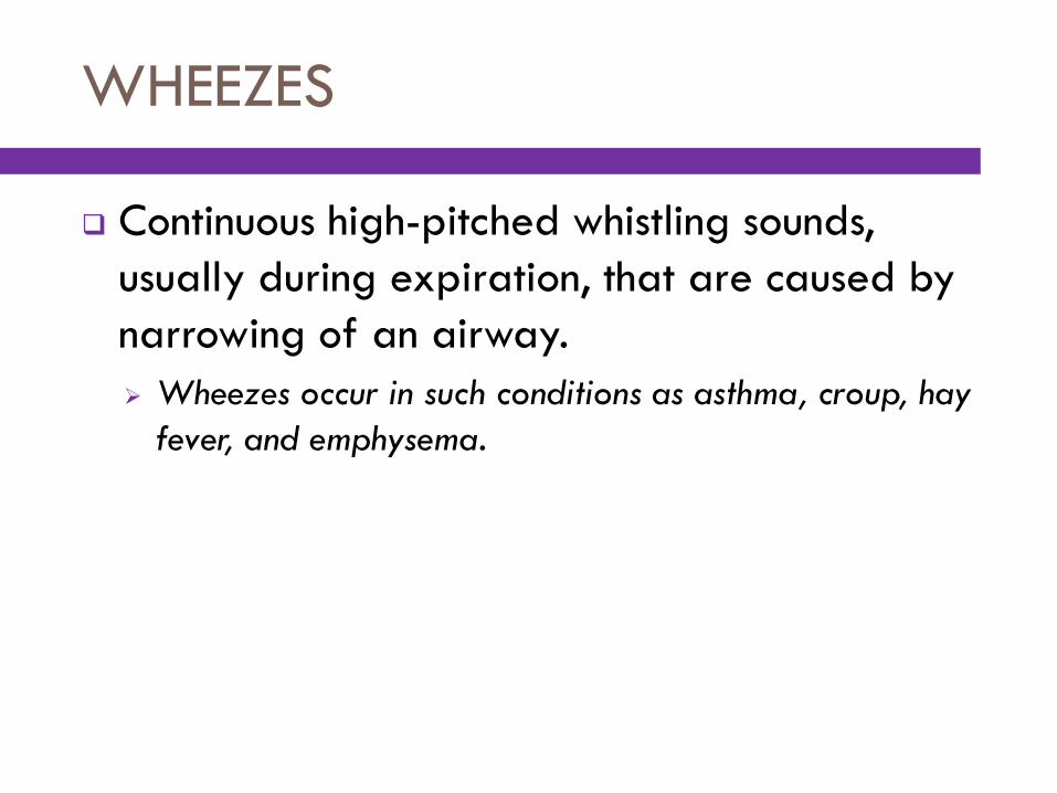
WHEEZES
Continuous high-pitched whistling sounds,
usually during expiration, that are caused by
narrowing of an airway.
Wheezes occur in such conditions as asthma, croup, hay
fever, and emphysema.

ACIDOSIS
Excessive acidity of blood due to an
accumulation of acids or an excessive loss of
bicarbonate.
Respiratory acidosis is caused by unusually high levels of
carbon dioxide (CO2) in the body.

ACUTE RESPIRATORY DISTRESS SYNDROME (ARDS)
Respiratory insufficiency marked by
progressive hypoxia.
ARDS is due to severe inflammatory damage that causes
abnormal permeability of the alveolar-capillary
membrane. As a result, the alveoli fill with fluid, which
interferes with gas exchange.

ANOXIA
Total absence of oxygen in body tissues.
Anoxia is caused by a lack of O2 in inhaled air or by
obstruction that prevents O2 from reaching the lungs.

ATELECTASIS
Collapse of lung tissue, preventing respiratory
exchange of oxygen (O2) and carbon dioxide
(CO2)
Atelectasis can be caused by obstruction of foreign
bodies, excessive secretions, or pressure on the lung from
a tumor. In fetal atelectasis, the lungs fail to expand
normally at birth.

CONSOLIDATION
Process of becoming solid, especially in
connection with the lungs.
Solidification of the lungs is caused by a pathological
engorgement of lung tissues that occurs in acute
pneumonia.

CORYZA
Acute inflammation of nasal passages
accompanied by profuse nasal discharge; also
called a cold.

CROUP
Acute respiratory syndrome that occurs
primarily in children and infants and is
characterized by laryngeal obstruction and
spasm, barking cough, and stridor.

CYSTIC FIBROSIS (CF)
Genetic disease of exocrine glands
characterized by excessive secretions of thick
mucus that do not drain normally, causing
obstruction of passageways (including
pancreatic and bile ducts and bronchi).
CF leads to chronic airway obstruction, recurrent
respiratory infection, bronchiectasis and, eventually,
respiratory failure.

EMPYEMA
Pus in a body cavity, especially in the pleural
cavity (pyothorax).
Empyema is usually the result of a primary infection in the
lungs.

EPIGLOTTITIS
In acute form, a severe, life-threatening
infection of the epiglottis and surrounding area
that occurs most commonly in children between
ages 2 and 12.
In the classic form, epiglottitis involves a sudden onset of
fever, dysphagia, inspiratory stridor, and severe
respiratory distress that commonly requires intubation or
tracheotomy to open the obstructed airway.

EPISTAXIS
Hemorrhage from the nose; also called
nosebleed.

HYPOXEMIA
Deficiency of oxygen in the blood, usually a
sign of respiratory impairment.

HYPOXIA
Deficiency of oxygen in body tissues, usually a
sign of respiratory impairment.
In hypoxia, body tissues have a decreased amount of
oxygen, which results in cyanosis.

INFLUENZA
Acute, contagious respiratory infection
characterized by sudden onset of fever, chills,
headache, and muscle pain.

LUNG CANCER
Pulmonary malignancy commonly attributable
to cigarette smoking.
Lung cancer comprises various malignant neoplasms that
may appear in the trachea, bronchi, or air sacs of the
lungs. Survival rates are low in lung cancer, due to rapid
metastasis and late detection.

PERTUSSIS
Acute infectious disease characterized by a
“whoop”-sounding cough; also called whooping
cough.
Immunization of infants as part of the diphtheria,
pertussis, tetanus (DPT) vaccine prevents the spread of
pertussis.

PLEURAL EFFUSION
Abnormal presence of fluid in the pleural cavity.
The fluid may contain blood (hemothorax), serum
(hydrothorax), or pus (pyothorax). Treatment includes a
surgical puncture of the chest using a hollow-bore needle
(thoracentesis, thoracocentesis) to remove excess fluid.
PNEUMOTHORAX
Collection of air in the pleural cavity, causing
the complete or partial collapse of a lung.
Pneumothorax can occur with pulmonary disease
(emphysema, lung cancer, or tuberculosis) when
pulmonary lesions rupture near the pleural surface,
allowing communication between an alveolus or bronchus
and the pleural cavity. It may also be the result of an
open chest wound or a perforation of the chest wall that
permits entrance of air.

SUDDEN INFANT DEATH SYNDROME (SIDS)
Completely unexpected and unexplained
death of an apparently well, or virtually well,
infant; also called crib death.
SIDS is the most common cause of death between the
second week and first year of life.

ARTERIAL BLOOD GAS (ABG)
Measurement of oxygen (O2) and carbon
dioxide (CO2) content of arterial blood by
various methods.
ABG analysis is used to assess adequacy of ventilation
and oxygenation and the acid-base status of the body.

BRONCHOSCOPY
Visual examination of the interior bronchi using a bronchoscope, a flexible fiberoptic instrument with a light, which can be inserted through the nose or mouth.
Bronchoscopy may be performed to remove obstructions, obtain a biopsy specimen, or observe directly for pathological changes.

CHEST X-RAY
Radiograph of the chest taken from the
anteroposterior (AP), posteroanterior (PA), or
lateral projections.
Chest x-ray is used to diagnose atelectasis, tumors,
pneumonia, emphysema, and many other lung diseases.

COMPUTED TOMOGRAPHY (CT)
Radiographic technique that uses a narrow
beam of x-rays that rotates in a full arc
around the patient to acquire multiple views of
the body that a computer interprets to produce
cross-sectional images of that body part.
CT scanning is used to detect lesions in the lungs and
thorax, blood clots, and pulmonary embolism (PE). CT
scan may be performed with or without a contrast
medium.

MAGNETIC RESONANCE IMAGING (MRI)
Radiographic technique that uses
electromagnetic energy to produce multiplanar
cross-sectional images of the body.
In the respiratory system, MRI is used to produce a scan
of the chest and lungs. MRI does not require a contrast
medium, but it may be used to enhance visualization of
internal structures.

PULMONARY FUNCTION TESTS (PFTs)
Variety of tests to determine the capacity of
the lungs to exchange oxygen (O2) and carbon
dioxide (CO2) efficiently.
Respiratory function is assessed by measuring the
capacity of the lungs and the volume of air during
inhalation and exhalation.

FORCED VITAL CAPACITY (FVC)
Measurement of the amount of air that can be
forcefully exhaled from the lungs after the
deepest inhalation.

FORCED EXPIRATORY VOLUME IN ONE SECOND (FEV1)
Measurement of the volume of air that can be
forcefully exhaled during the first second of
measuring the FVC.
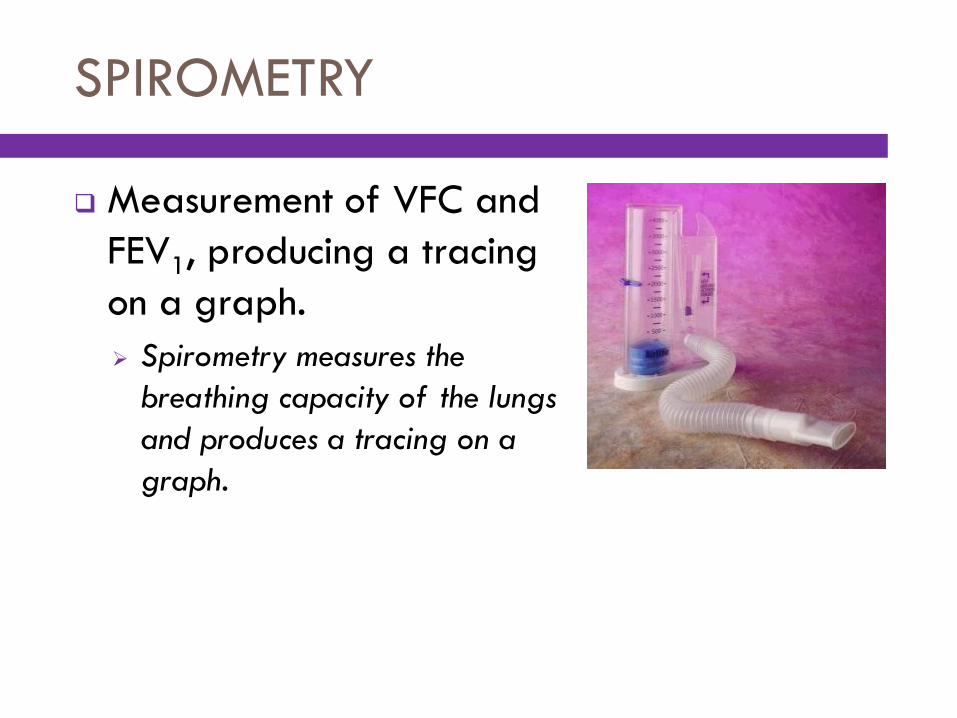
SPIROMETRY
Measurement of VFC and
FEV1, producing a tracing
on a graph.
Spirometry measures the
breathing capacity of the lungs
and produces a tracing on a
graph.

POSTURAL DRAINAGE
Use of body positioning to assist in removal of
secretions from specific lobes of the lung,
bronchi, or lung cavities.
BRONCHODILATORS
Drugs used to increase airflow by dilating
constricted airways through relaxation of the
smooth muscles that surround the bronchioles
and bronchi.
Bronchodilators are used to treat asthma, emphysema,
chronic obstructive pulmonary disease (COPD), and
exercise-induced bronchospasm. Most bronchodilators
provide metered doses of the medication and may
employ a spacer as a reservoir for the medication.

CORTICOSTEROIDS
Hormonal agents that reduce tissue edema and
inflammation associated with chronic lung
disease.

NEBULIZED MIST TREATMENT
Therapy that uses a device
to produce a fine spray
(nebulizer) that delivers
medication directly into the
lungs.