Unit 4: Cell Theory - Doc O's Wacky World at Bishop · PDF fileUnit 4: Cell Theory Discovery...
13
Go on to the next page. Unit 4: Cell Theory Discovery of cells Cells are far too small to see with the naked eye Invention of the microscope 1590 – Zacharias and Hans Janssen made one of the first compound microscopes 1660 – Robert Hooke made a compound microscope Hooke looked at cork and noticed room like structures he called ‘cells’ He calculated that a square inch of cork contained about 1 200 000 000 cells Drawing by Rita Greer - The original is a pencil drawing by Rita Greer, history painter, 2006. This was digitized by Rita and sent via email to the Department of Engineering Science, Oxford University, where it was subsequently uploaded to Wikimedia., FAL, https://commons.wikimedia.org/w/index.php?curid=7667256 1660 – Anton van Leeuwenhoek made a simple microscope (only one lens) that could magnify a specimen 266 times He viewed many samples of water from lakes, gums and gutters finding many small organisms that moved in different ways and named these organisms animalcules (little animals) Microscope picture: see page for author [CC BY 4.0 (http://creativecommons.org/licenses/by/4.0)], via Wikimedia Commons Development of cell theory 1838 – Matthias Schleiden Concluded all plants are made of cells based on his own research 1839 – Theodor Schwann Concluded all animals are made of cells, therefore all living things are made up of cells 1855 – Rudolf Virchow Proposed that new cells are formed only from already existing cells Cell theory All living things are composed of cells Cells are the basic units of structure and functions in living things All cells are produced from other cells Cells are usually very small It is very tricky to count the number of cells in the human body Not all of us are the same size We contain microbes The number changes as we recycle cells Best current estimate is 37.2 trillion cells in the human body
Transcript of Unit 4: Cell Theory - Doc O's Wacky World at Bishop · PDF fileUnit 4: Cell Theory Discovery...

Go on to the next page.
Unit 4: Cell Theory
Discovery of cells Cells are far too small to see with the naked eye
Invention of the microscope 1590 – Zacharias and Hans Janssen made one of the
first compound microscopes
1660 – Robert Hooke made a compound microscope Hooke looked at cork and noticed room like
structures he called ‘cells’ He calculated that a square inch of cork contained
about 1 200 000 000 cells Drawing by Rita Greer - The original is a pencil drawing by Rita Greer, history painter, 2006. This was digitized by Rita and sent via email to the Department of Engineering Science, Oxford University, where it was subsequently uploaded to Wikimedia., FAL, https://commons.wikimedia.org/w/index.php?curid=7667256
1660 – Anton van Leeuwenhoek made a simple microscope (only one lens) that could magnify a specimen 266 times
He viewed many samples of water from lakes, gums and gutters finding many small organisms that moved in different ways and named these organisms animalcules (little animals)
Microscope picture: see page for author [CC BY 4.0 (http://creativecommons.org/licenses/by/4.0)], via Wikimedia Commons
Development of cell theory 1838 – Matthias Schleiden
Concluded all plants are made of cells based on his own research 1839 – Theodor Schwann
Concluded all animals are made of cells, therefore all living things are made up of cells 1855 – Rudolf Virchow
Proposed that new cells are formed only from already existing cells
Cell theory
All living things are composed of cells
Cells are the basic units of structure and functions in living things
All cells are produced from other cells
Cells are usually very small It is very tricky to count the number of cells in the human body
Not all of us are the same size We contain microbes The number changes as we recycle cells
Best current estimate is 37.2 trillion cells in the human body

Go on to the next page.
Why are the cells so small? Very small cells are easier to replace without
disruption to the organism Very small cells can specialize which also makes
replacement much easier Mathematics
Surface to volume ratio Compare two cubic cells
Cell 1 – 10 μm (micrometers) on a side Surface area = 10 μm x 10 μm x 6 = 600 μm2
Volume= 10 μm x 10 μm x 10 μm = 1000 μm3 Surface area / Volume = 600 / 1000 = 0.6
Cell 2 – 20 μm on a side Surface area = 20 μm x 20 μm x 6 = 2400 μm2
Volume = 20 μm x 20 μm x 20 μm = 8000 μm3 Surface area / Volume = 2400 / 8000 = 0.3
By BallenaBlanca - https://commons.wikimedia.org/wiki/File:Esquema_del_epitelio_del_intestino_delgado.png, CC BY-SA 4.0, https://commons.wikimedia.org/w/index.php?curid=48093230
Notice that Cell 1 has twice the surface area to volume ratio and therefor can transport needed materials in the cell and wastes out of the cell twice as fast
Speed of diffusion – this limits cell size because large cells cannot transport things from one part of the cell to another part fast enough to survive
Limiting surface area / volume ratios Some cells that specialize in exchanging materials have many, tiny, finger-like extensions called
microvilli Microvilli (see picture inset of small intestine villi) greatly increase surface area without adding
much to the volume of a cell
Comparing eubacteria and archaebacteria Not too long ago, archaea were classified as bacteria (hence the name archaebacterial) but now we
know that they have a distinct evolutionary history and a much different biochemistry than bacteria
Similarities Both are prokaryotes (mostly single celled but never with a nucleus or membraned organelles) Both have cell walls Both have ribosomes Both have similar shapes – rods, cocci (spherical), and spirals Both have flagella to move Both reproduce asexually through binary fission, budding, and fragmentation
Differences Eubacteria
Cell membrane is ester linked lipid (peptidoglycan)
Flagella are like a hollow stalk subunits move up the central pore and add on at the tip
Can form spores that are dormant for years Are found almost everywhere in normal
environments
Archaebacteria Cell membrane is ether linked lipid (no
peptidoglycan) Flagella add on at the base Do not form spores Are found in harsh conditions like hot vents,
salty areas, and very acidic environments
Go on to the next page.
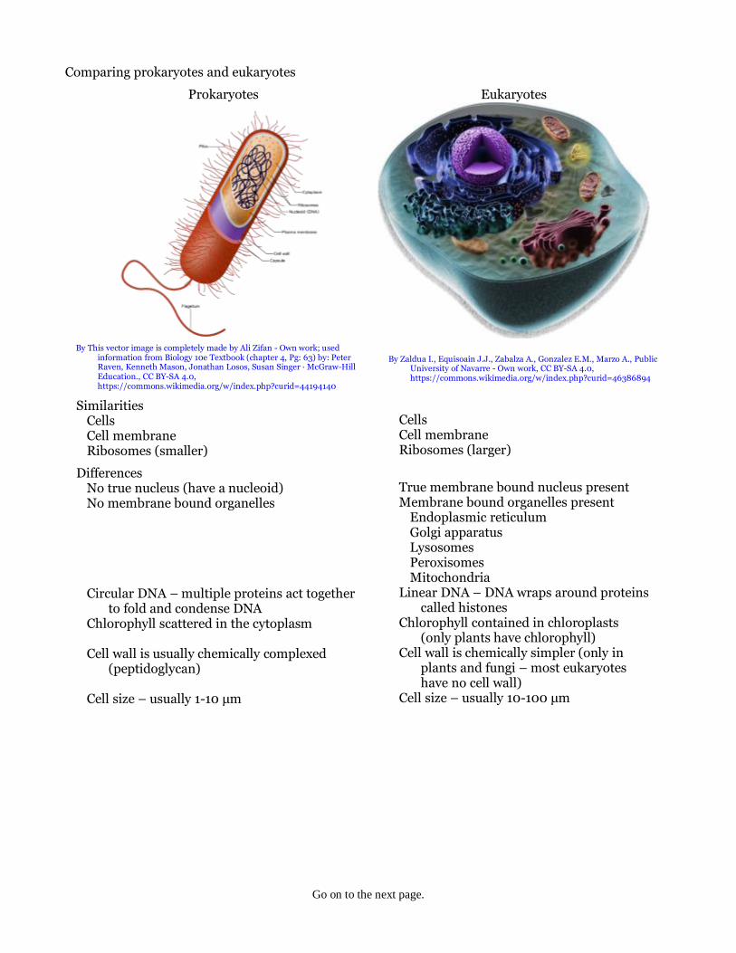
Comparing prokaryotes and eukaryotes
Prokaryotes
By This vector image is completely made by Ali Zifan - Own work; used
information from Biology 10e Textbook (chapter 4, Pg: 63) by: Peter Raven, Kenneth Mason, Jonathan Losos, Susan Singer · McGraw-Hill Education., CC BY-SA 4.0, https://commons.wikimedia.org/w/index.php?curid=44194140
Similarities Cells Cell membrane Ribosomes (smaller)
Differences No true nucleus (have a nucleoid) No membrane bound organelles Circular DNA – multiple proteins act together
to fold and condense DNA Chlorophyll scattered in the cytoplasm Cell wall is usually chemically complexed
(peptidoglycan) Cell size – usually 1-10 μm
Eukaryotes
By Zaldua I., Equisoain J.J., Zabalza A., Gonzalez E.M., Marzo A., Public University of Navarre - Own work, CC BY-SA 4.0, https://commons.wikimedia.org/w/index.php?curid=46386894
Cells Cell membrane Ribosomes (larger)
True membrane bound nucleus present Membrane bound organelles present
Endoplasmic reticulum Golgi apparatus Lysosomes Peroxisomes Mitochondria
Linear DNA – DNA wraps around proteins called histones
Chlorophyll contained in chloroplasts (only plants have chlorophyll)
Cell wall is chemically simpler (only in plants and fungi – most eukaryotes have no cell wall)
Cell size – usually 10-100 μm

Go on to the next page.
Parts of a eukaryotic cell Ribosomes – make proteins Endoplasmic reticulum
Rough – associated with ribosomes Smooth – makes lipids
Nucleus Nucleolus – condensed region where
ribosomes are formed Chromatin – DNA plus associated proteins Nuclear envelope – membrane around the
nucleus that has pores that allow materials to move in and out
Golgi body (or apparatus) – modifies proteins Centriole – important part of centrosomes
which are involved in organizing microtubules in the cytoplasm
Lysosomes – digest food Peroxisomes – metabolize wastes (not shown) Mitochondria – produce energy
By Mediran (Own work) [CC BY-SA 3.0 (http://creativecommons.org/licenses/by-sa/3.0)], via Wikimedia Commons
Comparing plant and animal cells
Animal cells Plant cells
Animal cells have centrioles, centrosomes, and lysosomes that plant cells do not have
Plant cells have cell walls, chloroplasts, plasmodesmata, plastids, and a large central vacuole that animal cells do not have
Source: Boundless. “Characteristics of Eukaryotic Cells.” Boundless Biology. Boundless, 26 May. 2016. Retrieved 26 Nov. 2016 from https://www.boundless.com/biology/textbooks/boundless-biology-textbook/cell-structure-4/eukaryotic-cells-60/characteristics-of-eukaryotic-cells-313-11446/

Go on to the next page.
Multicellular organisms
Advantages of multicellular organisms Longer lifespan An overall larger body size (usually fewer predators and better ability to maintain homeostasis) Cell differentiation (allows more structures, functions, and complexity)
Levels of organization in multicellular organisms Organelles – “little organs” typically, membrane bound structures or compartments in cells that
perform a specific function Examples: nucleus carries instructions for cells, peroxisomes metabolize wastes, and
mitochondria provide energy Cell – the basic structural, functional, and biological unit of living organisms
Often called the building blocks of life Examples: neurons (nerve cells), erythrocytes (red blood cells), epidermal cells (skin cells)
Tissue – groups of cells with a similar structure that work together for a specific function The four types of human tissues are: epithelial, connective, muscular, and nervous
Organ – collection of tissues joined in a structural unit to server a common function Examples: skeleton, muscles, teeth, stomach, intestines, liver, kidneys, lungs, brain, veins,
arteries, spleen, pancreas, spinal nerves, eye, ear, and skin System – groups of structures that perform the broadest functions in an animal
The eleven main types of human systems are: cardiovascular / circulatory, digestive / excretory, endocrine, integumentary / exocrine, lymphatic / immune, muscular / skeletal, nervous, reproductive, renal / urinary, respiratory, and vestibular
Structure vs Function Structure describes what something looks like or its makeup Function describes what a structure does or the job it performs
Example: mitochondrion Structure: a roughly ovoid organelle encased by an outer membrane and having an inner
membrane with many folds called cristae that contains its own DNA Function: energy production
Transport in cells
Diffusion
By JrPol - Own work, CC BY 3.0, https://commons.wikimedia.org/w/index.php?curid=4586487
Diffusion – the random mixing of substances due to the natural movement of particles
This process will always carry substances from areas of higher concentration to areas of lower concentration
Osmosis
By OpenStax - https://cnx.org/contents/[email protected]:fEI3C8Ot@10/Preface, CC BY 4.0, https://commons.wikimedia.org/w/index.php?curid=30131189
Osmosis – the process by which solvent molecules tend to pass through a semipermeable membrane from a less concentrated solution to a more concentrated solution
In cells, this process will always carry solvent (water) from areas of higher concentration to areas of lower concentration across the plasma (or cell) membrane

Go on to the next page.
Conditions for osmosis Osmosis is, basically, diffusion across a semipermeable membrane and requires:
A semipermeable membrane A concentration gradient
Cell walls are phospholipid bilayers that form a liposome
Water (and any charged particles that dissolve in water) cannot pass through the plasma membrane
except through pores (or holes in the membrane
Passive transport The movement of substances across cell membranes by osmosis
Passive transport always moves ions from higher concentrations to lower concentrations (with or down the concentration gradient)
Requires no energy input from the cell Typically, passive transport occurs through ion channels which are
composed of four proteins that form a pore (or hole) through the plasma (or cell) membrane
Ion channels are usually very fast (often a million ions per second or more)
Active transport Forcing the movement of substances across cell membranes against osmosis
Active transport always moves ions from lower concentrations to higher concentrations or up the concentration gradient
Typically, active transport occurs through ion transporters (or ion pumps like the Na+/K+ pump)
Ion pumps require cellular energy from some source (often ATP)
Example: the Na+/K+ pump Moves 3 Na+ ions out of the cell and
2 K+ ions into the cell In a typical cell, 1/5 the metabolic
energy is required for ion pumps
In a neuron, up to 2/3 the metabolic energy is required for ion pumps
By BruceBlaus. Blausen.com staff. "Blausen gallery 2014". Wikiversity Journal of Medicine.
DOI:10.15347/wjm/2014.010. ISSN 20018762. Derivative by Mikael Häggström - File:Blausen_0211_CellMembra
Schematic diagram of an ion channel. 1 - channel
domains (typically four per channel), 2 - outer
vestibule, 3 - selectivity filter, 4 - diameter of
selectivity filter, 5 - phosphorylation site, 6 - cell
membrane.

Go on to the next page.
Similarities and Differences between Active and Passive Transport
Similarities
Both involve movement of ions across a plasma (or cell) membrane Both require a pore to move ions through the plasma membrane Both are required to maintain proper cell functioning
Differences Passive Active
Moves from high to low concentration Moves from low to high concentration Requires no metabolic energy Requires metabolic energy from ATP Makes use of osmosis through ion channels Uses Na+/K+ pumps (or some ion pump)
Moving large particles in and out of cells Endocytosis – a form of active transport in which cells transport large molecules into the cell by
engulfing them The cell walls expand outward (or inward) and engulf the large particles The substances that enter the cell will be surrounded by membranes (vesicles or vacuoles) There are three types of endocytosis
Phagocytosis transports solid particles Pinocytosis transports particles in liquids Receptor-mediated endocytosis transports particles with specific sites that can bind to a
receptor in the cell membrane The vacuoles that form in this manner will be protein coated vacuoles
Exocytosis – a form of active transport in which cells transport large molecules out of the cell
Proteins that are to be transported out of the cell are surrounded by membranes and are called secretory vesicles
The membrane of the secretory vesicle fuses with the cell membrane then opens to the outside of the cell to push the protein out

Go on to the next page.
Basal Metabolic Rate (BMR) – the minimal amount of energy the body requires to function at rest BMR is expressed in food calories (kcal) Some resting body processes include breathing, blood circulation, maintaining body temperature,
brain function, nerve function, and muscle contraction (especially the heart), and cell metabolism Cell metabolism refers to all the processes a cell requires for functioning
Catabolism – processes which break larger molecules into smaller ones generally for body use to release energy
Anabolism – processes which assemble smaller molecules into larger ones whereby energy is stored in chemical bonds for later use
The BMR is most accurately determined by experimental measurement for an individual Usually, BMR is estimated by use of a mathematical formula
The Mifflin St Jeor Equation:
BMR = (10.0 𝑚
1 kg+
6.25 ℎ
1 𝑐𝑚−
5.0 𝑎
1 𝑦𝑟+ 𝑠)
𝑘𝑐𝑎𝑙
𝑑𝑎𝑦
Where m = mass in kg = wt lbs x 2.2
h = height in cm = h in x 2.54 a = age in years s = +5 for males and –161 for females
Photosynthesis Photo means “light” and synthesis means “putting together”
During photosynthesis, carbon dioxide (CO2) and water (H2O) are put together by plants using light energy from the sun to form sugars
The chemical that allows plants to do this is called chlorophyll In plant cells, chlorophyll is found in the chloroplasts
Plants break down some of the sugars they make into smaller molecules in order to release the energy they need for their cells to function
Some of the sugars are used to build cellulose Some of the sugars are stored for later use
Organisms that eat plants are using these stored sugars as food Nearly all living things obtain energy either directly or indirectly from the energy of sunlight captured
during photosynthesis
Chemical equations Scientists use chemical symbols and chemical equations as a kind of a shortcut to represent
processes such as photosynthesis Substances that are used in the reaction are called the reactants
Reactants are listed on the left of the equation Substances that are produced by the reaction are called the products
Products are listed on the right side of the equation
The photosynthesis equation:
Reactants Products
6 (CO2) + 6 (H2O) + sunlight → C6H12O6 + 6 O2 carbon dioxide + water + sunlight → glucose + oxygen
where the reactants are carbon dioxide and water and the products are glucose (a sugar) and oxygen as a waste product for the plant
The arrow in the equation is a yields sign and can be read as ‘yields’ or as ‘reacts to produce’ The entire equation would be read as:
Carbon dioxide plus water react to produce glucose plus oxygen.

Go on to the next page.
Steps of photosynthesis 1. Stage 1 – Capturing the sun's energy
Chlorophyll in the chloroplasts captures sunlight in two systems Photosystem I (PSI)
Sunlight causes chlorophyll to lose an electron (e–) The electron moves down the chloroplast electron transport chain This happens twice producing 2 e–
Photosystem II (PSII) Sunlight causes the splitting of water producing 2 e–, 2 H+ ions, and ½ O2 molecule These 2 e– replace the e– lost by chlorophyll in PSI
2. Stage 2 – Using energy to make food
Calvin cycle CO2 enters the stoma on the underside of leaves and H2O enters from the roots A complex set of reactions uses energy and the electrons from Stage 1, the H2O, and the CO2 to
produce the sugar glucose (C6H12O6) Plants throw out waste O2 formed in PSII through the stoma and use the glucose as food
Cellular respiration Although cellular respiration is often referred to as respiration it should not be confused with
breathing (which is also called respiration) Respiration is the process by which cells obtain energy from glucose
During respiration cells break down simple food molecules such as sugars to release the energy they contain
Respiration occurs in two stages Stage 1 occurs in the cytoplasm of the cells
Glucose is broken down into smaller molecules No oxygen is involved Only a small amount of energy is released
Stage 2 occurs in the mitochondria The small molecules from Stage 1 are broken down into even smaller molecules The chemical reactions in the mitochondria require oxygen A large amount of energy is released (explaining why mitochondria are called powerhouses) Carbon dioxide (CO2) and water (H2O) are also released in respiration
When humans breathe out they release CO2 and H2O
The respiration equation:
C6H12O6 + 6 O2 → 6 (CO2) + 6 (H2O) + energy glucose + oxygen → carbon dioxide + water + energy

Go on to the next page.
Note that the respiration equation is the reverse of the photosynthesis reaction Because these two reactions are opposite, they form a cycle that helps keep the CO2 and O2 levels
nearly constant on Earth
Fermentation Some single-celled organisms live deep in the ocean, in mud, or in other places where there is no
oxygen Such organisms use fermentation to obtain energy instead of respiration Fermentation – a process that produces energy for cells without using oxygen
The amount of energy released during fermentation is much less than during respiration
Alcoholic fermentation Yeast is an example of an organism that uses alcoholic fermentation
Bakers and brewers use alcoholic fermentation because this method also produces CO2 The CO2 bubbles cause bread to rise and form the bubbles in beer
Lactic acid fermentation When cells in the human body use oxygen faster than it can be replaced, the cells can generate
some energy by a fermentation process that also produces lactic acid Lactic acid causes the painful sensation that results in muscles that feel sore and weak
Similarities and Differences between Respiration and Fermentation
Stages of Respiration and Fermentation Respiration Fermentation
Stage 1: in the cytoplasm Stage 1: in the cytoplasm Glycolysis produces 2 ATP molecules and
2 pyruvate molecules Glycolysis produces 2 ATP molecules and
2 pyruvate molecules Stage 2: in the mitochondria Stage 2: no oxygen, can’t use mitochondria
Krebs cycle makes CO2 and 2 ATP Pyruvate converted to lactic acid Electron transport chain – about 34 ATP Repeat from Stage 1
Total: 38 ATP Total: 2 ATP
Similarities
Both methods produce energy for cells Both produce energy by creating adenosine triphosphate (ATP) Both use glycolysis in the cytoplasm
Differences Respiration Fermentation
Starts in cytoplasm moves to mitochondria Process never moves out of cytoplasm Requires oxygen Proceeds without use of oxygen Produces much more energy (38 ATP) Produces much less energy (2 ATP)
Life cycle of a cell Why cells reproduce
For single celled organisms, reproduction carries on the species For multicellular organisms:
Growth – increase the number of cells (growth of the organism) Maintenance – replace cells that grow old and die Repair – replace damaged cells
Stage 1: Interphase – the period before cell division Growth – cell reaches full size and copies of chloroplasts (plants) and mitochondria are made Replication – DNA is copied so that the cell has two identical sets of DNA Preparation for cell division – structures are made that are used for division

Go on to the next page.
Stage 2: Mitosis – the period when the cell nucleus divides into two new nuclei Prophase
The threadlike chromatin in the nucleus condenses to form chromosomes Chromosomes are double-rod structures
Chromatid – one single rod in a chromosome The two chromatids are identical because the cell DNA has replicated The chromatids are held together by a structure called a centromere
The pairs of centrioles move to opposite ends of the cell Spindle fibers form a bridge between the opposite ends of the cell The nuclear envelope breaks down
Metaphase Chromosomes line up across the center of the cell
This prepares the chromosomes so they can split with one daughter chromatid to each end Each chromosome attaches to a spindle fiber at its centromere
Anaphase The centromeres split One chromatid is drawn by its spindle fiber to one end of the cell and the other chromatid moves
to the opposite end of the cell drawn by its spindle fiber The cell stretches out as the opposite ends are pushed apart
Telophase The chromosomes unwind, stretch out, and lose their rod-like appearance A new nuclear envelope forms around each region of chromosomes
Stage 3: Cytokinesis – the period when the cell completes the process of division Cytokinesis starts at about the same time as the telophase

Go on to the next page.
Cytokinesis specifically for animal and plant cells: Animal cells
The cell membrane pinches in around the middle of the cell The cell splits in two Each daughter cell ends up with an identical set of chromosomes and about half the organelles
Plant cells The cell wall cannot pinch together in the middle of the cell Instead, the cell makes a structure called a cell plate which eventually forms into new cell
membranes that separate the two daughter cells A new cell wall forms around the two cell membranes
Time required for the cell cycle to occur in human liver cells (time measured in hours)
Binary fission in prokaryotic cells 1. The DNA replicates 2. Each copy of the DNA attaches to the cell membrane at opposite ends of the cell 3. The cell splits and each end pulls its copy of the DNA into its part of the new cell
Similarities in mitosis and binary fission:
Both are asexual forms of cell reproduction Both replicate DNA into two exact copies Both split cells into two exact copy daughter cells
9
10
2
0.83 0.17
Length of the Cell Cycle
Interphase Growth
Interphase DNA Replication
Interphase Division Prep
Mitosis
Cytokinesis

This is the last page.
Differences in mitosis and binary fission Mitosis Binary Fission
Occurs in eukaryotic cells Occurs in prokaryotic cells Many chromosomes are involved Only one single strand of DNA is involved Proceeds in five complex steps Proceeds in three simple steps