Unit 3 Cell Biologyswcontent.spokaneschools.org/cms/lib/WA01000970...138 UNIT 3 CELL BIOLOGY cell...
60
Cell Biology Chapter 7 Cell Structure and Function Chapter 8 Cell Processes Chapter 9 The Microscopic World Take a magnifying lens home and examine a leaf. First look at the leaf with your normal vision. Sketch a picture of the leaf and identify its structures. Next examine the surface of the leaf with a magnifying glass. Sketch what you see. Predict what you would see if you looked at the leaf through a powerful microscope. Make a sketch of your prediction.
Transcript of Unit 3 Cell Biologyswcontent.spokaneschools.org/cms/lib/WA01000970...138 UNIT 3 CELL BIOLOGY cell...

T H I S A T H O M E
Cell Biology
Chapter 7 Cell Structure and Function
Chapter 8 Cell Processes
Chapter 9 The Microscopic World
Take a magnifying lens home and examine a leaf.
First look at the leaf with your normal vision.
Sketch a picture of the leaf and identify its
structures. Next examine the surface of the leaf
with a magnifying glass.
Sketch what you see. Predict what you would
see if you looked at the leaf through a
powerful microscope. Make a sketch
of your prediction.

Chapter 7
Cell Structure and Function
Can you name something that you know exists even though you can’t
see it with your eyes? A drop of pond water has tiny swimming
organisms and small bits of plant material, but we can’t always see
them with our eyes. How do we know there are tiny things in a drop
of pond water? We can use a microscope to view the pond water.
There are instruments people use every day to help them see things
they wouldn’t usually be able to see. Have you ever used a pair of
binoculars or a magnifying glass? Have you ever had an X-ray taken
of an injury? Do you need to wear glasses or contact lenses to see
clearly? Vision systems are even being developed to restore vision to
blind people. In this chapter, you will take a journey into a small
world that was discovered when the microscope was invented—the
world of the cell. Imagine you could shrink yourself and walk into a
tiny cell. What is it like inside a cell? It’s a fascinating journey!
1. What is a cell and how do we know cells
exist?
2. Are human cells, animal cells, and plant
cells all the same?
3. What is inside a cell, and how is a cell like
a cookie factory?
136 UNIT 3 CELL BIOLOGY
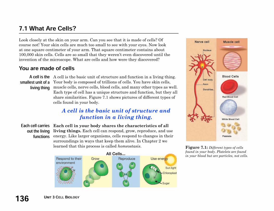
Figure 7.1: Different types of cells
found in your body. Platelets are found
in your blood but are particles, not cells.
7.1 What Are Cells?Look closely at the skin on your arm. Can you see that it is made of cells? Of
course not! Your skin cells are much too small to see with your eyes. Now look
at one square centimeter of your arm. That square centimeter contains about
100,000 skin cells. Cells are so small that they weren’t even discovered until the
invention of the microscope. What are cells and how were they discovered?
You are made of cellsA cell is thesmallest unit of aliving thing
A cell is the basic unit of structure and function in a living thing.
Your body is composed of trillions of cells. You have skin cells,
muscle cells, nerve cells, blood cells, and many other types as well.
Each type of cell has a unique structure and function, but they all
share similarities. Figure 7.1 shows pictures of different types of
cells found in your body.
A cell is the basic unit of structure and
function in a living thing.
Each cell carriesout the livingfunctionsEach cell in your body shares the characteristics of all
living things. Each cell can respond, grow, reproduce, and use
energy. Like larger organisms, cells respond to changes in their
surroundings in ways that keep them alive. In Chapter 2 we
learned that this process is called homeostasis.

1377.1 WHAT ARE CELLS?
CHAPTER 7: CELL STRUCTURE AND FUNCTION
Finding out about cells
Robert Hooke
discovered cells
How did we learn about cells? It all started with the invention of
the microscope in the late 1500s. English scientist Robert Hooke
(1635–1703) was the first to record his observations of cells. In
1663, he took a thin slice of cork and placed it under a microscope
that he built. Cork is made from the bark of the cork oak tree, but
its cells are no longer alive. Hooke made detailed sketches of his
observations. An artist’s version of one of his sketches is shown in
Figure 7.2. Hooke called each of the square structures a cell
because they reminded him of tiny rooms.
Some organisms
are made of a
single cell
Anton van Leeuwenhoek (1632–1723) was not a scientist. He was a
Dutch craftsman who made lenses. Yet with skill and curiosity,
Leeuwenhoek made some of the most important discoveries in
biology. He used his lenses to build a simple microscope. With his
microscope, he looked at pond water, blood, and scrapings from his
teeth. He was the first to observe single-celled protists, blood cells,
and bacteria.
All living things
are made
from cells
As microscopes improved, scientists made more discoveries. In
1839, two German scientists, Matthais Schleiden and Theodore
Schwann, viewed plant and animal tissues under a microscope.
They concluded that all plants and animals were made up of cells.
Fluorescence
microscopy
Cells usually do not glow. Scientists use fluorescent proteins to
make cells glow. The cells absorb these proteins like stains. The
fluorescence microscope uses filters that only let in light that
matches the fluorescing material being studied. All other types of
light are blocked out. The fluorescing areas shine out against a
dark background, making certain cell structures glow. The mouse
egg cells in Figure 7.3 have been treated to show DNA as a glowing
blue.
Figure 7.2: Robert Hooke’s sketch of
cork cells looked like this.
Figure 7.3: Mouse egg cells. The
DNA is the glowing blue.

138 UNIT 3 CELL BIOLOGY
cell theory - a theory that
explains the relationship between
cells and living things.
The cell theory
Cells only come
from other cells
Schleiden and Schwann’s theory was widely accepted by other
scientists. But where did cells come from? In the 1800s it was
believed that living things came from nonliving objects. Did cells
come from some tiny, nonliving objects? In 1855, a German
physician named Rudolf Virchow (1821–1902) proposed that cells
can only come from other cells.
Statements of
the cell theory
The work of Hooke, Leeuwenhoek, Schleiden, Schwann, Virchow,
and others led to an important theory in life science. The cell
theory explains the relationship between cells and living things.

1397.1 WHAT ARE CELLS?
CHAPTER 7: CELL STRUCTURE AND FUNCTION
Similarities among cells
There are many
different types
of cells
Some organisms are made of only a single cell. You are made of
billions of cells. In multicellular organisms like you, there are many
different types of specialized cells. For example, the cells that line
the retina of your eye have a structure and function that is very
different from your skin cells. About 200 different types of
specialized cells make up the tissues and organs of your body.
There are different types of cells but all cells
share similar characteristics.
All cells share
some similarities
Even though there are many different types of cells, they all share
similar characteristics (Figure 7.4). These include:
1. All cells are surrounded by a cell membrane. The cell
membrane is a barrier between the inside of the cell and its
environment. It also controls the movement of materials into
and out of the cell.
2. All cells contain organelles. An organelle is a structure
inside of a cell that helps the cell perform its functions.
Although all cells contain organelles, they don’t all contain
the same kinds. You’ll learn more about the organelles in the
next section.
3. All cells contain cytoplasm. The cytoplasm is a fluid
mixture that contains the organelles. It also contains the
compounds cells need to survive such as water, salts, enzymes,
and other carbon compounds.
4. All cells contain DNA. The cell theory states that all cells
come from other cells. When cells reproduce, they make copies
of their DNA and pass it on to the new cells. DNA contains the
instructions for making new cells and controls all cell functions.
Figure 7.4: All cells have a cell
membrane, organelles, cytoplasm, and
DNA.
cell membrane - a separating
barrier that controls movement of
materials into and out of the cell.
organelle - a structure inside of a
cell that helps it perform its
functions.
cytoplasm - a fluid mixture that
contains the organelles and the
compounds the cell needs.

140 UNIT 3 CELL BIOLOGY
Eukaryotic cell
Membrane
bound
nucleus
Cell
membrane
Various
membrane bound
organelles
Cytoplasm
Figure 7.5: Comparing prokaryotic
and eukaryotic cells.
prokaryotic cell - a cell that
does not have a nucleus or
membrane-covered organelles.
eukaryotic cell - a cell that has a
nucleus and membrane-covered
organelles.
Prokaryotic
cells
Eukaryotic
cells
Bacteria All other cells
No nucleus Nucleus
Organelles not
membrane-
covered
Membrane-
covered
organelles
DNA is
bunched up in
the center of the
cell
DNA is found in
the nucleus
Classifying cells
Two types of
cells
Based on the organization of their structures, all living cells can be
classified into two groups: prokaryotic and eukaryotic (Figure 7.5).
Animals, plants, fungi, and protozoans all have eukaryotic cells.
Only bacteria have prokaryotic cells.
Prokaryotic cells Prokaryotic cells do not have a nucleus.
The word prokaryotic means “before
nucleus” in Greek. Scientists believe
that all life on Earth came from these
cells. The oldest fossils of bacteria are
estimated to be 3.5 billion years old.
The DNA in a prokaryotic cell is
bunched up in the center of the cell. The
organelles are not covered with a
membrane. All prokaryotic cells are
much smaller than eukaryotic cells.
Eukaryotic cells Eukaryotic cells have a nucleus and
membrane-covered organelles
(with the exception of the red
blood cells of mammals). The
word eukaryotic means “true
nucleus” in Greek. The oldest
fossils of eukaryotic cells are
about 2 billion years old. There is
more DNA in these types of cells
and it is found in the nucleus.
These cells have membrane-
covered organelles. They tend to
be about ten times larger than
prokaryotic cells.
1417.1 WHAT ARE CELLS?
CHAPTER 7: CELL STRUCTURE AND FUNCTION
7.1 Section Review
1. What is the basic unit of structure and function in a living thing
called?
2. How did the invention of the microscope help scientists learn
more about living things?
3. Who was the first to discover cells?
4. Draw a timeline that shows the dates, discoveries, and
scientists involved in the development of the cell theory.
5. What are the four statements of the cell theory?
6. What are specialized cells? List three examples.
7. What are four similarities that all cells share?
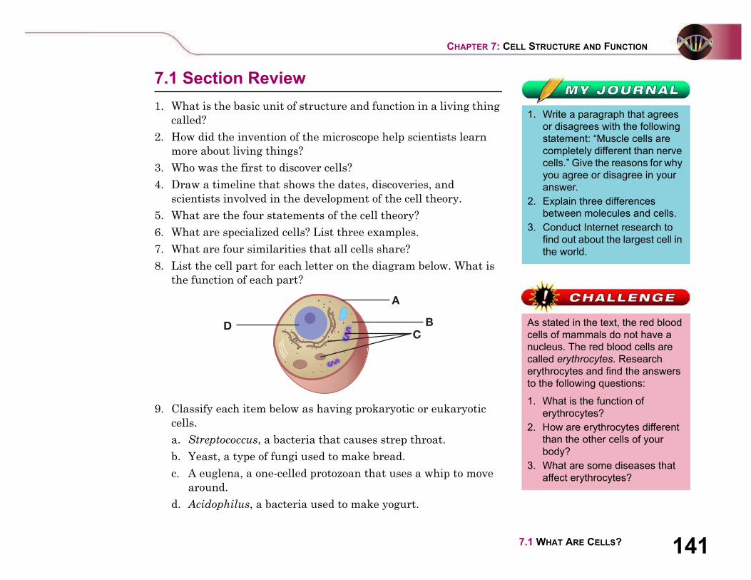
8. List the cell part for each letter on the diagram below. What is
the function of each part?
9. Classify each item below as having prokaryotic or eukaryotic
cells.
a. Streptococcus, a bacteria that causes strep throat.
b. Yeast, a type of fungi used to make bread.
c. A euglena, a one-celled protozoan that uses a whip to move
around.
d. Acidophilus, a bacteria used to make yogurt.
D
A
BC
1. Write a paragraph that agrees
or disagrees with the following
statement: “Muscle cells are
completely different than nerve
cells.” Give the reasons for why
you agree or disagree in your
answer.
2. Explain three differences
between molecules and cells.
3. Conduct Internet research to
find out about the largest cell in
the world.
As stated in the text, the red blood
cells of mammals do not have a
nucleus. The red blood cells are
called erythrocytes. Research
erythrocytes and find the answers
to the following questions:
1. What is the function of
erythrocytes?
2. How are erythrocytes different
than the other cells of your
body?
3. What are some diseases that
affect erythrocytes?

142 UNIT 3 CELL BIOLOGY
Figure 7.6: The parts of a cookie
factory.
An analogy is a comparison of one
thing to another different thing. The
cookie factory is a good analogy
for remembering cell parts and
their functions. After reading this
section, make another analogy
comparing your school to a cell.
7.2 Cells: A Look Inside
Imagine a factory that makes thousands of cookies a day. Ingredients come into the
factory, get mixed and baked, then the cookies are packaged. The factory has many
parts that contribute to the process. Can you name some of those parts and their
functions? A cell is a lot like a cookie factory. It too has many parts that contribute to
its processes. Let’s compare a cell to a cookie factory.
Comparing a cell to a cookie factory
Parts and
functions
A cookie factory has many parts. The cytoplasm of a cell has many
organelles. Figure 7.6 shows a fictional cookie factory. A typical
animal cell and its parts are shown on the next page. Table 7.1
compares a cookie factory to an animal cell. As you read this
section, refer to the table to help you remember the cell parts and
their functions.
Table 7.1: Comparing a cell and a cookie factory
Process Cookie factory part Cell part
Ingredients in/products out Factory gate and doors Cell membrane
Control center Manager’s office Nucleus
Energy Power plant Mitochondria
Storage Storage room Vacuole
Making the product Mixing/baking room Ribosome
Transport of materials Conveyer belts Endoplasmic reticulum
Packaging and distribution Shipping room Golgi body
Clean up and recycling Custodial staff Lysosome
Structure/support Walls and studs Cytoskeleton
1437.2 CELLS: A LOOK INSIDE
CHAPTER 7: CELL STRUCTURE AND FUNCTION
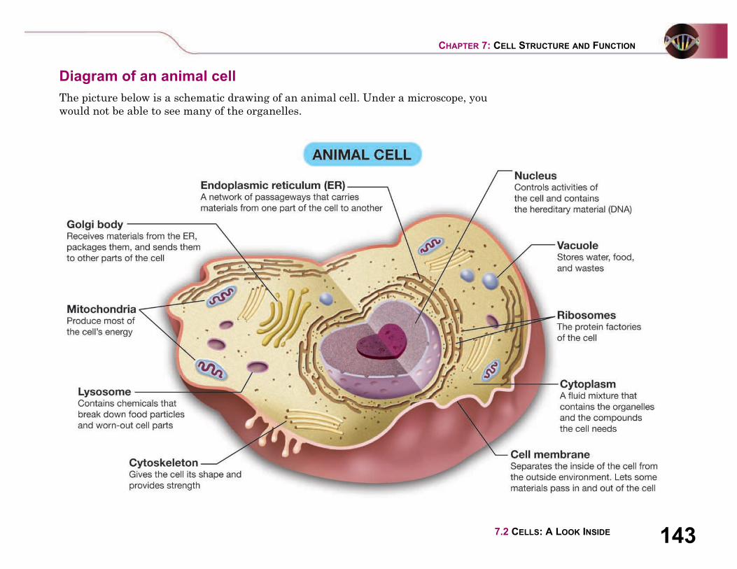
Diagram of an animal cell
The picture below is a schematic drawing of an animal cell. Under a microscope, you
would not be able to see many of the organelles.
144 UNIT 3 CELL BIOLOGY
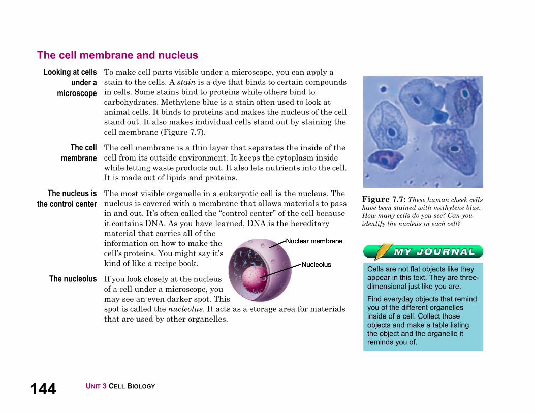
Figure 7.7: These human cheek cells
have been stained with methylene blue.
How many cells do you see? Can you
identify the nucleus in each cell?
Cells are not flat objects like they
appear in this text. They are three-
dimensional just like you are.
Find everyday objects that remind
you of the different organelles
inside of a cell. Collect those
objects and make a table listing
the object and the organelle it
reminds you of.
The cell membrane and nucleus
Looking at cells
under a
microscope
To make cell parts visible under a microscope, you can apply a
stain to the cells. A stain is a dye that binds to certain compounds
in cells. Some stains bind to proteins while others bind to
carbohydrates. Methylene blue is a stain often used to look at
animal cells. It binds to proteins and makes the nucleus of the cell
stand out. It also makes individual cells stand out by staining the
cell membrane (Figure 7.7).
The cell
membrane
The cell membrane is a thin layer that separates the inside of the
cell from its outside environment. It keeps the cytoplasm inside
while letting waste products out. It also lets nutrients into the cell.
It is made out of lipids and proteins.
The nucleus is
the control center
The most visible organelle in a eukaryotic cell is the nucleus. The
nucleus is covered with a membrane that allows materials to pass
in and out. It’s often called the “control center” of the cell because
it contains DNA. As you have learned, DNA is the hereditary
material that carries all of the
information on how to make the
cell’s proteins. You might say it’s
kind of like a recipe book.
The nucleolus If you look closely at the nucleus
of a cell under a microscope, you
may see an even darker spot. This
spot is called the nucleolus. It acts as a storage area for materials
that are used by other organelles.
1457.2 CELLS: A LOOK INSIDE
CHAPTER 7: CELL STRUCTURE AND FUNCTION
Organelles and their functions
Seeing the other
organelles
Even with a powerful microscope, it’s difficult to see organelles
other than the nucleus. Scientists use different techniques like
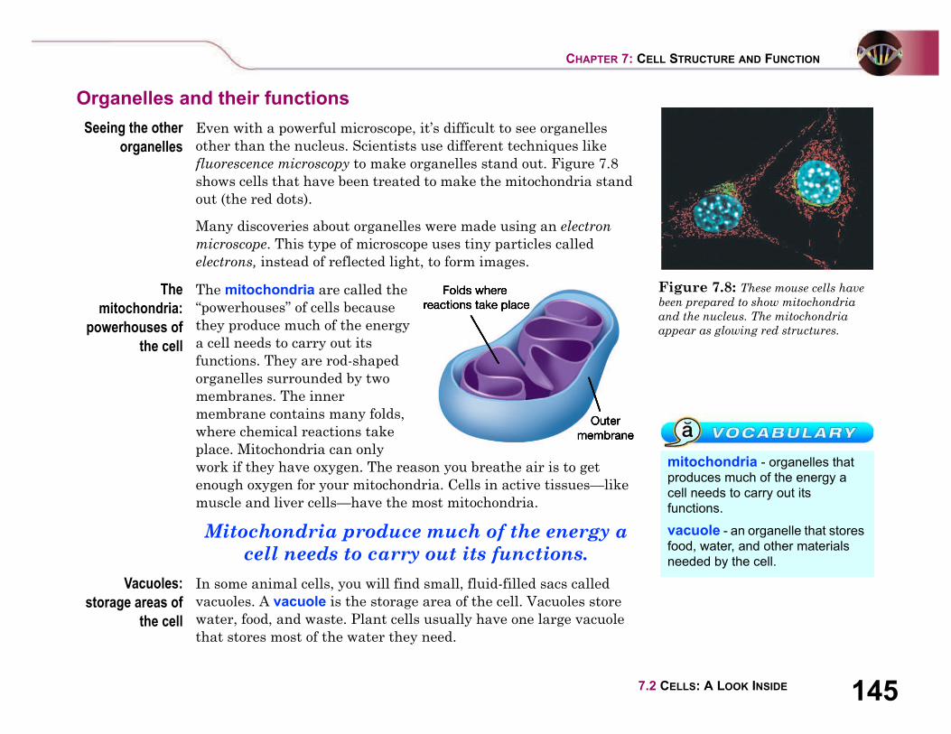
fluorescence microscopy to make organelles stand out. Figure 7.8
shows cells that have been treated to make the mitochondria stand
out (the red dots).
Many discoveries about organelles were made using an electron
microscope. This type of microscope uses tiny particles called
electrons, instead of reflected light, to form images.
The
mitochondria:
powerhouses of
the cell
The mitochondria are called the
“powerhouses” of cells because
they produce much of the energy
a cell needs to carry out its
functions. They are rod-shaped
organelles surrounded by two
membranes. The inner
membrane contains many folds,
where chemical reactions take
place. Mitochondria can only
work if they have oxygen. The reason you breathe air is to get
enough oxygen for your mitochondria. Cells in active tissues—like
muscle and liver cells—have the most mitochondria.
Mitochondria produce much of the energy a
cell needs to carry out its functions.
Vacuoles:
storage areas of
the cell
In some animal cells, you will find small, fluid-filled sacs called
vacuoles. A vacuole is the storage area of the cell. Vacuoles store
water, food, and waste. Plant cells usually have one large vacuole
that stores most of the water they need.
Figure 7.8: These mouse cells have
been prepared to show mitochondria
and the nucleus. The mitochondria
appear as glowing red structures.
mitochondria - organelles that
produces much of the energy a
cell needs to carry out its
functions.
vacuole - an organelle that stores
food, water, and other materials
needed by the cell.

146 UNIT 3 CELL BIOLOGY
Figure 7.9: This cell was treated to
make the cytoskeleton stand out.
endoplasmic reticulum - an
organelle that transports proteins
inside of the cell.
ribosome - an organelle that
makes proteins.
Golgi body - an organelle that
receives proteins, packages them,
and distributes them.
lysosome - an organelle that
contains enzymes that break
things down to be reused by
the cell.
cytoskeleton - a series of
protein fibers inside of a cell
that give structure and shape
to the cell.
Endoplasmic
reticulum
The endoplasmic reticulum (ER) is
a series of tunnels throughout the
cytoplasm. They transport proteins
from one part of the cell to another.
You can think of the ER as a series of
folded and connected tubes. There
are different places to enter and exit
in various locations.
Ribosomes If you look closely at the ER, you can
sometimes see little round grains all around it. Each of those tiny
grains is an individual ribosome. Ribosomes are the protein
factories of the cell. When ribosomes make proteins, they release
them into the ER. Some ribosomes are not attached to the ER, but
float in the cytoplasm.
Golgi bodies Golgi bodies receive proteins and other compounds
from the ER. They package these materials and
distribute them to other parts of the cell. They also
release materials outside of the cell. The
number and size of Golgi bodies found in a cell
depends on the quantity of compounds
produced in the cell. The more compounds
produced, the more and larger Golgi bodies there
are. For example, a large number of Golgi bodies are found in cells
that produce digestive enzymes.
Lysosomes Lysosomes contain enzymes that can break things down.
Lysosomes pick up foreign invaders such as bacteria, food, and old
organelles and break them into small pieces that can be reused.
Cytoskeleton The cytoskeleton is a series of fibers made from proteins. It
provides structure to the cell and gives it its shape. Figure 7.9
shows a cell that has been treated so the cytoskeleton stands out.
1477.2 CELLS: A LOOK INSIDE
CHAPTER 7: CELL STRUCTURE AND FUNCTION
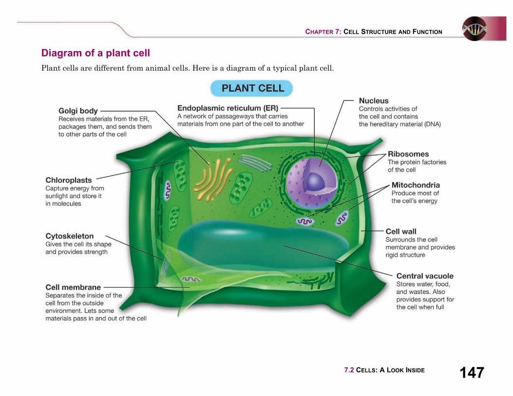
Diagram of a plant cell
Plant cells are different from animal cells. Here is a diagram of a typical plant cell.
148 UNIT 3 CELL BIOLOGY
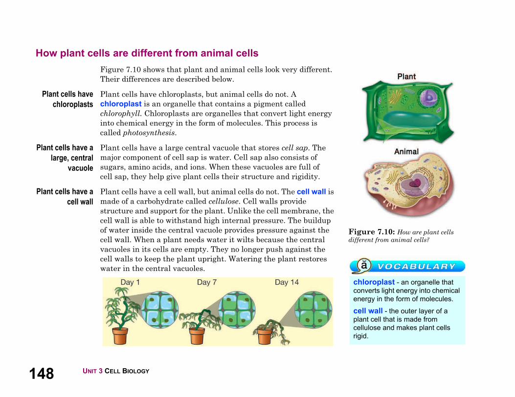
Figure 7.10: How are plant cells
different from animal cells?
chloroplast - an organelle that
converts light energy into chemical
energy in the form of molecules.
cell wall - the outer layer of a
plant cell that is made from
cellulose and makes plant cells
rigid.
How plant cells are different from animal cells
Figure 7.10 shows that plant and animal cells look very different.
Their differences are described below.
Plant cells have
chloroplasts
Plant cells have chloroplasts, but animal cells do not. A
chloroplast is an organelle that contains a pigment called
chlorophyll. Chloroplasts are organelles that convert light energy
into chemical energy in the form of molecules. This process is
called photosynthesis.
Plant cells have a
large, central
vacuole
Plant cells have a large central vacuole that stores cell sap. The
major component of cell sap is water. Cell sap also consists of
sugars, amino acids, and ions. When these vacuoles are full of
cell sap, they help give plant cells their structure and rigidity.
Plant cells have a
cell wall
Plant cells have a cell wall, but animal cells do not. The cell wall is
made of a carbohydrate called cellulose. Cell walls provide
structure and support for the plant. Unlike the cell membrane, the
cell wall is able to withstand high internal pressure. The buildup
of water inside the central vacuole provides pressure against the
cell wall. When a plant needs water it wilts because the central
vacuoles in its cells are empty. They no longer push against the
cell walls to keep the plant upright. Watering the plant restores
water in the central vacuoles.

1497.2 CELLS: A LOOK INSIDE
CHAPTER 7: CELL STRUCTURE AND FUNCTION
7.2 Section Review
1. Name the correct organelle for each function in the table below.
2. The plant cell wall is made of:
a. glucose
b. protein
c. cellulose
d. lipids
3. A Venn diagram shows how two or more things are similar
and different. Place the organelles into the Venn diagram in
Figure 7.11. What do your results tell you about the differences
between plant and animal cells?
4. What is the function of the cell wall? Why do plant cells need a
cell wall?
Organelle Function
Produces much of the energy a cell
needs to carry out its functions
Makes proteins.
Controls all activities of the cell and
contains the hereditary material
Packages proteins and distributes
them to other parts of the cell
Lets materials pass into or out of the cell
Stores water, food, and wastes
Transports proteins inside of the cell
Figure 7.11: Complete the Venn
diagram for question 3.
What effect on the function of a
cell would occur if one of the
following organelles was missing?
Write a sentence for each
organelle.
1. ribosome
2. lysosome
3. vacuole
4. mitochondria
5. chloroplast
6. cell membrane

150
Organ Transplants
How many ways do living things protect themselves? You
can probably think of dozens of examples. Roses have thorns.
Rabbits are quick. Pigeons fly in flocks. Have you ever
thought about this? What is the most important way that
many living things, including people, protect themselves?
The answer might surprise you.
All living things must protect themselves against disease.
Like other living things, people are under constant assault
from bacteria, viruses, and other organisms. Our immune
systems fight off these organisms.
What happens
when a foreign cell
enters your body?
It causes a quick
response from
your immune
system. A variety
of cells attack the
invader. At the
heart of your
immune system
are cells called
lymphocytes. These are a type of white blood cell.
Lymphocytes can grab onto foreign cells and help remove
them from your body.
For your immune system, the world divides into “us” and
“them.” “Us” means every cell in your body. “Them” means
almost everything else on Earth. The immune system
attacks “them.” This can be a problem with organ
transplants.
The problem with transplants
Hindu doctors in South Asia may have transplanted skin
2,600 years ago. Such grafts took skin from one part of a
person’s body. It replaced damaged skin in another part of
the same person’s body. This is still done today.
The immune system ignores this kind of transplant. The
tissues “match” exactly. All of the cells came from the same
body. For the same reason, heart bypass operations
use blood vessels from
the patient’s own
body to replace
blocked heart
arteries.
Modern
medicine is
able to
transplant
many organs
besides skin
and blood
vessels.
Kidneys, livers,
hearts, and even lungs
have been transplanted.
Transplants save people’s lives. In each case, the patient’s
immune system must be overcome. The immune system may
see the transplant as an invader. This is called “rejection.”
Antigens are on the surface of cells. They tell your immune
system whether a cell is “us” or “them.” Two types of
antigens cause rejection. One is found on red blood cells. The
other is called transplantation, or histocompatability,
antigens. These are found on every cell in your body except
red blood cells. The main transplantation antigens are called
the human leukocyte antigens, or HLA. Your genes
Ch
ap
ter
7 C
on
necti
on
151UNIT 3 CELL BIOLOGY
determine your HLA. Only identical twins have the same
genes. An organ could be transplanted from one identical
twin to another without rejection. In every other case,
doctors need to match organs. Doctors look for as close a
match as they can between the HLA of the patient and the
person who donated the organ.
Tissue-matching
Matching HLA antigens is more often called “tissue
matching” or “tissue typing.”
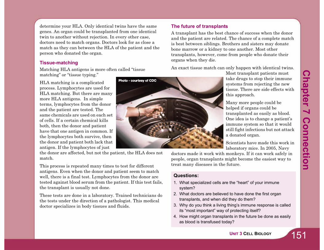
HLA matching is a complicated
process. Lymphocytes are used for
HLA matching. But there are many
more HLA antigens. In simple
terms, lymphocytes from the donor
and the patient are tested. The
same chemicals are used on each set
of cells. If a certain chemical kills
both, then the donor and patient
have that one antigen in common. If
the lymphocytes both survive, then
the donor and patient both lack that
antigen. If the lymphocytes of just
the donor are affected, but not the patient, the HLA does not
match.
This process is repeated many times to test for different
antigens. Even when the donor and patient seem to match
well, there is a final test. Lymphocytes from the donor are
tested against blood serum from the patient. If this test fails,
the transplant is usually not done.
These tests are done in a laboratory. Trained technicians do
the tests under the direction of a pathologist. This medical
doctor specializes in body tissues and fluids.
The future of transplants
A transplant has the best chance of success when the donor
and the patient are related. The chance of a complete match
is best between siblings. Brothers and sisters may donate
bone marrow or a kidney to one another. Most other
transplants, however, come from people who donate their
organs when they die.
An exact tissue match can only happen with identical twins.
Most transplant patients must
take drugs to stop their immune
systems from rejecting the new
tissue. There are side effects with
this approach.
Many more people could be
helped if organs could be
transplanted as easily as blood.
One idea is to change a patient’s
immune system so that it would
still fight infections but not attack
a donated organ.
Scientists have made this work in
laboratory mice. In 2005, Navy
doctors made it work with monkeys. If it can work safely in
people, organ transplants might become the easiest way to
treat many diseases in the future.
Questions:
1. What specialized cells are the “heart” of your immune
system?
2. What doctors are believed to have done the first organ
transplants, and when did they do them?
3. Why do you think a living thing’s immune response is called
its “most important” way of protecting itself?
4. How might organ transplants in the future be done as easily
as blood is transfused today?
Ch
ap
ter 7
Co
nn
ectio
n

152
Building a Scale Model of a Cell
Cells appear in all shapes
and sizes. In animals, cells
can be long like the motor
neurons that run from the
tips of your toes to the base
of the neck. Other cells in
your body can be small like
the red blood cells. Cell
models are a good way to
help you identify cell
structures. Often it is not clear how the size of the cell is
related to the size of the organelles. In this activity, you will
explore the relationship of cell size to organelle size by
creating a scale model.
What you will do
1. Complete the table (right). Use a scale factor of
1 micrometer = 1 centimeter. The calculation for the
diameter of the cell is completed for you.
2. Obtain a large sheet of paper from your teacher.
3. Measure the diameter of the cell (35 centimeters) and
draw a circle on your paper. This will be the outline of
your animal cell. Cut out the circle out of the paper.
4. Using your calculation, make a nucleus to scale using the
colored-construction paper your teacher has provided.
5. Make and add the rest of the organelles. Be sure to use
the animal cell diagram on page 143 as a guide in making
your organelles. For example, you could make a golgi
body that consists of 5 separate parts, 7 2 micrometers
each.
6. Once the model is complete, label the organelles. Or you
may wish to make a key that identifies each organelle.
Applying your knowledge
a. What is the smallest organelle in a typical animal cell?
b. What is the largest organelle in a typical animal cell?
c. How is your model of the cell different than models the
teacher used in class, or models you may see in a text
book?
d. This method does not apply only to cells. Can you think of
other examples where scale models are used?
e. How might you build a 3-dimensional scale model of a
cell? With a classmate, propose a method for creating a
scaled 3-dimensional model of a cell with all the
organelles. What types of things could one use to
represent the cell boundaries? What things might one use
to represent the organelles? Begin by writing up your
ideas in a proposal. Your teacher may ask you to build
your model as a project.
OrganelleAverage Size
( m)
Scaling Factor
(1 m = 1 cm)
Model
Size
(cm)
Cell diameter 35 35 m 1 cm/ m 35Nucleus 5
Mitochondria 6 2Lysosome 2
Endoplasmic reticulum 5 10Golgi body 7 2
Vacuole 2Ribosome .02
Ch
ap
ter
7 A
cti
vit
y

CHAPTER 7 CELL STRUCTURE AND FUNCTION 153
Chapter 7 Assessment
Vocabulary
Select the correct term to complete the sentences.
Section 7.1
1. Bacteria are _____ cells.2. The _____ controls what enters and exits the cell.
3. A structure inside a cell that does a certain job is called an _____.
4. The fluid mixture with organelles and other vital
compounds in cells is the _____ .
5. Eukaryotic cells all have a _____ that contains DNA.
Section 7.2
6. The _____ is the organelle that transports materials like
proteins around the cell.
7. Fibers inside the cell that give structure and shape are
called the _____.
8. Muscle cells have a lot of _____ to produce the large amounts
of energy necessary to do their work.
9. A _____ is a protein factory in the cell.
10. Enzymes found in a _____ are used to break down old cell
parts that are then recycled by the cell.
11. Proteins move from the ribosome to the _____ for packaging
before distribution around the cell.
12. Animal cells can change shape to move because they don't
have a _____, which is what makes plant cells rigid.
Concepts
Section 7.1
1. Which of the following is not part of the cell theory?
a. Cells only come from existing cells.
b. All of an organism's life functions occur within cells.
c. The two major types of cells are prokaryotic cells and
eukaryotic cells.
d. All living things are made of one or more cells.
2. Identify each characteristic as either a feature of
prokaryotic cells (P) or as a feature of eukaryotic cells (E).
a. _____ name means “before nucleus” in Greek
b. _____ believed to have originated 2 billion years ago
c. _____ DNA is contained in nucleus
d. _____ larger of the two types—10 times the size of the
other
e. _____ have organelles without membrane covers
Section 7.2
Match the organelles to the most appropriate item that performs
the same function to complete these analogies.
8. Which part of the cell is like a recipe book?
a. nucleolus
b. DNA
c. cell membrane
d. none of the above
9. The ____________________ is the largest organelle in the cell.
cell membrane
cell wall
nucleus
cytoplasm
cytoskeleton
endoplasmic reticulum
golgi body
lysosome
mitochondria
organelle
prokaryotic
ribosome
_____ 3. ER a. nutshell_____ 4. cell wall b. warehouse_____ 5. vacuole c. brain
_____ 6. cell membrane d. highway
7. nucleus e. skin
154 CHAPTER 7 CELL STRUCTURE AND FUNCTION
10. Cells can only have one of certain organelles like the
nucleus. Which organelles can a cell have many of the same
kind? Explain your answer.
11. Which organelle would cause a lot of damage to the cell if it
were to break open? Why?
12. Most potato cells don't have chloroplasts. If you saw these
cells under the microscope, how could you tell that they were
plant cells?
Math and Writing Skills
Section 7.1
1. Imagine that you are Anton van Leewenhoek and you have
just observed blood cells, bacteria, and single-celled protists
for the first time. Write a letter to a friend describing your
amazing discoveries.
2. Write an imaginary dialogue that could have taken place
between Matthais Schleiden and Theodore Schwann after
they observed plant and animal tissue under a microscope.
3. Many of the cells in your body are 0.01 mm long. Use that
measurement to complete these calculations.
a. An amoeba - a unicellular protist - is 1 mm long. How
many body cells would you have to stack end to end to
equal the size of an amoeba?
b. Figure out what your height is in millimeters by
multiplying your height in meters by 1000. How many
body cells would you have to stack end to end to equal
your height?
c. The length of a swimming pool is 25,000 mm. How
many body cells would you have to stack end to end to
equal the length of the pool?
d. Prokaryotic cells are approximately 1/10 the size of
eukaryotic cells. How big are prokaryotic cells?
4. If you were trying to classify an unknown organism by
looking at its cells, what could its cells tell you?
Section 7.2
5. Describe what goes on in a typical animal cell. Be sure to
mention all the organelles listed in the text.
6. Which organelles does a spinach cell have that a rabbit cell
does not? Explain your answer.
7. Explain the connection between a wilted plant and cell parts
like the vacuole and the cell wall.
Chapter Project—Cellular Song
Cells have organelles with weird names like Golgi body and
endoplasmic reticulum. It is often helpful to invent a way to help
you remember the names of the structures and their functions.
Create a song or poem about cell structure, using the guidelines
below. Record the song or poem and play it back for the class, or
perform it live. If you don't like solo work, join some classmates
and do this as a group project. Make sure everyone contributes
verses to the song or poem!
1. Choose one type of cell, either a plant cell or an animal cell.
2. Choose a popular song for the melody or rap. If you create a
poem, make the verses rhyme.
3. The song or poem must include each structure listed on the
animal or plant cell diagram in your book. In addition to
naming the structures, you must use the song or poem to
help you remember the function of each structure.
4. Submit your creation for approval, memorize it, and then
share the song or poem with your classmates. When it comes
time for a written test on cell structure, you might be
humming a tune to help you remember the answers!

Chapter 8 Cell Processes
How many cells are in the human body? Cells are so small that you
can only see them with a microscope; this means that the average
human body must many, many cells. Old cells are constantly being
replaced with new cells. Every minute you lose 30,000 to 40,000 worn-
out skin cells. If you live to be 80 years old, you have grown about
1000 skins in a lifetime! You can see how challenging it is to estimate
the number of cells. Most scientists agree that the human body
contains trillions of cells. If you had to individually count the cells in
your body, it would take over 2000 years! It is hard to imagine how
many cells there must be in a giant redwood tree. There is a redwood
tree in California that measures over 360 feet tall (110 meters)! How
can a massive tree like that come from a tiny seed? Read this chapter
on how cells work to satisfy your curiosity.
1. How do things move in and out of cells?
2. How do cells get energy?
3. Why are plants green?

156 UNIT 3 CELL BIOLOGY
Figure 8.1: Soap bubbles are similar
to cell membranes.
8.1 The Cell Membrane
The cell membrane is kind of like a soap bubble (Figure 8.1). A soap bubble consists
of a thin, flexible membrane. The soapy membrane seals the inside air from the
outside. Likewise the cell membrane is a thin, flexible layer that seals the inside of
the cell from its outside environment. In this section, you’ll learn about the structure
and function of the cell membrane.
A closer look at the cell membrane
The functions of
the cell
membrane
The cell membrane has many functions. It protects the cell from
its environment and takes in food and other compounds that the
cell needs. It also gets rid of waste from inside of the cell. The cell
membrane even allows cells to communicate and interact.
The structure of
the cell
membrane
The cell membrane is made of several types of molecules. Lipid
molecules form a double layer. This creates a thin, fluid layer like
a soap bubble. Embedded protein molecules can move around
within this layer. Carbohydrates attached to some proteins face
outward. Some of these serve as “identification cards” so cells can
recognize each other.
1578.1 THE CELL MEMBRANE
CHAPTER 8: CELL PROCESSES
Diffusion
What is
diffusion?
Cells live in a watery environment. The cytoplasm is 80% water.
Every cell in your body is also surrounded by a watery solution.
Solutions make it easier for molecules to move into or out of the
cell. Molecules move across the cell membrane by a process called
diffusion. Diffusion is the movement of molecules from areas of
greater concentration to areas of lesser concentration.
How diffusion
works in a cell
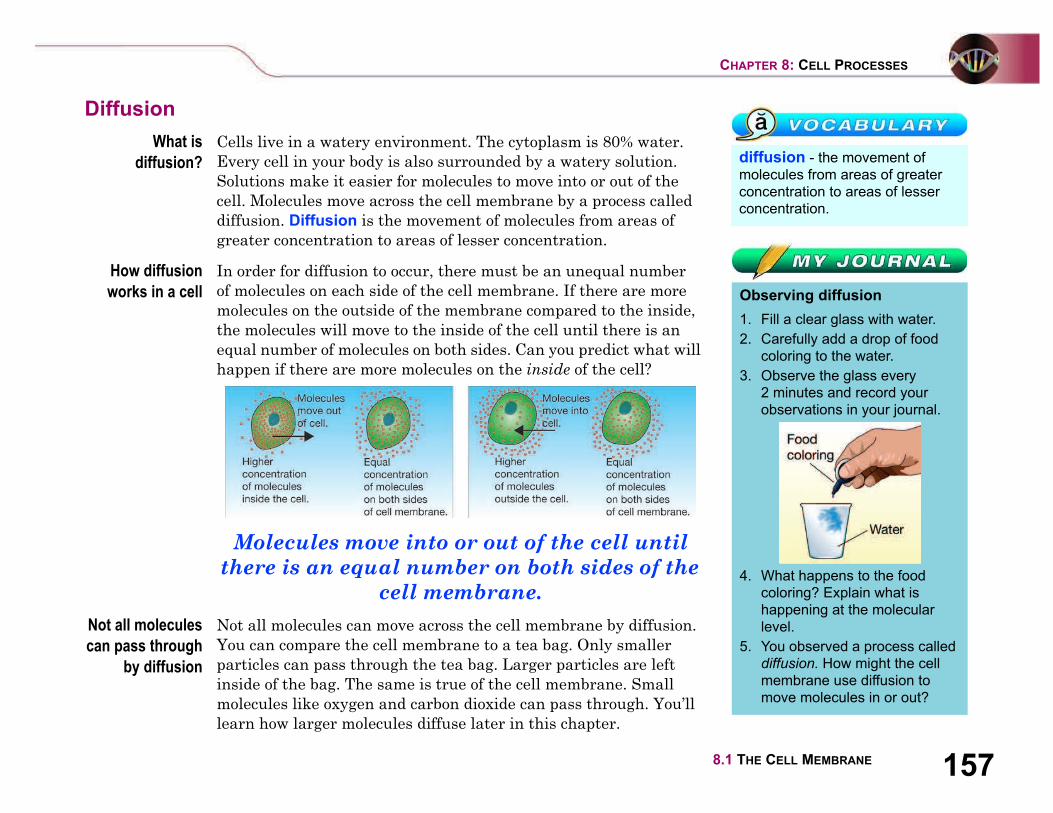
In order for diffusion to occur, there must be an unequal number
of molecules on each side of the cell membrane. If there are more
molecules on the outside of the membrane compared to the inside,
the molecules will move to the inside of the cell until there is an
equal number of molecules on both sides. Can you predict what will
happen if there are more molecules on the inside of the cell?
Molecules move into or out of the cell until
there is an equal number on both sides of the
cell membrane.
Not all molecules
can pass through
by diffusion
Not all molecules can move across the cell membrane by diffusion.
You can compare the cell membrane to a tea bag. Only smaller
particles can pass through the tea bag. Larger particles are left
inside of the bag. The same is true of the cell membrane. Small
molecules like oxygen and carbon dioxide can pass through. You’ll
learn how larger molecules diffuse later in this chapter.
diffusion - the movement of
molecules from areas of greater
concentration to areas of lesser
concentration.
Observing diffusion
1. Fill a clear glass with water.
2. Carefully add a drop of food
coloring to the water.
3. Observe the glass every
2 minutes and record your
observations in your journal.
4. What happens to the food
coloring? Explain what is
happening at the molecular
level.
5. You observed a process called
diffusion. How might the cell
membrane use diffusion to
move molecules in or out?
158 UNIT 3 CELL BIOLOGY
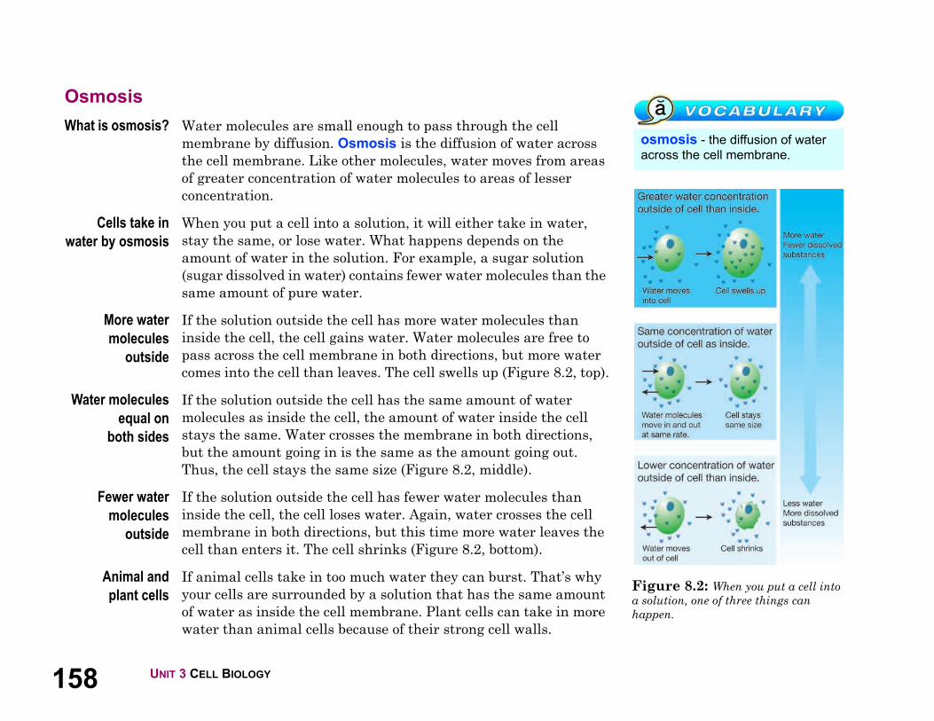
Figure 8.2: When you put a cell into
a solution, one of three things can
happen.
osmosis - the diffusion of water
across the cell membrane.
Osmosis
What is osmosis? Water molecules are small enough to pass through the cell
membrane by diffusion. Osmosis is the diffusion of water across
the cell membrane. Like other molecules, water moves from areas
of greater concentration of water molecules to areas of lesser
concentration.
Cells take in
water by osmosis
When you put a cell into a solution, it will either take in water,
stay the same, or lose water. What happens depends on the
amount of water in the solution. For example, a sugar solution
(sugar dissolved in water) contains fewer water molecules than the
same amount of pure water.
More water
molecules
outside
If the solution outside the cell has more water molecules than
inside the cell, the cell gains water. Water molecules are free to
pass across the cell membrane in both directions, but more water
comes into the cell than leaves. The cell swells up (Figure 8.2, top).
Water molecules
equal on
both sides
If the solution outside the cell has the same amount of water
molecules as inside the cell, the amount of water inside the cell
stays the same. Water crosses the membrane in both directions,
but the amount going in is the same as the amount going out.
Thus, the cell stays the same size (Figure 8.2, middle).
Fewer water
molecules
outside
If the solution outside the cell has fewer water molecules than
inside the cell, the cell loses water. Again, water crosses the cell
membrane in both directions, but this time more water leaves the
cell than enters it. The cell shrinks (Figure 8.2, bottom).
Animal and
plant cells
If animal cells take in too much water they can burst. That’s why
your cells are surrounded by a solution that has the same amount
of water as inside the cell membrane. Plant cells can take in more
water than animal cells because of their strong cell walls.

1598.1 THE CELL MEMBRANE
CHAPTER 8: CELL PROCESSES
Other types of transport
Protein channels Diffusion and osmosis do not require energy from the cell. This is
because the molecules move with a concentration difference (from
higher to lower). Larger molecules like sugars, starches, and
proteins sometimes diffuse through protein channels (Figure 8.3).
Because the molecules move from greater to lesser concentration
through the channels, this process also does not require energy.
Active transport Sometimes a cell needs to move molecules against a concentration
difference (from lower to higher concentration). Active transport is
a process that allows molecules to move across the cell membrane
from lower to higher concentrations. Active transport requires
energy. Protein molecules act as “pumps” to move the molecules
across the cell membrane as shown below. Your nerve cells have
lots of protein pumps to move ions across the cell membrane. This
is how signals travel through your nervous system.
Other types of
active transport
A cell can take in larger particles of food by “engulfing” them. The
cell membrane forms a pocket around the particle. Once inside the
cell, the pocket breaks loose from the cell membrane. It forms a
vacuole within the cytoplasm (Figure 8.4). Cells also send material
out of the cell in the same way. When this happens, a vacuole fuses
with the cell membrane and the contents are forced outside of the
cell. Both of these processes are types of active transport because
they require energy.
Figure 8.3: Larger molecules can
diffuse through protein channels.
Figure 8.4: A cell can also take in
larger amounts of material by engulfing
them.
160 UNIT 3 CELL BIOLOGY
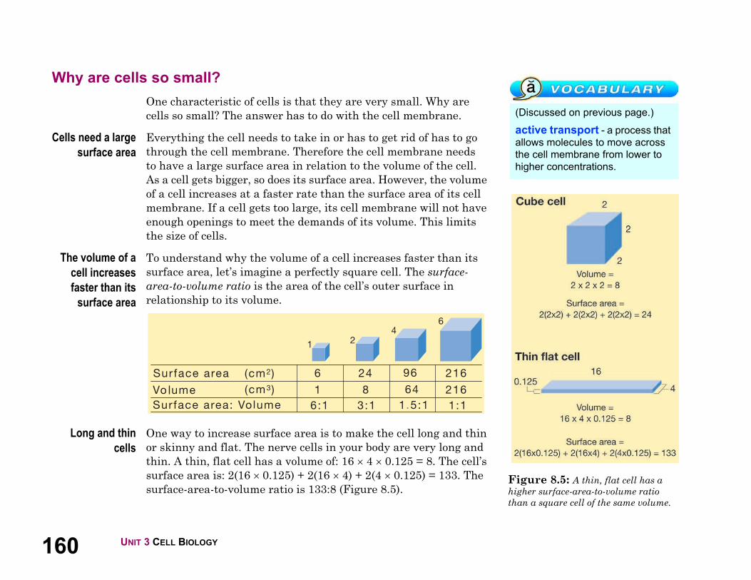
Figure 8.5: A thin, flat cell has a
higher surface-area-to-volume ratio
than a square cell of the same volume.
(Discussed on previous page.)
active transport - a process that
allows molecules to move across
the cell membrane from lower to
higher concentrations.
Why are cells so small?
One characteristic of cells is that they are very small. Why are
cells so small? The answer has to do with the cell membrane.
Cells need a large
surface area
Everything the cell needs to take in or has to get rid of has to go
through the cell membrane. Therefore the cell membrane needs
to have a large surface area in relation to the volume of the cell.
As a cell gets bigger, so does its surface area. However, the volume
of a cell increases at a faster rate than the surface area of its cell
membrane. If a cell gets too large, its cell membrane will not have
enough openings to meet the demands of its volume. This limits
the size of cells.
The volume of a
cell increases
faster than its
surface area
To understand why the volume of a cell increases faster than its
surface area, let’s imagine a perfectly square cell. The surface-
area-to-volume ratio is the area of the cell’s outer surface in
relationship to its volume.
Long and thin
cells
One way to increase surface area is to make the cell long and thin
or skinny and flat. The nerve cells in your body are very long and
thin. A thin, flat cell has a volume of: 16 4 0.125 = 8. The cell’s
surface area is: 2(16 0.125) + 2(16 4) + 2(4 0.125) = 133. The
surface-area-to-volume ratio is 133:8 (Figure 8.5).
1618.1 THE CELL MEMBRANE
CHAPTER 8: CELL PROCESSES
8.1 Section Review
1. List four functions of the cell membrane.
2. How is the cell membrane like a soap bubble?
3. What is diffusion? Name one example of diffusion.
4. What is osmosis? What structure in a plant cell helps protect it
from osmosis?
5. For each situation below, state whether water will move into
the cell, move out of the cell, or stay the same.
6. How is active transport different from diffusion?
7. Name two situations in which a cell would need to use active
transport instead of diffusion.
8. Explain why cells are so small.
9. Which figure below has the highest surface-area-to-volume
ratio? Explain your reasoning.
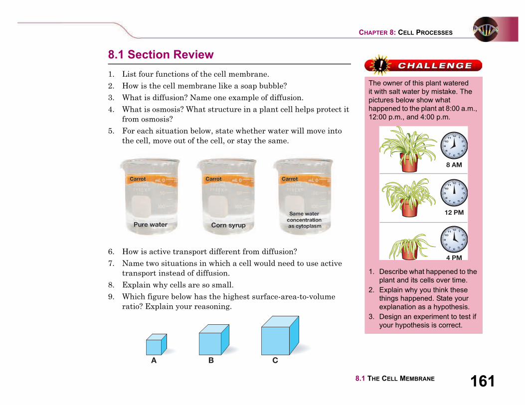
The owner of this plant watered
it with salt water by mistake. The
pictures below show what
happened to the plant at 8:00 a.m.,
12:00 p.m., and 4:00 p.m.
1. Describe what happened to the
plant and its cells over time.
2. Explain why you think these
things happened. State your
explanation as a hypothesis.
3. Design an experiment to test if
your hypothesis is correct.

162 UNIT 3 CELL BIOLOGY
Figure 8.6: The chemical reaction of
photosynthesis. What are the reactants
of the reaction? What are the products?
photosynthesis - a process
where plants use the energy of
sunlight to produce carbohydrates.
8.2 Cells and Energy
To stay alive, you need a constant supply of energy. You need energy to move, think,
grow, and even sleep. Where does that energy come from? It all starts with the sun.
Plant cells store energy from the sun in the form of molecules. In this section you’ll
learn about how cells store and release energy.
What is photosynthesis?
Solar cells and
chloroplasts
A solar calculator has solar cells that convert light into electrical
energy. The electrical energy powers the calculator. Some of
it is stored in a battery. A plant cell has chloroplasts that also
convert energy. Chloroplasts are where photosynthesis occurs.
Photosynthesis is a process where plants use the energy of
sunlight to produce energy-rich molecules (carbohydrates).
Photosynthesis takes place in the
chloroplasts.
How does a tiny
seed grow into a
massive tree?
Before our knowledge of photosynthesis, gardeners wondered how
a tiny seed could grow into a massive tree. Where did all of that
mass come from? In the 1600s, a Flemish scientist named Jan Van
Helmont (1580–1644) conducted an important experiment. He
grew a willow tree in a carefully weighed amount of soil. He
noticed that the mass of the soil barely changed while the mass of
the tree greatly increased. He concluded that the extra mass did
not come from the soil.
Photosynthesis
is a chemical
reaction
Later experiments carried out by other scientists showed that
plants use carbon dioxide (from the air) and water to make a
simple carbohydrate (glucose). They also release oxygen. This
chemical reaction (photosynthesis) takes place only in the
presence of light (Figure 8.6).

1638.2 CELLS AND ENERGY
CHAPTER 8: CELL PROCESSES
Light and color
Visible light The Sun provides Earth with a steady source of light. Your eyes
perceive sunlight as white light. However, it is really made up of
different colors of light. The colors that make up sunlight are called
visible light. There are other forms of light we cannot see such as
ultraviolet and infrared light.
Light is a wave Light is a wave, like a ripple on a pond. Waves can be described by
their wavelength (the length from peak to peak), and energy. Light
is part of a continuum of waves known as the electromagnetic
spectrum. Light waves have very short wavelengths. They range
from 800 nm (red light) to 400 nm (violet light). One nanometer
(nm) is equal to one-billionth of a meter!
Color A prism splits white light into all of its colors. Color is how we
perceive the energy of light. All of the colors of visible light have
different energies. Red light has the lowest energy and violet light
has the highest energy. As we move through the rainbow from red
to violet, the energy of the light increases (Figure 8.7).
Figure 8.7: A prism splits light
into all of its colors. All of the colors
of light have different energies and
wavelengths.
color - how we perceive the
energy of light.
Wavelength (Nm)
Increasing Energy
red
ora
ng
e
ye
llo
w
wh
ite
gre
en
blu
e
vio
let
Low
(800 nm)
High
(400 nm)
Prism
164 UNIT 3 CELL BIOLOGY
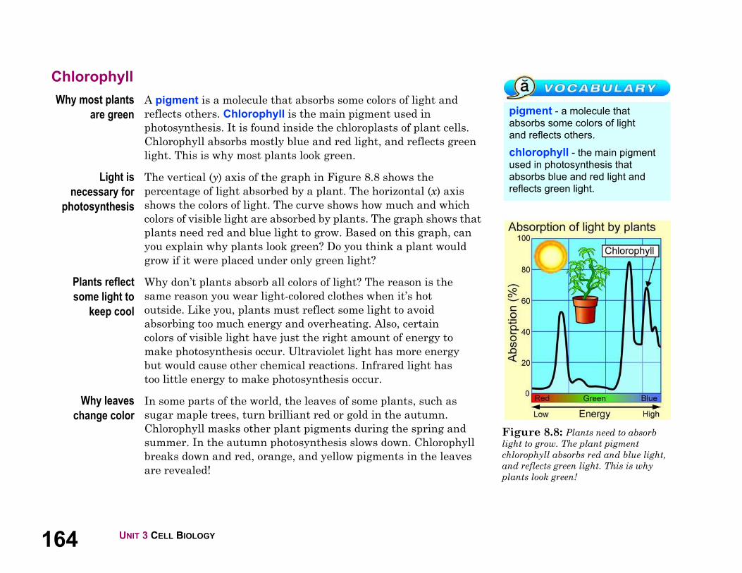
Figure 8.8: Plants need to absorb
light to grow. The plant pigment
chlorophyll absorbs red and blue light,
and reflects green light. This is why
plants look green!
pigment - a molecule that
absorbs some colors of light
and reflects others.
chlorophyll - the main pigment
used in photosynthesis that
absorbs blue and red light and
reflects green light.
Chlorophyll
Why most plants
are green
A pigment is a molecule that absorbs some colors of light and
reflects others. Chlorophyll is the main pigment used in
photosynthesis. It is found inside the chloroplasts of plant cells.
Chlorophyll absorbs mostly blue and red light, and reflects green
light. This is why most plants look green.
Light is
necessary for
photosynthesis
The vertical (y) axis of the graph in Figure 8.8 shows the
percentage of light absorbed by a plant. The horizontal (x) axis
shows the colors of light. The curve shows how much and which
colors of visible light are absorbed by plants. The graph shows that
plants need red and blue light to grow. Based on this graph, can
you explain why plants look green? Do you think a plant would
grow if it were placed under only green light?
Plants reflect
some light to
keep cool
Why don’t plants absorb all colors of light? The reason is the
same reason you wear light-colored clothes when it’s hot
outside. Like you, plants must reflect some light to avoid
absorbing too much energy and overheating. Also, certain
colors of visible light have just the right amount of energy to
make photosynthesis occur. Ultraviolet light has more energy
but would cause other chemical reactions. Infrared light has
too little energy to make photosynthesis occur.
Why leaves
change color
In some parts of the world, the leaves of some plants, such as
sugar maple trees, turn brilliant red or gold in the autumn.
Chlorophyll masks other plant pigments during the spring and
summer. In the autumn photosynthesis slows down. Chlorophyll
breaks down and red, orange, and yellow pigments in the leaves
are revealed!
1658.2 CELLS AND ENERGY
CHAPTER 8: CELL PROCESSES
Cellular respiration
What is cellular
respiration?
Your cells get the energy they need from the food you eat. Your
digestive system breaks down food into molecules. Your cells
convert those molecules into a form of energy they can use. Cellular
respiration is the process in which the chemical bonds of energy-
rich molecules (like glucose) are converted into a form of energy
that cells can use. In eukaryotic (including animal and plant) cells,
cellular respiration takes place in the mitochondria.
Cellular respiration takes place in the
mitochondria.
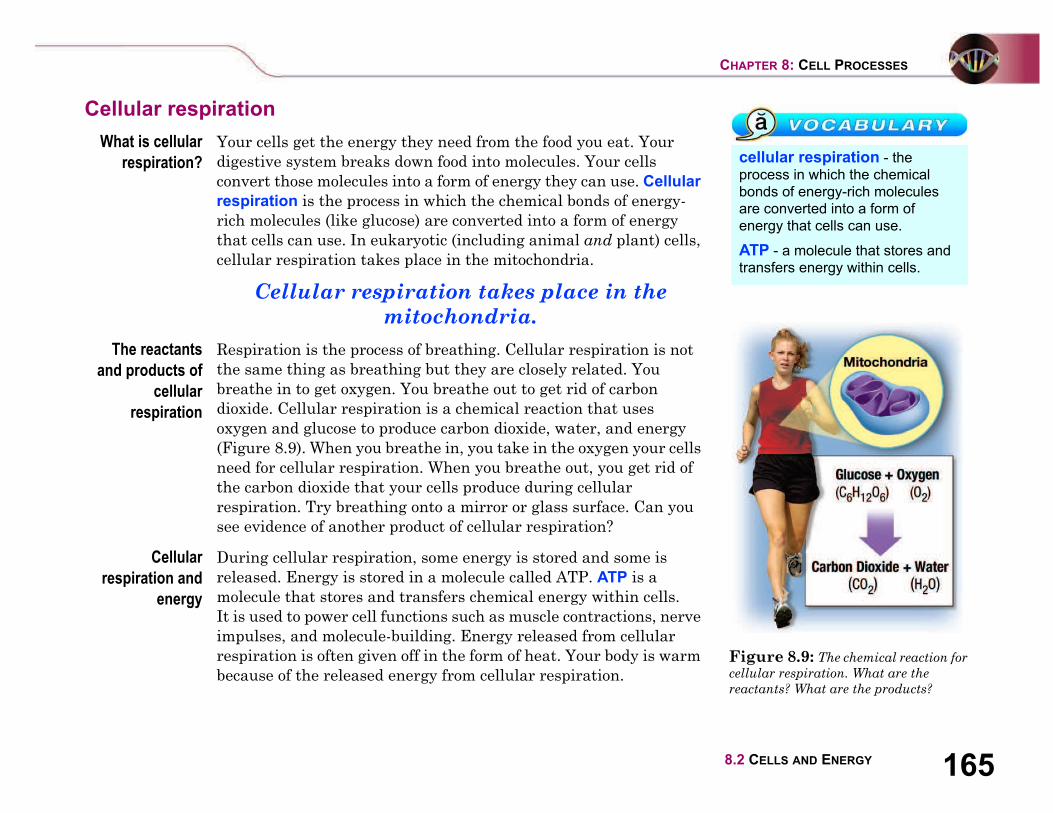
The reactants
and products of
cellular
respiration
Respiration is the process of breathing. Cellular respiration is not
the same thing as breathing but they are closely related. You
breathe in to get oxygen. You breathe out to get rid of carbon
dioxide. Cellular respiration is a chemical reaction that uses
oxygen and glucose to produce carbon dioxide, water, and energy
(Figure 8.9). When you breathe in, you take in the oxygen your cells
need for cellular respiration. When you breathe out, you get rid of
the carbon dioxide that your cells produce during cellular
respiration. Try breathing onto a mirror or glass surface. Can you
see evidence of another product of cellular respiration?
Cellular
respiration and
energy
During cellular respiration, some energy is stored and some is
released. Energy is stored in a molecule called ATP. ATP is a
molecule that stores and transfers chemical energy within cells.
It is used to power cell functions such as muscle contractions, nerve
impulses, and molecule-building. Energy released from cellular
respiration is often given off in the form of heat. Your body is warm
because of the released energy from cellular respiration.
Figure 8.9: The chemical reaction for
cellular respiration. What are the
reactants? What are the products?
cellular respiration - the
process in which the chemical
bonds of energy-rich molecules
are converted into a form of
energy that cells can use.
ATP - a molecule that stores and
transfers energy within cells.

166 UNIT 3 CELL BIOLOGY
Write the story of a carbon
atom as it travels through
photosynthesis and cellular
respiration. Include the following
information in your story:
• the molecules in which the
carbon atom is found.
• the organisms, cells, and
organelles through which it
travels.
Be creative!
Cellular respiration also
occurs in plants
It is important to understand that
both animal and plant cells
undergo cellular respiration. Plant
cells have mitochondria that
function just like the mitochondria
in animal cells. Plant cells use
some of the carbohydrates they
produce in photosynthesis for
cellular respiration. But animal
cells do not have chloroplasts and
cannot perform photosynthesis.
Comparing photosynthesis and cellular respiration
Comparing the reactions for photosynthesis and cellular
respiration shows how living things on Earth are connected.
The reactants in photosynthesis are the products in cellular
respiration. The reactants in cellular respiration are the products
in photosynthesis. The elements involved are carbon, hydrogen,
and oxygen.
1678.2 CELLS AND ENERGY
CHAPTER 8: CELL PROCESSES
8.2 Section Review
1. How are solar cells and chloroplasts similar?
2. What is the electromagnetic spectrum? Which part of the
electromagnetic spectrum do plants use for photosynthesis?
3. When white light is passed through a prism, what happens?
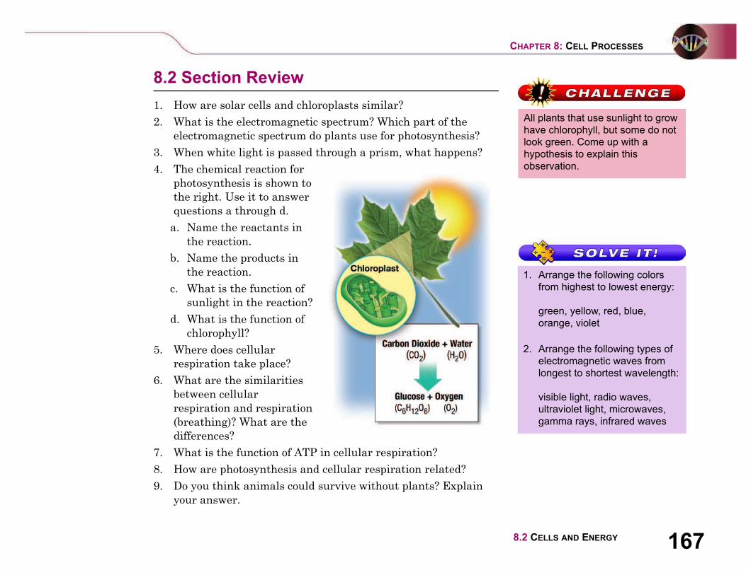
4. The chemical reaction for
photosynthesis is shown to
the right. Use it to answer
questions a through d.
a. Name the reactants in
the reaction.
b. Name the products in
the reaction.
c. What is the function of
sunlight in the reaction?
d. What is the function of
chlorophyll?
5. Where does cellular
respiration take place?
6. What are the similarities
between cellular
respiration and respiration
(breathing)? What are the
differences?
7. What is the function of ATP in cellular respiration?
8. How are photosynthesis and cellular respiration related?
9. Do you think animals could survive without plants? Explain
your answer.
All plants that use sunlight to grow
have chlorophyll, but some do not
look green. Come up with a
hypothesis to explain this
observation.
1. Arrange the following colors
from highest to lowest energy:
green, yellow, red, blue,
orange, violet
2. Arrange the following types of
electromagnetic waves from
longest to shortest wavelength:
visible light, radio waves,
ultraviolet light, microwaves,
gamma rays, infrared waves

168
Amazing Cells!
Did you know your body is made of trillions of cells? There
are millions of different types. Where did all of these
different types come from? Part of the answer is a special
type of cell called stem cells.
Many living things need stem cells including animals and
plants. An organism that is not fully developed is called an
embryo. In animal embryos, stem cells can develop into
different types of cells. Your body has over 200,000 different
types of cells. It has blood cells, muscle cells, skin cells, and
stomach cells just to name a few. Each type of cell has its
own structure and function.
The process of differentiation
All stem cells have some certain properties:
• Stem cells divide to make more stem cells. • Stem cells also have the ability to develop into different types of cells. A stem cell divides into two daughter cells. Each daughter
cell is identical to the original parent cell. When mature,
these cells also divide. This is how embryos get a supply of
stem cells. A growing embryo needs a lot of stem cells to
develop tissues and organs. In the laboratory, starting with a
few stem cells, scientists have grown millions in a few
months.
So how do
stem cells
change into
other types of
cells?
Scientists are
studying this
problem.
Something called a signal tells stem cells to become different
types of cells. Genes are pieces of DNA that carry
information from the parent cell to the offspring cells. The
genes inside stem cells provide internal signals. The
environment outside of the cell provides external signals.
The cell's environment includes chemicals from other cells.
Different types of specialized animal cells
There are two main types of
animal stem cells. More than
twenty years ago, scientists
extracted stem cells from the
embryos of mice. These stem
cells are described as
embryonic. The other main
type of stem cells is described
as adult. Embryonic stem
cells and adult stem cells are
very different.
Embryonic stem cells can divide to make more stem cells.
They wait for a signal. Then they start producing specialized
cells. These specialized cells form the tissues, which in turn
form the organs.Parent cell Two daughter cells
Ch
ap
ter
8 C
on
necti
on

169UNIT 3 CELL BIOLOGY
Embryonic stems cells are like new players on a soccer team.
Until the players are trained, they are reserves. They have
the potential to do a lot of different things. Once they are
trained, they become specialized in a position. The players
might be defenders or forwards. They might play goalie or
mid field. Similarly, embryonic stem cells are generic cells at
first. They get “training” from a signal. Then they develop
tissue for the kidneys, liver, or other organs.
While the main job of embryonic stem cells is growth, the
main job of adult stem cells is repair. They do not have as
much potential as embryonic stem cells. They seem to
already carry genetic information that determines which
type of cells they can become. They exist alongside the types
of cells they can produce. Adult stem cells in the skin, for
example, develop into skin cells to help new skin grow after
an injury.
The potential for treating diseases
Scientists think stem cells may help treat diseases. Can you
think how this might work? Embryonic stem cells can
develop into many other types of cells. If the right signals
can be discovered, these cells might be able to replace or
repair diseased tissue. Scientist's hope that diseases such as
diabetes and heart disease may be treated this way someday.
Adult stem cells are already used in medicine. For 30 years,
adult stem cells have been used in bone marrow transplants.
The potential of adult stem cells is more limited, but
scientists hope to use them to fight diseases. For example,
research in mice indicates that putting adult stem cells into
a damaged heart may help repair heart tissue.
Scientists are trying to better understand what triggers the
differentiation of stem cells. As knowledge and
understanding of stem cells increase, so does the potential
for many new disease therapies.
Questions:
1. What are the properties of stem cells?
2. Explain how stem cells change into different cell types.
3. What is the major difference between embryonic stem
cells and adult stem cells?
4. How are adult stem cells used in medicine today?
Ch
ap
ter 8
Co
nn
ectio
n

170
Making a Concept Map
A concept map is a way to represent information
visually. A concept map consists of nodes that contain
written concepts. The nodes are connected with lines to
to show relationships. The lines are labeled with an
arrowhead to describe the direction of the information.
In this activity you will create a concept map that
explains how cells get and use energy. Your concept
map should address the following question: How do
animal and plant cells use energy for life’s
processes?
What you will do
1. Write the concepts below on separate index cards or
sticky notes so they can be moved around.
2. Obtain a large sheet of paper or poster board from your
teacher.
3. Rank the concepts in order by placing the most general
concepts at the top to the most specific term at the
bottom. Think about the focus question to help rank the
concepts. Begin with only one to three of the most general
concepts at the top of the map.
4. Choose two to four sub concepts to place under each
general concept.
5. Connect the concepts by lines. Label the lines with one or
a few linking words that define the relationship between
the two concepts. These should read as a statement.
Draw arrow heads to show the direction of the
information.
6. Look at your map and revise any part if necessary.
7. Look for cross links between concepts in different sections
of the map. Draw and label these lines.
8. Present your concept map to the class and compare it to
others.
Applying your knowledge
a. Explain the relationship between photosynthesis and
growth.
b. Do plants take in organic food substances such as starch,
sugar or protein from the soil?
c. As a plant grows it gains weight (mass). Where does this
weight come from?
d. Where is carbon dioxide and water absorbed by most
plants?
e. What is the role of chlorophyll in a plant cell?
f. How does the food you eat aid in cellular respiration?
g. How did your concept map change as you made it?
h. Revise your concept map again if you wish, after your
class discussion.
mitochondria growth ATP energy
chloroplasts plant cell food carbon dioxide
oxygen carbohydrates energy pigments
photosynthesis carbon sunlight plants
animals aircellular
respirationchlorophyll
Ch
ap
ter
8 A
cti
vit
y

CHAPTER 8 CELL PROCESSES 171
Chapter 8 Assessment
Vocabulary
Select the correct term to complete the sentences.
Section 8.1
1. Movement of molecules that requires energy is called ____.
2. ____ is a kind of diffusion that involves water moving across
the cell membrane.
3. Osmosis and ____ are two types of passive transport because
they do not require energy.
Section 8.2
4. ____ stores and transfers chemical energy in cells.
5. Plant cells perform ____ to store energy from the Sun in the
form of molecules.
6. When ____ breaks down in the autumn, leaves change color
as red, orange, and yellow pigments become visible.
7. ____ uses oxygen and glucose to produce carbon dioxide,
water, and energy.
8. Chlorophyll is a ____, which is a molecule that absorbs some
colors of light and reflects others.
Concepts
Section 8.1
1. Draw and label a diagram of the cell membrane.
2. How do different cells recognize each other?
3. Distinguish between diffusion and osmosis.
4. Identify each situation as an example of diffusion, osmosis,
or active transport.
a. making a cup of tea
b. leftover salad wilting in the refrigerator
c. smoke escaping from the chimney
d. pumping up a tire with air
e. stained cotton t shirt soaking in sink
f. smell of perfume spreading through the room
Section 8.2
5. Why do plants look green?
6. How are breathing and cellular respiration related?
7. Do plant cells need to carry out respiration? Explain.
8. Create a table that compares photosynthesis and cellular
respiration including: definitions, reactants, products, what
organisms perform the process, and where it occurs in the
cell.
Math and Writing Skills
Section 8.1
1. The concentration of a solution can be expressed as a ratio -
a comparison of two numbers. For example if you dissolved
10 grams of sugar in one liter of water, you could say the
concentration as a ratio - 10 g: 1L or 10g/L. Calculate these
concentrations and ratios.
a. You dissolve 120 g of sugar in 2 L of water. What is the
concentration per liter? State the concentration as a
ratio.
b. You dissolve 50 g of salt in 3 L of water. What is the
concentration per liter? State the concentration as a
ratio.
chlorophyll
pigment
cellular respiration
photosynthesis
ATP
active transport
osmosis
diffusion

172 CHAPTER 8 CELL PROCESSES
2. Helium balloons float because helium is lighter than the
mixture of gases in the surrounding air. Use what you
learned about diffusion to explain why helium balloons
deflate after a few days. How is the balloon like the cell
membrane?
3. This chart shows the time (minutes) that it took for a
substance to diffuse completely in a liquid of increasing
temperature. (degrees Celsius). Use the data to help answer
the questions and predict the affect of temperature on the
rate of diffusion.
a. At what temperature was the rate of diffusion the
fastest?
b. At what temperature was the rate of diffusion the
slowest?
c. How does temperature affect the rate of diffusion?
Section 8.2
4. Fill in the greater than (>) or less than (<) symbol as
appropriate to complete these statements about energy and
wavelengths.
a. the energy used in x rays ____ the energy used in
microwaves
b. the wavelengths of gamma rays ____ the wavelengths of
radar
c. the energy of blue visible light ____ the energy of orange
visible light
d. the wavelength of tv remote controls ____ the
wavelengths of black lights
5. The word “photosynthesis” means “putting together with
light.” Explain how the meaning of the word photosynthesis
is related to the process.
6. Why are mitochondria sometimes called the powerhouses of
cells? Explain.
Chapter Project—Potato Experiment
Try this easy experiment, and then complete the project by
answering the questions. Carefully slice a potato into thin round
or oval pieces so that each slice has two flat, cut sides. USE
EXTREME CARE WHEN USING A SHARP KNIFE! Place
potatoes on a proper cutting surface such as a cutting board, and
be sure the potato can't roll and move around while you cut it. It
is best to have an adult do the cutting. Place a couple of potato
slices in a bowl filled with plain water. Place some different
potato slices in a second bowl of water to which 2 spoonfuls of
salt has been added and dissolved. Wait about 15 minutes, and
compare the potato slices. Pick them up out of the water and see
if they feel different. Try bending the slices.
1. Describe the differences you noticed in the potatoes after
they soaked for 15 minutes. Explain, using the terms cells
and osmosis, what happened to cause these results.
2. Draw a labeled "before and after" diagram that can explain
to someone who did not do the experiment exactly what
happened and what the science is that explains the results.
Use arrows to show movement of molecules.
Temperature
(degrees Celsius)
Time for diffusion
(minutes)
10 8
20 4
30 2
40 1.8
50 1.6
60 1.4
70 1.2
80 1
90 0.8
100 0.5

Chapter 9
The Microscopic World
In previous chapters, you learned what cells are like on the inside and
how they work. Most living things contain many cells. The human
body contains trillions of cells. Did you know that some living
organisms are made up of only ONE cell? Some of these single-celled
creatures have been found living in volcano openings, polar ice, and
even inside a human stomach! In this chapter, you will learn how
organisms made up of only one cell carry out necessary life functions.
You’ll also learn about invaders of cells called viruses. Viruses aren’t
considered alive by most scientists. They invade cells and turn them
into factories that make more viruses. It’s a strange world when you
start looking under a microscope!
1. What is a protozoan and how does it survive with
only one cell?
2. Are all bacteria harmful?
3. Is a virus alive or not?

174 UNIT 3 CELL BIOLOGY
Figure 9.1: What kind of life would
you encounter in a drop of pond water?
Figure 9.2: Fucus is a type of algae.
protozoan - a single-celled
eukaryote that has some animal-
like characteristics.
9.1 Protozoans
Imagine shrinking down to the size of a cell and going for a swim in a drop of pond
water (Figure 9.1). You enter a world filled with strange-looking creatures. One
propels itself with a long whip. Another has hairs all over its body and uses them to
swim. Watch out! There’s a blob coming toward you and he looks hungry! This world
might sound strange but it’s real. Just look at a drop of water from a pond under a
microscope. The creatures described are single-celled organisms known as
protozoans. In this section, you will learn about their structure and function.
What are protozoans?
Protozoans are
single-celled
eukaryotes
A protozoan (in Greek protos = first and zoon = animal) is a single-
celled eukaryote (an organism that has a membrane-bound
nucleus) that has some animal-like characteristics. Many
protozoans move about and feed like animals. Most protozoans
exist as a single, eukaryotic cell. Some gather together in groups
called colonies.
Protozoan
habitats
Protozoans need a moist environment to survive. Ponds are ideal
habitats for freshwater protozoans. They are also found in the
ocean, in moist soil, and in the cells and tissues of plants and
animals. In dry conditions, some protozoans can form a thick,
protective wall around their cells. In this form, they can be blown
about by the wind just like dandelion seeds. When they come in
contact with moist conditions, they return to their normal form.
Classification of
protozoans
Protozoans are most often placed in the Kingdom Protista.
This kingdom also includes the plant-like algae, and strange
fungus-like organisms called slime molds. Algae live in aquatic
environments and make their own food like plants (Figure 9.2).
Slime molds grow in damp environments and absorb their food.

1759.1 PROTOZOANS
CHAPTER 9: THE MICROSCOPIC WORLD
Structure and function of protozoans
Protozoans have
specialized
organelles
Protozoans come in an amazing variety of forms even though they
consist of a single cell. While animals and plants have specialized
cells and tissues, protozoans have specialized organelles. These
organelles are used for movement, feeding, and other functions.
Ciliates Ciliates are a group of protozoans that move by waving tiny,
hair-like organelles called cilia (Figure 9.3). A paramecium is
an example of a ciliate. It waves its cilia like tiny oars to move
through the water. It also uses its cilia to sweep food into an
organelle called a gullet. The contractile vacuole helps control
the amount of water inside the paramecium. Since paramecia live
in freshwater, there is a tendency for water to move into the cell
by osmosis. The contractile vacuole pumps out excess water.
Amoebas Amoebas are protozoans that move by means of pseudopods (in
Latin, “false feet.”) Amoeba proteus is a species found in ponds. An
amoeba stretches its cytoplasm in the direction it will move. The
stretched part becomes a pseudopod. The rest of the amoeba flows
into the pseudopod. Amoebas also use their pseudopods to get food.
An amoeba stretches out two pseudopods to surround a piece of
food. The food is then taken in to form a food vacuole.
Figure 9.3: A diagram of a
paramecium.
ciliates - a group of protozoans
that move by waving tiny, hair-like
organelles called cilia.
amoebas - a group of protozoans
that move by means of
pseudopods.
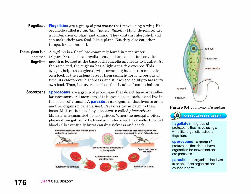
176 UNIT 3 CELL BIOLOGY
Figure 9.4: A diagram of a euglena.
flagellates - a group of
protozoans that move using a
whip-like organelle called a
flagellum.
sporozoans - a group of
protozoans that do not have
organelles for movement and
are parasites.
parasite - an organism that lives
in or on a host organism and
causes it harm.
Flagellates Flagellates are a group of protozoans that move using a whip-like
organelle called a flagellum (plural, flagella) Many flagellates are
a combination of plant and animal. They contain chlorophyll and
can make their own food, like a plant. But they also eat other
things, like an animal.
The euglena is a
common
flagellate
A euglena is a flagellate commonly found in pond water
(Figure 9.4). It has a flagella located at one end of its body. Its
mouth is located at the base of the flagella and leads to a gullet. At
the same end, the euglena has a light-sensitive eyespot. This
eyespot helps the euglena swim towards light so it can make its
own food. If the euglena is kept from sunlight for long periods of
time, its chlorophyll disappears and it loses the ability to make its
own food. Then, it survives on food that it takes from its habitat.
Sporozoans Sporozoans are a group of protozoans that do not have organelles
for movement. All members of this group are parasites and live in
the bodies of animals. A parasite is an organism that lives in or on
another organism called a host. Parasites cause harm to their
hosts. Malaria is caused by a sporozoan called plasmodium.
Malaria is transmitted by mosquitoes. When the mosquito bites,
plasmodium gets into the blood and infects red blood cells. Infected
blood cells eventually burst causing sickness and death.

1779.1 PROTOZOANS
CHAPTER 9: THE MICROSCOPIC WORLD
9.1 Section Review
1. To which kingdom do protozoans belong? What other organisms
are in that kingdom?
2. What are some animal-like characteristics of protozoans?
Which characteristic of protozoans is not animal-like?
3. What type of environment do protozoans need to survive?
4. Label the parts of the organisms below:
5. Complete the table below:
Protozoan groupType of
movement
Other
characteristicsExample
Ciliates paramecium
Amoebasuse pseudopods
to get food
Flagellates flagellum
Sporozoansmany species
are parasites
Each group of protozoans (ciliates,
amoebas, flagellates, and
sporozoans) has parasitic species.
Use the Internet and books to find
at least one disease that affects
humans and is caused by a
member of each group.
Next, pick one of the diseases and
make an informational brochure
about it. Include the following
information in your brochure.
1. What is the name and group of
the organism that causes the
disease?
2. How is the disease
transmitted?
3. What are the symptoms of the
disease?
4. What parts of the world does
the disease affect?
5. What are the treatments for the
disease?
6. How can the disease be
prevented?
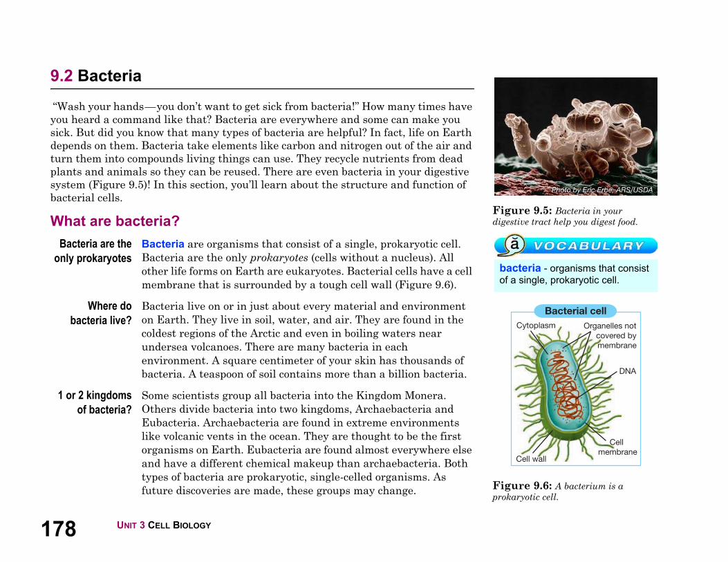
178 UNIT 3 CELL BIOLOGY
Figure 9.5: Bacteria in your
digestive tract help you digest food.
Figure 9.6: A bacterium is a
prokaryotic cell.
bacteria - organisms that consist
of a single, prokaryotic cell.
Bacterial cell
Organelles not
covered by
membrane
DNA
Cell
membraneCell wall
Cytoplasm
9.2 Bacteria
“Wash your hands—you don’t want to get sick from bacteria!” How many times have
you heard a command like that? Bacteria are everywhere and some can make you
sick. But did you know that many types of bacteria are helpful? In fact, life on Earth
depends on them. Bacteria take elements like carbon and nitrogen out of the air and
turn them into compounds living things can use. They recycle nutrients from dead
plants and animals so they can be reused. There are even bacteria in your digestive
system (Figure 9.5)! In this section, you’ll learn about the structure and function of
bacterial cells.
What are bacteria?
Bacteria are the
only prokaryotes
Bacteria are organisms that consist of a single, prokaryotic cell.
Bacteria are the only prokaryotes (cells without a nucleus). All
other life forms on Earth are eukaryotes. Bacterial cells have a cell
membrane that is surrounded by a tough cell wall (Figure 9.6).
Where do
bacteria live?
Bacteria live on or in just about every material and environment
on Earth. They live in soil, water, and air. They are found in the
coldest regions of the Arctic and even in boiling waters near
undersea volcanoes. There are many bacteria in each
environment. A square centimeter of your skin has thousands of
bacteria. A teaspoon of soil contains more than a billion bacteria.
1 or 2 kingdoms
of bacteria?
Some scientists group all bacteria into the Kingdom Monera.
Others divide bacteria into two kingdoms, Archaebacteria and
Eubacteria. Archaebacteria are found in extreme environments
like volcanic vents in the ocean. They are thought to be the first
organisms on Earth. Eubacteria are found almost everywhere else
and have a different chemical makeup than archaebacteria. Both
types of bacteria are prokaryotic, single-celled organisms. As
future discoveries are made, these groups may change.
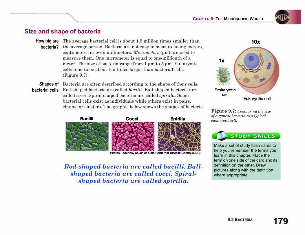
1799.2 BACTERIA
CHAPTER 9: THE MICROSCOPIC WORLD
Size and shape of bacteria
How big are
bacteria?
The average bacterial cell is about 1.5 million times smaller than
the average person. Bacteria are not easy to measure using meters,
centimeters, or even millimeters. Micrometers ( m) are used to
measure them. One micrometer is equal to one-millionth of a
meter. The size of bacteria range from 1 m to 5 m. Eukaryotic
cells tend to be about ten times larger than bacterial cells
(Figure 9.7).
Shapes of
bacterial cells
Bacteria are often described according to the shape of their cells.
Rod-shaped bacteria are called bacilli. Ball-shaped bacteria are
called cocci. Spiral-shaped bacteria are called spirilla. Some
bacterial cells exist as individuals while others exist in pairs,
chains, or clusters. The graphic below shows the shapes of bacteria.
Rod-shaped bacteria are called bacilli. Ball-
shaped bacteria are called cocci. Spiral-
shaped bacteria are called spirilla.
Figure 9.7: Comparing the size
of a typical bacteria to a typical
eukaryotic cell.
Make a set of study flash cards to
help you remember the terms you
learn in this chapter. Place the
term on one side of the card and its
definition on the other. Draw
pictures along with the definition
where appropriate.
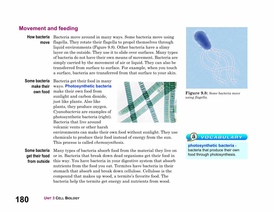
180 UNIT 3 CELL BIOLOGY
Figure 9.8: Some bacteria move
using flagella.
photosynthetic bacteria -
bacteria that produce their own
food through photosynthesis.
Movement and feeding
How bacteria
move
Bacteria move around in many ways. Some bacteria move using
flagella. They rotate their flagella to propel themselves through
liquid environments (Figure 9.8). Other bacteria have a slimy
layer on the outside. They use it to slide over surfaces. Many types
of bacteria do not have their own means of movement. Bacteria are
simply carried by the movement of air or liquid. They can also be
transferred from surface to surface. For example, when you touch
a surface, bacteria are transferred from that surface to your skin.
Some bacteria
make their
own food
Bacteria get their food in many
ways. Photosynthetic bacteria
make their own food from
sunlight and carbon dioxide,
just like plants. Also like
plants, they produce oxygen.
Cyanobacteria are examples of
photosynthetic bacteria (right).
Bacteria that live around
volcanic vents or other harsh
environments can make their own food without sunlight. They use
chemicals to produce their food instead of energy from the sun.
This process is called chemosynthesis.
Some bacteria
get their food
from outside
Many types of bacteria absorb food from the material they live on
or in. Bacteria that break down dead organisms get their food in
this way. You have bacteria in your digestive system that absorb
nutrients from the food you eat. Termites have bacteria in their
stomach that absorb and break down cellulose. Cellulose is the
compound that makes up wood, a termite’s favorite food. The
bacteria help the termite get energy and nutrients from wood.

1819.2 BACTERIA
CHAPTER 9: THE MICROSCOPIC WORLD
Bacteria and the beginning of life on Earth
Bacteria were the
first organisms
Scientists believe that bacteria were the first organisms on Earth.
Evidence comes from fossils of single-celled prokaryotes found in
rocks that are more than 3 billion years old. At that time, there was
little oxygen in the atmosphere. The earliest life was therefore
anaerobic (Latin for “without oxygen”). Anaerobic bacteria do not
require oxygen for cellular respiration. Today, anaerobic bacteria
thrive in places that have little or no oxygen, like swamps.
Bacteria
increased oxygen
in Earth’s
atmosphere
Over time, some bacteria developed the ability to use
photosynthesis. Cyanobacteria, still in existence today, were
one of the first photosynthetic bacteria. One of the products of
photosynthesis is oxygen. Over hundreds of millions of years, the
amount of oxygen in Earth’s atmosphere increased. This allowed
aerobic bacteria to develop. Aerobic bacteria use oxygen for
cellular respiration. There are many different species of aerobic
bacteria living today.
Eukaryotic cells
developed from
prokaryotic cells
Eventually, eukaryotic cells developed from bacteria. A scientific
theory states that long ago, smaller prokaryotic cells were engulfed
by larger prokaryotic cells. The smaller cells began to survive by
living inside of the larger cells. Over time they took on specific
functions inside the larger cells like producing energy. Eventually,
the smaller cells became the organelles (like mitochondria) inside
of eukaryotic cells (Figure 9.9).
Figure 9.9: How eukaryotic cells
developed from prokaryotic cells.
anaerobic bacteria - bacteria
that do not require oxygen to
survive.
aerobic bacteria - bacteria that
use oxygen for cellular respiration.
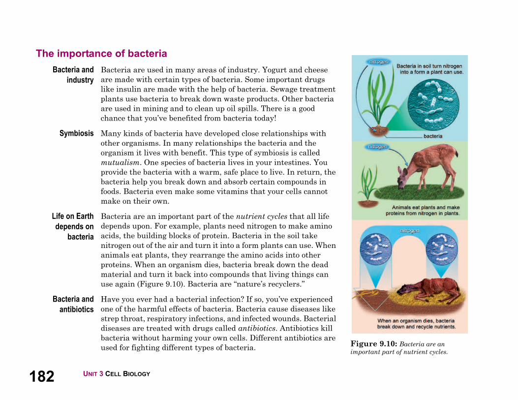
182 UNIT 3 CELL BIOLOGY
Figure 9.10: Bacteria are an
important part of nutrient cycles.
The importance of bacteria
Bacteria and
industry
Bacteria are used in many areas of industry. Yogurt and cheese
are made with certain types of bacteria. Some important drugs
like insulin are made with the help of bacteria. Sewage treatment
plants use bacteria to break down waste products. Other bacteria
are used in mining and to clean up oil spills. There is a good
chance that you’ve benefited from bacteria today!
Symbiosis Many kinds of bacteria have developed close relationships with
other organisms. In many relationships the bacteria and the
organism it lives with benefit. This type of symbiosis is called
mutualism. One species of bacteria lives in your intestines. You
provide the bacteria with a warm, safe place to live. In return, the
bacteria help you break down and absorb certain compounds in
foods. Bacteria even make some vitamins that your cells cannot
make on their own.
Life on Earth
depends on
bacteria
Bacteria are an important part of the nutrient cycles that all life
depends upon. For example, plants need nitrogen to make amino
acids, the building blocks of protein. Bacteria in the soil take
nitrogen out of the air and turn it into a form plants can use. When
animals eat plants, they rearrange the amino acids into other
proteins. When an organism dies, bacteria break down the dead
material and turn it back into compounds that living things can
use again (Figure 9.10). Bacteria are “nature’s recyclers.”
Bacteria and
antibiotics
Have you ever had a bacterial infection? If so, you’ve experienced
one of the harmful effects of bacteria. Bacteria cause diseases like
strep throat, respiratory infections, and infected wounds. Bacterial
diseases are treated with drugs called antibiotics. Antibiotics kill
bacteria without harming your own cells. Different antibiotics are
used for fighting different types of bacteria.
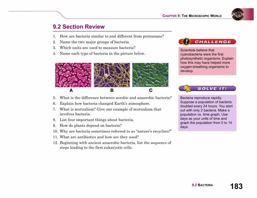
1839.2 BACTERIA
CHAPTER 9: THE MICROSCOPIC WORLD
9.2 Section Review
1. How are bacteria similar to and different from protozoans?
2. Name the two major groups of bacteria.
3. Which units are used to measure bacteria?
4. Name each type of bacteria in the picture below.
5. What is the difference between aerobic and anaerobic bacteria?
6. Explain how bacteria changed Earth’s atmosphere.
7. What is mutualism? Give one example of mutualism that
involves bacteria.
8. List four important things about bacteria.
9. How do plants depend on bacteria?
10. Why are bacteria sometimes referred to as “nature’s recyclers?”
11. What are antibiotics and how are they used?
12. Beginning with ancient anaerobic bacteria, list the sequence of
steps leading to the first eukaryotic cells.
Scientists believe that
cyanobacteria were the first
photosynthetic organisms. Explain
how this may have helped more
oxygen-breathing organisms to
develop.
Bacteria reproduce rapidly.
Suppose a population of bacteria
doubled every 24 hours. You start
out with only 2 bacteria. Make a
population vs. time graph. Use
days as your units of time and
graph the population from 0 to 14
days.

184 UNIT 3 CELL BIOLOGY
Figure 9.11: An image of flu viruses
bursting out of a cell. The image was
captured using an electron microscope.
Photo courtesy CDC Public Health
Image Library.
virus - a tiny, nonliving particle
made up of genetic material and
protein.
host cell - a cell that is, or
becomes, infected with a virus.
9.3 Viruses
Have you ever had the flu? Your muscles ache and your throat is sore. You also get a
fever and an upset stomach. The flu is a disease caused by a virus. Viruses infect
cells and cause many diseases, including smallpox, flu, AIDS, and the common cold.
To infect means to invade and produce an infection. Viruses infect virtually all types
of cells: bacterial cells, protozoan, fungal, plant, animal, and human. In this section
you will learn about viruses and how they infect cells.
The structure of viruses
What is a virus? A virus is a tiny, nonliving particle made up of genetic material
and protein. Viruses are not cells and are not made of cells. By
itself, a virus can do nothing. It does not eat, produce its own food,
or reproduce. All a virus can do is wait for a host cell to infect. A
host cell is a cell that is, or becomes, infected with a virus. Both
prokaryotic and eukaryotic cells can be hosts to viruses. Flu
viruses may infect cells of your respiratory tract (Figure 9.11).
When the virus spreads to many of your cells, you get sick.
The structure of
viruses
Viruses can be as much as 10,000 times smaller than bacteria. A
virus contains a core DNA.
Surrounding that core is a
protein coat. In some viruses, the
protein coat is covered by an
envelope made of proteins, lipids,
and carbohydrates. That
envelope may have spikes made
of carbohydrates and proteins
that help the virus particles
attach to host cells.

1859.3 VIRUSES
CHAPTER 9: THE MICROSCOPIC WORLD
How viruses infect cells
Host cells
become factories
for the virus
When some viruses come into contact with host cells, they trigger
the cells to engulf them. Other viruses fuse themselves to the cell
membrane and release their DNA into the cell. Once inside, the
viral DNA changes the function of the cell. The cell now becomes a
factory that produces new viruses. Eventually the infected cell dies
and bursts, freeing the new viruses. In some cases, new viruses just
pinch off so the cell remains alive.
Viruses and
host cells
A virus must be able to get its DNA inside of a cell before it can
multiply. The cell membrane controls what enters the cell. How
does a virus trick a cell into letting it enter? The “lock and key”
mechanism is the most common explanation. Certain proteins on
the virus’ protein coat must fit certain receptor sites on the host’s
cell membrane (Figure 9.12). If the proteins fit, the virus can enter
and infect the cell. If the proteins do not fit, the virus cannot enter
the cell or fuse with its cell membrane. Thus the viral DNA cannot
enter the cell and cause an infection.Figure 9.12: If the proteins fit, the
virus can infect the cell.
A tank crashes through the walls of
a car factory. People in the tank
get out and turn the car factory into
a tank factory. An analogy is a way
to find similarities between things
that are different. How does this
analogy explain how a virus
reproduces? Try and think of
another analogy for how a virus
reproduces.

186 UNIT 3 CELL BIOLOGY
Figure 9.13: Antibodies prevent
viruses from entering cells.
immune system - a system that
protects an organism from
unfamiliar objects like viruses.
antibodies - proteins that bind to
viruses and prevent them from
infecting cells.
The spread of viruses and immunity
The spread of a
viral infection
Once free from the host cell, new viruses infect other cells.
Because one virus causes a cell to produce thousands of new
viruses, viral infections spread quickly throughout the body.
Catching the flu is a good example of how this process works.
1. An infected person sneezes near you.
2. You inhale a virus, and it attaches to cells lining the inside of
your nose.
3. The virus attacks those cells and causes them to make new
viruses.
4. The host cells break and new viruses spread into your
bloodstream and also into your lungs. Infected tissues cause
different symptoms like muscle aches and sore throat.
The immune
system
Your immune system protects your cells from unfamiliar objects
like viruses and bacteria. With the flu virus, your immune system
produces chemicals that cause your body temperature to increase.
You get a fever. That fever slows down the production of new
viruses. This is because most of your body’s chemical reactions
work best at a temperature of 98.6 °F (37 °C). If your temperature
rises, the reactions slow down.
Antibodies Once the cells of your immune system recognizes a virus, they
make antibodies to stop further infections. Antibodies are
proteins that bind to viruses and prevent them from infecting cells
(Figure 9.13). If you come in contact with the same virus again,
the cells of your immune system recognize it and immediately
start producing antibodies to stop the virus’s spread. The cells of
your immune system produce different antibodies for different
viruses.

1879.3 VIRUSES
CHAPTER 9: THE MICROSCOPIC WORLD
Vaccines A vaccine causes your immune system to produce antibodies to a
particular virus. A vaccine is a preparation made from weakened
virus particles or their empty protein coats. That is why vaccines do
not make you sick. Your immune system mounts a response
against the particles and makes antibodies. When you come in
contact with the real virus, your immune system acts quickly to
prevent illness (Figure 9.14).
Changes in the
protein coat
New vaccines must be made each year to prevent some viral
infections. The flu is a good example. After multiplying many
times, flu viruses end up with mistakes in their genetic
instructions. These mistakes may alter the protein coat slightly.
With a different protein coat, the immune system may not
recognize the virus. This means that one year’s batch of flu
vaccines might not be as effective against the flu virus the
next year.
9.3 Section Review
1. Why is a virus not considered a living thing?
2. Explain using steps, how a virus multiplies.
3. Name three diseases caused by a virus.
4. Explain how a virus tricks a cell so it can enter it through the
cell membrane.
5. Describe, using several steps, how a virus infects cells and
spreads throughout an organism.
6. How does the immune system try to fight off a virus?
7. What is a vaccine? Why do you think a vaccine is sometimes
referred to as “artificial immunity?”
8. Explain why a new flu vaccine has to be produced each year. Figure 9.14: A vaccination helps
your immune system produce antibodies
quickly when exposed to a virus.
vaccine - a preparation of virus
particles that, when injected into
the body, causes the immune
system to produce antibodies.

188
The Good, The Bad, The Microbe
What are our bodies made of? Water, oxygen, tissue, yes. But
you might be surprised to learn that living creatures also
make up much of the human body. They are called microbes
and billions of them are swirling inside you and on your skin
right now. We cannot see or feel them, but microbes are all
over us. And we can't live without them.
What is a microbe?
Microbes are single-
celled organisms too
small to see with the
human eye. Scientists
use microscopes to
study them and
understand how they
work. There are four
major types of microbes:
bacteria, viruses, fungi,
and protozoa.
Microbes live all around us, in air and soil, in rocks and
water. They also live in plants, animals, and in our bodies.
Microbes are the oldest life form on Earth. Scientists
estimate that these creatures date more than 3.5 billion
years.
Microbiologists are scientists who study microbes. They
work in a variety of settings: helping to keep our food and
water from contamination, working in hospitals to determine
what germs make us sick, or trying to solve environmental
problems.
A friend and an enemy
Microbes are often called by the nickname “bugs.” Some
microbes can cause sicknesses like the common cold, strep
throat, and chicken pox. However, more than 95 percent of
microbes are harmless, despite their bad reputation. For
example, Escherichia coli (E. coli) lives safely in our
intestines. E. coli produces vitamins K and B-complex, two
essential nutrients we cannot make otherwise. We also have
many other useful bacteria living in our intestines that
prevent dangerous bacteria from infecting our bodies.
Although most E. coli is helpful to our bodies, a rare strain
causes severe food poisoning. It has a slightly different
genetic makeup than the E. coli in our intestines. That other
strain of E. coli is usually spread through contaminated
animal meat, but can be killed easily by heat. All the more
reason why the meat we eat be cooked to an internal
temperature of
160° Fahrenheit.
Bacteria play an
important role in
producing food
and medicine. For
example, yogurt,
sauerkraut, and
cheese are all
made with
bacteria.
Streptomyces, a
bacteria found in
soil, is used to make
the antibiotic streptomycin.
Ch
ap
ter
9 C
on
necti
on
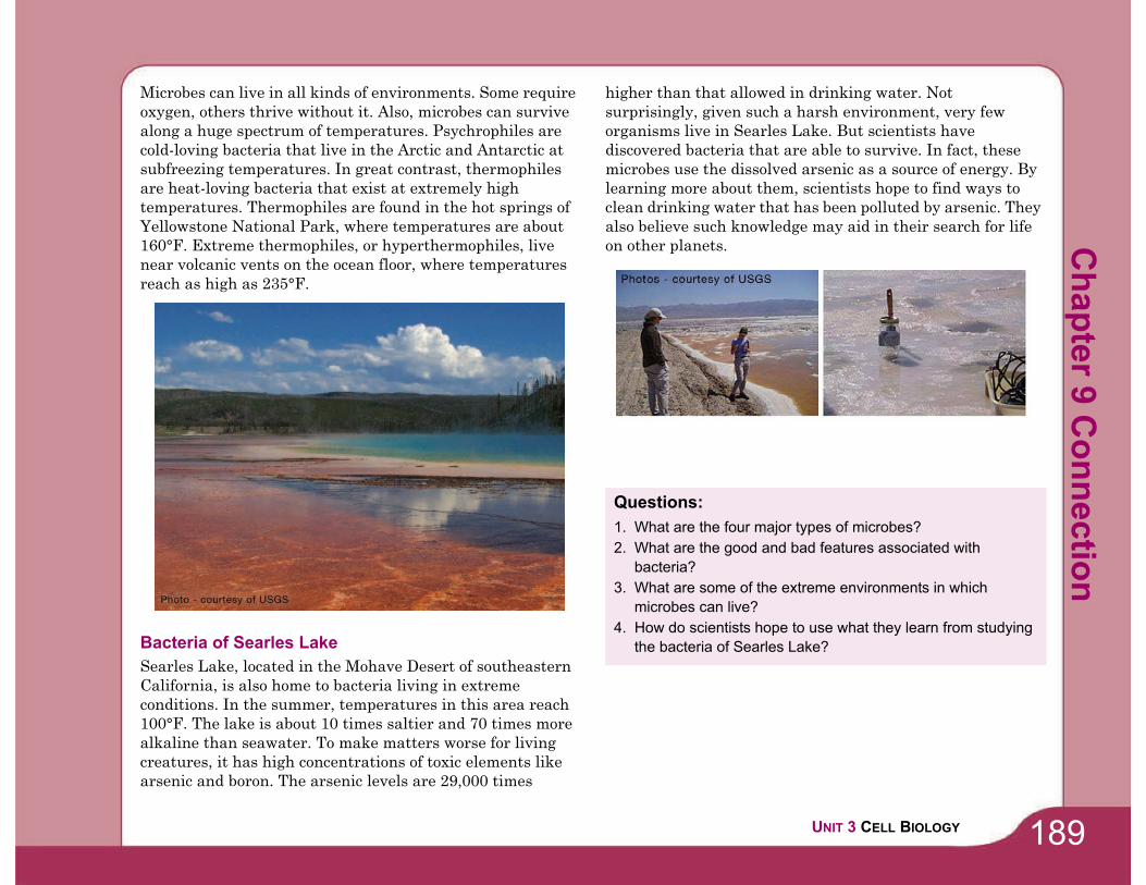
189UNIT 3 CELL BIOLOGY
Microbes can live in all kinds of environments. Some require
oxygen, others thrive without it. Also, microbes can survive
along a huge spectrum of temperatures. Psychrophiles are
cold-loving bacteria that live in the Arctic and Antarctic at
subfreezing temperatures. In great contrast, thermophiles
are heat-loving bacteria that exist at extremely high
temperatures. Thermophiles are found in the hot springs of
Yellowstone National Park, where temperatures are about
160°F. Extreme thermophiles, or hyperthermophiles, live
near volcanic vents on the ocean floor, where temperatures
reach as high as 235°F.
Bacteria of Searles Lake
Searles Lake, located in the Mohave Desert of southeastern
California, is also home to bacteria living in extreme
conditions. In the summer, temperatures in this area reach
100°F. The lake is about 10 times saltier and 70 times more
alkaline than seawater. To make matters worse for living
creatures, it has high concentrations of toxic elements like
arsenic and boron. The arsenic levels are 29,000 times
higher than that allowed in drinking water. Not
surprisingly, given such a harsh environment, very few
organisms live in Searles Lake. But scientists have
discovered bacteria that are able to survive. In fact, these
microbes use the dissolved arsenic as a source of energy. By
learning more about them, scientists hope to find ways to
clean drinking water that has been polluted by arsenic. They
also believe such knowledge may aid in their search for life
on other planets.
Questions:
1. What are the four major types of microbes?
2. What are the good and bad features associated with
bacteria?
3. What are some of the extreme environments in which
microbes can live?
4. How do scientists hope to use what they learn from studying
the bacteria of Searles Lake?
Ch
ap
ter 9
Co
nn
ectio
n

190
Outbreak! Patient Zero
An epidemic spreads rapidly by infection and affects many
individuals in a population at the same time. Examples of
epidemics could be the flu, measles, and strep throat. Patient
zero is the first patient in a population to become infected.
Imagine that there is an outbreak of a bacterial infection at
your school. You know that the infection spreads through
physical contact. Patient zero could have caught the bacteria
by touching an object that was infected. This person then
spread the bacteria by touching other items and individuals.
In fact, you could be patient zero and don’t even know it! In
order to contain the infection, you need to isolate its source.
See if you can figure out the source of the infection in the
following simulation.
For the activity, you will need:
• 1 cup of clear liquid from your teacher with a plastic dropper.• A few drops of indicator solution from your teacher (red cabbage juice)
What you will do
1. Obtain a cup with clear liquid and a dropper from your
teacher. The cup represents your body fluids. One of the
cups in the classroom is infected with a disease!
2. Move around the classroom and find a partner. Both
partners will place 5 drops of their liquid into the cup of
their partner. Do NOT dip your dropper into your
partner's cup. Let the liquid drop in to avoid
contamination.
3. Empty any remaining liquid back into your own cup.
4. Repeat the process with another partner and then return
to your seat.
5. Make a hypothesis about the number of infections in the
class. Record your hypothesis
6. Your teacher will place 1-2 drops of the indicator into
your cup. If your are infected your liquid will turn a blue
or greenish color.
7. Count and record the number of infections in the class.
8. Make a hypothesis about the person whom you think
started the outbreak in your classroom and why.
9. Make a flow chart showing how the infection possibly
spread. Your teacher will identify the student who spread
the disease.
Applying your knowledge
a. Identify at least 2 ways that the outbreak activity is
realistic.
b. You often come in contact with sick students but don't
become ill. What defenses does the human body have to
fight off an infection?
c. How can you prevent the spread of diseases that are
passed through excange of body fluids?
d. Not all diseases are spread by exchange of body fluids.
How else might diseases spread? List at least 3 other
forms of spreading a disease.
Ch
ap
ter
9 A
cti
vit
y
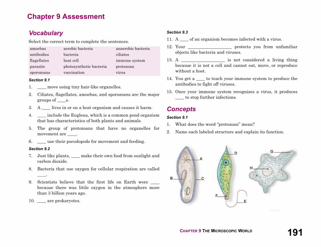
CHAPTER 9 THE MICROSCOPIC WORLD 191
Chapter 9 Assessment
Vocabulary
Select the correct term to complete the sentences.
Section 9.1
1. ____ move using tiny hair-like organelles.
2. Ciliates, flagellates, amoebas, and sporozoans are the major
groups of ____s.
3. A ____ lives in or on a host organism and causes it harm.
4. ____ include the Euglena, which is a common pond organism
that has characteristics of both plants and animals.
5. The group of protozoans that have no organelles for
movement are ____.
6. ____ use their pseudopods for movement and feeding.
Section 9.2
7. Just like plants, ____ make their own food from sunlight and
carbon dioxide.
8. Bacteria that use oxygen for cellular respiration are called
____.
9. Scientists believe that the first life on Earth were ____
because there was little oxygen in the atmosphere more
than 3 billion years ago.
10. ____ are prokaryotes.
Section 9.3
11. A ____ of an organism becomes infected with a virus.
12. Your ____________________ protects you from unfamiliar
objects like bacteria and viruses.
13. A ____________________ is not considered a living thing
because it is not a cell and cannot eat, move, or reproduce
without a host.
14. You get a ____ to teach your immune system to produce the
antibodies to fight off viruses.
15. Once your immune system recognizes a virus, it produces
____ to stop further infections.
Concepts
Section 9.1
1. What does the word “protozoan” mean?
2. Name each labeled structure and explain its function.
amoebas
antibodies
flagellates
parasite
sporozoans
aerobic bacteria
bacteria
host cell
photosynthetic bacteria
vaccination
anaerobic bacteria
ciliates
immune system
protozoan
virus
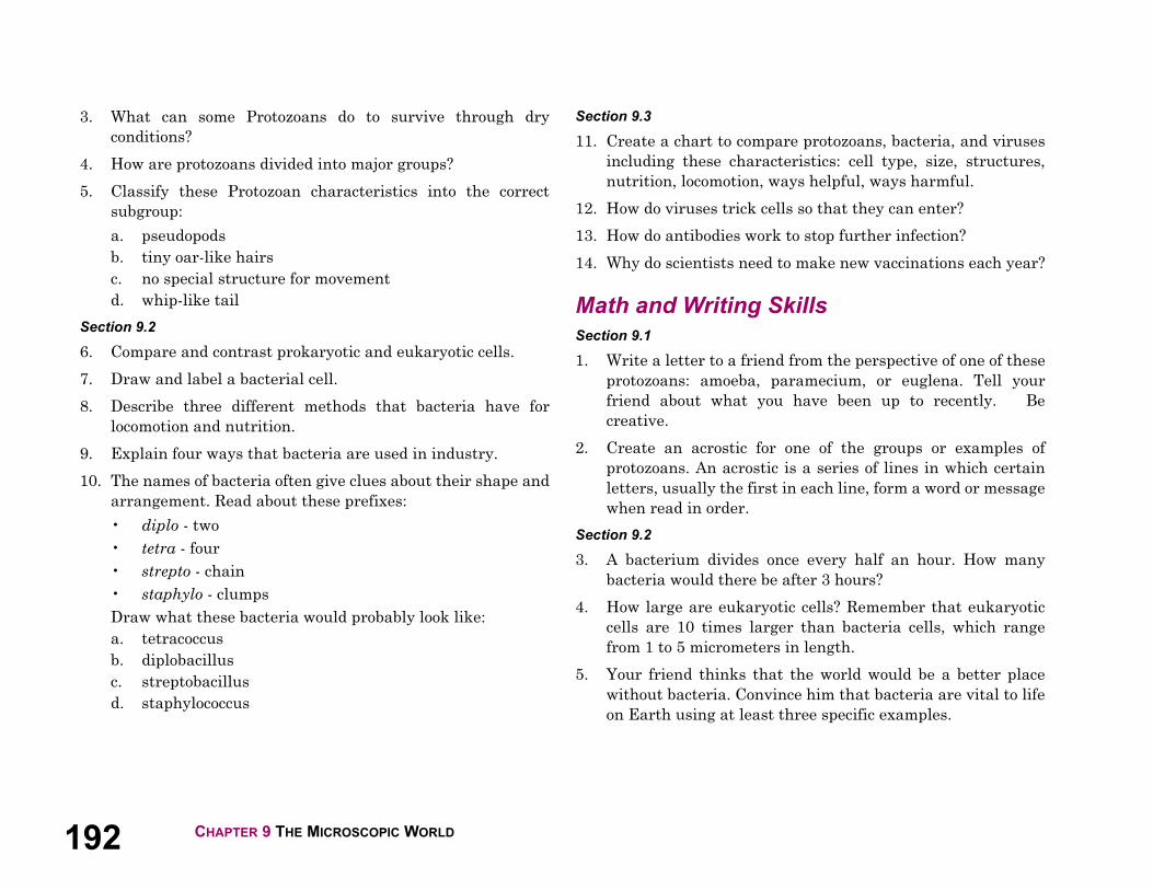
192 CHAPTER 9 THE MICROSCOPIC WORLD
3. What can some Protozoans do to survive through dry
conditions?
4. How are protozoans divided into major groups?
5. Classify these Protozoan characteristics into the correct
subgroup:
a. pseudopods
b. tiny oar-like hairs
c. no special structure for movement
d. whip-like tail
Section 9.2
6. Compare and contrast prokaryotic and eukaryotic cells.
7. Draw and label a bacterial cell.
8. Describe three different methods that bacteria have for
locomotion and nutrition.
9. Explain four ways that bacteria are used in industry.
10. The names of bacteria often give clues about their shape and
arrangement. Read about these prefixes:
• diplo - two
• tetra - four
• strepto - chain
• staphylo - clumps
Draw what these bacteria would probably look like:
a. tetracoccus
b. diplobacillus
c. streptobacillus
d. staphylococcus
Section 9.3
11. Create a chart to compare protozoans, bacteria, and viruses
including these characteristics: cell type, size, structures,
nutrition, locomotion, ways helpful, ways harmful.
12. How do viruses trick cells so that they can enter?
13. How do antibodies work to stop further infection?
14. Why do scientists need to make new vaccinations each year?
Math and Writing Skills
Section 9.1
1. Write a letter to a friend from the perspective of one of these
protozoans: amoeba, paramecium, or euglena. Tell your
friend about what you have been up to recently. Be
creative.
2. Create an acrostic for one of the groups or examples of
protozoans. An acrostic is a series of lines in which certain
letters, usually the first in each line, form a word or message
when read in order.
Section 9.2
3. A bacterium divides once every half an hour. How many
bacteria would there be after 3 hours?
4. How large are eukaryotic cells? Remember that eukaryotic
cells are 10 times larger than bacteria cells, which range
from 1 to 5 micrometers in length.
5. Your friend thinks that the world would be a better place
without bacteria. Convince him that bacteria are vital to life
on Earth using at least three specific examples.
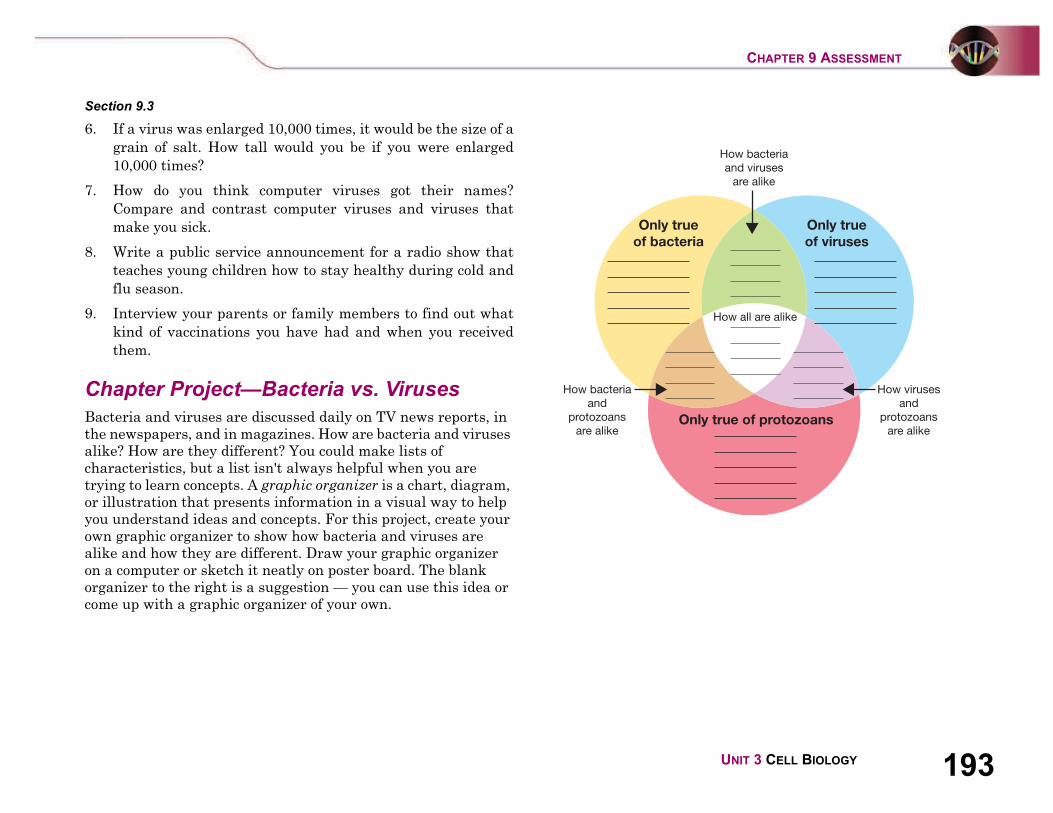
193UNIT 3 CELL BIOLOGY
CHAPTER 9 ASSESSMENT
Section 9.3
6. If a virus was enlarged 10,000 times, it would be the size of a
grain of salt. How tall would you be if you were enlarged
10,000 times?
7. How do you think computer viruses got their names?
Compare and contrast computer viruses and viruses that
make you sick.
8. Write a public service announcement for a radio show that
teaches young children how to stay healthy during cold and
flu season.
9. Interview your parents or family members to find out what
kind of vaccinations you have had and when you received
them.
Chapter Project—Bacteria vs. Viruses
Bacteria and viruses are discussed daily on TV news reports, in
the newspapers, and in magazines. How are bacteria and viruses
alike? How are they different? You could make lists of
characteristics, but a list isn't always helpful when you are
trying to learn concepts. A graphic organizer is a chart, diagram,
or illustration that presents information in a visual way to help
you understand ideas and concepts. For this project, create your
own graphic organizer to show how bacteria and viruses are
alike and how they are different. Draw your graphic organizer
on a computer or sketch it neatly on poster board. The blank
organizer to the right is a suggestion — you can use this idea or
come up with a graphic organizer of your own.
Only true
of bacteria
How bacteria
and viruses
are alike
How all are alike
How bacteria
and
protozoans
are alike
How viruses
and
protozoans
are alike
Only true
of viruses
Only true of protozoans