UNIT 2 - pages.stolaf.edu · Okazaki fragment 2 DNA pol III makes Okazaki fragment 2. 5 ...
51
© 2014 Pearson Education, Inc. UNIT 2 A. Mendel and the Gene Idea (Ch11) B. Chromosomal Basis of Inheritance (Ch12) C. Molecular Basis of Inheritance (Ch13) D. Gene Expression from Gene to Protein (Ch 14) Where are we going? CH13 More discoveries concerning the heritable material! What is it? What might its structure be? Replication and how it works Listen to an interview of Watson We will try to keep our eye on big picture as we look over details of replication!
-
Upload
truongduong -
Category
Documents
-
view
213 -
download
0
Transcript of UNIT 2 - pages.stolaf.edu · Okazaki fragment 2 DNA pol III makes Okazaki fragment 2. 5 ...

© 2014 Pearson Education, Inc.
UNIT 2 A. Mendel and the Gene Idea (Ch11) B. Chromosomal Basis of Inheritance (Ch12) C. Molecular Basis of Inheritance (Ch13) D. Gene Expression from Gene to Protein (Ch 14) Where are we going?
CH13More discoveries concerning the heritable material!What is it?What might its structure be?Replication and how it worksListen to an interview of WatsonWe will try to keep our eye on big picture as we look over details of replication!

© 2014 Pearson Education, Inc.
Figure 13.21b
Loops
30-nm fiber
300-nm fiber
Replicated chromosome (1,400 nm)
Scaffold
Chromatid (700 nm)
Reminder of structure!

© 2014 Pearson Education, Inc.
Figure 13.21a
Histone tail Histones
H1
DNA double helix (2 nm in diameter)
Nucleosome (10 nm in diameter)
Histones are a protein

© 2014 Pearson Education, Inc.
Figure 13.21
Histone tail Histones H1
DNA double helix (2 nm in diameter)
Nucleosome (10 nm in diameter)
Loops
30-nm fiber
300-nm fiber
Replicated chromosome (1,400 nm)
Scaffold
Chromatid (700 nm)

© 2014 Pearson Education, Inc.
Remember Morgan sex linked traits..He confirmed that genes are on chromosomes
But chromosomes are made up of protein AND DNA…which one is doing the important job of harboring information?
We will focus on these 3 classic experiments highlighted in this chapter……• Griffith (Fred)• Hershey and Chase (Alfred and Martha)• Meselson and Stahl (Matt and Frank)
© 2014 Pearson Education, Inc.
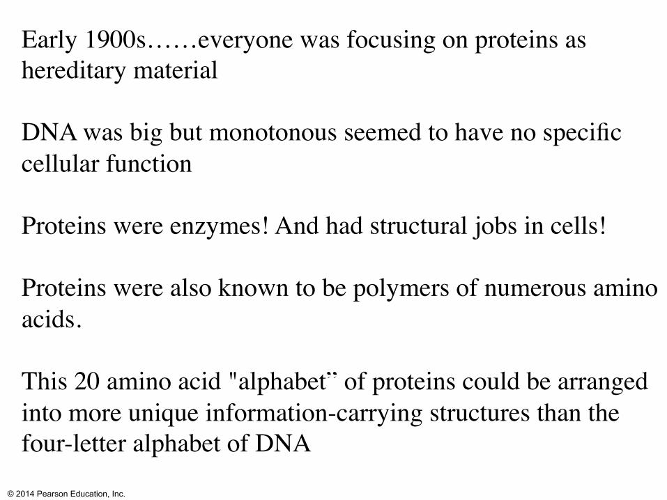
Early 1900s……everyone was focusing on proteins as hereditary material
DNA was big but monotonous seemed to have no specific cellular function
Proteins were enzymes! And had structural jobs in cells!
Proteins were also known to be polymers of numerous amino acids.
This 20 amino acid "alphabet” of proteins could be arranged into more unique information-carrying structures than the four-letter alphabet of DNA

© 2014 Pearson Education, Inc.
http://en.wikipedia.org/wiki/File:Fred_Griffith_and_%22Bobby%22_1936.jpg
Griffith (Trying to develop a vaccine)Turned to a bacterial pathogen…Streptococcus pneumoniae
Is it gram positive or negative?

© 2014 Pearson Education, Inc.
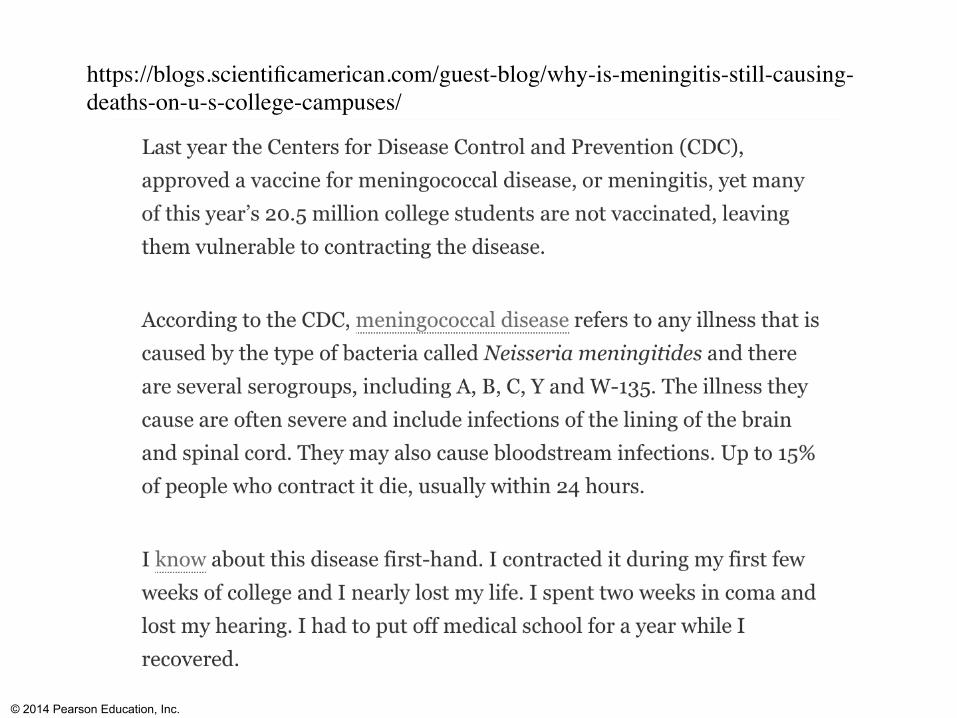
This species (Streptococcus pneumoniae) is one of the species that can cause meningitis (along with Neisseria meningitidis). What is meningitis?Did you get your meningitis vaccine?
Note you can also get viral meningitis but usually not as dangerous-no vaccine.
Streptococcus pneumoniae is also a cause of pneumonia along with a bunch of other organisms…
What is pneumonia?
© 2014 Pearson Education, Inc.
https://blogs.scientificamerican.com/guest-blog/why-is-meningitis-still-causing-deaths-on-u-s-college-campuses/

© 2014 Pearson Education, Inc.
So what did he do??
2 strains what do we mean by strains?
One virulent and not so virulent (What do we mean by virulent?)

© 2014 Pearson Education, Inc.
Figure 13.2
Living S cells
Mouse healthy Results
Experiment
Mouse healthy Mouse dies
Living S cells
Living R cells
Heat-killed S cells
Mixture of heat-killed S cells and living R cells
Mouse dies
Work by Avery identified the transforming substance as DNA (but others not convinced)

© 2014 Pearson Education, Inc.
Transformation-did not really understand mechanism
• Can we do this-pick up DNA from our environment?
• Why does this ability freak us out….what do we feed chickens pigs and cows to make them grow fast?
• Where do we put their manure waste??
• Where can this manure waste go?
© 2014 Pearson Education, Inc.
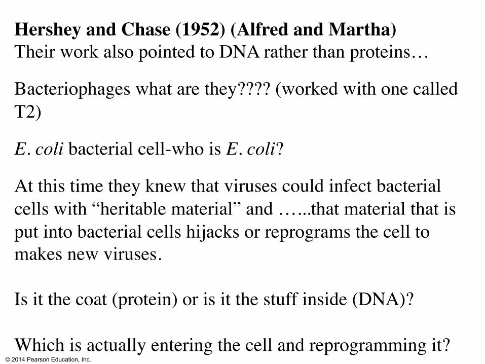
Hershey and Chase (1952) (Alfred and Martha)Their work also pointed to DNA rather than proteins…
Bacteriophages what are they???? (worked with one called T2)
E. coli bacterial cell-who is E. coli?
At this time they knew that viruses could infect bacterial cells with “heritable material” and …...that material that is put into bacterial cells hijacks or reprograms the cell to makes new viruses.
Is it the coat (protein) or is it the stuff inside (DNA)?
Which is actually entering the cell and reprogramming it?

© 2014 Pearson Education, Inc.
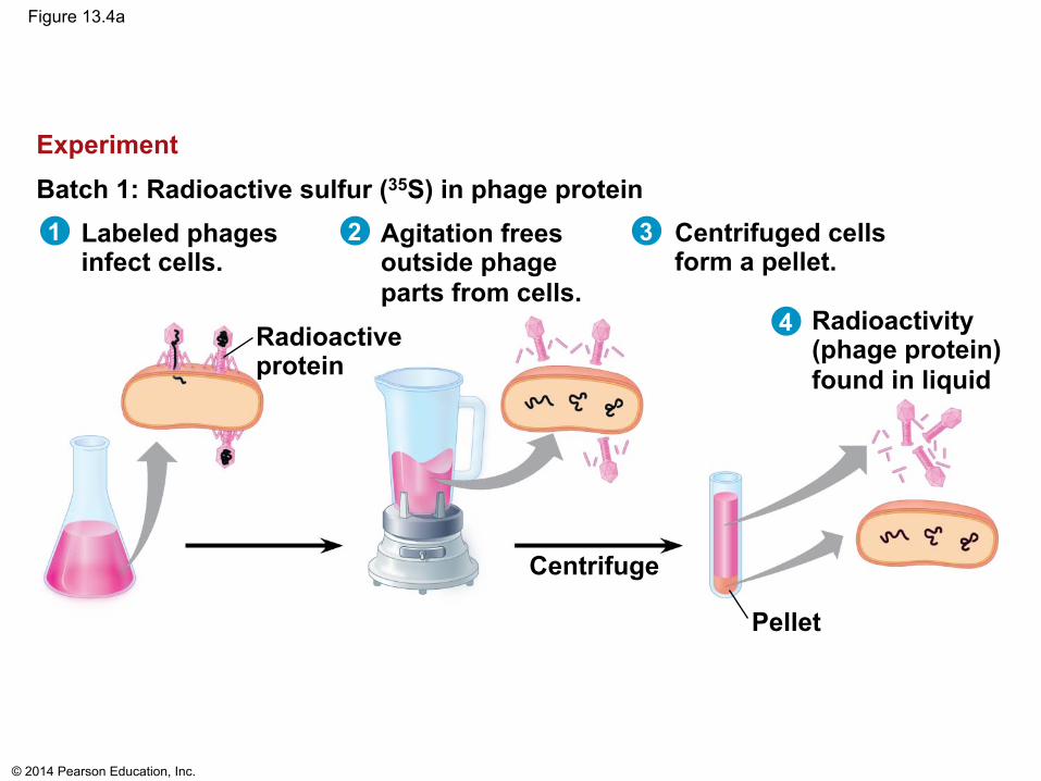
Some phages grown in media for a couple hrs with radioactive Sulphur…which should be incorporated into some coat proteins (Methionine, Cysteine)
Other phages grown in media for a couple hrs with radioactive Phosphorus….which should be incorporated into DNA

© 2014 Pearson Education, Inc.
These phages were allowed to infect bacteria.
Knocked off phage from cell and then centrifuged to concentrate cells. Where is Phosphorus (DNA)? Where is Sulfur (Protein Coat)??
Batch with P (DNA) labeled, found in cells.
Batch with S (Protein Coat) labeled, found in liquid
Was the protein coat passed on?
Thus, the Hershey–Chase experiment helped confirm that DNA, not protein, is the genetic material.
© 2014 Pearson Education, Inc.
Figure 13.4a
Labeled phages infect cells.
Batch 1: Radioactive sulfur (35S) in phage protein Experiment
Agitation frees outside phage parts from cells.
Centrifuged cells form a pellet.
Radioactivity (phage protein) found in liquid
Radioactive protein
Centrifuge
Pellet
1 2 3
4

© 2014 Pearson Education, Inc.
Figure 13.4b
Batch 2: Radioactive phosphorus (32P) in phage DNA
Radioactivity (phage DNA) found in pellet
Radioactive DNA
Centrifuge Pellet
Labeled phages infect cells.
Agitation frees outside phage parts from cells.
Centrifuged cells form a pellet.
1 2 3
4
Experiment

© 2014 Pearson Education, Inc.
Watson-Crick Model predicted….
Each of two daughter molecules would have one parental strand and one newly made!

© 2014 Pearson Education, Inc. http://www.dnaftb.org/20/gallery.html
The annual "degree granting" party of Max Delbrück's phage group held in the house shared by Meselson and Stahl at Caltech. (L-R) Harry Rubin, Max Delbrück, Rene Cohen, Matt Meselson, Frank Stahl
© 2014 Pearson Education, Inc.
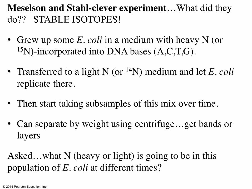
Meselson and Stahl-clever experiment…What did they do?? STABLE ISOTOPES!
• Grew up some E. coli in a medium with heavy N (or 15N)-incorporated into DNA bases (A,C,T,G).
• Transferred to a light N (or 14N) medium and let E. coli replicate there.
• Then start taking subsamples of this mix over time.
• Can separate by weight using centrifuge…get bands or layers
Asked…what N (heavy or light) is going to be in this population of E. coli at different times?

© 2014 Pearson Education, Inc.
Figure 13.11a
DNA sample centrifuged after first replication
DNA sample centrifuged after second replication
Bacteria cultured in medium with 15N (heavy isotope)
Bacteria transferred to medium with 14N (lighter isotope)
Less dense
More dense
Experiment
Results
1
3 4
2

© 2014 Pearson Education, Inc.
Figure 13.10
(a) Conservative model
(b) Semiconservative model
(c) Dispersive model
Parent cell First
replication Second
replication
Dark blue=heavy 15N
Move from heavy into light N media-what will happen when you centrifuge?

© 2014 Pearson Education, Inc.
Figure 13.10
(a) Conservative model
(b) Semiconservative model
(c) Dispersive model
Parent cell First
replication Second
replication
Dark blue=heavy 15N
Second Replication….How many bands?

© 2014 Pearson Education, Inc.
Figure 13.1
Watson and Crickhttp://www.ted.com/talks/james_watson_on_how_he_discovered_dna.html

© 2014 Pearson Education, Inc.
Watson interview!
What did you think was interesting?What was unusual?Surprising?Insights into how science works?

© 2014 Pearson Education, Inc.
First some info about structure of DNA!
© 2014 Pearson Education, Inc.
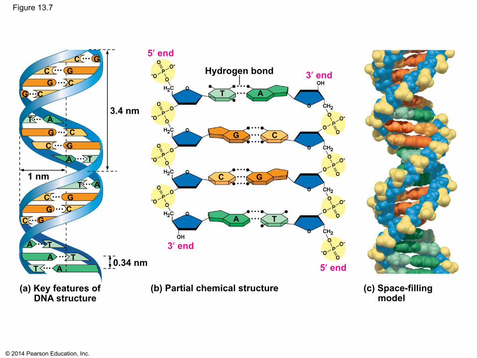
Figure 13.7
(c) Space-filling model
(a) Key features of DNA structure
(b) Partial chemical structure
3ʹ end
5ʹ end
3ʹ end
5ʹ end
Hydrogen bond
T A
C G
C G
3.4 nm
T A
T A
C G
C G
T
A
1 nm
0.34 nm
T A T
A
C G
C G
C
G
C G
T A
T A
C G C
G C G

© 2014 Pearson Education, Inc.
Figure 13.7a
(a) Key features of DNA structure
3.4 nm
T A
C G
C
G
T
A
1 nm
0.34 nm
T A T
A
C G
C G
C
G
C
G
T A
T A
C G C
G C G

© 2014 Pearson Education, Inc.
Figure 13.7b
3ʹ end
5ʹ end
3ʹ end
5ʹ end
Hydrogen bond
T A
C G
C G
T A
Phosphate group
12
What are the blue pentagons?
• A nucleotide is a phosphate molecule + a sugar molecule + a nitrogen rich base
Nitrogen rich bases A, T, G, C
A and G have double rings -two double rings would not fit so A bonds with T and G with C.

© 2014 Pearson Education, Inc.
Figure 13.5
Sugar– phosphate backbone
DNA nucleotide
Nitrogenous bases
3ʹ end
5ʹ end
Thymine (T)
Adenine (A)
Cytosine (C)
Guanine (G)

© 2014 Pearson Education, Inc.
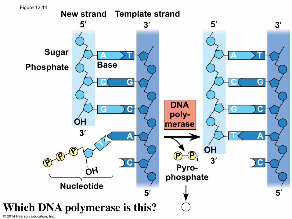
Figure 13.14
Pyro- phosphate
New strand
Phosphate
Nucleotide
5ʹ 3ʹ Template strand
Sugar Base
5ʹ
3ʹ
5ʹ
3ʹ
5ʹ 3ʹ
DNA poly-
merase T
A T
C G
A
C G
C
P
P i P
i 2
A T
C G
A
C G
C

© 2014 Pearson Education, Inc.
Figure 13.9-3
(a) Parental molecule
(b) Separation of parental strands into templates
(c) Formation of new strands complementary to template strands
through addition of nucleotides
T A
C G
C G
T A
T A T A
T A
C G
C G
T A
T A
C G
C G
T A
T A
T A
C G
C G
T A
T A

© 2014 Pearson Education, Inc.
DNA replicationWhere does it take place? When does it take place?How does it work??

© 2014 Pearson Education, Inc.
Figure 13.12
Replication fork
5ʹ
5ʹ 5ʹ 3ʹ
3ʹ
3ʹ
RNA primer
Origin of replication?Replication fork?3’ 5’? (skip)RNA primer (made by pink blob=primase). Other blobs (green=helicase=unzips)(blue-=topoisomerase)(gray=single stranded binding proteins)

© 2014 Pearson Education, Inc.
Figure 13.15
Parental DNA
5ʹ 3ʹ
5ʹ
3ʹ
5ʹ
3ʹ
Continuous elongation in the 5ʹ to 3ʹ direction
5ʹ 3ʹ
5ʹ
3ʹ
DNA pol III
RNA primer
5ʹ 3ʹ
Origin of replication
Origin of replication
Lagging strand
Lagging strand
Leading strand
Leading strand
Overview
Primer
DNA polymerase III (salmon blob)
Binds then ‘walks’ along, adding complementary nucleotide bases (A, C, G and T) to the strand of DNA (in the 5’ to 3’ direction-but you do not need to know this).It is making a leading strand!

© 2014 Pearson Education, Inc.
Figure 13.16a
Origin of replication Lagging strand Lagging
strand
Overall directions of replication
Leading strand
Leading strand
Overview
Lets zoom in to #1 and look at the lagging strand.

© 2014 Pearson Education, Inc.
Figure 13.16b-1
5ʹ 3ʹ
5ʹ
3ʹ RNA primer
Template strand
1
© 2014 Pearson Education, Inc.
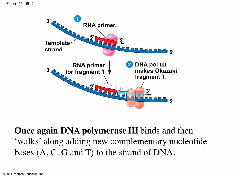
Figure 13.16b-2
5ʹ 3ʹ
5ʹ
3ʹ RNA primer.
RNA primer for fragment 1
Template strand
DNA pol III makes Okazaki fragment 1.
5ʹ 3ʹ
5ʹ
3ʹ
1
2
Once again DNA polymerase III binds and then ‘walks’ along adding new complementary nucleotide bases (A, C, G and T) to the strand of DNA.

© 2014 Pearson Education, Inc.
Figure 13.16b-3
5ʹ 3ʹ
5ʹ
3ʹ RNA primer.
RNA primer for fragment 1
Template strand
Okazaki fragment 1
DNA pol III makes Okazaki fragment 1.
DNA pol III detaches.
5ʹ 3ʹ
5ʹ
3ʹ
5ʹ
3ʹ 5ʹ
3ʹ
1
2
3
What happened? Why did it stop? What is fragment called? Where is DNA pol III going to go next??

© 2014 Pearson Education, Inc.
Figure 13.16c-1 RNA primer for fragment 2
Okazaki fragment 2 DNA pol III
makes Okazaki fragment 2.
5ʹ
3ʹ 5ʹ
3ʹ 4
Now you have all these bits what has to happen next? And who does that?
Can you leave the RNA primer pieces in there?

© 2014 Pearson Education, Inc.
Figure 13.16c-2 RNA primer for fragment 2
Okazaki fragment 2 DNA pol III
makes Okazaki fragment 2.
DNA pol I replaces RNA with DNA.
5ʹ
3ʹ 5ʹ
3ʹ
5ʹ
3ʹ 5ʹ
3ʹ 4
5
A different DNA polymerase comes in to replace them!(I vs III)

© 2014 Pearson Education, Inc.
Figure 13.17
3ʹ 5ʹ
Origin of replication
Lagging strand
Lagging strand
Overall directions of replication
Leading strand
Leading strand
Overview
5ʹ 3ʹ
5ʹ
3ʹ
Leading strand
Lagging strand
DNA ligase DNA pol I DNA pol III
Primase
DNA pol III Primer
5ʹ 3ʹ
5ʹ
3ʹ
Lagging strand template
Parental DNA
Helicase
Single-strand binding proteins
Leading strand template
© 2014 Pearson Education, Inc.
Figure 13.14
Pyro- phosphate
New strand
Phosphate
Nucleotide
5ʹ 3ʹ Template strand
Sugar Base
5ʹ
3ʹ
5ʹ
3ʹ
5ʹ 3ʹ
DNA poly-
merase T
A T
C G
A
C G
C P i P
A T
C G
A
C G
C
Which DNA polymerase is this?

© 2014 Pearson Education, Inc.
The diagram below shows a replication bubble with synthesis of the leading and lagging strands on both sides of the bubble. The parental DNA is shown in dark blue, the newly synthesized DNA is light blue, and the RNA primers associated with each strand are red. The origin of replication is indicated by the black dots on the parental strands.
Rank the primers (the red specks) in the order they were produced. If two primers were produced at the same time, overlap them.

© 2014 Pearson Education, Inc.
The diagram below shows a replication bubble with synthesis of the leading and lagging strands on both sides of the bubble. The parental DNA is shown in dark blue, the newly synthesized DNA is light blue, and the RNA primers associated with each strand are red. The origin of replication is indicated by the black dots on the parental strands.
Rank the primers in the order they were produced. If two primers were produced at the same time, overlap them.a and h then b and g then c and f and finally e and d

© 2014 Pearson Education, Inc.
True of Leading strand, Lagging strand, or Both????
Daughter strand elongates away from replication fork
Multiple primers needed
Made in segments
Made continuously
Daughter strand elongates toward replication fork

© 2014 Pearson Education, Inc.
True of Leading strand, Lagging strand, or Both????
Daughter strand elongates away from replication fork Lag
Multiple primers needed Lag
Made in segments Lag
Made continuouslyLead
Daughter strand elongates toward replication fork Lead

© 2014 Pearson Education, Inc.
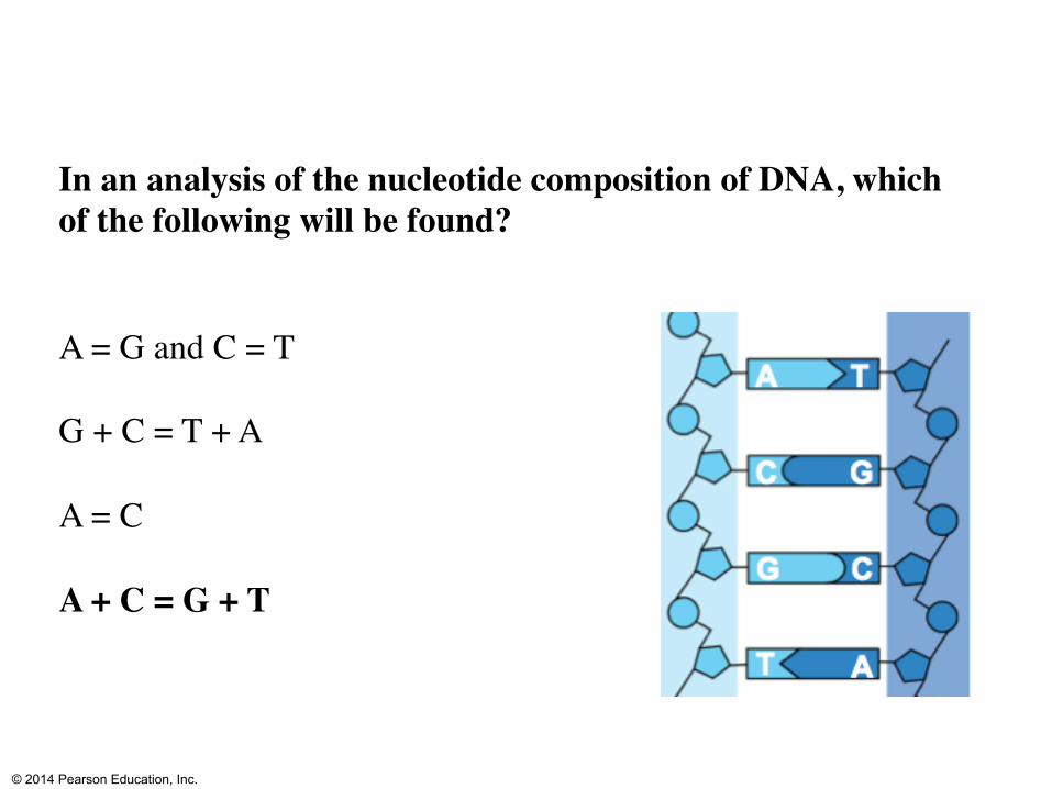
In an analysis of the nucleotide composition of DNA, which of the following will be found? (Imagine counting number of nucleotides of each type)
A = G and C = T
G + C = T + A
A = C
A + C = G + T
© 2014 Pearson Education, Inc.
In an analysis of the nucleotide composition of DNA, which of the following will be found?
A = G and C = T
G + C = T + A
A = C
A + C = G + T

© 2014 Pearson Education, Inc.
Cytosine makes up 42% of the nucleotides in a sample of DNA from an organism. Approximately what percentage of the nucleotides in this sample will be thymine?
31%
42%
8%
16%
It cannot be determined from the information provided.

© 2014 Pearson Education, Inc.
Cytosine makes up 42% of the nucleotides in a sample of DNA from an organism. Approximately what percentage of the nucleotides in this sample will be thymine?
31%
42%
8%
16%
It cannot be determined from the information provided.