Huanglongbing: the pathogen, the disease, its transmission and
Upload
bonnie-porterCategory
view
217download
0
UNIT 1 REVISION
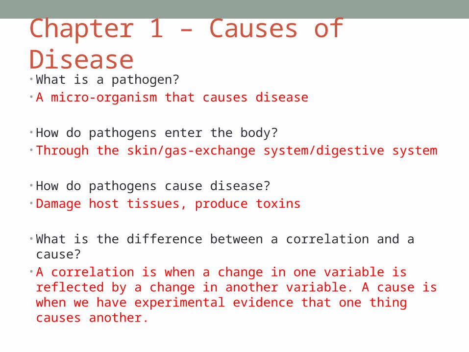
Chapter 1 – Causes of Disease• What is a pathogen?• A micro-organism that causes disease
• How do pathogens enter the body?• Through the skin/gas-exchange system/digestive system
• How do pathogens cause disease?• Damage host tissues, produce toxins
• What is the difference between a correlation and a cause?• A correlation is when a change in one variable is reflected
by a change in another variable. A cause is when we have experimental evidence that one thing causes another.

Chapter 1 – Causes of Disease• What is risk?• A measure of the probability that damage to health will
occur as the result of a given hazard
• List risk factors for cancer• Smoking, diet, obesity, physical activity, sunlight
• List risk factors for CHD• Smoking, high blood pressure, high blood cholesterol,
obesity, diet, physical activity

Chapter 2 – Enzymes and the Digestive System
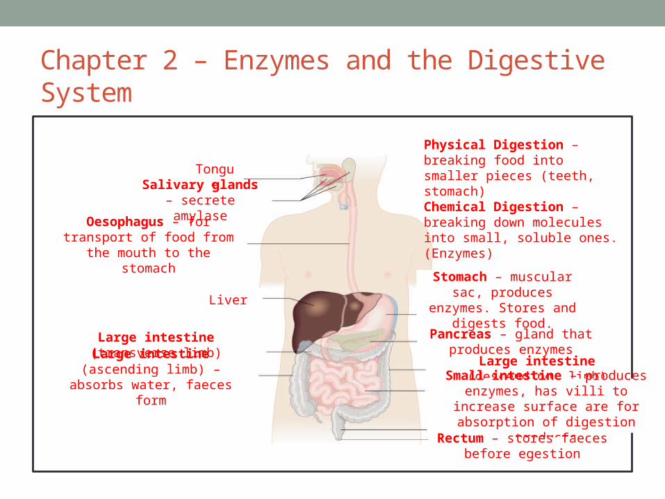
• Label the parts of the
digestive system and
explain the function
of each part:
• State what chemical
and physical digestion
are and where take place
Chapter 2 – Enzymes and the Digestive System
TongueSalivary glands – secrete amylase
Oesophagus – for transport of food from the mouth to the
stomach
Liver
Large intestine (transverse limb)
Large intestine (ascending limb) – absorbs water, faeces form
Stomach – muscular sac, produces enzymes. Stores
and digests food.
Pancreas – gland that produces enzymes
Large intestine (descending limb)
Small intestine – produces enzymes, has villi to increase surface are for absorption of digestion productsRectum – stores faeces
before egestion
Physical Digestion – breaking food into smaller pieces (teeth, stomach)Chemical Digestion – breaking down molecules into small, soluble ones. (Enzymes)
Chapter 2 - Enzymes
• What do enzymes do?• Break down large insoluble molecules into small soluble
ones by hydrolysis (splitting of molecules by adding water to them)
• Name the enzymes that break down carbohydrates, proteins and lipids
• Carbohydrases, proteases, lipases

Chapter 2 - CarbohydratesExplain how to carry out the Benedict's test:
Label the tubes below to show the result:
Draw the monomer α-glucose:
How are large molecules like carbohydrates made?
Made from a chain of smaller monomers. The longer chain is called a polymer. In carbohydrates, a single monomer is called a monosaccharide. A pair of monomers are called a disaccharide and a long chain of monomers is called a polysaccharide.
Grind up your sample with water (if it is not already a liquid. Add an equal volume of Benedict’s and boil for about 5 minutes.
None Very low Low Medium High
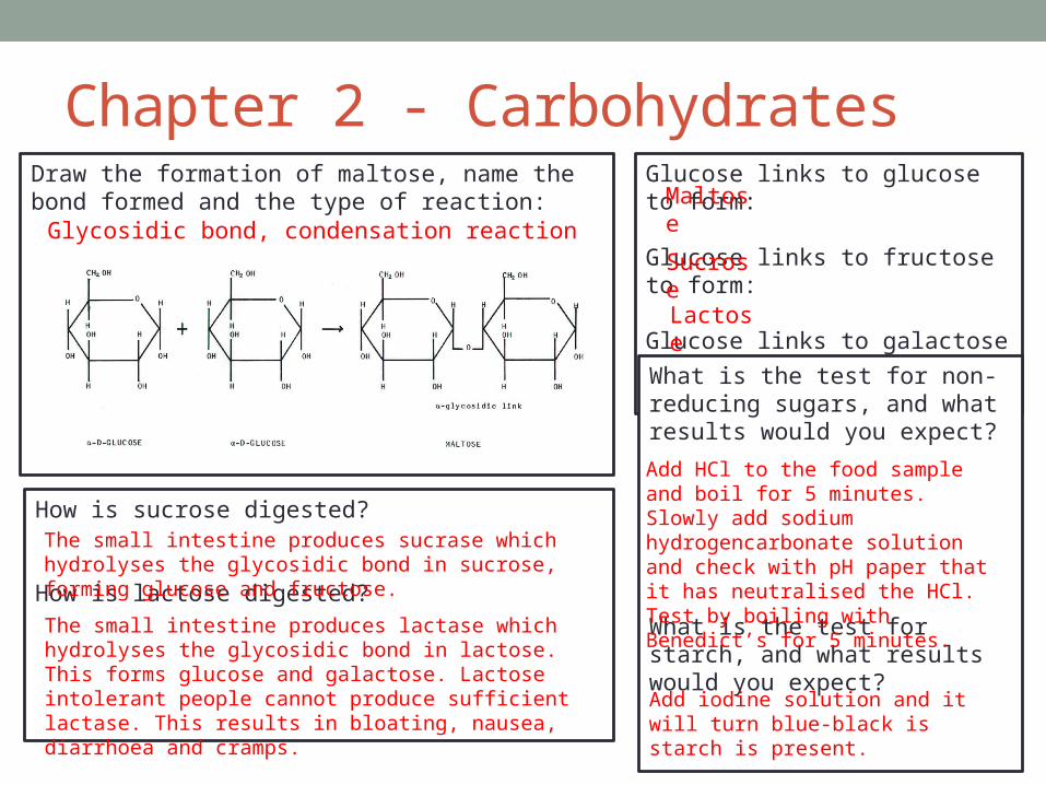
Chapter 2 - CarbohydratesGlucose links to glucose to form:
Glucose links to fructose to form:
Glucose links to galactose to form:
Draw the formation of maltose, name the bond formed and the type of reaction:
What is the test for non-reducing sugars, and what results would you expect?
What is the test for starch, and what results would you expect?
Glycosidic bond, condensation reaction
Maltose
Sucrose
Lactose
Add HCl to the food sample and boil for 5 minutes. Slowly add sodium hydrogencarbonate solution and check with pH paper that it has neutralised the HCl. Test by boiling with Benedict’s for 5 minutes.
Add iodine solution and it will turn blue-black is starch is present.
How is sucrose digested?
How is lactose digested?
The small intestine produces sucrase which hydrolyses the glycosidic bond in sucrose, forming glucose and fructose.
The small intestine produces lactase which hydrolyses the glycosidic bond in lactose. This forms glucose and galactose. Lactose intolerant people cannot produce sufficient lactase. This results in bloating, nausea, diarrhoea and cramps.

Chapter 2 – Proteins Draw and label an amino acid: What is the test for proteins and what
results would you expect?
What is the primary structure of a protein?The sequence of amino acids in a polypeptide chain.What is the secondary structure of a protein?The formation of hydrogen bonds which causes the polypeptide chain to twist into a 3D shape.What is the tertiary structure of a protein?Further twisting and folding of the chain to give a complex 3D structure. Bonds hold the structure in place – disulphide, ionic, hydrogen.What is the quaternary structure of a protein?Linking of more than one polypeptide chain (and often non-protein groups e.g. haem)
Add sodium hydroxide to the food sample. Add a few drops of copper sulphate solution. It will turn purple if a protein is present.
Chapter 2 - Enzymes• How do enzymes speed up reactions?• They act as catalysts by lowering the activation energy
needed for a reaction to occur.• How does an enzyme work?• The substrate fits into the active site and forms an enzyme-
substrate complex.• Explain the lock and key model of enzyme action• Each key (substrate) has a specific shape that is only
complementary to its lock (active site of the enzyme). The limitation of this model is that the enzyme is considered to be a rigid structure, but it is not, it can be flexible.

Chapter 2 - Enzymes• Explain the induced fit model of enzyme action• The enzyme changes shape slightly to fit the substrate.• List the factors that affect enzyme action• Temperature, pH, substrate concentration, inhibitors• Explain how temperature will affect an enzyme• As temperature increases molecules have more kinetic
energy, so move faster and collide more. This speeds up the rate of reaction (more enzyme-substrate complexes form). At some point the enzyme will become denatured – the higher temperature causes bonds holding the enzyme together to break. The active site changes shape and can no longer fit the substrate.

Chapter 2 - Enzymes• Explain the effect of a changing substrate concentration on the
rate of enzyme action• As you increase substrate concentration then the rate of reaction
increases (more enzyme-substrate complexes form). At a certain point all the active sites will be filled so adding more substrate will not increase the rate of reaction further.
• Explain the difference between competitive and non-competitive inhibitors.
• Competitive inhibitors have a shape similar to the substrate. They compete with the substrate for the active sites. This is not permanent.
• Non-competitive inhibitors attach to the enzyme at a place other than the active site. This alters the shape of the active site so the substrate cannot fit in, E-S complexes can’t form, the enzyme can’t function.
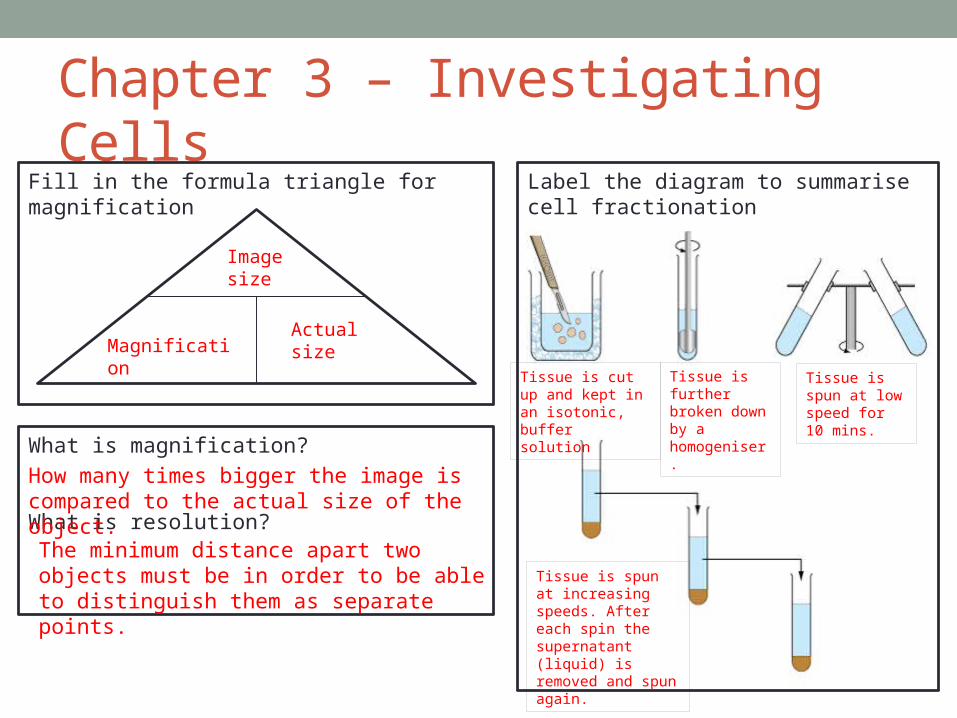
Label the diagram to summarise cell fractionation
Chapter 3 – Investigating CellsFill in the formula triangle for magnification
What is magnification?
What is resolution?
Image size
Actual sizeMagnification
How many times bigger the image is compared to the actual size of the object.
The minimum distance apart two objects must be in order to be able to distinguish them as separate points.
Tissue is cut up and kept in an isotonic, buffer solution
Tissue is further broken down by a homogeniser.
Tissue is spun at low speed for 10 mins.
Tissue is spun at increasing speeds. After each spin the supernatant (liquid) is removed and spun again.
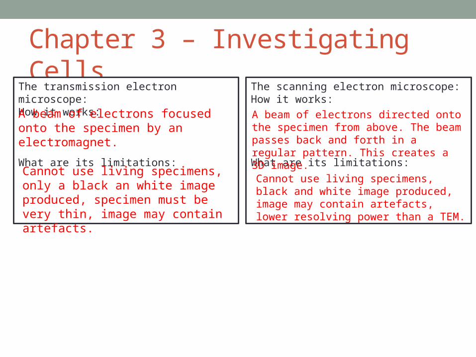
Chapter 3 – Investigating CellsThe transmission electron microscope:How it works:
What are its limitations:
The scanning electron microscope:How it works:
What are its limitations:
A beam of electrons focused onto the specimen by an electromagnet.
Cannot use living specimens, only a black an white image produced, specimen must be very thin, image may contain artefacts.
A beam of electrons directed onto the specimen from above. The beam passes back and forth in a regular pattern. This creates a 3D image.
Cannot use living specimens, black and white image produced, image may contain artefacts, lower resolving power than a TEM.
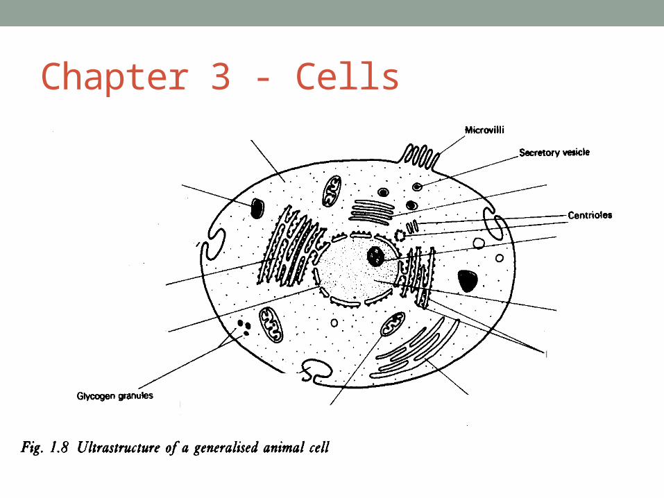
Chapter 3 - Cells
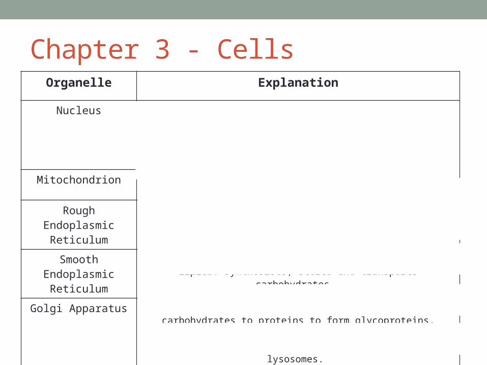
Chapter 3 - CellsOrganelle Explanation
Nucleus Contains the DNA. Has a nuclear envelope (double membrane to control the entry and exit of materials), nuclear pores (holes in the envelope to allow exit of large molecules) and nucleolus (manufactures ribosomal
RNA and assembles ribosomes)
Mitochondrion Site of respiration. Has a double membrane, cristae and matrix.
Rough Endoplasmic Reticulum
Has ribosomes for synthesis of proteins and glycoproteins. Provides a pathway for transport of materials out of the cell.
Smooth Endoplasmic Reticulum
Lacks ribosomes. Synthesises, stores and transports lipids. Synthesises, stores and transports carbohydrates.
Golgi Apparatus Stacks of flattened stacks of cisternae. Adds carbohydrates to proteins to form glycoproteins, produces secretory enzymes, secretes carbohydrates,
transports, modifies and stores lipids and forms lysosomes.
Lysosomes Contain enzymes. Break down material in phagocytes, release enzymes to the outside of the cell, digest worn out organelles and break down cells
after they have died.
Ribosomes 80s (eukaryotic) or 70s (prokaryotic). Important for protein synthesis.
Chapter 3 - Lipids• List the roles of lipids• Energy source, waterproofing, insulation, protection.• How does a triglyceride form?• Glycerol joins to 3 fatty acids in a condensation reaction.• Describe a phospholipid• They have a hydrophilic head (phosphate molecule) that
attracts to water and a hydrophobic tail (fatty acids) that orients itself away from water.
• How do we test for lipids?• Add ethanol to the food sample and shake. Add water and
shake. A cloudy-white colour indicates a lipids is present.
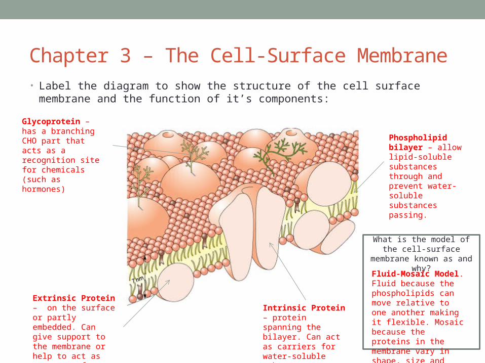
Chapter 3 – The Cell-Surface Membrane• Label the diagram to show the structure of the cell surface membrane and the function
of it’s components:
Phospholipid bilayer – allow lipid-soluble substances through and prevent water-soluble substances passing.
Intrinsic Protein – protein spanning the bilayer. Can act as carriers for water-soluble substances.
Glycoprotein – has a branching CHO part that acts as a recognition site for chemicals (such as hormones)
Extrinsic Protein – on the surface or partly embedded. Can give support to the membrane or help to act as receptors for chemicals.
What is the model of the cell-surface membrane known as
and why?Fluid-Mosaic Model. Fluid because the phospholipids can move relative to one another making it flexible. Mosaic because the proteins in the membrane vary in shape, size and pattern.

Chapter 3 - Diffusion• Definition of diffusion?• The net movement of molecules or ions from a region of
high concentration to a region of lower concentration• What affects the rate of diffusion?• Concentration gradient, area of the exchange surface and
thickness of the exchange surface• What is facilitated diffusion?• Diffusion through protein channels in the plasma
membrane. These channels are selective. Alternatively, it can be through carrier proteins that change shape in the presence of a particular molecule and release it to the inside. It is a passive process that occurs down a concentration gradient.

Chapter 3 - Osmosis• Definition of osmosis?• The passage of water from a region of higher water potential to a
region of lower water potential through a partially permeable membrane (less negative to more negative water potential).
• Describe how an animal cell changes when placed in solutions of different water potential
• In a solution with a higher (less negative) water potential the animal cell will take in water and burst. In a solution with a lower (more negative) water potential the animal cell will lose water and shrink/shrivel up.
• Describe how a plant cell changes when placed in solutions of different water potential
• In a solution with a higher (less negative) water potential the plant cell will take in water and become turgid. In a solution with a lower (more negative) water potential the plant cell will lose water and become plasmolysed.

Chapter 3 – Active Transport• Definition of active transport?• The movement of molecules or ions into or out of a cell
from a region of lower concentration to a region of higher concentration using energy and carrier molecules
• Explain how the carrier molecules help a molecule to enter the cell
• The molecule binds to a receptor on the carrier protein. Inside the cell ATP binds to the protein and splits into ADP + Pi, this changes the shape of the carrier protein. The carrier protein now opens to the inside of the membrane and releases the molecule to the inside.
Chapter 3 – Absorption in the Small Intestine
• How is the small intestine adapted for absorption?• Villi and microvilli increase the surface area, very thin walls,
good blood supply• Explain how absorption occurs in the small intestine
• Na+ ions are actively transported out of epithelial cells• The epithelial cell has a much lower Na+ concentration than inside the
lumen of the intestine• Na+ ions diffuse into epithelial cells down a concentration gradient
through a carrier protein• As Na+ ions diffuse in they couple with glucose molecules and carry
them into the epithelial cell too. • Glucose passes into the blood by facilitated diffusion.
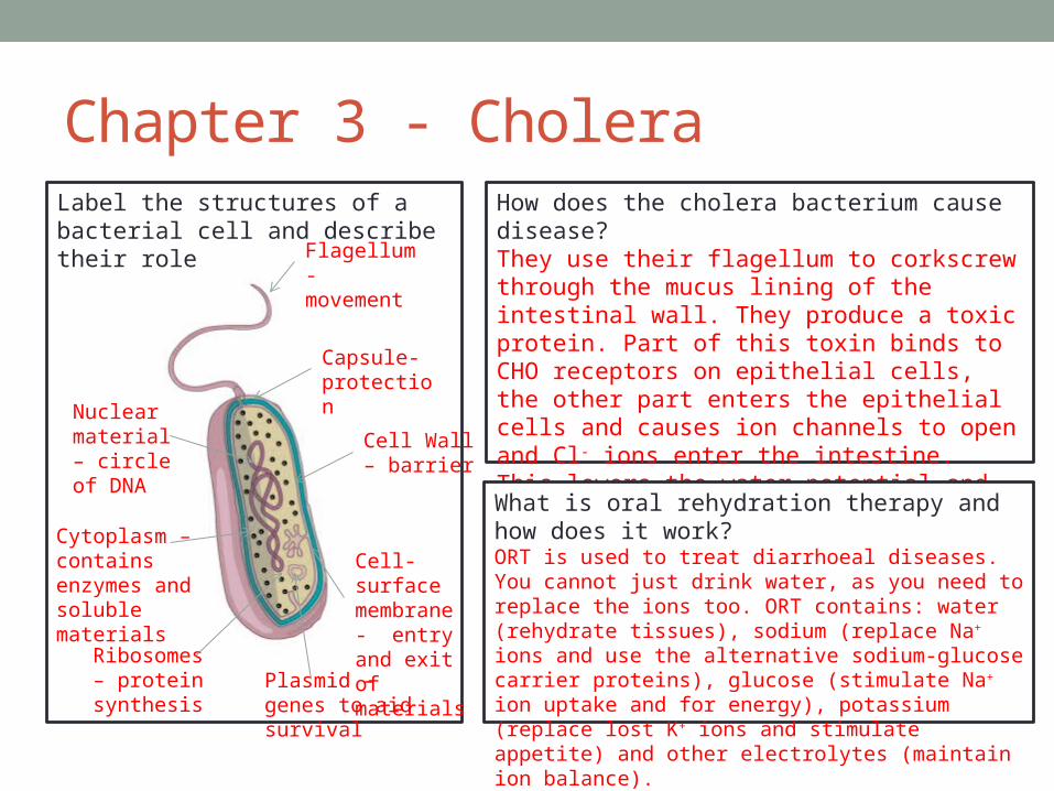
Label the structures of a bacterial cell and describe their role
Chapter 3 - Cholera
Flagellum- movement
Capsule- protection
Cell Wall – barrier
Cell-surface membrane - entry and exit of materials
Nuclear material – circle of DNA
Cytoplasm – contains enzymes and soluble materials
Ribosomes – protein synthesis
Plasmid – genes to aid survival
How does the cholera bacterium cause disease?They use their flagellum to corkscrew through the mucus lining of the intestinal wall. They produce a toxic protein. Part of this toxin binds to CHO receptors on epithelial cells, the other part enters the epithelial cells and causes ion channels to open and Cl- ions enter the intestine. This lowers the water potential and water floods in, causing diarrhoea.
What is oral rehydration therapy and how does it work?ORT is used to treat diarrhoeal diseases. You cannot just drink water, as you need to replace the ions too. ORT contains: water (rehydrate tissues), sodium (replace Na+ ions and use the alternative sodium-glucose carrier proteins), glucose (stimulate Na+ ion uptake and for energy), potassium (replace lost K+ ions and stimulate appetite) and other electrolytes (maintain ion balance).
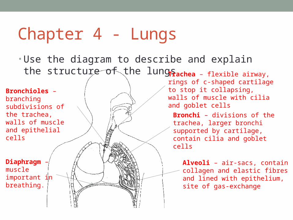
Chapter 4 - Lungs• Use the diagram to describe and explain the structure of
the lungs Trachea – flexible airway, rings of c-shaped cartilage to stop it collapsing, walls of muscle with cilia and goblet cells
Bronchi – divisions of the trachea, larger bronchi supported by cartilage, contain cilia and goblet cells
Bronchioles – branching subdivisions of the trachea, walls of muscle and epithelial cells
Alveoli – air-sacs, contain collagen and elastic fibres and lined with epithelium, site of gas-exchange
Diaphragm – muscle important in breathing.

Chapter 4 - Lungs• Describe inspiration• External intercostal muscles contract, pulling the ribs up and out.
Diaphragm contracts, flattening it. This increases the volume of the chest, decreasing pressure. Air moves in.
• Describe expiration• Internal intercostal muscles contract, pulling the ribs down and in.
Diaphragm relaxes and returns to its domed shape. This decreases the volume of the chest, increasing pressure. Air moves out.
• What is tidal volume?• The volume of air normally taken in in each breath.• How are the alveoli adapted for efficient diffusion?• Large surface area to volume ratio, very thin (short diffusion
pathway), partially permeable (allow selected material through easily), movement of air (maintain diffusion gradient), movement of blood (maintain diffusion gradient.
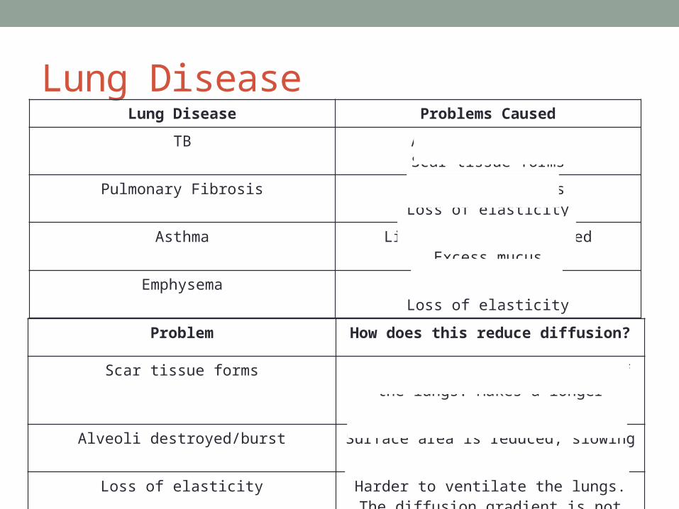
Lung DiseaseLung Disease Problems Caused
TB Alveoli destroyedScar tissue forms
Pulmonary Fibrosis Scar tissue formsLoss of elasticity
Asthma Linings become inflamedExcess mucus
Emphysema Alveoli burstLoss of elasticity
Problem How does this reduce diffusion?
Scar tissue forms This thickens the epithelium of the lungs. Makes a longer diffusion pathway.
Alveoli destroyed/burst Surface area is reduced, slowing down diffusion.
Loss of elasticity Harder to ventilate the lungs. The diffusion gradient is not maintained.
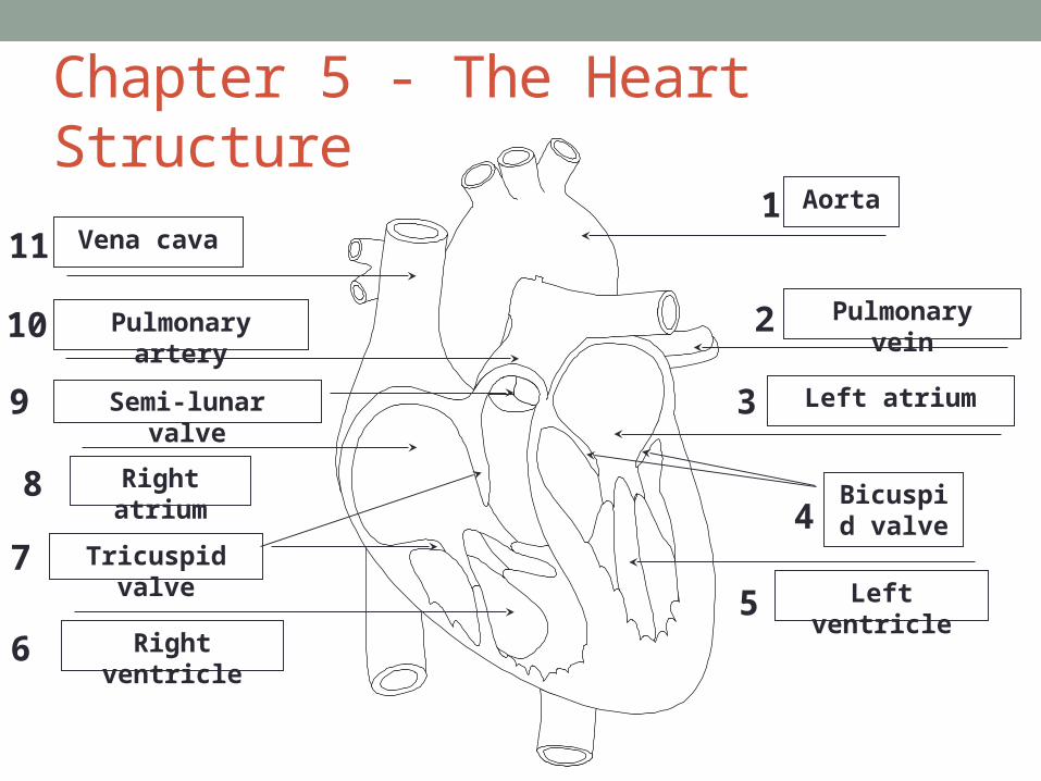
Chapter 5 - The Heart StructureAorta
Left atrium
Left ventricle
Pulmonary vein
Right atrium
Vena cava
Right ventricle
Pulmonary artery
Tricuspid valve
Semi-lunar valve
Bicuspid valve
1
2
3
4
5
11
10
9
8
7
6
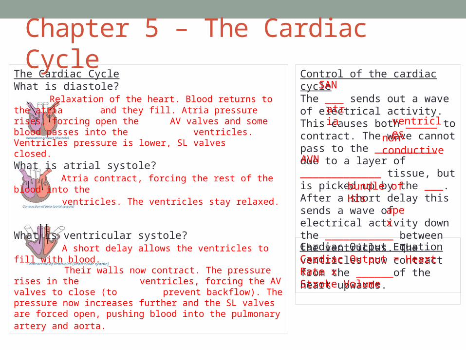
Chapter 5 – The Cardiac CycleThe Cardiac CycleWhat is diastole?
Relaxation of the heart. Blood returns to the atria and they fill. Atria pressure rises, forcing open the AV valves and some blood passes into the ventricles. Ventricles pressure is lower, SL valves closed.
What is atrial systole? Atria contract, forcing the rest of the blood into the ventricles. The ventricles stay relaxed.
What is ventricular systole? A short delay allows the ventricles to fill with blood. Their walls now contract. The pressure rises in the ventricles, forcing the AV valves to close (to prevent backflow). The pressure now increases
further and the SL valves are forced open, pushing blood into the pulmonary artery and aorta.
Control of the cardiac cycleThe ___ sends out a wave of electrical activity. This causes both _____ to contract. The wave cannot pass to the __________ due to a layer of _____________ tissue, but is picked up by the ___. After a short delay this sends a wave of electrical activity down the ___________ between the ventricles. The ventricles now contract from the ______of the heart upwards.
SAN
atriaventricles
non-conductive
AVN
bundle of His
apex
Cardiac Output EquationCardiac Output = Heart Rate x
Stroke Volume
Chapter 5 – Heart Disease• What is an atheroma?• A fatty deposit that forms within the wall of an artery. It narrows
the artery, reducing blood flow.• What is thrombosis?• If an atheroma breaks through the lining of the blood vessel then
it forms a rough surface that disrupts blood flow. This may result in a blood clot (thrombus) which may block the artery, depriving tissues of oxygen.
• What is an aneurysm?• Atheromas may weaken the artery walls. Weakened points may
swell and burst leading to haemorrhage.• What is a myocardial infarction?• A heart attack. Reduced supply of oxygen to the heart muscle
resulting from a blockage in the coronary arteries.

Chapter 6 – Defence Mechanisms
Barriers to Disease
HCl in the Stomach
Ciliated epithelia
Mucus membranes
Epidermis of the skin
What are the differences between specific and non-specific mechanisms?• Non-specific mechanisms do not distinguish
between types of pathogens, specific mechanisms do. Specific mechanism are slower but provide long-lasting immunity.
What is an antigen?• A molecule on the
surface of an organism that is recognised as foreign and triggers an immune response.

Chapter 6 - Phagocytosis

Chapter 6 - T Cells• Cell mediated immunity• Activated by antigen-presenting cells (e.g. Phagocyte)
• Mature in the thymus• Clone by mitosis
Do 4 main things:• Produce memory cells• Stimulate B cells to
divide• Stimulate
phagocytosis• Kill infected cells (by
making holes in the cells)
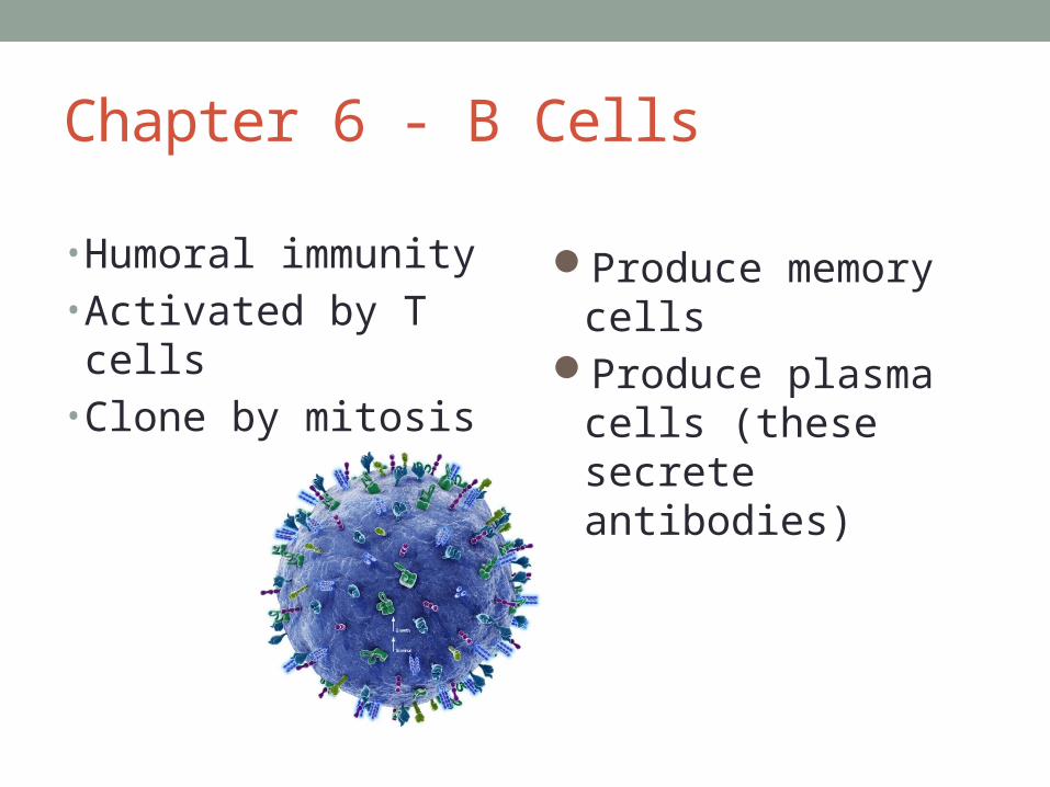
Chapter 6 - B Cells
• Humoral immunity• Activated by T cells• Clone by mitosis
Produce memory cellsProduce plasma cells
(these secrete antibodies)
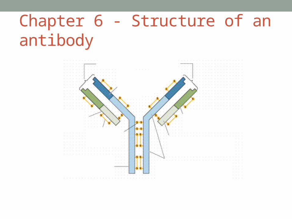
Chapter 6 - Structure of an antibody

Chapter 6 - Monoclonal Antibodies • The response of the immune system to any pathogen is
polyclonal. That is, the system manufactures antibodies of a range of structures (due to pathogens having a range of antigens).
• Isolating a single type of antibody and cloning it creates monoclonal antibodies
• Uses?• Separating a chemical from a mixture• Cancer treatment (antibodies only attach to cancer cells and activate a
drug that kills cells)• Immunoassay (e.g. pregnancy tests)• Transplant surgery (help to prevent rejection)

Chapter 6 - Vaccination• What is passive immunity?• The introduction of antibodies from an outside source. Short-
lived.• What is active immunity?• Stimulation of the production of antibodies by the individual’s
own immune system. Long-lasting.• What is vaccination?• Introduction of a substance into the body (dead or attenuated
pathogen) to stimulate active immunity.• List features of a successful vaccination programme.• Vaccine is cheap and readily available, few side effects, easy
to produce, store and transport, easy to administer, able to vaccinate most of the population (Herd Immunity).