UNIT 05: ASSESSMENT OF THE ABDOMEN, ANUS & · PDF fileUNIT 05: ASSESSMENT OF THE ABDOMEN, ANUS...
95
Chapter 21: Abdomen Elsevier items and derived items © 2008, 2004, 2000, 1996, 1992 by Saunders, an imprint of Elsevier Inc. Shahzad Bashir RN, BScN, DCHN, MScN (Std. DUHS) Instructor New Life College of Nursing September 26, 2015 In The Name of God (A PROJECT OF NEW LIFE COLLEGE OF NURSING KARACHI) UNIT 05: ASSESSMENT OF THE ABDOMEN, ANUS & RECTUM
Transcript of UNIT 05: ASSESSMENT OF THE ABDOMEN, ANUS & · PDF fileUNIT 05: ASSESSMENT OF THE ABDOMEN, ANUS...
Chapter 21: Abdomen
Elsevier items and derived items © 2008, 2004, 2000, 1996, 1992 by Saunders, an imprint of Elsevier Inc.
Shahzad Bashir
RN, BScN, DCHN, MScN (Std. DUHS)
Instructor
New Life College of Nursing
September 26, 2015
In The Name of
God
(A PROJECT OF NEW LIFE COLLEGE OF NURSING KARACHI)
UNIT 05:
ASSESSMENT OF THE ABDOMEN, ANUS & RECTUM

Chapter 21: Abdomen
Objectives.
By the end of the unit, learner will be able to:
• Discuss the pertinent health history questions necessary to perform the assessment of abdomen, anus & rectum.
• Describe the specific assessment to be made during the physical examination of the abdomen.
• Practice assessment skills of abdomen.
• Discuss component of a rectal examination.
• Documents finding.
• List the changes in abdomen that are characteristics of aging process.
Slide 21-2

Chapter 21: Abdomen
General Considerations
• The patient should have an empty bladder.
• The patient should be lying supine on the exam table and appropriately draped.
• The examination room must be quiet to perform adequate auscultation and percussion.
• Watch the patient's face for signs of discomfort during the examination
3
Chapter 21: Abdomen
General Considerations
• Disorders in the chest will often manifest with
abdominal symptoms. It is always wise to
examine the chest when evaluating an
abdominal complaint.
• Consider the inguinal/rectal examination in
males. Consider the pelvic/rectal examination
in females.
4
Chapter 21: Abdomen
• Structure and Function
• Subjective Data—Health History Questions
• Objective Data—The Physical Exam
• Abnormal Findings
Slide 21-5

Chapter 21: Abdomen
Structure and Function
• Surface landmarks
– Borders of abdominal cavity
– Abdominal muscles
• Internal anatomy (viscera)
– Solid viscera
• Liver
• Pancreas
• Spleen
• Adrenal glands
• Kidneys
• Ovaries
• Uterus
Slide 21-6

Chapter 21: Abdomen
Slide 21-7

Chapter 21: Abdomen
Slide 21-8

Chapter 21: Abdomen
Structure and Function (cont.)
• Internal anatomy (viscera) (cont.)
– Hollow viscera
• Stomach
• Gallbladder
• Small intestine
• Colon
• Bladder
Slide 21-9

Chapter 21: Abdomen
Internal Anatomy
Slide 21-10
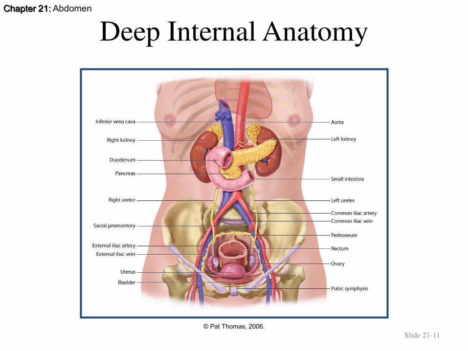
Chapter 21: Abdomen
Deep Internal Anatomy
Slide 21-11
© Pat Thomas, 2006.

Chapter 21: Abdomen
Structure and Function (cont.)
• Abdominal wall divided into four quadrants
– Right upper (RUQ)
– Left upper (LUQ)
– Right lower (RLQ)
– Left lower (LLQ)
Slide 21-12
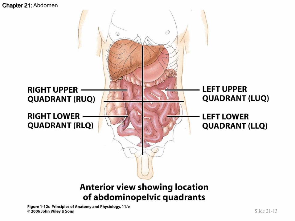
Chapter 21: Abdomen
Slide 21-13

Chapter 21: Abdomen
Location! Location! Location!
• RIGHT UPPER QUADRANT(RUQ)
• Liver
• Gallbladder
• Duodenum (small intestine)
• Head of Pancreas
• Right kidney and adrenal
• Hepatic flexure of colon
• Part of ascending and transverse colon
14

Chapter 21: Abdomen
Location! Location! Location!
• LEFT UPPER QUADRANT(LUQ)
• Left lobe of liver
• Stomach
• Spleen
• Left kidney and adrenal
• Body of pancreas
• Splenic flexure of colon
• Parts of transverse and descending colon
15
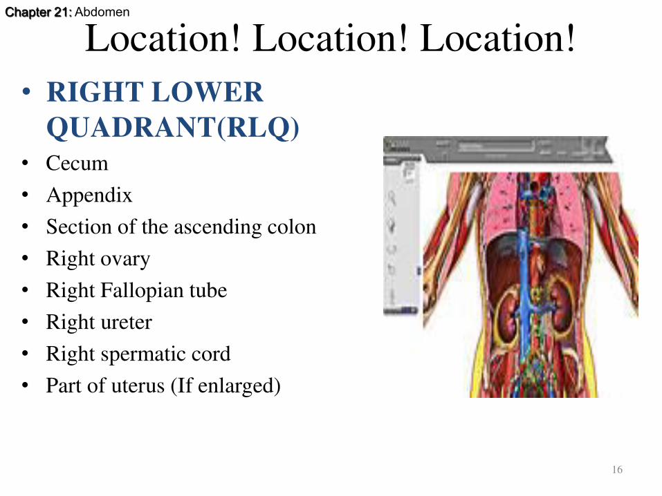
Chapter 21: Abdomen
Location! Location! Location!
• RIGHT LOWER
QUADRANT(RLQ)
• Cecum
• Appendix
• Section of the ascending colon
• Right ovary
• Right Fallopian tube
• Right ureter
• Right spermatic cord
• Part of uterus (If enlarged)
16

Chapter 21: Abdomen
Location! Location! Location!
• LEFT LOWER QUADRANT (LUQ)
• Sigmoid Colon
• Part of descending colon
• Left Ovary
• Left fallopian tube
• Left ureter
• Left spermatic cord
• Part of uterus(If enlarged)
17

Chapter 21: Abdomen
Subjective Data—
Health History Questions
• Appetite
• Dysphagia
• Food intolerance
• Abdominal pain
• Nausea/vomiting, Regurgitation
• Bowel habits
• Abdominal history
• Medications
• Nutritional assessment
Slide 21-18

Chapter 21: Abdomen
Subjective Data—
Health History Questions
• Heartburn
• Rectal bleeding
• Hemorrhoids
• Previous surgery
• Weight gain or loss
Slide 21-19

Chapter 21: Abdomen
Objective Data—The Physical Exam
• Preparation
– Lighting and draping
– Measures to enhance abdominal wall relaxation
• Equipment needed
– Stethoscope
– Small centimeter ruler
– Skin-marking pen
– Alcohol swab
Slide 21-20

Chapter 21: Abdomen
PREPARATION
• Equipment - stethoscope, marking pen,
ruler, paper for documentation
• Patient lie on back, pillow under head,
knees slightly flexed
• Empty bladder
• Short fingernails
• Proper light
• Privacy maintain e.g. side screen
Slide 21-21
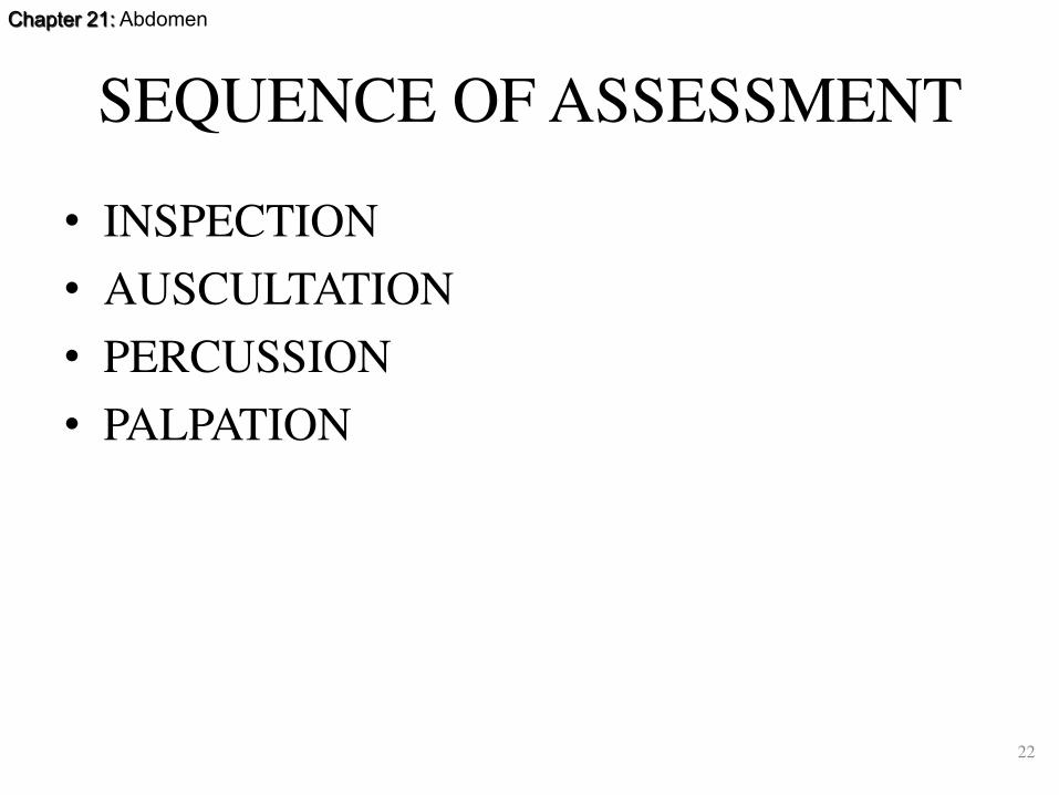
Chapter 21: Abdomen
SEQUENCE OF ASSESSMENT
• INSPECTION
• AUSCULTATION
• PERCUSSION
• PALPATION
22

Chapter 21: Abdomen
Objective Data—The Physical Exam (cont.)
Inspect the abdomen: • Contour
• Symmetry
• Umbilicus
• Skin (Pigmentation, Lesions, Striae
(elevated/depressed), Turgor
• Pulsation or movement
• Hair distribution
• Demeanor
Slide 21-23

Chapter 21: Abdomen
Contour
Slide 21-24
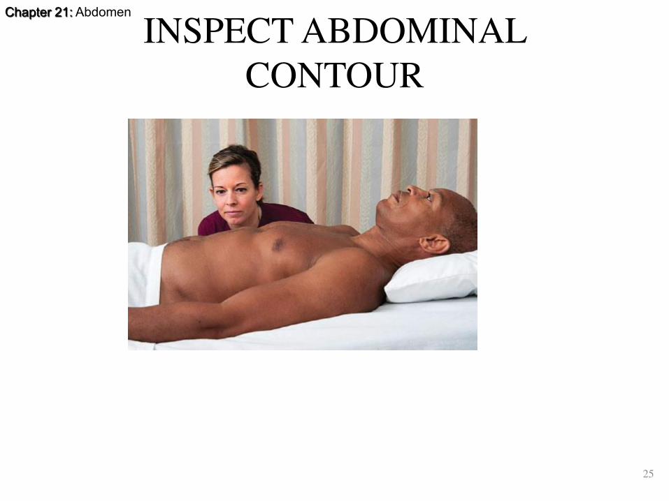
Chapter 21: Abdomen
INSPECT ABDOMINAL
CONTOUR
25

Chapter 21: Abdomen
Objective Data—The Physical Exam (cont.)
Auscultate the abdomen:
• Bowel sounds
• 5-35/min
• Increased, decreased, absent bowel sounds.
• Borborygymus
• Silent abdomen
• Vascular sounds (bruits)
Percuss the abdomen: • General tympany
• Liver span – Usual technique
– Scratch test
• Splenic dullness
• Costovertebral angle tenderness
• Special procedures – Fluid wave
– Shifting dullness
Slide 21-26

Chapter 21: Abdomen
AUSCULTATION
• Active bowel sounds 5-35/min
• Hypoactive 4/min or less( K+, Paralytic Ileus, Chronic use of
Laxative)
• Hyperactive 35 or more /min (Dysentery, Diarrhea, Early sign of
Intestinal Obstruction).
• Bruits (blowing sound)
– Aorta
– Renal
– Iliac
• Friction rub (Obstruction two layers of organs rubbing each other).
Slide 21-27

Chapter 21: Abdomen
GUT SOUNDS • Use the diaphragm of your stethoscope to listen to gut
sounds
• Normal gut sounds are gurgling, 5 to 35 per minute
• Borborygmi (Rumbling sounds caused by gas moving
through the intestines (stomach "growling“) are loud, easily
audible sounds. They are normal, too.
• High pitched , Tinkling (raindrops in a barrel) sounds are a
sign of early intestinal obstruction
• Succussion splash, A loud sound like splashing water, is
often heard without a stethoscope as the patient moves from
side to side. It occurs when the abdomen is filled with air or
fluid and indicates delayed gastric emptying from an
obstruction or gastric dilatation. 28

Chapter 21: Abdomen
GUT SOUNDS
• Decreased sounds: (none for a minute) are a sign of decreased gut activity. Gut sounds may be markedly decreased after abdominal surgery; abdominal infection (peritonitis) or injury.
• Absent Sounds : (no sounds for 5 minutes) are a bad sign. They can be caused by longer-lasting intestinal obstruction, intestinal perforation or intestinal (mesenteric) ischemia or infarction.
29

Chapter 21: Abdomen
Slide 21-30
BRUITS SOUNDS
• (A sound, especially an abnormal one. A bruit may be heard over an artery or vascular channel, reflecting turbulence of flow) OR VENOUS HUMS.
• Use the bell of your stethoscope to listen for bruits:
• Aortic bruits:
• Are heard in the epigastrium. They may be a sign of abdominal aortic (a sac formed by localized dilatation of the wall of an artery, a vein)

Chapter 21: Abdomen
BRUITS SOUNDS
• Renal artery bruits:
• Are in each upper quadrant. They may be a sign of renal artery stenosis (A narrowing), which is a potentially treatable cause of hypertension.
• Iliac/femoral bruits:
• Are in the lower quadrants. They may be a sign of peripheral atherosclerosis
Slide 21-31

Chapter 21: Abdomen
AUSCULTATION FOR BRUITS
32
Chapter 21: Abdomen
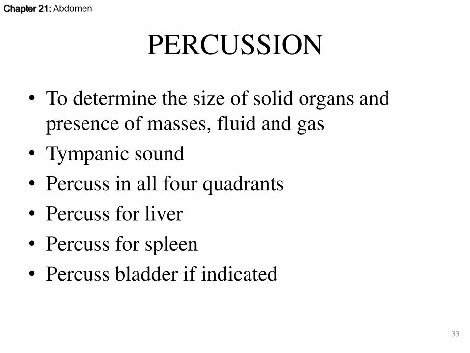
PERCUSSION
• To determine the size of solid organs and
presence of masses, fluid and gas
• Tympanic sound
• Percuss in all four quadrants
• Percuss for liver
• Percuss for spleen
• Percuss bladder if indicated
33
Chapter 21: Abdomen
THE TECHNIQUE FOR PERCUSSION
There are two basic sounds which can be elicited:
1. Tympanitic (drum-like) sounds produced by percussing over air filled structures.
2. Dull sounds that occur when a solid structure (e.g.
liver) or fluid (E.g. Ascites) lies beneath the region being examined.
Special note should be made if percussion produces pain, which may occur if there is underlying inflammation, as in peritonitis. This would certainly be supported by other historical and exam findings.
34
Chapter 21: Abdomen
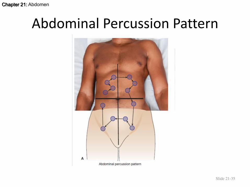
Abdominal Percussion Pattern
Slide 21-35
Chapter 21: Abdomen
PERCUSSION
Percussing the body gives one of three notes:
1. Tympany is found in most of the abdomen, caused by air in the gut. It has a higher pitch than the lung.
2. Resonance is found in normal lung. It is lower pitched and hollow.
3. Dullness is a flat sound, without echoes. The liver and spleen, and fluid in the peritoneum (ascites), give a dull note.
36

Chapter 21: Abdomen
Cont....
Slide 21-37

Chapter 21: Abdomen
38
PERCUSS THE LIVER
Chapter 21: Abdomen
PERCUSSION OF THE SPLEEN
• When significantly enlarged, percussion in the
left upper quadrant will produce a dull tone.
• Splenomegaly suggested by percussion should
then be verified by palpation
• Percuss in left anterior axillary line, just above
lowest rib
• Ask your patient to take a deep breath and
percuss again. Dullness with full inspiration
may be a sign of enlarged spleen.
Slide 21-39

Chapter 21: Abdomen
PERCUSSION OF SPLEEN
Slide 21-40

Chapter 21: Abdomen
PERCUSSION OF SPLEEN
Slide 21-41

Chapter 21: Abdomen
PERCUSSION
• If dullness in flank (on side) - check for
shifting dullness
• If indicated check for fluid wave
42

Chapter 21: Abdomen
SHIFTING DULLNESS
• With the patient supine, begin percussion at the level of the umbilicus and proceed down laterally. In the presence of ascites, you will reach a point where the sound changes from tympanitic to dull. This is the intestine-fluid interface and should be roughly equidistant(Central) from the umbilicus on the right and left sides as the fluid.
• Mark this point on both the right and left sides of the abdomen and then have the patient roll into a lateral decubitus position (i.e. onto either their right or left sides).
43

Chapter 21: Abdomen
44
SHIFTING DULLNESS (REAL PATIENT)
Chapter 21: Abdomen
Objective Data—The Physical Exam (cont.)
Palpate the liver: • Measures to enhance muscle
relaxation
• Light palpation
• Deep palpation
• Bimanual palpation
• Normally palpable structures
• Liver
– Usual technique
– Hooking technique
• Spleen
• Kidneys
• Aorta
Slide 21-45

Chapter 21: Abdomen
PALPATION • Light palpation to evaluate general condition,
• Four quadrants, 1-3cm. Special organs.
• Nature of any distention, and abnormalities and painfulness.
• E.g. Inguinal nodes, Hernia.
• Deep palpation 4-5cm, both hand technique or one hand, to detect any organ enlargement, abdominal masses or swellings
• Palpate for liver and spleen
• Rebound tenderness (Inflammation of appendix/ Peritoneal inflammation
46
Chapter 21: Abdomen
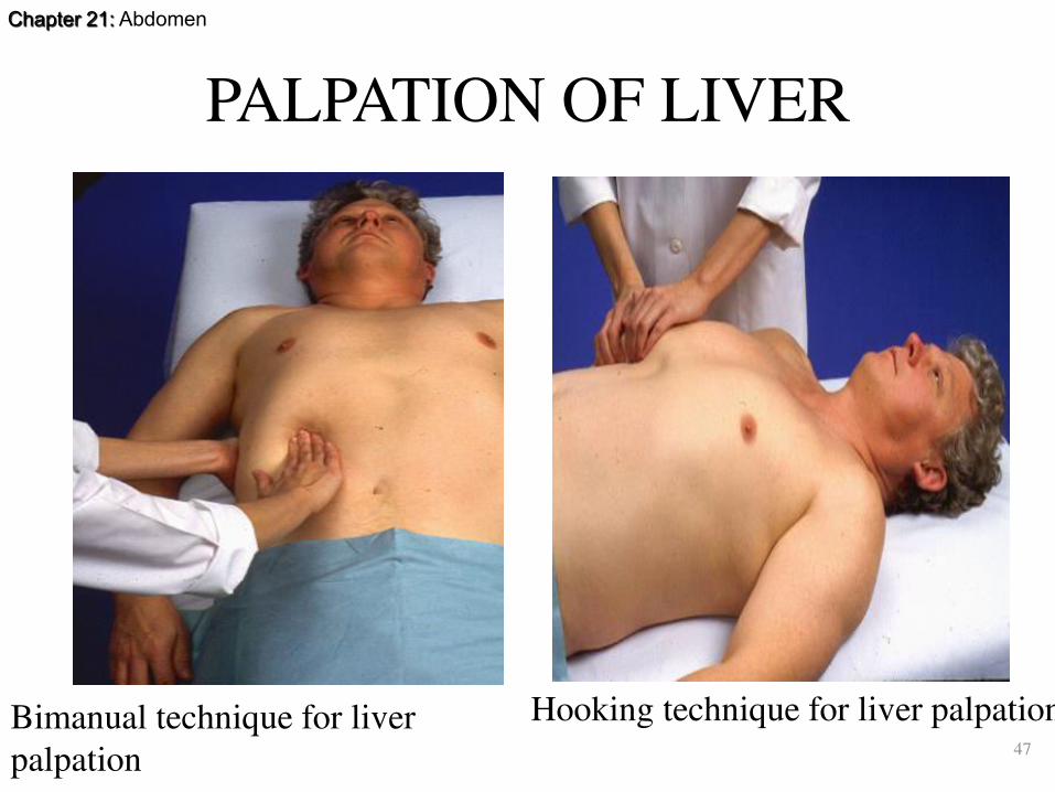
PALPATION OF LIVER
47
Hooking technique for liver palpationBimanual technique for liver
palpation

Chapter 21: Abdomen
Normal Liver Span
Slide 21-48
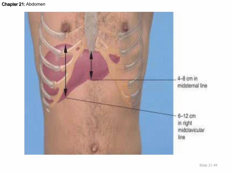
Chapter 21: Abdomen
Slide 21-49
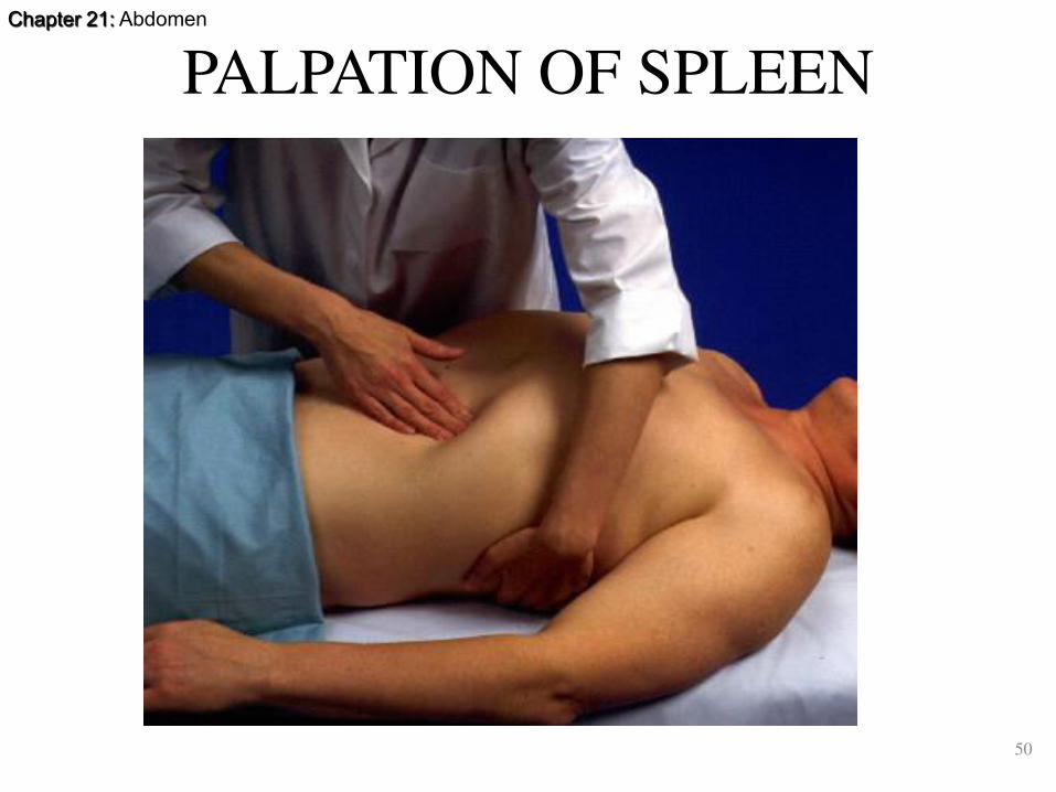
Chapter 21: Abdomen
PALPATION OF SPLEEN
50

Chapter 21: Abdomen
REBOUND TENDERNESS/
Blumberg’s sign
51
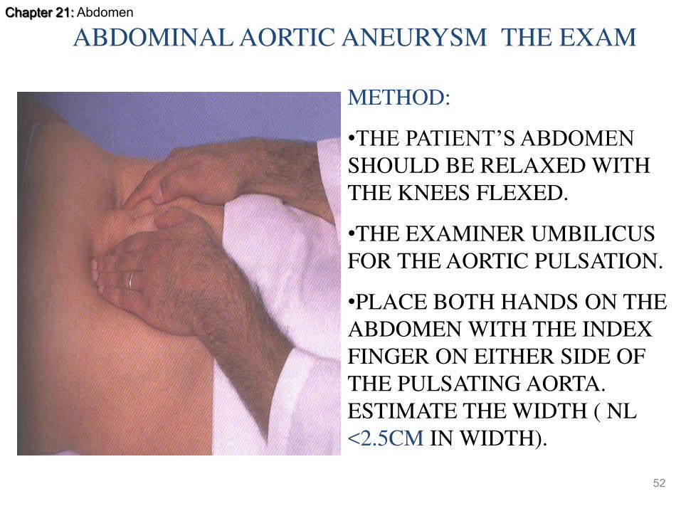
Chapter 21: Abdomen
52
ABDOMINAL AORTIC ANEURYSM THE EXAM
METHOD:
•THE PATIENT’S ABDOMEN SHOULD BE RELAXED WITH
THE KNEES FLEXED.
•THE EXAMINER UMBILICUS
FOR THE AORTIC PULSATION.
•PLACE BOTH HANDS ON THE
ABDOMEN WITH THE INDEX
FINGER ON EITHER SIDE OF
THE PULSATING AORTA.
ESTIMATE THE WIDTH ( NL
<2.5CM IN WIDTH).

Chapter 21: Abdomen
ON BACK
• CHECK FOR RENAL BRUITS
• COSTOVERTEBRAL ANGLE
TENDERNESS
53

Chapter 21: Abdomen
Slide 21-54

Chapter 21: Abdomen
BLUNT PERCUSION OF KIDNEY
55

Chapter 21: Abdomen
56
POSTERIOR VIEW: LOCATION OF THE
KIDNEYS
Chapter 21: Abdomen
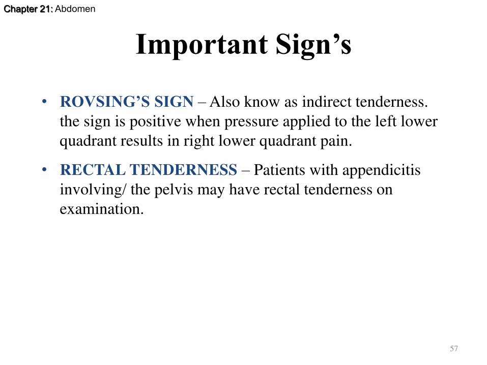
Important Sign’s
• ROVSING’S SIGN – Also know as indirect tenderness.
the sign is positive when pressure applied to the left lower
quadrant results in right lower quadrant pain.
• RECTAL TENDERNESS – Patients with appendicitis
involving/ the pelvis may have rectal tenderness on
examination.
57
Chapter 21: Abdomen
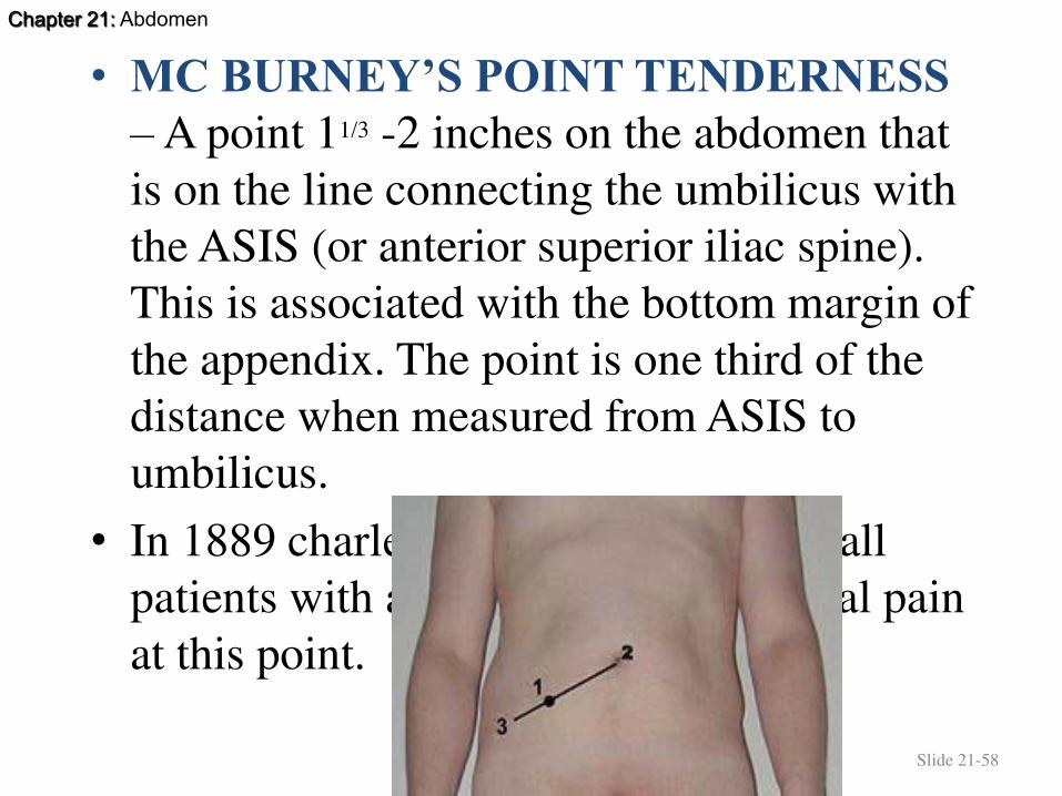
• MC BURNEY’S POINT TENDERNESS
– A point 11/3 -2 inches on the abdomen that
is on the line connecting the umbilicus with
the ASIS (or anterior superior iliac spine).
This is associated with the bottom margin of
the appendix. The point is one third of the
distance when measured from ASIS to
umbilicus.
• In 1889 charles mcburney stated that all
patients with appendicitis had maximal pain
at this point.
Slide 21-58

Chapter 21: Abdomen
Special Procedures for
Advanced Practice
• Rebound tenderness (Blumberg’s sign)
• Inspiratory arrest (Murphy’s sign)
• Iliopsoas muscle test
• Obturator test
Slide 21-59
Chapter 21: Abdomen
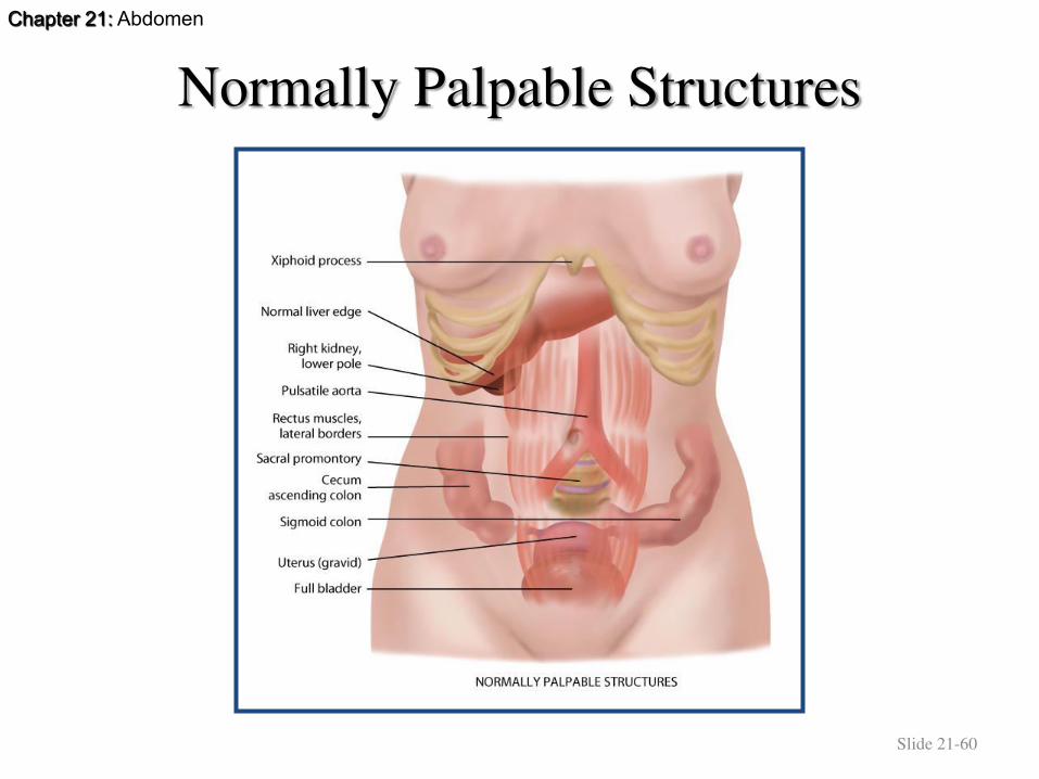
Normally Palpable Structures
Slide 21-60

Chapter 21: Abdomen
Sample Charting
Slide 21-61

Chapter 21: Abdomen
Sample Charting (cont.)
Slide 21-62

Chapter 21: Abdomen
Abnormal Findings Abdominal Distention
• Obesity
• Air or gas
• Ascites
• Ovarian cyst
• Pregnancy
• Feces
• Tumor
Slide 21-63
Chapter 21: Abdomen
Abnormal Findings Abnormalities on Inspection
• Umbilical hernia
• Epigastric hernia
• Incisional hernia
Slide 21-64

Chapter 21: Abdomen
Abnormal Findings Abnormal Bowel Sounds
• Succussion splash
• Hypoactive bowel sounds
• Hyperactive bowel sounds
Slide 21-65

Chapter 21: Abdomen
Abnormal Findings
Abnormalities on Palpation
of Enlarged Organs
• Enlarged liver (Hepatomegaly)
• Enlarged nodular liver
• Enlarged gallbladder (Cholecystitis, Cholithiasis)
• Enlarged spleen (Splenomegaly)
• Enlarged kidney (Pylonephritis, Polycystic Kidney)
• Aortic aneurysm
Slide 21-66

Chapter 21: Abdomen
EXAMINATION OF THE ANUS AND
RECTUM
• This information is sometimes included
with the abdominal assessment and at
times with assessment of the male and
female genitalia.
67

Chapter 21: Abdomen
GENERAL PRINCIPLES
• Anal canal is the final segment of digestive system.
• It measures from 2.5 cm to 4 cm long.
• It is lined with skin that contains no hair or sebaceous glands but does contain many somatic sensory nerves, making it very sensitive to touch.
• Within the anus are the two sphincters that normally hold the anal canal closed except when passing gas and feces.
68

Chapter 21: Abdomen
Examination of anus & Rectum.
• History: • Bowel habits(Changes).
• Character of stools(Blood).
• Rectal Pain
• C/O, Constipation, Diarrhoea
• Hemorrhoids
• Screening, (PR Proctoscopy)
• Use of Laxatives or medications
• Prostate problems
Slide 21-69
Chapter 21: Abdomen
RECTAL EXAMINATION
Assist patient into position:
• Male – left lateral, or standing upper body resting on a table.
• Female – Lithotomy
• Then …
I. Inspection
II. Palpation: Males
Females
Slide 21-70

Chapter 21: Abdomen
RECTAL EXAMINATION • Rectal Examination in Men. Inspect the perianal
areas. Palpate the anal canal, rectum, and prostate. If the patient cannot stand, examine the genitalia before doing the rectal examination.
• Genital and Rectal Examination in Women. Examine the external genitalia, vagina, and cervix. Obtain a Pap smear (a sample of secretions and superficial cells of the uterine cervix and uterus; examined with a microscope to detect any abnormal cells). Palpate the uterus and adnexa (ovaries). Do a rectovaginal and rectal examination.
71

Chapter 21: Abdomen
Examination of anus & Rectum.
• Inspection:
• Position- Side lying is preferred or lithotomy if genitalia exam in female or standing with upper body resting on a table for men.
• Inspect perianal tissue/ Sacrococygeal area by retracting buttocks.
• Look for skin characteristics, Lumps, lesions, hemorrhoids, ulcers, Rashes, Redness, inflammation, pigmentation.
• Ask client to bear down prolapse of rectum or hemorrhoids.
Slide 21-72
Chapter 21: Abdomen
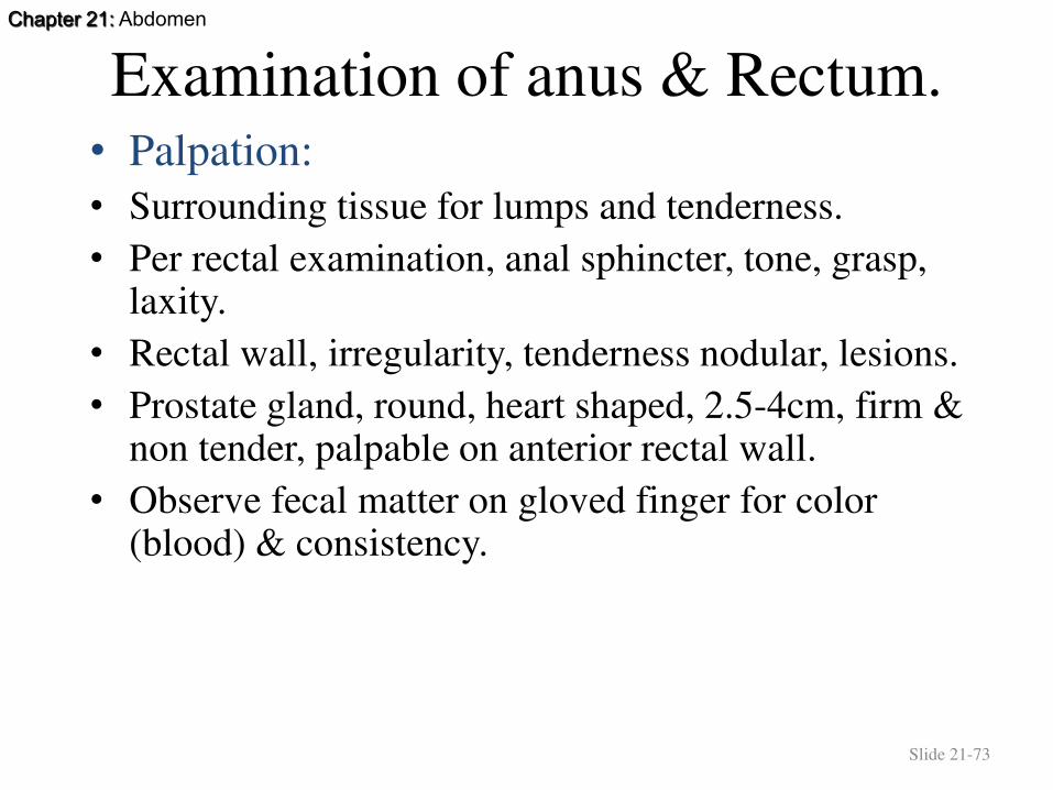
Examination of anus & Rectum. • Palpation:
• Surrounding tissue for lumps and tenderness.
• Per rectal examination, anal sphincter, tone, grasp, laxity.
• Rectal wall, irregularity, tenderness nodular, lesions.
• Prostate gland, round, heart shaped, 2.5-4cm, firm & non tender, palpable on anterior rectal wall.
• Observe fecal matter on gloved finger for color (blood) & consistency.
Slide 21-73

Chapter 21: Abdomen
HEMORRHOIDS
• With increased venous (portal) pressure, vein can enlarge.
• This is a hemorrhoid or a varicosity
• External hemorrhoids occur below the anorectal junction
• Itch and bleed with defecation
• Painful and swollen with thrombosis
• Resolve and leave flabby(Loose) skin top around
• Anal opening.
74

Chapter 21: Abdomen
HEMORRHOIDS
75

Chapter 21: Abdomen
continued
• Internal hemorrhoids originate above
anorectal junction
• Covered with mucosa
• May appear as red mass with pressure
(valsalva in heart abnormalities)
76

Chapter 21: Abdomen
THE NORMAL PROSTATE
77

Chapter 21: Abdomen
CANCER OF THE RECTUM
78

Chapter 21: Abdomen
FISTULA VERSUS FISSURE
79

Chapter 21: Abdomen
Slide 21-80
Chapter 21: Abdomen
81
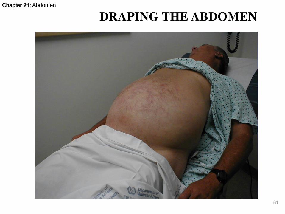
DRAPING THE ABDOMEN

Chapter 21: Abdomen
82
VARIOUS CAUSES OF ABDOMINAL
DISTENSION
• Obese abdomen

Chapter 21: Abdomen
83
VARIOUS CAUSES OF ABDOMINAL
DISTENSION
Hepatomegaly

Chapter 21: Abdomen
84
VARIOUS CAUSES OF ABDOMINAL
DISTENSION
Ascites

Chapter 21: Abdomen
85
VARIOUS CAUSES OF ABDOMINAL
DISTENSION
Markedly enlarged gall bladder
(labelled "GB")

Chapter 21: Abdomen
86
VARIOUS CAUSES OF ABDOMINAL
DISTENSION
Umbilical Hernia

Chapter 21: Abdomen
87
VARIOUS CAUSES OF ABDOMINAL
DISTENSION
Umbilical hernia
Chapter 21: Abdomen
88
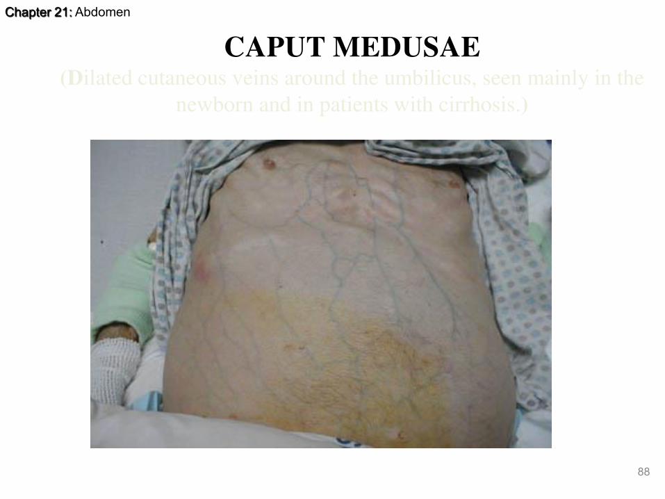
CAPUT MEDUSAE (Dilated cutaneous veins around the umbilicus, seen mainly in the
newborn and in patients with cirrhosis.)

Chapter 21: Abdomen
89
GYNECOMASTIA Abnormal Enlarged breast of male

Chapter 21: Abdomen
90
Auscultation
Adult Stethoscope: Diaphragm and Bell
Incorporated Into Single Side. Adult Stethoscope
Newborn Stethoscope Combination Adult & Pediatric Stethoscope

Chapter 21: Abdomen
91
EXAMINING FOR A FLUID WAVE:
Chapter 21: Abdomen
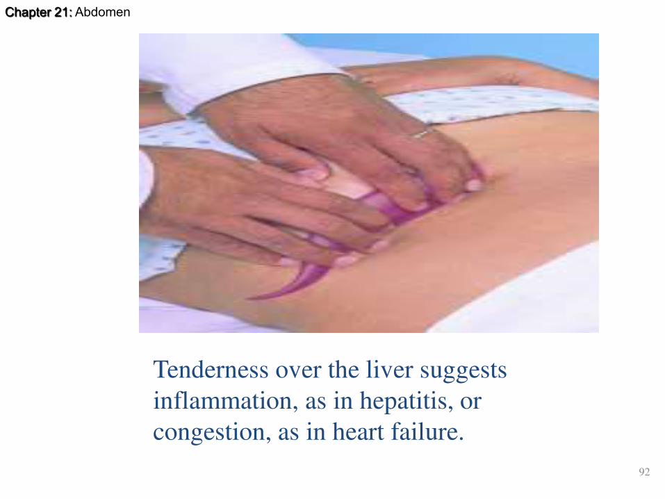
Tenderness over the liver suggests
inflammation, as in hepatitis, or
congestion, as in heart failure.
92

Chapter 21: Abdomen
GI Variations Due to Age
• Aging- should not affect GI function unless associated with a disease process.
• Decreased: salivation, sense of taste, gastric acid secretion, esophageal emptying, liver size, bacterial flora.
• Increased: constipation!
2/26/2016 93

Chapter 21: Abdomen
GI Variations with pregnancy • Decrease in gastric
motility • High incidence of N, V
(r/t pregnancy hormones) and “heartburn” or acid reflux
• Bowel sounds diminished r/t enlarged uterus displacing intestines
• Linea nigra- increased pigmentation of abd midline
• Striae Gravidarum 2/26/2016 94
Chapter 21: Abdomen
1. Anderson, K. (1996). Mosby’s Medical, Nursing and Allied
Health Dictionary, ed. 4. St. Louis: C.V. Mosby.
2. Barkauskas,V.H., Stoltenberg-Allen, K., Baumann, L.C., and
Darling- Fisher. (1998). Health and Physical Assessment, ed.
2. St. Louis: Mosby—Year Book.
3. Bates, B., Bickley, L.S., and Hoekelman, R.A. (1995).Physical
Examination and History Taking, ed. 6. Philadelphia:
Lippincott-Raven.
4. Becker, K.L., and Stevens, S.A. (1988). Performing in-depth
abdominal assessment. Nursing88 18(6):59–63.
5. Zator Estes, M.E. (1998). Health Assessment and Physical
Examination. Albany,N.Y.: Delmar Publishers.
References
95