Une etude des voies de signalisation impliquees dans … · Figure 8 Taux de survie sans...
144
HAL Id: tel-01058194 https://tel.archives-ouvertes.fr/tel-01058194 Submitted on 26 Aug 2014 HAL is a multi-disciplinary open access archive for the deposit and dissemination of sci- entific research documents, whether they are pub- lished or not. The documents may come from teaching and research institutions in France or abroad, or from public or private research centers. L’archive ouverte pluridisciplinaire HAL, est destinée au dépôt et à la diffusion de documents scientifiques de niveau recherche, publiés ou non, émanant des établissements d’enseignement et de recherche français ou étrangers, des laboratoires publics ou privés. Une étude des voies de signalisation impliquées dans la carcinogénèse et le traitement des fibromatoses agressives Armelle Dufresne To cite this version: Armelle Dufresne. Une étude des voies de signalisation impliquées dans la carcinogénèse et le traite- ment des fibromatoses agressives. Médecine humaine et pathologie. Université Claude Bernard - Lyon I, 2014. Français. <NNT: 2014LYO10082>. <tel-01058194>
Transcript of Une etude des voies de signalisation impliquees dans … · Figure 8 Taux de survie sans...

HAL Id: tel-01058194https://tel.archives-ouvertes.fr/tel-01058194
Submitted on 26 Aug 2014
HAL is a multi-disciplinary open accessarchive for the deposit and dissemination of sci-entific research documents, whether they are pub-lished or not. The documents may come fromteaching and research institutions in France orabroad, or from public or private research centers.
L’archive ouverte pluridisciplinaire HAL, estdestinée au dépôt et à la diffusion de documentsscientifiques de niveau recherche, publiés ou non,émanant des établissements d’enseignement et derecherche français ou étrangers, des laboratoirespublics ou privés.
Une étude des voies de signalisation impliquées dans lacarcinogénèse et le traitement des fibromatoses
agressivesArmelle Dufresne
To cite this version:Armelle Dufresne. Une étude des voies de signalisation impliquées dans la carcinogénèse et le traite-ment des fibromatoses agressives. Médecine humaine et pathologie. Université Claude Bernard - LyonI, 2014. Français. <NNT : 2014LYO10082>. <tel-01058194>
N° ordre 82-2014 Année 2014
UNIVERSITE CLAUDE BERNARD LYON 1
THESE
pour l’obtention du
DIPLOME DE DOCTORAT
(arrêté du 7 Août 2006)
présentée et soutenue publiquement
par
Melle Armelle Dufresne
Le 6 Juin 2014
Une étude des voies de signalisation impliquées dans la
carcinogénèse et le traitement des fibromatoses agressives
Directeur de thèse : Pr Jean-Yves Blay
JURY Mr Jean-Michel COINDRE, Président
Mr Jean-Yves BLAY
Mr Charles DUMONTET
Mr Jean-Emmanuel KURTZ
Mme Florence PEDEUTOUR, Rapporteur
Mr Eric RAYMOND, Rapporteur

2
UNIVERSITE CLAUDE BERNARD - LYON 1
Président de l’Université
Vice-président du Conseil d’Administration
Vice-président du Conseil des Etudes et de la Vie Universitaire
Vice-président du Conseil Scientifique
Directeur Général des Services
M. François-Noël GILLY
M. le Professeur Hamda BEN HADID
M. le Professeur Philippe LALLE
M. le Professeur Germain GILLET
M. Alain HELLEU
COMPOSANTES SANTE
Faculté de Médecine Lyon Est – Claude Bernard
Faculté de Médecine et de Maïeutique Lyon Sud – Charles
Mérieux
Faculté d’Odontologie
Institut des Sciences Pharmaceutiques et Biologiques
Institut des Sciences et Techniques de la Réadaptation
Département de formation et Centre de Recherche en Biologie
Humaine
Directeur : M. le Professeur J. ETIENNE
Directeur : Mme la Professeure C. BURILLON
Directeur : M. le Professeur D. BOURGEOIS
Directeur : Mme la Professeure C. VINCIGUERRA
Directeur : M. le Professeur Y. MATILLON
Directeur : Mme. la Professeure A-M. SCHOTT
COMPOSANTES ET DEPARTEMENTS DE SCIENCES ET TECHNOLOGIE
Faculté des Sciences et Technologies
Département Biologie
Département Chimie Biochimie
Département GEP
Département Informatique
Département Mathématiques
Département Mécanique
Département Physique
UFR Sciences et Techniques des Activités Physiques et Sportives
Observatoire des Sciences de l’Univers de Lyon
Polytech Lyon
Ecole Supérieure de Chimie Physique Electronique
Institut Universitaire de Technologie de Lyon 1
Ecole Supérieure du Professorat et de l’Education
Institut de Science Financière et d'Assurances
Directeur : M. le Professeur F. DE MARCHI
Directeur : M. le Professeur F. FLEURY
Directeur : Mme Caroline FELIX
Directeur : M. Hassan HAMMOURI
Directeur : M. le Professeur S. AKKOUCHE
Directeur : M. Georges TOMANOV
Directeur : M. le Professeur H. BEN HADID
Directeur : M. Jean-Claude PLENET
Directeur : M. Y.VANPOULLE
Directeur : M. B. GUIDERDONI
Directeur : M. P. FOURNIER
Directeur : M. G. PIGNAULT
Directeur : M. C. VITON
Directeur : M. A. MOUGNIOTTE
Directeur : M. N. LEBOISNE

3
Remerciements
A Monsieur le Professeur Jean-Yves BLAY, qui, par son intelligence, la confiance sans faille
avec laquelle il nous confie des projets et la richesse des portes qu’il nous ouvre nous mène
bien plus loin que ce que l’on aurait jamais pu imaginer, qu’il trouve ici l’expression de ma
sincère amitié et de ma profonde gratitude,
A Madame le Professeur Florence PEDEUTOUR et Monsieur le Professeur Eric RAYMOND,
qui m’ont fait l’honneur d’accepter de prendre de leur précieux temps pour juger mon travail
et l’enrichir de leurs remarques et conseils en tant que rapporteurs, qu’ils soient assurés de
mon entière reconnaissance,
A Messieurs les Professeurs Jean-Michel COINDRE, Charles DUMONTET et Jean-
Emmanuel KURTZ, qui ont accepté d’examiner et d’évaluer cette thèse, et à l’expertise
desquels soumettre mon travail est certainement un peu stressant mais sera forcément très
enrichissant, qu’ils en soient profondément remerciés.
Ce travail a été possible car soutenu financièrement par l’Institut National du Cancer que je
tiens à remercier vivement.
A tous les membres de l’équipe 11 du Centre de Recherche en Cancérologie de Lyon pour
leur accueil et leur soutien non seulement scientifique mais surtout amical, pour la finesse
constante des blagues fusant dans l’open space, pour avoir remis à jour mes compétences
musicales en pièce de culture, pour ne s’être jamais moqué des décès cellulaires massifs
constatés dans mes boites de culture….
A ma famille, à mes amis, à toi, et à mes 3 petits, qui n’ont pas souvent vu leur maman à la
sortie de l’école ces derniers temps mais qui, j’espère, comprendrons un jour que
l’aboutissement de ce travail représente surtout un choix délibéré de vie, d’orientation de
carrière, qui, loin d’être le plus simple mais soutenu par les bonnes personnes, est arrivé à
terme, porteur d’épanouissement et de confiance personnels.

4
Liste des abréviations et anglicismes ABL Abelson
ADN Acide Désoxyribonucléique
AINS Anti-Inflammatoire Non Stéroïdien
APC Adenomatous Polyposis Coli
ARN Acide Ribonucléique
BCL2 B-Cell Lymphoma 2
β-TrCP Beta-Transducin repeat Containing Protein
CAST Competitive Allele Specific Taqman
CCND1 Cyclin D1
CDK Cyclin-dependent Kinase
CK1 α Casein Kinase 1 α
CSMD1 CUB and Sushi Multiple Domains 1
COX 2 Cyclo-oxygenase 2
CSF R Colony Stimulating Factor Receptor
E2 Estrogènes
ESMO European Society of Medical Oncology
FA Fibromatose Agressive
FFPE Formalin Fixed Paraffin Embedded
FGF Fibroblast Growth Factor
FZD Frizzled
GIST Gastro-Intestinal Stromal Tumor
GSK-3β Glycogen Synthase Kinase-3β
IHC Immuno-histo-chimie
IRM Imagerie par Résonance Magnétique
LEF Lymphoid Enhancer-binding Factor
LHRH Luteinizing Hormone Releasing Hormone
LKB1 Liver Kinase B1
MCR Minor Cluster Region
miRNA Micro ARN
NF2 Neurofibrome 2
NCCN National Comprehensive Cancer Network
NRCAM Neuronal Cell Adhesion Molecule
PAF Polypose Adénomateuse Familiale
PBMC Peripheral Blood Monocyte Cell
PCR Polymerase Chain Reaction
PDGFR Platelet-derived Growth Factor Receptor
Pet scan Tomography par Emission de Positrons
PGE2 Prostaglandine
PI3K Phospho-inositol 3 Kinase
PTEN Phosphatase and Tensin homolog
Rb Rétinoblastome
RCP Réunion de Concertation Pluridisciplinaire
RE Récepteur aux Estrogènes
RMS Rhabdomyosarcome
RPg Récepteur à la Progestérone
RECIST Response Evaluation Criteria in Solid Tumours

5
SNP Single Nucleotide Polymorphism
KO Knock Out
SUV Standardized Uptake Value
TCF T-cell Factor
TGF Transforming Growth Factor
TMA Tissue Micro Array
TSP-1 Thrombospondin-1
VEGF Vascular Endothelial Growth Factor
WHO World Health Organization

6
Table des matières
Liste des figures et tableaux .................................................................................................. 7 Préambule ............................................................................................................................. 8 I. INTRODUCTION ..........................................................................................................10
A. Présentation ...........................................................................................................10 B. Prise en charge ......................................................................................................13
1. Stratégie .............................................................................................................13 2. Radiothérapie .....................................................................................................15 3. Traitement médical .............................................................................................16
C. Facteurs pronostiques et prédictifs .........................................................................20 D. Biologie des FA ......................................................................................................23
1. Nature néoplasique des FA ................................................................................23 2. Carcinogénèse : implication de la voie Wnt .........................................................27 3. APC ....................................................................................................................29 4. Beta-caténine .....................................................................................................33 5. Cytogénétique ....................................................................................................35 6. Identification de nouvelles cibles thérapeutiques ................................................35
II. TRAVAUX DE RECHERCHE .......................................................................................37 A. FACTEURS PRONOSTIQUES ..............................................................................37 1. Facteurs pronostiques cliniques .................................................................................37 2. Facteurs pronostiques biologiques .............................................................................43 B. FACTEURS PREDICTIFS ......................................................................................49
Identification de facteurs biologiques prédictifs de la réponse à l’imatinib mésylate dans les fibromatoses agressives ..........................................................................................49
III. DISCUSSION ............................................................................................................58 A. Evolution des concepts de prise en charge ............................................................58 B. Contribution de nos travaux à l’évolution de ces concepts ......................................59
1. Facteurs pronostiques cliniques ..........................................................................60 2. Facteurs pronostiques biologiques......................................................................60 3. Facteurs prédictifs ..............................................................................................60
C. Les limites de notre travail ......................................................................................61 D. Propositions stratégiques .......................................................................................62
1. Stratégie clinique ................................................................................................62 2. Stratégie de recherche biologique ......................................................................63
IV. CONCLUSION ...........................................................................................................65 Bibliographie ........................................................................................................................69 ANNEXES ............................................................................................................................81 Annexe 1 Liste des 138 “drivers genes” répertoriés par Vogelstein ......................................81 Annexe 2 Publications présentées dans ces travaux ............................................................86 Résumé ........................................................................................ Erreur ! Signet non défini.

7
Liste des figures et tableaux Figure 1 Répartition par sexe et tranche d’âge des diagnostics de FA en France actualisé au mois d’Août 2013 (données RRePS) ....................................................................................11 Figure 2 Images de scanner de FA, de la mandibule à gauche et abdominal à droite ..........12 Figure 3 Représentation schématique des modes évolutifs de FA (d’après Gronchi et al. 2013) ....................................................................................................................................12 Figure 4 Illustration photographique pré (à gauche) et post opératoire (à droite) d’une FA de la paroi abdominale ..............................................................................................................13 Figure 5 Schématisation de stratégie de prise en charge des patients atteints de FA proposée par Gronchi et al. ..................................................................................................19 Figure 6 Schématisation de la voie Wnt/β caténine en conditions physiologique (à gauche) et pathologique (à droite) (d’après T. Reya & H. Clevers, Nature 2005) ...................................27 Figure 7 Représentation schématique de la protéine APC et des sites de mutations génétiques correspondant. APC comporte 2843 codons. La MCR se situe entre les codons 1286 et 1513 et regroupe la majorité des sites de mutation somatique ................................30 Figure 8 Taux de survie sans progression en fonction du sous-type de mutation CTNNB1 dans 3 études .......................................................................................................................45 Figure 9 Survie sans récidive de 167 patients atteints de FA en fonction du statut du variant M541L de l’exon 10 de KIT ...................................................................................................56
Tableau 1 Etudes et séries rapportant les taux d’efficacité des chimiothérapies dans les FA .............................................................................................................................................18 Tableau 2 Principaux résultats des études rétrospectives identifiant les facteurs prédictifs de récidive après exérèse chirurgicale des FA (analyse multivariée). ........................................21 Tableau 3 Taux de survie sans récidive en fonction du statut primitif ou récidivant de FA ...22 Tableau 4 Expression des « marqueurs de malignité » en IHC ............................................24 Tableau 5 Correspondance entre les sites de mutations germinale et somatique sur le gène APC ......................................................................................................................................31 Tableau 6 Principales caractéristiques des modèles murins étudiant le rôle d’APC dans l’étiologie des FA. Adapté à partir de Fodde 2001 Nat Rev et Lips 2009 ..............................32 Tableau 7 Etude du caractère mutuellement exclusif des mutations de CTNNB1 et APC dans les FA sporadiques ...............................................................................................................34 Tableau 8 Fréquence et corrélation clinique des sous types de mutation de CTNNB1 .........44 Tableau 9 Etude IHC des cibles connues de l’imatinib des patients inclus dans les essais de phase II évaluant l’imatinib dans les FA (nombre cas positifs/nombre de cas étudiés) .........53 Tableau 10 Répartition de la présence du variant M541L de l’exon 10 de KIT en fonction du caractère hormonodépendant des FA ..................................................................................55

8
Préambule
La fibromatose agressive était une maladie mal connue, tant sur le plan moléculaire que
thérapeutique lorsque j’ai initié ce travail. Quand ces travaux ont débuté, l’essai clinique de
phase II, Desminib, évaluant la toxicité et l’efficacité de l’imatinib dans les FA non
accessibles à un traitement local, venait de s’achever et les résultats cliniques étaient
disponibles. Nous avons entrepris un programme de recherche de transfert dédié à cet essai
clinique, visant à identifier des marqueurs biologiques prédictifs de réponse à l’imatinib dans
cette population. Nous avons dans un premier temps rapatrié les blocs de tumeur inclus en
paraffine (« Formalin Fixed Paraffin Embedded ») des patients ayant participé à l’essai et
avons pu en récupérer 34 (sur 40 patients inclus dans l’étude). Ces blocs FFPE ont servi aux
différentes expériences menées ensuite.
Dans un premier temps, nous avons étudié en immunohistochimie sur tissue micro-array
l’expression de récepteurs de l’imatinib et d’éléments de voies de signalisation
potentiellement impliqués dans la réponse au traitement. Nous n’avons pas observé de
corrélation entre l’expression de ces protéines et les réponses objectives enregistrées dans
l’essai. Nous avons parallèlement étudié, par séquençage, la présence du variant M541L de
l’exon 10 de KIT sur l’ADN tumoral issu de ces mêmes blocs en paraffine. Différents « case
reports » publiés à ce moment-là évoquaient l’impact positif de la présence de ce variant sur
la réponse à l’imatinib. La qualité médiocre du matériel ne nous a permis de conclure que sur
10 patients : le variant était présent dans 3 échantillons, dont un patient en réponse partielle
et le seul patient en réponse complète de l’étude. Ce résultat n’était pas statistiquement
significatif mais il nous a semblé judicieux d’élargir la cohorte analysable en utilisant une
technique de PCR quantitative (CAST PCR) pour améliorer le rendement de nos résultats.
L’analyse statistique n’a pu conclure que la présence du variant M541L puisse être prédictive
d’une meilleure réponse à l’imatinib. Au même moment, nous avons eu des échanges avec
l’équipe du Pr Kurtz de Strasbourg qui avait identifié un nouveau variant de l’exon 10 de KIT
(V530I), associé à une réponse d’excellente qualité d’une FA à l’imatinib.
Parallèlement, dans le cadre du programme européen « PERSEUS 1 » initié sous
l’impulsion de l’association de patients « SOS Desmoïdes », un vaste recueil de données
multicentrique a été réalisé en 2008, auquel j’ai activement participé. Quatre cent vingt six
patients atteints de FA diagnostiquées entre 1965 et 2008 ont été recensés dans la «
Conticabase ». Après confirmation du diagnostic par relecture des lames histologiques par le
Groupe Sarcome Français (GSF), les données cliniques décrivant les antécédents du
patient, les caractéristiques de la tumeur, toutes les étapes de son évolution et les
traitements administrés étaient collectées de manière rétrospective. L’analyse de ces
données a identifié l’âge du patient, la taille et la localisation tumorales comme étant des
facteurs pronostiques de récidive. Cette base de données a également été utilisée par une
équipe du MSKCC comme cohorte de validation pour établir un nomogramme pronostique
de récidive des FA à partir de ces mêmes facteurs.
L’échec de l’identification de facteurs pronostiques et prédictifs aux étapes précédentes a
conduit à explorer une nouvelle approche via l’analyse du profil d’expression des miRNAs
des tumeurs, développé au sein du laboratoire d’accueil depuis le programme européen
Conticanet. Il a été décidé d’analyser ce profil à partir de l’ARN des FA des patients inclus
dans l’essai Desminib avec un double objectif : identifier des voies biologiques permettant de
rechercher des facteurs prédictifs de réponse à l’imatinib et également des facteurs

9
pronostiques. Une signature pronostique a été identifiée et confirmée sur une cohorte de
validation indépendante de FA inclus dans la base de données nationale, pour lesquelles on
disposait des blocs inclus en paraffine.
Dans ce manuscrit, je présenterai tout d’abord en introduction, les caractéristiques
spécifiques des FA et les particularités de leur prise en charge et de leur traitement.
J’exposerai ainsi à la fois l’importance et le manque de marqueurs cliniques et/ou
biologiques, pronostiques et prédictifs. Je rapporterai ensuite les principales données sur les
connaissances actuelles de la biologie de ces tumeurs pour appréhender les avancées
possibles dans cette thématique.
Dans une seconde partie, j’exposerai les principaux résultats de mes travaux, menés dans le
but de progresser dans l’identification de nouveaux marqueurs pronostiques et prédictifs.

10
I. INTRODUCTION
A. Présentation
Les fibromatoses agressives (FA) sont également appelées « tumeurs desmoïdes ». Ce sont
des tumeurs localement agressives du tissu conjonctif, caractérisées par une prolifération de
fibroblastes et une production abondante de collagène hyalinisé analogue au tendon
(« desmos » = tendon). Ces tumeurs sont rares puisqu’elles représentent 0.03% de toutes
les tumeurs et 3% des tumeurs des tissus mous. Leur incidence annuelle est de 2-4
nouveaux cas/1 000 000 habitants (Reitamo et al. 1982). Compte-tenu de cette rareté, leur
prise en charge doit se faire préférentiellement en centre spécialisé (“Soft Tissue and
Visceral Sarcomas: ESMO Clinical Practice Guidelines for Diagnosis, Treatment and Follow-
Up.” 2012).
Depuis 1997, il a été établi que ces FA doivent être considérées comme un processus
néoplasique (et non un processus uniquement réactionnel) puisqu’elles sont composées
d’une prolifération cellulaire issue d’un clone unique (Alman et al. 1997), des progéniteurs
des cellules mésenchymateuses en l’occurrence (Wu et al. 2010). Elles sont classées par la
« WHO Classification of Tumours of Soft Tissue and Bone » de 2013 comme étant de
malignité intermédiaire. En effet, et comme quelques rares autres tumeurs, elles présentent
cette particularité très intrigante de ne jamais donner de métastases à distance alors qu’elles
sont responsables d’une agressivité loco-régionale parfois très marquée et de récidives
fréquentes après exérèse.
Les FA se développent dans différents contextes, soit dans le cadre d’un syndrome de
Gardner quand elles sont associées à une polypose adénomateuse familiale et liées à la
mutation du gène APC (≈ 2% des cas), soit, le plus souvent, de manière sporadique. Des
cas de développant sur des zones cicatricielles (post-traumatiques ou post-chirurgicales)
sont fréquemment décrits. Le lien biologique entre cicatrice et FA a été établi par Carothers
et al. (Carothers et al. 2012) démontrant que les FA sont composées de cellules dérivées
des cellules souches mésenchymateuses ayant acquis des caractéristiques de cicatrisation,
d’angiogénèse et de fibrose (cf § D 1.).
Les séries rétrospectives de patients atteints de FA retiennent classiquement un sex ratio en
faveur des femmes, de l’ordre de 2 :1. Quand ce sex ratio est étudié plus précisément au
cours de la vie, il évolue en fait avec le nombre des années (Reitamo et al. 1982) (Skene et
al. 1998): si il y a nettement plus de femmes que d’hommes atteints pendant la période
d’activité génitale (≈ 20-40 ans), la fréquence entre les 2 sexes est équivalente pendant
l’enfance et après la ménopause. Ces différences sont considérées comme sous-tendues
par l’influence des stéroïdes sexuels sur ces tumeurs.
Les dernières données issues du « Réseau de Référence en Pathologie des Sarcomes des
tissus mous et des viscères » recensaient 735 cas de FA au mois d’Août 2013 et illustrent
bien cette répartition spécifique par sexe en fonction de l’âge :

11
Figure 1 Répartition par sexe et tranche d’âge des diagnostics de FA en France actualisé au mois
d’Août 2013 (données RRePS)
Si le pic d‘incidence se situe chez l’adulte, entre 20 et 50 ans, les FA peuvent être
diagnostiquées à tout âge : on retrouve, dans la littérature, un cas rapporté chez un bébé de
2 mois. Les formes pédiatriques ne sont pas rares et font l’objet de séries rétrospectives et
de traitements spécifiques (Skapek et al. 2013). Les séries publiées décrivent également
fréquemment des cas de patients âgés de plus de 80 ans.
Les FA sont des tumeurs du tissu conjonctif qui peuvent survenir partout dans l’organisme,
au niveau de tous les tissus de soutien. Leur localisation est habituellement organisée en 3
catégories : extra-abdominale, intra-abdominale et au niveau de la paroi abdominale.
A partir de ces 3 caractéristiques d’âge, de sexe du patient et de localisation de la tumeur, a
été déterminée une classification clinique en 4 groupes (Reitamo et al. 1986):
- les FA juvéniles : tumeur extra-abdominale chez la petite fille
- les FA de la période d’activité génitale : tumeur de la paroi abdominale chez la femme
- les FA d’âge moyen : tumeur plutôt abdominale chez les hommes autant que chez les
femmes
- les FA des personnes âgées : tumeur abdominale ou extra-abdominale, chez les
hommes autant que chez les femmes
La taille des tumeurs au moment du diagnostic est très variable, liée à la localisation et au
caractère infiltrant de la tumeur et donc à ses conséquences fonctionnelles (i. e.
symptomatologie). Une tumeur de 2 cm érodant la mandibule sera beaucoup plus
rapidement symptomatique qu’une lésion de 20 cm intra-abdominale (cf images). Ce
caractère infiltrant des tumeurs est représenté par leurs limites : si certaines tumeurs
semblent radiologiquement et macroscopiquement encapsulées refoulant les organes de
voisinage, d’autres ont des limites radiologiques imprécises à leur périphérie et envahissent
les structures adjacentes.
0
50
100
150
200
250
300
0-20 20-40 40-60 60 et +
femmes 477
hommes 258

12
Figure 2 Images de scanner de FA, de la mandibule à gauche et abdominal à droite
Enfin, le caractère sûrement le plus représentatif de la variabilité des tumeurs est l’évolutivité
des FA observée en clinique. Il est impressionnant de constater que certaines FA puissent
avoir une évolutivité indolente au point de demeurer stable en taille voire même de régresser
spontanément alors que d’autres évoluent rapidement et récidivent de manière répétée
après les traitements. Entre ces deux modalités évolutives, tous les modes d’évolution sont
rencontrés et ont été schématisés par Gronchi et al. (Gronchi et al. 2014).
Figure 3 Représentation schématique des modes évolutifs de FA (d’après Gronchi et al. 2013)
Cette grande variabilité observée au niveau de la présentation clinique, du contexte de
survenue et de l’évolutivité des tumeurs sous-tend sûrement différentes caractéristiques
moléculaires, non encore identifiées à ce jour.

13
B. Prise en charge
1. Stratégie
La prise en charge des FA est actuellement débattue et les anciens standards de prise en
charge largement remis en question depuis 5 ans. Auparavant, ces tumeurs étaient gérées
au diagnostic comme des tumeurs malignes : l’exérèse chirurgicale en était systématique
dans la mesure où les séquelles prévisibles étaient acceptables ; sinon une radiothérapie
seule était proposée. La radiothérapie adjuvante était recommandée en cas de marges
chirurgicales positives, mais, en l’absence d’essais randomisés, ne constituait pas un
standard (Ballo et al. 1999).
Il était délicat de proposer des règles univoques de prise en charge chirurgicale des FA dans
la mesure où elles peuvent survenir dans des sites anatomiques très divers de l’organisme,
être de toutes tailles et avoir des limites variables avec les organes adjacents (infiltration ou
non). Les seules recommandations qui peuvent éventuellement guider le geste portent sur le
la qualité de l’exérèse, i.e. R0, R1 ou 2. Après relecture et analyse de toute la littérature
portant sur ce sujet, il apparaît que le caractère macroscopiquement complet de l’exérèse
est important mais que le risque de récidive n’est pas différent entre une exérèse R0 ou R1
(données détaillées dans § I C).
Compte-tenu de plusieurs facteurs cliniques spécifiques des FA :
- absence de métastases secondaires,
- fréquence relativement importante des récidives locales après résection chirurgicale
(taux de récidive locale : 15 à 50% à 5 ans dans les 9 études rétrospectives),
- absence d’impact de la chirurgie sur le risque de reprogression après rechute,
- séquelles chirurgicales fonctionnelles et/ou esthétiques non négligeables,
Figure 4 Illustration photographique pré (à gauche) et post opératoire (à droite) d’une FA de la paroi
abdominale
- régressions spontanées non exceptionnelles,
- non évolutivité d’un certain nombre de tumeurs observée en clinique,
plusieurs ont proposé la possibilité de simplement surveiller les patients atteints de FA à la
prise en charge.
En 2007, Gouin et al. publient le premier article remettant en question la stratégie agressive
de traitement des FA (Gouin et al. 2007). Ces travaux ont le grand intérêt de porter sur une
AVANT
Chirurgie
APRES
14
cohorte prospective de 17 patients atteints de fibromatoses agressive extra-abdominale et
simplement surveillés : après 42 mois de suivi médian, 3 tumeurs avaient régressé et 12
étaient restées stables. Il est particulièrement intéressant de noter qu’aucune évolution
n’avait été observée après 36 mois. Par la suite, plusieurs études rétrospectives défendant
une attitude d’observation sans traitement ont été publiées: en 2008, le Dr Bonvalot rapporte
les résultats d’une étude rétrospective portant sur 112 patients, montrant que la survie sans
événement à 3 ans était identique entre les patients opérés avec marges saines et les
patients non opérés. Les 2/3 des patients simplement surveillés étaient non évolutifs à 3 ans
(Bonvalot et al. 2008). En augmentant la cohorte à 142 patients, la moitié des patients
surveillés n’évoluaient pas à 5 ans (Fiore et al. 2009). D’autres publications concluent dans
le même sens : Honeyman et al. (Honeyman et al. 2013) ont étudié une population de 93 cas
pédiatriques de FA primitives et récidivantes et retrouve des taux de survie sans évènements
à 5 ans de 21% sans traitement et 34% avec traitement (p = 0.09). Barbier et al. (Barbier et
al. 2010) rapportent 26 cas de FA primitives et récidivantes simplement surveillés, dont 24
restent stables après un suivi médian de 14 mois et 1 régresse spontanément. Stoeckle et
al. (Stoeckle et al. 2009) retrouvent des temps médians de stabilité de 48 à 137 mois selon
que la tumeur soit primitive ou récidivante.
Ces observations ont amené les principales instances européenne (ESMO) et nord-
américaines (NCCN) à conseiller la simple surveillance des FA asymptomatiques et non
évolutives dans leurs recommandations (von Mehren et al. 2012) (“Soft Tissue and Visceral
Sarcomas: ESMO Clinical Practice Guidelines for Diagnosis, Treatment and Follow-Up.”
2012).
Cette stratégie de surveillance a rapidement été intégrée aux pratiques quotidiennes de prise
en charge des patients. Toutefois, elle ne repose pour l’instant que sur des données
rétrospectives, par définition biaisées, et qui peuvent être critiquables dans la mesure où le
choix de traitement des patients a forcément été influencé par la présentation des tumeurs.
On ne retrouve jamais, à la lecture de ces articles, de description des patients surveillés. De
plus, plusieurs limites apparaissent dans ces études :
- la taille globale des cohortes et surtout le nombre de patients uniquement surveillés est
souvent faible (15 patients surveillés sur 93 au total dans l’étude de Honeyman),
- le suivi médian des patients est parfois très court (14 mois pour l’étude de Barbier),
- les critères établissant les groupes comparés sont très variables (patients opérés versus
patients surveillés ou bénéficiant de traitements médicaux dans l’étude de Bonvalot, patients
surveillés versus patients bénéficiant de traitements médicaux dans l’étude de Fiore),
- le choix de l’objectif peut être discutable (pas d’influence de la chirurgie sur la survie
globale dans l’étude de Stoeckle).
Seul un essai prospectif pourra répondre clairement à cette question de stratégie de
traitement ; cet essai est en cours, mené par le Dr Bonvalot. Il s’agit d’un essai de phase II,
non randomisé, multicentrique, qui prévoit d’inclure 100 patients atteints de fibromatoses
périphériques (membres ou paroi abdominale ou thoracique) primitives non opérées ou en
exérèse R2. L’objectif principal est d’évaluer la survie sans progression locale à 3 ans et l’un
des objectifs secondaires est de tenter d’identifier des facteurs biologiques, de l’hôte et/ou de
la tumeur, prédictifs de l’évolution. Cet essai est très innovant conceptuellement, proposant
la simple surveillance d’une tumeur (plutôt que la chirurgie), d’autant plus qu’il est soutenu
par un chirurgien.
15
En attendant les résultats de cette étude, les décisions prises actuellement en réunions de
concertation pluridisciplinaires ne retiennent la résection chirurgicale qu’en cas de tumeur
symptomatique, évolutive ou mettant en jeu le pronostic vital du patient. Tous les autres cas
sont dans un premier temps surveillés et une thérapeutique spécifique n’est proposée qu’en
cas de lésion évolutive. En tout état de cause, ces décisions sont probabilistes en l’absence
de facteurs pronostiques fiables permettant de sélectionner efficacement les patients qui
bénéficient réellement d’une telle stratégie attentiste.
Aucune étude n’a recherché les facteurs prédisant l’évolution naturelle de la maladie, en
dehors tout traitement. Or ceci est maintenant essentiel si on veut sélectionner les patients
pouvant bénéficier d’une simple surveillance.
Quels que soient les résultats finaux de cet essai, on sait dès à présent que certains de ces
patients vont évoluer et donc devoir être traités : les repérer précocement serait un objectif
important, et cette question a été une des objectifs de notre travail.
2. Radiothérapie
La place de la radiothérapie dans la thérapeutique des FA peut se discuter à 2 étapes de la
prise en charge : en tant que traitement complémentaire d’une chirurgie (à marges saines ou
non) et en tant que traitement isolé d’une tumeur en place. Dans la 1ère situation, des
éléments de réponse peuvent être apportés par les résultats de la méta-analyse portant sur
22 études et publiée en 2000 (Nuyttens et al. 2000). Ces résultats rapportent des taux de
contrôle local par une chirurgie seule de 72% en cas de marges saines et de 41% en cas de
marges positives. Ces résultats sont statistiquement inférieurs à ceux obtenus par une
radiothérapie complétant la résection chirurgicale qui permet d’obtenir un contrôle local de
94% en cas de marges saines (R0) et à 75% en cas de marges positives (R1 ou R2). Le
taux de récidive de 75% obtenu par chirurgie à marges positives et radiothérapie est
comparable à celui de 72% obtenu par chirurgie seule à marges saines. On peut considérer
que la radiothérapie « rattrape » une chirurgie à marges envahies. Toutefois, les conclusions
des études de stratégie thérapeutique portant sur des études rétrospectives sont en général
biaisées. En l’absence d’études randomisées, la radiothérapie complémentaire d’une
chirurgie incomplète n’est actuellement pas un standard, ni pour l’ESMO ni pour le NCCN.
De plus, les séquelles de la radiothérapie et le risque de seconde tumeur sont à prendre en
compte. Dans la méta-analyse, 9 articles rapportaient des complications à type de fibrose,
parfois invalidante, paresthésies, cellulite, fracture pathologique. Plus graves, un
ostéosarcome et un sarcome utérin sont survenus après irradiation, respectivement d’une
tumeur mandibulaire et d’une FA abdominale.
Dans la 2ème situation, un essai de phase II récemment publié rapporte un taux de contrôle
local de 81,5% à 3 ans avec une radiothérapie de 56 Gray pour des tumeurs primitives ou
récidivantes en place, ou résidus macroscopiques inopérables et évolutifs (Keus et al. 2013).
La meilleure réponse était parfois observée au-delà de 3 ans. Le suivi médian de la cohorte
était de 4.8 ans et la survie sans récidive à 10 ans de 70%. La radiothérapie peut ainsi
légitimement être proposée pour contrôler des tumeurs non opérables.

16
3. Traitement médical
Lorsque les FA sont évolutives et/ou symptomatiques et non accessibles à un traitement
local par chirurgie ou radiothérapie, différents traitements systémiques peuvent être
proposés, d’efficacité très variable. Aucune étude randomisée n’a jamais été menée pour
déterminer précisément le taux d’efficacité de ces thérapies. L’ESMO et le NCCN retiennent,
comme possibilité de traitement médical des FA, des hormonothérapies (tamoxifène,
torémifène, analogues de LHRH), des anti-inflammatoires non stéroidiens (sulindac,
celecoxib), des chimiothérapies (schémas à base d’anthracyclines, méthotrexate et
vinblastine ou vinorelbine, interféron à faible dose) et plus récemment, des thérapies ciblées
(imatinib, sorafenib). L’efficacité de ces traitements est globalement modeste et surtout
excessivement variable d’un patient à l’autre sans qu’aucun facteur prédictif formel pouvant
guider le choix du traitement n’ait à ce jour été identifié.
L’influence des hormones sur les FA justifiant l’utilisation des anti-oestrogènes, analogues de
la LH-RH ou inhibiteurs de l’aromatase, repose sur plusieurs constatations cliniques :
- incidence de cette pathologie plus élevée chez les femmes que chez les hommes,
particulièrement en période d’activité génitale (20-40 ans),
- survenue de FA de la paroi abdominale en cours de grossesse ou en post-partum,
- régression spontanée de FA à distance de l’accouchement ou en période de
ménopause.
Une analyse rétrospective des données de la littérature parue en 2011 recensait, entre 1983
et 2009, 41 articles portant sur l’utilisation d’hormonothérapie dans 168 cas FA (30 « case
report » et 11 séries) (Bocale et al. 2011). Bien évidemment, seuls les cas répondeurs sont
généralement publiés dans la littérature et ces données ne peuvent refléter l’efficacité réelle
de ce traitement. Les taux de réponse objective étaient comparés entre différents groupes
de patients reconstitués à partir de ces observations rétrospectives. Si une telle
méthodologie est discutable, elle peut fournir des pistes de réflexion à partir de ses
conclusions. Les auteurs retenaient que les taux de réponse des FA à l’hormonothérapie
étaient:
- identiques entre les FA associées à un syndrome de Gardner versus les sporadiques
- supérieurs chez les patients traités par hormonothérapie seule versus en association
avec d’autres traitements, notamment les AINS (cette constatation peut être liée à un
biais de méthodologie rétrospective où les tumeurs les plus agressives sont traitées
par des associations de traitements)
- identiques entre les traitements par tamoxifène versus torémifène
- identique en fonction de l’expression des récepteurs aux estrogènes (RE)
Plusieurs précisions peuvent être apportées à ces conclusions. Deux études cliniques
rapportent les résultats d’association de tamoxifène haute dose et sulindac, respectivement
chez des adultes (Hansmann et al. 2004) et une population pédiatrique (Skapek et al. 2013).
Parmi les 21 adultes traités par l’association, on retrouve 5 réponses objectives, 2 maladies
progressives et 11 stabilisations. Parmi les enfants, l’association permet d’obtenir 5 réponses
objectives sur 59 cas.
Concernant l’analyse des taux d’expression des RE, le nombre de cas étudiés est très faible
(9). De plus, il n’est pas précisé, dans les publications concernées, si les RE dosés sont de

17
type α ou β. Or, si le RE α est le plus communément mesuré, notamment dans les tumeurs
classiquement hormonodépendantes comme le cancer du sein, il semble que ce soit le sous
type β qui soit plus important dans les FA. Plusieurs publications rapportent une expression
quasi constante de ce récepteur dans les FA (Deyrup et al. 2006; Zhang et al. 2010; Santos
et al. 2010; Mignemi et al. 2012; Colombo et al. 2012). Toutefois, aucune n’a corrélé sa
présence à l’efficacité clinique de l’hormonothérapie. Actuellement, le dosage des REβ ne
peut être considéré comme facteur prédictif de réponse à l’hormonothérapie.
Biologiquement, les effets génomiques et non génomiques des 2 sous-types du récepteur
aux estrogènes ne sont pas clairement établis. Les 2 sous-types peuvent coexister dans les
tissus, avoir une activité transcriptionnelle variable et opposée : si REα semble plutôt avoir
un effet prolifératif, REβ aurait plutôt un effet anti-prolifératif.
Depuis la publication de cette revue, il faut signaler la publication d’un « case report »
rapportant une réponse complète de FA sous exemestane (Debled et al. 2012). La seule
série disponible sur l’efficacité de l’hormonothérapie dans les FA est parue en 2010. Cette
série rétrospective a recensé 26 lignes d’hormonothérapie administrées permettant d’obtenir
6 réponses partielles, 17 stabilisations et 3 maladies progressives pour un temps à
progression de 12 mois (de Camargo et al. 2010).
Les anti-inflammatoires non stéroidiens ont été développés dans cette indication sur des
bases pré cliniques associant des études in vitro et in vivo démontrant le rôle de la COX2 et
de son inhibition dans ces tumeurs. La COX2 est une enzyme induite par différents stimuli
cellulaires, dont les facteurs de croissance et les cytokines. Les taux de ses métabolites
(prostaglandine E2) ont été retrouvés élevés dans différents processus néoplasiques,
notamment dans les polypes intestinaux développés dans des modèles de souris avec
mutation de APC (souris KO APC∆716) où l’expression de COX2 est augmentée (Oshima et
al. 1996). L’administration d’anti COX2 chez ces souris réduit significativement le nombre de
polypes. COX2 est également surexprimée dans des prélèvements tumoraux de FA (Poon et
al. 2001; Signoroni et al. 2007; Mignemi et al. 2012) comparativement au tissu normal des
mêmes patients. Des expériences sur des cultures cellulaires primaires de FA ont montré
une diminution des taux de PGE2 et de la prolifération cellulaire en présence d’AINS. Les
taux de COX2 n’ont jamais été corrélés à l’efficacité des AINS. De plus, la découverte d’une
cible commune entre cette classe de médicaments et la voie de signalisation Wnt-APC-β
caténine a permis d’établir un lien entre l’effet anti-tumoral des AINS et les altérations
génétiques de la voie Wnt-APC-β caténine (He et al. 1999). Le récepteur activé par les
proliférateurs de peroxysome de type δ (PPARδ) fait partie de la super famille des
récepteurs nucléaires. Les membres de cette famille activent la transcription de séquences
spécifiques après fixation de leur ligand. APC réprime l’expression de PPARδ qui, à
contrario, est élevée dans les cancers colo-rectaux. Cette régulation se fait par la voie β
caténine/Tcf. Dans des cultures cellulaires de carcinomes colo-rectaux, l’activité de PPARδ
est fortement diminuée par le sulindac. Cliniquement, les cas de 22 patients atteints de FA
extra-abdominale et traités par meloxicam ont été examinés rétrospectivement : 95% d’entre
eux présentaient au moins une stabilisation de la maladie (Nishida et al. 2010). Toutefois, les
auteurs eux-mêmes concluent que « ce résultat ne puisse être totalement attribuable à
l’efficacité de l’AINS et que seul un essai randomisé contre placebo pourrait prouver l’effet
inhibiteur du meloxicam ».
Beaucoup plus toxique, l’utilisation de la chimiothérapie cytotoxique anti-cancéreuse dans
les FA ne repose pas sur beaucoup plus de données. Seule une étude prospective, de
phase II, est disponible (Skapek et al. 2007). Sur 28 enfants traités par une association de

18
méthotrexate et vinblastine, 8 ont présenté une réponse objective et 10, une stabilisation de
la maladie. Dix-huit patients ont évolué avec une médiane de 9,1 mois. Toutes les autres
études sont rétrospectives, présentées dans le tableau ci-dessous.
Auteur, année N Protocole Taux de réponse PFS
Weiss et al., 1989 8 MTX-VBL 75% NA
Patel et al., 1993 12 Doxorubicine-dacarbazine 67%
Skapek et al., 1998 10 MTX-VBL 50% NA
Weiss et al., 1999 13 MTX-NVB 60% NA
Reich et al., 1999 5 MTX-VBL 60% NA
Azzarelli et al., 2001 30 MTX-VBL 40% Non atteinte avec
10 ans suivi
Gega et al., 2006 7 Doxorubicine-dacarbazine 100% 74
De Camargo et al., 2010 10 MTX-VBL 20% NA
35 Anthracycline 37% NA
Garbay et al., 2011 27 MTX-VBL 15%
40.8 13 Anthracycline 54%
Constantinidou et al., 2011 18 MTX-VBL 11% 9
14 Doxo Peg. 33% NA
Abréviations : N taille de la cohorte ; MTX méthotrexate ; VLB vinblastine ; Doxo Peg doxorubicine
pégylée
Tableau 1 Etudes et séries rapportant les taux d’efficacité des chimiothérapies dans les FA
(Weiss & Lackman 1989; Patel et al. 1993; Skapek et al. 1998; Weiss et al. 1999; Reich et al. 1999;
Azzarelli et al. 2001; Gega et al. 2006; Garbay et al. 2012; Constantinidou et al. 2011; de Camargo et
al. 2010)
Si les schémas à base d’anthracyclines semblent les plus efficaces (Garbay et al. 2012), ceci
doit être mis en balance avec les toxicités à court, moyen et long terme qu’ils induisent.
Le développement des inhibiteurs de tyrosine-kinase dans les FA a démarré avec plus
d’études prospectives : l’imatinib a été évalué dans 3 essais de phase II incluant
respectivement 19, 40 et 51 patients (Heinrich et al. 2006; Chugh et al. 2010; Penel et al.
2011). Les taux de réponse objective sont de 15.7%, 12% et 6% et les taux de survie sans
récidive à 1 an à 36.8%, 67% et 66%. Kasper et al. ont tenté de rechercher une corrélation
entre les modifications de SUV d’un pet-scan et la réponse objective sous imatinib évaluée
par IRM selon les critères RECIST (Kasper et al. 2013). Une valeur initiale de SUV élevée
serait corrélée à une réponse partielle ou stabilisation (une valeur initiale de SUV plus basse
semble prévoir une maladie progressive). Ce résultat est en attente d’études confirmatives.
Le sorafenib induit 25% de réponse partielle dans une étude rétrospective portant sur 26
patients (Gounder et al. 2011). Le sunitinib a été évalué dans un essai de phase II incluant
19 patients (Jo et al. 2014). Un quart des patients ont présenté une réponse partielle et la

19
moitié, une stabilisation de la maladie. La survie sans progression à 2 ans était de 75%.
Deux cas ont rapporté une efficacité intéressante du pazopanib, avec des réponses
objectives de qualité et durant plus d’un an (Martin-Liberal et al. 2013). Ces observations ont
motivé la mise en route d’un essai de phase III (le premier !) multicentrique français
randomisant des patients atteints de FA évolutive entre une chimiothérapie par
méthotrexate-vinblastine et un traitement par pazopanib.
Il est assez surprenant de constater, lorsque l’on tente de faire la synthèse de l’efficacité des
traitements médicaux dans les FA, que l’on retrouve, grossièrement, et quel que soit le
traitement étudié, environ la moitié des patients présentant une stabilisation de la maladie,
un quart répondeurs et un quart progressifs. On peut être tenté de se poser la question de
savoir dans quelle mesure ces taux sont le reflet d’une activité de la thérapeutique ou
simplement liés à l’évolution naturelle de la maladie.
Actuellement, aucun facteur prédictif clinique ou biologique n’a été identifié, permettant de
sélectionner les patients bénéficiant au mieux d’un type de traitement pour leur proposer une
séquence thérapeutique qui leur soit adaptée et donc, plus utile. En l’absence de
recommandations, et compte-tenu de l’absence de potentiel métastatique des FA et de la
très faible mortalité dont elles sont responsables, le prescripteur tend naturellement à
proposer en premier lieu les traitements ayant le moins d’effets secondaires (AINS et
hormonothérapie avant chimiothérapie et traitements ciblés). En 2014, Gronchi et al.
proposent l’algorithme suivant pour la prise en charge des FA (Gronchi et al. 2014):
Figure 5 Schématisation de stratégie de prise en charge des patients atteints de FA proposée par
Gronchi et al.
Bien évidemment, à chacune de ces étapes doit se poser la question de la possibilité
d’inclusion dans un essai clinique, que ce soit un essai de stratégie ou de thérapeutique.

20
C. Facteurs pronostiques et prédictifs
L’expérience clinique et bibliographique montre à quel point le pronostic des FA est variable
d’un patient à l’autre, et le panel des traitements actifs ne permet pas de proposer une
approche rationnelle en l’absence de biomarqueurs ou d’arguments moléculaires pour guider
le choix du traitement. Dans la prise en charge de ces tumeurs, le praticien manque ainsi de
biomarqueurs cliniques et/ou biologiques clairement identifiés comme influençant le
pronostic. Dans le cas des FA, 2 situations principales requièrent la détermination de leur
pronostic :
- l’évolution d’une tumeur en place, diagnostiquée, non opérée. La recherche du pronostic à
ce stade semble particulièrement pertinente dans la mesure où si certaines FA augmentent
naturellement de volume avec des conséquences fonctionnelles qui peuvent être lourdes
(envahissement nerveux ou vasculaires, par exemple, avec les douleurs et risques
neurologiques ou hémorragiques qui en découlent), d’autres n’évoluent pas dans le temps,
même en l’absence de traitement, voire régressent spontanément.
- le risque de récidive loco-régionale après exérèse chirurgicale.
Toutes les études publiées recherchant à identifier des facteurs pronostiques de l’évolution
des FA sont des études rétrospectives pour lesquelles, l’immense majorité des patients était
opéré. On ne dispose pas de description de l’évolution naturelle de la maladie, en dehors de
tout traitement. Une telle description sera apportée par les résultats de l’étude de phase II du
Dr Bonvalot actuellement en cours et mentionnée ci-dessus (ayant fait l’objet du
PHRC10_02-10 et intitulée « Fibromatoses primitives périphériques: Etude de phase 2
évaluant une surveillance simple initiale avec recherche des facteurs prédictifs d’évolutivité
et enregistrement des traitements en cas de progression »). On ne peut donc actuellement
se baser sur aucun facteur pour déterminer le pronostic évolutif d’une tumeur en place. En
pratique, il est recommandé dans un premier temps, de simplement surveiller l’évolution
d’une tumeur nouvellement diagnostiquée par des examens cliniques et radiologiques
répétés pour se faire une idée de son évolutivité.
Les facteurs pronostiquant la rechute après exérèse chirurgicale ont été largement étudiés et
les résultats ne sont pas forcément concordants. Toutes ces études pronostiques portent sur
des cohortes rétrospectives de patients opérés, d’environ 100 à 200 patients, et analysent,
en multivarié, l’influence éventuelle de facteurs cliniques sur la survie sans récidive. Les
résultats sont très difficilement interprétables car souvent contradictoires.

21
Auteur, année n Age Sexe Taille Site tumoral Qualité marges Radiothérapie
Huang, 2013 214 non non oui non non -
Van Broekhoven, 2013 132 non non non oui non Non
Shin, 2013 119 non non non non oui non (sf délai)
Mullen, 2012 177 non non non non oui Non
Peng, 2012 211 oui - non oui non Non
Bertani, 2012 62 non non non oui non -
Lev, 2007 189 non non non
Gronchi, 2003 203 - - oui oui non -
Merchant, 1999 105 non non non non non -
Spear, 1998 107 oui non - non oui Oui
n : taille de la cohorte
Tableau 2 Principaux résultats des études rétrospectives identifiant les facteurs prédictifs de récidive
après exérèse chirurgicale des FA (analyse multivariée).
(Huang et al. 2013; van Broekhoven et al. 2013; Shin et al. 2013; Mullen et al. 2012; Peng et al. 2012;
Bertani et al. 2012; Lev et al. 2007; (Gronchi et al. 2003)Merchant et al. 1999)(Spear et al. 1998)
De toutes ces données, on peut tenter d’extraire l’analyse suivante :
- le sexe ne semble pas avoir d’impact pronostique sur la récidive
- l’âge n’est retrouvé que très rarement comme facteur pronostique (les plus jeunes
ayant tendance à avoir les pathologies les plus récidivantes) mais les analyses étaient très
hétérogènes : Lev l’étudie comme variable continue alors que les autres auteurs ont plutôt
tendance à faire des groupes par tranches d’âge (2 groupes à 4 groupes).
- lorsque la localisation tumorale influence le pronostic, ce sont toujours les tumeurs
extra abdominales, principalement des membres et des ceintures qui sont les plus graves.
- la qualité des marges chirurgicales est matière à controverse, a été analysée dans
de multiples études rétrospectives et même des méta-analyses. Pourtant, en les analysant,
les résultats semblent bien tous concordants. Toutes les études retrouvant un impact
statistiquement significatif de la qualité des marges sur la récidive, ont comparé des marges
positives à des marges négatives, c’est à dire R0 vs R1 et R2 (études rétrospectives de Shin
2013, Mullen 2012, Spear 1998 et méta-analyses de Nuyttens et Leithner) (Nuyttens et al.
2000)(Leithner et al. 2004). Dans les études de Huang, Van Broekhoven, Peng et Bertani,
qui ne retrouvent pas d’influence de la qualité des marges chirurgicales, la résection
complète R0 était comparée à l’envahissement microscopique des marges R1. Il ne semble
donc pas y avoir de différence sur le risque de récidive entre les résections R0 et R1.
- l’administration de radiothérapie améliore rarement le pronostic mais on n’a très peu
de renseignements sur les conditions et les modalités de son administration.
L’analyse rétrospective de ces données est largement critiquable. Dans des tumeurs de
présentation aussi hétérogène, la discussion de l’acte chirurgical prend en compte de

22
multiples facteurs comme l’âge du patient, mais aussi le risque de séquelles prévisibles
beaucoup plus que la taille ou la localisation tumorale. Les rapports de la tumeur avec les
organes adjacents (surtout s’il s’agit de nerfs ou de vaisseaux) sont essentiels dans la
qualité de la résection. De même, en l’absence de recommandations claires, on n’a aucune
notion dans ces études, des traitements adjuvants qui ont éventuellement pu être proposés
aux patients après des exérèses incomplètes ou marginales. L’entrainement du chirurgien,
les habitudes des centres sont autant de biais qui ont forcément influencé ces résultats.
De plus, dans l’interprétation de ces résultats, il faut garder à l’esprit l’hypothèse que l’acte
chirurgical lui-même puisse être un propre facteur favorisant la récidive. Ce concept repose
sur le fait qu’on retrouve fréquemment dans la littérature et en clinique des cas de FA qui se
développent sur des cicatrices ou des sites opératoires d’une part, et des cas de FA
multirécidivants après des chirurgies itératives d’autre part (parfois plus d’une dizaine
d’opérations sur une même FA). Cette notion est également (faiblement) soutenue par le fait
que, dans ces mêmes études rétrospectives, les taux de récidive sont presque toujours
supérieurs pour les patients présentant une tumeur récidivante comparés à ceux présentant
une tumeur primitive.
Auteur, n cohorte Taux de survie sans récidive
Tumeur primitive Tumeur récidivante
Huang, 214 84.9% à 5 ans 63% à 5 ans
Van Broekhoven, 132 que des tumeurs primitives. Taux de récidive locale à 5 ans : 17.6% (très bas)
Shin, 119 85% à 5 ans 47% à 5 ans
85% à 10 ans 31% à 10 ans
Mullen, 177 32% taux récidive 20% taux récidive ……
Bertani, 62 20% taux récidive 10% taux récidive …...
Gronchi, 203 76% à 10 ans 59% à 10 ans
Merchant, 105 que des tumeurs primitives. Taux de récidive locale à 5 ans : 25%
Spear, 107 79% 67%
Tableau 3 Taux de survie sans récidive en fonction du statut primitif ou récidivant de FA
(Huang et al. 2013; van Broekhoven et al. 2013; Shin et al. 2013; Mullen et al. 2012; Bertani et al.
2012; Gronchi et al. 2003; Merchant et al. 1999; Spear et al. 1998)
Il commence à apparaître dans la littérature des articles rapportant la recherche de facteurs
biologiques comme facteurs pronostiques (Romero et al. 2012; Colombo et al. 2013). La
mutation 45F de CTNNB1 ou la présence de macrophages associés à la tumeur ont été
identifiés comme facteurs pronostiques mais là encore, prédisant la rechute après exérèse.

23
D. Biologie des FA
Les mécanismes biologiques impliqués dans la transformation des cellules de FA ont été
étudiés ces dernières années et de grands progrès ont été réalisés dans leur
compréhension. D’une manière générale, la voie Wnt est altérée, par des mutations
activatrices ou inhibitrices de deux gènes principaux, APC et β-caténine.
1. Nature néoplasique des FA
Dans les années 90, suite à l’individualisation des FA comme entité ayant des
caractéristiques morphologiques et une histoire naturelle bien définis, la pathogénie de ces
tumeurs demeurait inconnue. La nature infiltrative des tumeurs et leur évolution parfois
agressive les rapprochaient de processus néoplasiques mais les cas de régression
spontanée faisaient plutôt penser qu’elles appartenaient au groupe des processus
réactionnels (comme les cicatrices chéloïdes).
La nature néoplasique d’une prolifération tumorale est définie par sa clonalité. Deux
publications ont démontré cette clonalité dans les années 1996-1997 en utilisant la même
méthode, celle de l’inactivation du chromosome X. Cette méthode a été clairement établie
comme capable de démontrer l’origine clonale des tumeurs humaines (Vogelstein et al.
1985) (Allen et al. 1992). Elle se base sur le fait qu’un seul chromosome X est actif chez la
femme. Le second est inactivé précocement au cours de l'embryogenèse. Cette inactivation
survient au hasard. Elle reste stable durant toute la vie de la cellule et est transmise aux
cellules filles après division cellulaire. Elle est également stable après culture in vitro ou
lorsque la cellule devient néoplasique. L'inactivation de nombreux gènes sur le chromosome
X s'accompagne de variations dans la méthylation des régions CpG en 5' des gènes. Cette
méthylation permet de reconnaître le chromosome X actif du chromosome X inactif en
étudiant l'action différentielle d'enzymes de restriction sensibles à la méthylation. Quand le
site est méthylé, l'enzyme ne coupe pas l'ADN. Par PCR, on étudie ainsi les sites de
méthylation du gène humain du récepteur aux androgènes (HUMARA). Dans un processus
polyclonal, on retrouvera une inactivation du chromosome X aléatoire dans les cellules
(d’origine paternelle et maternelle) alors que dans un processus monoclonal, on retrouvera
l’inactivation du même chromosome X dans toutes les cellules.
A partir, respectivement, de 4 cas et 20 cas de FA chez des femmes, Li et Alman ont
démontré ainsi la clonalité de ces tumeurs (Li et al. 1996)(Alman et al. 1997). Non
seulement, les caractéristiques d’inactivation sont identiques dans les cellules de FA mais
elles sont aussi similaires entre la tumeur primitive et sa récidive, confirmant que la tumeur
récidivante provient du même clone que la tumeur primitive. Cette clonalité a également été
démontrée par des analyses cytogénétiques montrant que les trisomies 8 et 20 sont des
anomalies stables dans les FA primitives et leurs récidives et non pas des anomalies
aléatoires (Fletcher et al. 1995).
Toutefois, la clonalité démontrée par les 2 techniques pré-citées peut également se
rencontrer dans des tumeurs bénignes et ne signe pas la malignité d’une tumeur (Bridge et
al. 1999) (Kopp et al. 1997). Bridge et al. ont retrouvé des trisomies 8 dans des dysplasies
ostéofibreuses et Kopp et al. des inactivations non aléatoires du chromosome X dans des
leucocytes sains. La question de la malignité ou de la bénignité des FA n’est pas résolue.
Cliniquement, l’absence de métastases oriente vers un caractère bénin mais l’agressivité

24
loco-régionale plutôt vers un caractère malin. Il n’existe aucun critère biologique ou
histologique signant de manière formelle la malignité d’une tumeur.
L’article référence de Hanahan et Weinberg (Hanahan and Weinberg 2011) retient 6
caractéristiques biologiques fondamentales du cancer, dont certaines ont été explorés dans
les FA :
- Le maintien de signaux de prolifération regroupe plusieurs mécanismes : activation
des voies de signalisation (e.g. mutations activatrices de BRAF ou PI3K), inhibition
de rétrocontrôle négatif (e.g. mutation RAS ou PTEN), activation d’oncogènes (e.g.
RAS MYC RAF).
La présence de ces mutations n’a pas été recherchée dans les FA. PTEN est exprimé en
IHC dans 4/7 FA (Chugh et al. 2010), KRAS n’est pas muté dans 8 FA (Miyaki et al. 1993).
- L’échappement aux inhibiteurs de croissance regroupe les mécanismes de perte
d’inhibition de contact (e.g. NF2 ou LKB1), d’échappement aux effets anti-prolifératifs
de TGFβ et d’inactivation des gènes suppresseurs de tumeur (e.g. Rb ou p53).
L’expression de Rb et p53 a été recherchée dans les FA et comparée à celle d’autres
tumeurs d’origine fibroblastiques, certaines bénignes (Muller et al. 1996) et d’autres malignes
(Hoos et al. 2001). Muller et al. concluent que la perte d’expression de Rb puisse avoir un
rôle dans la prolifération des FA. Hoos et al. retrouvent que l’expression de ces 2 gènes
suppresseurs de tumeur est corrélée à l’agressivité biologique des tumeurs.
Auteur, année Tumeurs pRb Ki67 p53 Bcl 2
Muller et al. 1996 FA 0/13 - 0/13 -
Fibromatose plantaire 6/6 - 0/6 -
Hoos et al. 2001
FA 24/24 24/24 23/24 0/24
FS-BG 11/25 17/25 20/25 12/25
FS-HG 1/14 2/14 10/14 8/14
Gebert et al. 2007 FA - 36/37 12/37 -
Stalinska et al. 2009 FA 35/50 13/50 - -
FS-BG/HG : Fibrosarcome de bas grade/haut grade
Tableau 4 Expression des « marqueurs de malignité » en IHC
(C. Gebert et al. 2007) (Stalinska et al. 2009)
L’expression du récepteur de TGFβ et de SMAD 2/3 (reflétant l’activité de la voie de TGFβ) a
été comparée en IHC entre 27 échantillons de FA, 14 de cicatrices et 6 de tissus fibreux
normal. Elle est élevée dans les FA et les cicatrices et absente dans les tissus normaux. Ces
résultats sont cohérents avec ceux de Bacac (Bacac et al. 2006) étudiant l’expression
génique de TGFβ.
- La résistance à la mort cellulaire regroupe les phénomènes d’augmentation de
signaux de survie (e.g. IGF) et l’augmentation de signaux anti-apoptotiques (e.g.
Bcl2).
L’étude de Hoos précitée confirme la surexpression de Bcl2 dans les cancers contrairement
aux FA.

25
- L’induction d’une angiogénèse est médiée positivement par le VEGFA et
négativement par TSP-1.
Plusieurs publications rapportent une augmentation des phénomènes d’angiogénèse dans
les FA. Le taux de VEGF est plus élevé dans les prélèvements de FA que dans les
prélèvements de cicatrice ou de peau (Mills et al. 2000) et la densité de microvaisseaux est
un facteur de récidive (Romero et al. 2012) et (Matono et al. 2011).
- L’activation des phénomènes d’invasion et de métastases : c‘est un élément clé de la
description clinique des FA et une question majeure. Dans l’article de Hanahan
comme dans les autres articles portant sur ce sujet, les phases d’invasion et de
métastases sont traitées comme une seule entité sous tendue par les mêmes
facteurs biologiques (perte de E cadhérine, transition épithélio-mésenchymateuse),
l’invasion étant le premier stade avant les métastases (Talmadge and Fidler 2010).
Or, dans le cas des FA, les tumeurs peuvent avoir des phénomènes d’invasion locale
très importants sans jamais donner de métastases. Les mécanismes biologiques
précis discriminants ces 2 phases sont inconnus. Les FA pourraient représenter un
excellent modèle à étudier pour répondre à cette question.
- La possibilité d’une immortalité réplicative par échappement aux mécanismes qui
contrôlent la réplication infinie. Le rôle des télomères n’ jamais été exploré dans les
FA.
Dans la même idée de typer la nature cancéreuse ou non des FA, une publication a rapporté
des travaux portant sur la comparaison de profils d’expression génique de FA avec ceux de
tumeurs bénignes : les fasciites nodulaires (Bacac et al. 2006). Les gènes surexprimés chez
les FA comparativement aux fasciites nodulaires peuvent se regrouper en 3 grands
groupes :
- les gènes impliqués dans la voie de signalisation Wnt : les protéines de cette voie
(AXIN2, SFRP), les régulateurs de cette voie (MITF, PTPRF) et les gènes cibles de
cette voie (EPHB3, PITX2, WISP-1).
- les gènes impliqués dans la voie TGF-β (surexpression de TGF-β2 et sous-
expression des inhibiteurs de TGF : BAMBI et SMAD7).
- les gènes impliqués dans la différenciation : MDK, NRG1, NPTX2 et NEFH.
Une autre publication qui avait également rapporté les profils d’expression génique de FA
(en les comparant à différents prélèvements de tissu sain cette fois), retrouvait des
similitudes dans la surexpression de gènes dans les FA : EPHB3, NRG1, NPTX2, TGF-β3,
WISP-1 (Skubitz and Skubitz 2004).
Il n’est pas possible de rattacher de manière formelle l’expression de ces protéines ou de
ces gènes à un caractère bénin ou malin des FA.
En 2010, l’équipe d’Alman a approfondi les travaux pour caractériser le clone cellulaire
formant les FA (Wu et al. 2010). Ils ont mené des expériences in vitro et in vivo leur
permettant d’aboutir à un faisceau d’arguments montrant que ce clone était issu de cellules
souches mésenchymateuses :
- les cellules de FA expriment des gènes et des marqueurs de surface caractéristiques des
cellules souches mésenchymateuses,
- dans les modèles murins porteurs de mutation APC (Apcwt/1638N) et donc prédisposés au
développement de FA, le nombre de tumeurs développées est proportionnel au nombre de
cellules souches mésenchymateuses,
26
- ces mêmes souris Apcwt/1638N croisées avec des souris déficientes en progéniteurs
mésenchymateux développent moins de FA comparées à celles ayant des taux standards de
progéniteurs (et alors que le nombre de polypes intestinaux développés restait identique)
- l’injection de cellules souches mésenchymateuses porteuses d’une mutation oncogénique
dans des souris immunodéficientes induit des proliférations cellulaires aberrantes.
Carothers (Carothers et al. 2012) est allé plus loin en montrant que les FA sont composées
non seulement de cellules souches mésenchymateuses mais aussi de fibrocytes CD34+ qui
stimulent l’angiogénèse pendant les phases de cicatrisation ou de progression tumorale. Il a
également montré que les cellules souches mésenchymateuses des FA sont des cellules
pluripotentes ayant la capacité de se différentier en chondrocytes, adipocytes et ostéocytes.
Les FA sont donc des tumeurs monoclonales issues de cellules souches
mésenchymateuses, présentant certaines mais pas toutes les caractéristiques biologiques
de cellules cancéreuses.

27
2. Carcinogénèse : implication de la voie Wnt
En 1982, Harold Varmus découvre que l’activation d’un gène, nommé initialement Int1
(integration 1), induit la formation de tumeur dans la glande mammaire de souris (Nusse and
Varmus 1982). Il est l’homologue, chez le mammifère, du gène dont la mutation induit la
formation de drosophiles sans ailes : Wingless (gènes Int et Wing : gène Wnt). Cette
découverte représente l’identification du premier élément de la voie Wnt. Dans les années 80
puis 90, les différents éléments de cette voie sont identifiés et leur rôle se précise. Il est
intéressant de noter que souvent, ces différents éléments sont découverts parallèlement, par
différentes équipes, et que leur connexion avec la voie Wnt se fait dans un second temps.
Telle qu’elle est connue aujourd’hui, cette voie peut se schématiser ainsi (Klaus and
Birchmeier 2008; Reya and Clevers 2005) :
Figure 6 Schématisation de la voie Wnt/β caténine en conditions physiologique (à gauche) et
pathologique (à droite) (d’après T. Reya & H. Clevers, Nature 2005)
La β-caténine est une protéine intra cellulaire, intégrée dans un complexe contenant
également la E-cadhérine et l’α-caténine, impliqué dans les jonctions avec les cellules
voisines et le cytosquelette. APC est une protéine cytoplasmique, régulant la signalisation
intracellulaire, de la membrane cellulaire au noyau. En condition physiologique, la β-caténine
nouvellement synthétisée se lie à APC et Axine dans un complexe de destruction. CK1α et
GSK-3β, inclus dans le même complexe, entrainent la phosphorylation des résidus Sérine et
Thréonine de la partie N-terminale de la β-caténine. La β-caténine est alors dégradée par le
protéasome via une interaction avec la β-TrCP, un composant du complexe E3 ubiquitine
ligase. Au niveau nucléaire, les protéines de la famille Groucho/TLE se lient aux facteurs de
transcription de la famille TCF-LEF et agissent comme co-répresseurs de la transcription.
Quand le ligand Wnt se fixe sur son récepteur Frizzled, ou en cas de mutation des gènes qui
contrôlent cette stabilité de la β-caténine (APC, axine ou la β-caténine elle-même), ce

28
système est déficient ; stabilisée, la β-caténine s’accumule dans le cytoplasme puis est
transloquée vers le noyau. Là, elle entre en compétition directe avec les protéines de la
famille Groucho/TLE et les déplace. Elle se lie aux facteurs de transcription TCF et LEF et
induit ainsi la transcription de gènes cibles responsables de la carcinogénèse.
Ces gènes cibles de la voie Wnt sont des gènes impliqués dans :
- la différenciation cellulaire,
- la signalisation: VEGF, FGF4, FGF18,
- la prolifération: MYC, cycline D1,
- l’adhésion : E-cadhérine, NRCAM,
- la voie Wnt elle-même : Fzd, Axine 2, β-TrCP, TCF-LEF, démontrant la capacité
d’auto-régulation (positive et négative) de la voie.
Au total, plus d’une cinquantaine de gènes sont actuellement recensés comme cibles de la
voie Wnt chez l’homme. Roel Nusse, qui a mené les travaux aboutissant à la découverte du
gène Wnt avec H. Varmus précité, a construit un site internet entièrement dédié à la voie
Wnt dans lequel cette liste de gènes cibles est régulièrement mise à jour
(http://www.stanford.edu/group/nusselab/cgi-bin/wnt/).
La voie Wnt fait partie des grandes voies de signalisation dont la fonction est essentielle lors
du développement embryonnaire, impliquée dans les processus de prolifération cellulaire,
survie et différentiation. Pendant le développement, son activité est régulée précisément et
plus tard, l’échappement à ce contrôle peut induire des cancers et d’autres maladies. Des
mutations activatrices d’oncogènes et des mutations inhibitrices de gènes suppresseurs de
tumeurs d’éléments de la voie Wnt ont été identifiés comme responsables de la
carcinogénèse de nombreux cancers.
Ce n’est pas tant par l’action de son ligand Wnt, du récepteur Frizzled lui-même ou des
interactions avec les cellules voisines que s’explique le mécanisme par lequel cette voie de
signalisation est impliquée dans la carcinogénèse mais plutôt par des modifications de ses
deux composants protéiques intracellulaires principaux : APC et β caténine, induisant la
transcription de gènes cibles. APC agit comme un gène suppresseur de tumeur alors que
CTNNB1 agit comme un oncogène.
Les FA peuvent se développer dans 3 types de contexte clinique :
- de manière sporadique : la FA survient de manière spontanée, sans être associée à
d’autres pathologies particulières
- dans le cadre d’un syndrome de Gardner (MIM 175100) : la FA survient dans un
contexte familial de polypose colique, le sujet et sa famille présentent des centaines de
polypes intestinaux, des cancers coliques et des tumeurs bénignes comme des fibromes,
des ostéomes du crâne ou des maxillaires, des lipomes sous-cutanés, des myomes ; des
tumeurs thyroïdiennes ou surrénaliennes ; des hypertrophies congénitales de l’épithélium
pigmentaire rétinien
- dans le cadre de « fibromatose infiltrative familiale » également appelée « maladie
de desmoïde héréditaire » (MIM135290) : la FA se développe dans un contexte familial de
FA, le plus souvent intra-abdominal et sans polypose colique associée
Le syndrome de Gardner et la fibromatose infiltrative familiale sont des maladies génétiques
liées à des mutations du gène APC.

29
3. APC
En comparant les taux d’incidence de carcinomes coliques se développant ou non sur des
polypes en fonction de l’âge, Ashley évoque en 1969 et pour la première fois, que le gène de
la polypose puisse diminuer le nombre d’évènements nécessaires à la muqueuse intestinale
pour qu’elle devienne néoplasique (Ashley 1969), faisant écho à la théorie de Knudson,
établie sur le rétinoblastome (Knudson 1971). Il suggère également que le développement
d’un carcinome lieberkühnien sur un polype adénomateux dépende des mêmes facteurs
externes que pour les cancers non précédés de polypose. La théorie de Knudson est
aujourd’hui totalement validée par les découvertes qui ont été faites sur les mécanismes de
carcinogénèse dans un contexte de polypose : pour survenir, un cancer nécessite 2
évènements mutationnels. Dans les cancers héréditaires, une mutation germinale est
transmise et une seconde mutation, de type somatique, survient et enclenche le processus
de carcinogénèse. Dans les cancers non héréditaires, 2 mutations somatiques sont
nécessaires.
De plus, et contrairement à ce qu’avait initialement décrit Knudson qui pensait que ces 2
mutations étaient indépendantes l’une de l’autre, il a été démontré dans les PAF que la
nature et la localisation de la mutation germinale détermine la nature et la localisation de la
mutation somatique et qu’à elles 2, elles influencent le phénotype de la maladie.
Vers 1990, plusieurs publications majeures (Leppert et al. 1987; Ashton-Rickardt et al. 1989;
Kinzler et al. 1991; Groden et al. 1991) (Cottrell et al. 1992) établissent des mutations (de
type délétion) du gène APC en 5q21-22 comme étant caractéristiques de la polypose
adénomateuse familiale. Ces anomalies sont également décrites dans les FA développées
dans le cadre d’un syndrome de Gardner (Okamoto et al. 1990; Miyaki et al. 1993). Ces
mutations représentent la mutation germinale, premier évènement conduisant vers la
carcinogénèse. Elles entrainent la formation d’un codon STOP qui aboutit à l’expression
d’une protéine APC tronquée. Elles se situent principalement dans la moitié 5’ du gène ;
elles sont donc situées en amont des sites régulateurs de la β caténine qui sont de 2 sortes :
- 7 sites de répétition de 20 acides aminés qui correspondent aux sites de fixation et
de « down regulation » de β caténine
- 3 sites de répétition d’environ 20 acides aminés contenant un motif Ser-Ala-Met-Pro,
qui assurent la fixation avec l’Axine. Ces 3 « SAMP repeats » sont distribués au milieu des
sites de « down regulation » de la β caténine. Ces sites de fixation à l’axine sont essentiels
dans la régulation des taux intra cellulaires de β caténine. Un modèle murin de protéine APC
tronquée mais conservant un seul « SAMP repeat » donne des souris viables et ne
développant pas de tumeurs (Smits et al. 1999). Dans la même idée, un autre modèle murin
se basant sur la transfection uniquement de la partie centrale d’APC, contenant tous les sites
de fixation à la β caténine et à l’axine, montre que ces sites sont suffisants pour éviter le
développement de tumeur (Shih et al. 2000).
Il y a 2 sites de mutation germinale prédominants : au niveau des codons 1061 (a) et 1309
(b). Lorsque la mutation germinale survient sur le codon 1601, ou en amont de la MCR
(Mutation Cluster Region), la seconde mutation, somatique, survient préférentiellement au
sein de la MCR (hot-spots 1 à 5). La protéine tronquée conserve alors 1 voire 2 sites de
« down regulation » de la β caténine (ΔAPC 1). Lorsque la mutation germinale survient au
sein de la MCR (b ou c), il y a, le plus souvent, une perte de l’allèle sauvage (ΔAPC 2).
(Lamlum et al. 1999)

30
Les mutations d’APC aboutissent ainsi à une protéine tronquée qui conserve 1 à 2 sites de
« down regulation » de la β caténine. En effet, les cellules tumorales doivent conserver une
« down regulation » basale de la β caténine car l’accumulation excessive de β caténine dans
le noyau induit la mort cellulaire (Kim et al. 2000).
Figure 7 Représentation schématique de la protéine APC et des sites de mutations génétiques
correspondant. APC comporte 2843 codons. La MCR se situe entre les codons 1286 et 1513 et
regroupe la majorité des sites de mutation somatique
(d’après Klaus Nature Review 2008)
Les FA qui se développent dans le contexte d’un syndrome de Gardner sont génétiquement
comparables aux carcinomes colo-rectaux auxquelles elles sont associées. Plusieurs études
ont bien établi cette concordance entre mutations germinale, somatique, et le phénotype des
FA. Lamlum et al. (Lamlum et al. 1999) ont recensé, dans les FA survenant dans un contexte
de PAF, les concordances entre mutations germinale et somatiques. Ils en concluent que la
région de la mutation germinale d’APC associée à la perte d’allèle dans les FA est plus
distale que la région correspondante dans les adénomes. Ceci explique la différence
observée dans la sélection de la mutation d’APC entre les 2 types de tumeurs.

31
Tableau 5 Correspondance entre les sites de mutations germinale et somatique sur le gène APC
De nombreuses publications ont étudié cette corrélation entre la localisation de la mutation
germinale d’APC et le risque de survenue de FA dans un contexte de syndrome de Gardner.
Globalement, il semble que les FA se développent principalement lorsque cette mutation est
située au-delà du codon 1444, et associé à une perte de l’allèle sauvage.
Bertario (Bertario et al. 2001) établit une corrélation statistique entre le site de la mutation
germinale d’APC et le risque de développer une FA : pour une mutation d’APC située au-
delà du codon 1444, le risque de développer une FA est augmentée de 12 fois, comparé aux
patients pour lesquels la mutation est située en amont. Les mutations situées au-delà du
codon 1309 sont associées à un risque 17 fois plus élevé de développer une FA que les
mutations situées en amont du codon 452. Davies, Gebert et Caspari retrouvent eux aussi
une incidence particulièrement élevée de FA chez les patients dont la mutation germinale se
situe au niveau du codon 1444 (Davies et al. 1995; Gebert et al. 1999; Caspari et al. 1995).
Wallis établit 3 groupes de patients en fonction de la localisation de la mutation APC : groupe
1 - codons 177 à 452 ; groupe 2 – codons 457 à 1309 ; groupe 3 – codons 1395 à 1493
(Wallis et al. 1999). C’est dans le groupe 3 que se développent le plus de FA. Dobbie retient
lui le codon 1403 comme étant associé avec le plus de manifestations extra-coliques de
PAF, dont les FA (Dobbie et al. 1996).
Plusieurs modèles murins ont été développés pour explorer ce mécanisme. Les mutations
situées sur les codons 716 ou 850, du côté 5’ du gène APC, aboutissent à l’expression d’une
protéine tronquée, stable qui n’entraine pas le développement de FA. Lorsque la mutation
Apc1638N est présente, la protéine est indétectable et la pénétrance de FA est de 100%.
Son équivalent Apc1638T aboutit lui à une protéine tronquée mais stable et active ; aucune
FA ne se développe.

32
Protéine APC
Modèle Mutation
génétique Produit du gène Phénotype Référence
APC 1638N/+
Mutation
déphasante sur
codon 1638
Protéine tronquée indétectable
Pénétrance
100% FA
multifocales
Smits et al.
1998
APC 1638T/1638T
Mutation
déphasante sur
codon 1638
Protéine tronquée stable Pas de FA
Smits et al.
1999
APC Min/+
Mutation non-
sens sur codon
850
Protéine tronquée stable
Pas de FA
Tumeurs intest. Su et al. 1992
APC Δ716/+
Mutation
tronquante sur
codon 716
Protéine tronquée stable
Pas de FA
Tumeurs intest.
Oshima et al.
1995
Tableau 6 Principales caractéristiques des modèles murins étudiant le rôle d’APC dans l’étiologie des
FA. Adapté à partir de Fodde 2001 Nat Rev et Lips 2009
(Smits et al. 1999; Su et al. 1992; Oshima et al. 1995; Smits et al. 1998)
Une méta-analyse publiée en 2011 et étudiant 4625 patients avec PAF, identifie 3 facteurs
comme étant statistiquement associés au risque de survenue de FA : la mutation d’APC au-
delà du codon 1399 (OR 4.37), une chirurgie abdominale (OR 3.35) mais surtout, avec un
OR à 7.02, des antécédents familiaux de FA (Sinha et al. 2011). Les auteurs concluent
qu’au-delà de cette corrélation génotype/phénotype lié à la mutation d’APC, d’autres gènes
modifiés sous-tendent sûrement le développement de FA.
A côté des FA qui se développent dans un contexte de syndrome de Gardner, il existe un
autre groupe de FA d’origine génétique : celles qui se développent dans le contexte d’une
« fibromatose infiltrative familiale », également appelée « maladie des desmoïdes
héréditaires ». Elles sont liées à des mutations germinales d’APC situées en 3’ (codons
1924, 1962, 1987, 2643) associées à une perte de l’allèle sauvage (Eccles et al. 1996;
Benoit et al. 2007; Couture et al. 2000). Le phénotype de ces maladies associe une
pénétrance de 100% de FA multifocales et une polypose colique quasi inexistante.
33
4. Beta-caténine
L’accumulation intra cellulaire de β-caténine est l’évènement-clé de la carcinogénèse médiée
par un dysfonctionnement de la voie Wnt. Les articles rapportant les résultats d’analyses
immunohistochimiques de FA sporadiques montrent que l’expression de β-caténine est plus
élevée dans les cellules tumorales que dans les cellules avoisinantes et que la β-caténine
est alors localisée au niveau nucléaire plutôt que cytoplasmique (Alman et al. 1997; Tejpar et
al. 1999; Saito et al. 2002; Gebert et al. 2007; Huss et al. 2013). Cette surexpression
protéique a été caractérisée de plusieurs manières :
- elle a été identifiée comme facteur pronostique péjoratif de récidive locale des tumeurs
(Gebert et al. 2007).
- son mécanisme est lié à une diminution de la dégradation protéique (i. e. stabilisation) pour
les uns, puisque les taux d’ARNm relatifs à cette surexpression restent stables (Alman et al.
1997). D’autres retrouvent au contraire des taux d’ARNm supérieurs suggérant également
une hyper expression par augmentation de la transcription du gène (Saito et al. 2002).
- dans les FA sporadiques, elle serait la conséquence de mutations du gène de la β-caténine
(CTNNB1) ou d’APC. CTNNB1 est situé sur le chromosome 3p22, une région qui est
fréquemment le siège de mutations dans les pathologies tumorales (Trent 1995). Trois
mutations (T41A, S45F et S45P) et 1 délétion situées sur l’exon 3 de CTNNB1 sont
retrouvées dans environ 80% des FA sporadiques (Miyoshi et al. 1998) (Tejpar et al. 1999;
Tsuyoshi Saito et al. 2002; Lazar et al. 2008; Le Guellec et al. 2012; Huss et al. 2013). Ces
codons 41 et 45 sont situés au niveau des sites de phosphorylation de la protéine par le
GSK-3β.
Ces mutations sont spécifiques puisque lorsqu’elles ont été recherchées parallèlement sur
260 FA et 191 autres tumeurs à cellules fusiformes (comprenant des sarcomes, des
processus réactionnels et des tumeurs bénignes), aucune mutation n’a été détectée dans le
groupe des tumeurs autres que les FA (Le Guellec et al. 2012). Leur intérêt diagnostique est
donc maintenant bien établi.
L’impact pronostique de ces mutations sur les FA sporadiques a été étudié : la survie sans
récidive est plus longue chez les patients présentant une tumeur CTNNB1 sauvage que chez
ceux présentant une tumeur CTNNB1 mutée, d’autant plus lorsqu’il s’agit de la mutation
S45F (Lazar et al. 2008; Colombo et al. 2013; Mullen et al. 2013).
Un seul modèle murin a été développé sur le sujet : la stabilisation de la β-caténine dans un
modèle de souris transgénique aboutit à la formation de FA chez 18 souris sur 24 (75%) au
bout de 3 mois (Cheon et al. 2002).
Le caractère mutuellement exclusif des mutations APC et CTNNB1 n’est pas clairement
établi. Chez les patients présentant des FA sporadiques, plusieurs études publiées ont
recherché la présence de mutations d’APC parmi les tumeurs qui ne présentaient pas de
mutation de CTNNB1.
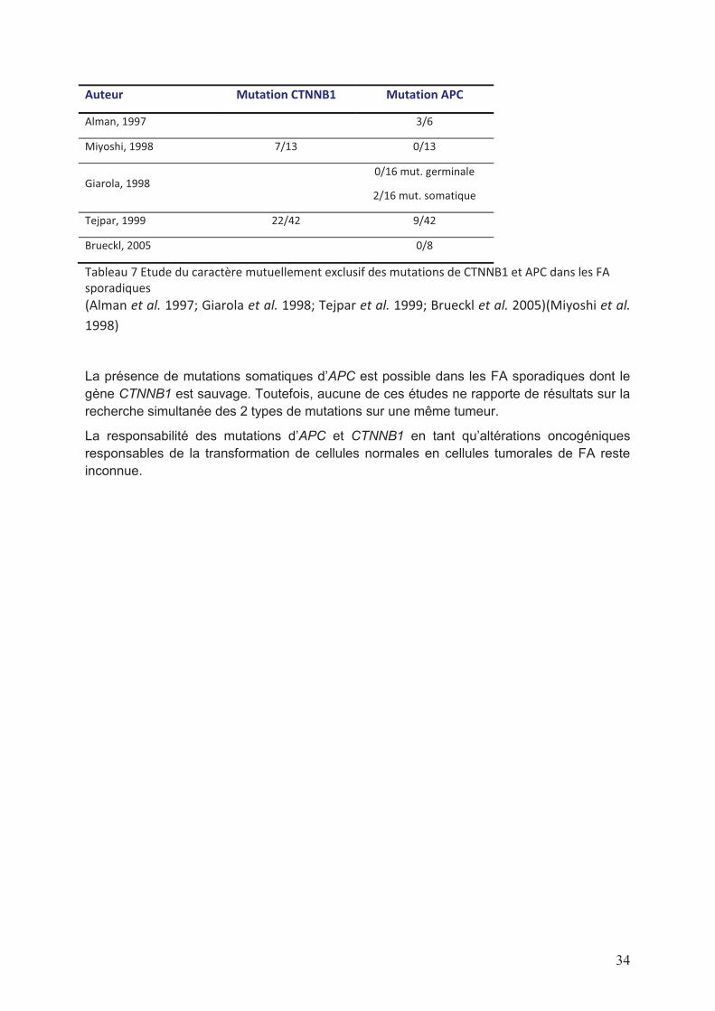
34
Auteur Mutation CTNNB1 Mutation APC
Alman, 1997 3/6
Miyoshi, 1998 7/13 0/13
Giarola, 1998 0/16 mut. germinale
2/16 mut. somatique
Tejpar, 1999 22/42 9/42
Brueckl, 2005 0/8
Tableau 7 Etude du caractère mutuellement exclusif des mutations de CTNNB1 et APC dans les FA
sporadiques
(Alman et al. 1997; Giarola et al. 1998; Tejpar et al. 1999; Brueckl et al. 2005)(Miyoshi et al.
1998)
La présence de mutations somatiques d’APC est possible dans les FA sporadiques dont le
gène CTNNB1 est sauvage. Toutefois, aucune de ces études ne rapporte de résultats sur la
recherche simultanée des 2 types de mutations sur une même tumeur.
La responsabilité des mutations d’APC et CTNNB1 en tant qu’altérations oncogéniques
responsables de la transformation de cellules normales en cellules tumorales de FA reste
inconnue.

35
5. Cytogénétique
Plusieurs publications rapportent les résultats d’étude des anomalies chromosomiques des
FA. Leur interprétation doit tenir compte du fait que certaines ont été réalisées sur des
prélèvements de tumeurs inclues en paraffine et d’autres sur des prélèvements congelés et
qu’il a été démontré que cela puisse induire des biais. Globalement, les anomalies
chromosomiques clonales sont présentes dans 27 à 46% des cas de FA. La première
conclusion de ces études est de confirmer la nature clonale de la prolifération de FA. La
seconde est d’identifier un certain nombre d’anomalies récurrentes :
- perte du chromosome 5 (Bridge et al. 1999; Larramendy et al. 1998; De Wever et al.
2000; Salas et al. 2010; Erben et al. 2012): la région perdue du chromosome 5 contient
le locus du gène APC. Elle est identifiée à la fois dans les FA survenant dans un contexte
de syndrome de Gardner mais aussi dans des FA sporadiques. Cette donnée confirme
l’importance des anomalies du gène APC dans la carcinogénèse des FA.
- gain des chromosomes 8 et 20 (Fletcher et al. 1995; Mertens et al. 1995; Qi et al. 1996;
Larramendy et al. 1998; De Wever et al. 2000; Brandal et al. 2003; Mayer et al. 2007;
Salas et al. 2010; Erben et al. 2012) : les trisomies 8 et 20 sont des anomalies
fréquemment retrouvées dans les FA. Il a même été évoqué que la présence d’une
trisomie 8 soit un facteur de mauvais pronostic (Fletcher et al. 1995). Toutefois, ces
anomalies sont rares dans les 2 études les plus récentes menées par CGH (Salas et al.
2010; Brandal et al. 2003) et plus fréquentes dans les anciennes études qui utilisaient
des caryotypes, et pour lesquelles la mise en culture cellulaire préalable au caryotype a
pu sélectionner et surestimer la présence des clones porteurs de trisomies.
- perte du chromosome 6 (Salas et al. 2010; Brandal et al. 2003; Larramendy et al. 1998):
les régions délétées ne sont pas les mêmes dans les différentes études et leur
interprétation est difficile.
L’interprétation et la signification de ces anomalies cytogénétiques dans la
carcinogénèse des FA restent compliquées.
6. Identification de nouvelles cibles thérapeutiques
Dans la recherche de nouvelles cibles thérapeutiques dans les FA, 2 approches semblent
intéressantes : l’exploration du micro environnement et la recherche par les nouvelles
méthodes à haut débit (protéomique, génomique….).
Mignemi et al. (Mignemi et al. 2012) ont étudié, par immunohistochimie, l’expression de
TGFβ qui est toujours présente dans les FA (par rapport au tissu fibreux représentant un
témoin négatif). L’expression parallèle de la forme phosphorylée de SMAD 2/3 confirme
l’activation de la voie du TGFβ. Le TGFβ est une cytokine sécrétée par de nombreuses
cellules, dont les macrophages, sous l’influence de stimuli inflammatoires. Il a une action
anti-proliférative et contrôle la différenciation cellulaire. Il a déjà été démontré qu’il pouvait
activer la voie Wnt/β caténine dans des fibroblastes tumoraux et réactionnels (Sato 2006;
Cheon et al. 2006). Romero et al. (Romero et al. 2012) se sont intéressés aux macrophages
associés à la tumeur dans les FA. Leur nombre est directement corrélé à la densité de
microvaisseaux de l’angiogénèse et tous les 2 sont des marqueurs de mauvais pronostic.
Enfin, le niveau d’expression de la protéine COX-2 dans les mastocytes est corrélé à la

36
douleur liée aux FA, correspondant très probablement à l’importance des phénomènes
inflammatoires (Emori et al. 2014).
Une approche radicalement différente et qui s’est énormément développée ces dernières
années, consiste à rechercher des pistes d’exploration de la maladie par des techniques à
haut débit.
L’expression de protéines détectée par spectométrie de masse et analysée de manière
différentielle entre des prélèvements d’ostéosarcomes et de FA a identifié principalement le
collagène II et la périostine comme étant spécifiques des FA (Rao et al. 2013). Une signature
d’expression génique a été établie à partir de l’ARN de 12 FA et 6 fasciites nodulaires
analysés grâce à 10 000 spots d’ADNc (Bacac et al. 2006). Plusieurs grands groupes de
transcrits ont été retenus surexprimés dans les FA : des protéines à activité tyrosine-kinase
(récepteurs ou non), des facteurs de transcription, des membres des voies de signalisation
Wnt, TGF-β, IFN, TNF. L’expression de 33 000 gènes a également été explorée dans les FA
et comparée à celle de tissus sains (Skubitz and Skubitz 2004). Les auteurs ont retenu
principalement l’expression des gènes ADAM12, WISP-1, SOX11 et FAP-α comme étant
spécifiques des FA. La même équipe a confirmé plus tard que l’expression de ces gènes
correspondait à une expression des protéines en IHC (Misemer et al. 2014). Le niveau
d’expression (sauf celui de SOX11) semblait également corrélé avec le niveau d’activité
tumorale représenté par le ratio de densité de la chromatine nucléaire. ADAM12 est impliqué
dans les interactions cellules/cellules et cellules/matrice et son rôle a été démontré dans
différents types de cancers. FAP a une activité gélatinase et collagénase mais son rôle
précis dans les cancers n’est pas connu. WISP est une protéine sécrétée dans la matrice
extra cellulaire qui agit comme facteur de croissance et dont la présence a été identifiée
comme spécifique des FA dans une autre étude d’expression génique présentée ci dessus
(Bacac et al. 2006). SOX 11 est un facteur de transcription nucléaire dont l’expression est
dérégulée dans de nombreux cancers. Une autre approche, originale, a consisté à tester
l’effet de 2 000 molécules sur la viabilité de cellules de FA en culture (Raymond Poon et al.
2012). Les auteurs n’ont étudié que les molécules qui inhibaient la viabilité cellulaire des
cultures primaires de FA sans effet sur les fibroblastes sains également en culture. Ils ont
retenu le Nefopam, capable de diminuer la prolifération cellulaire et les taux de β caténine in
vitro, et d’induire une diminution du nombre et du volume tumoral dans les modèles in vivo.
Enfin, une analyse des SNP de 9 FA a mis en évidence une nouvelle délétion en 8p23 dans
4 cas, correspondant au gène CSMD1, un gène suppresseur de tumeur (Erben et al. 2012).
Au total, les FA sont des tumeurs clonales issues de cellules souches mésenchymateuses
dans lesquelles une dérégulation de la voie Wnt aboutit à la transcription de gènes cibles.
Les mutations pilotes identifiées sont des mutations du gène APC quand les FA se
développent dans un contexte de syndrome de Gardner et des mutations du gène CTNNB1
quand les FA se développent de manière sporadique. Ces mutations sont devenues des
outils diagnostiques puissants, fiables et intégrés dans les pratiques quotidiennes.

37
II. TRAVAUX DE RECHERCHE
A. FACTEURS PRONOSTIQUES
1. Facteurs pronostiques cliniques
A partir de: “Prognostic factors influencing progression-free survival determined from a series
of sporadic desmoid tumors: a wait-and-see policy according to tumor presentation. Salas S,
Dufresne A, Bui B, Blay JY, Terrier P, Ranchere-Vince D, Bonvalot S, Stoeckle E, Guillou L,
Le Cesne A, Oberlin O, Brouste V, Coindre JM. J Clin Oncol. 2011 Sep 10;29(26):3553-8 »
Et « A prognostic nomogram for prediction of recurrence in desmoid fibromatosis. Crago AM,
Denton B, Salas S, Dufresne A, Mezhir JJ, Hameed M, Gonen M, Singer S, Brennan MF.
Ann Surg. 2013 Aug;258(2):347-53 »
Contexte et objectifs:
Dans le cadre du programme européen « PERSEUS 1 » initié sous l’impulsion de
l’association de patients « SOS Desmoïdes », un vaste de recueil de données national
multicentrique a été réalisé en 2008. Quatre-cent-vingt-six patients atteints de FA (dont 25
dans le cadre d’un syndrome de Gardner, non étudiés) diagnostiquées entre 1965 et 2008
ont été récencés dans la « Conticabase ». Après confirmation du diagnostic par relecture
des lames histologiques par le Groupe Sarcome Français (GSF), les données cliniques
décrivant les antécédents du patient, les caractéristiques de la tumeur, toutes les étapes de
son évolution et les traitements administrés étaient collectées de manière rétrospective.
L’article 1 présente la description de toutes ces données et la recherche de facteurs
pronostiques de récidive après exérèse chirurgicale qui lui a été associée. Lors de sa
publication, il représentait la plus vaste série de FA jamais publiée.
Dans l’article 2, une cohorte de 495 patients du MSKCC atteints de FA et opérés était
étudiée pour déterminer les facteurs pronostiques influençant la survie sans récidive. La
cohorte française multicentrique présentée ci-dessus a été utilisée comme cohorte de
validation indépendante dans cette étude. Tous les patients de cette étude ont bénéficié
d’une chirurgie d’exérèse et là encore, l’analyse a porté sur les facteurs prédisant la récidive.

38
Article 1
Facteurs pronostiques influençant la survie sans progression déterminés à partir d’une
cohorte de tumeurs desmoïdes sporadiques : une stratégie de surveillance selon la
présentation de la tumeur.
Rationnel Les tumeurs desmoïdes sont des proliférations mésenchymateuses
fibroblastiques/myofibroblastiques avec une agressivité loco-régionale et une forte capacité à
récidiver après le traitement initial. Nous présentons les résultats de la plus grande cohorte
de tumeurs desmoïdes sporadiques jamais publiées pour déterminer les facteurs
pronostiques de ces tumeurs rares.
Méthodes Quatre-cent-vingt-six patients avec un diagnostic de tumeur desmoïde étaient
inclus, et les paramètres suivants ont été étudiés : âge, sexe, délai entre les premiers
symptômes et le diagnostic, taille tumorale, localisation tumorale, chirurgie ou traumatisme
préalable dans le site où se développe la tumeur, qualité des marges chirurgicales et
contexte de desmoïde de la paroi abdominale chez une femme pendant ou dans les suites
d’une grossesse.
Résultats En analyse univariée, l’âge, la taille tumorale, le site tumoral et la qualité des
marges chirurgicales (R2 vs R0/R1) ont un impact significatif sur la survie sans progression.
Les courbes de survie sans progression ne sont pas significativement différentes pour
l’évaluation microscopique de la qualité de la résection chirurgicale (R0 vs R1). En analyse
multivariée, l’âge, la taille tumorale, et la localisation tumorale ont des valeurs
indépendantes. Trois groupes pronostiques pour la survie sans progression ont été définis
sur la base du nombre de facteurs pronostiques défavorables indépendants (0 ou 1, 2 et 3).
Conclusion Cette étude démontre clairement qu’il y a différents sous-groupes pronostiques
de tumeurs desmoïdes qui peuvent bénéficier de stratégies thérapeutiques différentes, dont
une stratégie de surveillance.
39
Article 2
Un nomogramme pronostique pour prédire la récidive des fibromatoses desmoïdes
Objectif Construire un nomogramme post-opératoire pour estimer le risque de récidive
locale pour les patients atteints de tumeur desmoïde.
Rationnel La prise en charge standard des tumeurs desmoïdes est la résection chirurgicale
mais les récidives locales sont fréquentes. Les autres options sont l’observation ou les
nouveaux traitements, mais peu de recommandations existent pour guider le choix des
traitements.
Méthodes Les patients bénéficiant d’une résection chirurgicale entre 1982 et 2011 pour une
desmoïde primitive ou localement récidivante étaient identifiés à partir d’une base de
données prospective d’une seule institution. Des analyses par régression de Cox ont été
utilisées pour évaluer les facteurs de risque et élaborer un nomogramme de récidive, qui a
été validé en utilisant une cohorte internationale et multicentrique de patients.
Résultats Les desmoïdes étaient opérées chez 495 patients (suivi médian de 60 mois).
Parmi les 439 patients bénéficiant d’une résection complète, 100 (23%) ont récidivé. La
survie sans récidive locale à 5 ans était de 69%. Huit patients sont décédés de leur maladie,
tous après une résection R2. La radiothérapie adjuvante n’était pas associée à une
amélioration de la survie sans récidive locale. En analyse multivariée, les facteurs associés à
la récidive étaient la localisation au niveau des extrémités, le jeune âge et la grande taille
tumorale mais pas la qualité des marges. Les tumeurs de la paroi abdominale ont le meilleur
pronostic (taux de survie sans récidive locale à 5 ans de 91%). L’âge, la localisation et la
taille étaient utilisés pour construire un nomogramme avec un indice de concordance de
0.703 en validation interne et 0.659 en validation externe. L’intégration de variables
supplémentaires (marges R1, sexe, profondeur et présentation d’une tumeur primitive vs
récidivante) n’améliorait pas de manière significative la concordance (indice de concordance
interne de 0.707).
Conclusions Un nomogramme post-opératoire incluant seulement la taille, la localisation et
l’âge prédit la récidive locale et peut aider à conseiller les patients. Les thérapies
systémiques peuvent être appropriées chez les patients jeunes présentant de grandes
desmoïdes des extrémités mais la chirurgie seule est curative pour la plupart des lésions de
la paroi abdominale.

40
Résultats, discussion et perspectives :
Les 2 points communs essentiels de ces 2 études sont :
- le nombre de cas inclus exceptionnellement élevé (des cohortes de respectivement 426
et 495 patients, validés par une cohorte de 274 patients) pour cette tumeur
mésenchymateuse rare, leur conférant une puissance d’analyse particulière,
- l’analyse multivariée identifiant les mêmes facteurs pronostiques: le jeune âge du patient,
la localisation de la tumeur au niveau des extrémités et une grande taille tumorale sont
des facteurs de récidive après exérèse chirurgicale.
Au-delà des résultats discordants rapportés jusqu’ici par les différentes études portant sur ce
sujet et présentés dans le § IC, la puissance et la concordance des résultats de ces 2 études
peut nous permettre d’établir de manière formelle ces 3 facteurs cliniques (âge, localisation
et taille) comme prédictifs de la rechute après exérèse d’une FA.
Ces 2 études apportent ensuite chacune des réponses à des questions plus spécifiques.
L’étude de l’article 1 présente des résultats d’une puissance particulière de par les conditions
d’inclusions et d’analyse des cas étudiés:
- la multiplicité des centres ayant participé (24) permet une vision globale de
l’hétérogénéité des pratiques de prise en charge de ces tumeurs,
- la relecture collégiale des lames histologiques par un groupe expert assure un diagnostic
de certitude des cas inclus, alors que les diagnostics anatomopathologiques erronés se
rencontrent régulièrement pour cette tumeur difficile à analyser,
- l’exclusion des cas de FA survenant dans le cadre d’un syndrome de Gardner permet
d’établir une cohorte homogène,
- la saisie des données collectées au dossier source de chaque patient et par peu
d’intervenants améliore la qualité des informations recueillies.
Quelques notions, qui sont fréquemment évoquées dans la littérature mais très rarement
analysées, ont été étudiées ici : le temps médian entre l’apparition des premiers symptômes
et le diagnostic de la maladie est relativement court puisqu’il est inférieur à 5 mois. Il ne
semble pas influencer le pronostic des patients, ce qui n’est pas étonnant pour une tumeur
qui ne métastase pas. De nombreux cas rapportés dans la littérature décrivent des FA se
développant sur des sites chirurgicaux, cicatrices ou traumatismes. Dans cette étude, on
retrouve effectivement que 8.4% des patients inclus avaient bénéficié, avant le diagnostic de
FA, d’un acte chirurgical et 3.9% avaient subi un traumatisme sur le site où s’est ensuite
développée la tumeur. Ces pourcentages suggèrent un possible lien de causalité.
Biologiquement, plusieurs publications établissent un parallèle entre les phénomènes de
cicatrisation et les FA : en augmentant le taux et l’activité de la β-caténine dans les
fibroblastes, tumoraux ou non, le TGFβ est impliqué dans la formation des cicatrices
hypertrophiques et chéloïdes et des FA (amini 2007, sato 2006).
Enfin, on recense dans cette étude 33 cas de femmes développant une FA en cours de
grossesse ou dans les mois qui suivent un accouchement. Les liens entre les hormones et
les FA sont très probablement en jeu ici.
La description et l’analyse des cas nous permettent de confirmer certaines données de la
littérature :

41
- parmi 27 patients surveillés, seuls 6 progressent (avec un long temps médian à
progression de 19.7 mois) quand la grande majorité d’’entre eux présente une maladie
stable voire régressant spontanément (16 cas de stabilisation et 5 cas de rémission).
- le taux de récidive, même après exérèse complète de la tumeur, est très élevé : 44%.
Ceci se retrouve dans les bas taux de survie sans récidive : 35% à 5 ans et 22.8% à 10
ans.
- dans cette large cohorte, aucun patient n’est décédé de cette tumeur.
Ces tumeurs ne sont quasiment jamais mortelles. Elles ne peuvent être graves que par les
symptômes dont elles sont responsables au niveau loco-régional. Elles récidivent
fréquemment après une exérèse chirurgicale, même d’excellente qualité. Toutes ces notions
doivent absolument être intégrées dans les discussions de prise en charge des patients
atteints de ces tumeurs. Ces notions sont exceptionnelles par rapport aux tumeurs
cancéreuses dont s’occupent habituellement les oncologues, radiothérapeutes et chirurgiens
cancérologiques.
Les facteurs pronostiques ont été établis sur l’analyse de la survie sans progression. Dans la
mesure où près de 87% des patients de la cohorte ont initialement été opérés, cette analyse
correspond essentiellement à des facteurs influençant la récidive après exérèse chirurgicale.
En analyse univariée, l’âge du patient inférieur à 37 ans, la taille tumorale supérieure à 7 cm,
la localisation de la tumeur au niveau d’un membre ou de la fesse, et les marges
chirurgicales macroscopiquement envahies sont des facteurs péjoratifs de la survie sans
récidive. Seuls les résidus macroscopiques sont de mauvais pronostic mais il n’y a pas de
différence en taux de récidive entre une exérèse de qualité R0 ou R1 (ces données sont
concordantes avec l’analyse des données de la littérature détaillée dans le § IC). Ceci doit
guider le geste chirurgical, s’il est décidé, pour limiter les séquelles fonctionnelles et/ou
esthétiques de ces tumeurs qui ne métastasent pas. En analyse multivariée, seuls l’âge du
patient, la taille et le site tumoral restent significatifs. Ceci confirme que la qualité des marges
chirurgicales est forcément dépendante de la taille et de la localisation tumorale.
Un des caractères originaux de ce travail est lié au fait que les auteurs ont présenté la PFS
en fonction de la présence d’un ou plusieurs facteurs pronostiques ce qui a permis d’établir
la courbe suivante :
A
A
B

42
Remarque A : La médiane de survie sans récidive est de 41 mois. Ensuite, lorsqu’il y a 0 ou
1 facteur de mauvais pronostique, la courbe décroit très doucement, correspondant à un
nombre très faible de récidive. Il n’y a plus aucune récidive après 120 mois. Ceci concorde
avec les publications qui rapportent que presque toutes les récidives surviennent dans les 3
ans après la chirurgie.
Remarque B : Les patients qui présentent 2 ou 3 facteurs de mauvais pronostique récidivent
quasiment tous, plus ou moins tôt en fonction du nombre de facteurs.
Quand il n’y pas ou peu de facteurs de mauvais pronostique, 50% des patients sont
définitivement guéris par la chirurgie. Quand il y a 2 ou 3 facteurs de mauvais pronostiques,
la récidive est quasi systématique et donc, la chirurgie seule ne semble pas adaptée. Ces
patients devraient être soit simplement surveillés, soit opérés et bénéficier d’un traitement
complémentaire.
Pour établir la meilleure stratégie pour ces patients, les données scientifiques manquent. Il
nous faut maintenant connaitre les facteurs d’évolutivité d’une tumeur en place, non
réséquée et surveillée. Il n’est pas certain que ces facteurs soient les mêmes que les
facteurs de récidive après chirurgie. Cette stratégie de surveillance est trop récente pour
nous permettre d’établir une cohorte rétrospective analysant ces facteurs d’évolutivité.
Actuellement, se discute la mise en place d’un observatoire des nouveaux cas de FA, ce qui
nous permettrait d’analyser ces facteurs. L’essai clinique de phase II du Dr Bonvalot pourra
également permettre de répondre à cette question.
En pratique, si les facteurs d’évolutivité sont identiques aux facteurs de récidive, il faudra
développer des stratégies de traitements adjuvants : mieux définir la place de la
radiothérapie à ce niveau et discuter des traitements médicaux. Si ces facteurs sont
différents, on établira la stratégie de prise en charge du patient en fonction de
caractéristiques tumorales.
Dans l’étude de l’article 2, la totalité des patients ont été opérés de leur FA ; l’évolution
spontanée des tumeurs ou les traitements médicaux n’ont pas été étudiés.
Un des intérêts spécifiques de cette étude est d’avoir analysé précisément le rôle de la
radiothérapie complémentaire à la chirurgie. La radiothérapie ne diminue pas le risque de
rechute, ni globalement, ni dans l’analyse en sous-groupes (selon l’âge du patient, la taille et
le site tumoral). Seuls les patients atteints de FA située au niveau des extrémités semblent
pouvoir bénéficier de la radiothérapie (bénéfice absolu de baisse du taux de récidive locale
de 15%). L’administration de la radiothérapie a également été analysée au cours du temps :
il est intéressant de constater que :
- Entre les périodes avant et après 1997, le nombre de patients recevant une radiothérapie
adjuvante s’est effondré, passant de 30% à 7%
- cette baisse d’utilisation ne s’est pas associée à une augmentation du taux de récidive
locale (71% vs 70%).
L’autre grand intérêt de cette étude est d’avoir établi un nomogramme permettant d’estimer
de manière chiffré ce risque de récidive que l’on sait globalement élevé. On peut ainsi
orienter la discussion de la stratégie thérapeutique pour un patient en fonction du risque de
récidive, calculé sur ses caractéristiques tumorales.
43
2. Facteurs pronostiques biologiques
A partir de “High frequency of b-catenin heterozygous mutations in extra-abdominal fibromatosis: a potential molecular tool for disease management. J. Domont, S Salas, L Lacroix, V Brouste, P Saulnier, P Terrier, D Ranchere, A Neuville, A Leroux, L Guillou, R Sciot, F Collin, A Dufresne, J-Y Blay, A Le Cesne, J-M Coindre, S Bonvalot, and J Benard. British Journal of Cancer 2010; 102: 1032 - 1036”
Contexte et objectifs:
Lorsque cette étude a été initiée, en 2008, 3 publications portant sur 12 à 42 patients
rapportaient des mutations du gène CTNNB1 dans les FA sporadiques. Tejpar retrouvait 22
mutations sur les codons 45 ou 41 parmi 42 cas (Tejpar et al. 1999), Miyoshi en retrouvait 7
sur 13 cas étudiés (Miyoshi et al. 1998) et Saito en retrouvait 3 sur 12 (Tsuyoshi Saito et al.
2002). Ces travaux portaient sur de petites cohortes et l’impact des différents sous-types de
mutations n’était pas analysé. Seul Lazar (Lazar et al. 2008) avait analysé une plus large
cohorte de 138 FA sporadiques pour déterminer plus précisément la fréquence des
mutations de CTNNB1 dans ces pathologies. Il retrouvait 85% de mutations et un pronostic
plus grave pour le sous-type S45F.
Le but des travaux présentés ici était double : établir la fréquence de ces mutations
(fréquence globale et des sous-types) sur une plus large cohorte et rechercher une
éventuelle corrélation avec des caractéristiques cliniques et/ou pronostiques. Les cas
étudiés provenaient de la base de données « Conticanet » ce qui a permis d’effectuer ces
analyses sur des prélèvements de tumeur congelés et pour lesquels on disposait des
données cliniques correspondantes.
Article 3
Fréquence des mutations hétérozygotes de la β-caténine dans les fibromatoses extra-
abdominales : un outil moléculaire potentiel dans la prise en charge de la maladie
Rationnel Les fibromatoses comprennent des entités cliniques distinctes, dont les
fibromatoses sporadiques extra abdominales qui ont un fort potentiel de récidive, même
après une exérèse de qualité. Il n’existe pas de biomarqueurs moléculaires de la récidive
locale. Nous avons étudié les mutations de la β-caténine dans une cohorte multicentrique
européenne de tumeurs fibromateuses pour relier le statut mutationnel de la β-caténine à
l’évolution de la maladie.
Méthodes Le séquençage direct de l’exon 3 de la β-caténine a été réalisé sur 155 cas de
fibromatoses congelées de différentes localisations. La corrélation du devenir avec le taux et
le type de mutation était recherchée dans le groupe de fibromatoses extra abdominales (101
patients).
Résultats Des mutations de β-caténine ont été retrouvées dans 83% des cas totaux. Parmi
les 101 cas de fibromatoses extra abdominales, des taux identiques de mutations étaient
observés (87%), dont la T41A (39.5%), S45P (9%), S45F (36.5%) et des délétions (2%).
Aucun des paramètres clinico-pathologiques n’a été retrouvé significativement associé au
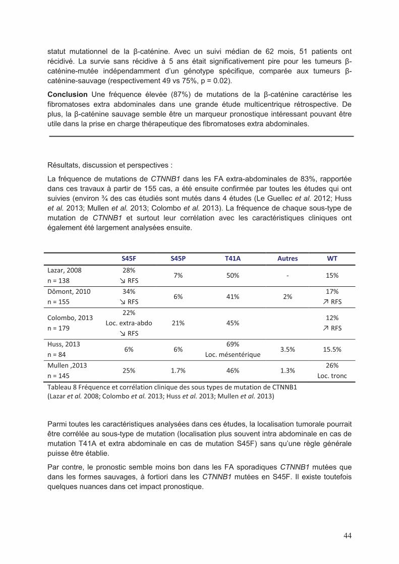
44
statut mutationnel de la β-caténine. Avec un suivi médian de 62 mois, 51 patients ont
récidivé. La survie sans récidive à 5 ans était significativement pire pour les tumeurs β-
caténine-mutée indépendamment d’un génotype spécifique, comparée aux tumeurs β-
caténine-sauvage (respectivement 49 vs 75%, p = 0.02).
Conclusion Une fréquence élevée (87%) de mutations de la β-caténine caractérise les
fibromatoses extra abdominales dans une grande étude multicentrique rétrospective. De
plus, la β-caténine sauvage semble être un marqueur pronostique intéressant pouvant être
utile dans la prise en charge thérapeutique des fibromatoses extra abdominales.
Résultats, discussion et perspectives :
La fréquence de mutations de CTNNB1 dans les FA extra-abdominales de 83%, rapportée
dans ces travaux à partir de 155 cas, a été ensuite confirmée par toutes les études qui ont
suivies (environ ¾ des cas étudiés sont mutés dans 4 études (Le Guellec et al. 2012; Huss
et al. 2013; Mullen et al. 2013; Colombo et al. 2013). La fréquence de chaque sous-type de
mutation de CTNNB1 et surtout leur corrélation avec les caractéristiques cliniques ont
également été largement analysées ensuite.
S45F S45P T41A Autres WT
Lazar, 2008
n = 138
28%
↘ RFS 7% 50% - 15%
Dômont, 2010
n = 155
34%
↘ RFS 6% 41% 2%
17%
↗ RFS
Colombo, 2013
n = 179
22%
Loc. extra-abdo
↘ RFS
21% 45% 12%
↗ RFS
Huss, 2013
n = 84 6% 6%
69%
Loc. mésentérique 3.5% 15.5%
Mullen ,2013
n = 145 25% 1.7% 46% 1.3%
26%
Loc. tronc
Tableau 8 Fréquence et corrélation clinique des sous types de mutation de CTNNB1
(Lazar et al. 2008; Colombo et al. 2013; Huss et al. 2013; Mullen et al. 2013)
Parmi toutes les caractéristiques analysées dans ces études, la localisation tumorale pourrait
être corrélée au sous-type de mutation (localisation plus souvent intra abdominale en cas de
mutation T41A et extra abdominale en cas de mutation S45F) sans qu’une règle générale
puisse être établie.
Par contre, le pronostic semble moins bon dans les FA sporadiques CTNNB1 mutées que
dans les formes sauvages, à fortiori dans les CTNNB1 mutées en S45F. Il existe toutefois
quelques nuances dans cet impact pronostique.
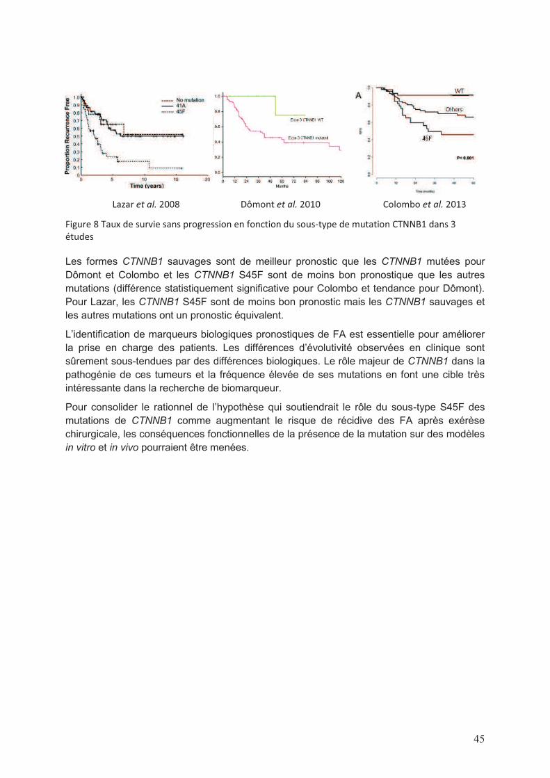
45
Lazar et al. 2008 Dômont et al. 2010 Colombo et al. 2013
Figure 8 Taux de survie sans progression en fonction du sous-type de mutation CTNNB1 dans 3
études
Les formes CTNNB1 sauvages sont de meilleur pronostic que les CTNNB1 mutées pour
Dômont et Colombo et les CTNNB1 S45F sont de moins bon pronostique que les autres
mutations (différence statistiquement significative pour Colombo et tendance pour Dômont).
Pour Lazar, les CTNNB1 S45F sont de moins bon pronostic mais les CTNNB1 sauvages et
les autres mutations ont un pronostic équivalent.
L’identification de marqueurs biologiques pronostiques de FA est essentielle pour améliorer
la prise en charge des patients. Les différences d’évolutivité observées en clinique sont
sûrement sous-tendues par des différences biologiques. Le rôle majeur de CTNNB1 dans la
pathogénie de ces tumeurs et la fréquence élevée de ses mutations en font une cible très
intéressante dans la recherche de biomarqueur.
Pour consolider le rationnel de l’hypothèse qui soutiendrait le rôle du sous-type S45F des
mutations de CTNNB1 comme augmentant le risque de récidive des FA après exérèse
chirurgicale, les conséquences fonctionnelles de la présence de la mutation sur des modèles
in vitro et in vivo pourraient être menées.
46
A partir de « miRNA expression profile of desmoid tumors predicts their aggressiveness. A.
Dufresne, M. Paturel, L. Alberti, H. Philippon, AV Decouvelaere, P. Cassier, JY Blay ».
Soumis à « International Journal of Cancer »
Contexte et objectifs
Cette étude des profils miRNAs a débuté dans le cadre de l’analyse de la cohorte de l’essai
clinique de phase II Desminib à la recherche de facteurs biologiques prédictifs de réponse à
l’imatinib. Les analyses supervisées menées dans un premier temps visaient donc à
identifier des biomarqueurs prédictifs. Parallèlement, comme pour les autres études de
données à large échelle, des analyses non supervisées ont été menées, nous orientant plus
vers un poids pronostique que prédictif de ces profils miRs.
Article 4
Prédiction de la progression des tumeurs desmoïdes par l’utilisation des profils d’expression
des microRNAs
Rationnel La tumeur desmoïde (TD) est une tumeur rare du tissue conjonctif avec une
agressivité loco-régionale mais une évolution imprévisible. Les microRNAs sont des petits
ARN non codants qui contrôlent l’expression des gènes et dont l’expression est dérégulée
dans de nombreux cancers. Cette étude a été menée pour définir la valeur prédictive et/ou
pronostique des profils d’expression miRNA dans les TD.
Méthodes L’ARN a été extrait de prélèvements de tissus inclus en paraffine de 41 patients
inclus dans 2 cohortes distinctes : une cohorte de 26 patients inclus dans l’essai de phase II
« Desminib » et une cohorte indépendante de validation de 15 patients atteints de TD
diagnostiquée au Centre Léon Bérard. Le profil miRNA était établit par « Taqman Low
Density Array » à partir de 500 ng d’ ARN total. Des analyses supervisées et non supervisées
ont été réalisées sur les 2 cohortes.
Résultats Les analyses supervisées prédictives et pronostiques menées sur la cohorte
Desminib n’ont pas pu identifier de miRNAs exprimés différentiellement respectivement entre
les patients progressifs et non progressifs sous imatinib et les patients progressifs et non
progressifs après arrêt de l’imatinib. Un clustering hiérarchique non supervisé de la cohorte
Desminib a identifié 2 groupes (A et B) de 13 patients chacun. Seul le nombre de lignes
thérapeutiques avant l’inclusion dans l’essai a été retrouvé significativement différent entre
les 2 groupes (p = 0.0387). Le délai à progression après arrêt de l’imatinib est plus long dans
le groupe B que dans le groupe A (p = 0.05). Quinze miRs étaient exprimés
différentiellement statistiquement entre les groupes A et B, ciblant plus de 3000 gènes (dont
AGO1, BCL2, CDK6, SMAD4, PTEN, CCND1, VEGFA, RB1 ciblés par au moins 3 miRs).
Ces résultats ont été confirmés sur une cohorte de validation indépendante : le clustering
hiérarchique de ces 15 miRs a identifié 2 groupes, dont le temps à récidive est
statistiquement différent (28.8 versus 68.8 mois ; p = 0.0213).
Conclusion Ces résultats nous apportent la première démonstration d’une valeur
pronostique des profils d’expression de miRNAs avec un possible impact direct sur la prise
en charge des patients. L’objectif suivant sera de valider une signature miRNA précise dans
une cohorte prospective pour sélectionner les patients qui ne bénéficient pas du tout de la
résection chirurgicale de leur tumeur et devraient être simplement surveillés.

47
Résultats, discussion et perspectives :
Par l’expérience globale issue de notre laboratoire et le travail mené sur la cohorte
« Desminib », l’analyse des profils d’expression des miRNAs se révèle très longue et difficile.
Le recueil des données, l’analyse statistique et l’interprétation des résultats sont complexes
et il est frappant de constater, dans la littérature, le peu d’informations disponibles dans les
parties « Matériels et Méthodes » des très nombreuses publications portant sur les miRs. Il
n’existe aucune standardisation des méthodes; pourtant, à de nombreuses étapes, il nous a
fallu faire des choix pour avancer dans l’analyse des données et les résultats étaient
modifiés par :
- la qualité du matériel tumoral disponible : nous avons extrait l’ARN total des tumeurs
depuis les blocs inclus en paraffine. Les prélèvements fixés en Bouin ne sont
clairement pas analysables en parallèle avec des tumeurs fixées en formol,
- la méthode d’extraction de l’ARN, sur colonne ou par Trizol®/phenol-chloroforme
influence également les résultats,
- la méthode de détection des miRs : elle peut être qualitative ou semi-quantitative (la
technique TLDA que nous avons choisi donne un résultat semi-quantitatif),
- en plus de l’analyse des courbes d’amplification par le logiciel de TLDA, nous
vérifions « à la main » et éliminons de manière complètement subjective les courbes
d’amplification d’aspect non satisfaisant,
- l’interprétation des « données manquantes » a été très débattue : un miR non détecté
est-il fortement sous-exprimé ou absent à l’état de base ? Ceci est un constat
d’ignorance : on ne sait pas quelle est l’expression physiologique d’un miR et si, en
condition pathologique, les miRs évoluent de manière qualitative (ré-expression de
miRs et disparition d’autres) ou quantitative (sur et sous-expression de miRs),
- la méthode de normalisation : 4 grandes méthodes de normalisation ont été testées
en parallèle sur notre cohorte par nos biostatisticiens. La méthode de la médiane a
été retenue car elle semblait la plus pertinente et la plus fiable pour notre cohorte,
- le choix des groupes de patients des analyses supervisés : ou comment déterminer
la qualité de réponse d’une FA à un traitement : les patients stables et répondeurs
sont-ils à considérer comme sensibles au traitement ou leur évolution reflète-t-elle
uniquement l’évolution naturelle de leur maladie ? De même sur le choix des
groupes pronostiques, tous les patients inclus dans l’essai « Desminib » étaient
censés avoir une maladie récidivante et progressive à l’inclusion dans l’essai, donc
tous avoir une maladie de mauvais pronostic,
- le déséquilibre en taille des groupes comparés : lorsque le test Anova était réalisé sur
des groupes de taille trop différente, le biostatisticien réalisait des simulations (1000 à
10 000) de test Anova entre le groupe le plus petit et un groupe de taille équivalente
pris au hasard au sein du plus grand groupe,
- le choix des bases de données : nous avons d’emblée éliminé les bases dites
« prédites » dans lesquelles l’influence d’un miR sur un transcrit est supposée à partir
d’une homologie de séquence. Nous n’avons travaillé que sur les 2 bases dites
« validées » pour lesquelles l’influence du miR sur le transcrit a été confirmée par
différentes méthodes (western blot, northern blot, qRT-PCR, microarray, NGS…).
48
Même avec les bases validées, il faut être conscient qu’elles se modifient en
permanence et que d’une version à une autre, les résultats sont différents,
- la modification des données pour un seul patient : la petite taille de nos cohortes,
d’étude et de validation, rend les analyses très sensibles puisque toute modification
des données cliniques ou biologiques d’un seul patient peut modifier les conclusions.
Toutefois, et malgré toutes ces limites, le poids pronostique des miRs révélé par notre étude
me semble robuste. La cohorte « Desminib » incluant des patients récidivants n’est pas
adaptée à l’analyse du pronostic de ces tumeurs. Et pourtant, les courbes de survie sans
progression entre les 2 groupes de patients identifiés par l’analyse non supervisée après
arrêt de l’imatinib étaient très nettement distinctes, à la limite de la significativité statistique.
La cohorte de validation, moins biaisée, confirme clairement cet impact pronostique.
Après avoir identifié la signification clinique des profils d’expression miR, l’étape suivante
consiste à étudier les miRs justifiant la formation de ces groupes pronostiques pour analyser
leurs gènes cibles et ouvrir la voie à de nouvelles perspectives de recherche. On réalise des
tests Anova qui nous donnent un niveau de significativité des miRs différentiellement
exprimés entre nos groupes. Statistiquement, ces tests identifient 15 miRs qui ciblent 3040
gènes ( !) selon les bases de données. On se retrouve alors confronté au problème
d’interprétation commun aux analyses à large échelle : parmi toutes les cibles identifiées,
laquelle ou lesquelles sont fiables, pertinentes, intéressantes à explorer pour tenter de
découvrir un mécanisme d’oncogénèse encore inconnu pour ces tumeurs voire une
potentielle cible thérapeutique ? Dans quelle mesure la significativité statistique est corrélée
à la significativité biologique ? Plus la p value d’un test est petite, plus on est sûr que la
différence étudiée existe ; cela ne veut pas dire que cette différence soit importante, et donc,
biologiquement pertinente. Dans le cas de miRs, nous n’avons pu identifier de miRs qui
soient « toujours » versus « jamais » exprimés dans un groupe par rapport à l’autre. Il nous a
donc semblé intéressant de tenir compte du différentiel de Ct entre les 2 groupes pour un
miR donné.
Dans le but de réduire cette liste de gènes (en conservant les plus intéressants), nous avons
donc sélectionné les miRs puis les gènes ainsi:
- nous n’avons conservé que les miRs dont le différentiel d’expression de la médiane
des Ct normalisés entre les 2 groupes identifiés est supérieur à 2 (la valeur de 2 Ct
d’écart est considérée comme étant minimum pour soutenir une différence
biologique) : on retient alors 9 miRs, qui ciblent encore 2623 gènes
- nous n’avons ensuite conservé que les gènes ciblés par au moins 3 miRs : il reste
alors 23 gènes dont BCL2 PTEN VEGFA CCND1 CDK6.
Il ne semble pas juste de tenter de corréler directement le niveau d’expression d’un miR à
l’activité biologique de son gène cible (i.e. miR sous exprimé, gène sur exprimé). En effet,
compte-tenu de la complexité des systèmes biologiques et du caractère particulier et encore
imprécis de la biologie des FA qui se situe à la frontière entre des tumeurs malignes et
bénignes, l’analyse doit en être très prudente. Cette liste de gènes peut seulement orienter
vers des pistes de travail et des voies de signalisation à explorer, et se poursuivre, par
exemple, par la recherche d’expression de protéines correspondantes en IHC.

49
B. FACTEURS PREDICTIFS
Identification de facteurs biologiques prédictifs de la réponse à l’imatinib
mésylate dans les fibromatoses agressives
A partir de « Identification of biological factors predictive of response to imatinib mesylate in
aggressive fibromatosis. Dufresne A, Bertucci F, Penel N, Le Cesne A, Bui B, Tubiana-Hulin
M, Ray-Coquard I, Cupissol D, Chevreau C, Perol D, Goncalves A, Jimenez M, Bringuier PP,
Blay JY. Br J Cancer. 2010 Aug 10;103(4):482-5 »
Et « Impact of KIT exon 10 M541L allelic variant on the response to imatinib in aggressive
fibromatosis: analysis of the desminib series by competitive allele specific Taqman PCR
technology. A. Dufresne, L. Alberti, M. Brahmi, H. Phlippon, D. Perol and J.-Y. Blay. Under
review for BMC Cancer”
Ces 2 études ont été menées sur la cohorte de patients inclus dans l’essai « Desminib ». Les
résultats de cet essai randomisé de phase I/II ont été publiés en 2011 (Penel et al. 2011). Il
s’agissait d’un essai multicentrique, national, dont l’objectif principal était de déterminer
l’efficacité et la tolérance de l’imatinib mésylate chez des patients atteints de FA récidivante
ou progressive. Quarante patients ont été inclus entre Septembre 2004 et Octobre 2005,
issus de 15 centres français. Les caractéristiques des patients étaient similaires à celles
décrites dans les séries littéraires. Trente-trois patients (85%) présentaient une progression
radiologique objectivée lors de leur entrée dans l’essai. Les résultats ont rapporté une
grande majorité de stabilisations de la maladie (3/4 des patients, au moins) et seulement 4
réponses objectives. Le taux de survie sans progression à 1 an est de 66%, identique à celui
publié par Chugh dans les mêmes conditions (Chugh et al. 2010).
L’un des objectifs secondaires de l’étude était d’identifier des facteurs biologiques, prédictifs
de la réponse à l’imatinib.

50
Article 5
Identification de facteurs biologiques prédictifs de la réponse à l’imatinib mésylate dans les
fibromatoses agressives.
Rationnel L’imatinib induit des réponses et des stabilisations de la maladie chez les patients
porteurs de fibromatoses agressives (FA) non opérables. La cible précise de l’imatinib dans
les FA et les facteurs prédictifs de réponse ne sont pas connus.
Méthodes Nous avons étudié les facteurs potentiellement prédictifs de réponse à l’imatinib
dans une série de 40 patients porteurs de FA progressive inclus dans un essai de phase II
d’imatinib : nous avons testé la présence du variant M541L de l’exon 10 de KIT, l’expression
des kinases sensibles à l’imatinib et des protéines du cycle cellulaire par immunohistochimie,
ainsi que d’autres facteurs cliniques et biologiques.
Résultats Parmi les 10 patients dont on a pu extraire l’ADN, 3 étaient porteurs du variant de
l’exon 10 de KIT (30%), sans corrélation avec la réponse ou la survie sans progression.
L’expression des autres cibles de l’imatinib (PDGFRA/B, MCSF-R), et des éléments en aval
du cycle cellulaire, de la prolifération cellulaire et des voies de prolifération (cycline D1, ERK,
MEK 1-2) n’étaient pas corrélés à la survie sans progression. La lymphopénie avant
traitement (<1500/µl) et la taille tumorale > 120 mm étaient corrélés à une survie sans
progression plus courte dans les analyses uni et multivariées.
Conclusion Nos travaux montrent que des caractéristiques biologiques de base du patient
sont le paramètre majeur influençant la réponse à l’imatinib dans les FA. Les caractéristiques
de la tumeur, incluant la présence du variant M541L de l’exon 10 de KIT, peuvent influencer
le contrôle tumoral mais cela doit être confirmé et mieux expliqué.

51
Article 6
Impact du variant allélique M541L de l’exon 10 de KIT sur la réponse à l’imatinib dans les
fibromatoses agressives: analyse de la série Desminib par la technologie PCR de
competitive allele specific Taqman
Rationnel La fibromatose aggressive (FA) est une maladie proliférative fibroblastique rare
avec un comportement local agressif et pas de métastases à distance, caractérisée par des
mutations des gènes CTNNB1 ou APC. Quand les FA progressives et/ou symptomatiques,
ne sont pas accessibles à un traitement local, différents traitements médicaux peuvent être
efficaces, dont l’imatinib mésylate. L’essai de phase II « Desminib » a inclu 40 patients
atteints de FA pour évaluer la toxicité et l’efficacité de l’imatinib et a conclu à un taux de
contrôle tumoral de 65% à 1 an. Nous avons étudié la potentielle valeur prédictive du variant
M541L de l’exon 10 de KIT (KIT M541L) sur cette série prospective.
Méthodes L’ADN a été extrait en quantité suffisante pour 33 patients inclus dans l’essai
Desminib. La détection du KIT M541L a été réalisée par la technologie PCR de Competitive
Allele-Specific Taqman®. Le statut de l’allèle de KIT a été analysé par rapport à la
présentation clinique et à l’évolution du patient.
Résultats Dans 6 des 33 cas (18%), la technique n’a pu conclure au statut mutationnel ; 5
patients (19%) étaient porteurs du KIT L541 et 22 patients (81%) étaient classés
« sauvages ». Par rapport à la cohorte globale, KIT M541L était observé à des fréquences
identiques selon les caractéristiques cliniques. Dans les sous-groupes KIT M541L et KIT
« sauvage », les taux de contrôle tumoral à 1 an sont respectivement de 100% et 68% (p =
0.316). La médiane de survie sans progression des patients porteurs de KIT M541L ou non
est respectivement de 29.9 et 24.5 mois (p = 0.616), la médiane de survie globale n’est pas
atteinte, dans aucun des 2 groupes.
Conclusion Nos résultats ne sont pas en faveur d’un effet prédictif de KIT M541L sur
l’efficacité de l’imatinib pour les patients atteints de FA.
52
A partir de « A V530I Mutation in c-KIT Exon 10 Is Associated to Imatinib Response in
Extraabdominal Aggressive Fibromatosis. Kurtz JE, Asmane I, Voegeli AC, Neuville A,
Dufresne A, Litique V, Chevreau C, Bergerat JP. Sarcoma. 2010;2010:458156 »
Cette publication rapporte le cas d’une patiente présentant une FA et traitée par imatinib
permettant d’obtenir une réponse partielle d’excellente qualité associée à une amélioration
complète des symptômes fonctionnels et algiques. Cherchant à expliquer biologiquement
cette réponse, le séquençage de l’exon 10 de KIT réalisé a permis d’identifier la présence
d’une mutation V503I.
Article 7
Une mutation V530I de l’exon 10 de KIT est associée à la réponse à l’imatinib dans une
fibromatose agressive extra abdominale.
Les fibromatoses agressives (FA) ou tumeurs desmoïdes sont des tumeurs rares,
caractérisées par une invasion des tissus profonds par une prolifération néoplasique
monoclonale de fibroblastes, développée à partir des structures musculo-aponévrotiques. La
chirurgie est le traitement de référence mais les marges négatives sont difficiles à obtenir
dans le cas de volumineuses tumeurs et peuvent entrainer des séquelles fonctionnelles
importantes. Le traitement médical des FA comporte des anti-inflammatoires non
stéroïdiens, du tamoxifène avec des résultats inconstants. Plusieurs publications rapportant
une efficacité de l’imatinib dans les FA apparaissent dans la littérature. Ici, nous décrivons
pour la première fois un variant V530I de l’exon 10 de KIT qui est associé à une réponse
majeure à l’imatinib dans une FA extra abdominale. La substitution V530I précédemment
découverte a été caractérisée dans les leucémies myéloïdes aigües, mais n’a jamais été
décrites dans d’autres pathologies, jusqu’ici. Dans cet article, nous discutons les mutations
ou polymorphismes de l’exon 10 de KIT qui ont été décrits dans plusieurs situations liées à
KIT, dont la leucémie myéloïde aigüe, la mastocytose et la fibromatose agressive.

53
L’imatinib mésylate est un inhibiteur de récepteurs tyrosine-kinase pour lequel les cibles
clairement identifiées sont aujourd’hui au nombre de 4 : ABL, KIT, CSF-1R et PDGFR α et β.
Logiquement, la recherche de facteurs prédictifs dans l’essai Desminib a commencé par la
recherche de l’expression des cibles connues de l’imatinib pour tenter de corréler leur
présence à la réponse au traitement. Deux autres publications ont abordé ce sujet sous le
même angle : celle de Heinrich (Heinrich et al. 2006) qui a étudié l’expression de KIT,
PDGFR α et β et PDGF-AA et –BB ainsi que la présence de mutations de KIT, PDGFRA et
PDGFRB et celle de Chugh (Chugh et al. 2010) qui a recherché l’expression et les mutations
des mêmes cibles chez respectivement 19 et 51 patients atteints de FA et traités par imatinib
dans 2 essais de phase II.
Heinrich et al. Chugh et al. Dufresne et al.
KIT 3/19 1/7 0/29
PDGFR α 0/19 7/7 0/29
PDGFR β 19/19 7/7 29/29
PDGF-AA 14/14 ND ND
PDGF-BB 14/14 ND ND
Tableau 9 Etude IHC des cibles connues de l’imatinib des patients inclus dans les essais de phase II
évaluant l’imatinib dans les FA (nombre cas positifs/nombre de cas étudiés)
(Heinrich et al. 2006; Chugh et al. 2010)
L’expression d’aucune de ces cibles n’a pu être corrélée à la réponse tumorale à l’imatinib.
Des mutations ont également été recherchées au niveau de ces cibles. Dans l’étude de
Heinrich, le séquençage des exons 9, 11, 13 et 17 de KIT, des exons 12, 14 et 18 de
PDGFR α et des exons 11 et 17 de PDGFR β n’a retrouvé aucune mutation. Les mêmes
exons étaient séquencés dans l’étude de Chugh et on retrouvait une délétion sur l’exon 12
de PDGFR α et deux délétions sur l’exon 12 de PDGFR β. Deux patients sur 19 étaient
porteurs du variant M541L sur l’exon 10 de KIT. Aucune de ces anomalies n’était corrélée à
la réponse à l’imatinib. Dans notre étude, la présence du variant M541L de l’exon 10 de KIT
n’était pas corrélée à la réponse tumorale.
La démarche « officielle » pour identifier une thérapie ciblée efficace consiste à 1) identifier
une cible moléculaire exprimée et/ou activée dans une pathologie tumorale pour 2)
déterminer l’efficacité d’une thérapie ciblant cette molécule anormale. Dans les travaux
présentés ici, la démarche inverse a été menée : 1) la thérapie ciblée a été administrée à
une cohorte de patients non sélectionnés, atteints de cette pathologie et 2) les cibles de
cette thérapie ont été analysées dans la cohorte de patients pour tenter d’établir une
corrélation entre leur présence et l’efficacité du traitement.
Si cette démarche semble surtout motivée par le fait qu’elle permette au praticien de faire
bénéficier le plus rapidement possible à ses patients d’une nouvelle molécule, l’absence des
étapes de « preuve du mécanisme » et « preuve du concept » peut freiner voire stopper le
développement d’une thérapie ciblée dans une pathologie (e. g. gefitinib dans les cancers
bronchiques non à petites cellules). Toutefois, avec un peu de chance, cette méthode
d’analyse rétrospective peut parfois donner des résultats. Lorsque l’imatinib a été administré
pour la première fois à une patiente atteinte de GIST en 2001 (Joensuu et al. 2001), on
savait juste depuis 2-3 ans que les GISTs exprimaient KIT qui semblait activé par des

54
mutations spécifiques (Sarlomo-Rikala et al. 1998; Hirota et al. 1998; Lux et al. 2000). Ces
mutations n’avaient pas encore été identifiées comme puissants biomarqueurs de l’efficacité
de l’imatinib.
Dans le modèle idéal, une cible thérapeutique est une protéine exprimée, modifiée par une
mutation génétique et dont le mécanisme d’action est impliqué dans la carcinogénèse du
modèle tumoral étudié. Si en plus il existe un médicament capable d’inhiber cette protéine,
on a toutes les chances d’avoir identifié une cible prometteuse (Fletcher, Blay). Mais la
conjonction de tous ces critères est rarement obtenue et, en pratique, l’identification d’une
cible doit se concevoir à différents niveaux.
Elle peut se rechercher au niveau génomique : c’est dans ce cadre qu’Heinrich et Chugh ont
recherché les mutations des exons 9, 11, 13 et 17 de KIT, des exons 12, 14 et 18 de PDGFR
α et des exons 11 et 17 de PDGFR β. Ces mutations ont été identifiées comme activatrices
du récepteur c-kit et biomarqueurs de l’efficacité de l’imatinib dans les GISTs ou les
leucémies myéloïdes chroniques (Apperley et al. 2002; Heinrich, Corless, Duensing, et al.
2003; Heinrich, Corless, Demetri, et al. 2003). Ces mutations juxta-membranaires entrainent
une activation constitutionnelles du récepteur qui active plusieurs voies de signalisation
jouant un rôle crucial dans la prolifération ou la survie cellulaire telles que les voies
Ras/Raf/MAP kinases, Janus/Stat, PI3K/Akt/mTOR et SRC. Aucune de ces mutations n’a
été retrouvée dans les FA.
Dans la même idée, nous avons recherché la présence du variant M541L de l’exon 10 de
KIT pour évaluer l’éventuelle corrélation de sa présence à l’efficacité de l’imatinib. Au
moment où ces travaux ont débuté, en 2006, 2 « case report », l’un portant sur une patiente
(Gonçalves et al. 2006) et l’autre sur 4 (Seinfeld et al. 2006) rapportaient une réponse
intéressante à l’imatinib chez des patients porteurs de FA et chez qui la présence de ce
variant était retrouvée. Notre première analyse portant sur les patients inclus dans l’essai
Desminib était motivante puisque, parmi les 10 patients pour lesquels l’ADN avait pu être
amplifié et séquencé, le variant était présent dans 3 cas, dont la seule patiente de l’étude
présentant une réponse complète à l’imatinib (et 2 stabilisations). Devant la difficulté
technique et le faible rendement du séquençage chez ces patients, l’étude s’est poursuivie
par une technique de PCR quantitative : la « Competitive-Allele Specific Taqman® PCR ». En
effet, les FA sont des tumeurs peu cellulaires, très fibrotiques, dont l’ADN est altéré au cours
du temps par les méthodes de fixation et conservation. La fixation en formol et à fortiori en
Bouin, l’inclusion en paraffine et les méthodes d’extraction entrainent la dégradation et la
fragmentation des acides nucléiques. Le rendement du séquençage classique (dit de
Sanger) est très mauvais pour ces tumeurs. L’utilisation de la CAST PCR semble avoir
contourné en partie ces problèmes. Dans cette technique, l’amplification de seul l’allèle muté
(après blocage spécifique de l’allèle « wild type ») permet d’augmenter la sensibilité de
détection d’une mutation. Cette méthode nous a permis de déterminer le statut de l’exon 10
de KIT pour 27 patients (variant M541L détecté dans 5 cas). Si aucun patient porteur du
variant n’a progressé sous imatinib, les analyses statistiques de taux de réponse et de survie
n’ont pu identifier la présence de ce variant comme facteurs prédictif de réponse au
traitement. Dans l’étude de Chugh, 2 patients sur 19 étaient porteurs du variant et ne
répondaient pas mieux à l’imatinib que les autres.
Si le rationnel de ces études génomiques est discutable, l’interprétation des résultats
négatifs doit se faire au regard des limites statistiques liées à la faible taille globale des

55
cohortes de cette maladie rare et au faible nombre d’évènements (grande majorité de
stabilisations).
Depuis, ce variant a été identifié comme étant un polymorphisme (Grabellus et al. 2011). En
effet, sa fréquence est de 7/42 soit 16.7% des FA ce qui n’est pas statistiquement différent
de sa fréquence chez des donneurs sains (26/166 soit 15.7%). De plus, comme on peut
l’attendre d’un polymorphisme, le variant est présent dans les tissus sains adjacents à 4
échantillons de tumeur porteurs du variant. Des travaux menés ensuite dans notre équipe
ont concluent dans le même sens en retrouvant la présence du variant dans des couples de
blocs FFPE de GIST et de PBMC correspondant, pour 5 patients. Ces données ne sont pas
en faveur d’un potentiel rôle du variant dans la carcinogénèse des tumeurs. Toutefois, ceci
n’excluait pas à priori son rôle dans l’efficacité d’un traitement.
La significativité de ce variant demeure inexpliquée et s’ils ne sont pas publiés, des travaux
ont depuis été menés sur ce sujet.
Parmi les 426 patients de la base de données nationale de FA (mentionnée ci-dessus),
l’exon 10 a été séquencé pour 167 d’entre eux permettant d’identifier la présence du variant
chez 34 patients (soit 19%). La présence du variant a été analysée par rapport à de
nombreux critères cliniques et biologiques : sexe du patient, contexte d’hormonodépendance
ou de syndrome de Gardner, présence d’une mutation du gène APC, localisation et taille
tumorales, meilleure réponse observé à un traitement, récidive, délai à récidive. L’analyse de
la survie globale a été abandonnée car le nombre trop faible d’évènements ne la rend pas
fiable.
Aucun de ces critères n’était corrélé à la présence du variant M541L sauf le contexte
d’hormonodépendance, celui-ci étant défini comme une FA se développant au cours d’une
grossesse ou dans les 6 mois suivant l’accouchement. Le taux de corrélation était hautement
significatif avec une p-value du test chi-deux à 0.006.
Kit exon 10 mutation Number (%)
Wild type M541-L Total
Hormonal dependency
Number (%)
No 130 (84) 24 (16) 154
Yes 7 (54) 6 (46) 13
Total 137 (82) 30 (18) 167
Tableau 10 Répartition de la présence du variant M541L de l’exon 10 de KIT en fonction du caractère
hormonodépendant des FA
Le variant est présent dans plus de la moitié des FA qui se développent dans un contexte
d’hormonodépendance. Nous avons émis l’hypothèse que le variant M541L puisse
sensibiliser les cellules à la stimulation hormonale. Nous avons donc développé un modèle
in vitro et étudié pendant de longs mois l’influence éventuelle d’hormones et traitements anti-
hormonaux sur la prolifération et la mort cellulaire de lignées cellulaires de fibroblastes 3T3
transfectées par le variant M541L. Comparativement aux 3T3 transfectées par le vecteur, la
prolifération cellulaire des 3T3 M541L semble effectivement stimulée par l’ajout d’E2 dans
les milieux de culture. Toutefois, ce résultat a été difficile à obtenir et à reproduire pour
conclure de manière formelle. Nous avons remis en cause le modèle utilisé de 3T3
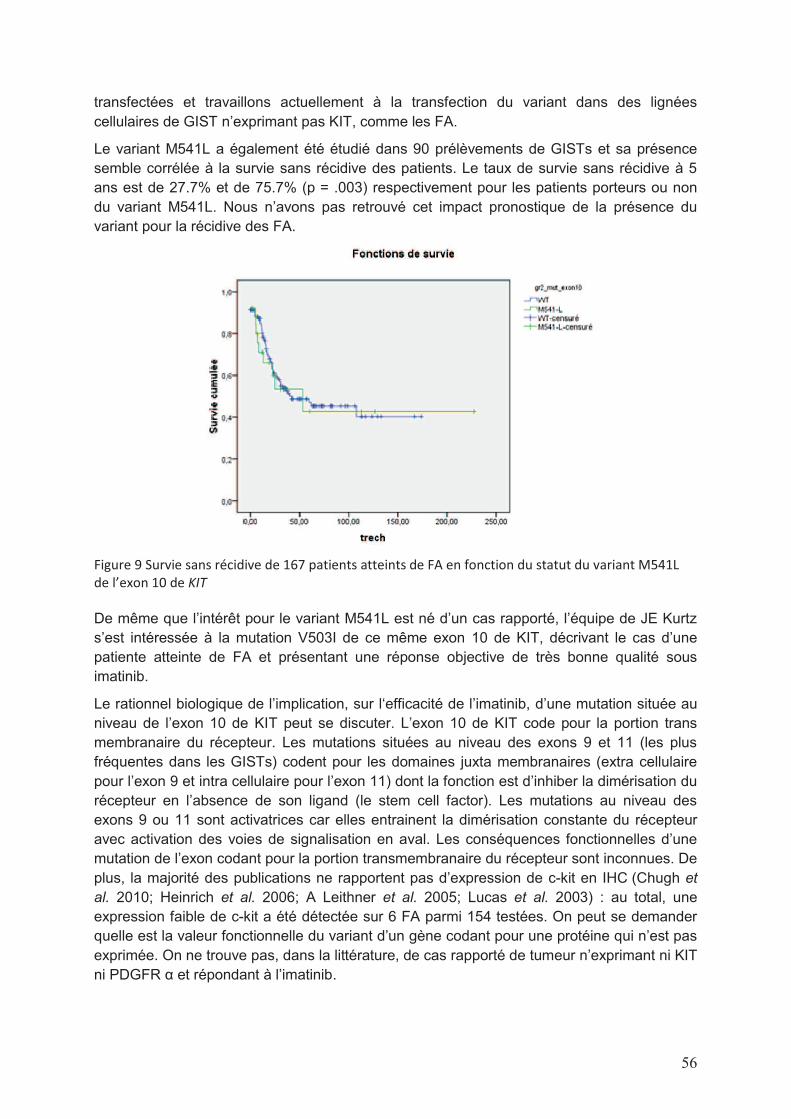
56
transfectées et travaillons actuellement à la transfection du variant dans des lignées
cellulaires de GIST n’exprimant pas KIT, comme les FA.
Le variant M541L a également été étudié dans 90 prélèvements de GISTs et sa présence
semble corrélée à la survie sans récidive des patients. Le taux de survie sans récidive à 5
ans est de 27.7% et de 75.7% (p = .003) respectivement pour les patients porteurs ou non
du variant M541L. Nous n’avons pas retrouvé cet impact pronostique de la présence du
variant pour la récidive des FA.
Figure 9 Survie sans récidive de 167 patients atteints de FA en fonction du statut du variant M541L
de l’exon 10 de KIT
De même que l’intérêt pour le variant M541L est né d’un cas rapporté, l’équipe de JE Kurtz
s’est intéressée à la mutation V503I de ce même exon 10 de KIT, décrivant le cas d’une
patiente atteinte de FA et présentant une réponse objective de très bonne qualité sous
imatinib.
Le rationnel biologique de l’implication, sur l‘efficacité de l’imatinib, d’une mutation située au
niveau de l’exon 10 de KIT peut se discuter. L’exon 10 de KIT code pour la portion trans
membranaire du récepteur. Les mutations situées au niveau des exons 9 et 11 (les plus
fréquentes dans les GISTs) codent pour les domaines juxta membranaires (extra cellulaire
pour l’exon 9 et intra cellulaire pour l’exon 11) dont la fonction est d’inhiber la dimérisation du
récepteur en l’absence de son ligand (le stem cell factor). Les mutations au niveau des
exons 9 ou 11 sont activatrices car elles entrainent la dimérisation constante du récepteur
avec activation des voies de signalisation en aval. Les conséquences fonctionnelles d’une
mutation de l’exon codant pour la portion transmembranaire du récepteur sont inconnues. De
plus, la majorité des publications ne rapportent pas d’expression de c-kit en IHC (Chugh et
al. 2010; Heinrich et al. 2006; A Leithner et al. 2005; Lucas et al. 2003) : au total, une
expression faible de c-kit a été détectée sur 6 FA parmi 154 testées. On peut se demander
quelle est la valeur fonctionnelle du variant d’un gène codant pour une protéine qui n’est pas
exprimée. On ne trouve pas, dans la littérature, de cas rapporté de tumeur n’exprimant ni KIT
ni PDGFR α et répondant à l’imatinib.

57
L’identification d’une cible thérapeutique peut également se rechercher au niveau protéique.
Les protéines sont les effecteurs des processus cellulaires dont les modifications de nombre
ou de fonction peuvent être caractéristiques de tumeurs et représenter ainsi des cibles
thérapeutiques. Dans notre étude comme dans celles de Chugh et Heinrich, les récepteurs
Kit, PDGFR α et M-CSF R, cibles connues de l’imatinib, ne sont pas exprimés à la surface
des cellules de FA. Ils ne semblent donc pas pouvoir médier la réponse à l’imatinib dans ces
tumeurs. PDGFR β, par contre, détecté par IHC dans notre étude et celle de Chugh et par
immunoblot dans celle d’Heinrich, est exprimé chez tous les patients, à différents niveaux. Il
n’a pas été possible de corréler le niveau d’expression à la réponse à l’imatinib.
En aval de ces récepteurs, différentes voies de signalisation peuvent être activées et ont été
également analysées dans les 3 études. Les récepteurs tyrosine-kinase activent les voies
Ras/Raf/MEK/ERK, PI3K/AKT/PTEN et JAK/STAT3. L’expression des formes phosphorylées
d’AKT, MAPK, STAT3, ERK et MEK et non phosphorylées AKT, MAPK, STAT3, PTEN et
PI3K ne sont pas corrélées à la réponse à l’imatinib. De plus, les niveaux de phosphorylation
ne changent pas lorsqu’on les compare avant et après traitement par imatinib. Les
jonctions adhérentes intercellulaires incluent E-cadhérine et β-caténine ; E-cadhérine n’est
exprimée dans aucun cas et β-caténine dans tous les cas. Plus que l’expression globale de
β-caténine, on sait que c’est sa localisation intra cellulaire qui est informative : sous
membranaire en conditions physiologiques elle est transloquée en intra nucléaire en
conditions pathologiques. Ce facteur n’a pas été étudié.
Dans notre étude, l’expression de la cycline D1 est retrouvée chez 5 patients sur 33. Aucun
des patients dont la tumeur exprime la Cycline D1 n’est progressif à 1 an. La cycline D1 est
un régulateur du cycle cellulaire dont le poids pronostique péjoratif a été démontré dans
plusieurs types de cancers (cancer bronchique, prostatique, oesophagien, carcinome
urothélial de vessie…). De plus, il a été démontré, dans 38 cas de FA sporadiques, que sa
surexpression était corrélée à la localisation intra cellulaire de β-caténine (T Saito et al.
2001).
L’analyse des profils d’expression des miRNAs n’a pas pu identifier de miRs exprimés
différentiellement et avec un niveau de significativité statistique suffisante entre les patients
répondeurs et non répondeurs à l’imatinib dans l’essai Desminib.
Au total, l’objectif initial de nos travaux qui était d’identifier dans les FA des facteurs
biologiques de réponse à l’imatinib n’a pas abouti. Toutefois, lorsque l’on connaît la lenteur
naturelle d’évolution des FA et les cas de régression spontanée, on peut se poser la
question de savoir si les réponses au traitement quantifiées selon les critères RECIST dans
ces études sont réellement dues à une éventuelle efficacité du traitement ou simplement le
reflet de l’évolution naturelle de la maladie. En d’autres termes, la recherche de facteurs
prédictifs équivaut peut-être, dans ces tumeurs, à rechercher des facteurs pronostiques.
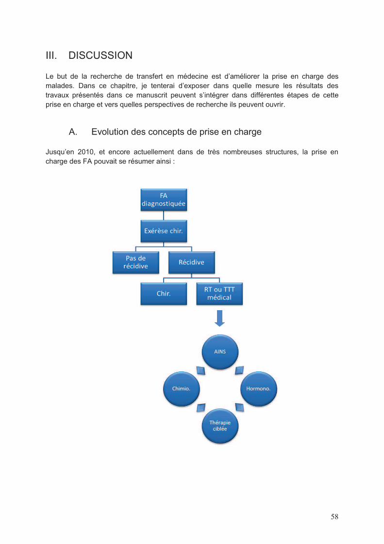
58
III. DISCUSSION Le but de la recherche de transfert en médecine est d’améliorer la prise en charge des
malades. Dans ce chapitre, je tenterai d’exposer dans quelle mesure les résultats des
travaux présentés dans ce manuscrit peuvent s’intégrer dans différentes étapes de cette
prise en charge et vers quelles perspectives de recherche ils peuvent ouvrir.
A. Evolution des concepts de prise en charge
Jusqu’en 2010, et encore actuellement dans de très nombreuses structures, la prise en
charge des FA pouvait se résumer ainsi :

59
Cette stratégie n’est pas satisfaisante, à plusieurs niveaux :
- La résection chirurgicale est souvent réalisée d’emblée, sans diagnostic anatomo-
pathologique préalablement établi sur biopsie,
- Le diagnostic anatomo-pathologique est difficile à établir uniquement sur les analyses macro
et microscopique. De nombreuses erreurs diagnostiques de FA sont corrigées par la
relecture centralisée des sarcomes (RRePs). (Ray-Coquard et al. 2012)
- Il n’existe pas de recommandations sur le type d’exérèse chirurgicale (manque de données
notamment sur l’influence de la qualité des marges sur la récidive). Le geste chirurgical est
pourtant souvent compliqué par une taille volumineuse et l’envahissement des organes de
voisinage,
- Plus de la moitié des patients opérés récidivent, et souvent plusieurs fois de suite,
- La place de la radiothérapie reste mal définie,
- Les traitements médicaux sont multiples mais globalement peu efficaces ; ils sont donc
prescrits dans le cas de tumeurs évolutives et/ou symptomatiques, non accessibles à un
traitement local de chirurgie ou radiothérapie. Il n’existe pas de recommandations sur la
séquence optimale des traitements systémiques. Il n’existe pas non plus de facteur prédictif
orientant vers le choix d’un traitement plus qu’un autre. Le praticien a donc naturellement et
logiquement tendance à prescrire les traitements les moins toxiques en 1ère intention pour les
faire se succéder au rythme des évolutions de la maladie, en espérant qu’ils puissent
soulager le patient.
Cette stratégie de prise en charge a été bouleversée par l’introduction de la notion de
surveillance dans la prise en charge initiale des tumeurs.
B. Contribution de nos travaux à l’évolution de ces concepts
Le pronostic des FA reste mystérieux et représente pourtant un élément essentiel dans la
prise en charge des patients. L’immense hétérogénéité des modes évolutifs des FA
observée en clinique et rapportée dans les séries rétrospectives (de la régression spontanée
à l’invasion et la récidive loco–régionales invalidantes) est forcément sous tendue par des
caractéristiques biologiques différentes, de la tumeur et/ou de son micro environnement, et
non identifiées à ce jour.
Dans la nouvelle stratégie de prise en charge des patients, la question du pronostic doit être
abordée sous 2 angles : identification de facteurs de récidive après exérèse chirurgicale
d’une tumeur et pronostic évolutif d’une tumeur en place. On ne dispose pas d’éléments
actuellement pour penser que ces facteurs soient les mêmes dans les 2 situations mais ce
n’est pas impossible. Les 1ers ont été beaucoup plus étudiés car ils correspondaient à la
stratégie de prise en charge adoptée jusqu’à présent. Les 2nds seront issus d’une stratégie
plus récemment développée mais devraient rapidement se révéler indispensables.
60
1. Facteurs pronostiques cliniques
On a, jusqu’à présent, beaucoup plus étudié de facteurs pronostiques cliniques que
biologiques, essentiellement du fait de la simplicité/complexité des études à mener. Des
résultats des 2 études cliniques portant sur ce sujet et auxquelles nous avons participé, il
ressort que l’âge jeune du patient, la taille importante de la tumeur et sa localisation extra-
abdominale soient des facteurs de récidive après exérèse chirurgicale. Bien qu’issues de
séries rétrospectives, on peut considérer que ces résultats soient valables, compte-tenu de
la très grande taille des effectifs étudiés et de la concordance des résultats entre les 2
études. Aujourd’hui, il me semble légitime de commencer à intégrer dans nos discussions en
RCP ces 3 facteurs cliniques : la présence de ces facteurs de récidive chez un patient
devrait orienter la stratégie de prise en charge vers une surveillance plutôt qu’une chirurgie
d’exérèse, compte-tenu du risque élevé de récidive.
2. Facteurs pronostiques biologiques
La présence de la mutation S45F de CTNNB1 est le seul facteur de récidive biologique
identifié à ce jour. Ce résultat doit être confirmé de 2 manières avant de s’intégrer à nos
discussions :
- confirmation de l’impact pronostique de S45F par des études cliniques plus larges et
prospectives dans l’idéal,
- identification du mécanisme biologique sous-tendant cet impact pronostique : les
conséquences fonctionnelles de la présence de cette mutation doivent être explorées
in vitro et in vivo par l’étude de lignées cellulaires transfectées par cette mutation.
L’étude des miRs comme facteur pronostique abordée dans nos travaux, doit être
approfondie, pour les FA mais aussi de manière plus générale. L’impact pronostique des
miRs a été une surprise dans notre étude initialement élaborée pour identifier des facteurs
prédictifs de réponse au traitement médical. De cela découle plusieurs conséquences :
statistiquement significative, la corrélation entre le profil miR et la survie sans récidive en fait
une piste très intéressante. Toutefois, la cohorte de patients analysés, tous récidivants et
multitraités, et la survie sans récidive étudiés (survie sans progression après l’arrêt de
l’imatinib) ne sont pas pertinents. Même si la cohorte rétrospective de validation, plus
adaptée, confirme ces résultats, la validation sur une cohorte large, indépendante et
prospective sera indispensable.
L’impact réel, biologique, des miRs sur les gènes dont le transcrit est ciblé et l’expression
protéique qui en découle doit être précisé.
3. Facteurs prédictifs
L’utilisation des traitements systémiques dans les FA doit être globalement repensée.
L’efficacité réelle des traitements reste à déterminer. Il est évident que, face à ces tumeurs
qui présentent spontanément des stabilisations dans 50% des cas et des réponses
objectives dans 20% des cas, l’utilisation des critères RECIST, habituelle dans l’évaluation
des cancers, ne soit pas pertinente. Etudiant une tumeur qui ne métastase pas, les

61
symptômes et la qualité de vie du patient devraient être beaucoup plus pris en compte. De
plus, il est perturbant de constater que ces taux d’évolution naturelle ne soient finalement
pas très différents des taux de réponse observés sous traitement : taux de réponse partielle
de 20-50% dans les essais randomisés de chimiothérapie, 25% de réponse objective et 50%
de stabilisations dans les essais de phase II évaluant l’hormonothérapie ou une association
hormonothérapie/AINS. Les 3 essais de phase II évaluant l’imatinib rapportent 6 à 15% de
réponses objectives. On manque actuellement de grandes séries rétrospectives et
prospectives pour évaluer précisément l’évolution naturelle de la maladie. En attendant, on
est en droit de se demander si l’efficacité rapportée dans ces essais reflète réellement une
activité du médicament ou tout simplement cette évolution naturelle. Seul des essais
randomisés, contre placebo en l’absence de bras standard, permettront de trancher cette
question de manière formelle. Une manière d’optimiser les résultats de ces essais serait
d’identifier des cibles pour sélectionner les patients, que ce soit les cibles de traitements
actuels ou les cibles de nouveaux médicaments.
Nos travaux ne nous ont pas permis d’identifier de facteurs biologiques prédictifs de réponse
à l’imatinib.
C. Les limites de notre travail
L’avancée de nos travaux s’est heurtée à différentes caractéristiques cliniques et biologiques
liées aux FA :
- Les démonstrations statistiques sont rendues très difficiles par le faible nombre de
cas étudiés et le faible nombre d’évènements. Ces tumeurs sont globalement rares.
Elles sont mal connues, les erreurs diagnostiques sont fréquentes. Peu d’essais
cliniques sont développés. L’absence de métastases, et donc de décès liés à la
maladie nous empêche d’étudier la survie globale des patients. La survie sans
progression est biaisée par la proportion non négligeable de patients dont la tumeur
est spontanément stable voire régressive.
- Le matériel biologique disponible est souvent difficilement exploitable. Comme pour
beaucoup de tumeurs, on dispose essentiellement de blocs FFPE (et non pas de
matériel congelé) qui altère la conservation des acides nucléiques et des protéines
phosphorylées. De plus, ces tumeurs sont peu cellulaires et très fibrotiques (le
collagène inhibe les PCR).
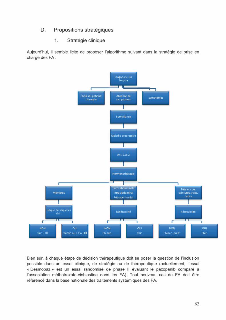
62
D. Propositions stratégiques
1. Stratégie clinique
Aujourd’hui, il semble licite de proposer l’algorithme suivant dans la stratégie de prise en
charge des FA :
Bien sûr, à chaque étape de décision thérapeutique doit se poser la question de l’inclusion
possible dans un essai clinique, de stratégie ou de thérapeutique (actuellement, l’essai
« Desmopaz » est un essai randomisé de phase II évaluant le pazopanib comparé à
l’association méthotrexate-vinblastine dans les FA). Tout nouveau cas de FA doit être
référencé dans la base nationale des traitements systémiques des FA.
Diagnostic sur biopsie
Choix du patient: chirurgie
Absence de symptomes
Surveillance
Maladie progressive
Anti Cox 2
Hormonothérapie
Membres
Risque de séquelles chir.
NON
Chir. ± RT
OUI
Chimio ou ILP ou RT
Paroi abdominale
Intra abdominal
Rétropéritonéal
Résécabilité
NON
Chimio.
OUI
Chir.
Tête et cou, ceintures,tronc,
pelvis
Résécabilité
NON
Chimio. ou RT
OUI
Chir.
Symptomes

63
Les résultats de l’essai clinique de phase II du Dr Bonvalot (en cours) évaluant de manière
prospective l’évolution des FA simplement surveillées seront fondamentaux pour
définitivement intégrer cette stratégie dans nos pratiques et apporter des données inédites
sur l’évolution naturelle de la maladie. L’identification de facteurs cliniques pronostiques,
permettant de prédire l’évolutivité d’une tumeur en place, seront essentiels dans la
discussion de stratégie de prise en charge des patients, d’autant plus qu’ils seront associés
aux facteurs cliniques de récidive identifiés dans nos travaux.
Enfin, le projet développé au niveau national, d’établir une base clinico-biologique des FA,
prospective sur 3 ans, permettra de répondre à plusieurs questions :
- incidence exacte de la maladie par un recueil exhaustif des cas,
- identification de facteurs associés au développement des FA (traumatisme, intervention
chirurgicale…),
- analyse du caractère mutuellement exclusif des anomalies moléculaires de CTNNB1 et
APC et identification d’autres anomalies génétiques,
- facteurs de progression d’une tumeur en place,
- contrôle local après traitement focal,
- résultat en population réelle des traitements systémiques utilisés hors essai,
- évolutivité pendant/après grossesse,
- particularités des formes pédiatriques.
2. Stratégie de recherche biologique
De multiples travaux de recherche peuvent être développés pour poursuivre l’exploration des
mécanismes biologiques des FA.
Le diagnostic de FA est maintenant confirmé grâce à l’aide de la biologie moléculaire. Dans
le cadre d’un syndrome de Gardner, les FA sont liées à une mutation du gène APC et dans
le cadre de FA sporadiques, le gène CTNNB1 est muté. La recherche de ces mutations est
maintenant effectuée en routine pour confirmer le diagnostic de FA. Trois questions
persistent:
- les mutations d’APC et CTNNB1 sont-elles mutuellement exclusives ? (ou faut-il les
rechercher toutes les 2 systématiquement ?).
- D’autres mutations spécifiques des FA existent-elles ? Cette notion est défendue par le fait
qu’un certain nombre (≈ 10%) de FA ne contiennent ni mutation d’APC ni de CTNNB1. On
ne peut aujourd’hui exclure qu’il y ait d’autres mutations que APC ou CTNNB1 impliquées
dans la carcinogénèse des FA et qui surviennent seules ou associées aux mutations déjà
connues. Leur identification pourrait avoir un intérêt diagnostique et orienter vers d’autres
mécanismes de carcinogénèse.
- Les mutations d’APC et CTNNB1 sont des mutations pilotes mais sont-elles suffisantes au
développement des FA ? Dans ce cas, elles pourraient représenter des cibles
thérapeutiques de choix, d’autant plus que les inhibiteurs de la voie Wnt/β caténine
commencent à se développer.
Le rôle pronostique du sous-type de mutation de CTNNB1 peut être exploré de 2 manières :
son impact réel pourra être déterminé à partir de l’essai prospectif du Dr Bonvalot et de la

64
base clinico-biologique. On pourra rechercher une corrélation statistique entre sa présence
et l’évolutivité des tumeurs. On peut également imaginer développer un modèle pré-clinique,
avec transfection de cellules par le gène CTNNB1 porteur de la mutation S45F. Le type de
lignées cellulaires à tranfecter reste un problème : il n’existe pas, actuellement, de lignées
cellulaires de FA, qui meurent après quelques dizaines de passages. La tranfection de
fibroblastes est un modèle imparfait (un temps utilisé dans notre laboratoire). Nous
travaillons actuellement à la transfection de lignées de GIST (48B) ayant la caractéristique
de ne pas exprimer le récepteur KIT à leur surface (ce qui est aussi le cas des FA). La
prolifération cellulaire des lignées CTNNB1 S45F pourra être analysée dans des modèles in
vitro et in vivo et comparée à celle des lignées transfectées par les autres mutations ou
porteuses du vecteur vide.
Pour déterminer réellement si le profil d’expression des miR a un poids pronostique dans ces
tumeurs, il conviendrait de l’évaluer en prospectif, par exemple dans l’essai du Dr Bonvalot.
La constatation que les FA ne donnent jamais de métastases reste totalement inexpliquée.
Quelques autres tumeurs, dites « à malignité intermédiaire » présentent également cette
caractéristique. Ce qui est surprenant dans les FA est le potentiel d’agressivité loco-régional
qui peut être très important avec envahissement majeur des structures de voisinage. Dans
l’article référence sur les caractéristiques du cancer, Hanahan et Weinberg reprennent la
thèse de Talmadge et Findler (Talmadge and Fidler 2010) qui considèrent que « invasion »
et « métastases » sont 2 étapes successives d’un seul et même processus. Sans beaucoup
de rationnel scientifique, Vogelstein évoque également l’idée qu’il puisse exister des
mutations génétiques qui transforment un cancer primitif en un cancer métastatique comme
il existe des mutations qui transforment une cellule normale en tumeur bénigne et une
tumeur bénigne en tumeur maligne (Bert Vogelstein et al. 2013). Vogelstein reconnaît
également que ces mutations, qui seraient spécifiques des phénomènes de métastatisation,
sont parfaitement inconnues à ce jour. L’observation des FA tend plutôt à défendre cette 2nde
théorie. Elles représenteraient un excellent modèle pour étudier ces anomalies.

65
IV. CONCLUSION
Devant la multiplicité des anomalies des cellules cancéreuses et la complexité de leur étude,
le schéma ci-après a été élaboré pour tenter de coordonner ces anomalies entre elles.
La conception de ce schéma m’a aidée à articuler entre eux les différents acteurs d’une
cellule cancéreuse et les différents niveaux de lecture qu’il faut distinguer. Ces acteurs
peuvent se répartir en 3 catégories : le gène, le transcrit et la protéine. Normalement, il doit y
avoir une corrélation entre les 3 mais une anomalie génétique ne se traduit pas forcément
par une anomalie protéique.
Il me semble licite de distinguer les anomalies génétiques initiatrices de tumeurs (ou
carcinogènes ou « driver mutation » ou mutation pilote) des anomalies génétiques
secondaires de l’activation anormale des voies de signalisation, qui confèrent leurs
caractéristiques tumorales aux cellules. Via l’ARN messager, ces anomalies initiales sont
transmises aux protéines qui acquièrent ainsi des modifications quantitatives et/ou
qualitatives. Vogelstein a listé 138 gènes pilotes (annexe 1) et considère cette liste
quasiment exhaustive. Les thérapies ciblées ayant démontré des résultats d’efficacité tels
qu’ils ont pu obtenir des autorisations de mise sur le marché ciblent des protéines dont le
gène est souvent présent dans cette liste (ABL, ALK, BRAF, EGFR, ERBB2, KIT…). Il faut
garder à l’esprit qu’un même gène puisse être oncogène ou gène suppresseur de tumeur en
fonction du type de tumeur dans lequel il est présent. Il faut également intégrer
l’hétérogénéité des tumeurs décrites par Vogelstein, et présente à plusieurs niveaux :
hétérogénéité intra-tumorale, inter-métastatique, intra-métastatique, inter-patients.
Aujourd’hui, l’attitude clinique de plus en plus pratiquée qui consiste à biopsier un site
métastatique pour adapter la stratégie thérapeutique ne prend en compte qu’une toute petite
partie de cette hétérogénéité. Parallèlement, ce système est sous le contrôle de
phénomènes épigénétiques (méthylation, miRNA, longs ARN non codants).
Les anomalies quantitatives/qualitatives des protéines entrainent des modifications des voies
de signalisation qui aboutissent à la transcription d’autres gènes conférant leur
caractéristiques aux cellules tumorales.
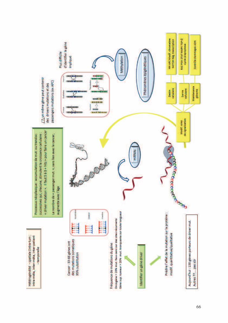
66

67
Davoli et al. ont récemment encore compliqué ces notions autour des gènes pilotes en y
intégrant l’aneuploïdie: ils ont analysé l’impact fonctionnel des mutations génétiques de 8200
tumeurs qui ont permis d’identifier environ 320 gènes suppresseurs de tumeur et 250
oncogènes (Davoli et al., 2013). La dérégulation des cellules cancéreuses proviendrait de la
somme de mutations d’oncogènes, et de perte de gènes suppresseurs de tumeurs. La
distribution et la puissance (variable) de ces gènes pilotes sur les chromosomes serait
l'élément essentiel pour prédire les pertes et gains caractéristiques de l’aneuploïdie.
Le but initial du travail de cette thèse était d’identifier un ou des facteurs prédictifs de
réponse à l’imatinib dans les FA. L’analyse protéique en IHC, épigénétique par étude des
miRs et génétique par CAST-PCR n’a donné aucun résultat concluant. Comment, alors,
orienter les recherches pour augmenter les chances de résultat, c’est-à-dire, comment
identifier un (bon) facteur prédictif ?
Ow et al. (Ow et al. 2013) proposent la schématisation d’une démarche très complète pour
intégrer la génomique du cancer dans la sélection de traitement :

68
Si la découverte d’Her2 et trastuzumab, d’Alk et crizotinib, ou de BRCA1 et inhibiteurs de
PARP ont suivi en grande partie ces étapes, d’autres exemples passés ont montré que l’on
pouvait identifier des biomarqueurs particulièrement efficaces en court-circuitant tout ce
processus (KIT et imatinib mésylate).
Entre ces 2 extrêmes, JY Blay et JA Fletcher proposent la démarche ci-dessous :
Le fait de baser les recherches sur l’expression de cibles ne prend pas en compte les gènes
suppresseurs de tumeurs qui sont pourtant présent en nombre bien plus grand que les
oncogènes dans les cellules tumorales. L’analyse de l’activation de la cible recherche des
anomalies qualitatives de la protéine mais pas quantitatives (qui peuvent pourtant être
importantes, e. g. Her2). Le mécanisme dans lequel la cible est impliquée me semble
fondamental ; s’il s’agit d’un mécanisme de carcinogénèse, elle sera sûrement pertinente. Le
fait qu’une molécule ciblante existe déjà accélèrera forcément le transfert vers la
thérapeutique.
Au-delà de la recherche développée sur de solides concepts scientifiques, basés sur la
continuité logique d’un processus initié par des analyses à large échelle permettant
d’identifier de potentiels biomarqueurs confirmés dans des modèles pré-cliniques et
aboutissant à la conception d’essais cliniques sur des populations sélectionnées de
patients, la pratique m’est apparue toute autre : 40 patients atteints de FA ont été traités par
de l’imatinib sans vrai rationnel scientifique mais plutôt motivé par un laboratoire
pharmaceutique développant une thérapie ciblée dans plusieurs indications. Une fois les
patients traités, on s’est posé la question de savoir par quel mécanisme biologique le
traitement pouvait potentiellement être efficace. Toutefois, l’exercice intellectuel qui consiste
à intégrer les résultats ainsi obtenus dans les données scientifiques n’en est pas moins
intéressant et utile.
Aujourd’hui, dans les FA, on peut évoquer que l’évolution des tumeurs et la réponse au
traitement dépendent de :
- La mutation primaire (CTNNB1 ou APC)
- Les SNP (M541L de l’exon 10 de KIT par exemple)
- Les caractéristiques cliniques du patient (âge, activité génitale…) et de la tumeur
(taille, topographie, envahissement...).

69
Bibliographie
Allen, R.C. et al., 1992. Methylation of HpaII and HhaI sites near the polymorphic CAG
repeat in the human androgen-receptor gene correlates with X chromosome inactivation.
American journal of human genetics, 51(6), pp.1229–39.
Alman, B.A. et al., 1997. Aggressive fibromatosis (desmoid tumor) is a monoclonal disorder.
Diagnostic molecular pathology : the American journal of surgical pathology, part B,
6(2), pp.98–101.
Anon, 2012. Soft tissue and visceral sarcomas: ESMO Clinical Practice Guidelines for
diagnosis, treatment and follow-up. Annals of oncology : official journal of the
European Society for Medical Oncology / ESMO, 23 Suppl 7, pp.vii92–9.
Apperley, J.F. et al., 2002. Response to imatinib mesylate in patients with chronic
myeloproliferative diseases with rearrangements of the platelet-derived growth factor
receptor beta. The New England journal of medicine, 347(7), pp.481–7.
Ashley, D.J., 1969. Colonic cancer arising in polyposis coli. Journal of medical genetics, 6(4),
pp.376–8.
Ashton-Rickardt, P.G. et al., 1989. High frequency of APC loss in sporadic colorectal
carcinoma due to breaks clustered in 5q21-22. Oncogene, 4(10), pp.1169–74.
Azzarelli, A. et al., 2001. Low-dose chemotherapy with methotrexate and vinblastine for
patients with advanced aggressive fibromatosis. Cancer, 92(5), pp.1259–64.
Bacac, M. et al., 2006. A gene expression signature that distinguishes desmoid tumours from
nodular fasciitis. The Journal of pathology, 208(4), pp.543–53.
Ballo, M.T. et al., 1999. Desmoid tumor: prognostic factors and outcome after surgery,
radiation therapy, or combined surgery and radiation therapy. Journal of clinical
oncology : official journal of the American Society of Clinical Oncology, 17(1),
pp.158–67.
Barbier, O. et al., 2010. Primary or recurring extra-abdominal desmoid fibromatosis:
assessment of treatment by observation only. Orthopaedics & traumatology, surgery &
research : OTSR, 96(8), pp.884–9. Available at:
http://www.ncbi.nlm.nih.gov/pubmed/21075698 [Accessed March 25, 2014].
Benoit, L. et al., 2007. 3’ Mutation of the APC gene and family history of FAP in a patient
with apparently sporadic desmoid tumors. Journal of clinical gastroenterology, 41(3),
pp.297–300.
Bertani, E. et al., 2012. Recurrence and prognostic factors in patients with aggressive
fibromatosis. The role of radical surgery and its limitations. World journal of surgical
oncology, 10, p.184.

70
Bertario, L. et al., 2001. Genotype and phenotype factors as determinants of desmoid tumors
in patients with familial adenomatous polyposis. International journal of cancer. Journal
international du cancer, 95(2), pp.102–7.
Bocale, D. et al., 2011. Anti-oestrogen therapy in the treatment of desmoid tumours: a
systematic review. Colorectal disease : the official journal of the Association of
Coloproctology of Great Britain and Ireland, 13(12), pp.e388–95.
Bonvalot, S. et al., 2008. Extra-abdominal primary fibromatosis: Aggressive management
could be avoided in a subgroup of patients. European journal of surgical oncology : the
journal of the European Society of Surgical Oncology and the British Association of
Surgical Oncology, 34(4), pp.462–8.
Brandal, P. et al., 2003. Molecular cytogenetic characterization of desmoid tumors. Cancer
genetics and cytogenetics, 146(1), pp.1–7.
Bridge, J.A. et al., 1992. Clonal chromosomal abnormalities in desmoid tumors. Implications
for histopathogenesis. Cancer, 69(2), pp.430–6.
Bridge, J.A. et al., 1999. Trisomies 8 and 20 characterize a subgroup of benign fibrous lesions
arising in both soft tissue and bone. The American journal of pathology, 154(3), pp.729–
33.
Van Broekhoven, D.L.M. et al., 2013. Local recurrence after surgery for primary extra-
abdominal desmoid-type fibromatosis. The British journal of surgery, 100(9), pp.1214–
9.
Brueckl, W.M. et al., 2005. Genetic testing for germline mutations of the APC gene in
patients with apparently sporadic desmoid tumors but a family history of colorectal
carcinoma. Diseases of the colon and rectum, 48(6), pp.1275–81.
Brueckl, W.M. et al., Ki-67 expression and residual tumour (R) classification are associated
with disease-free survival in desmoid tumour patients. Anticancer research, 21(5),
pp.3615–20.
De Camargo, V.P. et al., 2010. Clinical outcomes of systemic therapy for patients with deep
fibromatosis (desmoid tumor). Cancer, 116(9), pp.2258–65.
Carothers, A.M. et al., 2012. Mesenchymal stromal cell mutations and wound healing
contribute to the etiology of desmoid tumors. Cancer research, 72(1), pp.346–55.
Caspari, R. et al., 1995. Familial adenomatous polyposis: desmoid tumours and lack of
ophthalmic lesions (CHRPE) associated with APC mutations beyond codon 1444.
Human molecular genetics, 4(3), pp.337–40.
Cheon, S.S. et al., 2002. beta-Catenin stabilization dysregulates mesenchymal cell
proliferation, motility, and invasiveness and causes aggressive fibromatosis and
hyperplastic cutaneous wounds. Proceedings of the National Academy of Sciences of the
United States of America, 99(10), pp.6973–8.

71
Cheon, S.S. et al., 2006. Beta-catenin regulates wound size and mediates the effect of TGF-
beta in cutaneous healing. FASEB journal : official publication of the Federation of
American Societies for Experimental Biology, 20(6), pp.692–701.
Chugh, R. et al., 2010. Efficacy of imatinib in aggressive fibromatosis: Results of a phase II
multicenter Sarcoma Alliance for Research through Collaboration (SARC) trial. Clinical
cancer research : an official journal of the American Association for Cancer Research,
16(19), pp.4884–91.
Colombo, C. et al., 2013. CTNNB1 45F mutation is a molecular prognosticator of increased
postoperative primary desmoid tumor recurrence: an independent, multicenter validation
study. Cancer, 119(20), pp.3696–702.
Colombo, C. et al., 2012. FAP-related desmoid tumors: a series of 44 patients evaluated in a
cancer referral center. Histology and histopathology, 27(5), pp.641–9.
Constantinidou, A. et al., 2011. Advanced aggressive fibromatosis: Effective palliation with
chemotherapy. Acta oncologica (Stockholm, Sweden), 50(3), pp.455–61.
Cottrell, S. et al., 1992. Molecular analysis of APC mutations in familial adenomatous
polyposis and sporadic colon carcinomas. Lancet, 340(8820), pp.626–30.
Couture, J. et al., 2000. A germline mutation at the extreme 3’ end of the APC gene results in
a severe desmoid phenotype and is associated with overexpression of beta-catenin in the
desmoid tumor. Clinical genetics, 57(3), pp.205–12.
Davies, D.R. et al., 1995. Severe Gardner syndrome in families with mutations restricted to a
specific region of the APC gene. American journal of human genetics, 57(5), pp.1151–8.
Davoli T, et al., 2013. Cumulative haploinsufficiency and triplosensitivity drive aneuploidy
patterns and shape the cancer genome Cell, 155(4), pp 948-62.
Debled, M. et al., 2012. Complete response to exemestane in a patient with a desmoid tumor.
Future oncology (London, England), 8(4), pp.483–6.
Deyrup, A.T., Tretiakova, M. & Montag, A.G., 2006. Estrogen receptor-beta expression in
extraabdominal fibromatoses: an analysis of 40 cases. Cancer, 106(1), pp.208–13.
Dobbie, Z. et al., 1996. Correlation between the development of extracolonic manifestations
in FAP patients and mutations beyond codon 1403 in the APC gene. Journal of medical
genetics, 33(4), pp.274–80.
Eccles, D.M. et al., 1996. Hereditary desmoid disease due to a frameshift mutation at codon
1924 of the APC gene. American journal of human genetics, 59(6), pp.1193–201.
Emori, M. et al., 2014. Desmoid tumor-associated pain is dependent on mast cell expression
of cyclooxygenase-2. Diagnostic pathology, 9, p.14.
72
Erben, P. et al., 2012. Molecular analysis of desmoid tumors with a high-density single-
nucleotide polymorphism array identifies new molecular candidate lesions. Onkologie,
35(11), pp.684–9.
Fiore, M. et al., 2009. Desmoid-type fibromatosis: a front-line conservative approach to select
patients for surgical treatment. Annals of surgical oncology, 16(9), pp.2587–93.
Fletcher, J.A. et al., 1995. Chromosome aberrations in desmoid tumors. Trisomy 8 may be a
predictor of recurrence. Cancer genetics and cytogenetics, 79(2), pp.139–43.
Fodde, R., Smits, R. & Clevers, H., 2001. APC, signal transduction and genetic instability in
colorectal cancer. Nature reviews. Cancer, 1(1), pp.55–67.
Garbay, D. et al., 2012. Chemotherapy in patients with desmoid tumors: a study from the
French Sarcoma Group (FSG). Annals of oncology : official journal of the European
Society for Medical Oncology / ESMO, 23(1), pp.182–6.
Gebert, C. et al., 2007. Expression of beta-catenin and p53 are prognostic factors in deep
aggressive fibromatosis. Histopathology, 50(4), pp.491–7.
Gebert, J.F. et al., 1999. Combined molecular and clinical approaches for the identification of
families with familial adenomatous polyposis coli. Annals of surgery, 229(3), pp.350–61.
Gega, M. et al., 2006. Successful chemotherapeutic modality of doxorubicin plus dacarbazine
for the treatment of desmoid tumors in association with familial adenomatous polyposis.
Journal of clinical oncology : official journal of the American Society of Clinical
Oncology, 24(1), pp.102–5.
Giarola, M. et al., 1998. Mutations of adenomatous polyposis coli (APC) gene are uncommon
in sporadic desmoid tumours. British journal of cancer, 78(5), pp.582–7.
Gonçalves, A. et al., 2006. Response of a KIT-positive extra-abdominal fibromatosis to
imatinib mesylate and KIT genetic analysis. Journal of the National Cancer Institute,
98(8), pp.562–3.
Gouin, F. et al., 2007. [Rating of tumoral growth in non-operated primary or recurrent extra-
abdominal aggressive fibromatosis]. Revue de chirurgie orthopédique et réparatrice de
l’appareil moteur, 93(6), pp.546–54.
Gounder, M.M. et al., 2011. Activity of Sorafenib against desmoid tumor/deep fibromatosis.
Clinical cancer research : an official journal of the American Association for Cancer
Research, 17(12), pp.4082–90.
Grabellus, F. et al., 2011. The prevalence of the c-kit exon 10 variant, M541L, in aggressive
fibromatosis does not differ from the general population. Journal of clinical pathology,
64(11), pp.1021–4.
Groden, J. et al., 1991. Identification and characterization of the familial adenomatous
polyposis coli gene. Cell, 66(3), pp.589–600.

73
Gronchi, A. et al., 2003. Quality of surgery and outcome in extra-abdominal aggressive
fibromatosis: a series of patients surgically treated at a single institution. Journal of
clinical oncology : official journal of the American Society of Clinical Oncology, 21(7),
pp.1390–7.
Gronchi, A. et al., 2014. Sporadic desmoid-type fibromatosis: a stepwise approach to a non-
metastasising neoplasm--a position paper from the Italian and the French Sarcoma
Group. Annals of oncology : official journal of the European Society for Medical
Oncology / ESMO, 25(3), pp.578–83.
Le Guellec, S. et al., 2012. CTNNB1 mutation analysis is a useful tool for the diagnosis of
desmoid tumors: a study of 260 desmoid tumors and 191 potential morphologic mimics.
Modern pathology : an official journal of the United States and Canadian Academy of
Pathology, Inc, 25(12), pp.1551–8.
Hanahan, D. & Weinberg, R.A., 2011. Hallmarks of cancer: the next generation. Cell, 144(5),
pp.646–74.
Hansmann, A. et al., 2004. High-dose tamoxifen and sulindac as first-line treatment for
desmoid tumors. Cancer, 100(3), pp.612–20. He, T.C. et al., 1999. PPARdelta is an
APC-regulated target of nonsteroidal anti-inflammatory drugs. Cell, 99(3), pp.335–45.
Heinrich, M.C., Corless, C.L., Demetri, G.D., et al., 2003. Kinase mutations and imatinib
response in patients with metastatic gastrointestinal stromal tumor. Journal of clinical
oncology : official journal of the American Society of Clinical Oncology, 21(23),
pp.4342–9.
Heinrich, M.C., Corless, C.L., Duensing, A., et al., 2003. PDGFRA activating mutations in
gastrointestinal stromal tumors. Science (New York, N.Y.), 299(5607), pp.708–10.
Heinrich, M.C. et al., 2006. Clinical and molecular studies of the effect of imatinib on
advanced aggressive fibromatosis (desmoid tumor). Journal of clinical oncology :
official journal of the American Society of Clinical Oncology, 24(7), pp.1195–203.
Hirota, S. et al., 1998. Gain-of-function mutations of c-kit in human gastrointestinal stromal
tumors. Science (New York, N.Y.), 279(5350), pp.577–80. Honeyman, J.N. et al., 2013.
Desmoid fibromatosis in children and adolescents: a conservative approach to
management. Journal of pediatric surgery, 48(1), pp.62–6.
Hoos, A. et al., 2001. Characterization of molecular abnormalities in human fibroblastic
neoplasms: a model for genotype-phenotype association in soft tissue tumors. Cancer
research, 61(7), pp.3171–5.
Huang, K. et al., 2013. Prognostic factors influencing event-free survival and treatments in
desmoid-type fibromatosis: analysis from a large institution. American journal of
surgery.
Huss, S. et al., 2013. β-catenin (CTNNB1) mutations and clinicopathological features of
mesenteric desmoid-type fibromatosis. Histopathology, 62(2), pp.294–304.
74
Jo, J.-C. et al., 2014. A prospective multicenter phase II study of sunitinib in patients with
advanced aggressive fibromatosis. Investigational new drugs, 32(2), pp.369–76.
Joensuu, H. et al., 2001. Effect of the tyrosine kinase inhibitor STI571 in a patient with a
metastatic gastrointestinal stromal tumor. The New England journal of medicine,
344(14), pp.1052–6.
Kasper, B. et al., 2013. Positron emission tomography as a surrogate marker for evaluation of
treatment response in patients with desmoid tumors under therapy with imatinib. BioMed
research international, 2013, p.389672.
Keus, R.B. et al., 2013. Results of a phase II pilot study of moderate dose radiotherapy for
inoperable desmoid-type fibromatosis--an EORTC STBSG and ROG study (EORTC
62991-22998). Annals of oncology : official journal of the European Society for
Medical Oncology / ESMO, 24(10), pp.2672–6.
Kim, K. et al., 2000. Overexpression of beta-catenin induces apoptosis independent of its
transactivation function with LEF-1 or the involvement of major G1 cell cycle
regulators. Molecular biology of the cell, 11(10), pp.3509–23.
Kinzler, K.W. et al., 1991. Identification of FAP locus genes from chromosome 5q21. Science
(New York, N.Y.), 253(5020), pp.661–5.
Kinzler, K.W. & Vogelstein, B., 1996. Lessons from hereditary colorectal cancer. Cell, 87(2),
pp.159–70.
Klaus, A. & Birchmeier, W., 2008. Wnt signalling and its impact on development and cancer.
Nature reviews. Cancer, 8(5), pp.387–98.
Knudson, A.G., 1971. Mutation and cancer: statistical study of retinoblastoma. Proceedings of
the National Academy of Sciences of the United States of America, 68(4), pp.820–3.
Kopp, P. et al., 1997. Clonal X-inactivation analysis of human tumours using the human
androgen receptor gene (HUMARA) polymorphism: a non-radioactive and
semiquantitative strategy applicable to fresh and archival tissue. Molecular and cellular
probes, 11(3), pp.217–28.
Lamlum, H. et al., 1999. The type of somatic mutation at APC in familial adenomatous
polyposis is determined by the site of the germline mutation: a new facet to Knudson’s
“two-hit” hypothesis. Nature medicine, 5(9), pp.1071–5.
Larramendy, M.L. et al., 1998. Chromosome band 1q21 is recurrently gained in desmoid
tumors. Genes, chromosomes & cancer, 23(2), pp.183–6.
Lazar, A.J.F. et al., 2008. Specific mutations in the beta-catenin gene (CTNNB1) correlate
with local recurrence in sporadic desmoid tumors. The American journal of pathology,
173(5), pp.1518–27.
Leppert, M. et al., 1987. The gene for familial polyposis coli maps to the long arm of
chromosome 5. Science (New York, N.Y.), 238(4832), pp.1411–3.

75
Leithner, A. et al., 2004. Margins in extra-abdominal desmoid tumors: a comparative
analysis. Journal of surgical oncology, 86(3), pp.152–6.
Leithner, A. et al., 2005. Immunohistochemical analysis of desmoid tumours. Journal of
clinical pathology, 58(11), pp.1152–6.
Lev, D. et al., 2007. Optimizing treatment of desmoid tumors. Journal of clinical oncology :
official journal of the American Society of Clinical Oncology, 25(13), pp.1785–91.
Li, M. et al., 1996. Desmoid fibromatosis is a clonal process. Human pathology, 27(9),
pp.939–43.
Lucas, D.R. et al., 2003. c-Kit expression in desmoid fibromatosis. Comparative
immunohistochemical evaluation of two commercial antibodies. American journal of
clinical pathology, 119(3), pp.339–45.
Lux, M.L. et al., 2000. KIT extracellular and kinase domain mutations in gastrointestinal
stromal tumors. The American journal of pathology, 156(3), pp.791–5.
Maher, E.R. et al., 1992. Phenotypic variation in hereditary nonpolyposis colon cancer
syndrome. Association with infiltrative fibromatosis (desmoid tumor). Cancer, 69(8),
pp.2049–51.
Martin-Liberal, J. et al., 2013. Pazopanib is an active treatment in desmoid tumour/aggressive
fibromatosis. Clinical sarcoma research, 3(1), p.13.
Matono, H. et al., 2011. Abnormalities of the Wnt/β-catenin signalling pathway induce
tumour progression in sporadic desmoid tumours: correlation between β-catenin
widespread nuclear expression and VEGF overexpression. Histopathology, 59(3),
pp.368–75.
Mayer, M. et al., 2007. Molecular cytogenetic analysis of chromosome aberrations in desmoid
tumors. Polish journal of pathology : official journal of the Polish Society of
Pathologists, 58(3), pp.167–71.
Von Mehren, M. et al., 2012. Soft tissue sarcoma, version 2.2012: featured updates to the
NCCN guidelines. Journal of the National Comprehensive Cancer Network : JNCCN,
10(8), pp.951–60.
Merchant, N.B. et al., 1999. Extremity and trunk desmoid tumors: a multifactorial analysis of
outcome. Cancer, 86(10), pp.2045–52.
Mertens, F. et al., 1995. Trisomy 20 is a primary chromosome aberration in desmoid tumors.
International journal of cancer. Journal international du cancer, 63(4), pp.527–9.
Mignemi, N.A. et al., 2012. Signal transduction pathway analysis in desmoid-type
fibromatosis: transforming growth factor-β, COX2 and sex steroid receptors. Cancer
science, 103(12), pp.2173–80.

76
Mills, B.G., Frausto, A. & Brien, E., 2000. Cytokines associated with the pathophysiology of
aggressive fibromatosis. Journal of orthopaedic research : official publication of the
Orthopaedic Research Society, 18(4), pp.655–62.
Misemer, B.S. et al., 2014. Expression of FAP, ADAM12, WISP1, and SOX11 is
heterogeneous in aggressive fibromatosis and spatially relates to the histologic features
of tumor activity. Cancer medicine, 3(1), pp.81–90.
Miyaki, M. et al., 1993. Coexistence of somatic and germ-line mutations of APC gene in
desmoid tumors from patients with familial adenomatous polyposis. Cancer research,
53(21), pp.5079–82.
Miyoshi, Y. et al., 1998. Frequent mutations in the beta-catenin gene in desmoid tumors from
patients without familial adenomatous polyposis. Oncology research, 10(11-12), pp.591–
4.
Mullen, J.T. et al., 2012. Desmoid tumor: analysis of prognostic factors and outcomes in a
surgical series. Annals of surgical oncology, 19(13), pp.4028–35.
Mullen, J.T. et al., 2013. β-Catenin mutation status and outcomes in sporadic desmoid tumors.
The oncologist, 18(9), pp.1043–9.
Muller, E. et al., 1996. Molecular genetic and immunohistochemical analysis of the tumor
suppressor genes Rb and p53 in palmar and aggressive fibromatosis. Diagnostic
molecular pathology : the American journal of surgical pathology, part B, 5(3),
pp.194–200.
Nieuwenhuis, M.H. & Vasen, H.F.A., 2007. Correlations between mutation site in APC and
phenotype of familial adenomatous polyposis (FAP): a review of the literature. Critical
reviews in oncology/hematology, 61(2), pp.153–61.
Nishida, Y. et al., 2010. Successful treatment with meloxicam, a cyclooxygenase-2 inhibitor,
of patients with extra-abdominal desmoid tumors: a pilot study. Journal of clinical
oncology : official journal of the American Society of Clinical Oncology, 28(6),
pp.e107–9.
Nusse, R. & Varmus, H.E., 1982. Many tumors induced by the mouse mammary tumor virus
contain a provirus integrated in the same region of the host genome. Cell, 31(1), pp.99–
109.
Nuyttens, J.J. et al., 2000. Surgery versus radiation therapy for patients with aggressive
fibromatosis or desmoid tumors: A comparative review of 22 articles. Cancer, 88(7),
pp.1517–23.
Okamoto, M. et al., 1990. Molecular nature of chromosome 5q loss in colorectal tumors and
desmoids from patients with familial adenomatous polyposis. Human genetics, 85(6),
pp.595–9.

77
Oshima, M. et al., 1995. Loss of Apc heterozygosity and abnormal tissue building in nascent
intestinal polyps in mice carrying a truncated Apc gene. Proceedings of the National
Academy of Sciences of the United States of America, 92(10), pp.4482–6.
Oshima, M. et al., 1996. Suppression of intestinal polyposis in Apc delta716 knockout mice
by inhibition of cyclooxygenase 2 (COX-2). Cell, 87(5), pp.803–9.
Ow, T.J. et al., 2013. Integration of cancer genomics with treatment selection: From the
genome to predictive biomarkers. Cancer, 119(22), pp.3914–28.
Patel, S.R., Evans, H.L. & Benjamin, R.S., 1993. Combination chemotherapy in adult
desmoid tumors. Cancer, 72(11), pp.3244–7.
Penel, N. et al., 2011. Imatinib for progressive and recurrent aggressive fibromatosis
(desmoid tumors): an FNCLCC/French Sarcoma Group phase II trial with a long-term
follow-up. Annals of oncology : official journal of the European Society for Medical
Oncology / ESMO, 22(2), pp.452–7.
Peng, P.D. et al., 2012. Management and recurrence patterns of desmoids tumors: a multi-
institutional analysis of 211 patients. Annals of surgical oncology, 19(13), pp.4036–42.
Poon, R. et al., 2001. Cyclooxygenase-two (COX-2) modulates proliferation in aggressive
fibromatosis (desmoid tumor). Oncogene, 20(4), pp.451–60.
Poon, R. et al., 2012. A high throughput screen identifies Nefopam as targeting cell
proliferation in β-catenin driven neoplastic and reactive fibroproliferative disorders. PloS
one, 7(5), p.e37940.
Qi, H. et al., 1996. Trisomies 8 and 20 in desmoid tumors. Cancer genetics and cytogenetics,
92(2), pp.147–9.
Rao, U.N.M. et al., 2013. Distinct profiles of oxidative stress-related and matrix proteins in
adult bone and soft tissue osteosarcoma and desmoid tumors: a proteomics study. Human
pathology, 44(5), pp.725–33.
Ray-Coquard, I. et al., 2012. Sarcoma: concordance between initial diagnosis and centralized
expert review in a population-based study within three European regions. Annals of
oncology : official journal of the European Society for Medical Oncology / ESMO,
23(9), pp.2442–9.
Reich, S. et al., 1999. Low-dose chemotherapy with vinblastine and methotrexate in
childhood desmoid tumors. Journal of clinical oncology : official journal of the
American Society of Clinical Oncology, 17(3), p.1086.
Reitamo, J.J. et al., 1982. The desmoid tumor. I. Incidence, sex-, age- and anatomical
distribution in the Finnish population. American journal of clinical pathology, 77(6),
pp.665–73.

78
Reitamo, J.J., Scheinin, T.M. & Häyry, P., 1986. The desmoid syndrome. New aspects in the
cause, pathogenesis and treatment of the desmoid tumor. American journal of surgery,
151(2), pp.230–7.
Reya, T. & Clevers, H., 2005. Wnt signalling in stem cells and cancer. Nature, 434(7035),
pp.843–50.
Robanus-Maandag, E. et al., 2011. Familial adenomatous polyposis-associated desmoids
display significantly more genetic changes than sporadic desmoids. PloS one, 6(9),
p.e24354.
Romero, S. et al., 2012. Role of tumor-associated macrophages and angiogenesis in desmoid-
type fibromatosis. Virchows Archiv : an international journal of pathology, 461(2),
pp.117–22.
Saito, T. et al., 2001. beta-catenin nuclear expression correlates with cyclin D1
overexpression in sporadic desmoid tumours. The Journal of pathology, 195(2), pp.222–
8.
Saito, T. et al., 2002. Possible association between higher beta-catenin mRNA expression and
mutated beta-catenin in sporadic desmoid tumors: real-time semiquantitative assay by
TaqMan polymerase chain reaction. Laboratory investigation; a journal of technical
methods and pathology, 82(1), pp.97–103.
Salas, S. et al., 2010. Molecular characterization by array comparative genomic hybridization
and DNA sequencing of 194 desmoid tumors. Genes, chromosomes & cancer, 49(6),
pp.560–8.
Santos, G.A.C. et al., 2010. Evaluation of estrogen receptor alpha, estrogen receptor beta,
progesterone receptor, and cKIT expression in desmoids tumors and their role in
determining treatment options. Bioscience trends, 4(1), pp.25–30.
Sarlomo-Rikala, M. et al., 1998. CD117: a sensitive marker for gastrointestinal stromal
tumors that is more specific than CD34. Modern pathology : an official journal of the United States and Canadian Academy of Pathology, Inc, 11(8), pp.728–34.
Sato, M., 2006. Upregulation of the Wnt/beta-catenin pathway induced by transforming
growth factor-beta in hypertrophic scars and keloids. Acta dermato-venereologica, 86(4),
pp.300–7.
Seinfeld, J. et al., 2006. Desmoid-type fibromatoses involving the brachial plexus: treatment
options and assessment of c-KIT mutational status. Journal of neurosurgery, 104(5),
pp.749–56.
Shih, I.M. et al., 2000. The beta-catenin binding domain of adenomatous polyposis coli is
sufficient for tumor suppression. Cancer research, 60(6), pp.1671–6.
Shin, S.H. et al., 2013. Surgical outcome of desmoid tumors: adjuvant radiotherapy delayed
the recurrence, but did not affect long-term outcomes. Journal of surgical oncology,
108(1), pp.28–33.

79
Signoroni, S. et al., 2007. Cyclooxygenase-2 and platelet-derived growth factor receptors as
potential targets in treating aggressive fibromatosis. Clinical cancer research : an
official journal of the American Association for Cancer Research, 13(17), pp.5034–40.
Sinha, A. et al., 2011. Risk factors predicting desmoid occurrence in patients with familial
adenomatous polyposis: a meta-analysis. Colorectal disease : the official journal of the
Association of Coloproctology of Great Britain and Ireland, 13(11), pp.1222–9.
Skapek, S.X. et al., 1998. Combination chemotherapy using vinblastine and methotrexate for
the treatment of progressive desmoid tumor in children. Journal of clinical oncology :
official journal of the American Society of Clinical Oncology, 16(9), pp.3021–7.
Skapek, S.X. et al., 2007. Vinblastine and methotrexate for desmoid fibromatosis in children:
results of a Pediatric Oncology Group Phase II Trial. Journal of clinical oncology :
official journal of the American Society of Clinical Oncology, 25(5), pp.501–6.
Skapek, S.X. et al., 2013. Safety and efficacy of high-dose tamoxifen and sulindac for
desmoid tumor in children: results of a Children’s Oncology Group (COG) phase II
study. Pediatric blood & cancer, 60(7), pp.1108–12.
Skene, A.I. et al., 1998. Multimodality treatment in the control of deep musculoaponeurotic
fibromatosis. The British journal of surgery, 85(5), pp.655–8.
Skubitz, K.M. & Skubitz, A.P.N., 2004. Gene expression in aggressive fibromatosis. The
Journal of laboratory and clinical medicine, 143(2), pp.89–98.
Smits, R. et al., 1998. Apc1638N: a mouse model for familial adenomatous polyposis-
associated desmoid tumors and cutaneous cysts. Gastroenterology, 114(2), pp.275–83.
Smits, R. et al., 1999. Apc1638T: a mouse model delineating critical domains of the
adenomatous polyposis coli protein involved in tumorigenesis and development. Genes
& development, 13(10), pp.1309–21.
Spear, M.A. et al., 1998. Individualizing management of aggressive fibromatoses.
International journal of radiation oncology, biology, physics, 40(3), pp.637–45.
Stalinska, L. et al., 2009. Analysis of pRb, p16INK4A proteins and proliferating antigens:
PCNA, Ki-67 and MCM5 expression in aggressive fibromatosis (desmoid tumor).
Histology and histopathology, 24(3), pp.299–308.
Stoeckle, E. et al., 2009. A critical analysis of treatment strategies in desmoid tumours: a
review of a series of 106 cases. European journal of surgical oncology : the journal of
the European Society of Surgical Oncology and the British Association of Surgical
Oncology, 35(2), pp.129–34.
Su, L.K. et al., 1992. Multiple intestinal neoplasia caused by a mutation in the murine
homolog of the APC gene. Science (New York, N.Y.), 256(5057), pp.668–70.
Talmadge, J.E. & Fidler, I.J., 2010. AACR centennial series: the biology of cancer metastasis:
historical perspective. Cancer research, 70(14), pp.5649–69.
80
Tejpar, S. et al., 1999. Predominance of beta-catenin mutations and beta-catenin
dysregulation in sporadic aggressive fibromatosis (desmoid tumor). Oncogene, 18(47),
pp.6615–20.
Vogelstein, B. et al., 1985. Use of restriction fragment length polymorphisms to determine the
clonal origin of human tumors. Science (New York, N.Y.), 227(4687), pp.642–5.
Vogelstein, B. et al., 2013. Cancer genome landscapes. Science (New York, N.Y.), 339(6127),
pp.1546–58.
Wallis, Y.L. et al., 1999. Molecular analysis of the APC gene in 205 families: extended
genotype-phenotype correlations in FAP and evidence for the role of APC amino acid
changes in colorectal cancer predisposition. Journal of medical genetics, 36(1), pp.14–
20.
Weiss, A.J. et al., 1999. Therapy of desmoid tumors and fibromatosis using vinorelbine.
American journal of clinical oncology, 22(2), pp.193–5.
Weiss, A.J. & Lackman, R.D., 1989. Low-dose chemotherapy of desmoid tumors. Cancer,
64(6), pp.1192–4.
De Wever, I. et al., 2000. Cytogenetic, clinical, and morphologic correlations in 78 cases of
fibromatosis: a report from the CHAMP Study Group. CHromosomes And Morphology.
Modern pathology : an official journal of the United States and Canadian Academy of
Pathology, Inc, 13(10), pp.1080–5. Available
Wu, C. et al., 2010. Aggressive fibromatosis (desmoid tumor) is derived from mesenchymal
progenitor cells. Cancer research, 70(19), pp.7690–8.
Zhang, H.-Y. et al., 2010. [Expression of beta-catenin and estrogen receptor in desmoid-type
fibromatosis]. Sichuan da xue xue bao. Yi xue ban = Journal of Sichuan University.
Medical science edition, 41(1), pp.101–5.
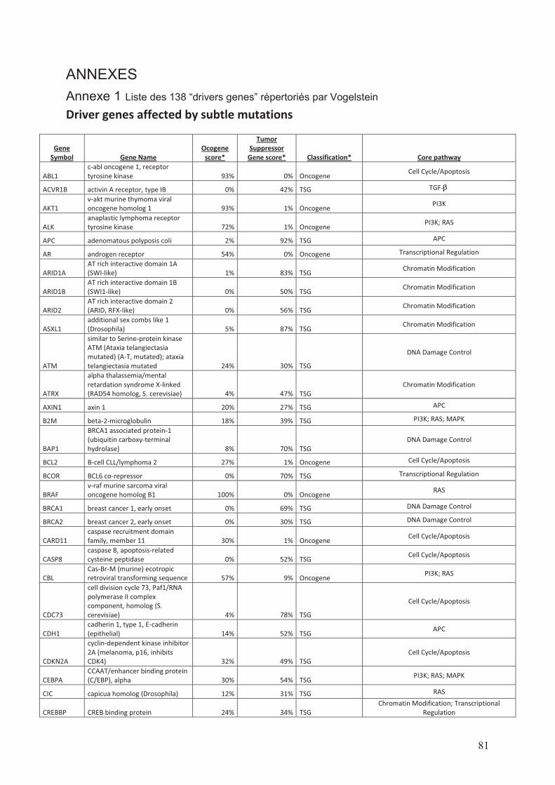
81
ANNEXES
Annexe 1 Liste des 138 “drivers genes” répertoriés par Vogelstein
Driver genes affected by subtle mutations
Gene
Symbol Gene Name
Ocogene
score*
Tumor
Suppressor
Gene score* Classification* Core pathway
ABL1
c-abl oncogene 1, receptor
tyrosine kinase 93% 0% Oncogene Cell Cycle/Apoptosis
ACVR1B activin A receptor, type IB 0% 42% TSG TGF-
AKT1
v-akt murine thymoma viral
oncogene homolog 1 93% 1% Oncogene PI3K
ALK
anaplastic lymphoma receptor
tyrosine kinase 72% 1% Oncogene PI3K; RAS
APC adenomatous polyposis coli 2% 92% TSG APC
AR androgen receptor 54% 0% Oncogene Transcriptional Regulation
ARID1A
AT rich interactive domain 1A
(SWI-like) 1% 83% TSG Chromatin Modification
ARID1B
AT rich interactive domain 1B
(SWI1-like) 0% 50% TSG Chromatin Modification
ARID2
AT rich interactive domain 2
(ARID, RFX-like) 0% 56% TSG Chromatin Modification
ASXL1
additional sex combs like 1
(Drosophila) 5% 87% TSG Chromatin Modification
ATM
similar to Serine-protein kinase
ATM (Ataxia telangiectasia
mutated) (A-T, mutated); ataxia
telangiectasia mutated 24% 30% TSG
DNA Damage Control
ATRX
alpha thalassemia/mental
retardation syndrome X-linked
(RAD54 homolog, S. cerevisiae) 4% 47% TSG
Chromatin Modification
AXIN1 axin 1 20% 27% TSG APC
B2M beta-2-microglobulin 18% 39% TSG PI3K; RAS; MAPK
BAP1
BRCA1 associated protein-1
(ubiquitin carboxy-terminal
hydrolase) 8% 70% TSG
DNA Damage Control
BCL2 B-cell CLL/lymphoma 2 27% 1% Oncogene Cell Cycle/Apoptosis
BCOR BCL6 co-repressor 0% 70% TSG Transcriptional Regulation
BRAF
v-raf murine sarcoma viral
oncogene homolog B1 100% 0% Oncogene RAS
BRCA1 breast cancer 1, early onset 0% 69% TSG DNA Damage Control
BRCA2 breast cancer 2, early onset 0% 30% TSG DNA Damage Control
CARD11
caspase recruitment domain
family, member 11 30% 1% Oncogene Cell Cycle/Apoptosis
CASP8
caspase 8, apoptosis-related
cysteine peptidase 0% 52% TSG Cell Cycle/Apoptosis
CBL
Cas-Br-M (murine) ecotropic
retroviral transforming sequence 57% 9% Oncogene PI3K; RAS
CDC73
cell division cycle 73, Paf1/RNA
polymerase II complex
component, homolog (S.
cerevisiae) 4% 78% TSG
Cell Cycle/Apoptosis
CDH1
cadherin 1, type 1, E-cadherin
(epithelial) 14% 52% TSG APC
CDKN2A
cyclin-dependent kinase inhibitor
2A (melanoma, p16, inhibits
CDK4) 32% 49% TSG
Cell Cycle/Apoptosis
CEBPA
CCAAT/enhancer binding protein
(C/EBP), alpha 30% 54% TSG PI3K; RAS; MAPK
CIC capicua homolog (Drosophila) 12% 31% TSG RAS
CREBBP CREB binding protein 24% 34% TSG
Chromatin Modification; Transcriptional
Regulation
82
CRLF2 cytokine receptor-like factor 2 100% 0% Oncogene STAT
CSF1R
colony stimulating factor 1
receptor 50% 15% Oncogene PI3K; RAS
CTNNB1
catenin (cadherin-associated
protein), beta 1, 88kDa 92% 1% Oncogene APC
CYLD
cylindromatosis (turban tumor
syndrome) 0% 85% TSG Cell Cycle/Apoptosis
DAXX death-domain associated protein 7% 61% TSG Chromatin Modification; Cell Cycle/Apoptosis
DNMT1
DNA (cytosine-5-)-
methyltransferase 1 36% 5% Oncogene Chromatin Modification
DNMT3A
DNA (cytosine-5-)-
methyltransferase 3 alpha 74% 12% Oncogene Chromatin Modification
EGFR
epidermal growth factor
receptor (erythroblastic leukemia
viral (v-erb-b) oncogene
homolog, avian) 97% 0% Oncogene
PI3K; RAS
EP300 E1A binding protein p300 12% 32% TSG Chromatin Modification; APC; TGF- ; NOTCH
ERBB2
v-erb-b2 erythroblastic leukemia
viral oncogene homolog 2,
neuro/glioblastoma derived
oncogene homolog (avian) 67% 3% Oncogene
PI3K; RAS
EZH2
enhancer of zeste homolog 2
(Drosophila) 67% 12% Oncogene Chromatin Modification
FAM123B
family with sequence similarity
123B 4% 66% TSG APC
FBXW7
F-box and WD repeat domain
containing 7 55% 18% TSG NOTCH
FGFR2
fibroblast growth factor receptor
2 49% 6% Oncogene PI3K; RAS ; STAT
FGFR3
fibroblast growth factor receptor
3 99% 0% Oncogene PI3K; RAS ; STAT
FLT3 fms-related tyrosine kinase 3 98% 0% Oncogene RAS; PI3K; STAT
FOXL2 forkhead box L2 100% 0% Oncogene TGF-
FUBP1
far upstream element (FUSE)
binding protein 1 0% 70% TSG Cell Cycle/Apoptosis
GATA1
GATA binding protein 1 (globin
transcription factor 1) 8% 84% TSG NOTCH, TGF-
GATA2 GATA binding protein 2 53% 4% Oncogene NOTCH, TGF-
GATA3 GATA binding protein 3 9% 66% TSG Transcriptional Regulation
GNA11
guanine nucleotide binding
protein (G protein), alpha 11 (Gq
class) 92% 1% Oncogene
PI3K; RAS; MAPK
GNAQ
guanine nucleotide binding
protein (G protein), q
polypeptide 95% 1% Oncogene
PI3K;RAS; MAPK
GNAS GNAS complex locus 93% 2% Oncogene APC; PI3K; TGF- , RAS
H3F3A
H3 histone, family 3B (H3.3B); H3
histone, family 3A pseudogene;
H3 histone, family 3A; similar to
H3 histone, family 3B; similar to
histone H3.3B 93% 0% Oncogene
Chromatin Modification
HIST1H3B
histone cluster 1, H3j; histone
cluster 1, H3i; histone cluster 1,
H3h; histone cluster 1, H3g;
histone cluster 1, H3f; histone
cluster 1, H3e; histone cluster 1,
H3d; histone cluster 1, H3c;
histone cluster 1, H3b; histone
cluster 1, H3a; histone cluster 1,
H2ad; histone cluster 2, H3a;
histone cluster 2, H3c; histone
cluster 2, H3d 60% 0% Oncogene
Chromatin Modification
HNF1A HNF1 homeobox A 29% 55% TSG APC
HRAS
v-Ha-ras Harvey rat sarcoma viral
oncogene homolog 96% 0% OncogeneRAS

83
IDH1
isocitrate dehydrogenase 1
(NADP+), soluble 100% 0% Oncogene Chromatin Modification
IDH2
isocitrate dehydrogenase 2
(NADP+), mitochondrial 99% 0% Oncogene Chromatin Modification
JAK1 Janus kinase 1 26% 18% Oncogene STAT
JAK2 Janus kinase 2 100% 0% Oncogene STAT
JAK3 Janus kinase 3 60% 6% Oncogene STAT
KDM5C
lysine (K)-specific demethylase
5C 0% 62% TSG Chromatin Modification
KDM6A
lysine (K)-specific demethylase
6A 0% 72% TSG Chromatin Modification
KIT
similar to Mast/stem cell growth
factor receptor precursor (SCFR)
(Proto-oncogene tyrosine-
protein kinase Kit) (c-kit) (CD117
antigen); v-kit Hardy-Zuckerman
4 feline sarcoma viral oncogene
homolog 90% 0% Oncogene
PI3K; RAS; STAT
KLF4 Kruppel-like factor 4 80% 4% Oncogene Transcriptional Regulation; WNT
KRAS
v-Ki-ras2 Kirsten rat sarcoma
viral oncogene homolog 100% 0% Oncogene RAS
MAP2K1
mitogen-activated protein kinase
kinase 1 67% 0% Oncogene RAS
MAP3K1
mitogen-activated protein kinase
kinase kinase 1 0% 63% TSG RAS; MAPK
MED12 mediator complex subunit 12 84% 0% Oncogene Cell Cycle/Apoptosis; TGF-
MEN1 multiple endocrine neoplasia I 7% 68% TSG Chromatin Modification
MET
met proto-oncogene (hepatocyte
growth factor receptor) 61% 4% Oncogene PI3K; RAS
MLH1
mutL homolog 1, colon cancer,
nonpolyposis type 2 (E. coli) 18% 37% TSG DNA Damage Control
MLL2
myeloid/lymphoid or mixed-
lineage leukemia 2 1% 70% TSG Chromatin Modification
MLL3
myeloid/lymphoid or mixed-
lineage leukemia 3 5% 44% TSG Chromatin Modification
MPL
myeloproliferative leukemia virus
oncogene 96% 0% Oncogene STAT
MSH2
mutS homolog 2, colon cancer,
nonpolyposis type 1 (E. coli) 0% 65% TSG DNA Damage Control
MSH6 mutS homolog 6 (E. coli) 3% 68% TSG DNA Damage Control
MYD88
myeloid differentiation primary
response gene (88) 92% 1% Oncogene Cell Cycle/Apoptosis
NCOR1 nuclear receptor co-repressor 1 11% 32% TSG Chromatin Modification
NF1 neurofibromin 1 2% 73% TSG RAS
NF2 neurofibromin 2 (merlin) 4% 89% TSG APC
NFE2L2
nuclear factor (erythroid-derived
2)-like 2 74% 1% Oncogene Cell Cycle/Apoptosis
NOTCH1
Notch homolog 1, translocation-
associated (Drosophila) 44% 27% TSG NOTCH
NOTCH2 Notch homolog 2 (Drosophila) 0% 27% TSG NOTCH
NPM1
nucleophosmin 1 (nucleolar
phosphoprotein B23, numatrin)
pseudogene 21; hypothetical
LOC100131044; similar to
nucleophosmin 1;
nucleophosmin (nucleolar
phosphoprotein B23, numatrin) 2% 98% TSG
Cell Cycle/Apoptosis
NRAS
neuroblastoma RAS viral (v-ras)
oncogene homolog 99% 0% Oncogene RAS
PAX5 paired box 5 42% 26% TSG Chromatin Modification
PBRM1 polybromo 1 0% 83% TSG Chromatin Modification
PDGFRA
platelet-derived growth factor
receptor, alpha polypeptide 84% 1% Oncogene PI3K; RAS

84
PHF6 PHD finger protein 6 18% 61% TSG Transcriptional Regulation
PIK3CA
phosphoinositide-3-kinase,
catalytic, alpha polypeptide 95% 1% Oncogene PI3K
PIK3R1
phosphoinositide-3-kinase,
regulatory subunit 1 (alpha) 14% 37% TSG PI3K
PPP2R1A
protein phosphatase 2 (formerly
2A), regulatory subunit A, alpha
isoform 85% 2% Oncogene
Cell Cycle/Apoptosis
PRDM1
PR domain containing 1, with
ZNF domain 0% 64% TSG Chromatin Modification
PTCH1 patched homolog 1 (Drosophila) 7% 60% TSG HH
PTEN
phosphatase and tensin
homolog; phosphatase and
tensin homolog pseudogene 1 30% 55% TSG
PI3K
PTPN11
protein tyrosine phosphatase,
non-receptor type 11; similar to
protein tyrosine phosphatase,
non-receptor type 11 90% 0% Oncogene
RAS
RB1 retinoblastoma 1 4% 80% TSG Cell Cycle/Apoptosis
RET ret proto-oncogene 86% 1% Oncogene RAS; PI3K
RNF43 ring finger protein 43 7% 43% TSG APC
RUNX1
runt-related transcription factor
1 34% 41% TSG Transcriptional Regulation
SETD2 SET domain containing 2 3% 47% TSG Chromatin Modification
SETBP1 SET binding protein 1 25% 4% Oncogene Chromatin Modification; Replication
SF3B1
splicing factor 3b, subunit 1,
155kDa 91% 0% Oncogene Transcriptional Regulation
SMAD2 SMAD family member 2 0% 41% TSG TGF-
SMAD4 SMAD family member 4 24% 39% TSG TGF-
SMARCA4
SWI/SNF related, matrix
associated, actin dependent
regulator of chromatin,
subfamily a, member 4 22% 22% TSG
Chromatin Modification
SMARCB1
SWI/SNF related, matrix
associated, actin dependent
regulator of chromatin,
subfamily b, member 1 16% 74% TSG
Chromatin Modification
SMO
smoothened homolog
(Drosophila) 51% 3% Oncogene HH
SOCS1
suppressor of cytokine signaling
1 15% 46% TSG STAT
SOX9
SRY (sex determining region Y)-
box 9 0% 70% TSG APC
SPOP speckle-type POZ protein 66% 3% Oncogene Chromatin Modification; HH
SRSF2
SRSF2 serine/arginine-rich
splicing factor 2 95% 2% Oncogene Transcriptional Regulation
STAG2 stromal antigen 2 0% 33% TSG DNA Damage Control
STK11 serine/threonine kinase 11 24% 52% TSG mTOR
TET2 tet oncogene family member 2 14% 70% TSG Chromatin Modification
TNFAIP3
tumor necrosis factor, alpha-
induced protein 3 1% 80% TSG Cell Cycle/Apoptosis; MAPK
TRAF7 TNF receptor-associated factor 7 61% 9% TSG Apoptosis
TP53 tumor protein p53 73% 20% TSG Cell Cycle/Apoptosis; DNA Damage Control
TSC1 tuberous sclerosis 1 0% 45% TSG PI3K
TSHR
thyroid stimulating hormone
receptor 86% 0% Oncogene PI3K; MAPK
U2AF1
U2 small nuclear RNA auxiliary
factor 1 92% 1% Oncogene Transcriptional Regulation
VHL
von Hippel-Lindau tumor
suppressor 27% 60% TSG PI3K; RAS; STAT
WT1 Wilms tumor 1 10% 79% TSG Chromatin Modification
85
*Genes were classified as Oncogenes if they had an Oncogene Score >20% and classified as a Tumor Suppressor Gene (TSG) if the TSG
Score was >20% (the 20/20 rule). The Oncogene Score was defined as the number of clustered mutations (i.e., missense mutations at the
same amino acid or identical in-frame insertions or deletions) divided by the total number of mutations. The TSG Score was defined as the
number of truncating mutations divided by the total number of mutations. Truncating mutations included nonsense mutations, insertions
or deletions that alter the reading frame, splice-site mutations, or mutations at the normal stop codon predicted to result in a longer
protein. When a gene had an oncogene score >20% and a TSG Score >5%, it was classified as a TSG because well-studied oncogenes rarely
harbor stop codons. The major data source for this classification was the COSMIC database (www.sanger.ac.uk/genetics/CGP/cosmic/).
To be classified as an oncogene, there had to be >10 clustered mutations in this database. To be classified as a tumor suppressor gene,
there had had to be at least 7 inactivating mutations recorded in this database. In those cases in which 7 to 20 inactivating mutations
were recorded in the COSMIC database, manual curation was performed. This curation was used to identify other examples of mutations
not yet recorded in the COSMIC database and to exclude the most common artifacts encountered in next-generation sequencing, such as
mapping errors and high mutation frequencies observed in normal tissues. Genes with mutations occurring predominantly in tumors with
very high rates of mutation, such as in mismatch-repair deficient tumors or melanomas, were excluded. As more individual tumors are
sequenced in the future, the 20/20 rule can be improved by (i) considering mutations only in particular tumor types, rather than in all
tumor types combined (as done here); (ii) requiring a higher number (e.g., 15) of clustered or inactivating mutations as a threshold for
inclusion; and (iii) for genes with thousands of recorded mutations, choose a random subset to calculate the Oncogene Score (if enough
tumors are sequenced, all mutations will appear to be clustered)
Driver genes affected by amplification or homozygous deletion
Gene
Symbol Gene Name Genetic alteration Classification Core pathway
CCND1 cyclin D1 Amplification Oncogene Cell Cycle/Apoptosis
CDKN2C cyclin-dependent kinase inhibitor 2C (p18, inhibits CDK4) Homozygous deletion TSG Cell Cycle/Apoptosis
IKZF1 IKAROS family zinc finger 1 (Ikaros) Homozygous deletion TSG Transcriptional Regulation
LMO1 LIM domain only 1 (rhombotin 1) Amplification Oncogene Transcriptional Regulation
MAP2K4 mitogen-activated protein kinase kinase 4 Homozygous deletion TSG MAPK
MDM2 Mdm2 p53 binding protein homolog (mouse) Amplification Oncogene Cell Cycle/Apoptosis
MDM4 Mdm4 p53 binding protein homolog (mouse) Amplification Oncogene Cell Cycle/Apoptosis
MYC v-myc myelocytomatosis viral oncogene homolog (avian) Amplification Oncogene Cell Cycle/Apoptosis
MYCL1
v-myc myelocytomatosis viral oncogene homolog 1, lung
carcinoma derived (avian) Amplification Oncogene Cell Cycle/Apoptosis
MYCN
v-myc myelocytomatosis viral related oncogene,
neuroblastoma derived (avian) Amplification Oncogene Cell Cycle/Apoptosis
NCOA3 nuclear receptor coactivator 3 Amplification Oncogene Chromatin Modification
NKX2-1 NK2 homeobox 1 Amplification Oncogene PI3K; MAPK
SKP2 S-phase kinase-associated protein 2 (p45) Amplification Oncogene Cell Cycle/Apoptosis

86
Annexe 2 Publications présentées dans ces travaux
- Article 1
Prognostic factors influencing progression-free survival determined from a series of sporadic
desmoid tumors: a wait-and-see policy according to tumor presentation. Salas S, Dufresne A,
Bui B, Blay JY, Terrier P, Ranchere-Vince D, Bonvalot S, Stoeckle E, Guillou L, Le Cesne A,
Oberlin O, Brouste V, Coindre JM. J Clin Oncol.2011 Sep 10;29(26):3553-8
- Article 2 A prognostic nomogram for prediction of recurrence in desmoid fibromatosis. Crago AM, Denton B, Salas S, Dufresne A, Mezhir JJ, Hameed M, Gonen M, Singer S, Brennan MF. Ann Surg. 2013 Aug;258(2):347-53
- Article 3 High frequency of b-catenin heterozygous mutations in extra-abdominal fibromatosis: a potential molecular tool for disease management. J. Domont, S Salas, L Lacroix, V Brouste, P Saulnier, P Terrier, D Ranchere, A Neuville, A Leroux, L Guillou, R Sciot, F Collin, A
Dufresne, J-Y Blay, A Le Cesne, J-M Coindre, S Bonvalot, and J Benard. British Journal of Cancer 2010; 102: 1032 - 1036
- Article 4
Identification of biological factors predictive of response to imatinib mesylate in aggressive
fibromatosis. Dufresne A, Bertucci F, Penel N, Le Cesne A, Bui B, Tubiana-Hulin M, Ray-
Coquard I, Cupissol D, Chevreau C, Perol D, Goncalves A, Jimenez M, Bringuier PP, Blay
JY. Br J Cancer. 2010 Aug 10;103(4):482-5
- Article 5
A V530I Mutation in c-KIT Exon 10 Is Associated to Imatinib Response in Extraabdominal
Aggressive Fibromatosis. Kurtz JE, Asmane I, Voegeli AC, Neuville A,Dufresne A, Litique V,
Chevreau C, Bergerat JP. Sarcoma. 2010;2010:458156
- Article 6 Impact of KIT exon 10 M541L allelic variant on the response to imatinib in aggressive fibromatosis: analysis of the desminib series by competitive allele specific Taqman PCR technology. A. Dufresne, L. Alberti, M. Brahmi, H. Phlippon, D. Perol and J.-Y. Blay. En cours de revision à BMC Cancer
- Article 7 miRNA expression profile of desmoid tumors predicts their aggressiveness. A. Dufresne, M.
Paturel, L. Alberti, H. Philippon, AV Decouvelaere, P. Cassier, JY Blay. Soumis à
International Journal of Cancer

Prognostic Factors Influencing Progression-Free SurvivalDetermined From a Series of Sporadic Desmoid Tumors:A Wait-and-See Policy According to Tumor PresentationSebastien Salas, Armelle Dufresne, Binh Bui, Jean-Yves Blay, Philippe Terrier, Dominique Ranchere-Vince,Sylvie Bonvalot, Eberhard Stoeckle, Louis Guillou, Axel Le Cesne, Odile Oberlin, Veronique Brouste,and Jean-Michel Coindre
Sebastien Salas, L’Institut National de
la Sante et de la Recherche Medicale
(INSERM) U916, Bergonie Institute,
Bordeaux; Binh Bui, Eberhard Stoeckle,
Veronique Brouste, and Jean Michel
Coindre, Bergonie Institute; Jean-
Michel Coindre, University Victor Sega-
len, Bordeaux; Sebastien Salas, Timone
Hospital, Marseille; Armelle Dufresne,
Jean-Yves Blay, and Dominique
Ranchere-Vince, Leon Berard Center,
Lyon; Philippe Terrier, Sylvie Bonvalot,
Axel Le Cesne and Odile Oberlin,
Gustave Roussy Institute, Villejuif; and
Louis Guillou, University Institute of
Pathology, Lausanne, Switzerland.
Submitted November 3, 2010; accepted
June 23, 2011; published online ahead
of print at www.jco.org on August 15,
2011.
Supported by the Connective Tissue
Cancer Network and La Ligue National
Contre le Cancer.
Authors’ disclosures of potential con-
flicts of interest and author contribu-
tions are found at the end of this
article.
Corresponding author: Sebastien Salas,
MD, PhD, Departement de Pathologie,
Institut Bergonie. 229, cours de
l’Argonne, Bordeaux, France CEDEX
33076; e-mail:
© 2011 by American Society of Clinical
Oncology
0732-183X/11/2926-3553/$20.00
DOI: 10.1200/JCO.2010.33.5489
A B S T R A C T
PurposeDesmoid tumors are mesenchymal fibroblastic/myofibroblastic proliferations with locoregionalaggressiveness and high ability to recur after initial treatment. We present the results of thelargest series of sporadic desmoid tumors ever published to determine the prognostic factors ofthese rare tumors.
Patients and MethodsFour hundred twenty-six patients with a desmoid tumor at diagnosis were included, and thefollowing parameters were studied: age, sex, delay between first symptoms and diagnosis, tumorsize, tumor site, previous history of surgery or trauma in the area of the primary tumor, surgicalmargins, and context of abdominal wall desmoids in women of child-bearing age during or shortlyafter pregnancy. We performed univariate and multivariate analysis for progression-free sur-vival (PFS).
ResultsIn univariate analysis, age, tumor size, tumor site, and surgical margins (R2 v R0/R1) had asignificant impact on PFS. PFS curves were not significantly different for microscopic assessmentof surgical resection quality (R0 v R1). In multivariate analysis, age, tumor size, and tumor site hadindependent values. Three prognostic groups for PFS were defined on the basis of the number ofindependent unfavorable prognostic factors (0 or 1, 2, and 3).
ConclusionThis study clearly demonstrates that there are different prognostic subgroups of desmoid tumorsthat could benefit from different therapeutic strategies, including a wait-and-see policy.
J Clin Oncol 29:3553-3558. © 2011 by American Society of Clinical Oncology
INTRODUCTION
Desmoid tumors (or aggressive fibromatosis) are
soft tissue tumors of clonal origin and mesenchymal
fibroblastic/myofibroblastic proliferations, with an
incidence of two to four new cases per million peo-
ple per year.1,2 Tumorigenesis is linked to beta-
catenin stabilization involving beta-catenin/WNT/
TCF signaling.3 These benign tumors behave
aggressively, deeply infiltrate tissues, and may re-
lapse locoregionally but never metastasize.4,5 They
arise in the abdominal wall, in the abdominal cavity
or, more frequently, in the extremities or the trunk.6
Most desmoid tumors develop sporadically in
young adults, although some cases occur in the con-
text of Gardner’s syndrome (a variant of familial
adenomatous polyposis), and their location is more
often intra-abdominal compared with the sporadic
form.7 Abdominal wall desmoids occur most com-
monly in women of child-bearing age during or
shortly following pregnancy.8
Complete surgical removal remains the opti-
mal treatment but may be difficult or mutilating
according to the tumor location or local extension.
Moreover, a significant proportion of patients will
relapse locally and/or regionally after initial surgery.
Recurrence rates ranging from 30% to 40% have
been reported in the major published series.9 In this
case, mutilating surgery and/or radiotherapy are of-
ten used with a large risk of functional consequences
in the case of musculoskeletal or organ resections.
When local treatments are not feasible, systemic
therapies, including antiestrogen, nonsteroidal anti-
inflammatory drugs, chemotherapy and, more re-
cently, targeted therapy, may induce responses.10 In
addition, some desmoids can spontaneously regress
JOURNAL OF CLINICAL ONCOLOGY O R I G I N A L R E P O R T
VOLUME 29 � NUMBER 26 � SEPTEMBER 10 2011
© 2011 by American Society of Clinical Oncology 3553
193.54.110.33Information downloaded from jco.ascopubs.org and provided by at CENTRE LEON BERARD on September 5, 2013 from
Copyright © 2011 American Society of Clinical Oncology. All rights reserved.

or present growth arrest, so some patients should benefit from a
wait-and-see policy without the use of aggressive therapies. For this
reason, the benefit of surgery in curing desmoids has recently been
questioned.4,11-15 The purpose of this study is to investigate the prog-
nostic factors influencing progression-free-survival (PFS) of spo-
radic desmoids.
PATIENTS AND METHODS
Patient Selection
From February 1, 1965, to March 6, 2008, 426 consecutive patients withsporadic aggressive fibromatosis were diagnosed for their first tumoral event in24 participating cancer centers and were entered into the European database.16
Twenty-five patients were excluded from this study because they presenteddesmoid tumors in the context of Gardner’s syndrome. Patients considered ashaving Gardner’s syndrome were those having an association of a desmoidtumor and family history of colorectal polyposis. We excluded this minoritypopulation to retain a homogeneous group knowing that PFS in these patientsis significantly different from PFS in those with a sporadic desmoid tumor(Appendix Fig A1, online only). The diagnosis of desmoid tumors was con-
firmed in each case by collegial histologic analysis (mesenchymal fibroblastic/myofibroblastic proliferations).
Pathology Review
Histologic slides of all patients entered in this study were reviewed bythe pathology subcommittee of the French Sarcoma Group (GSF). Thesubcommittee included 20 pathologists, and a monthly slide review sessionwas performed. For each tumor, one to eight slides were collegially re-viewed. Histologic typing was based on the WHO histologic typing of softtissue tumors.17
Data Collection
Data regarding patients’ characteristics, tumor description, treatmentmodalities and their results, and outcome were obtained from a retrospectivereview of medical records. These records and histologic data were entered intoa centralized computerized database.16 The following eight variables wereanalyzed for their potential prognostic value: age at presentation, sex, delaybetween first symptoms and diagnosis, tumor size, tumor site, previous historyof surgery or trauma in the area of the primary tumor, surgical margins(macroscopic incomplete resection as R2 resection; microscopic incomplete
Table 1. Patient and Disease Characteristics at Baseline
Characteristic
OverallPatients
(N � 426)
No. %
Age at diagnosis, years
Median 37
Range 0.3-83
Sex
Male 142 33.3
Female 284 66.7
Tumor localization
Abdominal wall 74 17.4
Abdominal cavity 46 10.8
Extra-abdominal 296 69.5
Multifocal 10 2.3
Tumor size, cm
Median 7
Range 0.8-30
Delay between first symptoms and diagnosis, months
Median 4.74
Range 0.1-142
Extra-abdominal localization
Trunk 119 40.2
Upper and lower limbs 109 36.8
Head and neck 27 9.1
Buttocks 22 7.4
Unknown 19 6.5
Limb localization
Proximal 52 47.7
Distal 57 52.3
Context of abdominal wall desmoids in women ofchild-bearing age during or shortly afterpregnancy
Yes 33 7.7
Previous history of surgery in area of primary tumor
Yes 36 8.4
Previous history of trauma in area of primary tumor
Yes 17 3.9
Table 2. Treatment Characteristics and Outcome
Characteristic
Overall Patients (N �
426)
No. % 95% CI
Surgery
Yes 370 86.9
No 56 13.1
Wait-and-see policy
Yes 27 6.3
No 399 93.7
Surgical margins
Microscopically complete tumor resection (R0) 111 30
Microscopically incomplete tumor resection (R1) 110 29.7
Macroscopically incomplete resection (R2) 37 8.6
Unknown 112 30.3
Radiotherapy
No radiotherapy 376 88.3
Radiotherapy 43 10.1
Unknown 7 1.6
Medical treatment
None 363 85.1
Preoperative 6 1.5
Postoperative 29 6.8
Exclusive 23 5.4
Unknown 5 1.2
Median follow-up, months 52 43 to 60
Outcome
Spontaneous remission after wait-and-see policy 5 1.2
Stable disease after wait-and-see policy 16 3.8
Progression after wait-and-see policy 6 1.4
Progression during treatment 9 1.9
Absence of residual tumor after treatment 323 75.8
Residual tumor after treatment 63 14.7
Currently under treatment 1 0.02
Status unknown 4 1
Median progression-free survival, months 41 29 to 53
Local recurrence for patients with absence ofresidual tumor after treatment 143 44.3
Progression for patients with residual tumor aftertreatment 42 66.6
Death from desmoid tumor 0
Death 15 3.5
Salas et al
3554 © 2011 by American Society of Clinical Oncology JOURNAL OF CLINICAL ONCOLOGY
193.54.110.33Information downloaded from jco.ascopubs.org and provided by at CENTRE LEON BERARD on September 5, 2013 from
Copyright © 2011 American Society of Clinical Oncology. All rights reserved.
resection as R1 resection; microscopic complete resection as R0 resection), andcontext of abdominal wall desmoids in women of child-bearing age during orshortly after pregnancy. The status of resection margins in surgically treatedpatients was classified according to the International Union Against Cancer(UICC) R classification.18 Absence of residual tumor after local treatmentmeant that the patients had no disease visible on imaging after surgery and/or radiotherapy.
Statistical Analysis
PFS is defined as time from the date of initial diagnosis to the date ofprogression or recurrence or last follow-up. Follow-up times were described asmedians by using the inverse Kaplan-Meier estimator.19 Continuous variableswere expressed as medians and range, and categorical variables were expressedas percentage. Survival curves were obtained by using the Kaplan-Meiermethod and were compared with the log-rank test. The Cox proportionalhazards model was used to calculate adjusted hazard ratios (HRs) and their95% CIs. All statistical tests were two-sided, and the threshold for statisticalsignificance was P � .05. Variables with a P value of less than .05 in univariateanalyses were tested in the multivariate analysis. Analyses were performed withSPSS software, version 17.0 (SPSS, Chicago, IL).
RESULTS
Patient and Disease Characteristics
Patient characteristics are listed in Table 1. The median age was
37 years (range, 0.3 to 83 years). Fifty-six patients (13%) were younger
than age 18 years. Two thirds of the 426 patients were female. Tumor
locations were as follows: abdominal wall, 74 (17.4%); abdominal
cavity, 46 (10.8%); and extra-abdominal 296 (69.5%). Ten tumors
(2.3%) were multifocal (lesions affecting more than one primary site).
Among patients with an extra-abdominal tumor, 119 (40.2%) had
fibromatosis arising in the trunk, 109 (36.8%) in the upper and lower
limbs, 27 (9.1%) in the head and neck region, and 22 (7.4%) in the
buttocks. Among patients with limb tumors, 57 (52.3%) had distal
limb tumors and 52 (47.7%) had proximal limb tumors. Tumor size
was known in 329 patients (77.2%), and the median largest diameter
was 7.0 cm. Median time between first symptoms and diagnosis was
4.74 months (range, 0.1 to 142 months). Thirty-six patients (8.4%)
had a previous history of surgery in the area of the primary tumor, and
17 (3.9%) had a previous history of trauma in the area of the primary
tumor. Thirty-three (7.7%) abdominal wall desmoids occurred in
women of childbearing age during or shortly after pregnancy.
Local Treatment
Patients’ treatment characteristics and outcomes are described in
Table 2.Three hundred seventy patients (86.9%) had an initial surgical
resection. Histologic evaluation of surgical margins was available in
258 patients (70%). One hundred eleven patients (30%) had R0 resec-
tion, 110 (29.7%) had R1 resection, and 37 (8.6%) had R2 resection.
Radiotherapy generally included photons or electrons with a median
dose of 50 Gy. Forty-three patients received radiotherapy. Surgery was
followed by radiotherapy in 37 patients. Among these patients, one
had R0 resection, 24 had R1 resection, five had R2 resection, and seven
had unknown margins. Six patients received radiotherapy as the only
treatment for their disease.
Medical Treatment
Only 23 patients (5.4%) received medical treatment exclusively
as first-line treatment. Ten patients received chemotherapy: six pa-
tients were treated with chemotherapy only, three patients were
treated with chemotherapy and tamoxifen or toremifene, and one
patient was treated with chemotherapy and a nonsteroidal anti-
inflammatory drug. Twelve patients were treated with tamoxifen or
toremifene and/or a nonsteroidal anti-inflammatory drug. One pa-
tient was treated with imatinib. Medical treatment was given as adju-
vant treatment in 29 patients (6.8%) and as neoadjuvant treatment in
six patients (1.5%). Twenty patients who received chemotherapy were
treated with an anthracycline or with a methotrexate-vinblastine–
containing regimen. Thirty-three patients were treated with tamox-
ifen or toremifene, 22 with a nonsteroidal anti-inflammatory drug,
and one with imatinib. Association of different medical treatments
was possible.
Wait-and-See Policy
A nonaggressive approach was adopted in a subgroup of patients in
an attempt to avoid surgery. Twenty-seven patients (6.3%) were thus
placed under initial attentive medical surveillance. A wait-and-see policy
Patients
(N = 426)
Wait and see
(n = 27)
Spontaneous
remission
(n = 5)
Stable disease
(n = 16)
Progressive
disease
(n = 6)
Residual tumor
(n = 63)
No residual
tumor
(n = 323)
Locoregional TTT
(surg and/or RT)
(n = 376)
Medical
treatment
(n = 23)
Progressive
disease
(n = 42)
No progression
(n = 21)
Local
recurrence
(n = 143)
No local
recurrence
(n = 180)
Fig 1. Diagram of patient outcomes. RT,
radiotherapy; surg, surgery; TTT, treatment.
Desmoid Tumors: Prognostic Factors of Progression-Free Survival
www.jco.org © 2011 by American Society of Clinical Oncology 3555
193.54.110.33Information downloaded from jco.ascopubs.org and provided by at CENTRE LEON BERARD on September 5, 2013 from
Copyright © 2011 American Society of Clinical Oncology. All rights reserved.

waschosenmostlyaccordingtothesiteofprogression,whichcouldnotbe
life-threatening or at risk for mutilation (adjacent nerves or vessels).
Outcome in Patients With the Wait-and-See Policy
Patients’ outcomes are shown in Figure 1. Median follow-up was
52 months (95% CI, 43.6 to 61.6 months). Among 27 patients with the
wait-and-see policy, six presented with progression during follow-up.
Median delay to progression was 19.7 months (range, 7.8 to 46.2
months). Sixteen patients had stable disease. Spontaneous tumor re-
gressions occurred in five patients. None of these patients presented
with progression at the end of the follow-up.
Response
Absence of residual tumor after local treatment (surgery and/or
radiotherapy) was observed in 323 patients (75.8%). Among patients
treated with chemotherapy, partial responses were reported in six
patients, stable disease in three patients, and progression in one pa-
tient. At the time of data analysis, local recurrences had been observed
in 143 (44.3%) of 323 patients with no residual tumor after first-
line treatment.
Survival Analysis
Overall survival is shown in Appendix Figure A4 (online only)
and PFS is shown in Appendix Figure A5 (online only). PFS rates at 5
and 10 years were 35% and 22.8%, respectively. Two hundred forty-
eight patients were alive without disease at the end of the follow-up.
The mean number of recurrences was two (range, one to 20). No
patient died of the disease. Fifteen patients died of other causes. Me-
dian PFS was 41 months (range, 29 to 53 months).
Table 3. Univariate Analysis for Prognostic Factors in Progression-Free Survival
FactorNo. of
Patients
Progression-Free Survival Rate
Log-Rank P2-Year 5-Year 10-Year
Age, years
� 37 199 57.4 36.3 28.7 .005
� 37 205 70.9 49.5 42.1
Sex
Male 130 63.1 41.3 23 .35
Female 276 64.4 43.3 39.9
Context of abdominal wall desmoids in women of child-bearingage during or shortly after pregnancy
No 374 63.1 41.2 32.9 .06
Yes 32 75 62.3 62.3
Previous history of surgery in area of primary tumor
No 371 63.3 41.4 34 .264
Yes 35 70.5 54.5 48.5
Previous history of trauma in area of primary tumor
No 389 64.3 42.9 35 .456
Yes 17 55.1 44.1 —
Tumor localization
Abdominal wall 91 78.2 61.7 57.2 � .001
Abdominal cavity 43 70 50.2 50.2
Extra-abdominal 267 59.2 36.5 26.9
Extra-abdominal localization
Trunk 93 65.5 53.9 42.6 � .001
Upper and lower limbs 105 50.3 23.9 21.7
Head and neck 28 71.9 60.4 —
Buttocks 20 62.3 16.6 —
Limb localization
Proximal 52 68.8 54.5 49.5 .006
Distal 59 54.4 22.8 18.3
Delay between first symptoms and diagnosis, months
� 4.74 114 69.3 54.9 47.8 .942
� 4.74 114 71.9 52.7 46.2
Tumor size, cm
� 7 181 75.1 57.4 47 .004
� 7 136 58.3 40.6 32.8
Surgical margins (R0 v R1 v R2)
R0 110 76.5 62.5 47.5 � .001
R1 107 73.7 60.5 48.1
R2 35 43.4 22 16.5
Surgical margins (R0 v R1)
R0 110 76.5 62.5 47.5 .867
R1 107 73.7 60.5 48.1
Salas et al
3556 © 2011 by American Society of Clinical Oncology JOURNAL OF CLINICAL ONCOLOGY
193.54.110.33Information downloaded from jco.ascopubs.org and provided by at CENTRE LEON BERARD on September 5, 2013 from
Copyright © 2011 American Society of Clinical Oncology. All rights reserved.
Prognostic Factors for PFS
In univariate analysis (Table 3), poor prognostic factors were age
younger than 37 years (median of the whole cohort; P � .005; Appen-
dix Fig A6, online only), size more than 7 cm (median of the whole
cohort; P � .004; Appendix Fig A7, online only), extra-abdominal
localization (P � .001; Appendix Fig A8, online only), and macro-
scopic residual disease after surgery (R0/R1 v R2; P � .001; Appendix
Fig A9, online only). Among extra-abdominal tumors, the worst out-
come was observed in limb and buttocks tumors. Among limb tu-
mors, distal tumors were those with the worst prognosis. PFS curves
were not significantly different for the microscopic assessment of
surgical margin (R0 v R1). In multivariate analysis, age (HR, 1.97; 95%
CI, 1.36 to 2.84; P � .010), tumor size (HR, 1.64; 95% CI, 1.13 to 2.36;
P � .008), and tumor site (extra-abdominal tumor; HR, 2.55; 95% CI,
1.48 to 4.4; P � .001) remained significant prognostic variables (Table
4). Three prognostic groups for PFS were defined on the basis of the
number of unfavorable prognostic factors. Since only 26 patients had
zero poor prognostic factors, we grouped patients with zero or one
poor prognostic factors together to constitute three prognostic
groups: good prognosis, fair prognosis, and poor prognosis.
Figure 2 shows the PFS curves in patients with zero or one, two,
and three unfavorable prognostic factors, respectively. Patients with
zero or one prognostic factor were mainly treated with surgery alone
(data not shown).
DISCUSSION
We present here the results of the largest series of sporadic desmoid
tumors ever published to determine the prognostic factors of these
rare tumors. One of the main problems in managing desmoids tumor
is their locoregional aggressiveness and their high ability to recur after
initial treatment. In this study, no patients died of their disease prob-
ably because we excluded desmoid tumors in the context of Gardner’s
syndrome. Seventy-five percent of patients were in complete remis-
sion after initial management but nearly 50% did relapse. This ex-
plains the choice of PFS and not overall survival in this study to search
for prognostic factors. Surgical resection is often responsible for severe
functional and esthetic consequences. Moreover, the observation of
spontaneous regression in some rare cases of desmoid tumor led us to
conduct this study to search for factors of progression and thus iden-
tify subgroups of patients eligible for individualized management,
including a wait-and-see policy, early surgery, and neoadjuvant or
adjuvant treatments.20-22 In this cohort of patients under the wait-
and-see policy, nearly 20% spontaneously regressed and 60% had
stable disease, and only 20% had tumor progression. However, this
retrospective study cannot provide conclusions regarding the out-
come of these patients since the sample was small and the patients
were selected.
In contrast with other studies, prognosis was not significantly
different for microscopic assessment of surgical margins (R0 v
R1).9,13,23-26 In other studies, however, the presence of microscopic
disease did not necessarily affect long-term disease-free survival.5,14
These results are probably due to the fact that desmoid tumors are
extremely infiltrative locally, making it difficult to assess microscopic
resection margins. Thus, function-sparing surgery should be pre-
ferred to aggressive surgery seeking negative margins.14
Our results show that age (older than 37 years) is statistically
associated with longer PFS. Age as a prognostic factor for local recur-
rence of desmoids has already been studied in several publications. In
one series, age (older than 30 years) was a negative prognostic factor,27
in two series it was a positive factor (age younger than 18 years in the
study by Spear et al28; age younger than 32 years in the study by
Sorensen et al25), and in most of the other series, it had no prognostic
value.5,9,13,24,29 Recently, the identification of biologic pathways in-
volved in the tumorigenesis of desmoids emphasized these age differ-
ences, genomic alterations being more common in older patients. This
could explain the prognostic role of age.3
Tumor location as a prognostic factor has already been demon-
strated, with tumors of the extremities having the worst progno-
sis.9,13,25,27,30 In our study, among extra-abdominal tumors, the worst
outcomes were observed in limb tumors, especially in distal locations.
A subset of patients with extra-abdominal fibromatosis could be man-
aged with a different policy: head and neck and trunk tumors have a
better prognosis. Thus, location is an important factor in the assess-
ment of patients at diagnosis and for stratifying patients taking part in
randomized trials. It is unclear, however, whether these differences are
related to biologic or to surgical management differences.
These findings therefore point to three prognostic factors of PFS
(age, tumor size, and tumor site) based on the results of the largest
series ever published. The natural history of these tumors is unique.
Although considered nonmalignant because of their inability to me-
tastasize, their locoregional aggressiveness and recurrence rate after
resection are particularly high. With nearly 50% of recurrences and
the likelihood of accelerating the evolution of the disease, the value of
surgery (ie, the mainstay in initial management) is under debate.
Table 4. Multivariate Progression-Free Survival Analysis
Variable Crude HR 95% CI P
Median age 1.97 1.36 to 2.84 � .001
Median size 1.64 1.13 to 2.36 .008
Tumor site
Abdominal wall
Intra-abdominal tumor 1.95 0.92 to 4.15 .084�
Extra-abdominal tumor 2.55 1.48 to 4.4 � .001
Abbreviation: HR, hazard ratio.�Not significant.
0/1 Factors
2 Factors
3 Factors
0
Pro
ba
bili
ty
Time (months)
1.0
0.8
0.6
0.4
0.2
14412096724824 168 192 216 240
Fig 2. Probability of survival of patients with zero, one, two, or three factors.
Desmoid Tumors: Prognostic Factors of Progression-Free Survival
www.jco.org © 2011 by American Society of Clinical Oncology 3557
193.54.110.33Information downloaded from jco.ascopubs.org and provided by at CENTRE LEON BERARD on September 5, 2013 from
Copyright © 2011 American Society of Clinical Oncology. All rights reserved.

Several physicians now adopt a wait-and-see policy as initial strategy
for some selected patients.
This study clearly demonstrates that there are different prognos-
tic subgroups of desmoid tumors that could benefit from different
therapeutic strategies. The main question raised by our findings is how
patients should be managed. Should we consider that patients with
good prognostic factors (older than 37 years, tumor size � 7 cm, and
tumor located in the abdominal wall or intra-abdominally) have less
risk of recurrence after surgery and should they thus undergo surgery
straightaway in a curative intent? Or that such patients must have
indolent tumors that could benefit from a wait-and-see policy? It
would be interesting to take subjective morbidity into account as well
as the morbidity associated with treatment, which could have an
impact on therapeutic management.
This study could be the starting point for prospective studies, the
only way to answer these questions and optimize the management
(surgery versus wait-and-see policy) of desmoid tumors. While await-
ing such results, it seems logical to carefully watch the evolution of a
desmoid tumor after its diagnosis and propose local treatment only in
the case of progressive and/or symptomatic disease.
AUTHORS’ DISCLOSURES OF POTENTIAL CONFLICTSOF INTEREST
The author(s) indicated no potential conflicts of interest.
AUTHOR CONTRIBUTIONS
Conception and design: Sebastien Salas, Jean-Michel Coindre
Administrative support: Jean-Michel Coindre
Provision of study materials or patients: Binh Bui, Jean-Yves Blay,
Sylvie Bonvalot, Axel Le Cesne, Odile Oberlin, Eberhard Stoeckle
Collection and assembly of data: Sebastien Salas, Armelle Dufresne,
Philippe Terrier, Dominique Ranchere-Vince, Louis Guillou, Odile
Oberlin, Jean-Michel Coindre
Data analysis and interpretation: Sebastien Salas, Armelle Dufresne,
Binh Bui, Jean-Yves Blay, Sylvie Bonvalot, Eberhard Stoeckle, Louis
Guillou, Odile Oberlin, Veronique Brouste, Jean-Michel Coindre
Manuscript writing: All authors
Final approval of manuscript: All authors
REFERENCES
1. Shields CJ, Winter DC, Kirwan WO, et al:
Desmoid tumours. Eur J Surg Oncol 27:701-706,
2001
2. Li M, Cordon-Cardo C, Gerald WL, et al:
Desmoid fibromatosis is a clonal process. Hum
Pathol 27:939-943, 1996
3. Salas S, Chibon F, Noguchi T, et al: Molecular
characterization by array comparative genomic hy-
bridization and DNA sequencing of 194 desmoid
tumors. Genes Chromosomes Cancer 49:560-568,
2010
4. Phillips SR, A’Hern R, Thomas JM: Aggres-
sive fibromatosis of the abdominal wall, limbs and
limb girdles. Br J Surg 91:1624-1629, 2004
5. Merchant NB, Lewis JJ, Woodruff JM, et al:
Extremity and trunk desmoid tumors: A multifacto-
rial analysis of outcome. Cancer 86:2045-2052, 1999
6. Weiss RJ, Treiber M, Zahlten-Hinguranage A,
et al: Improving local control in patients with aggres-
sive fibromatosis by combined surgery and radio-
therapy [in German]. Chirurg 73:615-621, 2002
7. Hizawa K, Iida M, Mibu R, et al: Desmoid
tumors in familial adenomatous polyposis/Gardner’s
syndrome. J Clin Gastroenterol 25:334-337, 1997
8. Reitamo JJ, Scheinin TM, Hayry P: The des-
moid syndrome. New aspects in the cause, patho-
genesis and treatment of the desmoid tumor. Am J
Surg 151:230-237, 1986
9. Ballo MT, Zagars GK, Pollack A, et al: Des-
moid tumor: Prognostic factors and outcome after
surgery, radiation therapy, or combined surgery and
radiation therapy. J Clin Oncol 17:158-167, 1999
10. Penel N, Le Cesne A, Bui BN, et al: Imatinib
for progressive and recurrent aggressive fibromato-
sis (desmoid tumors): An FNCLCC/French Sarcoma
Group phase II trial with a long-term follow-up. Ann
Oncol 22:452-457. 2011
11. Nuyttens JJ, Rust PF, Thomas CR Jr, et al:
Surgery versus radiation therapy for patients with
aggressive fibromatosis or desmoid tumors: A com-
parative review of 22 articles. Cancer 88:1517-1523,
2000
12. Fiore M, Rimareix F, Mariani L, et al: Desmoid-
type fibromatosis: A front-line conservative ap-
proach to select patients for surgical treatment. Ann
Surg Oncol 16:2587-2593, 2009
13. Bonvalot S, Eldweny H, Haddad V, et al:
Extra-abdominal primary fibromatosis: Aggressive
management could be avoided in a subgroup of
patients. Eur J Surg Oncol 34:462-468, 2008
14. Gronchi A, Casali PG, Mariani L, et al: Quality
of surgery and outcome in extra-abdominal aggres-
sive fibromatosis: A series of patients surgically
treated at a single institution. J Clin Oncol 21:1390-
1397, 2003
15. Smith AJ, Lewis JJ, Merchant NB, et al:
Surgical management of intra-abdominal desmoid
tumours. Br J Surg 87:608-613, 2000
16. Conticanet: Conticabase: European sarcoma
database and tumour bank. www.conticabase.org
17. Fletcher CDM, Unni KK, Mertens F (eds):
World Health Organization classification of tumours:
Pathology and genetics of tumours of soft tissue
and bone. Lyon, France, IARC Press, 2002
18. Tumor of bone and soft tissues, in Sobin LH,
Wittekind C (eds): TNM Classification of Malignant
Tumors (UICC) (ed 6). New York, NY, Wiley Liss,
2002
19. Shuster JJ: Median follow-up in clinical trials.
J Clin Oncol 9:191-192, 1991
20. Dalen BP, Geijer M, Kvist H, et al: Clinical and
imaging observations of desmoid tumors left with-
out treatment. Acta Orthop 77:932-937, 2006
21. Barbier O, Anract P, Pluot E, et al: Primary or
recurring extra-abdominal desmoid fibromatosis: As-
sessment of treatment by observation only. Orthop
Traumatol Surg Res 96:884-889, 2010
22. Nakayama T, Tsuboyama T, Toguchida J, et al:
Natural course of desmoid-type fibromatosis. J Or-
thop Sci 13:51-55, 2008
23. Huang PW, Tzen CY: Prognostic factors in
desmoid-type fibromatosis: A clinicopathological and
immunohistochemical analysis of 46 cases. Pathology
42:147-150, 2010
24. Huang K, Fu H, Shi YQ, et al: Prognostic
factors for extra-abdominal and abdominal wall des-
moids: A 20-year experience at a single institution.
J Surg Oncol 100:563-569, 2009
25. Sørensen A, Keller J, Nielsen OS, et al: Treat-
ment of aggressive fibromatosis: A retrospective
study of 72 patients followed for 1-27 years. Acta
Orthop Scand 73:213-219, 2002
26. Faulkner LB, Hajdu SI, Kher U, et al: Pediatric
desmoid tumor: Retrospective analysis of 63 cases.
J Clin Oncol 13:2813-2818, 1995
27. Rock MG, Pritchard DJ, Reiman HM, et al:
Extra-abdominal desmoid tumors. J Bone Joint Surg
Am 66:1369-1374, 1984
28. Spear MA, Jennings LC, Mankin HJ, et al:
Individualizing management of aggressive fibroma-
toses. Int J Radiat Oncol Biol Phys 40:637-645, 1998
29. Ozger H, Eralp L, Toker B, et al: Evaluation
of prognostic factors affecting recurrences and
disease-free survival in extra-abdominal desmoid
tumors [in Turkish]. Acta Orthop Traumatol Turc
41:291-294, 2007
30. Stoeckle E, Coindre JM, Longy M, et al: A
critical analysis of treatment strategies in desmoid
tumours: A review of a series of 106 cases. Eur
J Surg Oncol 35:129-134, 2009
■ ■ ■
Salas et al
3558 © 2011 by American Society of Clinical Oncology JOURNAL OF CLINICAL ONCOLOGY
193.54.110.33Information downloaded from jco.ascopubs.org and provided by at CENTRE LEON BERARD on September 5, 2013 from
Copyright © 2011 American Society of Clinical Oncology. All rights reserved.
ORIGINAL ARTICLE
A Prognostic Nomogram for Prediction of Recurrence inDesmoid Fibromatosis
Aimee M. Crago, MD, PhD,∗ Brian Denton, MS,† Sebastien Salas, MD, PhD,‡ Armelle Dufresne, MD,§
James J. Mezhir, MD,∗ Meera Hameed, MD,¶ Mithat Gonen, PhD,† Samuel Singer, MD, FACS,∗
and Murray F. Brennan, MD, FACS∗
Objective: To construct a postoperative nomogram to estimate the risk of
local recurrence for patients with desmoid tumors.
Background: The standard management of desmoid tumors is resection, but
many recur locally. Other options include observation or novel chemothera-
peutics, but little guidance exists on selecting treatment.
Methods: Patients undergoing resection during 1982–2011 for primary or
locally recurrent desmoids were identified from a single-institution prospective
database. Cox regression analysis was used to assess risk factors and to create
a recurrence nomogram, which was validated using an international, multi-
institutional data set.
Results: Desmoids were treated surgically in 495 patients (median follow-up
of 60 months). Of 439 patients undergoing complete gross resection, 100
(23%) had recurrence. Five-year local recurrence–free survival was 69%.
Eight patients died of disease, all after R2 resection. Adjuvant radiation was
not associated with improved local recurrence–free survival. In multivariate
analysis, factors associated with recurrence were extremity location, young
age, and large tumor size, but not margin. Abdominal wall tumors had the best
outcome (5-year local recurrence–free survival rate of 91%). Age, site, and
size were used to construct a nomogram with concordance index of 0.703 in
internal validation and 0.659 in external validation. Integration of additional
variables (R1 margin, sex, depth, and primary vs recurrent presentation) did
not importantly improve concordance (internal concordance index of 0.707).
Conclusions: A postoperative nomogram including only size, site, and age
predicts local recurrence and can aid in counseling patients. Systemic therapies
may be appropriate for young patients with large, extremity desmoids, but
surgery alone is curative for most abdominal wall lesions.
Keywords: aggressive fibromatosis, desmoid, nomogram
(Ann Surg 2013;258: 347–353)
D esmoid tumors are clonal proliferations that may arise frommesenchymal stem cells.1,2 Desmoids are associated with
β-catenin mutations in more than 75% of cases; a smaller propor-tion are associated with APC mutations and familial adenomatous
From the Departments of ∗Surgery; †Biostatistics and Epidemiology, MemorialSloan-Kettering Cancer Center, New York, NY; ‡Aix-Marseille Universite,INSERM U911 and Timone Hospital, Department of Medicine, Division ofAdult Oncology, Marseille, France; §Leon Berard Center, INSERM U590,69373, Lyon, France; and ¶Department of Pathology, Memorial Sloan-Kettering Cancer Center, New York, NY.
Disclosure: Funding support was received from Cycle for Survival, MemorialSloan-Kettering Cancer Center, the Kristen Ann Carr Fund, the National In-stitutes of Health through Soft Tissue Sarcoma Program Project grant P01CA047179, and the National Cancer Institute of the National Institutes ofHealth through a SPORE in soft tissue sarcoma. The authors declare noconflicts of interest.
Reprints: Aimee M. Crago, MD, PhD, Gastric and Mixed Tumor Service, Depart-ment of Surgery, Memorial Sloan-Kettering Cancer Center, 1275 York Avenue,H1220, New York, NY 10065. E-mail: [email protected].
Copyright C© 2013 by Lippincott Williams & WilkinsISSN: 0003-4932/13/25802-0347DOI: 10.1097/SLA.0b013e31828c8a30
polyposis (FAP).3–7 The lesions do not have the ability to metasta-size, but progressive disease and local recurrence can lead to signifi-cant morbidity.8 Intra-abdominal tumors may cause intestinal obstruc-tion or fistulization and extremity tumors may result in neuropathicpain and limited mobility. For these reasons, aggressive treatmentstrategies were historically employed to completely extirpate disease.Patients were often managed with radical surgery and adjuvant ordefinitive radiation therapy. For many tumors, however, the anatomiclocation can make resection impossible or extremely morbid, andpostoperative recurrence rates have been reported as more than 70%in some series.8–15
The clinical course of desmoid fibromatosis varies widely; insome instances, the lesions remain stable for long periods with no in-tervention, whereas in others the tumors grow rapidly.8,16,17 The diffi-culty of surgical eradication of desmoids, together with their variableclinical course, has led to exploration of alternative treatment strate-gies. In instances in which operation carries substantial risk, systemictherapies have been prescribed.18 Up to 70% of patients treated withnovel systemic therapies may have improvement of desmoid-relatedsymptoms.19 In several series, patients with asymptomatic desmoidwere observed for 3 to 6 months after presentation.16 In these series,up to 65% of patients did not progress, suggesting that some patientswith desmoid do not require any intervention.
Although observation, systemic therapy, and surgery are cur-rently options for management of desmoid tumors, an optimal man-agement strategy and how best to select patients for therapy have yetto be defined. We sought to identify patients ideally suited for sur-gical intervention versus those who might be candidates for neoad-juvant/definitive systemic therapies or nonoperative management. Todo so, we examined characteristics of a large cohort of patients withsurgically resected desmoids. We identified characteristics that pre-dict local recurrence, and we constructed a clinical nomogram thatpredicts postoperative tumor recurrence.
METHODS
Patient CohortApproval for evaluation of patients with desmoid managed at
Memorial Sloan-Kettering Cancer Center (MSKCC) was obtainedfrom the institutional review board. Patients who underwent surgicalresection between July 1, 1982 and November 1, 2011 were identifiedfrom the institutional soft tissue sarcoma database, in which clinico-pathologic characteristics and survival outcomes were prospectivelyrecorded. Diagnosis and margin status were determined by a sarcomapathologist at the time of surgical resection.
Clinicopathologic characteristics were defined as follows. Le-sions at the axilla, shoulder joint, buttock, and groin were classifiedas extremity. Lesions at the neck, because of their small number inour cohort (n = 29), were considered collectively with extremity le-sions. Chest wall and abdominal wall lesions were differentiated onthe basis of their position relative to the tenth rib. Visceral, intra-abdominal, and retroperitoneal lesions were considered collectively;each of these lesions was considered deep to the fascia. Tumor depth
Copyright © 2013 Lippincott Williams & Wilkins. Unauthorized reproduction of this article is prohibited.
Annals of Surgery � Volume 258, Number 2, August 2013 www.annalsofsurgery.com | 347
Crago et al Annals of Surgery � Volume 258, Number 2, August 2013
in the extremity or trunk was defined as superficial or deep accordingto the relationship to the investing fascia. Margin was classified asgross positive (R2), microscopic positive (R1), or negative (R0).
An external validation set included 274 patients who under-went complete surgical resection between February 1, 1965 to March6, 2008 in 24 cancer centers from the French Sarcoma Group; clinico-pathologic correlates were entered into the European database (www.conticabase.org).20
Statistical MethodsCharacteristics of primary versus recurrent tumors and tumors
with complete versus incomplete gross resection were compared us-ing χ 2 analysis. Local recurrence–free survival (LRFS) was definedfrom the time of first surgery at MSKCC. Patients who underwentR2 resection were not included in the analysis of LRFS. Deaths inpatients without documented recurrence were considered events; pa-tients who were alive at most recent follow-up were censored. LRFSprobabilities were estimated by the Kaplan-Meier method and strati-fied according to various clinicopathologic variables. Age and tumorsize (at primary presentation) were considered on a continuous scalefor nomogram construction and were considered by category (age:16–25, 26–45, 46–65, and >65 years; size: ≤5, 5–10, and >10 cm) inthe univariate and multivariate analyses; the categories were chosenbefore modeling.
Cox proportional hazards regression was used to perform uni-variate and multivariate analyses; all variables (excluding treatment-related variables) were included in the multivariate analysis. No cor-relation was found between significant variables when evaluated bySpearman rank correlation. Two nomograms were constructed, one
including only those variables significant to 0.05 on multivariate anal-ysis and the second including all variables. The Cox model of nomo-gram construction has been described.21 Validation, in this case, wasperformed internally by creating 100 bootstrap resamples and ex-ternally on the European database set. Nomogram calibration waschecked using the calibration plot.
RESULTS
Characteristics of Surgically Resected PatientsThe study cohort consisted of 495 individual patients who
underwent surgical resection of a desmoid tumor at MSKCC (Table 1).The cohort was predominantly female (67%, n = 329). Overall, 382patients (77%) had primary disease, whereas 113 (23%) had recurrentdisease. Patients with primary and recurrent disease were similar interms of sex and depth. Patients with recurrent disease tended to beyounger (79% vs 66% younger than 45 years, P = 0.074; median age,34 vs 38 years). They more commonly presented with extremity orchest wall lesions (83% vs 54%, P < 0.001). Patients presenting withprimary disease were more likely to undergo complete microscopicresection (57% vs 45% in recurrent disease, P = 0.05). They wereless likely to undergo treatment with radiation (12% vs 32%, P <
0.001) or chemotherapy (9% vs 19%, P = 0.002) than were patientspresenting with recurrent disease.
FAP had been documented in 15 patients, 6 of them male and9 female. Their age at resection ranged from 16 to 43 years. Ten hadintra-abdominal lesions, 4 had chest wall desmoids, and one had aparaspinal lesion.
TABLE 1. Characteristics of Patients
Characteristic Primary (n = 382) Recurrent (n = 113) P
Sex 0.734Male 130 (34%) 36 (32%)Female 252 (66%) 77 (68%)
Age, yrs 0.07415–25 67 (18%) 28 (25%)26–45 184 (48%) 60 (53%)46–65 92 (24%) 18 (16%)>65 39 (10%) 7 (6.2%)
Location of primary tumor <0.001Abdominal wall 77 (20%) 11 (9.7%)Chest wall 50 (13%) 26 (23%)GI/intra-abdominal 94 (25%) 6 (5.3%)Extremity 138 (36%) 68 (60%)Other 23 (6.0%) 2 (2%)
Depth 0.914Superficial 28 (7.3%) 7 (6.2%)Deep 351 (92%) 105 (93%)Unknown 3 (0.79%) 1 (0.88%)
Margin status 0.05R0 216 (57%) 51 (45%)R1 130 (34%) 43 (38%)R2 35 (9.2%) 18 (16%)Unknown 1 (0.26%) 1 (0.88%)
Size of primary tumor <0.001≤5 cm 112 (29%) 36 (32%)>5 cm, ≤10 cm 155 (41%) 45 (40%)>10 cm 113 (30%) 12 (11%)Unknown 2 (0.52%) 20 (18%)
Any chemotherapy 34 (8.9%) 22 (19%) 0.002Adjuvant RT 46 (12%) 36 (32%) <0.001
RT indicates radiation therapy.
Copyright © 2013 Lippincott Williams & Wilkins. Unauthorized reproduction of this article is prohibited.
348 | www.annalsofsurgery.com C© 2013 Lippincott Williams & Wilkins

Annals of Surgery � Volume 258, Number 2, August 2013 A Nomogram to Predict Desmoid Recurrence
Determinants of Complete Gross ResectionComplete gross resection was confirmed in 440 patients (89%),
whereas 53 patients (11%) underwent R2 resection with gross residualdisease in place after surgery. The R2-resected tumors were morecommonly large (38% vs 24% were >10 cm; P = 0.001) and locatedin the abdomen (47% vs 17%, P < 0.001). Twenty-five percent of R2resections were in extremity tumors. Of 15 patients with documentedFAP, 8 underwent incomplete gross resection.
Predictors of Local RecurrenceMedian follow-up of the cohort was 60 months (range, 0–327
months). Overall, 44 patients were dead at last follow-up, but only 8died of disease. All of these patients had undergone R2 resection; 6of them had intra-abdominal tumors and 3 were patients with FAP.Two patients died after intestinal transplant.
FIGURE 1. Recurrence-free survival in 439 patients undergoingcomplete gross resection of a desmoid tumor analyzed by theKaplan-Meier method.
Of 439 patients undergoing complete gross resection, 100 hadrecurrence. Among these, 92 (92%) had their recurrence within 5years. Five-year LRFS was 69% (Fig. 1).
The clinicopathologic characteristics were examined for asso-ciation with LRFS after complete gross resection (Table 2). Patientsex, tumor depth, and presentation status (primary vs recurrent) werenot associated with LRFS. However, patients younger than 26 yearsat the time of diagnosis had a significantly higher rate of local re-currence (hazard ratio: 4.27, P = 0.006; Fig. 2A). Five-year LRFSamong these patients was 52% compared with 81% among patientsolder than 65 years.
Size of the patient’s primary tumor was associated with out-come. Tumors larger than 10 cm in largest diameter were associatedwith 5-year LRFS rates of 57% compared with 72% for tumors of5 cm or smaller and 74% for tumors larger than 5 cm and 10 cmor smaller (Fig. 2B). There was a striking association between siteand LRFS. Intra-abdominal, chest wall, and extremity tumors hadrelatively poor outcomes (5-year LRFS rates of 76%, 72%, and 60%,respectively). Desmoids in the neck, which were considered collec-tively with extremity lesions in the main analyses, behaved nearlyidentically to extremity tumors (5-year LRFS rates of 43% for neckvs 47% for lower extremity). Abdominal wall tumors had the bestoutcome [5-year LRFS rates of 90% (Fig. 2C) vs 34% in patientsaged 25 years or younger with large, extremity tumors (n = 11)].Only 8 of 79 patients with abdominal wall tumors had recurrence. Allwere 40 years of age or younger, and 3 of the 8 patients had tumorslarger than 10 cm. Only 1 abdominal wall tumor that recurred, a rectussheath lesion, was smaller than 5 cm.
A multivariate analysis demonstrated that age, tumor size, andtumor site are independent predictors of recurrence (Table 2). Marginstatus (R1 vs R0) was not associated with altered outcome in thecohort as a whole or in subgroups defined by age or tumor site.However, among patients with small tumors (<5 cm), R0 resectionwas associated with longer LRFS compared with R1 resection (76%vs 60%, P = 0.007). Margin was not associated with recurrence inlarger lesions.
Adjuvant Radiation TherapyThe role of radiation therapy in desmoid fibromatosis has been
debated. In our series, there was no improvement in outcomes inpatients who had received radiation therapy during the course oftheir treatment (5-year LRFS rates of 68% vs 72% without radiation;Fig. 3A). In addition, adjuvant radiation therapy was not significantlyassociated with improved LRFS in any subset of tumors (stratified
TABLE 2. Analysis of Factors Predicting Local Recurrence After Desmoid Resection
Univariate Multivariate
Factor Hazard Ratio (95% CI) P Hazard Ratio (95% CI) P
Margin status (R1 vs R0) 1.18 (0.78–1.80) 0.42 0.99 (0.65–1.52) 0.97Presentation status (recurrent vs primary) 1.32 (0.82–2.14) 0.28 1.16 (0.70–1.90) 0.57Depth (deep vs superficial) 1.56 (0.63–3.84) 0.34 1.37 (0.54–3.45) 0.51Sex (female vs male) 1.17 (0.75–1.84) 0.49 1.32 (0.82–2.10) 0.25Location (vs abdominal wall)
Extremity 5.21 (2.25–12.1) <0.001 5.02 (2.14–8.42) <0.001Chest wall 3.31 (1.27–8.62) 0.014 3.12 (1.20–8.42) 0.02Intra-abdominal 2.79 (1.03–7.55) 0.043 2.73 (0.98–7.59) 0.054Other 1.52 (0.31–7.56) 0.61 1.54 (0.31–7.63) 0.6
Size (>10 cm vs ≤10 cm) 1.87 (1.22–2.86) 0.005 1.94 (1.23–3.05) 0.004Age (vs >65 yrs)
≤25 yrs 4.27 (1.51–12.2) 0.006 3.55 (1.23–10.3) 0.01326–65 yrs 1.99 (0.72–5.48) 0.19 2.02 (0.72–5.70) 0.21
Copyright © 2013 Lippincott Williams & Wilkins. Unauthorized reproduction of this article is prohibited.
C© 2013 Lippincott Williams & Wilkins www.annalsofsurgery.com | 349

Crago et al Annals of Surgery � Volume 258, Number 2, August 2013
FIGURE 2. Recurrence-free survival in 439 patients undergoingcomplete gross resection of a desmoid tumor stratified by (A)patient age, (B) tumor size, and (C) tumor location.
by size, site, and patient age at diagnosis) except extremity tumors.Adjuvant radiation was most commonly used for extremity tumors(in 23% of these patients), and radiation for extremity tumors wasassociated with a 15% absolute reduction in local recurrence; LRFSamong patients with extremity tumors was 71% with radiation versus56% without (P = 0.029).
To eliminate the contribution of selection bias to analysis of therole radiation plays in management of desmoid tumors, we analyzedoutcomes over time. Before 1997, 30% of patients received adjuvantradiation therapy, but in 1997 and later, only 7% of patients receivedthis treatment (P < 0.0001). The decreased use of radiation was notaccompanied by a statistically significant change in LRFS (71% vs70%; Fig. 3B).
A Nomogram to Predict Local Recurrence AfterSurgical Resection
On the basis of the univariate and multivariate models, we in-tegrated clinicopathologic risk factors into a nomogram to predictpostoperative recurrence of desmoid tumors at 3, 5, and 7 years aftersurgery. Because prescription of radiation had no association withoutcome, we retained patients receiving this therapy in our analysis.Included in the model were the significant predictors of local recur-rence on univariate analysis, that is, patient age and tumor site andsize. The nomogram is shown in Figure 4. The bootstrapping concor-dance index of this model was 0.703. Addition of margin, depth, sex,or presentation status to the model did not improve concordance sig-nificantly (0.707). Actual incidence of recurrence was plotted versuspredicted recurrence, confirming good calibration of the nomogram(Fig. 5).
External validation of this nomogram was performed on acohort of patients (n = 274) treated at sarcoma specialty centers inEurope. Patients within this cohort were of similar age as those treatedat MSKCC (mean 38.6 vs 37.5 years). Tumors in the validation setwere slightly smaller (mean 6 vs 7 cm; P = 0.001) and were morecommonly seen in the chest wall (27% vs 16%) and less often inthe extremity (36% vs 41%; P = 0.009). The concordance index inthe validation set was 0.659 (confidence interval, 0.598–0.712). Theexternal validation set was previously used to create a 3-point scoringsystem to predict progression-free survival after desmoid resection.Compared with the nomogram, the 3-point tool had substantiallylower concordance index for both in the MSKCC cohort (0.532) andin the external validation cohort (0.570). Calibration of the nomogramin the validation cohort demonstrated that the tool is particularlyaccurate for high-risk lesions (predicted vs actual 3-year survival inthe quintile with poorest prognosis being 51% vs 49%). The tooloverestimated recurrence-free survival rates for the tumors with goodprognosis (89% predicted vs 69% actual risk of local recurrence at5 years in the upper quintile).
DISCUSSIONDesmoid tumors can be managed by observation, surgery, or
chemotherapy, but little information exists on how to select the besttreatment of the individual patient. Given promising responses tosome novel chemotherapies and the high rate of local recurrence afterresection, we sought to define subgroups of patients with desmoidtumors who should be considered for surgical resection and subsetsin whom a multimodality approach to management can be explored.To accomplish this, we have used a large, prospectively collecteddatabase to find risk factors for local recurrence after complete grossresection and to create a clinical nomogram that predicts risk ofrecurrence.
In a multivariate analysis, only tumor site and size and patientage were associated with recurrence. A nomogram that uses these 3
Copyright © 2013 Lippincott Williams & Wilkins. Unauthorized reproduction of this article is prohibited.
350 | www.annalsofsurgery.com C© 2013 Lippincott Williams & Wilkins
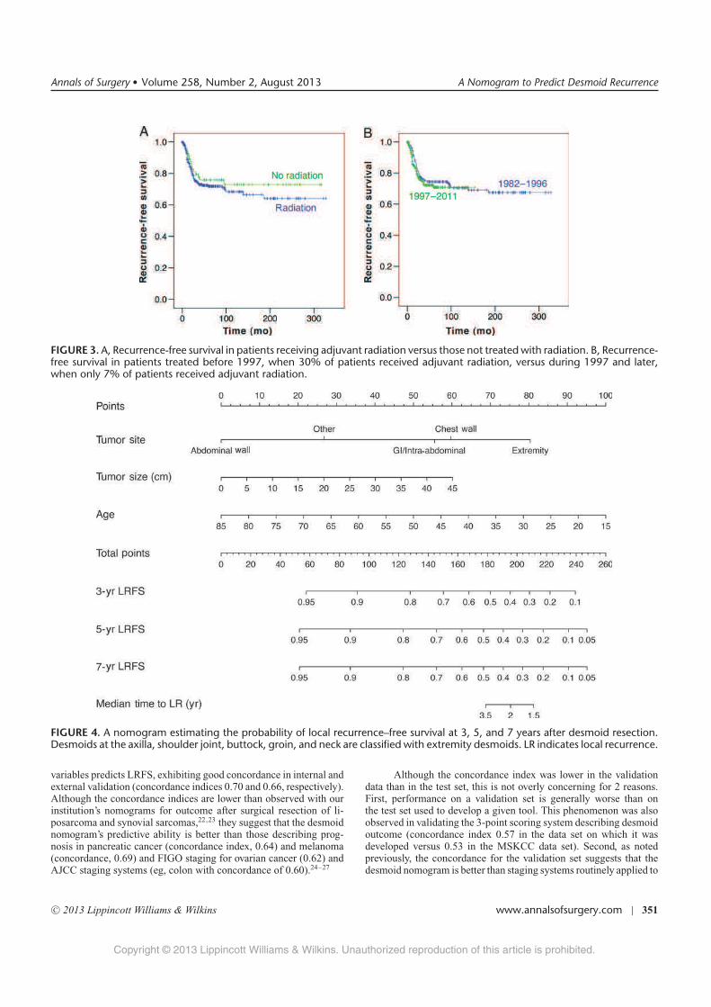
Annals of Surgery � Volume 258, Number 2, August 2013 A Nomogram to Predict Desmoid Recurrence
FIGURE 3. A, Recurrence-free survival in patients receiving adjuvant radiation versus those not treated with radiation. B, Recurrence-free survival in patients treated before 1997, when 30% of patients received adjuvant radiation, versus during 1997 and later,when only 7% of patients received adjuvant radiation.
FIGURE 4. A nomogram estimating the probability of local recurrence–free survival at 3, 5, and 7 years after desmoid resection.Desmoids at the axilla, shoulder joint, buttock, groin, and neck are classified with extremity desmoids. LR indicates local recurrence.
variables predicts LRFS, exhibiting good concordance in internal andexternal validation (concordance indices 0.70 and 0.66, respectively).Although the concordance indices are lower than observed with ourinstitution’s nomograms for outcome after surgical resection of li-posarcoma and synovial sarcomas,22,23 they suggest that the desmoidnomogram’s predictive ability is better than those describing prog-nosis in pancreatic cancer (concordance index, 0.64) and melanoma(concordance, 0.69) and FIGO staging for ovarian cancer (0.62) andAJCC staging systems (eg, colon with concordance of 0.60).24–27
Although the concordance index was lower in the validationdata than in the test set, this is not overly concerning for 2 reasons.First, performance on a validation set is generally worse than onthe test set used to develop a given tool. This phenomenon was alsoobserved in validating the 3-point scoring system describing desmoidoutcome (concordance index 0.57 in the data set on which it wasdeveloped versus 0.53 in the MSKCC data set). Second, as notedpreviously, the concordance for the validation set suggests that thedesmoid nomogram is better than staging systems routinely applied to
Copyright © 2013 Lippincott Williams & Wilkins. Unauthorized reproduction of this article is prohibited.
C© 2013 Lippincott Williams & Wilkins www.annalsofsurgery.com | 351
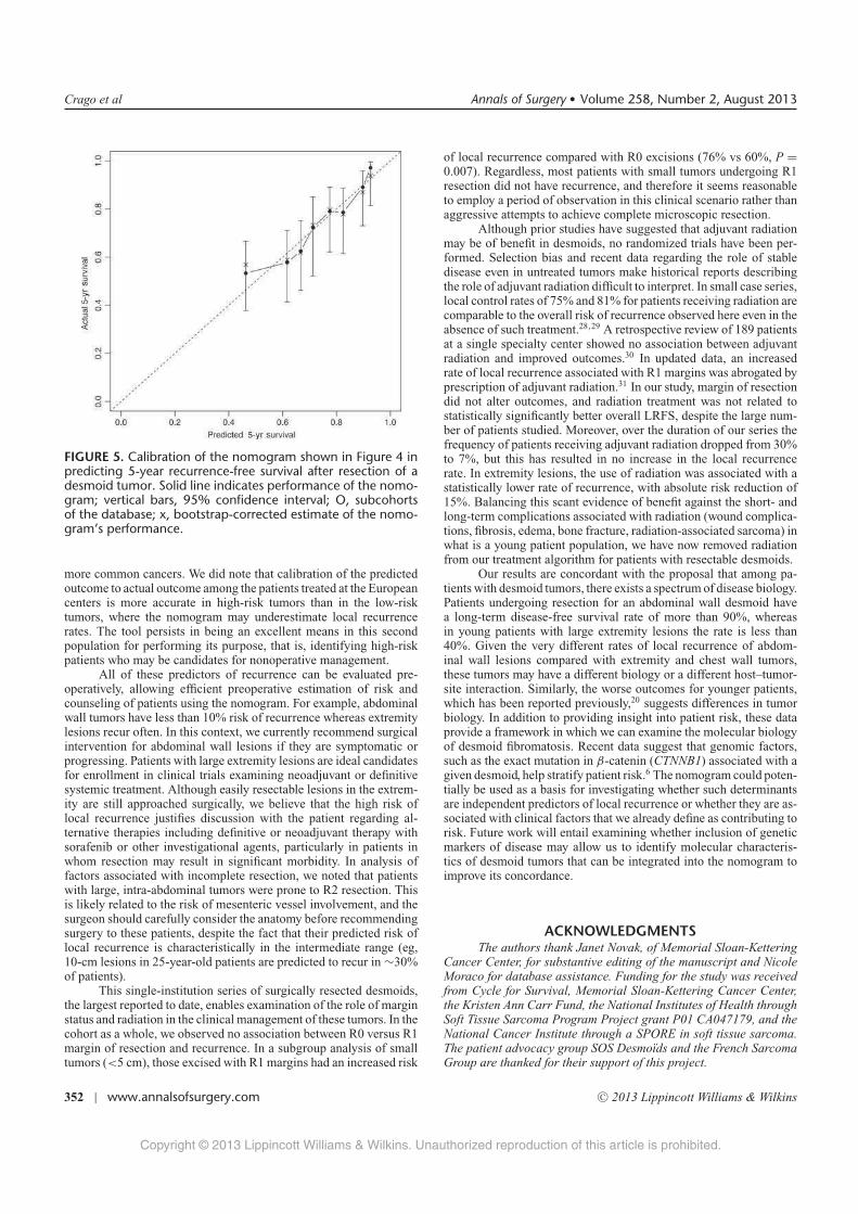
Crago et al Annals of Surgery � Volume 258, Number 2, August 2013
FIGURE 5. Calibration of the nomogram shown in Figure 4 inpredicting 5-year recurrence-free survival after resection of adesmoid tumor. Solid line indicates performance of the nomo-gram; vertical bars, 95% confidence interval; O, subcohortsof the database; x, bootstrap-corrected estimate of the nomo-gram’s performance.
more common cancers. We did note that calibration of the predictedoutcome to actual outcome among the patients treated at the Europeancenters is more accurate in high-risk tumors than in the low-risktumors, where the nomogram may underestimate local recurrencerates. The tool persists in being an excellent means in this secondpopulation for performing its purpose, that is, identifying high-riskpatients who may be candidates for nonoperative management.
All of these predictors of recurrence can be evaluated pre-operatively, allowing efficient preoperative estimation of risk andcounseling of patients using the nomogram. For example, abdominalwall tumors have less than 10% risk of recurrence whereas extremitylesions recur often. In this context, we currently recommend surgicalintervention for abdominal wall lesions if they are symptomatic orprogressing. Patients with large extremity lesions are ideal candidatesfor enrollment in clinical trials examining neoadjuvant or definitivesystemic treatment. Although easily resectable lesions in the extrem-ity are still approached surgically, we believe that the high risk oflocal recurrence justifies discussion with the patient regarding al-ternative therapies including definitive or neoadjuvant therapy withsorafenib or other investigational agents, particularly in patients inwhom resection may result in significant morbidity. In analysis offactors associated with incomplete resection, we noted that patientswith large, intra-abdominal tumors were prone to R2 resection. Thisis likely related to the risk of mesenteric vessel involvement, and thesurgeon should carefully consider the anatomy before recommendingsurgery to these patients, despite the fact that their predicted risk oflocal recurrence is characteristically in the intermediate range (eg,10-cm lesions in 25-year-old patients are predicted to recur in ∼30%of patients).
This single-institution series of surgically resected desmoids,the largest reported to date, enables examination of the role of marginstatus and radiation in the clinical management of these tumors. In thecohort as a whole, we observed no association between R0 versus R1margin of resection and recurrence. In a subgroup analysis of smalltumors (<5 cm), those excised with R1 margins had an increased risk
of local recurrence compared with R0 excisions (76% vs 60%, P =
0.007). Regardless, most patients with small tumors undergoing R1resection did not have recurrence, and therefore it seems reasonableto employ a period of observation in this clinical scenario rather thanaggressive attempts to achieve complete microscopic resection.
Although prior studies have suggested that adjuvant radiationmay be of benefit in desmoids, no randomized trials have been per-formed. Selection bias and recent data regarding the role of stabledisease even in untreated tumors make historical reports describingthe role of adjuvant radiation difficult to interpret. In small case series,local control rates of 75% and 81% for patients receiving radiation arecomparable to the overall risk of recurrence observed here even in theabsence of such treatment.28,29 A retrospective review of 189 patientsat a single specialty center showed no association between adjuvantradiation and improved outcomes.30 In updated data, an increasedrate of local recurrence associated with R1 margins was abrogated byprescription of adjuvant radiation.31 In our study, margin of resectiondid not alter outcomes, and radiation treatment was not related tostatistically significantly better overall LRFS, despite the large num-ber of patients studied. Moreover, over the duration of our series thefrequency of patients receiving adjuvant radiation dropped from 30%to 7%, but this has resulted in no increase in the local recurrencerate. In extremity lesions, the use of radiation was associated with astatistically lower rate of recurrence, with absolute risk reduction of15%. Balancing this scant evidence of benefit against the short- andlong-term complications associated with radiation (wound complica-tions, fibrosis, edema, bone fracture, radiation-associated sarcoma) inwhat is a young patient population, we have now removed radiationfrom our treatment algorithm for patients with resectable desmoids.
Our results are concordant with the proposal that among pa-tients with desmoid tumors, there exists a spectrum of disease biology.Patients undergoing resection for an abdominal wall desmoid havea long-term disease-free survival rate of more than 90%, whereasin young patients with large extremity lesions the rate is less than40%. Given the very different rates of local recurrence of abdom-inal wall lesions compared with extremity and chest wall tumors,these tumors may have a different biology or a different host–tumor-site interaction. Similarly, the worse outcomes for younger patients,which has been reported previously,20 suggests differences in tumorbiology. In addition to providing insight into patient risk, these dataprovide a framework in which we can examine the molecular biologyof desmoid fibromatosis. Recent data suggest that genomic factors,such as the exact mutation in β-catenin (CTNNB1) associated with agiven desmoid, help stratify patient risk.6 The nomogram could poten-tially be used as a basis for investigating whether such determinantsare independent predictors of local recurrence or whether they are as-sociated with clinical factors that we already define as contributing torisk. Future work will entail examining whether inclusion of geneticmarkers of disease may allow us to identify molecular characteris-tics of desmoid tumors that can be integrated into the nomogram toimprove its concordance.
ACKNOWLEDGMENTSThe authors thank Janet Novak, of Memorial Sloan-Kettering
Cancer Center, for substantive editing of the manuscript and NicoleMoraco for database assistance. Funding for the study was receivedfrom Cycle for Survival, Memorial Sloan-Kettering Cancer Center,the Kristen Ann Carr Fund, the National Institutes of Health throughSoft Tissue Sarcoma Program Project grant P01 CA047179, and theNational Cancer Institute through a SPORE in soft tissue sarcoma.The patient advocacy group SOS Desmoıds and the French SarcomaGroup are thanked for their support of this project.
Copyright © 2013 Lippincott Williams & Wilkins. Unauthorized reproduction of this article is prohibited.
352 | www.annalsofsurgery.com C© 2013 Lippincott Williams & Wilkins
Annals of Surgery � Volume 258, Number 2, August 2013 A Nomogram to Predict Desmoid Recurrence
REFERENCES1. Wu C, Nik-Amini S, Nadesan P, et al. Aggressive fibromatosis (desmoid tumor)
is derived from mesenchymal progenitor cells. Cancer Res. 2010;70:7690–7698.
2. Li M, Cordon-Cardo C, Gerald WL, et al. Desmoid fibromatosis is a clonalprocess. Hum Pathol. 1996;27:939–943.
3. Alman BA, Li C, Pajerski ME, et al. Increased beta-catenin protein and somaticAPC mutations in sporadic aggressive fibromatoses (desmoid tumors). Am JPathol. 1997;151:329–334.
4. Li C, Bapat B, Alman BA. Adenomatous polyposis coli gene mutation altersproliferation through its beta-catenin-regulatory function in aggressive fibro-matosis (desmoid tumor). Am J Pathol. 1998;153:709–714.
5. Tejpar S, Nollet F, Li C, et al. Predominance of beta-catenin mutations and beta-catenin dysregulation in sporadic aggressive fibromatosis (desmoid tumor).Oncogene. 1999;18:6615–6620.
6. Lazar AJ, Tuvin D, Hajibashi S, et al. Specific mutations in the beta-cateningene (CTNNB1) correlate with local recurrence in sporadic desmoid tumors.Am J Pathol. 2008;173:1518–1527.
7. Robanus-Maandag E, Bosch C, Amini-Nik S, et al. Familial adenomatouspolyposis-associated desmoids display significantly more genetic changes thansporadic desmoids. PLoS One. 2011;6:e24354.
8. Lewis JJ, Boland PJ, Leung DH, et al. The enigma of desmoid tumors. AnnSurg. 1999;229:866–872; discussion 872–873.
9. Merchant NB, Lewis JJ, Woodruff JM, et al. Extremity and trunk desmoidtumors: a multifactorial analysis of outcome. Cancer. 1999;86:2045–2052.
10. Easter DW, Halasz NA. Recent trends in the management of desmoid tumors.Summary of 19 cases and review of the literature. Ann Surg. 1989;210:765–769.
11. Higaki S, Tateishi A, Ohno T, et al. Surgical treatment of extra-abdominaldesmoid tumours (aggressive fibromatoses). Int Orthop. 1995;19:383–389.
12. Lopez R, Kemalyan N, Moseley HS, et al. Problems in diagnosis and manage-ment of desmoid tumors. Am J Surg. 1990;159:450–453.
13. Markhede G, Lundgren L, Bjurstam N, et al. Extra-abdominal desmoid tumors.Acta Orthop Scand. 1986;57:1–7.
14. Plukker JT, van Oort I, Vermey A, et al. Aggressive fibromatosis (non-familialdesmoid tumour): therapeutic problems and the role of adjuvant radiotherapy.Br J Surg. 1995;82:510–514.
15. Rock MG, Pritchard DJ, Reiman HM, et al. Extra-abdominal desmoid tumors.J Bone Joint Surg Am. 1984;66:1369–1374.
16. Fiore M, Rimareix F, Mariani L, et al. Desmoid-type fibromatosis: a front-line conservative approach to select patients for surgical treatment. Ann SurgOncol. 2009;16:2587–2593.
17. Smith AJ, Lewis JJ, Merchant NB, et al. Surgical management of intra-abdominal desmoid tumours. Br J Surg. 2000;87:608–613.
18. de Camargo VP, Keohan ML, D’Adamo DR, et al. Clinical outcomes ofsystemic therapy for patients with deep fibromatosis (desmoid tumor). Cancer.2010;116:2258–2265.
19. Gounder MM, Lefkowitz RA, Keohan ML, et al. Activity of sorafenib againstdesmoid tumor/deep fibromatosis. Clin Cancer Res. 2011;17:4082–4090.
20. Salas S, Dufresne A, Bui B, et al. Prognostic factors influencing progression-free survival determined from a series of sporadic desmoid tumors: a wait-and-see policy according to tumor presentation. J Clin Oncol. 2011;29:3553–3558.
21. Kattan MW, Leung DH, Brennan MF. Postoperative nomogram for 12-yearsarcoma-specific death. J Clin Oncol. 2002;20:791–796.
22. Canter RJ, Qin LX, Maki RG, et al. A synovial sarcoma-specific preoper-ative nomogram supports a survival benefit to ifosfamide-based chemother-apy and improves risk stratification for patients. Clin Cancer Res. 2008;14:8191–8197.
23. Dalal KM, Kattan MW, Antonescu CR, et al. Subtype specific prognosticnomogram for patients with primary liposarcoma of the retroperitoneum, ex-tremity, or trunk. Ann Surg. 2006;244:381–391.
24. Brennan MF, Kattan MW, Klimstra D, et al. Prognostic nomogram for pa-tients undergoing resection for adenocarcinoma of the pancreas. Ann Surg.2004;240:293–298.
25. Wong SL, Kattan MW, McMasters KM, et al. A nomogram that predicts thepresence of sentinel node metastasis in melanoma with better discriminationthan the American Joint Committee on Cancer staging system. Ann Surg Oncol.2005;12:282–288.
26. Barlin JN, Yu C, Hill EK, et al. Nomogram for predicting 5-year disease-specific mortality after primary surgery for epithelial ovarian cancer. GynecolOncol. 2012;125:25–30.
27. Weiser MR, Gonen M, Chou JF, et al. Predicting survival after curativecolectomy for cancer: individualizing colon cancer staging. J Clin Oncol.2011;29:4796–4802.
28. Guadagnolo BA, Zagars GK, Ballo MT. Long-term outcomes for desmoid tu-mors treated with radiation therapy. Int J Radiat Oncol Biol Phys. 2008;71:441–447.
29. Jelinek JA, Stelzer KJ, Conrad E, et al. The efficacy of radiotherapy as postoper-ative treatment for desmoid tumors. Int J Radiat Oncol Biol Phys. 2001;50:121–125.
30. Ballo MT, Zagars GK, Pollack A, et al. Desmoid tumor: prognostic factors andoutcome after surgery, radiation therapy, or combined surgery and radiationtherapy. J Clin Oncol. 1999;17:158–167.
31. Lev D, Kotilingam D, Wei C, et al. Optimizing treatment of desmoid tumors.J Clin Oncol. 2007;25:1785–1791.
Copyright © 2013 Lippincott Williams & Wilkins. Unauthorized reproduction of this article is prohibited.
C© 2013 Lippincott Williams & Wilkins www.annalsofsurgery.com | 353

High frequency of b-catenin heterozygous mutations in
extra-abdominal fibromatosis: a potential molecular tool for
disease management
J Domont1,13, S Salas2,13, L Lacroix3,4, V Brouste2, P Saulnier3, P Terrier1, D Ranchere5, A Neuville6, A Leroux7,
L Guillou8, R Sciot9, F Collin10, A Dufresne5, J-Y Blay5, A Le Cesne1, J-M Coindre*,2,11, S Bonvalot*,1
and J Benard*,1,4,12
1Sarcoma Committee, Institut Gustave Roussy, Villejuif, France; 2Institut Bergonie, Bordeaux, France; 3Translational Laboratory, Institut Gustave Roussy,
Villejuif, France; 4Department of Clinical Biology and Pathology, Institut Gustave Roussy, Villejuif, France; 5Centre Leon Berard, Lyon, France; 6Department
of Pathology, Hopital Hautepierre, Strasbourg, France; 7Department of Pathology, Centre Alexis Vautrin, Nancy, France; 8University Institute of Pathology,
Lausanne, Switzerland; 9Department of Pathology, KU Leuven, Belgium; 10Department of Pathology, Centre Georges-Francois Leclerc, Dijon, France;11INSERM U916, Pathology Department, University Victor Segalen, Bordeaux, France; 12CNRS-UMR 8126, IFR54, Institut Gustave Roussy, Villejuif,
France
BACKGROUND: Fibromatosis comprises distinct clinical entities, including sporadic extra-abdominal fibromatosis, which have a high
tendency for recurrence, even after adequate resection. There are no known molecular biomarkers of local recurrence. We searched
for b-catenin mutations in a European multicentre series of fibromatosis tumours to relate b-catenin mutational status to disease
outcome.
METHODS: Direct sequencing of exon 3 b-catenin gene was performed for 155 frozen fibromatosis tissues from all topographies.
Correlation of outcome with mutation rate and type was performed on the extra-abdominal fibromatosis group (101 patients).
RESULTS: Mutations of b-catenin were detected in 83% of all cases. Among 101 extra-abdominal fibromatosis, similar mutation rates
(87%) were observed, namely T41A (39.5%), S45P (9%), S45F (36.5%), and deletion (2%). None of the clinico-pathological
parameters were found to be significantly associated with b-catenin mutational status. With a median follow-up of 62 months,
51 patients relapsed. Five-year recurrence-free survival was significantly worse in b-catenin-mutated tumours regardless of a specific
genotype, compared with wild-type tumours (49 vs 75%, respectively, P¼ 0.02).
CONCLUSION: A high frequency (87%) of b-catenin mutation hallmarks extra-abdominal fibromatosis from a large multicentric
retrospective study. Moreover, wild-type b-catenin seems to be an interesting prognostic marker that might be useful in the
therapeutic management of extra-abdominal fibromatosis.
British Journal of Cancer (2010) 102, 1032–1036. doi:10.1038/sj.bjc.6605557 www.bjcancer.com
Published online 2 March 2010
& 2010 Cancer Research UK
Keywords: b-catenin mutation; fibromatosis; prognostic factor
��
�
�
�
�
�
�
�
�
�
�
�
�
�
�
�
�
�
�
�
�
�
�
�
�
�
�
�
�
�
�
�
�
�
�
�
�
�
�
�
�
�
�
�
�
�
�
�
�
�
�
�
�
�
�
�
�
�
�
�
�
�
�
Fibromatoses are rare, locally invasive soft-tissue tumourscomposed of spindle cells that display a wide spectrum ofaggressiveness. These star-shaped soft tissues, devoid of a capsule,invade surrounding structures and have a high propensity for localrecurrence (Sobin and Wittekind, 2002).Clinical forms of the disease depend on the tumour site:
abdominal fibromatosis (deep or desmoid), which occurs on theabdominal wall of women in association with hormonal factors;intra-abdominal fibromatosis, some forms of which are associatedwith germ-like mutations of the adenomatous polyposis coli (APC)gene-associated Gardner syndrome; and extra-abdominal fibro-
matosis, which occurs in the head and neck, trunk, and limbs.Sporadic extra-abdominal fibromatosis has a high tendency forlocal recurrence, even after apparently adequate resection. Ininstitutional retrospective studies, local failure rates at 5 years haveranged from 25 to 60% (Bonvalot et al, 2008). This wide rangereflects the great variability of accrual, treatments, and follow-upin this rare disease, which has not yet been investigated in arandomised controlled study. Traditionally, patients undergostandard surgery, with the primary goal always being to achievecomplete resection with negative margins, as indicated forsarcomas. However, Bonvalot et al (2008) recently questionedthe need for systematically including surgery and other aggressivetreatments in the first-line treatment of primary extra-abdominaldesmoid tumours. In their study, tumour growth arrest occurredin two-thirds of patients managed non-surgically. A larger, multi-institutional study conducted recently also showed that a wait-and-see approach avoided the need for aggressive surgery andradiotherapy in the majority of patients with primary tumours
Received 21 October 2009; revised 5 January 2010; accepted 8 January2010; published online 2 March 2010
*Correspondence: Dr J Benard; E-mail: [email protected] or Dr S Bonvalot;E-mail: [email protected] or Dr J-M Coindre; E-mail: [email protected] authors contributed equally to this work
British Journal of Cancer (2010) 102, 1032 – 1036
& 2010 Cancer Research UK All rights reserved 0007 – 0920/10 $32.00
www.bjcancer.com
MolecularDiagnostic
s

(Fiore et al, 2009). Intrinsic biological characteristics of tumourcells and the host microenvironment, for example, could accountfor highly diverse outcomes.To date, no predictive criteria (clinical, histological, or
biological) that might help oncologists in the clinical managementof patients with extra-abdominal fibromatosis have been identi-fied. From a biological point of view, desmoids and extra-abdominal fibromatosis tumours typically harbour mutations ofAPC and/or b-catenin genes (Alman et al, 1997; Tejpar et al,1999). Adenomatous polyposis coli is known to regulate thecytoplasmic level of b-catenin, which is involved in cell adhesionand has a key role in the Wnt (Wingless) signalling pathway.The phosphorylation of b-catenin on threonine 41 and serine 33,37, and 45, encoded by exon 3 of the b-catenin gene, occurswhen b-catenin binds to APC and leads to protein degradationthrough the ubiquitin–proteasome pathway (Orford et al, 1997;Rubinfeld et al, 1997). Adenomatous polyposis coli mutationsgive rise to b-catenin protein accumulation and induce Wntpathway activation. b-Catenin gene missense mutations, in turn,lead to post-translational stabilisation of the protein andtranslocation from the cytoplasm to the nucleus, where b-cateninbinds to the T-cell factor lymphoid enhancer family, resultingin transcription transactivation (Morin et al, 1997). Such muta-tions had only been described in small series of desmoid tumoursuntil very recently when Lazar et al (2008) reported a highfrequency of b-catenin mutations in the vast majority offibromatoses.In this study, we analysed a large series of extra-abdominal
fibromatoses (with follow-up data spanning over 5 years) collectedthrough the connective tissue cancer network Conticanet inFrance, Belgium, and Switzerland to evaluate whether b-cateninmutational status might constitute a valuable biomarker forrecurrence that could improve the therapeutic management ofsuch disabling tumours.
MATERIALS AND METHODS
Tumour material of patients
All tumour samples were collected through CONTICANET andstored in a centralised tumour data bank at the Bergonie Institutein Bordeaux, France, after obtaining approval from the appro-priate ethics committees. This European fibromatosis tumourdatabase contained exhaustive clinical information and follow-updata for over 5 years. From this tumour database, 187 eligiblefibromatosis tumour tissues collected between December 1987and January 2007 were tested in this retrospective study. Most ofthe patients with extra-abdominal fibromatosis had been surgi-cally treated with curative intent. The tumour material comprisedtumour samples from biopsies or microbiopsies performed using16- or 18-gauge needles. The tissues had been obtained aftersurgery at diagnosis and snap-frozen in liquid nitrogen. Twenty-three formalin-fixed, paraffin-embedded (FFPE) tumour samplespaired to frozen tumour samples were also obtained, as were35-mm-thick tumour slices flanked by 4-mm sections from eitherfrozen or FFPE fibromatosis tumours for use as histolo-gical controls (haematoxylin–eosin–saffron staining). The histo-logical controls were analysed by pathologists specialised in soft-tissue tumours (Ph T and JMC). Over 90% of the histologicalfeatures identified were typical of fibromatosis in all tumoursanalysed.
b-Catenin gene sequencing
Following lysis of the 35 mm tumour slices, nucleic acids wereobtained using Qiagen column separation according to the manu-facturer’s instructions (Qiagen, Hilden, Germany). Available
DNA for sequencing was obtained in 155 desmoid-typefibromatosis tumour biopsy specimens including all clinical formsof the disease. DNA lymphocytes from 96 anonymous individualswere used as non-mutated controls. The CTNNB1 mutationslocated in exon 3 were analysed by direct DNA sequencing. Thegenomic DNA was first amplified by polymerase chain reaction(PCR) of over 35 cycles at an annealing temperature of 551C(forward primers: 50-ATTTGATGGAGTTGGACATGGC-30 andreverse: 50-CCAGCTACTTGTTCTTGAGTGAAGG-30) as previouslydescribed (Delmas et al, 2007). All PCR amplimers were checkedusing 1% agarose gel electrophoresis. Sequencing reactions wereperformed using a BigDye terminator cycle sequencing kitand analysed on a 48-capillary 3730 DNA Analyzer (AppliedBiosystems, Foster City, CA, USA). Exonic sequences were readand aligned using SeqScapeR software (Applied Biosystems).As reference for b-catenin gene, NM 00109822091 aligned on asequence of chromosome 3 (NCBI 36) was used.
Patient population and clinical data
The varying clinical forms of fibromatosis (sporadic tumours,intra-abdominal tumours associated with hereditary syndrome,and deep and extra-abdominal tumours) were all represented inthe Conticanet series of tumours used for this study. Of 155fibromatosis tissues from all topologies available for DNAsequencing, 101 extra-abdominal fibromatoses were used toinvestigate the correlation between b-catenin mutational statusand clinical characteristics: age, sex, tumour size, tumourlocation (head and neck, abdominal and thoracic walls and limbs),therapy (including quality of surgical resection as assessed bytype of resection (R0, R1, or R2 according to the UICC-Rclassification system)) (Lev et al, 2007), and outcome (for 101patients treated with a curative intent). R0 resection was obtainedfor 42 patients.
Statistical analysis
Survival times were calculated from the date of diagnosis to thedate of relapse (disease-free survival). Log-rank tests were used forsurvival outcome analysis. Descriptive statistics were used tocorrelate patient characteristics to the b-catenin genotype, and therelationship between clinical characteristics and b-catenin muta-tional status was analysed using logistic regression analysis.
RESULTS
High frequency of b-catenin gene mutation in frozentumour specimens
Of the 155 sequenced DNA from tumour samples obtained fromthe Conticanet fibromatosis tumour database, 129 (83%) containeda mutation of b-catenin exon 3. No mutations were detected in theDNA extracted from lymphocytes of the 96 controls. All sequenceswere controlled in an independent experiment. The rate ofmutation identified (83%) confirms and updates the findings ofa preliminary report by our group on the basis of 95 cases fromthe same series (Domont et al, 2009).b-Catenin missense mutations were essentially found at codons
41 and 45, identified as c.121 A4G or pThr-41-Ala (T41A),c.133T4C or pSer45Pro (S45P), and c.134C4T or pSer45Phe(S45F). The 41A and S45F mutations were relatively common,occurring in 41% (64 out of 155) and 34% (53 out of 155) of cases,whereas the S45P mutation occurred in just 6% (9 out of 155)(Figure 1). In three cases (2%), we also detected a small in-framedeletion of 3, 6, and 30 base pairs, which removed codon 36,codons 33 and 34, and codons 45–54, respectively.
High frequency of b-catenin heterozygous mutations in EAF
J Domont et al
1033
British Journal of Cancer (2010) 102(6), 1032 – 1036& 2010 Cancer Research UK
MolecularDiagnostics

Potential diagnostic interest of mutation analysis onformalin-fixed tissues
The clinical potential of the above results obtained from frozentumour tissues led us to test the feasibility of sequencing usingFFPE tissues, which are more readily available in daily practice. Wetherefore compared sequencing results from the FFPE samplespaired to the frozen tumour specimens of the series. We observedidentical data in 20 out of 23 (87%) samples, with no false positivesand just three false negatives from FFPE tissues. Archival FFPEtumour tissues might thus provide useful and easily obtainablematerial for determining b-catenin mutational status in dailypractice.
Characteristics of patients with extra-abdominalfibromatosis by b-catenin genotype
The European fibromatosis tumour database contained exhaustiveclinical information, with follow-up data for over 5 years, for 101patients (36 men) with extra-abdominal fibromatosis that had beensurgically treated with curative intent. Fifty-seven patients hadbeen treated for primary fibromatosis (57%) and 40 for relapse(40%) (Table 1). The b-catenin mutation rate detected in thisgroup (87% (88 out of 101)) was similar to that detected for thewhole series, as was the distribution of mutation types (T41A,39.5% (40 out of 101), S45F, 36.5% (37 out of 101), and S45P, 9% (9out of 101)) (Figure 1). We also found two deletions (2%). Table 1shows patient characteristics according to b-catenin mutationalstatus. Most of the surgical margins were R0 (42%) or R1 (34%).Few patients had received postoperative radiation therapy (18%)or medical treatment (8%). With a median follow-up of 62 months(range 3–452), 51 patients (51%) relapsed. b-Catenin mutationalstatus was not found to be significantly associated with sex, age,tumour site, presentation (primary or relapse), or therapy. Nocorrelation could be established between surgical resection qualityand mutation rate.
Correlation of outcome with mutation rate and type
To test whether b-catenin mutational status correlated with clinicaloutcome in patients with extra-abdominal fibromatosis, we
compared the frequency of disease-free survival (estimatedfrom the date of diagnosis to the date of relapse) in patientswith wild-type and b-catenin-mutated tumours. Among 51relapses (51% of the cohort), 1 and 50 were from patients withwild-type tumour and mutated tumour, respectively. Five-yearrecurrence-free survival (Figure 2) was significantly shorter in allb-catenin-mutated tumours (43%), compared with wild-typetumours (75%) (P¼ 0.02). We then attempted to relate pooroutcome to a specific genotype but found no significant differencesfor T41A, S45F, or S45P mutants, although the 45F mutation didseem to show a weak trend of significance for poorer outcome.Given the possible prognostic impact of surgical margin quality,we analysed the b-catenin genotype in patients with R0resection (n¼ 42) and found relapse to be significantly higher inpatients with mutated tumours (18 out of 33) than wild-typetumours (0 out of 9) (P¼ 0.02), as shown in the Kaplan–Meiergraph (Figure 3).
DISCUSSION
As dysregulation of expression and mutation of b-cateninhallmarks sporadic aggressive fibromatosis (Tejpar et al, 1999),we searched for exon 3 b-catenin gene mutations in a largeEuropean multicentre series of extra-abdominal fibromatosistumours. Indeed, mutations of exon 3 of b-catenin, whichharbours phosphorylable serine/threonine residues, induce stabi-lisation of b-catenin and subsequent biological properties such asproliferation and invasiveness.Our study revealed a high frequency of b-catenin mutations in
fibromatosis tumours (n¼ 155), and in extra-abdominal forms inparticular (n¼ 101), results similar to that reported by anotherstudy (Lazar et al, 2008) that included a similar-sized series offibromatosis tumours (n¼ 138). Remarkably, all mutationsclustered at Threonine 41 (T41A) and Serine 45 (S45F and S45P)in all topographies, suggesting that these residues have crucialfunctions in the b-catenin/Wnt signalling pathway required for themaintenance of fibroblast/myofibroblast homoeostasis (Cheonet al, 2002; Clevers, 2006; Polakis, 2007). Irrespective of themolecular mechanism at work behind these two specific residues(Xu et al, 2007), a high frequency of mutations (87%) hallmarks
c.134 C>T; p.S45F
WT ACA CCT TCT CTG AGT GGT
Mut ... ...
c.121 A>G; p.T41A
WT GGT GCC ACT ACC CCT
Mut
c.133 T>C; p.S45P
WT ACA GCT CCT TCT CTG AGT GGT
Mut ... ... ... .........C.........T.... ...
GCT
.........G........ ...
GCTACA
Figure 1 b-Catenin mutations detected in extra-abdominal fibromatosis tumours. Typical heterozygous missense mutations found in exon 3 b-cateningene at codons 41 (T41A) and 45 (S45F and S45P).
High frequency of b-catenin heterozygous mutations in EAF
J Domont et al
1034
British Journal of Cancer (2010) 102(6), 1032 – 1036 & 2010 Cancer Research UK
MolecularDiagnostic
s

extra-abdominal fibromatosis at a molecular level. On studying aseries of 101 such tumours, we found that 5-year recurrence-freesurvival was significantly worse in all mutated tumours (43%)compared with wild-type tumours (75%) (P¼ 0.02). However, nospecific mutant genotypes (T41A, S45F, or S45P mutants) wererelated to prognosis, although we did observe a weak trend forS45F, but it was far from significant. Our results contrast withthose of a very recent American study proposing that patients withfibromatosis harbouring an S45F mutation are at particular risk ofrecurrence (Lazar et al, 2008). The discrepancy may reflectdifferences in the clinical entities studied. Although our studywas restricted to extra-abdominal tumours, Lazar et al (2008) alsoincluded deep abdominal tumours.Evidence that b-catenin mutations influence the risk of relapse
is further strengthened by our analysis of patients with R0resections, in which relapse-free survival was significantlyhigher in wild-type than in mutated tumours. Hence, asextra-abdominal fibromatosis relapse is clinically unpredictable,its risk of occurrence could be evaluated using b-cateninmutational status: a wild-type tumour, for example, wouldpredict good outcome, whereas a b-catenin mutation wouldpredict a potential risk of relapse. It is probably not the onlyfactor involved, however, as wild-type tumours represent 16%of cases in our study, whereas indolent extra-abdominal fibro-matosis represents roughly 50% of the patients accordingto a recent multi-institutional European analysis (Fiore et al,2009). Therefore, several mutated tumours harbouring stabilisedb-catenin do not relapse. Various mechanisms could explainthis paradox. First of all, it is possible that deregulationof additional molecular actors of the canonical Wnt pathway –from the receptor (with agonists and antagonists) to cyto-plasmic effectors and finally to the target gene – may contributeto the tumour phenotype. Second, alteration of the non-canonicalWnt pathway and effectors must also be considered. Hence,additional prospective studies are needed to find more discrimi-nant markers.Finally, from a practical point of view, the feasibility test
comparing sequences from frozen and FFPE tumour tissuesshowed that b-catenin mutational status can be assessed usingeither fresh or archival extra-abdominal tumour specimens in
Table 1 Characteristics of patients with extra-abdominal fibromatosis by
b-catenin genotype
CTNNB1 Total WT Mutated 41A 45F 45P del
characteristics n n n n n n
N 101 13 88 40 37 9 2
Age
Median, years 37 39.5 37.5 36.7 37 45.5 43
Range 0.1 – 77 0.5 – 66 0.1 – 77 10 –73 0.1 – 77 14–65 19–68
Sex
Male 36 4 32 14 15 3 —
Female 65 9 56 26 22 6 2
Presentation
Primary 57 12 45 16 20 8 1
Relapse 40 1 39 22 15 1 1
NA 4 — 4 2 2 — —
Tumour site
Head/Neck 8 1 7 2 5 — —
Trunk 54 5 49 20 19 8 2
Limb 37 7 30 17 12 1 —
NA 2 — 2 1 1 — —
Tumour size, mm
Median, mm 80 40 80 80 85 75 40
Range 10–300 15–120 10 –300 21–300 25 –200 25 –190 10–70
Therapy
Surgery 101 13 88 40 37 9 2
Radiation therapy 18 1 17 7 8 1 1
Medical therapy 8 1 7 3 4 — —
Surgical margin
RO 42 9 33 14 13 5 1
R1 33 3 30 15 12 3 —
R2 8 — 8 1 5 1 1
NA 18 1 17 10 7 — —
Outcome
Relapse 51 1 50 22 23 4 1
Patient clinical data and b-catenin genotype tumour data from the series of
extra-abdominal fibromatosis tumours. WT, wild b-catenin gene (exon 3) del,
deletion in exon 3; 41A, 45F, 45P, mutated residues; n, number of patients; NA, not
available; mm, millimiter; R0, R1, and R2, quality of surgical margins according to the
UICC-R classification system.
Exon 3 CTTNB1–wild-type EAF
Exon 3 CTTNB1–mutated EAF
Re
lap
se
-fre
e s
urv
iva
l
1.0
0.8
0.6
0.4
0.2
0.0
0 12 24 36 48 60 72 84 96 108 120
Months
Figure 2 Kaplan–Meier graph of relapse-free rate– time as a function ofa mutation event. Plots of time from time of first surgery to remove tumour(primary or relapse) of extra-abdominal fibromatosis (EAF) to the firstrecurrence event according to Kaplan–Meier for the entire cohort(n¼ 101). Plot of time from tumour surgery (primary or relapse) torecurrence event. Analysis reveals that the mutated genotype, irrespectiveof the b-catenin mutation (T41A, S45F, or S45P), is a significant predictor ofrelapse (P¼ 0.02).
Exon3 CTTNB1–wild-type EAF
Exon3 CTTNB1–mutated EAF
Re
lap
se
-fre
e s
urv
iva
l
1.0
0.8
0.6
0.4
0.2
0.0
Months
0 12 24 36 48 60 72 84 96 108 120
Figure 3 Kaplan–Meier representation of relapse-free rate– time as afunction of a mutation event for patients operated for extra-abdominalfibromatosis (EAF) with RO resection (n¼ 42). Plot of time from tumoursurgery (primary or relapse) to recurrence event. Multivariate analysisreveals that the b-catenin-mutated genotype, irrespective of the mutation(T41A, S45F, or S45P), is a significant predictor of relapse (P¼ 0.02).
High frequency of b-catenin heterozygous mutations in EAF
J Domont et al
1035
British Journal of Cancer (2010) 102(6), 1032 – 1036& 2010 Cancer Research UK
MolecularDiagnostics

accordance with the tumour collection system used in eachpathology laboratory. This simple yet efficient molecular testmight prove to be a very interesting complementary diagnostictool to regular histological examination in extra-abdominalfibromatosis. Indeed, in cases in which there is insufficienttumour material for histological analysis, b-catenin sequencingcould be performed, as it requires just a few ng of gDNA. The testcould also be helpful to resolve puzzling cases in which thehistological interpretation of primary tumour tissue obtained byfine-needle aspiration is complicated.A final application would be to use molecular tumour findings
to individualise the choice of management protocols. Thisapplication of personalised medicine in patients with extra-abdominal fibromatosis could help to identify the right treatmentat the right time for optimal outcome.
ACKNOWLEDGEMENTS
This study was supported by CONTICANET. The data and materialsused in this publication were provided by the Conticanet database(http://www.conticabase.org). We are grateful to the SOS DesmoidAssociation (Marieke Podevin, President) and to the FrenchDesmoid Patients Association (http://www.sos-desmoide.asso.fr),in the framework of the PERSEUS Program (‘Patients andOncologists’ network facing a fibers’ network’) launched by theassociations in 2008 with the support of the Ligue Nationale contrele Cancer (http://www.ligue-cancer.net). We also thank EmilieChope for her preliminary indicative experiments, Simon Baconnier(Conticanetmanager) for his constant input and support, and DavidCappellen and Lionel Larue for stimulating discussions. Edited byEnglishBooster, http://www.englishbooster.com
REFERENCES
Alman BA, Li C, Pajerski ME, Diaz-Cano S, Wolfe HJ (1997) Increased beta-catenin protein and somatic APC mutations in sporadic aggressivefibromatoses. Am J Pathol 151: 329–334
Bonvalot S, Eldweny H, Haddad V, Rimareix F, Missenard G, Oberlin O,Vanel D, Terrier P, Blay JY, Le Cesne A, Le Pechoux C (2008)Extra-abdominal primary fibromatosis: aggressive managementcould be avoided in a subgroup of patients. Eur J Surg Oncol 34:462–468
Cheon S, Cheah A, Turley S, Nadesan P, Poon R, Clevers H, Alman BA(2002) B-catenin stabilization dysregulates mesenchymal cell prolifera-tion, motility and invasiveness and causes aggressive fibromatosis andhyperplastic cutaneous wounds. PNAS 99: 6973–6978
Clevers H (2006) Wnt/beta-catenin signalling in development and disease.Cell 127: 469–480
Delmas V, Beermann F, Martinozzi S, Carreira S, Ackermann J, KumasakaM, Denat L, Goodall J, Luciani F, Viros A, Demirkan N, Bastian BC,Goding Cr, Larue L (2007) Beta-catenin induces immortalization ofmelanocytes by suppressing p16INK4a expression and cooperates withN-Ras in melanoma development. Genes Dev 21: 2923–2935
Domont J, Salas S, Lacroix L, Brouste V, Dufresne A, Terrier P, Blay J-Y,Le Cesne A, Coindre JM, Benard J, Bonvalot S (2009) High incidence ofb-catenin heterozygous mutations in extra-abdominal fibromatosis: asurrogate marker for disease management. J Clin Oncol 27: 15s,(suppl; abstr 10501)
Fiore M, Rimareix F, Mariani L, Domont J, Collini P, Le Pechoux C,Casali PG, Le Cesne A, Gronchi A, Bonvalot S (2009) Desmoid-typefibromatosis: a front-line conservative approach to select patients forsurgical treatment. Ann Surg Oncol 16: 2587–2593
Lazar AJ, Tuvin D, Hajibashi S, Habeeb S, Bolshakov S, Mayordomo-Aranda E, Warneke C, Lopez-Terrada D, Pollock RE, Lev D (2008)Specific mutations in the b-catenin gene correlate with local recurrencein sporadic desmoid tumors. Am J Pathol 173: 1518–1527
Lev D, Kotilingam D, Wei C, Ballo M, Zagars G, Pisters P, Lazar AA, Patel S,Benjamin R, Pollock R (2007) Optimizing treatment of desmoid tumors.J Clin Oncol 25: 1785–1791
Morin PJ, Sparks AB, Korinek V, Barker N, Clevers H, Vogelstein B, KinzlerKW (1997) Activation ofbeta-catenin-Tcf signaling in colon cancer bymutations in beta catenin or APC. Science 275(5307): 1787–1790
Orford K, Crockett C, Jensen JP, Weissman AM, Byers SW (1997) Serinephosphorylation-regulated ubiquitination and degradation of beta-catenin. J Biol Chem 272(40): 24735–24738
Polakis P (2007) The many ways of Wnt in cancer. Curr Opin Genet Dev 17:45–51
Rubinfeld B, Albert I, Porfiri E, Munemitsu S, Polakis P (1997) Loss ofbeta-catenin regulation by the APC tumor suppressor protein correlateswith loss of structure due to common somatic mutations of the gene.Cancer Res 57(20): 4624–4630
Sobin LH, Wittekind Ch (2002) TNM classification of malignant tumoursUICC, 6th edn, Tumor of bone and soft tissues p110–p116. Wiley Liss:New York
Tejpar S, Nollet F, Li C, Wunder JS, Michils G, dal Cin P, Van Cutsem E,Bapat B, van Roy F, Cassiman JJ, Alman BA (1999) Predominance ofb-catenin mutations and b-catenin dysregulation in sporadic aggressivefibromatosis. Oncogene 18(47): 6615–6620
Xu W, Kimelman D (2007) Mechanistic insights from structural studies ofb-catenin and its binding partners. J Cell Sci 120: 3337–3344
High frequency of b-catenin heterozygous mutations in EAF
J Domont et al
1036
British Journal of Cancer (2010) 102(6), 1032 – 1036 & 2010 Cancer Research UK
MolecularDiagnostic
s
Identification of biological factors predictive of response to
imatinib mesylate in aggressive fibromatosis
A Dufresne1,2, F Bertucci3, N Penel4, A Le Cesne5, B Bui6, M Tubiana-Hulin7, I Ray-Coquard2, D Cupissol8,
C Chevreau9, D Perol2, A Goncalves3, M Jimenez10, PP Bringuier1 and JY Blay*,1,2
1Department of Medical Oncology and Department of Pathological Anatomy, Hospital Edouard Herrriot, Lyon, France; 2Department of Medical
Oncology, Biostatistics Unit, Centre Leon Berard, INSERM U590, Lyon, France; 3Department of Medical Oncology, Institut Paoli Calmette, INSERM
UMR891, Marseille, France; 4Centre Oscar Lambret, Lille, France; 5Department of Medical Oncology, Institut Gustave Roussy, Villejuif Cedex, France;6Institut Bergonie, Bordeaux, France; 7Centre Rene Huguenin, St Cloud, France; 8Centre Val d’Aurelle, Montpellier, France; 9Centre Claudius Regaud,
Toulouse, France; 10FNCLCC, Paris, France
BACKGROUND: Imatinib induces responses and disease stabilisations in non-resectable patients with aggressive fibromatosis (AF).
The precise target of imatinib in AF and predictive factors for response to treatment are unknown.
METHODS: We investigated factors potentially predictive of response to imatinib in a series of 40 patients with progressive AF included
in a phase II trial of imatinib: we tested the presence of KIT exon 10 variant (M541L), the expression of imatinib-sensitive kinases and
cell cycle proteins by immunohistochemistry (IHC), and other clinical and biological factors.
RESULTS: Of 10 patients for whom DNA could be extracted, 3 had a KIT exon 10 variant (30%), with no correlation with response or
progression-free survival (PFS). The expression of other imatinib targets (PDGFRA/B, macrophage colony-stimulating factor receptor
(M-CSFR)) and of downstream components of the cell cycle, cell proliferation and proliferation pathway (cyclin D1, ERK, MEK 1–2)
did not correlate with PFS. Pre-treatment lymphopenia (o1500/ml) and tumour size 4120mm correlated with shorter PFS in
univariate and multivariate analyses.
CONCLUSION: Our findings show that a baseline biological characteristic of the patient is the major parameter influencing response to
imatinib in aggressive fibromatosis. Tumour characteristics, including the presence of a KIT exon 10 M541L variant, may influence
tumour control but this needs to be confirmed and better explained.
British Journal of Cancer (2010) 103, 482–485. doi:10.1038/sj.bjc.6605783 www.bjcancer.com
Published online 27 July 2010
& 2010 Cancer Research UK
Keywords: aggressive fibromatosis; imatinib mesylate; predictive factors
��
�
�
�
�
�
�
�
�
�
�
�
�
�
�
�
�
�
�
�
�
�
�
�
�
�
�
�
�
�
�
�
�
�
�
�
�
�
�
�
�
�
�
�
�
�
�
�
�
�
�
�
�
�
�
�
�
Aggressive fibromatosis (AF), also known as desmoid tumour, is arare neoplasm arising from deep musculo-aponeurotic structures.The disease, which affects 2 to 4 per 100 000 persons annually, mayoccur sporadically or be associated with familial adenomatouspolyposis in Gardner syndrome. This slow-growing fibroustumour may arise at any site in the body. Desmoid tumours havebeen classified into three main subsets, according to their location:extra-abdominal (60% of cases), abdominal wall (25%), and intra-abdominal (15%) tumours. They are considered non-malignant,with loco regional aggressiveness and a potential for recurrencebut no risk of metastatic spread (Nuyttens et al, 2000; Enzingerand Weiss, 2001; Janinis et al, 2003).When treatment is decided upon, complete surgical removal
remains the best option but may be difficult or mutilating,depending on the tumour location or disease extension. Moreover,a significant proportion of patients relapse locally and/orregionally after initial surgery; in these patients, mutilating surgeryand/or radiotherapy are often used, with important functionalconsequences in case of musculoskeletal or organ resection. Whenlocal treatment is not feasible, systemic therapies including
hormonal treatments such as with anti-estrogens, administrationof non-steroidal anti-inflammatory drugsor chemotherapy mayinduce responses, whereas some authors propose a watch and waitpolicy (Fiore et al, 2009). Imatinib mesylate (Gleevec) is a specifictyrosine-kinase inhibitor targeting kit, bcr-abl, platelet-derivedgrowth factor receptors and, as more recently reported, M-CSFR orCSF1R. Responses and prolonged disease stabilisations withimatinib have been reported in different series of patients withrelapsing AF, with 1-year progression-free survival (PFS) ratesclose to 60–70% (Mace et al, 2002; Goncalves et al, 2006; Heinrichet al, 2006). However, the biological mechanisms underlying thecytostatic effect of imatinib in AF remain unclear.In this work, we analysed potential predictors of response to
imatinib in AF using samples from patients included in the phaseII study of the French Sarcoma Group, DESMINIB (Fayette et al,2007; Dufresne et al, 2009). The presence of the M541L KITvariant, the expression of imatinib targets and cell cycle proteinson tissue microarrays (TMAs), as well as more classical clinicaland biological factors were investigated.
MATERIALS AND METHODS
Patients with progressive or recurrent AF not amenable to curativesurgery or radiotherapy were included in a phase II multicenter
Revised 21 May 2010; accepted 15 June 2010; published online 27 July2010
*Correspondence: JY Blay; E-mail: [email protected]
British Journal of Cancer (2010) 103, 482 – 485
& 2010 Cancer Research UK All rights reserved 0007 – 0920/10
www.bjcancer.com
Transla
tionalTherapeutic
s

study evaluating the efficacy and toxicity of imatinib. Patients withadequate end-organ function were treated with a dose of 400mgimatinib, increasing to 800mg in case of disease progression.Best clinical response to imatinib (400 or 800mg daily) wasdefined according to the RECIST criteria. Evaluation was carriedout every 3 months. All on-treatment tumour evaluations but twowere reviewed by an independent radiological review committee.Patients whose best response was complete response (CR), partialresponse (PR) or stable disease (SD) lasting more than 6 monthswere considered responsive. Patients for whom disease stabilisa-tion lasted less than 6 months or who had disease progression wereconsidered non-responsive. One of the secondary end points of thestudy was the determination of clinical and biological criteriapotentially predictive of response or prolonged disease stabilisa-tion on treatment with imatinib. Informed consent was obtainedfrom all patients before their enrolment in the study and collectionof archival pathology specimens.In all, 40 patients were included in this study. Two patients who
participated in the initial clinical study refused consent for use oftheir tumour samples for biological research. Paraffin-embeddedtissue samples from 34 of the 40 patients (85%) were collectedfrom the pathology centres.
Immunohistochemistry (IHC)
A total of 5 of the 34 tissue samples were not suitable for theconstruction of TMAs. In all, 29 specimens were thus used asdonor blocks. Sections from each donor block were stained withhaematoxylin, eosin, and saffron. Morphologically representativeregions were located and circled on each slide. Finally, three 1mmcylindrical cores were extracted from the circled areas andprecisely arrayed into new recipient paraffin blocks (35� 22�3mm), with a distance of 1.5mm between cores. TMA blocks werecut into 4 mm sections with a microtome.Table 1 presents the different antibodies tested with their dilution,
the duration of antigen retrieval and the duration of incubation withprimary antibody. All reactions were realised using a BenchMark XTautomated stainer (Ventana, Tuscon, AZ, USA). All slides weredeparaffinised by heating at 751C for 16min. Antigen retrieval wascarried out by treatment with EDTA (pH 8.0) between 30 and 60minat 951C. The samples were incubated for 24 or 32min at 371C with theprimary antibody, and then 8min at 371C with the secondaryantibody directly combined with horseradish peroxidase. All sampleswere counterstained with haematoxylin.
Mutation analysis
We analysed the previously reported KIT exon 10 mutation(Goncalves et al, 2006), which corresponds to an A to C pointmutation (positions 1621 on mRNA and 69304 on DNA) resulting inthe substitution of methionine for leucine (position 541). Genomic
DNA was extracted from slides of 34 paraffin-embedded specimensusing the MasterPure complete DNA and RNA purification kit(Epicentre, Madison, WI, USA). DNA of suitable quality for mutationanalysis was obtained in only 10 cases, mainly because of improperfixation of paraffin-embedded tissues. Polymerase chain reaction(PCR) was used to amplify KIT exon 10. The primers used weregenerated by Invitrogen (Carlsbad, CA, USA): 50-GAG TGG CTGTGG TAG AGA TC-30 (sense) and 30-GAG AAA GGG AAA AAT AGATCA-50 (antisense). PCR was carried out using Taq DNA polymerase(Transgenomic, Omaha, NE, USA) with the following cycleconditions: denaturation at 951C, followed by 10 cycles at decreasingtemperatures between 661C and 611C with a decrement of 0.51C percycle, and additional extension at 721C. The primers used forsequencing were the same as those used for amplification. Beforesequencing, PCR products were purified using Centriprep centrifugalfilter device (Millipore, Billerica, MA, USA). The purified PCRproduct was then subjected to automated sequencing using a DNAanalyser (Applied Biosystem, Carlsbad, CA, USA).
Statistical analysis
The duration of PFS is defined as the time from the beginning oftreatment to the date of disease progression or death. PFS isa continuous variable, censured at the date of disease progressionor death. PFS rates were estimated using the Kaplan–Meiermethod and compared using the log-rank test. The Cox propor-tional hazards model was used to identify the factors possiblyinfluencing the duration of PFS. Variables found significant inunivariate analysis were used for multivariate analysis.Together with protein expression and presence of KIT muta-
tions, we assessed the following variables: gender, age, site ofprimary tumour, tumour size, performance status (PS), number ofprevious surgeries, PNN count, haemoglobin levels and lympho-penia before imatinib.
RESULTS
Complete clinical results of the desminib phase II study arecurrently being submitted for publication and will only be brieflydescribed here. Table 2 presents the major clinical data of thestudy. In all, 40 patients, 12 (30%) males and 28 (70%) females,with a median age of 41 years were included in the desminib phaseII trial between August 2004 and November 2005. In six cases(15%), AF was associated with familial adenomatous polyposis.Tumour sites were the limbs (n¼ 24, 60%), the abdominal wall(n¼ 7, 18%), the mesentery (n¼ 8, 20%) and the head and neck(n¼ 4, 10%). Previous treatments included surgery (n¼ 33, 80%),radiotherapy (n¼ 9, 23%), hormonal treatment (n¼ 18, 45%),non-steroidal anti-inflammatory drugs (n¼ 12, 30%), andchemotherapy (n¼ 8, 20%). All patients had recurrent orprogressive disease at the time of inclusion. Imatinib was given
Table 1 Antibodies characteristics
Antibody Reference Dilution Ag retrieval (min) Primary Ab (min)
Anti-c-kit A4502; Dako, Glostrup, Denmark 1 : 100 30 24
Anti-PDGFR a AF 307 NA; R&D, Minneapolis, MN, USA 1 : 50 30 32
Anti-PDGFR b sc-339; Santa Cruz, Santa Cruz, CA, USA 1 : 300 30 32
Anti-M-CSFR 49C10; Cell Signaling, Danvers, MA, USA 1 : 50 30 32
Anti-E cadherin 18-0223; Zymed, Carlsbad, CA, USA 1 : 70 30 24
Anti-b catenin M3539; Dako 1 : 200 30 32
Anti-p42 map Kinase ERK 9108; Cell Signaling 1 : 100 30 32
Anti-phospho-AKT Ser 473 3787; Cell Signaling 1 : 50 30 32
Anti-phospho-MEK 1/2 2338; Cell Signaling 1 : 50 30 32
Anti-cyclin D1 RM-9104-R7; Neomarkers, Fremont, CA, USA 1 30 24
Abbreviations: Ab¼ antibody; Ag¼ antigen; ERK¼ extracellular signal regulated kinase; M-CSFR¼macrophage colony-stimulating factor receptor; PDGFR¼ platelet derived
growth factor.
Indicators of response to imatinib in aggressive fibromatosis
A Dufresne et al
483
British Journal of Cancer (2010) 103(4), 482 – 485& 2010 Cancer Research UK
TranslationalTherapeutics
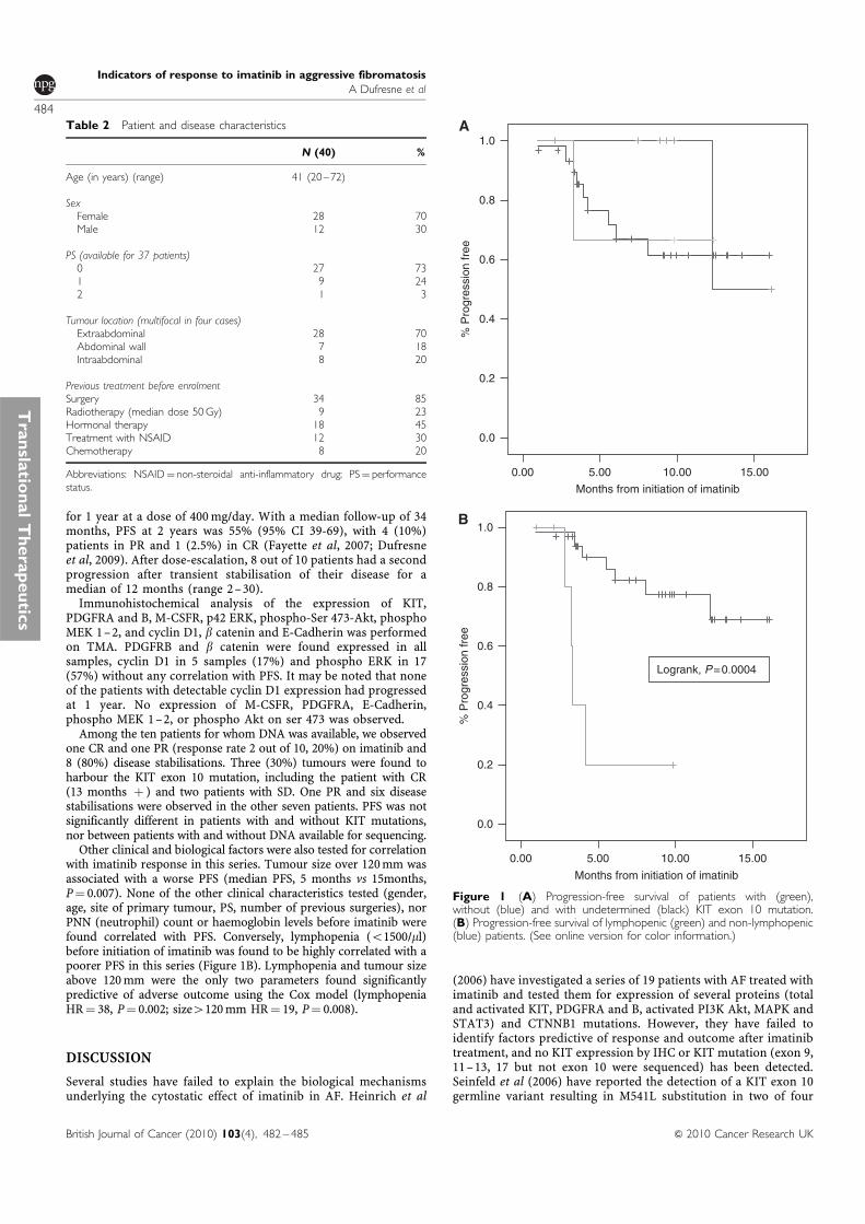
for 1 year at a dose of 400mg/day. With a median follow-up of 34months, PFS at 2 years was 55% (95% CI 39-69), with 4 (10%)patients in PR and 1 (2.5%) in CR (Fayette et al, 2007; Dufresneet al, 2009). After dose-escalation, 8 out of 10 patients had a secondprogression after transient stabilisation of their disease for amedian of 12 months (range 2–30).Immunohistochemical analysis of the expression of KIT,
PDGFRA and B, M-CSFR, p42 ERK, phospho-Ser 473-Akt, phosphoMEK 1–2, and cyclin D1, b catenin and E-Cadherin was performedon TMA. PDGFRB and b catenin were found expressed in allsamples, cyclin D1 in 5 samples (17%) and phospho ERK in 17(57%) without any correlation with PFS. It may be noted that noneof the patients with detectable cyclin D1 expression had progressedat 1 year. No expression of M-CSFR, PDGFRA, E-Cadherin,phospho MEK 1–2, or phospho Akt on ser 473 was observed.Among the ten patients for whom DNA was available, we observed
one CR and one PR (response rate 2 out of 10, 20%) on imatinib and8 (80%) disease stabilisations. Three (30%) tumours were found toharbour the KIT exon 10 mutation, including the patient with CR(13 months þ ) and two patients with SD. One PR and six diseasestabilisations were observed in the other seven patients. PFS was notsignificantly different in patients with and without KIT mutations,nor between patients with and without DNA available for sequencing.Other clinical and biological factors were also tested for correlation
with imatinib response in this series. Tumour size over 120mm wasassociated with a worse PFS (median PFS, 5 months vs 15months,P¼ 0.007). None of the other clinical characteristics tested (gender,age, site of primary tumour, PS, number of previous surgeries), norPNN (neutrophil) count or haemoglobin levels before imatinib werefound correlated with PFS. Conversely, lymphopenia (o1500/ml)before initiation of imatinib was found to be highly correlated with apoorer PFS in this series (Figure 1B). Lymphopenia and tumour sizeabove 120mm were the only two parameters found significantlypredictive of adverse outcome using the Cox model (lymphopeniaHR¼ 38, P¼ 0.002; size4120mm HR¼ 19, P¼ 0.008).
DISCUSSION
Several studies have failed to explain the biological mechanismsunderlying the cytostatic effect of imatinib in AF. Heinrich et al
(2006) have investigated a series of 19 patients with AF treated withimatinib and tested them for expression of several proteins (totaland activated KIT, PDGFRA and B, activated PI3K Akt, MAPK andSTAT3) and CTNNB1 mutations. However, they have failed toidentify factors predictive of response and outcome after imatinibtreatment, and no KIT expression by IHC or KIT mutation (exon 9,11–13, 17 but not exon 10 were sequenced) has been detected.Seinfeld et al (2006) have reported the detection of a KIT exon 10germline variant resulting in M541L substitution in two of four
Months from initiation of imatinib
0.0
0.2
0.4
0.6
0.8
1.0
% P
rog
ressio
n f
ree
Months from initiation of imatinib
0.0
0.2
0.4
0.6
0.8
1.0
% P
rog
ressio
n f
ree
Logrank, P=0.0004
0.00 15.0010.005.00
0.00 15.0010.005.00
Figure 1 (A) Progression-free survival of patients with (green),without (blue) and with undetermined (black) KIT exon 10 mutation.(B) Progression-free survival of lymphopenic (green) and non-lymphopenic(blue) patients. (See online version for color information.)
Table 2 Patient and disease characteristics
N (40) %
Age (in years) (range) 41 (20–72)
Sex
Female 28 70
Male 12 30
PS (available for 37 patients)
0 27 73
1 9 24
2 1 3
Tumour location (multifocal in four cases)
Extraabdominal 28 70
Abdominal wall 7 18
Intraabdominal 8 20
Previous treatment before enrolment
Surgery 34 85
Radiotherapy (median dose 50Gy) 9 23
Hormonal therapy 18 45
Treatment with NSAID 12 30
Chemotherapy 8 20
Abbreviations: NSAID¼ non-steroidal anti-inflammatory drug; PS¼ performance
status.
Indicators of response to imatinib in aggressive fibromatosis
A Dufresne et al
484
British Journal of Cancer (2010) 103(4), 482 – 485 & 2010 Cancer Research UK
Transla
tionalTherapeutic
s

extraabdominal AF samples. Some of us have later reported on apatient with AF who responded to imatinib treatment andpresented with the same KIT exon 10 variant (Goncalves et al,2006). This is the reason why we searched for M541L exon 10variant in our series of patients treated with imatinib.To our knowledge, this is the largest series investigating pre-
imatinib tumour- and host-related molecular and clinical factors,including KIT exon 10 mutations. Even though only a limitednumber of cases could be investigated, response rates and PFSwere not found significantly different in patients with and withoutthe M541L KIT allele. It may be noted though, all three patientswith M541L allele achieved tumour control with imatinib,including one patient with a CR. Even if not statistically confirmed,the results of the present study strongly support the clinicalimpression that AF patients harbouring the M541L variant aremore sensitive to imatinib. The lack of statistical correlationbetween the presence of the M541L variant and response rate orsurvival is probably because of the low number of patients tested.This observation is of major interest and should be confirmed ina larger series. Moreover, the biological rationale for such amechanism remains to be explored.By examining cell models transfected with the exon 10 M541L
variant, Tamborini et al (2006) and Bertucci et al (2007) havefailed to show that the substitution could result in KIT activationor inactivation and actually corresponded to a KIT polymorphism.Both groups have concluded that the sensitivity of AF to imatinibrequires an alternative explanation and possibly involves anautocrine mechanism, possibly associated with a hypersensitivityto SCF, related to the induction of a ligand-independentdimerisation induced by the exon 10 variant. The lack ofcorrelation between expression on IHC and outcome is fullyconsistent with the previous report by Heinrich et al. Interestingly,Tabone-Eglinger et al (2008) have shown that GIST-type KIT
mutations induce an activation-dependent alteration of normalmaturation and trafficking, resulting in the intracellular retentionof the activated kinase within the cell. Imatinib-induced inhibitionof the phosphorylation of immature and mature mutant KITproteins has resulted in the restoration of KIT expression at thecell surface. They conclude that these observations likely accountfor the absence of correlation between response to imatinib andKIT expression using IHC and may deserve to be investigated inother tyrosine kinase-activated tumours. More recently, anotherKIT exon 10 mutation possibly linked to imatinib response in AFhas been identified, V530I (Kurtz et al, 2010).The only biological parameter correlated with PFS in our
patients was pre-treatment lymphopenia, whereas anaemia andPNN count had no predictive value (Figure 1A). Of note,lymphopenia was not found correlated to PS in this series.Imatinib has been previously reported to exert an anti-tumouractivity in animal models through the modulation of immuneresponse (Borg et al, 2004).The present observation shows that a baseline biological
characteristic of the host, not of the tumour, is the majorparameter influencing response to imatinib in aggressive fibro-matosis. Tumour characteristics, including the presence of the KITexon 10 M541L variant, may have influenced tumour control inthis small series but this needs to be confirmed and betterexplained.
ACKNOWLEDGEMENTS
Supported by grants from the INCa, the Comites du Rhone et del’Ain de la Ligue contre le Cancer, and the Conticanet Network ofExcellence (FP6-018806).
REFERENCES
Bertucci F, Goncalves A, Viens P, Monges G, Dubreuil P (2007) Desmoid-type fibromatosis. J Neurosurg 107: 473–475
Borg C, Terme M, Taieb J, Menard C, Flament C, Robert C, Maruyama K,Wakasugi H, Angevin E, Thielemans K, Le Cesne A, Chung-Scott V, LazarV, Tchou I, Crepineau F, Lemoine F, Bernard J, Fletcher JA, Turhan A,Blay JY, Spatz A, Emile JF, heinrich MC, Mecheri S, Tursz T, Zitvogel L(2004) Novel mode of action of c-kit tyrosine kinase inhibitors leadingto NK cell-dependent antitumor effects. J Clin Invest 114(3): 379–388
Dufresne A, Penel N, Salas S, Le Cesne A, Perol D, Bui B, Brain E,Ray-Coquard I, Jimenez M, Blay JY (2009) Updated outcome with long-term follow-up of imatinib for the treatment of progressive or recurrentaggressive fibromatosis: a FNCLCC/French Sarcoma Group phase II trial.ASCO Abstract #10518
Enzinger FM, Weiss SW (2001) Fibromatosis. In Enzinger and Weiss’s SoftTissue Tumors Sharon Weiss, John R. Goldblum (eds). 4th edn, Mosby:St-Louis, MO
Fayette J, Dufresne A, Penel N, Le Cesne A, Bui B, Tubiana-Hulin M,Guillemet C, Chabaud S, Jimenez M, Blay JY (2007) Imatinib for thetreatment of aggressive fibromatosis/desmoid tumors (AF/DT) failinglocal treatment: updated outcome and predictive factors for progressionfree survival. A FNCLCC French Sarcoma Group-GETO study. ASCOAbstract # 10062
Fiore M, Rimareix F, Mariani L, Domont J, Collini P, Le Pechoux C, CasaliPG, Le Cesne A, Gronchi A, Bonvalot S (2009) Desmoid-typefibromatosis: a front-line conservative approach to select patients forsurgical treatment. Ann Surg Oncol 16: 2587–2593
Goncalves A, Monges G, Yang Y, Palmerini F, Dubreuil P, Noguchi T,Jacquemier J, Di Stefano D, Delpero JR, Sobol H, Bertucci F (2006)Response of a KIT-positive extra-abdominal fibromatosis to imatinibmesylate and KIT genetic analysis. J Nat Cancer Inst 98: 562–563
Heinrich MC, McArthur GA, Demetri GD, Joensuu H, Bono P, Herrmann R,Hirte H, Cresta S, Koslin DB, Corless CL, Dirnhofer S, Van Oosterom AT,Nikolova Z, Dimitrijevic S, Fletcher JA (2006) Clinical and molecularstudies of the effect of imatinib on advanced aggressive fibromatosis(desmoid tumor). J Clin Oncol 24: 1195–1203
Janinis J, Patriki M, Vini L, Aravantinos G, Whelan JS (2003) Thepharmacological treatment of aggressive fibromatosis: a systematicreview. Ann Oncol 14: 181–190
Kurtz JE, Asmane I, Voegeli AC, Neuville A, Dufresne A, Litique V,Chevreau C, Bergerat JP (2010) A V530I Mutation in c-KIT Exon 10 IsAssociated to Imatinib Response in Extraabdominal Aggressive Fibro-matosis. Sarcoma 2010: 458156
Mace J, Sybil Biermann J, Sondak V, McGinn C, Hayes C, Thomas D,Baker L (2002) Response of extra-abdominal desmoid tumors to therapywith imatinib mesylate. Cancer 95: 2373–2379
Nuyttens JJ, Ryst PF, Thomas CR, Turrisi AT (2000) Surgery vsradiotherapy for patients with aggressive fibromatosis or desmoidtumors. A comparative review of 22 articles. Cancer 88: 1517–1523
Seinfeld J, Kleinschmidt-Demasters BK, Tayal S, Lillehei KO (2006)Desmoid-type fibromatoses involving the plexus brachial: treatmentoptions and assessment of c-kit mutational status. J Neurosurg 104(5):749–756
Tabone-Eglinger S, Subra F, El Sayadi H, Alberti L, Tabone E, Michot JP,Theou-Anton N, Lemoine A, Blay JY, Emile JF (2008) KIT mutationsinduce intracellular retention and activation of an immature form of theKIT protein in gastrointestinal stromal tumors. Clin Cancer Res 14(8):2285–2294
Tamborini E, Negri T, Miselli F, Lagonigro MS, Pricl S, Pilotti S (2006)Re: response of a KIT-positive extra-abdominal fibromatosis to imatinibmesylate and KIT genetic analysis. J Natl Cancer Inst 98: 1583–1584
Indicators of response to imatinib in aggressive fibromatosis
A Dufresne et al
485
British Journal of Cancer (2010) 103(4), 482 – 485& 2010 Cancer Research UK
TranslationalTherapeutics

Hindawi Publishing CorporationSarcomaVolume 2010, Article ID 458156, 4 pagesdoi:10.1155/2010/458156
Case Report
A V530I Mutation in c-KIT Exon 10 Is Associated to ImatinibResponse in Extraabdominal Aggressive Fibromatosis
Jean-Emmanuel Kurtz,1 Irene Asmane,1 Anne-Claire Voegeli,2 Agnes Neuville,3
Armelle Dufresne,4 Valere Litique,1 Christine Chevreau,5 and Jean-Pierre Bergerat1
1 Pole d’Hematologie et d’Oncologie, Hopitaux Universitaires de Strasbourg, 67098 Strasbourg, France2 Pole de Biologie, Hopitaux Universitaires de Strasbourg, 67098 Strasbourg, France3 Service d’Anatomie pathologique, Hopitaux Universitaires de Strasbourg, 67098 Strasbourg, France4 Service d’Oncologie Medicale, Centre Hospitalier de Chambery, 73011, France5 Service d’Oncologie Medicale, Centre Hospitalier Universitaire, 31052 Toulouse, France
Correspondence should be addressed to Jean-Emmanuel Kurtz, [email protected]
Received 5 October 2009; Revised 4 January 2010; Accepted 25 January 2010
Academic Editor: Irene Andrulis
Copyright © 2010 Jean-Emmanuel Kurtz et al. This is an open access article distributed under the Creative Commons AttributionLicense, which permits unrestricted use, distribution, and reproduction in any medium, provided the original work is properlycited.
Aggressive fibromatosis (AF) or desmoid tumor is a rare condition, characterized by deep tissue invasion by a monoclonalfibroblastic neoplasm, developed from musculoaponeurotic structures. Surgery is the treatment of choice, but negative marginscan hardly been achieved in large tumors, and can lead to major functional disability. AF medical therapy includes nonsteroids anti-inflammatory drugs, tamoxifen, with inconsistent results. Several reports of imatinib efficacy in AF appear in the literature. Here,we describe for the first time a V530I KIT exon 10 mutant that was associated to a dramatic imatinib response in an extraabdominalaggressive fibromatosis. The previously discovered V530I substitution was characterized in the core binding factor AML, but hadnever been reported in any other condition, so far. In this paper, we discuss the KIT exon 10 mutations or polymorphisms thathave been described in a variety of KIT-related conditions, including acute myelogenous leukemia, mastocytosis, and aggressivefibromatosis.
1. Case Report
A 22-year-old Caucasian female without any particularmedical history suffered from a traumatic fracture of theleft humerus great tuberosity in 2002. She underwent anon-operative treatment with a Mayo-Clinic splint and wasdischarged from the hospital. Radiologic outcome was unre-markable, though she complained with persistent shoulderpain. Three years later, a painful mass of the shoulder rapidlyappeared, impairing shoulder mobility. She was referred toour institution. Clinical examination revealed a hard, bulkymass of the shoulder, with collateral circulation. The tumorinvolved the whole curving contour of the shoulder, andreached the axilla at its posterior limit. Mobilization of thearm was almost impossible due to both pain and stiffness(Figure 1(a)). A shoulder MRI revealed a 12× 6 cm irregular,heterogeneous mass of the posterior part of the shoulder
(Figure 1(b)). The tumor invaded subcutaneous tissue aswell as the deltoid and infrasupinatous muscles. Necroticareas diagnosed as T1-hyposignal were found, as well as othertumor areas that were strongly enhanced by contrast injec-tion. There was no apparent bone destruction. A surgicalbiopsy was performed, diagnosing aggressive fibromatosis(extraabdominal desmoids tumor).
Due to the tumor burden, curative surgery could notbe performed, and a medical therapy consisting in imatinibmesylate (Glivec) was started at a daily dose of 400 mg. After4 weeks of treatment, the patient noticed an improvementin the abduction of the arm as well as a slight tumor sizedecrease. The treatment was continued at the same dosing,with consistent improvement in mobility, tumor measure-ments and analgesics consumption. There was no significantside-effects. The treatment was finally stopped at month34, in the setting of tumor regression, complete recovery
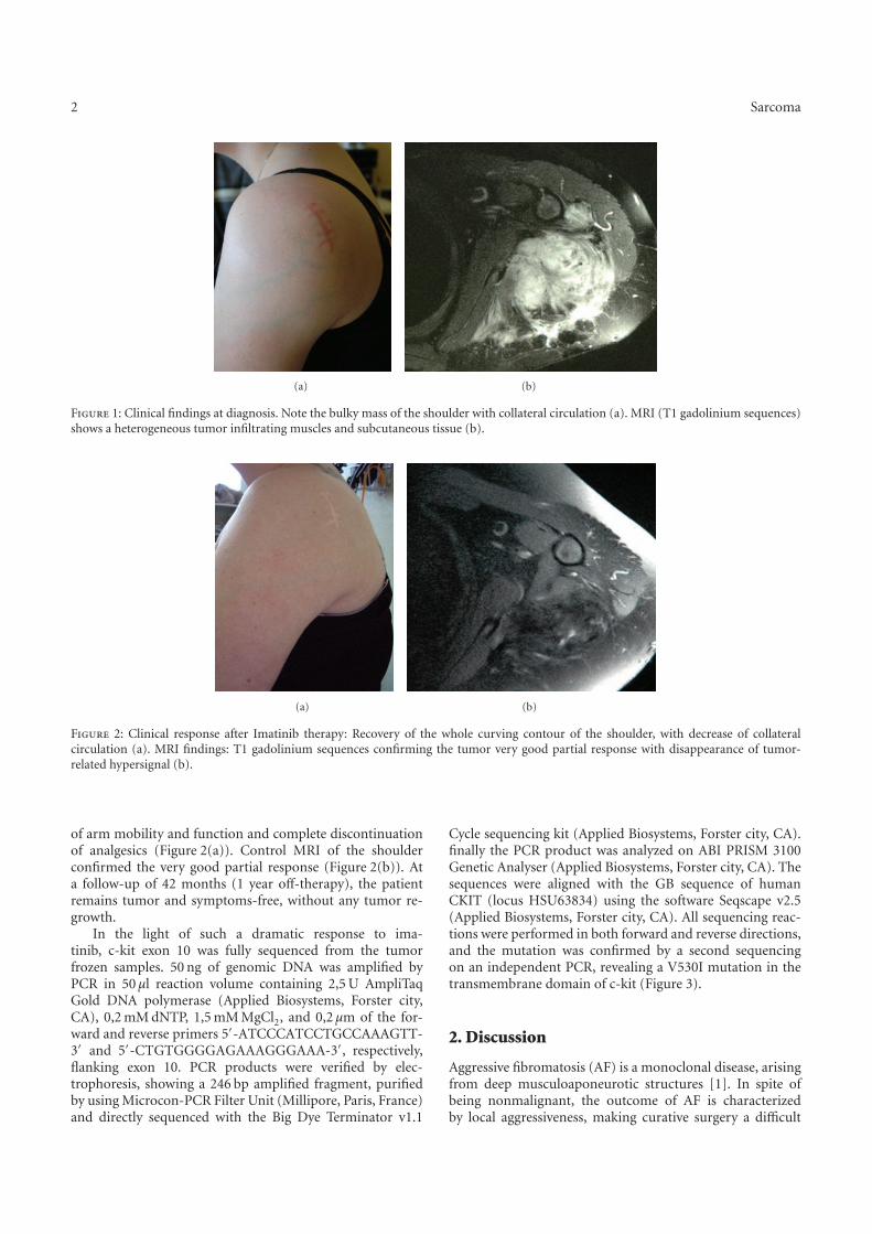
2 Sarcoma
(a) (b)
Figure 1: Clinical findings at diagnosis. Note the bulky mass of the shoulder with collateral circulation (a). MRI (T1 gadolinium sequences)shows a heterogeneous tumor infiltrating muscles and subcutaneous tissue (b).
(a) (b)
Figure 2: Clinical response after Imatinib therapy: Recovery of the whole curving contour of the shoulder, with decrease of collateralcirculation (a). MRI findings: T1 gadolinium sequences confirming the tumor very good partial response with disappearance of tumor-related hypersignal (b).
of arm mobility and function and complete discontinuationof analgesics (Figure 2(a)). Control MRI of the shoulderconfirmed the very good partial response (Figure 2(b)). Ata follow-up of 42 months (1 year off-therapy), the patientremains tumor and symptoms-free, without any tumor re-growth.
In the light of such a dramatic response to ima-tinib, c-kit exon 10 was fully sequenced from the tumorfrozen samples. 50 ng of genomic DNA was amplified byPCR in 50 µl reaction volume containing 2,5 U AmpliTaqGold DNA polymerase (Applied Biosystems, Forster city,CA), 0,2 mM dNTP, 1,5 mM MgCl2, and 0,2 µm of the for-ward and reverse primers 5′-ATCCCATCCTGCCAAAGTT-3′ and 5′-CTGTGGGGAGAAAGGGAAA-3′, respectively,flanking exon 10. PCR products were verified by elec-trophoresis, showing a 246 bp amplified fragment, purifiedby using Microcon-PCR Filter Unit (Millipore, Paris, France)and directly sequenced with the Big Dye Terminator v1.1
Cycle sequencing kit (Applied Biosystems, Forster city, CA).finally the PCR product was analyzed on ABI PRISM 3100Genetic Analyser (Applied Biosystems, Forster city, CA). Thesequences were aligned with the GB sequence of humanCKIT (locus HSU63834) using the software Seqscape v2.5(Applied Biosystems, Forster city, CA). All sequencing reac-tions were performed in both forward and reverse directions,and the mutation was confirmed by a second sequencingon an independent PCR, revealing a V530I mutation in thetransmembrane domain of c-kit (Figure 3).
2. Discussion
Aggressive fibromatosis (AF) is a monoclonal disease, arisingfrom deep musculoaponeurotic structures [1]. In spite ofbeing nonmalignant, the outcome of AF is characterizedby local aggressiveness, making curative surgery a difficult

Sarcoma 3
G RCTTT T AA T C
123
Figure 3: KIT exon 10 point mutation 1609 G→A (resulting in theamino-acid substitution V530I).
challenge, as patients are exposed to local recurrence. Theclinical outcome of AF is unpredictable, and many treatmentoptions have been considered, including surgery, radiationtherapy and medical therapy. Systemic medical therapy hasfocused on NSAIDs such as Sulindac and anti-Cox2 as wellas hormonal therapy (tamoxifen) although AF inconsistentlyexpress estrogen receptors [2].
The KIT gene encodes the stem cell growth factorreceptor, a type III transmembrane receptor tyrosine kinasethat has been involved in the pathogenesis of various con-ditions, including gastro-intestinal stromal tumors (GISTs),mastocytosis, acute myelogenous leukemia (AML) as wellas piebaldism, a rare autosomal inherited skin disorder.The tyrosine kinase inhibitor imatinib mesylate (Glivec)has become a cornerstone of advanced GIST, in which KITexon 11 mutations are associated with imatinib efficacy, asopposed to these that are present in exon 9 [3]. However, KITexon 10 mutations have not been reported so far in GISTs, asopposed to deletions at the intron10-exon11 boundary thatmay impact response to imatinib [4].
Imatinib has been incorporated in the therapeutic arma-mentarium of AF, in the light of several case reports of long-lasting responses [5–8]. However, all AF do not respond toimatinib treatment, as shown in a retrospective analysis of 19cases of heavily pretreated patients in whom the response ratewas only 15.7% (3/19 PR) [8]. Interestingly, it is unlikely thatpositive KIT staining would predict imatinib sensitivity as inthis series, most of the cases were KIT negative. Moleculardeterminants of tumor response to tyrosine kinase inhibitorsare well known in gastro-intestinal stromal tumors, whereKIT mutations are predictive for imatinib efficacy [3]. Inthe retrospective series reported by Heinrich et al., no KITmutation was however found, even in the three respondersto imatinib [8]. This data contrasts with previous reportsof KIT exon 10 point mutation 1621 A→C (resultingin the amino-acid substitution M541L) that could predictimatinib sensitivity [6]. Similar findings were reported in aFrench Sarcoma Group study, where 3 out of 10 patientsexperiencing tumor control with imatinib actually had anM541L mutation [9]. Moreover, Seinfeld et al. reported theoccurrence of a limited clinical response, again in a M541L
patient [10]. However, contradictory data also appear inthe literature. In the light of experiments conducted byTamborini et al., as well as Bertucci et al., it was hypothesizedthat the exon 10 M541L variant is not activating neitherinactivating and is actually a KIT polymorphism [11, 12].Consistent findings were reported by Kruger et al., whofound that the allele frequency of the KIT variant M541Lwas as high as 8.1% in the healthy population, not differingfrom chronic myelogenous patients (8.3%) [13]. The V530Ivariant has to our knowledge, never been described so far, ineither AF or other KIT-related diseases such as mastocytosis,GISTs or piebaldism, [14, 15]. The exon 10 V530I substi-tution alters the transmembrane domain of KIT (containedbetween amino acids 521–543), and was described in acutemyelogenous leukemia [16, 17]. Similar and rare (<5%)exon 10 KIT-activating transmembrane mutations such asA533D or F522C have been reported in mastocytosis [15].KIT deletions or insertions appear in 7 to 17% of AMLcases (raising to 33% in the setting of chromosome 16inversion), affecting members of the core binding factor(CBF) gene family [16–18]. In a series of 103 AML patients,those harboring a KIT mutation had a shorter event-free andrelapse-free survival [18]. There is little data with regard toimatinib therapy in AML patients with CBF KIT mutations.In a series of 3 patients, imatinib had transient activityin a patient with exon 8 in-frame deletion, but none inthe 2 patients carrying D816Y and D816V mutations [19].In a series of published by Cortes et al., 18 patients withrecurrent or refractory AML or myelodysplastic syndromewere challenged with imatinib, with very transient results[20]. Unfortunately, no KIT analysis was performed in thisstudy. A trial investigating dasatinib, another tyrosine kinaseinhibitor is currently recruiting AML patients harboringCBF mutations in France. Interestingly, V530I variantswere associated, in this condition with a greater imatinibsensitivity, as the IC50 was lower than the wild-type KITby a 5-fold factor. Moreover, inhibition of V530I variantsresulted in both proliferation abrogation and increasedapoptosis in vitro [16]. We have not investigated whether ourpatient exhibited a germline or a somatic mutation as tissuesampling consisted in tumor biopsy only, and no consent wasobtained to test for any germline mutation in our patientwho lacked significant familial history of cancer. There is,moreover, no data in the literature supporting the hypothesisthat the V530I KIT (as opposed to the aforementionedM541L) mutation could be a polymorphism rather than arare, highly penetrating alteration.
Our patient exhibited a typical medical history oftrauma, preceding the onset of AF by an interval of threeyears, a delay consistent with literature data [2]. There is littledata in the literature regarding imatinib treatment durationin AF. Of note, in our case, evidence for treatment efficacywas present as soon as 4 weeks after treatment started.Moreover, imatinib was administered for 3 years as the tumorsize constantly decreased during this period. However, andas opposed to the report by Wcislo et al., imatinib wasthen stopped without subsequent local relapse [7]. In otherreports, the duration of imatinib therapy varied between 34weeks and 16 months [5, 6, 10].
4 Sarcoma
Based on the literature data, as well as this report, itappears mandatory that AF patients be screened not only forM541L substitution, which physiopathology remains at theleast, controversial, but should undergo systematic exon 10sequencing to detect transmembrane variants, such as V530I.Hence, the presence of such a mutation could support front-line imatinib therapy, rather than more classic strategies,including tamoxifen or NSAIDs. Dramatic response to ima-tinib is clearly associated with this substitution, and providedsuch a mutation has been detected, not only imatinib can beconsidered, but also other TK inhibitors, such as sunitinibor others [21]. Whether the V530I variant might successfullybe challenged with tyrosine kinase inhibitors other thanimatinib is unknown. Growney et al. tried to assess theefficacy of PKC412, a novel tyrosine kinase inhibitor in avariety of KIT mutants, including V530I, but this variantcould not transform the Ba/F3 cells in their experiment[21]. Interestingly, reports of sunitinib efficacy in imatinib-resistant AF patients appear in the literature, suggesting thatas in GISTs, treatment strategies with “rescue” alternate TKinhibitors can be considered in selected patients [22].
References
[1] B. A. Alman, M. E. Pajerski, S. Diaz-Cano, K. Corboy, andH. J. Wolfe, “Aggressive fibromatosis (desmoid tumor) is amonoclonal disorder,” Diagnostic Molecular Pathology, vol. 6,no. 2, pp. 98–101, 1997.
[2] E. Stoeckle, J. M. Coindre, M. Longy, et al., “A critical analysisof treatment strategies in desmoid tumours: a review of a seriesof 106 cases,” European Journal of Surgical Oncology, vol. 35,no. 2, pp. 129–134, 2009.
[3] F. Duffaud and A. Le Cesne, “Imatinib in the treatment of solidtumours,” Targeted Oncology, vol. 4, no. 1, pp. 45–56, 2009.
[4] C. L. Corless, L. McGreevey, A. Town, et al., “KIT genedeletions at the intron 10-exon 11 boundary in GI stromaltumors,” Journal of Molecular Diagnostics, vol. 6, no. 4, pp.366–370, 2004.
[5] J. Mace, J. Sybil Biermann, V. Sondak, et al., “Response ofextraabdominal desmoid tumors to therapy with imatinibmesylate,” Cancer, vol. 95, no. 11, pp. 2373–2379, 2002.
[6] A. Goncalves, G. Monges, Y. Yang, et al., “Response of a KIT-positive extra-abdominal fibromatosis to imatinib mesylateand KIT genetic analysis,” Journal of the National CancerInstitute, vol. 98, no. 8, pp. 562–563, 2006.
[7] G. Wcislo, K. Szarlej-Wcislo, and C. Szczylik, “Control ofaggressive fibromatosis by treatment with imatinib mesylate.A case report and review of the literature,” Journal of CancerResearch and Clinical Oncology, vol. 133, no. 8, pp. 533–538,2007.
[8] M. C. Heinrich, G. A. McArthur, G. D. Demetri, et al.,“Clinical and molecular studies of the effect of imatinib onadvanced aggressive fibromatosis (desmoid tumor),” Journalof Clinical Oncology, vol. 24, no. 7, pp. 1195–1203, 2006.
[9] J. Fayette, A. Dufresne, N. Penel, et al., “Imatinib for the treat-ment of aggressive fibromatosis/desmoid tumors (AF/DT)failing local treatment: updated outcome and predictive fac-tors for progression free survival. A FNCLCC French SarcomaGroup-GETO study,” Journal of Clinical Oncology, vol. 25, no.18, supplement, 2007, abstract no. 10062.
[10] J. Seinfeld, B. K. Kleinschmidt-Demasters, S. Tayal, and K. O.Lillehei, “Desmoid-type fibromatoses involving the brachialplexus: treatment options and assessment of c-KIT mutationalstatus,” Journal of Neurosurgery, vol. 104, no. 5, pp. 749–756,2006.
[11] E. Tamborini, T. Negri, F. Miselli, M. S. Lagonigro, S. Pricl,and S. Pilotti, “Re: response of a KIT-positive extra-abdominalfibromatosis to imatinib mesylate and KIT genetic analysis,”Journal of the National Cancer Institute, vol. 98, no. 21, pp.1583–1584, 2006.
[12] F. Bertucci, A. Goncalves, P. Viens, G. Monges, and P.Dubreuil, “Desmoid-type fibromatosis,” Journal of Neuro-surgery, vol. 107, no. 2, pp. 473–475; author reply 475, 2007.
[13] S. Kruger, M. Emig, P. Lohse, G. Ehninger, A. Hochhaus,and H. K. Schackert, “The c-kit (CD117) sequence variationM541L, but not N564K, is frequent in the general population,and is not associated with CML in Caucasians,” Leukemia, vol.20, no. 2, pp. 354–355, 2006.
[14] X.-Y. Yin, Y.-Q. Ren, S. Yang, et al., “A novel KIT missensemutation in one Chinese family with piebaldism,” Archives ofDermatological Research, vol. 301, no. 5, pp. 387–389, 2009.
[15] A. Orfao, A. C. Garcia-Montero, L. Sanchez, and L. Escribano,“Recent advances in the understanding of mastocytosis: therole of KIT mutations,” British Journal of Haematology, vol.138, no. 1, pp. 12–30, 2007.
[16] J. Cammenga, S. Horn, U. Bergholz, et al., “ExtracellularKIT receptor mutants, commonly found in core bindingfactor AML, are constitutively active and respond to imatinibmesylate,” Blood, vol. 106, no. 12, pp. 3958–3961, 2005.
[17] M. Gari, A. Goodeve, G. Wilson, et al., “c-kit proto-oncogeneexon 8 in-frame deletion plus insertion mutations in acutemyeloid leukaemia,” British Journal of Haematology, vol. 105,no. 4, pp. 894–900, 1999.
[18] N. Boissel, H. Leroy, B. Brethon, et al., “Incidence andprognostic impact of c-Kit, FLT3, and Ras gene mutationsin core binding factor acute myeloid leukemia (CBF-AML),”Leukemia, vol. 20, no. 6, pp. 965–970, 2006.
[19] R. Cairoli, A. Beghini, E. Morello, et al., “Imatinib mesylatein the treatment of Core Binding Factor leukemia’s with KITmutations: a report of three cases,” Leukemia Research, vol. 29,no. 4, pp. 397–400, 2005.
[20] J. Cortes, F. Giles, S. O’Brien, et al., “Results of imatinibmesylate therapy in patients with refractory or recurrent acutemyeloid leukemia, high-risk myelodysplastic syndrome, andmyeloproliferative disorders,” Cancer, vol. 97, no. 11, pp.2760–2766, 2003.
[21] J. D. Growney, J. J. Clark, J. Adelsperger, et al., “Activationmutations of human c-KIT resistant to imatinib mesylate aresensitive to the tyrosine kinase inhibitor PKC412,” Blood, vol.106, no. 2, pp. 721–724, 2005.
[22] K. M. Skubitz, J. C. Manivel, D. R. Clohisy, and J. W. Frolich,“Response of imatinib-resistant extra-abdominal aggressivefibromatosis to sunitinib: case report and review of theliterature on response to tyrosine kinase inhibitors,” CancerChemotherapy and Pharmacology, vol. 64, no. 3, pp. 635–640,2009.

Impact of KIT exon 10 M541L allelic variant on the response to imatinib in aggressive
fibromatosis: analysis of the desminib series by competitive allele specific Taqman PCR
technology
Armelle Dufresne1,2
, Laurent Alberti1, Mehdi Brahmi
1, Héloïse Philippon
1, David Pérol
3, Jean
Yves Blay1,2
1. Cancer Research Center of Lyon, INSERM UMR 1052, CNRS UMR 5286; 2. Medical
Oncology Department; 3. Biostatistics unit
Anticancer Center Leon Berard, Lyon, France
This study was funded by grant from the Institut National du Cancer (INCa)
Corresponding author:
Dr Armelle Dufresne, MD, Cancer Research Center of Lyon, INSERM UMR 1052, CNRS
UMR 5286; Centre Leon Berard, 28 rue Laënnec, Lyon, France; e-mail:
[email protected]; phone: + 33 4 78 78 28 28, fax: + 33 4 78 78 27 20

2
Abstract
Aggressive fibromatosis (AF) is a rare fibroblastic proliferative disease with a locally
aggressive behaviour and no distant metastasis, characterized by driver mutations in CTNNB1
or in APC gene. When progressive and/or symptomatic AF is not amenable to local
management, a variety of medical treatments may be efficient, including imatinib mesylate.
The phase II “Desminib trial” included 40 patients with AF to evaluate the toxicity and
efficacy of imatinib and conclude to a 65% tumor control rate at 1 year. We investigated a
potential predictive value of KIT exon 10 M541L variant (KITL541
) on this prospective series.
DNA was extracted in sufficient quantity for 33 patients included in the Desminib trial. The
detection of KITL541
was performed by Competitive Allele-Specific Taqman® PCR
technology. KIT allele status was analyzed regarding clinical presentation and patient
outcome. In 6 of 33 cases (18%), the technique failed to conclude to the mutational status; 5
patients (19%) harboured KITL541
and 22 patients (81%) were classified as KIT wild type.
Compared with total cohort, KITL541
was observed at similar frequencies according to clinical
characteristics. In the KITL541
and the KITWT
subgroups, the tumor control rate at 1 year was
100% and 68%, respectively (p = 0,316). The median PFS of patients harbouring KITL541
or
not is 29,9 and 24,5 months, respectively (p = 0,616), and the median overall survival is not
reached, in any of the groups. Our results do not support a predictive effect of KITL541
in the
efficacy of imatinib for patients with AF.

3
Introduction
Aggressive fibromatosis (AF) is a rare fibroblastic proliferative disease characterized by
driver mutations in CTNNB1, at specific sites of the exon 3, or in the APC gene (in the context
of Gardner syndrome). The management of aggressive fibromatosis has substantially evolved
in the last 10 years [1]. AF are characterized by an aggressive local behaviour, yet
unpredictable, with a risk of relapse after surgical excision but a lack of distant metastasis.
These tumors are characterized by heterogeneity in their clinical presentation with an
unpredictable clinical course. The classical strategy of aggressive front-line therapy with
surgery and radiotherapy is now debated and a wait-and-see policy at initial presentation is
now often proposed (NCCN 2012 Guidelines) [2]. Systemic treatments such as non steroid
anti inflammatory drugs (NSAIDs), hormonal treatment, cytotoxic chemotherapy, imatinib, or
sorafenib are often used to control tumor growth and/or to relieve symptomatic AF, all with
moderate and variable efficacy [3-8]. This observation raises the need to identify biomarkers,
to effectively select patients who benefit from a treatment.
In 2 prospective series of patients treated with imatinib, progression free survival (PFS) was
66% and 67% at 1 year [7, 8]. The phase II “Desminib trial” included 40 patients to evaluate
the toxicity and efficacy of imatinib mesylate administered to patients with AF not amenable
to radiotherapy or non-mutilating surgery. The results showed a disease control by imatinib in
a large proportion of patients with 4 (10%) complete or partial confirmed responses and 28
(70%) stable disease as best response, leading to a 1 year PFS of 67% [7].
KIT is one of the major target of imatinib; mutations of KIT predicts for the efficacy of the
drug in GIST [9], but also in melanoma and thymic carcinoma [10, 11]. Several cases reports
have suggested a potential role of the KIT exon 10 M541L variant (KITL541
) in sensitivity of
AF to imatinib [12, 13]. The present study was conducted on Desminib series to search for a
potential predictive value of KITL541
.
4
Material and methods
Patients
This study was performed as a retrospective translational research program on tumor samples
of patients included in Desminib trial [7]. Forty patients with progressive or recurrent AF that
could not be treated with curative surgery or radiotherapy were included in Desminib phase
I/II trial to evaluate the efficacy and toxicity of imatinib. Patients with adequate end organ
function were treated with 400mg of imatinib daily, increasing to 800mg in case of
progressive disease. Best clinical response to imatinib was defined according to RECIST
criteria. Evaluations were performed every 3 months. All evaluations of tumor responses to
imatinib were reviewed by a radiological independent validation committee. Informed consent
was obtained from each patient to enroll them in the study and collect archival pathology
specimens.
Tissue samples
The analysis of present study was performed on the initial tumor of patients, obtained by
biopsy or surgical excision to establish the diagnosis of the disease. Paraffin-embedded tissues
samples of patients included in the study were obtained from pathology centers, all from
tumors at initial diagnosis.
DNA extraction
Total DNA was extracted from tumor using QIAamp DNA kit N° 56404 (Qiagen, France)
according to the manufacturer’s instructions and quantified by spectrophotometry (NanoDrop
ND-100 instrument, Thermo Fisher Scientific, Waltham, MA). Briefly, FFPE tumours were
lysed for 24h in ATL buffer supplemented with proteinase K at 60°C in rotative agitation
5
after different washes with toluene and ethanol, in this order. Genomic DNA was isolated
with a QIAamp MiniElute column.
Competitive Allele-Specific Taqman®
PCR (CAST-PCR)
The detection of KIT541
status was performed by Competitive Allele-Specific Taqman®
PCR
technology provided by Applied Biosystems®
(Fig. 1). Each mutant allele assay detects
specific mutant alleles. Each assay contains: an allele-specific primer that detects the mutant
allele, a MGB oligonucleotide blocker that suppresses the wild type allele, a locus specific
primer and a locus specific TaqMan® FAM™ dye-labeled MGB probe. Gene reference assays
detect the genes that the target mutations reside in. They are designed to amplify a mutation-
free and polymorphism-free region of the target gene. Each assay contains: a locus-specific
pair of forward and reverse primers and a locus specific TaqMan® FAM™ dye-labeled MGB
probe.
In a mutation detection experiment, a sample of unknown mutation status is run in individual
real-time PCRs with one assay that target mutant alleles within a gene and the corresponding
gene reference assay. After amplification, the Ct (Cycle threshold) values are determined by
the Applied Biosystems® real-time PCR instrument software, the Ct value of each mutant
allele assay and the Ct value of the gene reference assay.
A mutation is detected in the DNA sample if Ctmut < 38 AND Ctrf < 35. If Ctmut > 38 and/or
Ctrf > 35, the software classifies the gDNA sample as mutation not detected; the sample is
either mutation negative, or below the limit of detection for the TaqMan® Mutation Detection
Assays. Ct was also determined for exogenous IPC (Internal Passive Control) reagents added
to each reaction to evaluate PCR failure or inhibition in a reaction.

6
qPCR conditions
qPCR run were performed in 96-well plate, in a final volume of 20μl in each well containing
10μL of 2X Taqman Genotyping Mastermix (Applied Biosystems), 0.4μL of 500X
Exogenous IPC template DNA, 2μL of 10X Exogenous IPC mix, 2μL of each primer (KITL541
and Reference), 1.6μL of deionized water and 20 ng of DNA (in 4μL). Runs were performed
on the ViiA™ 7 Real-Time PCR System using the following set of reaction conditions: 95°C
10:00 [92°C
00:15; 58°C
01:00] 5 [92°C
00:15; 60°C
01:00] 40.
KIT541
validation
For 10 patients among the 33 patients tested by CAST PCR, the determination of KIT exon
10 status was also determined by sequencing. Sequencing method has been completely
described in previous publication [14].
Statistical analysis
Statistics were performed by using R software. Chi-2 analyses were performed in order to
search for a correlation between KIT status and tumor response. Progression free and overall
survival of patients harboring or not KITL541
variant were compared by log-rank test after
Kaplan-Meier analysis.
Results
DNA was obtained in sufficient quantity for 33 of the 40 patients included in the Desminib
trial. Characteristics of these patients and their tumor sample are presented in the table 1. The
clinical characteristics of patients are similar to those describe in the literature, with a majority
of female patients, a median age at diagnosis in the 5th
decade, and patients presenting mainly
large tumors. The FFPE blocks had been made from 7 to 15 years.

7
Among the 33 samples tested, 6 had Ctrf > 35 were considered non-informative (4 among
these 6 patients had tissue sample fixed in Bouin). The values of Ctmut and Ctrf are presented
in the chart (Fig. 2) for the 27 evaluable patients. Five patients (19%) had Ctmut < 38 AND Ctrf
< 35 and were considered to harbour KITL541
; 22 patients (81%) Ctmut > 38 AND Ctrf < 35
were classified as KIT wild type (KITWT)
status.
Ten patients of the cohort had double determination of KIT status by sequencing and CAST-
PCR. Figure 3 presents the determination of KIT status by the 2 methods for 1 case harbouring
KITL541
and 1 case harbouring KITWT
.
The distribution of clinical characteristics among the 5 patients harbouring KITL541
is not
different of that of the entire cohort. In this subgroup, there is 3 female and 2 male, with a
median age at diagnosis of 48 years. The tumor is extra abdominal in 3 cases and located in
the abdominal wall in 2 cases with median tumor size of 70 mm [60-189].
The table 2 presents the distribution of objective response according to KIT status. Among the
22 patients harbouring KITWT
status, 4 patients and 7 patients presented progressive disease at
6 months and 1 year, respectively, compared to no progressive disease at 1 year among the 5
patients harbouring KITL541
. By Chi-2 analysis, the presence of KIT
L541 was not statistically
associated with objective response observed at 6 months or at 1 year.
The median progression-free survival (PFS) of patients harbouring KITL541
and KITWT
is 29,9
and 24,5 months (p = 0,616), respectively and the median overall survival (OS) is not
reached, for both group (Fig. 4).

8
Discussion
The identification of a reliable biomarker to predict for treatment efficacy would be useful for
the management of AF patients. Whether KIT541
status predicts for response to imatinib in AF
had been suggested by previous single case studies. In 2010, we failed to precisely determine
the biological mechanisms involved in this efficacy but suggest, as others, a possible role of
KIT exon 10 M541L variant in the sensitivity of AF to imatinib mesylate [14]. Our
conclusions were limited by the small cohort analysed (10 patients), mainly due to the
difficulty to extract sufficient quality and quantity DNA material from FFPE samples to
perform sequencing. Taking advantage of the technological improvement, this biomarker
could be tested in 2012 in the Desminib phase II trial designed to evaluate the activity of
imatinib mesylate for patients with AF not amenable to local treatment.
Quantitative PCR (Q-PCR) technologies are quickly and largely developing, sustained by
their simplicity to generate robust data. It has already been established that Q-PCR methods
present several advantages, compared with classical sequencing [15]. The use of Taqman-
minor-groove-binder (Taqman-MGB) technology is more efficient and more accurate than
sequencing. Its selectivity ranges from 1 to 10% according to the level of fragmentation of
DNA (25-30% for sequencing). It is an easy one-step method, fast, requiring only basic
expertise and less than 2 fold more expensive than sequencing.
Because of these numerous advantages, publications using this method have increasingly
increased. The “MIQE précis” (minimum standard guidelines for fluorescence-based
quantitative real-time PCR experiments) were applied to the present study to ensure its quality
[16]. The superiority of Q-PCR methods on classical sequencing has been especially
established in case of unfavorable FFPE-DNA. Fixation, embedding and extraction methods
may lead to the degradation and fragmentation of nucleic acid, but remains the most frequent
storage condition of tissue samples. Q-PCR methods use small amplicon size allowing to

9
partially by-pass this problem of fragmented DNA. This is why Q-PCR method was chosen in
our study based on FFPE sample embedded from 7 to 15 years earlier.
It has already been demonstrated that Competitive Allele Specific Taqman (CAST) PCR
allows efficient amplification of nucleic acids from FFPE samples [17]. It was adapted to
analyze FFPE samples of Desminib trial since AF have a low cellular density, and with DNA
quality deteriorated by FFPE conditions of preservation. Moreover, AF tissues are
characterized by extracellular fibrous matrix known to inhibit PCR reactions. Indeed, the
efficiency of the CAST PCR method was confirmed in our context of FFPE of AF with the
validation of CAST PCR results by classical sequencing on 10 cases and allows us to
determine the KIT exon 10 mutational status in 33 cases.
Statistical analyses failed to demonstrate any correlation between KIT541
status and objective
response at 6 and 12 months or survival while treatment with imatinib mesylate. However, it
seems important to note that no patient with tumor harbouring KITL541
presented progressive
disease at 6 or 12 months, as compared to 4 and 7 patients presenting progressive disease at 6
and 12 months, respectively, in the KITWT
cohort. Based on these results, KITL541
was not
found to be a predictive biomarker for the efficacy of imatinib mesylate, but it must be noted
that the power of the study remained limited by the small size of the cohort; similar study is
ongoing in the lab on gastro intestinal stromal tumors.
Multiple activating KIT mutations have been described in the extra and intra cellular domain
of the receptor. Few mutations have been described in the transmembrane domain encoded by
exon 10, and one recently reported was associated with response to imatinib [18]. The
predictive value of a Single Nucleotide Polymorphism (SNP) has not been reported, even
though several reports show that the KITL541
variant may provide a positive signal in different
diseases. Foster and Rocha reported the presence of KITL541
in 5 patients with mastocytosis, in
2 pairs of twins (children) and in 1 adult, respectively [19, 20]. Foster combined this clinical
10
observation to in vitro analysis demonstrating that FDC-P1 cells transfected with KITL541
showed an enhanced proliferative response, only to low levels of stem cell factor (SCF) (≤
6,25 ng/ml), but did not confer factor independence. KITL541
cells were also around 2-fold
more sensitive to imatinib than those expressing KITWT
. Inokuchi et al. explored the role of
KITL541
in chronic myelogenous leukemia (CML) patients [21]. They first observed a
statistically significant higher frequency of the variant in patients (6/80, 7.5%) than in healthy
control (1/68, 1.5%: p < 0.05, Fisher’s exact test), partly due to newly occurring mutation at
blastic crises. They also performed in vitro experiments on KITL541
Ba/F3 cells showing that
tyrosine kinase activation and proliferative response of KITL541
cells was slightly higher than
KITWT
in medium containing 0,1 ng/ml of SCF. Krüger et al. were not able to confirm these
results screening 102 CML patients and 166 healthy controls in Caucasian population [22].
They found no differences in the allele frequencies for KITL541
variant among patients
(16/102, 15.7%) and controls (26/166, 15.7%). Grabellus et al. also detect no difference in
genotype frequency of KITL541
in cases of AF (7/42, 16.7%) compared with healthy
population (26/166, 15.7%) [23]. As expected for a SNP, they also detected KITL541
variant in
adjacent non-neoplastic tissue (muscle) in 4 of 4 KITL541
positive cases with normal tissue
available. The authors concluded to the characterization of KITL541
as a SNP devoided of
functional importance or role in tumorigenesis in AF.
Conclusion
Our results confirm the efficiency of CAST-PCR as a reliable Q-PCR method to determine
mutational status. Our analyses do not support a predictive value of KITL541
in efficacy of
imatinib for patients with AF. The significance of KITL541
variant remains unclear.
11
References
1- Bonvalot S, Desai A, Coppola S, Le Péchoux C, Terrier P, Dômont J, et al. The treatment
of desmoid tumors: a stepwise clinical approach. Ann Oncol. 2012;23 Suppl 10:x158-66.
2- von Mehren M, Benjamin RS, Bui MM, Casper ES, Conrad EU 3rd, DeLaney TF, et al.
Soft tissue sarcoma, version 2.2012: featured updates to the NCCN guidelines. J Natl Compr
Canc Netw. 2012;10:951-60.
3- Garbay D, Le Cesne A, Penel N, Chevreau C, Marec-Berard P, Blay JY, et al.
Chemotherapy in patients with desmoid tumors: a study from the French Sarcoma Group
(FSG). Ann Oncol. 2012;23:182-6.
4- Gega M, Yanagi H, Yoshikawa R, Noda M, Ikeuchi H, Tsukamoto K, et al. Successful
chemotherapeutic modality of doxorubicin plus dacarbazine for the treatment of desmoid
tumors in association with familial adenomatous polyposis. J Clin Oncol. 2006;24:102-5.
5- Hansmann A, Adolph C, Vogel T, Unger A, Moeslein G. High-dose tamoxifen and
sulindac as first-line treatment for desmoid tumors. Cancer 2004;100:612-20.
6- Gounder MM, Lefkowitz RA, Keohan ML, D'Adamo DR, Hameed M, Antonescu CR, et
al. Activity of Sorafenib against desmoid tumor/deep fibromatosis. Clin Cancer Res.
2011;17:4082-90.
7- Penel N, Le Cesne A, Bui BN, Perol D, Brain EG, Ray-Coquard I et al. Imatinib for
progressive and recurrent aggressive fibromatosis (desmoid tumors): an FNCLCC/French
Sarcoma Group phase II trial with a long-term follow-up. Ann Oncol 2011;22:452-457.
8- Chugh R, Wathen JK, Patel SR, Maki RG, Meyers PA, Schuetze SM, et al.; Sarcoma
Alliance for Research through Collaboration (SARC). Efficacy of imatinib in aggressive
fibromatosis: Results of a phase II multicenter Sarcoma Alliance for Research through
Collaboration (SARC) trial. Clin Cancer Res 2010 ;16:4884-4891.
12
9- Heinrich MC, Corless CL, Demetri GD, Blanke CD, von Mehren M, Joensuu H, et al.
Kinase mutations and imatinib response in patients with metastatic gastrointestinal stromal
tumor. J Clin Oncol. 2003;21:4342-9.
10- Guo J, Si L, Kong Y, Flaherty KT, Xu X, Zhu Y, et al. Phase II, open-label, single-arm
trial of imatinib mesylate in patients with metastatic melanoma harboring c-Kit mutation or
amplification. J Clin Oncol. 2011;29:2904-9.
11- Schirosi L, Nannini N, Nicoli D, Cavazza A, Valli R, Buti S, et al. Activating c-KIT
mutations in a subset of thymic carcinoma and response to different c-KIT inhibitors. Ann
Oncol. 2012;23:2409-14.
12- Gonçalves A, Monges G, Yang Y, Palmerini F, Dubreuil P, Noguchi T et al. Response of
a KIT-positive extra-abdominal fibromatosis to imatinib mesylate and KIT genetic analysis. J
Natl Cancer Inst 2006;98:562-563.
13- Seinfeld J, Kleinschmidt-Demasters BK, Tayal S, Lillehei KO. Desmoid-type
fibromatoses involving the brachial plexus: treatment options and assessment of c-KIT
mutational status. J Neurosurg 104:749-756, 2006
14- Dufresne A, Bertucci F, Penel N, Le Cesne A, Bui BN, Tubiana-Hulin M et al.
Identification of biological factors predictive of response to imatinib mesylate in aggressive
fibromatosis. Br J Cancer 2010;103:482-5.
15- Kotoula V, Charalambous E, Biesmans B, Malousi A, Vrettou E, Fountzilas G, et al.
Targeted KRAS mutation assessment on patient tumor histologic material in real time
diagnostics. PLoS One 2009;4:e7746.
16- Bustin SA, Beaulieu JF, Huggett J, Jaggi R, Kibenge FS, Olsvik PA, et al. MIQE précis:
Practical implementation of minimum standard guidelines for fluorescence-based quantitative
real-time PCR experiments. BMC Mol Biol. 2010;11:74.

13
17- Didelot A, Le Corre D, Luscan A, Cazes A, Pallier K, Emile JF, et al. Competitive allele
specific TaqMan PCR for KRAS, BRAF and EGFR mutation detection in clinical formalin
fixed paraffin embedded samples. Exp Mol Pathol. 2012;92:275-80.
18- Kurtz JE, Asmane I, Voegeli AC, Neuville A, Dufresne A, Litique V, et al. A V530I
Mutation in c-KIT Exon 10 Is Associated to Imatinib Response in Extraabdominal Aggressive
Fibromatosis. Sarcoma 2010;458156
19- Foster R, Byrnes E, Meldrum C, Griffith R, Ross G, Upjohn E, et al. Association of
paediatric mastocytosis with a polymorphism resulting in an amino acid substitution (M541L)
in the transmembrane domain of c-KIT. Br J Dermatol. 2008;159:1160-9.
20- Rocha J, Luz Duarte M, Marques H, Torres F, Tavares P, Silva A, et al. Association of
adult mastocytosis with M541L in the transmembrane domain of KIT. J Eur Acad Dermatol
Venereol. 2010;24:1118-9.
21- Inokuchi K, Yamaguchi H, Tarusawa M, Futaki M, Hanawa H, Tanosaki S et al.
Abnormality of c-kit oncoprotein in certain patients with chronic myelogenous leukemia--
potential clinical significance. Leukemia 2002;16:170-7.
22- Krüger S, Emig M, Lohse P, Ehninger G, Hochhaus A, Schackert HK. The c-kit (CD117)
sequence variation M541L, but not N564K, is frequent in the general population, and is not
associated with CML in Caucasians. Leukemia. 2006;20:354-5.
23- Grabellus F, Worm K, Sheu SY, Siffert W, Schmid KW, Bachmann HS. The prevalence
of the c-kit exon 10 variant, M541L, in aggressive fibromatosis does not differ from the
general population. J Clin Pathol 2011;64:1021-24.

14
Tables and figures
Table 1 Characteristics of the 33 patients and their FFPE sample analysed (%)
Patients n = 33 Gender Male 11 (33) Female 22 (67) Median age at diagnosis [range], years 40 [20-72] Tumor location Intra abdo 6 (18) Abdo wall 3 (9) Extra abdo 24 (73) Median tumor size [range], mm 100 [25-220] Familial Adenomatous Polyposis yes 5 (15) no 28 (85) Performans status 0 22 (67) 1 8 (24) 2 1 (3) Unknown 2 (6) Median TTP [range], months 24.6 [2.8-42.3] FFPE sample Block age 1997-1999 11 (33%) 2000-2005 22 (66%) Mean DNA quantity [range], ng/µl 782,14 [106,42-1748,86] Mean A260/280 ratio [range] 1,98 [1,76-2,05]
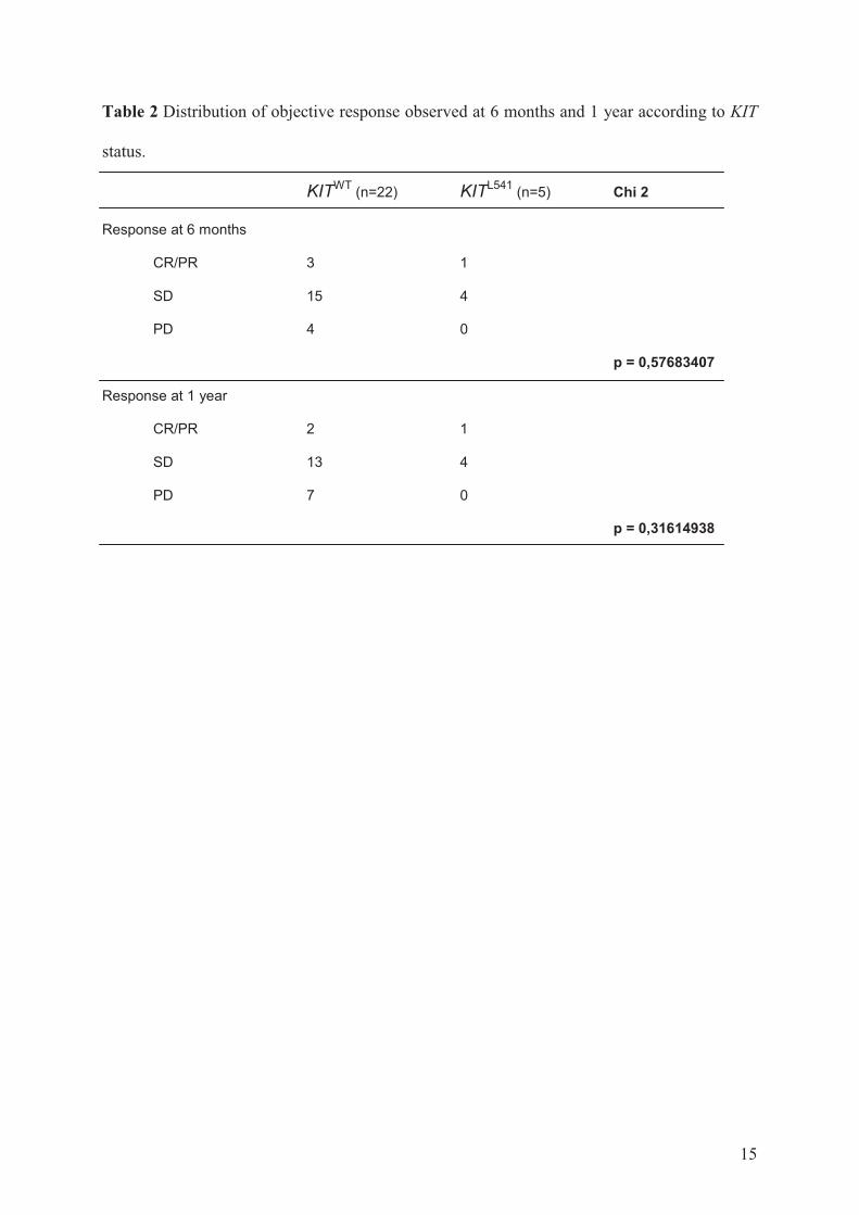
15
Table 2 Distribution of objective response observed at 6 months and 1 year according to KIT
status.
KITWT (n=22) KITL541
(n=5) Chi 2
Response at 6 months
CR/PR 3 1
SD 15 4
PD 4 0
p = 0,57683407
Response at 1 year
CR/PR 2 1
SD 13 4
PD 7 0
p = 0,31614938

16
Figure 1 In Competitive Allele-Specific Taqman®
PCR technology, each mutant allele assay
detects specific mutant alleles and a blocker suppresses the wild type allele
Figure 2 For each evaluable patient, the cross represents Ctrf and the point represents Ctmut.
Bars correspond to ΔCt. Surrounded bars correspond to cases KITL541
. Others bars correspond
to cases KITWT
.

17
Figure 3 Determination of KIT status by the 2 methods (sequencing and CAST PCR) for 1
case harbouring KITL541
and 1 case harbouring KITWT
. (A) Representative multicomponent
and amplification plots and sequencing of KITL541
(B) Representative multicomponent and
amplification plots and sequencing of KITWT
A
B
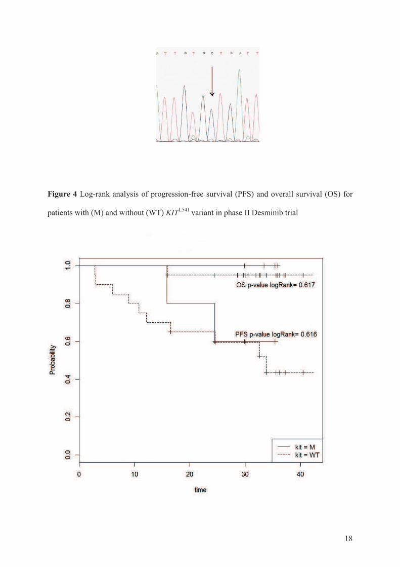
18
Figure 4 Log-rank analysis of progression-free survival (PFS) and overall survival (OS) for
patients with (M) and without (WT) KITL541
variant in phase II Desminib trial

Prediction of desmoid tumor progression using miRNA expression profiling
A. Dufresne, M. Paturel, L. Alberti, H. Philippon, A. Duc, AV Decouvelaere, P. Cassier, JY
Blay
Abstract
Rationale Desmoid tumor (DT) is a rare connective tissue tumor with locoregional
aggressiveness but unpredictable behaviour. MicroRNAs are noncoding small RNA
controlling the expression of a variety of genes and whose expression is deregulated in many
cancers. This study was conducted to define the predictive and/or prognostic value of miRNA
expression profile in DT.
Material and methods RNA was extracted from paraffin-embedded tissues sections of DT of
41 patients included in two distinct cohorts: a cohort of 26 patients included in the Desminib
phase II trial and an independent validation cohort of 15 patients with DT diagnosed in Leon
Berard Cancer Center. The miRNA profile was determined by Taqman Low Density Array
from 500 nanograms of total RNA. Supervised and unsupervised analyses were performed on
the 2 cohorts.
Results Predictive and prognostic supervised analysis on the Desminib cohort failed to
identify miRNAs differentially expressed between progressive and non-progressive patients
under imatinib treatment and between progressive and non-progressive patients after
discontinuation of imatinib, respectively. An unsupervised hierarchical clustering of Desminib
cohort identified however 2 groups (A and B) of 13 patients each. Only the number of
previous lines of treatment before inclusion in the study was significantly different between
the 2 groups (p = 0.0387). Time to progression after discontinuation of imatinib is longer in
group B than in group A (p = 0.05). Fifteen miRNAs were highly statistically differentially
expressed between groups A and B, targeting more than 3000 genes (whose AGO1, BCL2,
CDK6, SMAD4, PTEN, CCND1, VEGFA, RB1 targeted by at least 3 miRs). These results
were confirmed on the independent validation cohort: hierarchical clustering of these 15 miRs
identified 2 groups, whose time to recurrence is statistically different (28.8 months vs 68.8
months; p = 0.0213).
Conclusion These results bring the first demonstration of the prognostic value of miRNA
expression profile with a possible direct impact on the management of patients. The next
objective is to validate a precise miRNA signature in a prospective cohort to select patients
that do not benefit at all from surgical resection of their tumor and should be simply
monitored.
Introduction
Desmoid tumor (DT), also known as “aggressive fibromatosis”, is a rare fibroblastic
proliferative disease affecting about 2 to 4 persons per million per year [1]. Majority of DT
arise in a sporadic form related to CTNNB1 (β catenin gene) mutation [2]. DT may also occur
in the context of predisposing genetic condition called Gardner’s syndrome, characterized by
germline mutations of the APC gene and associated with familial adenomatous polyposis
(FAP) or also as rare forms of familial FAP [3]. Approximately 50% of DT occurs in the
extremities, other main locations being the trunk wall and mesentery [4]. These tumors never
metastasize and are considered as locally malignant tumors, with unpredictable behavior;
while it remains indolent in some patients, it also may involve nervous, vascular or muscular
structures leading to serious symptoms or even causing patient death in some cases (especially
with intra abdominal location).
Surgical resection of the tumor has been the mainstay treatment when planned surgery does
not imply serious functional and/or aesthetical sequelae. Nevertheless, spontaneous
stabilizations or regressions are regularly seen. Furthermore, despite initial surgical excision
of quality (R0 or R1 resection), recurrences are common (> 40%). A current debate calls into
question the utility of surgery as a treatment of these tumors. A retrospective study evaluating
a front-line conservative approach (no surgery and no radiotherapy) by watch-and-wait policy
or use of systemic treatment has resulted in prolonged progression-free survival in a
substantial proportion of patients (≈50%) [5]. The authors failed to determine prognostic
factors to select patients who benefit from this strategy. Recently, NCCN updated its
guidelines on Soft Tissue Sarcoma including “observation as an option for patients with
resectable desmoid tumors that are small and asymptomatic, not causing morbidity, pain, or
functional limitation”. [6]
Micro-RNAs (miRNAs) are noncoding small RNA molecules synthesized from intronic
regions with a size range from 16 to 35 nucleotides. They are processed by specific
complexes of proteins containing Drosha and Dicer to be matured and finally integrated in
RISC complexes. Mature miRNAs match with complementary sequences in messenger RNAs
resulting in translation inhibition and accelerated mRNA degradation [7]. miRNA expression
is deregulated in many cancers, resulting in a tumor miRNA signature, which could be useful
for their classification in line with their tissue origin and molecular alterations. Thus, they
currently constitute potent diagnosis and/or prognosis biomarkers with their tissue
specificities and their involvement in oncogenic process.
This study was conducted to define the potential predictive and/or prognostic value of miRNA
expression profile of DT to identify rapidly progressing and non progressing patients.
Material and methods
Patients
This study was performed as a retrospective translational research program on tumor samples
of patients included in Desminib trial [8]. Forty patients with progressive or recurrent DT that
could not be treated with curative surgery or radiotherapy were included in Desminib phase
I/II trial to evaluate the efficacy and toxicity of imatinib. Patients with adequate end organ
function were treated with 400mg of imatinib daily, increasing to 800mg in case of
progressive disease. Best clinical response to imatinib was defined according to RECIST
criteria. Evaluation was performed every 3 months. All the tumoral evaluations under
treatment but two have been reviewed by a radiological independent validation committee.

Informed consent was obtained from each patient to enroll them in the study and collect
archival pathology specimens.
A second cohort, completely independent from the first one, was established from 15 patients
with relapsing and non-relapsing DT treated at Center Leon Berard between 1990 and 2005.
Tissue samples
The analysis of present study was performed on the initial tumor of patients, sampled by
biopsy or surgical excision to establish the diagnosis of the disease. Paraffin-embedded tissues
sections of patients included in the study were obtained from pathology centers, all from
tumors at initial diagnosis. It has been already established that FFPE samples are good quality
material for the miRNA profile study [9, 10].
RNA extraction and quantitative real-time PCR
FFPE tumours were lysed for 24h in ATL buffer (Qiagen, France) supplemented with
proteinase K (Qiagen) at 60°C in rotative agitation after different washes with toluene,
ethanol, and tris/EDTA in this order. Total RNA was extracted from tumour using a single
phenol/chloroform extraction protocol with Trizol, according to the manufacturer’s
instructions (Life Technologies, Saint Aubin, France). Five hundred nanograms of total RNA
were subjected to the microfluidic PCR technology performed by Applied Biosystems (Life
Technologies, Saint Aubin, France). In brief, RNA was reversed transcribed, using
multiplexed specific looped miRNA primers from the Taqman MicroRNA Reverse
Transcription kit. The second step consists in a real-time quantitative PCR on Taqman Low
Density Array (TLDA) card A second version: RT products are introduced through
microchannels into miniature wells that are preloaded with dehydrated specific primers and
probes. Analyses were performed for 384 miRNAs.
Statistical analysis
For each miRNA, the threshold cycle (Ct) was calculated by the ABI 7900 Sequence
Detection System software. A cut-off of 37 was applied to discard the late Ct values
according to the manufacturer‘s instructions [11]. Median method was applied as
normalization [12, 13] and each miRNA was expressed by relative quantification (Rq = 2 –
ΔΔCt).
Supervised and unsupervised hierarchical clusterings were performed using TIBCO Spotfire
Decision Site software. Supervised analysis was performed on several groups of patients:
- responsive versus stable and progressive disease during the 1-year treatment by imatinib to
search for a predictive value
- progressive versus non progressive disease after the end of treatment by imatinib to search
for a prognostic value
Univariate logistic regression analysis was performed on groups of patients identified by
unsupervised hierarchical clustering with the following variables : sex (male vs female), age
at diagnosis (continuous variable), tumor size (at inclusion in Desminib trial, continuous
variable), tumor location (mesenteric vs abdominal wall vs extra-abdominal), context of FAP
(yes vs no), number of previous lines of treatment before inclusion in the study (patients
treated by radiotherapy (RT) and/or chemotherapy (CT) and/or hormone therapy and/or non
steroid anti-inflammatory drugs (NSAIDs) as an ordinal variable with 4 categories),
lymphocytes count (at inclusion in the trial, continuous variable), presence of the KIT M541L
exon 10 variant (yes vs no), level of immunohistochemical expression of β catenin, cyclin D1,
and PDGFRβ (0 vs 1 vs 2 vs 3).
Times to progression were compared by log-rank test after Kaplan-Meier analysis.

To detect differential expression of miRNAs between different groups of interest, we used a
one-way analysis of variance (ANOVA) with a p-value threshold of 5%.
To identify genes targeted by miRNAs of interest, we considered only the validated target in
our homemade database combining the 2 validated databases (TarBase v.6.0; last release on
Aug. 1 2012 [14] and miRTarbase; last release on Nov. 1 2013 [15]).
Results
Patients
Forty patients were included in Desminib phase II trial. Detailed results of the study were
published in 2010 [8].Two patients included in the clinical study refused to sign consent for
biological research on their tumor sample. Paraffin-embedded tissues samples of 34 patients
were obtained from pathology centers. They all were taken from specimen of initial diagnosis.
Six tissue samples were excluded from the study because of their fixation in Bouin. RNA was
extracted from 28 paraffin-embedded tissues and obtained in sufficient quantity (at least
500ng in 3μl) for 26 patients.
The 15 patients included in the validation cohort presented DT treated at Center Leon Berard.
All of them benefited from surgical resection of their initial tumor.
The tumor tissue sample analysed for each patient comes from the surgical excision of the
initial tumor, before the patient receive any medical treatment.
Table 1 presents the characteristics of the 41 patients analysed (26 patients in the desminib
cohort and 15 patients in the validation cohort).
The characteristics of the patients correspond to those describe in the literature. Among the 41
patients analysed, there is a majority of women with a median age at diagnosis around the 40
years old. The tumors mainly occur in an extra-abdominal location (the majority in the trunk
and few cases in upper and lower limbs or in head and neck). Median tumor size is around 9
cm at the inclusion in the Desminib study and a little smaller in the validation cohort. This is
the only criteria different between the 2 cohorts. More than half of patients received no
medical treatment or only one line of treatment and 37% of them received two, three or four
lines.
In the Desminib cohort, after a median follow-up of 68 months, median TTP was 2 years and
median OS is not reached. Majority of patients had stable (19 patients) or responsive (3
patients) disease under imatinib. After discontinuation of imatinib, 10 patients were
progressive within 23.5 months of follow-up and 14 were never progressive.
In the validation cohort, after a median follow-up of 56,8 months, 6 patients had recurrence
and 9 did not present any recurrence.
Supervised analysis
Predictive and prognostic supervised analysis on the desminib cohort failed to identify
miRNA differentially expressed between responsive versus non responsive patients to
imatinib and between progressive versus non progressive patients after discontinuation of
imatinib, respectively. Any miR reached the statistical significance after Bonferroni
correction to discriminate studied groups.
Unsupervised analysis
The unsupervised hierarchical clustering of the Desminib cohort identified 2 groups of 13
patients each, called group A and group B. To identify clinical and/or biological factors that

could explain the partition of patients in the 2 clusters, unvariate analysis by logistic
regression was performed.
Only the number of previous lines of treatment before inclusion in the Desminib trial was
found significantly associated with the belonging to one group of the clustering or another,
with an Odd Ratio of 2.44 [1.17-6.78] and a p-value of 0.0387. Patients of group B have been
treated with significantly less lines of treatment before inclusion in the Desminib trial.
Time to progression (TTP) after interruption of treatment by imatinib was compared between
the groups A and B by log rank analysis. Three patients were excluded of the study because of
lack of follow-up.
The median follow-up after interruption of imatinib was 23.6 months [12.4-39.0]. Six patients
among 11 in group A and 3 patients among 12 in group B relapsed.
Even not statistically significant, median TTP in group B is longer than median TTP in group
A.
By Anova test, 15 miRNA were found differentially expressed with statistical significance
between the 2 groups A and B. The table 2 presents the list of these 15 miRs and their p-
value.
All these miRNA are under-expressed in the group A that is associated with poor prognosis.
After interrogation of database, more than 3000 genes are targeted these 15 miRNAs. Among
them, 50 genes are targeted by at least 3 miRNAs, whose: AGO1, BCL2, CDK6, SMAD4,
PTEN, CCND1, VEGFA, RB1.
To validate these results, these 15 miRNAs were used to perform a hierarchical clustering of
the validation cohort. Two groups of 6 and 9 patients were identified, called group A’ and
group B’, respectively.
Univariate analysis by logistic regression failed to find any significant clinical parameters to
explain the distribution of patients in the 2 groups. Parameters tested were age and sex of the
patient, location of the tumor, number of recurrence and number of treatment lines.
After a median follow-up of 53 months [3-230], 4 patients among 6 in group B’ and 5 patients
among 9 in group A’ recurred. Time to recurrence (TTR) after initial surgery was compared
by log-rank analysis between the groups A’ and B’.
Time to recurrence is significantly longer in group B’ identified by the 15 miRNA profile
expression than in group A’.

Discussion
This study presents, to date, the first results of the analysis of miRNA profile expression in
desmoid tumor (DT). Although retrospective, it is based on a cohort of patients included in a
clinical phase II trial (Desminib trial) that ensures a good quality follow-up and
exhaustiveness of the data recorded. The main limitation of our study is linked to the small
size of its cohorts, but DTs are very rare mesenchymal tumors.
The characteristics of patients in the studied cohorts (Desminib and validation cohorts) are
similar to those described in the largest retrospective issue [4].
As miRNAs have never been studied in DT yet, we performed an explanatory approach of the
analysis with no assumption. First, predictive and prognostic supervised analyses were
conducted. The choice of groups to compare in the predictive analysis was difficult to
establish because the response evaluation to medical treatment of DT is not clearly
established: RECIST criteria are certainly not adapted to these slow growing tumors.
Predictive analysis failed to identify specific imatinib targets that could explain the sensitivity
of DT to this drug, but propose new targets to further explore (PARP, IGF1R, HSP90B).
Prognosis supervised analysis was difficult to interpret for the Desminib cohort as far as
majority of patients had recurrent and/or progressive disease at inclusion in the trial.
As more and more used with the emergence of “omics”, analysis that concern “high-
dimensional data”, we then chose to realize a clustering. A study upstream on the robustness
of clustering has shown the Euclidean distance and Ward's method gave the best results for
small cohorts of sarcoma tumors [Pissaloux in press]. It also presents the strong advantage not
to require any a priori on the groups to obtain. This analysis on Desminib cohort leads to
individualize 2 groups of patients.
The results of logistic regression showed a significantly association between the belonging to
group B and a lower number of lines of treatment administered before inclusion in the
Desminib trial, that may reflect the aggressiveness of the tumor. In clinical practice, it is
known that systemic treatments (radiotherapy, chemotherapy, hormone therapy, NSAID) are
little effective and not devoid of side effects. When several lines are administered, it usually
reflects an aggressive tumor, uncontrolled and/or symptomatic, for which the physician tests
successively all available treatments to relieve patient symptoms. This criterion of number of
treatment lines is used in clinical trials that evaluate other low grade tumor such as T-cell
lymphomas [16]. This data suggested a prognostic value of miRNA profile represented by the
tumor aggressiveness. It was confirmed by the comparison of TTP after discontinuation of
imatinib in the 2 groups: even not statistically significant (p = 0.0596), the TTP curves of the
2 groups were clearly distinct. The lack of statistical significance may be due to the limited
size of the cohort and to the fact that all patients included in Desminib trial had a recurrent so
aggressive tumor.
These results had to be validated in an independent cohort that included patients with
recurrent and not recurrent tumors after surgical excision. This cohort was more adapted than
the Desminib one to search for a prognostic value. Unsupervised clustering indeed led to 2
groups of patients that present statistically different time to recurrence, confirming the
prognostic value of miRNA profile expression.
Dozens of studies already demonstrated a prognostic value of miRNA in many different types
of malignancy [17-20].
The next step of this analysis was logically aimed to identify discriminating miRNAs between
the clusters in order to 1) define a prognosis signature and 2) determine genes whose
transcription is impacted by these miRNAs. By Anova test and Bonferroni correction, 15
miRNAs were found to be significant in the unsupervised analysis of Desminib cohort. These
15 miRs target more than 3000 genes. Then it was essential to select several genes known to
be involved in signalling pathways in cancer and that could represent interesting new targets
to study to explore biological mechanisms of DT and the development of new therapeutics.
To that, we decided to focus on genes targeted by several miRNAs, to increase their
biological relevance.
PTEN is a key regulator of cell cycle and a tumor suppressor gene. Genetic inactivations or
reduced expression of PTEN are frequently associated with cancers. It has been already
established that mutations in SMAD4 and in PTEN are involved in the development of
familial juvenile polyposis [21]. Angiogenesis is also directly impacted by VEGFA. Rb is
also considered as a tumor suppressor by inhibiting cell cycle progression. CDK6 and Cyclin
D1 phosphorylate Rb, inhibiting its activity. It has been demonstrated a lack of nuclear
staining by immunohistochemistry in 13 cases of DT (interestingly, the staining was present
in palmar fibromatosis s long as in the vascular tissue adjacent to the fibroproliferative lesion)
[22]. In a previous study looking for predictive factors of response to imatinib in Desminib
trial, we found by immunohistochemistry analysis on tissue micro array, inconsistent
expression of Cyclin D1. It’s interesting to see that the 5 patients (among 31 tested) with
histological staining for Cyclin D1 are classified in the group A of the present study. Bcl2 is
classified as an oncogene as long as it acts as an anti-apoptotic.
Even if interesting, these 15 miRNAs can be used as a prognostic signature only after their
validation in a larger and prospective cohort of DT.
Conclusion
These results bring the first demonstration of the prognostic value of miRNA expression
profile in DT. The impact of such a discovery is essential in the management of patients with
these tumors that are particularly over-treated. It is now evident that a substantial proportion
of patients (≈ 50%) do not benefit at all from surgical resection of their tumor, such a
resection leading to functional and/or esthetical sequels for tumor that frequently never
progress. This is now so well established, that a prospective phase II clinical trial is ongoing
to systematically propose to patients with DT an active surveillance, and surgical management
only in case of progressive disease. There’s a deep need of clinical and/or biological surrogate
markers of aggressiveness of these tumor to select patients who may benefit of such a wait-
and-see policy. The results of our study need to be confirmed and we plan to analyse tumor
sample of patients including in this ongoing phase II trial and to prospectively establish their
miRNA profile.
The ultimate goal will be to define a robust prognostic signature based on few miRNAs to
predict the behaviour of the tumor newly diagnosed and adapt the management of the tumor.
To go further, we also consider to evaluate miRNA profile in the serum of patients to develop
a non-invasive diagnostic tool.
References
1/ Biermann JS. Desmoid tumours. Curr Treat Options Oncol 2000; 1: 262–266.
2/ Dômont J, Salas S, Lacroix L, Brouste V, Saulnier P, Terrier P, Ranchère D, Neuville A,
Leroux A, Guillou L, Sciot R, Collin F, Dufresne A, Blay JY, Le Cesne A, Coindre JM,
Bonvalot S, Bénard J. High frequency of beta-catenin heterozygous mutations in extra-
abdominal fibromatosis: a potential molecular tool for disease management. Br J Cancer.
2010 Mar 16;102(6):1032-6
3/ Schiessling S, Kihm M, Ganschow P, Kadmon G, Büchler MW, Kadmon M. Desmoid
tumour biology in patients with familial adenomatous polyposis coli. Br J Surg. 2013
Apr;100(5):694-703
4/ Salas S, Dufresne A, Bui B, Blay JY, Terrier P, Ranchere-Vince D, Bonvalot S, Stoeckle E,
Guillou L, Le Cesne A, Oberlin O, Brouste V, Coindre JM. Prognostic factors influencing
progression-free survival determined from a series of sporadic desmoid tumors: a wait-and-
see policy according to tumor presentation. J Clin Oncol. 2011 Sep 10;29(26):3553-8.
5/ Fiore M, Rimareix F, Mariani L, Domont J, Collini P, Le Péchoux C, Casali PG, Le Cesne
A, Gronchi A, Bonvalot S. Desmoid-type fibromatosis: a front-line conservative approach to
select patients for surgical treatment. Ann Surg Oncol. 2009 Sep;16(9):2587-93
6/ von Mehren M, Benjamin RS, Bui MM, Casper ES, Conrad EU 3rd, Delaney TF, Ganjoo
KN, George S, Gonzalez R, Heslin MJ, Kane JM 3rd, Mayerson J, McGarry SV, Meyer C,
O'Donnell RJ, Paz B, Pfeifer JD, Pollock RE, Randall RL, Riedel RF, Schuetze S, Schupak
KD, Schwartz HS, Shankar S, Van Tine BA, Wayne J, Sundar H, McMillian NR. Soft Tissue
Sarcoma, Version 2.2012: Featured Updates to the NCCN Guidelines. J Natl Compr Canc
Netw. 2012 Aug 1;10(8):951-960.
7/ Amaia Lujambio & Scott W. Lowe The microcosmos of cancer Nature 482, 347-355
8/ Penel N, Le Cesne A, Bui BN, Perol D, Brain EG, Ray-Coquard I, Guillemet C, Chevreau
C, Cupissol D, Chabaud S, Jimenez M, Duffaud F, Piperno-Neumann S, Mignot L, Blay JY.
Imatinib for progressive and recurrent aggressive fibromatosis (desmoid tumors): an
FNCLCC/French Sarcoma Group phase II trial with a long-term follow-up. Ann Oncol. 2011
Feb;22(2):452-7.
9/ Xi Y, Nakajima G, Gavin E, Morris CG, Kudo K, Hayashi K, Ju J. Systematic analysis of
microRNA expression of RNA extracted from fresh frozen and formalin-fixed paraffin-
embedded samples. RNA. 2007 Oct;13(10):1668-74.
10/ Liu A, Tetzlaff MT, Vanbelle P, Elder D, Feldman M, Tobias JW, Sepulveda AR, Xu X.
MicroRNA expression profiling outperforms mRNA expression profiling in formalin-fixed
paraffin-embedded tissues. Int J Clin Exp Pathol. 2009 Mar 20;2(6):519-27.
11/ Witwer KW, Sarbanes SL, Liu J, Clements JE. A plasma microRNA signature of acute
lentiviral infection: biomarkers of central nervous system disease. AIDS. 2011 Nov
13;25(17):2057-67 (choix du ct à 37)
12/ Vandesompele J, De Preter K, Pattyn F, Poppe B, Van Roy N, De Paepe A, Speleman F.

Accurate normalization of real-time quantitative RT-PCR data by geometric averaging of
multiple internal control genes. Genome Biol. 2002 Jun 18;3(7):RESEARCH0034
13/ Andersen CL, Jensen JL, Ørntoft TF. Normalization of real-time quantitative reverse
transcription-PCR data: a model-based variance estimation approach to identify genes suited
for normalization, applied to bladder and colon cancer data sets. Cancer Res. 2004 Aug
1;64(15):5245-50.
14/ Vergoulis, T. I. Vlachos, P. Alexiou, G. Georgakilas, M. Maragkakis, M. Reczko, S.
Gerangelos, N. Koziris, T. Dalamagas, AG Hatzigeorgiou; Tarbase 6.0: Capturing the
Exponential Growth of miRNA Targets with Experimental Support. Nucl. Acids Res. (2012)
40 (D1): D222-D229.
15/ Hsu SD, Lin FM, Wu WY, Liang C, Huang WC, Chan WL, Tsai WT, Chen GZ, Lee CJ,
Chiu CM, Chien CH, Wu MC, Huang CY, Tsou AP, Huang HD. (2011) Nucleic acids
research
16/ Damaj G, Gressin R, Bouabdallah K, Cartron G, Choufi B, Gyan E, Banos A, Jaccard A,
Park S, Tournilhac O, Schiano-de Collela JM, Voillat L, Joly B, Le Gouill S, Saad A, Cony-
Makhoul P, Vilque JP, Sanhes L, Schmidt-Tanguy A, Bubenheim M, Houot R, Diouf M,
Marolleau JP, Béné MC, Martin A, Lamy T. Results From a Prospective, Open-Label, Phase
II Trial of Bendamustine in Refractory or Relapsed T-Cell Lymphomas: The BENTLY Trial.
J Clin Oncol. 2013 Jan 1;31(1):104-10.
17/ Yanaihara N, Caplen N, Bowman E, Seike M, Kumamoto K, Yi M, Stephens RM,
Okamoto A, Yokota J, Tanaka T, Calin GA, Liu CG, Croce CM, Harris CC. Unique
microRNA molecular profiles in lung cancer diagnosis and prognosis. Cancer Cell. 2006
Mar;9(3):189-98.
18/ Calin GA, Ferracin M, Cimmino A, Di Leva G, Shimizu M, Wojcik SE, Iorio MV,
Visone R, Sever NI, Fabbri M, Iuliano R, Palumbo T, Pichiorri F, Roldo C, Garzon R,
Sevignani C, Rassenti L, Alder H, Volinia S, Liu CG, Kipps TJ, Negrini M, Croce CM. A
MicroRNA signature associated with prognosis and progression in chronic lymphocytic
leukemia. N Engl J Med. 2005 Oct 27;353(17):1793-801.
19/ Nair VS, Maeda LS, Ioannidis JP. Clinical outcome prediction by microRNAs in human
cancer: a systematic review. J Natl Cancer Inst. 2012 Apr 4;104(7):528-40.
20/ Lu J, Getz G, Miska EA, Alvarez-Saavedra E, Lamb J, Peck D, Sweet-Cordero A, Ebert
BL, Mak RH, Ferrando AA, Downing JR, Jacks T, Horvitz HR, Golub TR. MicroRNA
expression profiles classify human cancers. Nature. 2005 Jun 9;435(7043):834-8.
21/ Howe JR, Bair JL, Sayed MG, Anderson ME, Mitros FA, Petersen GM, Velculescu VE,
Traverso G, Vogelstein B. Germline mutations of the gene encoding bone morphogenetic
protein receptor 1A in juvenile polyposis [PTEN SMAD4]. Nat Genet. 2001 Jun;28(2):184-7.
22/ Muller E, Castagnaro M, Yandel DW, Wolfe HJ, Alman BA.Molecular genetic and
immunohistochemical analysis of the tumor suppressor genes Rb and p53 in palmar and
aggressive fibromatosis. Diagn Mol Pathol. 1996 Sep;5(3):194-200.
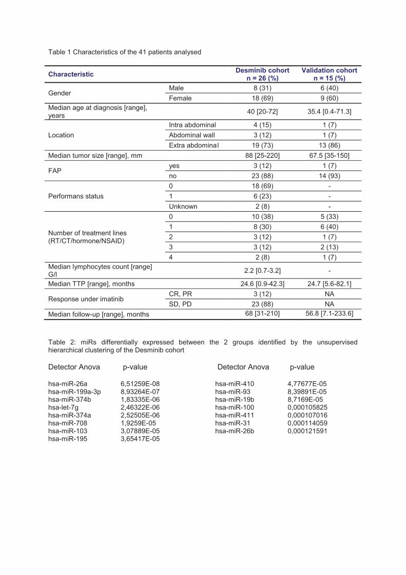
Table 1 Characteristics of the 41 patients analysed
Characteristic Desminib cohort
n = 26 (%) Validation cohort
n = 15 (%)
Gender Male 8 (31) 6 (40)
Female 18 (69) 9 (60)
Median age at diagnosis [range], years
40 [20-72] 35.4 [0.4-71.3]
Location
Intra abdominal 4 (15) 1 (7)
Abdominal wall 3 (12) 1 (7)
Extra abdomina l 19 (73) 13 (86)
Median tumor size [range], mm 88 [25-220] 67.5 [35-150]
FAP yes 3 (12) 1 (7)
no 23 (88) 14 (93)
Performans status
0 18 (69) -
1 6 (23) -
Unknown 2 (8) -
Number of treatment lines (RT/CT/hormone/NSAID)
0 10 (38) 5 (33)
1 8 (30) 6 (40)
2 3 (12) 1 (7)
3 3 (12) 2 (13)
4 2 (8) 1 (7)
Median lymphocytes count [range] G/l
2.2 [0.7-3.2] -
Median TTP [range], months 24.6 [0.9-42.3] 24.7 [5.6-82.1]
Response under imatinib CR, PR 3 (12) NA
SD, PD 23 (88) NA
Median follow-up [range], months 68 [31-210] 56.8 [7.1-233.6]
Table 2: miRs differentially expressed between the 2 groups identified by the unsupervised hierarchical clustering of the Desminib cohort
Detector Anova p-value Detector Anova p-value hsa-miR-26a 6,51259E-08 hsa-miR-199a-3p 8,93264E-07 hsa-miR-374b 1,83335E-06 hsa-let-7g 2,46322E-06 hsa-miR-374a 2,52505E-06 hsa-miR-708 1,9259E-05 hsa-miR-103 3,07889E-05 hsa-miR-195 3,65417E-05
hsa-miR-410 4,77677E-05 hsa-miR-93 8,39891E-05 hsa-miR-19b 8,7169E-05 hsa-miR-100 0,000105825 hsa-miR-411 0,000107016 hsa-miR-31 0,000114059 hsa-miR-26b 0,000121591

Figure 1 Right figure represents the heat map clustering of miRNA expression in desmoid tumors of the desminib cohort (26 samples). Each row represents the relative levels of expression for each miRNA and each column shows the expression levels for each patient sample. The red or green color indicates relatively high or low expression, respectively, while grey squares indicate absent data points Left figure represents the comparison by log-rank analysis, of time-to-progression after discontinuation of imatinib of groups identified by unsupervised analysis in the Desminib cohort.
Time to progression after imatinib interruption
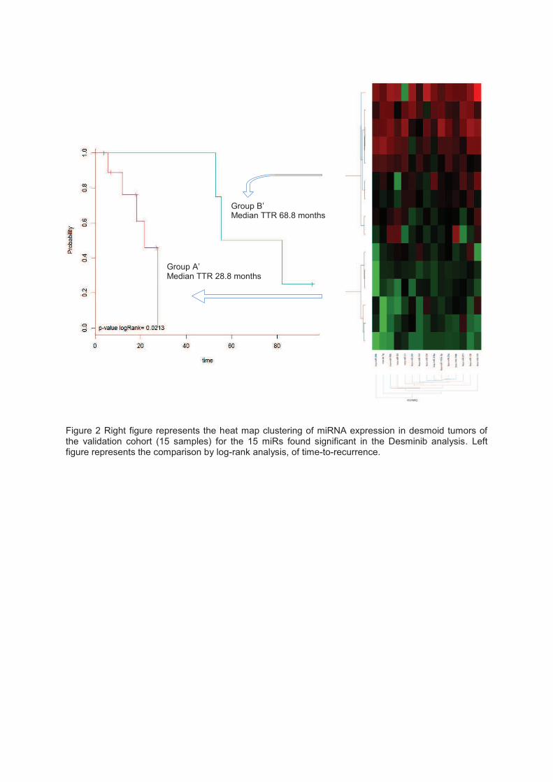
Figure 2 Right figure represents the heat map clustering of miRNA expression in desmoid tumors of the validation cohort (15 samples) for the 15 miRs found significant in the Desminib analysis. Left figure represents the comparison by log-rank analysis, of time-to-recurrence.
Group B’ Median TTR 68.8 months
Group A’ Median TTR 28.8 months
Résumé
Les fibromatoses agressives sont des tumeurs conjonctives rares, pouvant envahir les
structures adjacentes parfois de manière très agressive et responsables de fréquentes
récidives loco-régionales après exérèse mais dépourvues de potentiel métastatique. Elles
peuvent également se stabiliser voire régresser spontanément. Leur évolution est
imprévisible. Actuellement, la stratégie de leur prise en charge est remise en cause et ces
tumeurs sont de plus en plus souvent surveillées à leur diagnostic. En cas de tumeur
évolutive, les traitements systémiques disponibles sont multiples mais d’efficacité très
variable. Biologiquement, ces tumeurs sont issues de cellules souches mésenchymateuses
dont la voie Wnt/β-caténine est dérégulée. Cette dérégulation est liée à une mutation du
gène APC quand la tumeur survient dans un contexte de syndrome de Gardner et à une
mutation du gène CTNNB1 quand elle survient de manière sporadique. Aucun facteur
pronostique de récidive ou d’évolutivité spontanée et aucun facteur prédictif d’efficacité des
traitements médicaux n’a aujourd’hui été identifié.
Les travaux de deux premières publications présentés le manuscrit, ont étudié les facteurs
cliniques de récidive après exérèse chirurgicale sur de larges séries rétrospectives, française
et américaine, et ont retenu le jeune âge du patient, la grande taille tumorale et sa
localisation extra-abdominale comme étant de mauvais pronostic. Dans une troisième
publication, nous nous sommes appliqués à rechercher si le sous-type moléculaire de
mutation de CTNNB1 observé dans les fibromatoses sporadiques pouvait influencer la
récidive: les résultats concluent que la mutation S45F est de moins bon pronostic que les
autres, ce qui se retrouve dans la littérature, sans que l’explication biologique en soit définie.
Un autre article soumis à publication rapporte les résultats de l’analyse des profils
d’expression des miRNAs des fibromatoses qui semblent se corréler à leur pronostic mais
cela doit être confirmé. Les 3 articles suivant présentés, dont 2 sont publiés, présentent des
travaux recherchant des facteurs prédictifs de réponse des fibromatoses à l’imatinib :
l’expression d’aucune des cibles connues de l’imatinib n’a été retrouvée comme influençant
significativement la réponse au traitement. Le rôle des variants M541L et V530I de l’exon 10
de KIT reste à déterminer.
L’exploration des mécanismes biologiques sous-tendant les différences cliniques observées
tant au niveau du pronostic des fibromatoses que de leur réponse aux thérapies (ciblées ou
non) doit se poursuivre. Les axes de recherches doivent être développés en tenant compte
des nouveaux standards de stratégie de prise en charge, moins interventionnistes. C’est en
ciblant précisément les nouveaux défis de traitement de ces tumeurs que cette recherche
pourra être efficace.
![[Fr] Guide de survie du directeur digital dans le monde du multimédia (Celumium)](https://static.fdocuments.in/doc/165x107/5495de6cb479593c058b4615/fr-guide-de-survie-du-directeur-digital-dans-le-monde-du-multimedia-celumium.jpg)