Understanding the TMJ - edusymp.com
13
UNDERSTANDING THE TMJ Insight into how the form and function of the TMJ affect imaging findings DANIA TAMIMI, BDS, DMSc WHAT WE WILL DISCUSS • TMJ imaging anatomy • Effect of TMJ on growth and development of the craniofacial complex THE TMJ IS: • A ginglymo-diarthroidal joint – Hinge-sliding motion involving two joints • Freely moving articulation between mandible and squamous portion of the temporal bone • Special – that’s why you’re here TMJ COMPONENTS INTRACAPSULAR COMPONENTS PERIPHERAL COMPONENTS INTERNAL COMPONENTS 1. Articular surfaces 2. Disc and attachments 3. Joint compartments A discussion on the effect of the TMJ on the growth and development of the mandible OSSEOUS COMPONENTS
Transcript of Understanding the TMJ - edusymp.com

UNDERSTANDING THE TMJ
Insight into how the form and function of the TMJ affect imaging findings
DANIA TAMIMI, BDS, DMSc
WHAT WE WILL DISCUSS
• TMJ imaging anatomy
• Effect of TMJ on growth and development of the craniofacial
complex
THE TMJ IS:
• A ginglymo-diarthroidal joint – Hinge-sliding motion involving
two joints
• Freely moving articulation between mandible and squamous
portion of the temporal bone
• Special – that’s why you’re here
TMJ COMPONENTS
INTRACAPSULAR COMPONENTS PERIPHERAL COMPONENTS
INTERNAL COMPONENTS
1. Articular surfaces
2. Disc and attachments
3. Joint compartments
A discussion on the effect of the TMJ on the
growth and development of the mandible
OSSEOUS COMPONENTS

OSSEOUS COMPONENTS
1. Mandibular condyle
2. Glenoid fossa and
articular eminence
CBCT / CT ANATOMY
Sagittal Oblique
Coronal Oblique
Axially Corrected images
CBCT/ CT ANATOMY
Sagittal Oblique Coronal Oblique
Axially-Corrected images


MANDIBULAR CONDYLAR FIBROCARTILAGE
Articular Zone
Fibrous Zone
Proliferative Zone
Hypertrophic Zone
Mature Zone
Bone
MANDIBULAR CONDYLAR FIBROCARTILAGE
• Thin layer of fibrous, cartilage-like tissue unique to the TMJ
• Not innervated or vascularized – takes nourishment from
synovial fluid
• Lack of innervation and vascularity allows the articular surfaces
withstand the high dynamic load TMJ regime
• Load is exceeded � breakdown of cartilage Courtesy N. Varpniarsky
ARTICULAR EMINENCE FIBROCARTILAGE

TMJ
• TMJ is a Growth Site
• TMJ has a Fibro-Cartilagenous cap that Responds Biomechanical
and Biochemical Stimuli
• Mesenchymal Cell Differentiation into Articular Cartilage followed
by Endochondral Ossification Contributes to Condylar Growth
MANDIBULAR GROWTH
• Normal: Counter-clockwise direction
MANDIBULAR GROWTH
• Enchondral & Intra-membraneous growth
• Mandibular Growth Mirrors TMJ Growth
• Genetic and Epi-Genetic Growth
• Growth Fields
GROWTH SITE/ FIELDS
• Condyles, Rami, Alveolar processes, Coronoid processes, Body
• Sites have genetic potential for Growth through Mesenchymal
Cell Differentiation and Cell Division
• Can be Modulated by external factors including neighboring
growth sites, hormones, tissue stress/ strain and tissue damage
MANDIBULAR GROWTH
Growth directions involving periosteal resorption are indicated by arrows pointing into the bone surface, and
growth directions involving periosteal deposition are represented by arrows pointing out of the bone surface.
From Enlow, D. H. and D. B. Harris: A study of the postnatal growth of the human mandible. Am. J. Orthod. 50 (25), 1964

Stress Distribution
•Load Support
•Cortex
•Load Transfer
•Trabeculae
MANDIBULAR DEFORMATION
• Sagittal Bending (AP)
• Increased Vertical Dimension
• Transverse Bending (ML)
• Increased Transverse Dimension
• Torsion (Twisting)
• Cylindrical Shape
Morphological Adaptations
FEM Modeling
Dr. T. Korioth
ADAPTATION TOOLS SET
• Remodeling
• Modeling
• Displacement
• Positional
• Tooth Eruption
• Coupled specific sites of bone
resorption and formation
• Ongoing secondary process that
modifies internal distribution and
microstructure of bone to
mechanically optimize it
• Uncoupled bone deposition and
resorption
• Associated with periosteal
deposition or resorption of bone;
leads changes in size, shape,
and position of bone
DEFINITIONSBone modeling Bone remodeling
FACIAL GROWTH PATTERNS
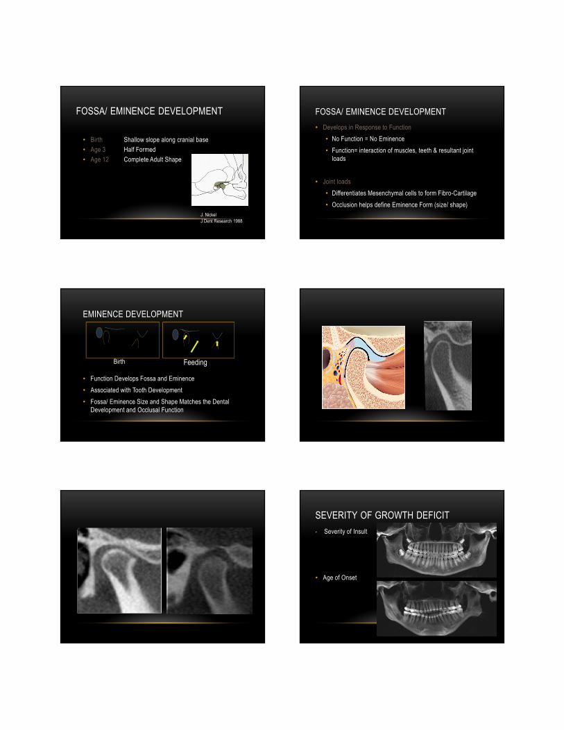
FOSSA/ EMINENCE DEVELOPMENT
• Birth Shallow slope along cranial base
• Age 3 Half Formed
• Age 12 Complete Adult Shape
J. Nickel
J Dent Research 1988
FOSSA/ EMINENCE DEVELOPMENT
• Develops in Response to Function
• No Function = No Eminence
• Function= interaction of muscles, teeth & resultant joint
loads
• Joint loads
• Differentiates Mesenchymal cells to form Fibro-Cartilage
• Occlusion helps define Eminence Form (size/ shape)
EMINENCE DEVELOPMENT
• Function Develops Fossa and Eminence
• Associated with Tooth Development
• Fossa/ Eminence Size and Shape Matches the Dental
Development and Occlusal Function
Birth Feeding
SEVERITY OF GROWTH DEFICIT
• Severity of Insult
• Age of Onset

MANDIBULAR GROWTH
Age
Gro
wth DD
Puberty
Post- PubertyAdult
Normal
PCR-2
PCR-1
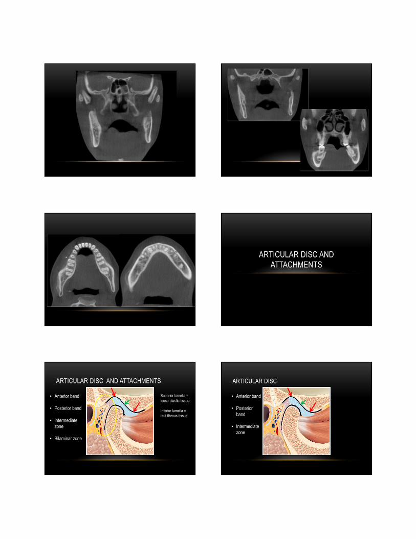
ARTICULAR DISC AND
ATTACHMENTS
ARTICULAR DISC AND ATTACHMENTS
• Anterior band
• Posterior band
• Intermediate
zone
• Bilaminar zone
Superior lamella =
loose elastic tissue
Inferior lamella =
taut fibrous tissue.
ARTICULAR DISC
• Anterior band
• Posterior
band
• Intermediate
zone

ARTICULAR DISC
Disc composed of:
• 70-75% water
• Fibroblast-like cells
• Chondrocyte-like cells
• Type 1 collagen: (90% dry
weight)
• Elastin: 5% dry weight
• Proteoglycans (enhance
compressive properties)
ARTICULAR DISC COLLAGEN ARRANGEMENT
Anterior band:
- Collagen fibers transversely
oriented
Intermediate zone:
- Collagen fibers are
anteroposteriorly oriented
Posterior band:
- Transverse and vertical
collagen fibers
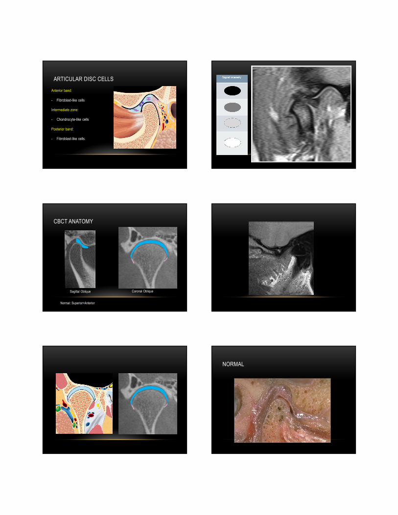
ARTICULAR DISC CELLS
Anterior band:
- Fibroblast-like cells
Intermediate zone:
- Chondrocyte-like cells
Posterior band:
- Fibroblast-like cells.
CBCT ANATOMY
Sagittal Oblique Coronal Oblique
Normal: Superior>Anterior
NORMAL

• Temporal (TPA) / Superior
Lamina
• Condylar (CPA) / Inferior
Lamina
• Intermediate (IPA)
POSTERIOR ATTACHMENT
PL Wesstesson
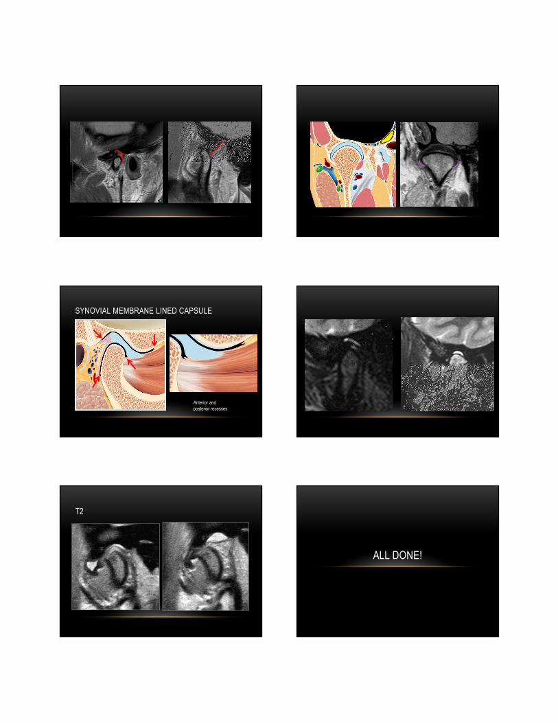
SYNOVIAL MEMBRANE LINED CAPSULE
Anterior and
posterior recesses
T2
ALL DONE!