Understanding the Basics of Clinical Oncology, from...
17
Understanding the Basics of Clinical Oncology, from Diagnosis to Treatment 5/21/13 Andrew Ko, MD BIOGRAPHY: Dr. Andrew Ko did his medical training at the Johns Hopkins School of Medicine, Beth Israel Hospital/Harvard Medical School, and Stanford University Medical Center. He is currently an Associate Professor in the Division of Hematology/Oncology at UCSF, where his primary clinical and research interests focus on gastrointestinal malignancies, with a particular emphasis on pancreatic cancer. He serves as the chair of the scientific Protocol Review Committee at the UCSF Comprehensive Cancer Center, and is also very involved in the UCSF School of Medicine, serving on the Admissions Committee and co‐directing the second‐year medical school course M3: Mechanisms, Methods, and Malignancies. Nationally, Dr. Ko is a member of the scientific program committee and specialty editorial board for the American Society of Clinical Oncology, sits on several editorial boards for peer‐reviewed oncology journals (including the leading clinical oncology journal, the Journal of Clinical Oncology), and is currently a member of the National Cancer Institute’s Pancreatic Cancer Task Force and the National Comprehensive Cancer Network (NCCN) Pancreatic Cancer guidelines committee. Outside of work, Andrew enjoys playing the piano, running, whitewater rafting, tennis, reading, cooking, and watching sports. He lives in San Francisco with his wife Christine and their two children, Naomi (age 6) and Elliott (age 3). BIBLIOGRAPHY: DeVita VT Jr, Lawrence TS, Rosenberg SA, DePinho RA, Weinberg RA, eds. Cancer: Principles and Practice of Oncology, 9 th ed. DeVita VT Jr, Rosenberg SA. Two hundred years of cancer research. N Engl J Med 2012; 366(23):2207‐14. Philadelphia: Lippincott Williams & Wilkins, 2011. Hanahan D, Weinberg RA. The hallmarks of cancer. Cell 2000;100(1):57‐70. The history of cancer. From American Cancer Society website (http://www.cancer.org) Siegel R, Naishadham D, Jemal A. Cancer statistics, 2013. CA Cancer J Clin. 2013;63(1):11‐30.
Transcript of Understanding the Basics of Clinical Oncology, from...
Understanding the Basics of Clinical Oncology, from Diagnosis to Treatment 5/21/13
Andrew Ko, MD
BIOGRAPHY: Dr. Andrew Ko did his medical training at the Johns Hopkins School of Medicine, Beth Israel Hospital/Harvard Medical School, and Stanford University Medical Center. He is currently an Associate Professor in the Division of Hematology/Oncology at UCSF, where his primary clinical and research interests focus on gastrointestinal malignancies, with a particular emphasis on pancreatic cancer. He serves as the chair of the scientific Protocol Review Committee at the UCSF Comprehensive Cancer Center, and is also very involved in the UCSF School of Medicine, serving on the Admissions Committee and co‐directing the second‐year medical school course M3: Mechanisms, Methods, and Malignancies. Nationally, Dr. Ko is a member of the scientific program committee and specialty editorial board for the American Society of Clinical Oncology, sits on several editorial boards for peer‐reviewed oncology journals (including the leading clinical oncology journal, the Journal of Clinical Oncology), and is currently a member of the National Cancer Institute’s Pancreatic Cancer Task Force and the National Comprehensive Cancer Network (NCCN) Pancreatic Cancer guidelines committee. Outside of work, Andrew enjoys playing the piano, running, whitewater rafting, tennis, reading, cooking, and watching sports. He lives in San Francisco with his wife Christine and their two children, Naomi (age 6) and Elliott (age 3).
BIBLIOGRAPHY: DeVita VT Jr, Lawrence TS, Rosenberg SA, DePinho RA, Weinberg RA, eds. Cancer: Principles and Practice of Oncology, 9th ed. DeVita VT Jr, Rosenberg SA. Two hundred years of cancer research. N Engl J Med 2012; 366(23):2207‐14. Philadelphia: Lippincott Williams & Wilkins, 2011. Hanahan D, Weinberg RA. The hallmarks of cancer. Cell 2000;100(1):57‐70. The history of cancer. From American Cancer Society website (http://www.cancer.org) Siegel R, Naishadham D, Jemal A. Cancer statistics, 2013. CA Cancer J Clin. 2013;63(1):11‐30.

5/21/2013
1
UNDERSTANDING THE BASICS OF CLINICAL ONCOLOGY(FROM DIAGNOSIS TO TREATMENT)
UCSF Mini Medical School for the Public
Andrew H. Ko, M.D.Associate Professor of Clinical MedicineUCSF Comprehensive Cancer Center
What is CANCER?
A term for diseases in which abnormal cells divide without control, can invade nearby tissues, and can spread to other parts of the body through the blood and lymph systems.
Examples:
Carcinoma: a cancer that begins in the skin or in tissues that line or cover internal organs. E.g.: breast, lung
Sarcoma: a cancer that begins in bone, cartilage, fat, muscle, blood vessels, or other connective or supportive tissue. E.g.: osteosarcoma
Leukemia: a cancer that starts in blood‐forming tissue, such as the bone marrow, and causes large numbers of abnormal blood cells to be produced and enter the blood
NCI website.
Other terms in oncology to be clear about:
Tumor: An abnormal mass of tissue that results when cells divide more than they should, or do not die when they should. Synonym: neoplasm
Tumors can either be
Malignant (= cancer; has the potential to invade and destroy nearby tissue and spread to other parts of the body)
or
Benign (may grow larger, but does not invade/ spread)
NCI website.
Cancer is a GENETIC disease
Sporadic = common >90%
Accumulation mutations in somatic cells over a lifetime
Develop at older age
Hereditary = less common 5‐10%
(but very common within affected family!)
Inherited susceptibility via germline mutation
Gives tumor a ‘head start’
Develop at younger age
Proliferation
Cell Death
Tissue homeostasis
Normal Cell
Proliferation
Cell Death
growth suppressors
growth factors
(mitogens)
death signals survival
factors
Normal cells have safeguards to maintain homeostasis

5/21/2013
2
Tumor formation results from a disruption of normal tissue homeostasis
Proliferation
Cell Death
Types of genes involved in cancer development
GO!Oncogenes: promote tumor progression
Tumor suppressor genes: inhibit
tumor progression
GO GO GO!!!
“Hallmarks of cancer”
Hanahan and Weinberg (2000, 2011)
10 leading causes of death in U.S.
Why do people die from cancer?
Local effects Central nervous system
involvement Brain (obtundation, increased intracranial pressure, herniation)
Spinal cord compression
Hemorrhage Gastrointestinal bleeding (upper and lower)
Massive hemoptysis (pulmonary source)
Intra‐abdominal
Bardia A and Rao R. N Engl J Med 2006;355:1357
Why do people die from cancer?
Local effects, cont’d.
Obstruction of:
Trachea/bronchi
Kidneys/ureters
GI tract
Biliary tract

5/21/2013
3
Why do people die from cancer?
Organ failure from extensive disease involvement(somewhat less common)
Why do people die from cancer?
Cancer patients are more likely to develop:
Thromboembolic events (deep venous thromboses, pulmonary emboli) – Trousseau’s syndrome
Infections (pneumonia, bacteremia, etc.)
Immunosuppression from both underlying cancer itself and from treatment (e.g. myelosuppressivechemotherapy)
Decreased mobility, disrupted integrity of mucosal barriers are set‐ups for infection
Why do people die from cancer?
Iatrogenic
toxicities associated with chemotherapy and radiation
perioperative complications
Why do people die from cancer?
SYSTEMIC EFFECTS:
Cancer anorexia = a loss of appetite or desire to eat
Cancer cachexia = a wasting syndrome resulting in weakness and involuntary weight loss, due to loss of adipose tissue and skeletal muscle mass
DON’T assume that cancer is a uniformly fatal diagnosis!
Many cancers, especially those caught at early stages, can be cured
Increasing focus on survivorship – how do individuals deal with post‐treatment physical and psychosocial issues after they’re ‘cured’?
Even metastatic disease is treatable and, in some instances, curable
Danger exists of taking a nihilistic and overly glum view of the disease
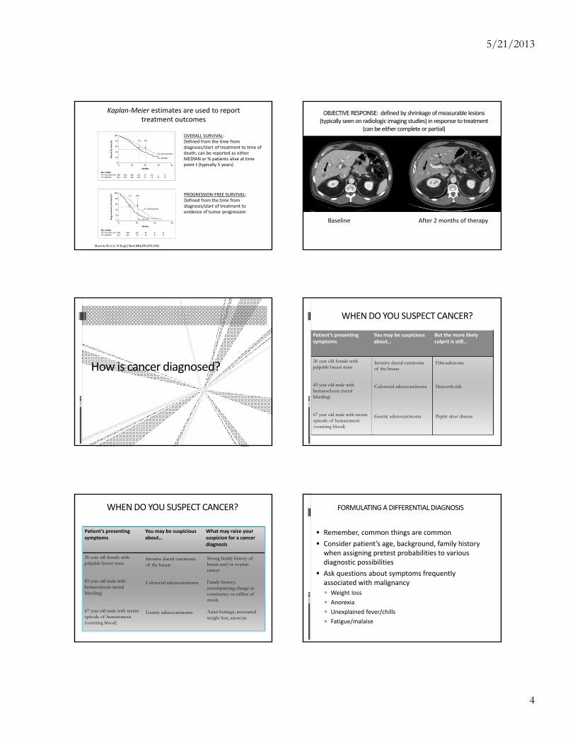
Clinical endpoints used in clinical oncology
OVERALL SURVIVAL The length of time someone is alive from diagnosis/start of treatment to time of death; reported as either MEDIAN or % patients alive at time point t (typically 1 or 5 years)
PROGRESSION‐FREE SURVIVAL The length of someone is alive and free from evidence of tumor progression
DISEASE‐FREE SURVIVAL The length of time someone is alive with no sign of the cancer returning
OVERALL RESPONSE RATE Percentage of patients who demonstrate shrinkage of measurable sites of disease (by radiologic imaging studies or physical exam) in response to treatment; can be complete or partial
QUALITY OF LIFE Overall enjoyment of life – measures aspects of an individual’s sense of well‐being and ability to carry out various activities
5/21/2013
4
Hurwitz H et al. N Engl J Med 2004;350:2335-2342
Kaplan‐Meier estimates are used to report treatment outcomes
OVERALL SURVIVAL:Defined from the time from diagnosis/start of treatment to time of death; can be reported as either MEDIAN or % patients alive at time point t (typically 5 years)
PROGRESSION‐FREE SURVIVAL:Defined from the time from diagnosis/start of treatment to evidence of tumor progression
OBJECTIVE RESPONSE: defined by shrinkage of measurable lesions (typically seen on radiologic imaging studies) in response to treatment
(can be either complete or partial)
Baseline After 2 months of therapy
How is cancer diagnosed?
WHEN DO YOU SUSPECT CANCER?
Patient’s presenting symptoms
You may be suspicious about…
But the more likely culprit is still…
26 year old female with palpable breast mass
45 year old male with hematochezia (rectal bleeding)
67 year old male with recent episode of hematemesis(vomiting blood)
Invasive ductal carcinoma of the breast
Colorectal adenocarcinoma
Gastric adenocarcinoma
Fibroadenoma
Hemorrhoids
Peptic ulcer disease
WHEN DO YOU SUSPECT CANCER?
Patient’s presenting symptoms
You may be suspicious about…
What may raise your suspicion for a cancer diagnosis
26 year old female with palpable breast mass
45 year old male with hematochezia (rectal bleeding)
67 year old male with recent episode of hematemesis (vomiting blood)
Invasive ductal carcinoma of the breast
Colorectal adenocarcinoma
Gastric adenocarcinoma
Strong family history of breast and/or ovarian cancer
Family history; accompanying change in consistency or caliber of stools
Asian heritage; associated weight loss, anorexia
FORMULATING A DIFFERENTIAL DIAGNOSIS
Remember, common things are common
Consider patient’s age, background, family history when assigning pretest probabilities to various diagnostic possibilities
Ask questions about symptoms frequently associated with malignancy
Weight loss
Anorexia
Unexplained fever/chills
Fatigue/malaise

5/21/2013
5
ESTABLISHING A CANCER DIAGNOSIS
“Tissue is the issue”
Different sampling techniques:
Fine needle aspiration cytology
Core needle biopsy
Different approaches:
Percutaneous (CT or ultrasound guidance)
Intra‐operative
Endoscopic
Examples of biopsy techniques
Normal cells of the colon
Poorly‐differentiatedAdenocarcinoma(high grade)
Well‐differentiated adenocarcinoma (low grade)
Tumors thenget an assignedGRADE based on how abnormal the cells look undera microscope
e.g.: ‐ Grade 1/2/3; ‐ Low/intermediate/high;‐Well‐/medium‐/ poorly‐differentiated
Next step: STAGINGPrimary Tumor (T)
TX primary tumor cannot be assessedT0 no evidence of primary tumorTis carcinoma in situT1,T2,T3,T4 increasing size +/or local extent of primary
tumorRegional lymph nodes (N)
Nx regional lymph nodes cannot be assessedN0 no regional lymph node metastasisN1,N2,N3 increasing involvement of regional lymph
nodesDistant metastasis (M)
Mx distant metastasis cannot be assessedM0 no distant metastasisM1 distant metastasis
TRANSLATES INTO TNM stage = overall stage I, II, III, or IV cancer
This can get very confusing…
5/21/2013
6
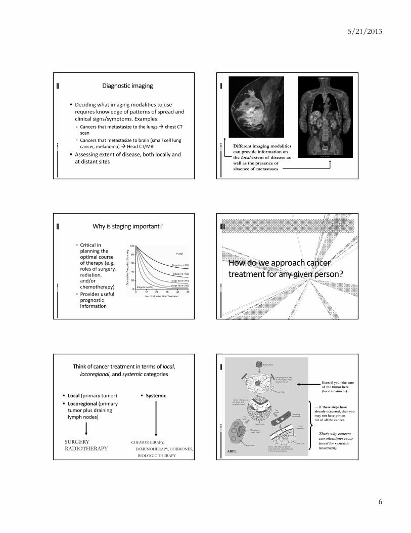
Diagnostic imaging
Deciding what imaging modalities to use requires knowledge of patterns of spread and clinical signs/symptoms. Examples:
Cancers that metastasize to the lungs chest CT scan
Cancers that metastasize to brain (small cell lung cancer, melanoma) Head CT/MRI
Assessing extent of disease, both locally and at distant sites
Different imaging modalitiescan provide information on the local extent of disease as well as the presence or absence of metastases
Why is staging important?
Critical in planning the optimal course of therapy (e.g. roles of surgery, radiation, and/or chemotherapy)
Provides useful prognostic information
How do we approach cancer treatment for any given person?
Think of cancer treatment in terms of local, locoregional, and systemic categories
Local (primary tumor)
Locoregional (primary tumor plus draining lymph nodes)
Systemic
SURGERY CHEMOTHERAPY,
RADIOTHERAPY IMMUNOHERAPY, HORMONES,
BIOLOGIC THERAPY
Even if you take careof the tumor here(local treatment)…
… if these steps havealready occurred, then youmay not have gottenrid of all the cancer.
That’s why cancerscan oftentimes recur(need for systemictreatment).
ABPI.
5/21/2013
7
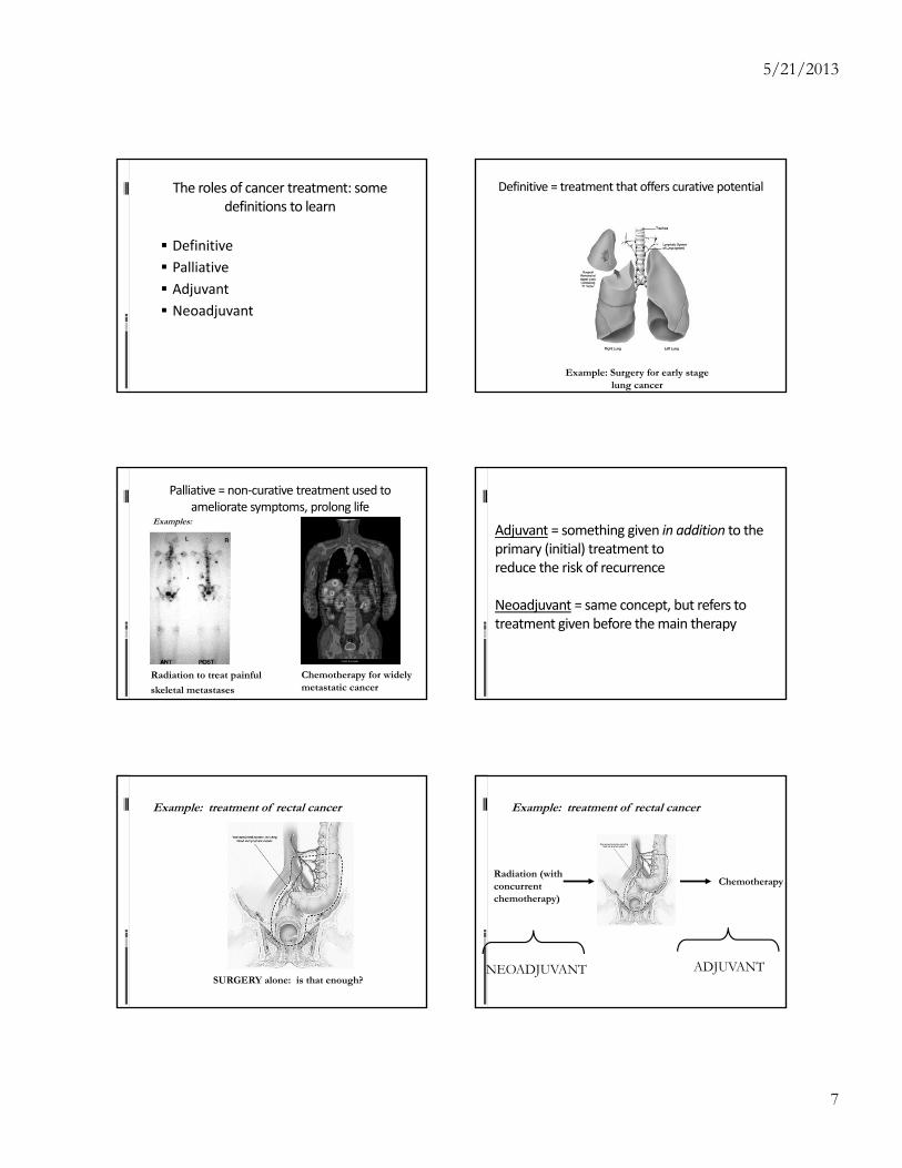
The roles of cancer treatment: some definitions to learn
Definitive
Palliative
Adjuvant
Neoadjuvant
Definitive = treatment that offers curative potential
Example: Surgery for early stagelung cancer
Palliative = non‐curative treatment used to ameliorate symptoms, prolong life
Radiation to treat painful
skeletal metastases
Chemotherapy for widely metastatic cancer
Examples:Adjuvant = something given in addition to the primary (initial) treatment to reduce the risk of recurrence
Neoadjuvant = same concept, but refers to treatment given before the main therapy
Example: treatment of rectal cancer
SURGERY alone: is that enough?
Radiation (withconcurrentchemotherapy)
Chemotherapy
Example: treatment of rectal cancer
NEOADJUVANT ADJUVANT

5/21/2013
8
NEOADJUVANT THERAPYPOTENTIAL ADVANTAGES? POTENTIAL DISADVANTAGES?
Earlier eradication of occult metastatic disease No need to wait for postoperative recovery Primary lesion still present and evaluable Downstaging/downsizing of tumor Previously undetectable metastases may become evident, sparing some patients from undergoing unnecessary operation
Delay of potentially curative operationMajor treatment‐associated side effects may weaken patient’s condition prior to surgeryObscuring accurate surgical pathologic staging
Diseases where adjuvant therapy is considered (in specific contexts)
Diseases where neoadjuvant therapy is considered (in specific contexts)
Breast
Colorectal
Lung
Pancreas
Stomach
Melanoma
Prostate
Breast
Rectal
Lung
Esophageal
Bladder
Soft tissue sarcoma
Radiation (withconcurrentchemotherapy)
Chemotherapy
ALSO LOOK AT IT THIS WAY…
The radiation is given to reducethe chances of local recurrence.
The chemotherapy is givento reduce the chances ofsystemic recurrence.
Cancer treatment typically requires a multimodality approach
Surgical oncology
Medical oncology
Radiation oncology
From : Devita VT and Rosenberg SA, Two hundred years of cancer research. N Engl J Med 366, 2012

5/21/2013
9
Question #1: Surgery is routinely used for curative intent in each of the following early‐stage malignancies except:
A. Bladder cancer
B. Gastric cancer
C. Kidney (renal cell) cancer
D. Non‐Hodgkin’s lymphoma
E. Thyroid cancer
I. SURGICAL ONCOLOGY
90% of patients with solid tumors require some surgical procedure for diagnosis, primary treatment, or management of complications during the course of treatment
Often represents the primary, and best, treatment option for patients with earlier stages of disease
Increasingly used in conjunction with other modalities (chemo, XRT) to optimize outcomes
MAJOR OBJECTIVES OF THE CANCER SURGEON:(1) SURGICAL INTERVENTION FOR CURE
MELANOMA COLON CANCER
BASIC PRINCIPLES OF SURGICAL RESECTION FOR CURE IN CANCER PATIENTS
Understanding anatomy is critical! Need to consider local extent of disease in relation to: Tissue layers Possible invasion into adjacent tissues/organs Maintaining appearance and function of the involved organ, e.g.: Breast conserving surgery vs. lumpectomy (breast cancer)
? Need for, and extent of, regional lymph node dissection (lymphadenectomy)
Resection en bloc (rather than piecemeal)
MAJOR OBJECTIVES OF THE CANCER SURGEON
2. Diagnosis and staging (incisional vs. excisionalbiopsy)
MAJOR OBJECTIVES OF THE CANCER SURGEON:3. Symptom palliation (e.g. bowel obstruction, uncontrollable bleeding) and acute surgical emergencies (e.g. spinal cord compression)

5/21/2013
10
MAJOR OBJECTIVES OF THE CANCER SURGEON
4. PREVENTION of cancer
(i.e., prophylactic operations for high‐risk individuals)
Double mastectomies for women who are BRCA mutation carriers
Total colectomy for individuals with FAP (familial adenomatous polyposis)
Total gastrectomy for individuals with CDH1 mutations (hereditary diffuse gastric cancer)
Meijers-Heijboer H et al. N Engl J Med 2001;345:159-164
Actuarial Incidence of Breast Cancer among Women with a BRCA1 or BRCA2 Mutation after Prophylactic Mastectomy or during Surveillance
MAJOR OBJECTIVES OF THE CANCER SURGEON: (5) Rehabilitation/reconstruction
Sir William Halstead and the pioneering of the
radical mastectomy (1889)
Example: Breast cancer surgery -- improvements over time in surgical approach and reconstruction
Modified radical mastectomy
http://www.breastcancer.org/images/tram_reconstruction1%5B2%5D.jpg
Modified radical mastectomy with TRAM flap
CANCER TREATMENT TYPICALLY REQUIRES A MULTIMODALITY APPROACH
Surgical oncology
Medical oncology
Radiation oncology

5/21/2013
11
Question #2: Each of the following cancers can oftentimes be cured with chemotherapy alone except:
A. Metastatic small cell lung cancer
B. Metastatic choriocarcinoma
C. Burkitt’s lymphoma
D. Metastatic testicular cancer
II. MEDICAL ONCOLOGY
Focuses on SYSTEMIC therapy
(= treatment that intends to reach cancer cells throughout the entire body)
Includes:
Chemotherapy
Hormonal therapy
Immunotherapy
Biologic/targeted therapy
THE ORIGINS OF CHEMOTHERAPY AND MEDICAL ONCOLOGY AS A SPECIALTY
Navy seamen in WWII developbone marrow hypoplasia following exposure to mustard gas leads to trial of nitrogen mustard in patients with malignant lymphoma by Gilman and Philips(1946)
BROAD CLASSES OF DRUGS USED IN MEDICAL ONCOLOGY
Category Examples
Chemotherapy antimetabolites, platinum agents, alkylating agents, taxanes, topoisomerase inhibitors, anthracyclines
Hormones Tamoxifen, anastrazole (breast ca);
GnRH agonists (prostate ca)
Drugs affecting the immune system
Interferon alfa, interleukin‐2
Monoclonal antibodies Rituximab (anti‐CD20), trastuzumab (anti‐Her2/neu), bevacizumab (anti‐VEGF), cetuximab (anti‐EGFR)
Small molecule inhibitors Imatinib, erlotinib, sorafenib, vemurafenib
Vaccines Sipulecuel‐T
Fisher R et al. N Engl J Med 1993;328:1002-1006
??
How are chemotherapy ‘cocktails’ developed? Chemotherapy: how treatment regimens are designed
Agents with demonstrated preclinical activity against particular tumor type (in vitro and animal models)
Combining chemotherapy agents should be based upon: Non‐overlapping mechanisms of action; Non‐overlapping toxicity profiles; Evidence of synergy?
Treatment cycles (e.g., every 3 weeks) are defined to allow optimal recovery time of normal tissue
5/21/2013
12
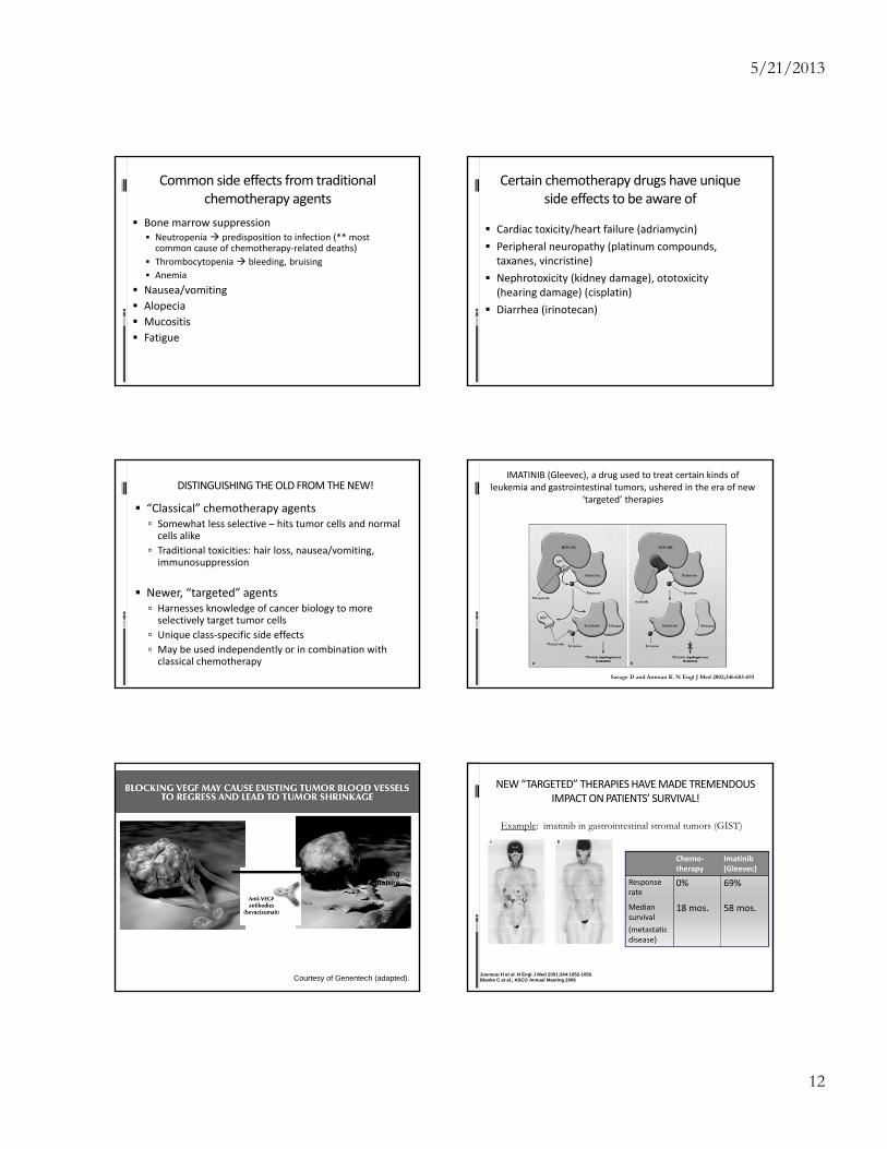
Common side effects from traditional chemotherapy agents
Bone marrow suppression Neutropenia predisposition to infection (** most common cause of chemotherapy‐related deaths)
Thrombocytopenia bleeding, bruising
Anemia
Nausea/vomiting
Alopecia
Mucositis
Fatigue
Certain chemotherapy drugs have unique side effects to be aware of
Cardiac toxicity/heart failure (adriamycin)
Peripheral neuropathy (platinum compounds, taxanes, vincristine)
Nephrotoxicity (kidney damage), ototoxicity (hearing damage) (cisplatin)
Diarrhea (irinotecan)
DISTINGUISHING THE OLD FROM THE NEW!
“Classical” chemotherapy agents Somewhat less selective – hits tumor cells and normal cells alike
Traditional toxicities: hair loss, nausea/vomiting, immunosuppression
Newer, “targeted” agents Harnesses knowledge of cancer biology to more selectively target tumor cells
Unique class‐specific side effects
May be used independently or in combination with classical chemotherapy
Savage D and Antman K. N Engl J Med 2002;346:683-693
IMATINIB (Gleevec), a drug used to treat certain kinds of leukemia and gastrointestinal tumors, ushered in the era of new
‘targeted’ therapies
Regressingvasculature
Shrinkingtumor cell
Courtesy of Genentech (adapted).
NEW “TARGETED” THERAPIES HAVE MADE TREMENDOUS IMPACT ON PATIENTS’ SURVIVAL!
Chemo‐therapy
Imatinib(Gleevec)
Response rate
0% 69%
Median survival
(metastaticdisease)
18 mos. 58 mos.
Joensuu H et al. N Engl J Med 2001;344:1052-1056Blanke C et al., ASCO Annual Meeting 2006
Example: imatinib in gastrointestinal stromal tumors (GIST)
5/21/2013
13
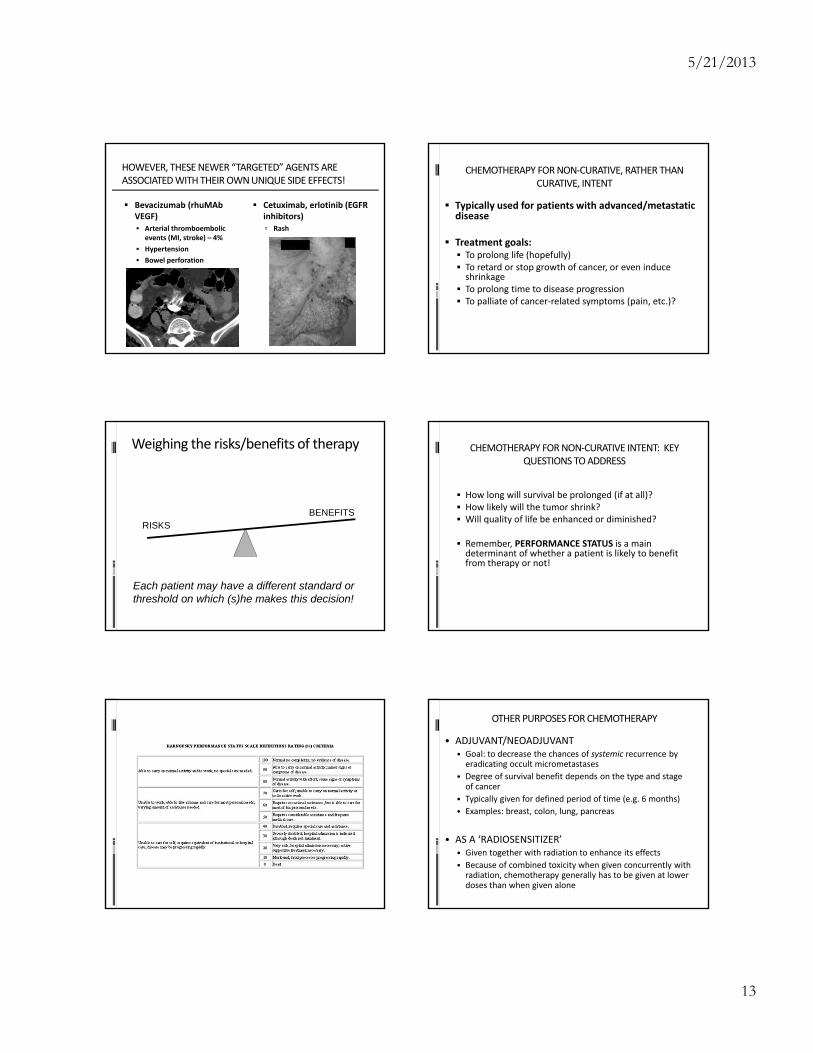
HOWEVER, THESE NEWER “TARGETED” AGENTS ARE ASSOCIATED WITH THEIR OWN UNIQUE SIDE EFFECTS!
Bevacizumab (rhuMAbVEGF) Arterial thromboembolic
events (MI, stroke) – 4%
Hypertension
Bowel perforation
Cetuximab, erlotinib (EGFR inhibitors) Rash
CHEMOTHERAPY FOR NON‐CURATIVE, RATHER THAN CURATIVE, INTENT
Typically used for patients with advanced/metastatic disease
Treatment goals: To prolong life (hopefully) To retard or stop growth of cancer, or even induce shrinkage
To prolong time to disease progression To palliate of cancer‐related symptoms (pain, etc.)?
Weighing the risks/benefits of therapy
RISKSBENEFITS
Each patient may have a different standard orthreshold on which (s)he makes this decision!
CHEMOTHERAPY FOR NON‐CURATIVE INTENT: KEY QUESTIONS TO ADDRESS
How long will survival be prolonged (if at all)? How likely will the tumor shrink? Will quality of life be enhanced or diminished?
Remember, PERFORMANCE STATUS is a main determinant of whether a patient is likely to benefit from therapy or not!
OTHER PURPOSES FOR CHEMOTHERAPY
• ADJUVANT/NEOADJUVANT• Goal: to decrease the chances of systemic recurrence by eradicating occult micrometastases
• Degree of survival benefit depends on the type and stage of cancer
• Typically given for defined period of time (e.g. 6 months)
• Examples: breast, colon, lung, pancreas
• AS A ‘RADIOSENSITIZER’• Given together with radiation to enhance its effects
• Because of combined toxicity when given concurrently with radiation, chemotherapy generally has to be given at lower doses than when given alone

5/21/2013
14
DRUG DEVELOPMENT: PHASES OF CLINICAL TRIALS
PHASE DESCRIPTION
I Often not disease‐specific; establishing correct dose, safety profile
II Efficacy against specific tumor types; further safety information. May be randomized or non‐randomized.
III Larger‐scale, randomized study comparing study treatment to standard treatment
IV Post‐FDA approval, additional testing primarily for marketing purposes
CANCER TREATMENT TYPICALLY REQUIRES A MULTIMODALITY APPROACH
Surgical oncology
Medical oncology
Radiation oncology
Question #3: In which of the following malignancies has radiation essentially replaced surgery as the primary modality for cure?
A. Anal squamous cell carcinoma
B. Malignant melanoma
C. Osteosarcoma
D. Ovarian carcinoma
E. Pancreatic cancer
III. RADIATION THERAPY
Radiation therapy (RT) has effectively treated cancer for >100 years X-rays discovered in 1895 by Röentgen First skin cancers cured by RT in 1896; first cervix cancer
cured by RT in 1903
RADIATION THERAPY (RT)
~60% of all cancer pts receive RT as part of their treatment
Can be administered: Definitively (often concurrently with chemotherapy)
Examples: anal, head and neck
Adjuvant/neoadjuvant – to reduce risk of local relapseExamples: breast, sarcoma, gastroesophageal
PalliativeExamples: bone metastases
Anal cancer: radiation (plus chemotherapy) can cure 60+ percent of patients – avoids need for permanent colostomy!

5/21/2013
15
What kind of radiation is used for treating cancer?
HOW DOES RADIATION WORK?
Photons interact with molecules in tissue to produce excitation and/or ionization
Ionization releases large amounts of energy, enough to break chemical bonds, and ejected electrons can interact with other molecules
The primary biological target of ionizing radiation is DNA (produces double-strand breaks)
Normal tissues have a substantial capacity to recover from radiation damage, whereas tumors often have defective radiation repair pathways
RADIATION DOSE
General goals Maximize dose to tumor Minimize dose to surrounding normal tissues
Units for dose 1 Gy (Gray) = 1 Joule/kg = 100 cGy = 100 rads
Fractionation The total radiation dose is usually split into smaller “fractions”
of radiation given over several weeks Higher dose per fraction can cause more toxicity, but too low
dose per fraction might not be enough to kill tumor cells Different total doses and doses per fraction have different
biological effects depending on the tissue irradiated
Tumorcontrol
Normaltissue
damage
50%
Dose (Gy)
Therapeutic ratio = % tumor control that can be achieved with a given level of (acceptable)
normal tissue damage
5%
80%95%
x
xx
x
Radiation treatment planningMODES OF RADIATION THERAPY
EXTERNAL BEAM RADIATION Traditional delivery system: Linear accelerator (LINAC)

5/21/2013
16
MODES OF RADIATION THERAPY, cont’d.
NEWER FORMS OF EXTERNAL BEAM RADIATION: greater precision, minimize exposure to normal surrounding structures
Cyberknife
MODES OF RADIATION THERAPY, cont’d.
Newer forms of external beam radiation: Gamma Knife
MODES OF RADIATION THERAPY
INTERNAL RADIATION (BRACHYTHERAPY)Examples:‐ Permanent radioactive seeds (e.g., prostate)‐ Temporary high‐dose rate implant catheters (e.g., gynecologic)
POTENTIAL COMPLICATIONS ASSOCIATED WITH RADIATION
Each organ/tissue can tolerate maximal lifetime dose of radiation, above which permanent damage can occur
Toxicities can be: Acute (occurring during and
shortly following RT) Skin irritation/breakdown, mucositis/enteritis, alopecia, fatigue
Chronic or delayed Cytopenias, scarring or stricture of affected organs, bladder or bowel urgency or incontinence, cytopenias, secondary malignancies, infertility
Cancer management: a multidisciplinary approach
Typically requires the input of different specialists including: Medical oncologist
Surgical oncologist
Radiation oncologist
Radiologist
Pathologist
Social worker
Nutritionist
Multidisciplinary tumor boards are frequently set up to address the management of specific patients
CONSIDERING A CAREER IN AN ONCOLOGIC SPECIALTY
Medical oncology
Internal medicine residency (3 years)
3‐year fellowship (+/‐ hematology)
Radiation oncology
One‐year preliminary (usually internal medicine)
4‐year radiation oncology residency
Surgical oncology
Surgical residency (5 years)
Surgical oncology fellowship (usually ~ 2 years)