Understanding & Responding Appropriately - UCLA CPC · Chest & Abdomen Trauma: Understanding &...
79
Chest & Abdomen Trauma: Understanding & Responding Appropriately Tony Melendez, RN BS, MICN EMS Educator UCLA Center for Pre-Hospital Care 2013
Transcript of Understanding & Responding Appropriately - UCLA CPC · Chest & Abdomen Trauma: Understanding &...

Chest & Abdomen Trauma: Understanding & Responding Appropriately
Tony Melendez, RN BS, MICN EMS Educator UCLA Center for Pre-Hospital Care
2013

2
Potentially Life Threatening Chest Injuries
Tracheo-bronchial rupture/laceration
open pneumothorax
tension pneumothorax
hemothorax flail chest cardiac tamponade aortic rupture
3
Tracheobronchial Injury
Actual airway damage ◦ 80% involve mainstem bronchus
◦ 15% trachea,
◦ 5% distal bronchus
◦ Usually within 2.5 cm of carina
◦ Air escapes into the thoracic cavity
◦ mortality up to 30% usu. Within 1st hour
Caused by penetrating or blunt mechanism

4
#1= Anterior (#2) #2= Middle (#3) #3= Posterior (#4) Side view: #1= Superior

5
Tracheobronchial Injury
Must be considered in all patients ◦ with penetrating injuries of the lower neck or upper chest
◦ AND any patient with evidence of violent blunt injury to the chest

6
Tracheobronchial Injury
2 groups: ◦ Free communication of injury with pleural space
OR
◦ Injury confined to peribronchial connective tissue sheath (more subtle symptoms: minimal pneumo or emphysema)

7
Signs/Symptoms
Hoarse voice Severe respiratory distress Stridor Cyanosis Subcutaneous emphysema
(cervical or mediastinal) Decreased/unequal/absent breath sounds Hemoptysis Shock

8
Field Treatment
Airway control ◦ Maintain spinal immobilization
IVs en route
Controlled airway ◦ closest trauma center
Uncontrolled airway ◦ closest facility/EDAP
◦ needle cricothyrotomy …?
9
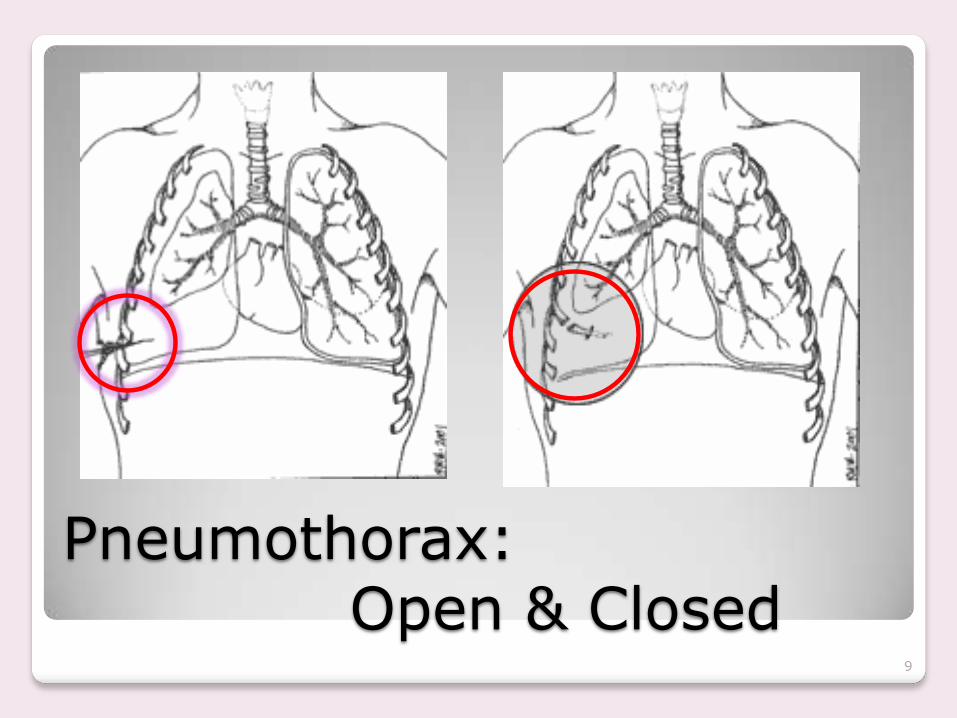
Pneumothorax: Open & Closed

10
Open Pneumothorax “Sucking Chest Wound”
Caused by penetrating or blunt mechanism
Hole in chest wall allows air to move freely in and out of the
pleural space Negative pressure is lost in Pleural Space
◦ causes lungs to passively collapse
Lung tissue usually remains intact

11
Signs and Symptoms
Sucking sound or “bubbling” at wound site on inspiration ◦ possible bubbling on expiration
Dyspnea/tachypnea
Decreased breath sounds on affected side
Unequal chest rise and fall
Possible subcutaneous air
Skins and vital signs reflect poor perfusion

12
Field Treatment
O2, monitor,
IVs en route
Spinal immobilization as indicated
Three-sided occlusive dressing
Rapid transport
In ED: Chest tube will be placed.

13
Monitoring Open Pneumothorax
Observe for signs and symptoms of tension pneumothorax (open pneumo can deteriorate)
If tension pneumothorax develops:
remove occlusive dressing &… prepare for emergent needle thoracostomy

14
Tension Pneumothorax
Blunt or penetrating mechanism Injury perforates chest wall
and/or pleural space Air becomes trapped in the pleural space
as it enters with each breath ◦ air cannot escape ◦ space enlarges ◦ lung collapses ◦ pressure builds

15
Tension Pneumothorax
Pressure builds pushing mediastinum to the opposite side
Pressure is put on heart & unaffected lung and.. will eventually kink the vena cava and deviate the trachea
Results in decreased right heart return, and thus decreased cardiac output, decreased BP

16
Tension Pneumo Deterioration

17
Tension Pneumo Deterioration

18
Signs and Symptoms
Unequal lung sounds ◦ decreased or absent on affected side
Progressive respiratory distress
Accessory muscle use
Dyspnea
May note subcutaneous air in upper chest wall
Signs of poor perfusion
Possibly decreased compliance with BVM

19
Late Signs and Symptoms
JVD
Tracheal deviation

20
Field Treatment
High flow oxygen/intubate prn
Immediate Needle Thoracostomy on affected side
Remove occlusive dressings
Spinal immobilization
Two large IVs en route/fluid resuscitate
Rapid transport
Shock position as tolerable

21
Needle Thoracostomy
Needle decompresses pleural space
Converts tension pneumothorax to open pneumothorax
Complications ◦ hemo/pneumothorax
◦ laceration of intercostal nerves/blood vessels
◦ infection

22
Needle Thoracostomy Landmarks
2nd intercostal space Mid clavicular line (2nd ICS, MCL)
On the affected side Large bore catheter: 14 ga. or larger
◦ with one-way flutter valve
Inserted perpendicular to chest wall “Walk” the needle over the top 3rd rib

Note N. thoracostomy placement

24
Needle Thoracostomy Stabilization
Listen for pop/rush of air
May get some bubbles of blood
Remove needle from catheter and take off syringe attach flutter valve
Secure cannula with 4x4s and tape
Immediate remove & apply pressure if punctured a spurting blood vessel

Heimlich valve & it’s function

Needle Thoracostomy Kit example

Questions…?
27
28
Hemothorax from blunt or penetrating trauma
Blood accumulates in pleural cavity
Caused from injury to heart, great vessels, or intercostal arteries
Will get Thoracotomy (cracked chest) in ED if blood loss through chest tube is greater than 2 to 4 ml / kg / hour

29
Hemothorax

30
Tension Pnemothorax vs. Hemothorax
May be difficult to distinguish if significant hemothorax
Both present with… signs of poor perfusion and unequal
breath sounds More common to see
hypotension before respiratory distress
Usually pleural space fills with air and blood

31
Cardiac Tamponade Caused by blunt or penetrating mechanism
Accumulation of blood into pericardial sac that surrounds heart
Blood in sac reduces chamber filling
Pressure backs up and results in decreased right heart return and thus decreased stroke volume and decreased cardiac output

32
Becks Triad Characteristic of Cardiac Tamponade
Hypotension (narrowed pulse pressure)
JVD
Muffled, distant heart sounds
Patient will also display…
Tachycardia
Dyspnea
Poor skin vitals
33
Field Treatment
Trauma field treatment
Pericardiocentesis
to be performed in ED

34
Tension Pnemothorax vs. Cardiac Tamponade
Both present with
signs of poor perfusion
Both have chest trauma
Difference is breath sounds ◦ tension pnemo=unequal BS
◦ C. tamponade=equal BS

Questions…? 35

36
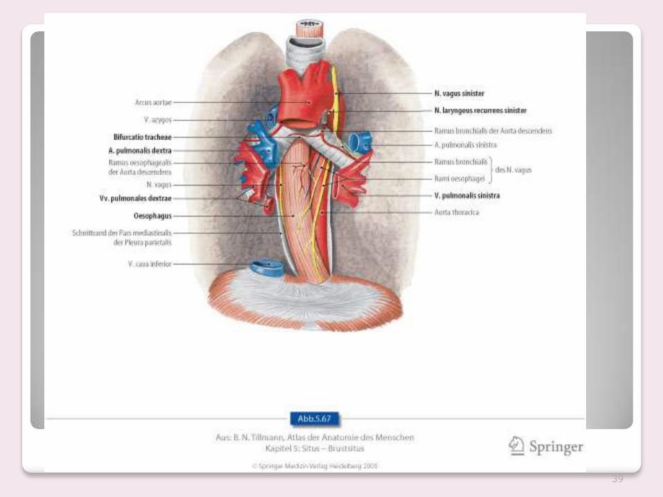
Aortic Rupture
Usually from a rapid deceleration mechanism ◦ Motor Vehicle Accidents
◦ falls
◦ crush injury
Usual site: the distal aortic arch just beyond left subclavian artery takeoff where it is firmly tethered

37
Aortic Rupture
Massive hemorrhage within few minutes ◦ severe injury; Rapid Deceleration Shearing ◦ 80-90% fatality rate within 1st hour
Most common cause of Sudden Death
post MVC Estimated that 1:6 who die in MVA’s
sustain an aortic rupture

38
39

40
Signs/Symptoms
Chest pain
Dyspnea
May have…
no obvious signs of chest trauma
(up to 50%!)

41
Trauma Field Treatment
◦ Hi-flow O2
◦ Support airway as needed
◦ cardiac monitoring
◦ spinal precautions
◦ shock position
◦ IVs (x2) en route
◦ fluid resuscitate (500 cc) with s/o shock
◦ Keep Warm (maintain body temperature)

42

43

44
Flail Chest
2 or more rib fractures in at least 2 places Paradoxical motion of flail segment Segment moves: in on inspiration (Normal is out on Insp.)
out on expiration (Normal is in on Exp.)

45
Flail chest
Risk for pneumothorax from fractured rib
Signs and Symptoms: ◦ dyspnea/tachypnea ◦ localized chest pain ◦ may have palpable crepitus ◦ s/o poor perfusion from poor oxygen exchange
Massive force needed to cause injury ◦ think about other injuries
(4-10) (8-12) (1-2)

46
Treatment for flail chest
High flow oxygen/IVs en route/monitor
Stabilize flail segment with bulky taped dressing
IVs en route

47
Diaphragmatic Rupture
Diaphragm separates abdominal and thoracic cavity
Abdominal contents rupture through thin diaphragm wall and enter chest cavity ◦ from sharp increase in
intra-abdominal pressure
L>R side from liver protection

48
Signs and Symptoms of Diaphragmatic Rupture
Restricted lung ventilation
decreased venous return
Dyspnea & hypotension ensues
May c/o abdominal pain Bowel sounds
heard in chest Multiple injuries
usually involved Historically
< 5% of blunt trauma

49
Diaphragmatic Rupture
80 – 90% occur from MVC’s
Lateral Impact 3x more likely to cause rupture. “T-Boned”
Postero-lateral aspect of Diaphragm is its embryologic weak point.
(L)>(R) rupture likely from (R) liver protection (80-90% occur on (L) side)
Preoperatively Dx’d in only 40-50% for (L)

50
Traumatic Asphyxia
Results from severe crushing injury to chest and abdomen
Rapid increased intra-thoracic pressure
Blood forced into veins of upper thorax, neck and face
Results in reddish-purple discoloration of face and neck, JVD, conjunctival hemorrhage

51
Traumatic Asphyxia

52
Pulmonary Contusion
Rapid deceleration forces most common
Hemoptysis from hemorrhaging alveoli
Pulmonary edema ensues
Injury to actual Lung Parenchyma.

53
Pulmonary Contusion
Among the most common result of Blunt Force Trauma along with rib fractures & pneumothorax (Think: Airbags!)
Occurs in 17% of multiple trauma patients
A form of hematoma to lung tissue.
Mortality 6 – 25% due to superimposed pneumonia, ARDS, embolic blood clots
54
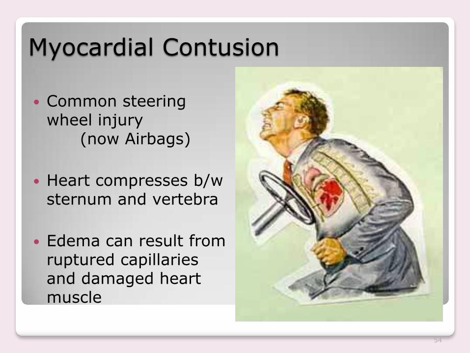
Myocardial Contusion
Common steering wheel injury (now Airbags)
Heart compresses b/w sternum and vertebra
Edema can result from ruptured capillaries and damaged heart muscle

55
Myocardial Contusion Presentations
Can range from minor to MI May be asymptomatic up to 8 hrs Can present in cardiogenic shock May occur with cardiac tamponade Need to assume contusion
with significant blunt chest trauma
Adequate paramedic report of mechanism essential
56
S/S Myocardial Contusion
Can present similar to angina/MI
Dyspnea: Ronchi, Wheezes, Hemoptysis
Palpitations
Possible dysrhythmias
Obvious chest trauma, or NOT!
Treatment may include Amiodarone for Ventricular Tachydysrhythmias

57
58
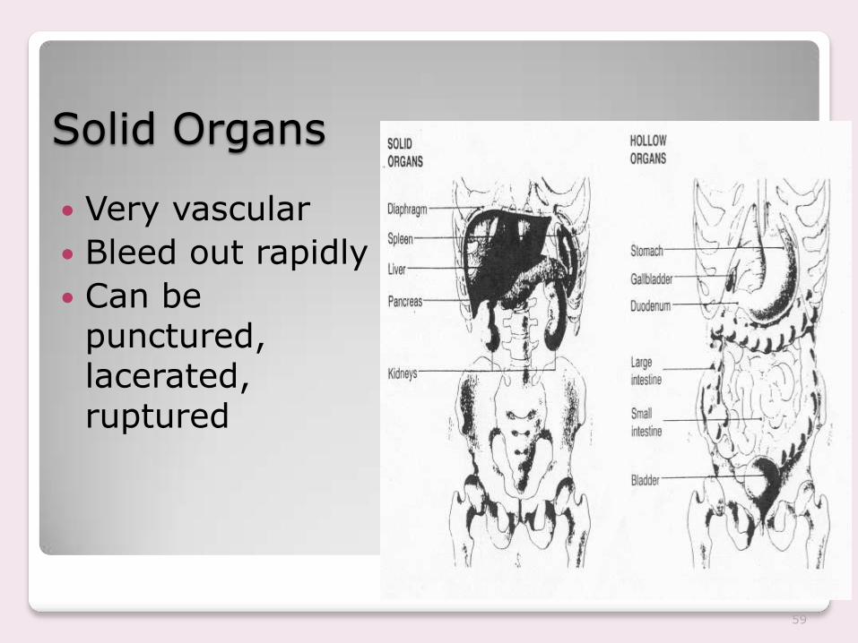
Abdominal Trauma Understanding Injury of Solid versus Hollow organs
59
Solid Organs
Very vascular
Bleed out rapidly
Can be punctured, lacerated, ruptured

60
Hollow Organs
Usually contain fluids
Fluids can spill into abdominal cavity after injury
Causes peritonitis and sepsis
Can rupture if full ◦ water balloon effect

61
Signs/Symptoms of BAT
Rigid abdomen
Distended abdomen
Abdominal guarding
Kerr’s sign ◦ pain referred to left shoulder from splenic injury
Or, soft, round, with minimal tenderness!!

62
Abdomen regions: What’s Involved
63
The “4” Abdomens
Intrathoracic Abdomen:
upper abdomen that lies beneath the rib cage (the ribcage makes complete abdomen exam difficult)
◦ Diaphragm
◦ Liver
◦ Spleen
◦ Stomach

64
The “True” Abdomen
Contains the intestines, Small & Large
(with highly omentum!)
The Uterus when gravid
The bladder when distended, full
Perforation in this area is associated with significant physical findings, pain & tenderness from peritonitis

66
The “Retroperitoneal” Abdomen
This area is “behind the guts” Problems here are difficult to diagnose
by physical exam alone ◦ Kidneys ◦ Adrenal glands ◦ Ureters ◦ Pancreas ◦ Aorta ◦ Vena Cava


68
The “Pelvic” Abdomen
The bony pelvis ◦ Urinary bladder ◦ Urethra ◦ Rectum ◦ Some small intestine ◦ Ovaries, Fallopian tubes ◦ Uterus
You can lose 50% of blood volume here!

“Pelvic” Abdomen
Female Pelvis Male Pelvis

70
Evisceration
Cover with moist , sterile dressing
Place patient supine with knees slightly flexed
Placing contents back inside Abdominal Cavity is contra-indicated.
71
Be guided by mechanisms & associated forces
Collisions by the injured person & the external environment results in sudden,massive increased intra-abdominal pressure. (external compression)
Next, acceleration & deceleration forces
directly affect the organs … and the bony body (create shear forces at fixed points of organ attachment)

72
And Thirdly….
Also, a crushing effect on intra-abdominal organs lying between the vertebral column & abdominal wall (solid viscera particularly vulnerable)
73
Physical exam can be… notoriously unreliable!
Don’t let the findings fool you
Although exam appears normal ◦ Soft, nontender, nondistended
Bleeding still may occur within the abdomen despite a normal exam
Also distracting injuries, abdominal Wall spasms, AMS, Intoxication all confuse the exam.

74
Trauma Center Criteria in LA Co.
Remember: Trauma patients with complaints of diffuse abdominal trauma go to a trauma center
Diffuse abdominal trauma is defined as pain in two or more of the four abdominal quadrants
The four quadrants of the abdomen are: ◦ RUQ, LUQ, RLQ, LLQ

75
Consider Pain Management

76
Who Should Receive Pain Medication
Patients who have an isolated traumatic extremity injury, burn, fractured hip, or chief complaint of pain.
Caution with: Head injuries
Multi-system trauma
Labor
Abdominal pain
Elderly

77
Morphine Pain Management
Adults: IV Dosage : 2-10 mg slow IVP titrated to
pain relief. May repeat to a max dose of 20mg.
IM Dosage : 10mg IM one time dose. IV is the recommended route.
If unable to start an IV or patient does not require IV, IM is an option.

78
Any Questions?

79
Thanks for Listening