Understanding Neurotransmission and the Disease … Neurotransmission and the Disease of Addiction 2...
16
www.EliteCME.com Page 1 Understanding Neurotransmission and the Disease of Addiction 2 CE Hours By: Kathryn Brohl, MA, LMFT Learning objectives This workshop is designed to help you: Assess neurotransmission in the context of drug use and research. Describe the neurotransmitter – neurotransmission interaction. Apply different research methodologies. Analyze certain medication and behavioral management interventions. Describe neurotransmitters and the drugs that affect them. Apply changes to the Criteria for Substance Dependence and Abuse in the new DSM-5. Assess the long term effects of drug use. Introduction The National Institute of Alcohol Abuse and Alcoholism (2013), nearly 88,000 people (approximately 62,000 men and 26,000 women) die from alcohol related causes annually, making it the third leading preventable cause of death in the United States. In 2006, alcohol misuse problems cost the United States $223.5 billion. ● Adults (ages 18+): Approximately 17 million adults ages 18 and older (7.2 percent of this age group) had an Alcohol use disorder (AUD) in 2012. This includes 11.2 million men (9.9 percent of men in this age group) and 5.7 million women (4.6 percent of women in this age group). ○ About 1.4 million adults received treatment for an AUD at a specialized facility in 2012 (8.4 percent of adults in need). This included 416,000 women (7.3 percent of women in need) and 1.0 million men (8.9 percent of men in need). ● Youth (ages 12–17): In 2012, an estimated 855,000 adolescents ages 12–17 (3.4 percent of this age group) had an AUD. This number includes 444,000 females (3.6 percent) and 411,000 males (3.2 percent). ○ An estimated 76,000 adolescents received treatment for an AUD at a specialized facility in 2012 (8.9 percent of adolescents in need). This included 28,000 females (6.3 percent of adolescent females in need) and 48,000 males (11.7 percent of adolescent males in need). Drug and alcohol dependence continues to challenge mental health professionals as they partner with responsible addictionologists (medical doctors who are board-eligible or certified by the American Society of Addiction Medicine) to utilize a two pronged approach to providing medical intervention/management and psychotherapeutic treatment for their patients/clients. During recent decades, the accumulation of research has enlightened the addictions field, while it has also provided patients/clients, validation with regard to their physical and mental cravings, and relapse triggers following their withdrawal from drugs or alcohol. In addition it has given interventionists a deeper understanding about the disease of addiction, and enhances medication management and psychotherapy approaches, to the benefit of their patients/clients. Overall, the field of addiction has gathered a substantial body of research that has identified specific drugs’ effects on neurotransmission, establishing that drug dependence and addiction are elements of organic brain disease. By altering neurotransmission, addictive drugs produce effects that make people want to continue to abuse them, and induce health problems that can be penetrating and long term. Notably, these effects are drug specific, and disrupt particular neurotransmitters in specific ways. However, some relevant effects, such as initial pleasurable feelings, subsequent dependence and addiction, are shared by all. The end result is a disruption of the dopamine neurotransmitter system. When neuroscientists seek to better understand why a drug is abused and the subsequent consequences of this abuse, they ask: 1. Which neurotransmitter or neurotransmitters does the drug affect? 2. How does the drug alter transmission? In this course, learners will gain further understanding about the interaction between neurotransmitters and addictive substances, and how they alter neurotransmission, and subsequent thinking and behaviors. Learners will also gain a deeper awareness about research methodologies about neurotransmission and drug interaction, as well as recent scientific findings. Abused drugs affect the way people think, feel, and behave. The long term effects ultimately interface with our overall society as well; at great emotional and financial cost to those affected. Long-term effects of drugs on the brain A normal question is to ask why drugs are bad. After all, the “high” or “rush” only lasts a little while, right? What else could be happening in the drug abuser’s brain? However, one must consider that the brain is continuously changing, and learning occurs because neurons are forming new synapses. Scientists say that the brain has plasticity. It does not mean the brain is made of a chemical plastic, but it refers to the brain’s ability to modify connections in response to experience. When a person learns something or has new experiences, some new synapses may form, or existing synapses may get stronger. Other synapses may disappear. When a person takes drugs repeatedly, the experience literally changes the brain. If a person takes drugs and then stops, he or she will “crave” the drug. In other words, the individual will have a strong desire to take more of the drug. Scientists can actually see evidence of cravings in the brain. For example, if a cocaine addict sees pictures of
Transcript of Understanding Neurotransmission and the Disease … Neurotransmission and the Disease of Addiction 2...
www.EliteCME.com Page 1
Understanding Neurotransmission and the Disease of Addiction
2 CE Hours
By: Kathryn Brohl, MA, LMFT
Learning objectives
This workshop is designed to help you: Assess neurotransmission in the context of drug use and research. Describe the neurotransmitter – neurotransmission interaction. Apply different research methodologies. Analyze certain medication and behavioral management
interventions.
Describe neurotransmitters and the drugs that affect them. Apply changes to the Criteria for Substance Dependence and
Abuse in the new DSM-5. Assess the long term effects of drug use.
Introduction
The National Institute of Alcohol Abuse and Alcoholism (2013), nearly 88,000 people (approximately 62,000 men and 26,000 women) die from alcohol related causes annually, making it the third leading preventable cause of death in the United States. In 2006, alcohol misuse problems cost the United States $223.5 billion.
● Adults (ages 18+): Approximately 17 million adults ages 18 and older (7.2 percent of this age group) had an Alcohol use disorder (AUD) in 2012. This includes 11.2 million men (9.9 percent of men in this age group) and 5.7 million women (4.6 percent of women in this age group).
○ About 1.4 million adults received treatment for an AUD at a specialized facility in 2012 (8.4 percent of adults in need). This included 416,000 women (7.3 percent of women in need) and 1.0 million men (8.9 percent of men in need).
● Youth (ages 12–17): In 2012, an estimated 855,000 adolescents ages 12–17 (3.4 percent of this age group) had an AUD. This number includes 444,000 females (3.6 percent) and 411,000 males (3.2 percent).
○ An estimated 76,000 adolescents received treatment for an AUD at a specialized facility in 2012 (8.9 percent of adolescents in need). This included 28,000 females (6.3 percent of adolescent females in need) and 48,000 males (11.7 percent of adolescent males in need).
Drug and alcohol dependence continues to challenge mental health professionals as they partner with responsible addictionologists (medical doctors who are board-eligible or certifi ed by the American Society of Addiction Medicine) to utilize a two pronged approach to providing medical intervention/management and psychotherapeutic treatment for their patients/clients.
During recent decades, the accumulation of research has enlightened the addictions fi eld, while it has also provided patients/clients, validation with regard to their physical and mental cravings, and relapse triggers following their withdrawal from drugs or alcohol. In addition it has given interventionists a deeper understanding about the disease of addiction, and enhances medication management and psychotherapy approaches, to the benefi t of their patients/clients.
Overall, the fi eld of addiction has gathered a substantial body of research that has identifi ed specifi c drugs’ effects on neurotransmission, establishing that drug dependence and addiction are elements of organic brain disease. By altering neurotransmission, addictive drugs produce effects that make people want to continue to abuse them, and induce health problems that can be penetrating and long term.
Notably, these effects are drug specifi c, and disrupt particular neurotransmitters in specifi c ways. However, some relevant effects, such as initial pleasurable feelings, subsequent dependence and addiction, are shared by all. The end result is a disruption of the dopamine neurotransmitter system.
When neuroscientists seek to better understand why a drug is abused and the subsequent consequences of this abuse, they ask:1. Which neurotransmitter or neurotransmitters does the drug affect? 2. How does the drug alter transmission?
In this course, learners will gain further understanding about the interaction between neurotransmitters and addictive substances, and how they alter neurotransmission, and subsequent thinking and behaviors. Learners will also gain a deeper awareness about research methodologies about neurotransmission and drug interaction, as well as recent scientifi c fi ndings. Abused drugs affect the way people think, feel, and behave. The long term effects ultimately interface with our overall society as well; at great emotional and fi nancial cost to those affected.
Long-term effects of drugs on the brain
A normal question is to ask why drugs are bad. After all, the “high” or “rush” only lasts a little while, right? What else could be happening in the drug abuser’s brain? However, one must consider that the brain is continuously changing, and learning occurs because neurons are forming new synapses. Scientists say that the brain has plasticity. It does not mean the brain is made of a chemical plastic, but it refers to the brain’s ability to modify connections in response to experience. When a person learns something or has new experiences, some new
synapses may form, or existing synapses may get stronger. Other synapses may disappear.
When a person takes drugs repeatedly, the experience literally changes the brain. If a person takes drugs and then stops, he or she will “crave” the drug. In other words, the individual will have a strong desire to take more of the drug. Scientists can actually see evidence of cravings in the brain. For example, if a cocaine addict sees pictures of
Page 2 www.EliteCME.com
drug paraphernalia: PET scans show that the part of the brain that is important for memory (called the amygdala) is activated. If the addict sees a video with mountains, trees, and animals, the amygdala is not stimulated. Thus, just seeing pictures of drugs or things associated with drugs can trigger an uncontrollable urge for drugs.
After taking drugs for a period of time, a person may need to take a higher dose to have the same feeling or “rush” that he or she did when fi rst taking the drug. This is called tolerance. The brain has adapted to having a certain amount of drug present and does not respond the same way it did initially. For this reason, drug abusers and addicts take increasingly higher amounts of an abused drug. Tolerance may develop because the body may become more effi cient at eliminating the chemical from the body, or because the cells of the body and brain become less responsive to the effect of the drug.
Drugs can also change the structure of the brain. Perhaps one of the most dramatic long-term effects of a drug is to kill neurons. Many people have heard that drinking alcohol will kill brain cells, and it’s true. If alcohol is abused over a period of time, neurons in the brain can die. Some neurons in the brain are more sensitive to alcohol than others. Neurons that make up the mammillary bodies, areas in the brain that are important for memory, are more vulnerable to the effects of alcohol than are some other neurons in the brain. The neurons in the cerebral cortex, the part of the brain that controls most of our mental functions and endows us with consciousness, may also die if a person frequently abuses alcohol in high doses.
Another drug that is toxic to neurons is an amphetamine derivative called MDMA, or ecstasy. In rats and non-human primates, MDMA appears to kill neurons that produce serotonin. In some parts of the brain, the axons of some of these neurons may regenerate (or re-grow) after drug use is stopped, but the new growth of the neurons is not normal. Some areas are not reinnervated (nerve fi bers do not grow
back into the area) as they were before the drug abuse and some areas have abnormally high regrowth of the neurons. Either way, the neurons are not normal. Studies have not yet been able to determine if MDMA has this same effect on humans, but some preliminary evidence indicates that MDMA may kill serotonin neurons in humans.
Cocaine also changes the brain in ways that may last for a long period of time. PET scans of human brains have shown that glucose metabolism is reduced even three months after the last use of cocaine. Remember, that glucose metabolism is an indicator of how active the brain cells are. If the neurons are using less glucose, they are not as active. The changes that cocaine causes in the brain last much longer than the pleasurable feelings it produces. Other drugs cause similar decreases in brain activity. Even two years after the last use of amphetamines, PET images show that the drug abuser’s brain is less active than the person who never used drugs.
While scientifi c studies have clearly shown that certain drugs can cause dramatic changes in the brain, not all questions have been answered. Scientists, for many reasons, don’t know all of the effects that a drug may have. First, the brain is such a complicated organ that, despite great scientifi c advances, understanding all that it does, will take many more years. Second, individuals may respond differently to drugs due to genetic differences among people. Third, many drug abusers abuse more than one drug. Many individuals who take cocaine, for example, also drink alcohol. The combination of the drugs makes it diffi cult to determine what the effect of one drug alone may be. Another complication is drug addicts may have other health problems, in addition to their drug problem. Heroin addicts, for example, spend most of their energy and activity trying to get their next “fi x.” Consequently, they do not eat well and may have impaired immune systems. Also, drug addicts often suffer from mental illnesses, such as depression. The changes that occur in the brain because of mental illness make it diffi cult to determine what changes the drugs have caused.
Understanding diagnosis criteria
Developed and published by the American Psychiatric Association (APA), the Diagnostic and Statistical Manual of Mental Disorders (DSM) is the manual used by clinicians and researchers to diagnose and classify mental disorders.
According to the APA (2013), within the fi fth edition of the Diagnostic and Statistical Manual of Mental Disorders (DSM-5), the revised chapter of “Substance-Related and Addictive Disorders” includes substantive changes to the disorders grouped there plus changes to the criteria of certain conditions.
Substance Use DisorderWhile the DSM-IV utilized two separate diagnoses of “Dependence” and “Abuse”, substance use disorder in DSM-5 combines the DSM-IV categories of substance abuse and substance dependence into a single disorder measured on a continuum from mild to severe. Each specifi c substance (other than caffeine, which cannot be diagnosed as a substance use disorder) is addressed as a separate use disorder (e.g., alcohol use disorder, stimulant use disorder, etc.), but nearly all substances are diagnosed based on the same overarching criteria. In this overarching disorder, the criteria have not only been combined, but strengthened.
Whereas a diagnosis of substance abuse previously required only one symptom, mild substance use disorder in DSM-5 requires two to three symptoms from a list of 11. Drug craving has also been added to the list, and the criteria detailing “problems with law enforcement” have been eliminated because of cultural considerations that make the criteria diffi cult to apply internationally.
In DSM-IV, the distinction between abuse and dependence was based on the concept of abuse as a mild or early phase and dependence as
the more severe manifestation. In practice, the abuse criteria were sometimes quite severe. The revised substance use disorder, a single diagnosis, may result in a better match the symptoms that patients experience.
Previously, the diagnosis of dependence caused much confusion. Most people link dependence with “addiction” when in fact dependence can be a normal body response to a substance.
Substance use disorders span a wide variety of problems arising from substance use, and cover 11 different criteria:1. Taking the substance in larger amounts or for longer than the you
meant to.2. Wanting to cut down or stop using the substance but not managing to.3. Spending a lot of time getting, using, or recovering from use of the
substance.4. Cravings and urges to use the substance.5. Not managing to do what you should at work, home or school,
because of substance use.6. Continuing to use, even when it causes problems in relationships.7. Giving up important social, occupational or recreational activities
because of substance use.8. Using substances again and again, even when it puts the you in
danger.9. Continuing to use, even when you know you have a physical or
psychological problem that could have been caused or made worse by the substance.
10. Needing more of the substance to get the effect you want (tolerance).11. Development of withdrawal symptoms, which can be relieved by
taking more of the substance.
www.EliteCME.com Page 3
Addictive disorders
The chapter also includes gambling disorder as the sole condition in a new category on behavioral addictions. DSM-IV listed pathological gambling but in a different chapter. This new term and its location in the new manual refl ect research fi ndings that gambling disorder is similar to substance-related disorders in clinical expression, brain origin, comorbidity, physiology, and treatment.
Recognition of these commonalities will help people with gambling disorder get the treatment and services they need, and others may better understand the challenges that individuals face in overcoming this disorder.
While gambling disorder is the only addictive disorder included in DSM-5 as a diagnosable condition, Internet gaming disorder will be included in Section III of the manual. Disorders listed there require further research before their consideration as formal disorders. This condition is included to refl ect the scientifi c literature on persistent and recurrent use of Internet games, and a preoccupation with them, can result in clinically signifi cant impairment or distress. Much of this literature comes from studies in Asian countries. The condition criteria do not include general use of the Internet, gambling, or social media at this time.
Additional DSM-5 information
DSM-5 no longer includes caffeine use disorder, although research shows that as little as two to three cups of coffee can trigger a withdrawal effect marked by tiredness or sleepiness. There is suffi cient evidence to support this as a condition, however it is not yet clear to what extent it is a clinically signifi cant disorder. To encourage further
research on the impact of this condition, caffeine use disorder is included in Section III of DSM-5.
Additionally, the DSM-5 eliminated “legal problems” as a criterion for both substance use disorder and addictive disorders.
Defi ning neurotransmission as a communicator
Simply put, the process of communication between brains cells is called neurotransmission. Information is relayed from cell to cell to regions that process and attach meaning and memory, taking the form, when within the cell, of an electrical signal.
Consequently, in order to cross the tiny intercellular gap that separates one cell from the next, the information takes the form of a chemical signal. The specialized chemicals that carry the signals across the intercellular gaps, or synapses, are, thus, called neurotransmitters.
These interactions are an essential component of the brain’s response to experience and the environment.
Communication between neurons is the foundation for brain function. Understanding how neurotransmission occurs is crucial to understanding how the brain processes and integrates information, as it interacts with drugs. Interruption of neural communication causes changes in cognitive processes and behavior.
The brain is made up of nerve cells and glial cells
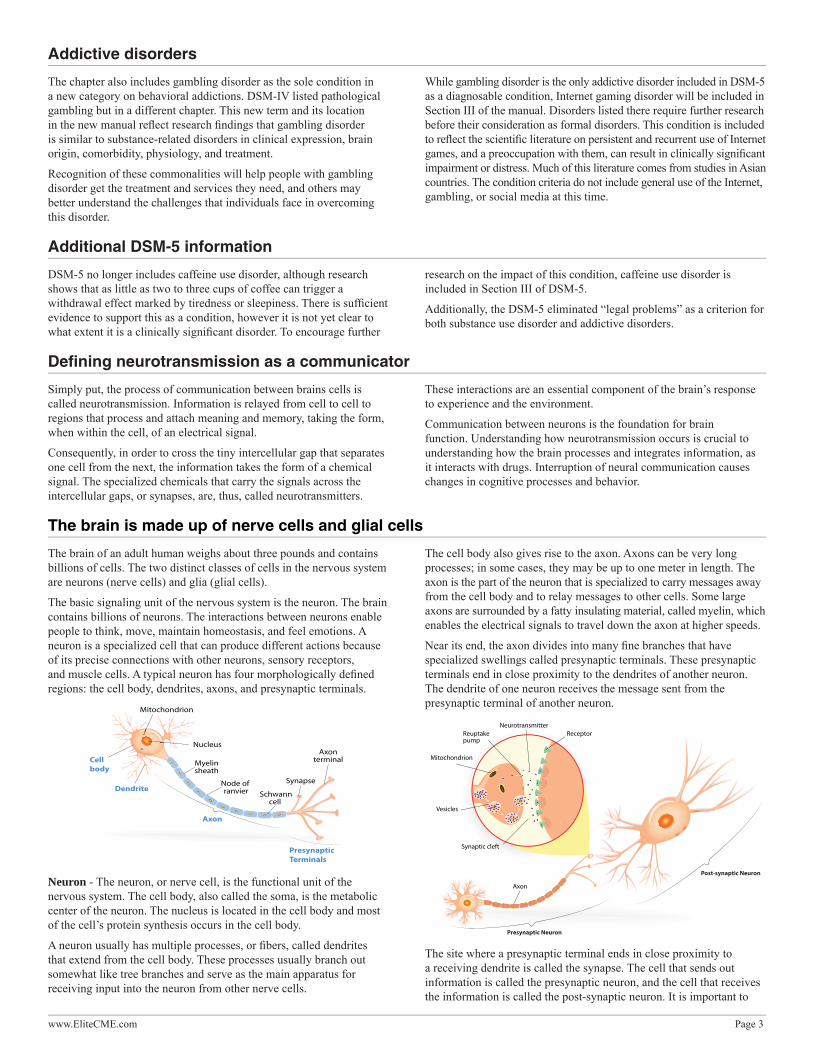
The brain of an adult human weighs about three pounds and contains billions of cells. The two distinct classes of cells in the nervous system are neurons (nerve cells) and glia (glial cells).
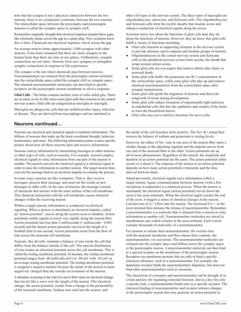
The basic signaling unit of the nervous system is the neuron. The brain contains billions of neurons. The interactions between neurons enable people to think, move, maintain homeostasis, and feel emotions. A neuron is a specialized cell that can produce different actions because of its precise connections with other neurons, sensory receptors, and muscle cells. A typical neuron has four morphologically defi ned regions: the cell body, dendrites, axons, and presynaptic terminals.
Presynaptic Terminals
Neuron - The neuron, or nerve cell, is the functional unit of the nervous system. The cell body, also called the soma, is the metabolic center of the neuron. The nucleus is located in the cell body and most of the cell’s protein synthesis occurs in the cell body.
A neuron usually has multiple processes, or fi bers, called dendrites that extend from the cell body. These processes usually branch out somewhat like tree branches and serve as the main apparatus for receiving input into the neuron from other nerve cells.
The cell body also gives rise to the axon. Axons can be very long processes; in some cases, they may be up to one meter in length. The axon is the part of the neuron that is specialized to carry messages away from the cell body and to relay messages to other cells. Some large axons are surrounded by a fatty insulating material, called myelin, which enables the electrical signals to travel down the axon at higher speeds.
Near its end, the axon divides into many fi ne branches that have specialized swellings called presynaptic terminals. These presynaptic terminals end in close proximity to the dendrites of another neuron. The dendrite of one neuron receives the message sent from the presynaptic terminal of another neuron.
Presynaptic Neuron
Post-synaptic Neuron
The site where a presynaptic terminal ends in close proximity to a receiving dendrite is called the synapse. The cell that sends out information is called the presynaptic neuron, and the cell that receives the information is called the post-synaptic neuron. It is important to
Page 4 www.EliteCME.com
note that the synapse is not a physical connection between the two neurons; there is no cytoplasmic continuity between the two neurons. The intercellular space between the presynaptic and postsynaptic neurons is called the synaptic space or synaptic cleft.
Researchers originally thought that electrical impulses jumped these gaps, like electricity jumps across the gap in a spark plug. Now scientists know this is false. Chemicals-not electrical impulses- travel across the gap.
An average neuron forms approximately 1,000 synapses with other neurons. It has been estimated that there are more synapses in the human brain than there are stars in our galaxy. Furthermore, synaptic connections are not static. Neurons form new synapses or strengthen synaptic connections in response to life experiences.
The synapse is the site where chemicals pass between neurons. Neurotransmitters are released from the presynaptic neuron terminals into the extracellular space called the synaptic cleft or synaptic space. The released neurotransmitter molecules can then bind to specifi c receptors on the postsynaptic neuron membrane to elicit a response.
Glial Cells - The brain contains another class of cells called glia. There are as many as ten to fi fty times more glial cells than neurons in the central nervous system. Glial cells are categorized as microglia or macroglia.
Microglia are phagocytic cells that are mobilized after injury, infection or disease. They are derived from macrophages and are unrelated to
other cell types in the nervous system. The three types of macroglia are oligodendrocytes, astrocytes, and Schwann cells. The oligodendrocytes and Schwann cells form the myelin sheaths that insulate axons and enhance conduction of electrical signals along the axons.
Scientists know less about the functions of glial cells than they do about the functions of neurons. However, they do know that glial cells fulfi ll a variety of functions including:
● Glial cells function as supporting elements in the nervous system to provide structure and to separate and insulate groups of neurons.
● Oligodendrocytes in the central nervous system and Schwann cells in the peripheral nervous system form myelin, the sheath that wraps around certain axons.
● Some glial cells are scavengers that remove debris after injury or neuronal death.
● Some glial cells buffer the potassium ion (K+) concentration in the extracellular space, while some glial cells take up and remove chemical neurotransmitters from the extracellular space after synaptic transmission.
● Some glial cells guide the migration of neurons and direct the outgrowth of axons during development.
● Some glial cells induce formation of impermeable tight junctions in endothelial cells that line the capillaries and venules of the brain to form the blood-brain barrier.
● Glial cells may serve nutritive functions for nerve cells.
Neurons continued…
Neurons use electrical and chemical signals to transmit information. The billions of neurons that make up the brain coordinate thought, behavior, homeostasis, and more. The following information paints a more specifi c picture about how all these neurons pass and receive information.
Neurons convey information by transmitting messages to other neurons or other types of cells, such as muscles. For example, neurons employ electrical signals to relay information from one part of the neuron to another. The neuron converts the electrical signal to a chemical signal in order to pass the information to another neuron. The target neuron then converts the message back to an electrical impulse to continue the process.
In some ways, neurons act like computers. That is, they receive messages, process their message, and send out the results as new messages to other cells. In the case of neurons, the message consists of chemicals that interact with the outer surface of the cell membrane. This chemical interaction with the cell membrane causes chemical changes within the receiving neuron.
Within a single neuron, information is conducted via electrical signaling. When a neuron is stimulated, an electrical impulse, called an “action potential”, moves along the neuron axon or dendrite. Action potentials enable signals to travel very rapidly along the neuron fi ber. Action potentials last less than 2 milliseconds (1 millisecond = 0.001 second) and the fastest action potentials can travel the length of a football fi eld in one second. Action potentials result from the fl ow of ions across the neuronal cell membrane.
Neurons, like all cells, maintain a balance of ions inside the cell that differs from the balance outside of the cell. This uneven distribution of ions creates an electrical potential across the cell membrane. This is called the resting membrane potential. In humans, the resting membrane potential ranges from -40 millivolts (mV) to -80 mV with –65 mV as an average resting membrane potential. The resting membrane potential is assigned a negative number because the inside of the neuron is more negatively charged than the outside environment of the neuron.
A stimulus occurring at the end of a nerve fi ber starts an electrical change that travels like a wave over the length of the neuron. This electrical change, the action potential, results from a change in the permeability of the neuronal membrane. Sodium ions rush into the neuron, and
the inside of the cell becomes more positive. The Na+-K+ pump then restores the balance of sodium and potassium to resting levels.
However, the infl ux of Na+ ions in one area of the neuron fi ber starts a similar change in the adjoining segment and the impulse moves from one end of the neuronal fi ber to the other. Action potentials are an all-or-none phenomenon. Regardless of the stimuli, the amplitude and duration of an action potential are the same. The action potential either occurs or it doesn’t. The response of the neuron to an action potential depends on how many action potentials it transmits and the time interval between them.
Stated previously, electrical signals carry information within a single neuron. Again, communication between neurons (with a few exceptions in mammals) is a chemical process. When the neuron is stimulated, the electrical signal (action potential) travels down the axon to the axon terminals. When the electrical signal reaches the end of the axon, it triggers a series of chemical changes in the neuron. Calcium ions (Ca++) fl ow into the neuron. The increased Ca++ in the axon terminal then initiates the release of neurotransmitter. Remember, a neurotransmitter is a molecule that is released from a neuron to relay information to another cell. Neurotransmitter molecules are stored in membranous sacs called vesicles in the axon terminal. Each vesicle contains thousands of molecules of a neurotransmitter.
For neurons to release their neurotransmitter, the vesicles fuse with the neuronal membrane and then release their contents, the neurotransmitter, via exocytosis. The neurotransmitter molecules are released into the synaptic space and diffuse across the synaptic space to the postsynaptic neuron. A neurotransmitter molecule can then bind to a special receptor on the membrane of the postsynaptic neuron. Receptors are membrane proteins that are able to bind a specifi c chemical substance, such as a neurotransmitter. For example, the dopamine receptor binds the neurotransmitter dopamine, but does not bind other neurotransmitters such as serotonin.
The interaction of a receptor and neurotransmitter can be thought of as a lock-and-key for regulating neuronal function. Just as a key fi ts only a specifi c lock, a neurotransmitter binds only to a specifi c receptor. The chemical binding of neurotransmitter and receptor initiates changes in the postsynaptic neuron that may generate an action potential in
www.EliteCME.com Page 5
the postsynaptic neuron. If it does trigger an action potential, the communication process continues.
After a neurotransmitter molecule binds to its receptor on the postsynaptic neuron, it comes off of (releases from) the receptor and diffuses back into the synaptic space. The released neurotransmitter, as well as any neurotransmitter that did not bind to a receptor, is either degraded by enzymes in the synaptic cleft, or it may be taken back up into the presynaptic axon terminal by active transport through a transporter or reuptake pump. Once the neurotransmitter is back inside the axon terminal, it is either destroyed or repackaged into new vesicles that may be released the next time the neuron is stimulated. Different neurotransmitters are inactivated in different ways.
Binding causes a set of chemical reactions within the receiving neuron. Those reactions start up the same kind of impulse that was fi red in the sending neuron. In this way, the original impulse is conducted through the sending neuron -and through the rest of the neurons in a nerve pathway. Eventually, the impulse reaches its fi nal destination, such as muscle, gland or organ. The result is a change in the way we think, feel or behave.
The chemical reactions inside the receiving neuron are called second messengers. Second messengers pass along the original message from the neurotransmitter. And, neurotransmitters are sometimes called fi rst messengers.
Neurotransmission and drugs
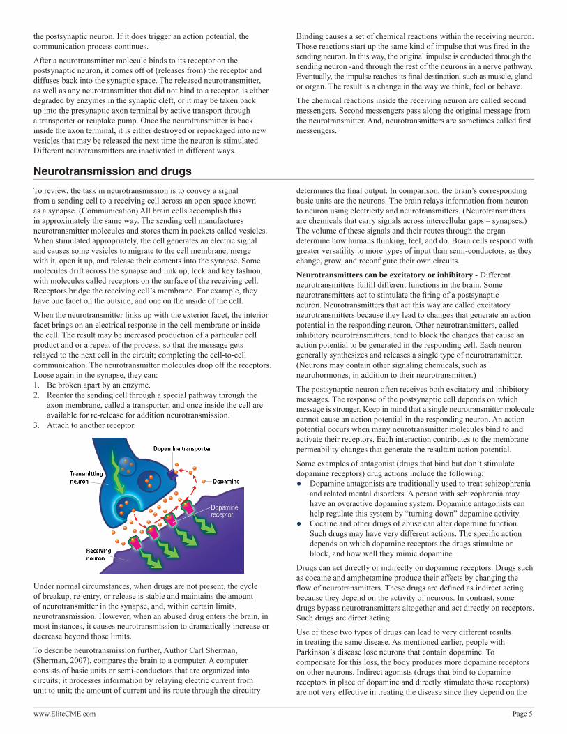
To review, the task in neurotransmission is to convey a signal from a sending cell to a receiving cell across an open space known as a synapse. (Communication) All brain cells accomplish this in approximately the same way. The sending cell manufactures neurotransmitter molecules and stores them in packets called vesicles. When stimulated appropriately, the cell generates an electric signal and causes some vesicles to migrate to the cell membrane, merge with it, open it up, and release their contents into the synapse. Some molecules drift across the synapse and link up, lock and key fashion, with molecules called receptors on the surface of the receiving cell. Receptors bridge the receiving cell’s membrane. For example, they have one facet on the outside, and one on the inside of the cell.
When the neurotransmitter links up with the exterior facet, the interior facet brings on an electrical response in the cell membrane or inside the cell. The result may be increased production of a particular cell product and or a repeat of the process, so that the message gets relayed to the next cell in the circuit; completing the cell-to-cell communication. The neurotransmitter molecules drop off the receptors. Loose again in the synapse, they can:1. Be broken apart by an enzyme.2. Reenter the sending cell through a special pathway through the
axon membrane, called a transporter, and once inside the cell are available for re-release for addition neurotransmission.
3. Attach to another receptor.
Under normal circumstances, when drugs are not present, the cycle of breakup, re-entry, or release is stable and maintains the amount of neurotransmitter in the synapse, and, within certain limits, neurotransmission. However, when an abused drug enters the brain, in most instances, it causes neurotransmission to dramatically increase or decrease beyond those limits.
To describe neurotransmission further, Author Carl Sherman, (Sherman, 2007), compares the brain to a computer. A computer consists of basic units or semi-conductors that are organized into circuits; it processes information by relaying electric current from unit to unit; the amount of current and its route through the circuitry
determines the fi nal output. In comparison, the brain’s corresponding basic units are the neurons. The brain relays information from neuron to neuron using electricity and neurotransmitters. (Neurotransmitters are chemicals that carry signals across intercellular gaps – synapses.) The volume of these signals and their routes through the organ determine how humans thinking, feel, and do. Brain cells respond with greater versatility to more types of input than semi-conductors, as they change, grow, and reconfi gure their own circuits.
Neurotransmitters can be excitatory or inhibitory - Different neurotransmitters fulfi ll different functions in the brain. Some neurotransmitters act to stimulate the fi ring of a postsynaptic neuron. Neurotransmitters that act this way are called excitatory neurotransmitters because they lead to changes that generate an action potential in the responding neuron. Other neurotransmitters, called inhibitory neurotransmitters, tend to block the changes that cause an action potential to be generated in the responding cell. Each neuron generally synthesizes and releases a single type of neurotransmitter. (Neurons may contain other signaling chemicals, such as neurohormones, in addition to their neurotransmitter.)
The postsynaptic neuron often receives both excitatory and inhibitory messages. The response of the postsynaptic cell depends on which message is stronger. Keep in mind that a single neurotransmitter molecule cannot cause an action potential in the responding neuron. An action potential occurs when many neurotransmitter molecules bind to and activate their receptors. Each interaction contributes to the membrane permeability changes that generate the resultant action potential.
Some examples of antagonist (drugs that bind but don’t stimulate dopamine receptors) drug actions include the following:
● Dopamine antagonists are traditionally used to treat schizophrenia and related mental disorders. A person with schizophrenia may have an overactive dopamine system. Dopamine antagonists can help regulate this system by “turning down” dopamine activity.
● Cocaine and other drugs of abuse can alter dopamine function. Such drugs may have very different actions. The specifi c action depends on which dopamine receptors the drugs stimulate or block, and how well they mimic dopamine.
Drugs can act directly or indirectly on dopamine receptors. Drugs such as cocaine and amphetamine produce their effects by changing the fl ow of neurotransmitters. These drugs are defi ned as indirect acting because they depend on the activity of neurons. In contrast, some drugs bypass neurotransmitters altogether and act directly on receptors. Such drugs are direct acting.
Use of these two types of drugs can lead to very different results in treating the same disease. As mentioned earlier, people with Parkinson’s disease lose neurons that contain dopamine. To compensate for this loss, the body produces more dopamine receptors on other neurons. Indirect agonists (drugs that bind to dopamine receptors in place of dopamine and directly stimulate those receptors) are not very effective in treating the disease since they depend on the
Page 6 www.EliteCME.com
presence of dopamine neurons. In contrast, direct agonists are more effective because they stimulate dopamine receptors, even when dopamine neurons are missing.
Once returned to the sending neuron by the reuptake system, dopamine is subject to an enzyme named monoamine oxidase (MAO). MAO also affects dopamine levels. MAO usually breaks down dopamine. If no other factors were at work, MAO would keep the amount of “used” dopamine, fairly low. However, dopamine taken back into the nerve ending can return to the vesicle for storage. Once inside the vesicle, dopamine is protected from MAO.
A drug named reserpine prevents the reuptake of dopamine and some other neurotransmitters. Administering reserpine causes dopamine to remain exposed within the cell and broken down by MAO. This profoundly reduces the available dopamine.
Changing the action of MAO can help physicians treat diseases that involve dopamine transmission. For instance, the drug deprenyl inhibits MAO. This increases the stores of dopamine and slows the progression of Parkinson’s disease. In higher doses, deprenyl enhances the effects of dopamine on behavior.
Interestingly, one form of MAO actually protects dopamine. This form of MAO, found in dopamine neurons, acts on substances in the neuron other than dopamine. Here MAO protects the “purity” of neurotransmission by breaking down other neurotransmitters. Inhibiting this form of MAO can increase levels of neurotransmitters such as serotonin, which seems to help people diagnosed with depression.
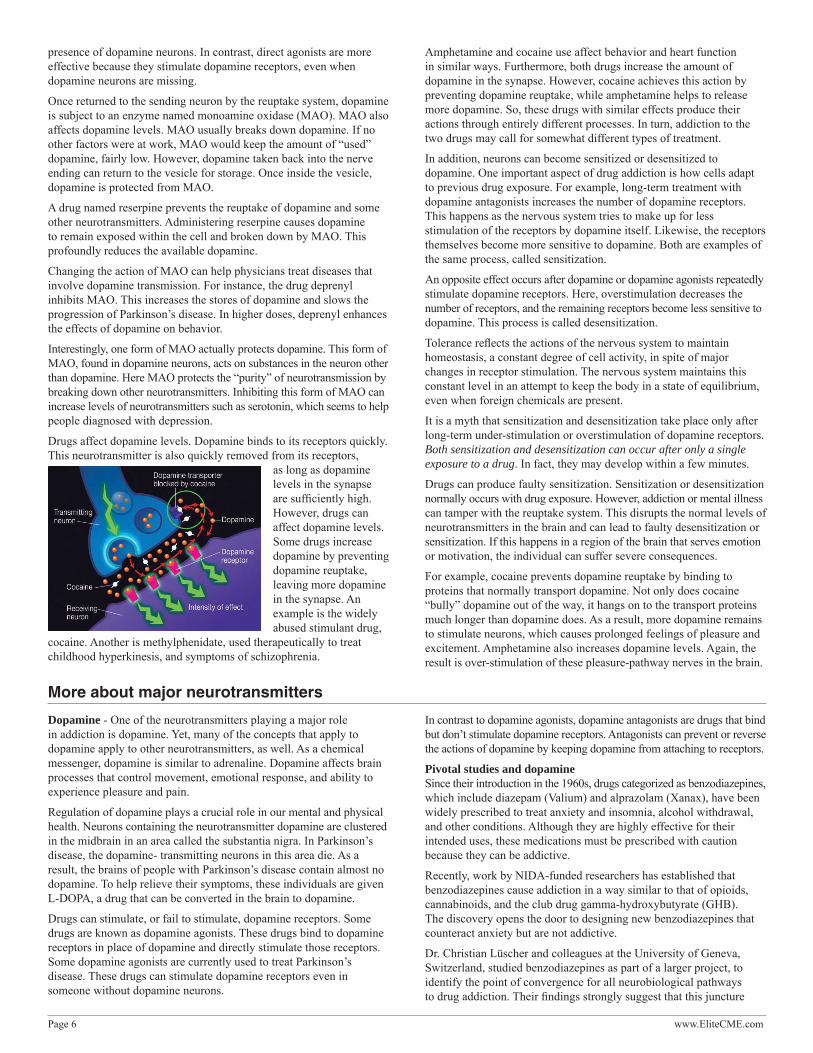
Drugs affect dopamine levels. Dopamine binds to its receptors quickly. This neurotransmitter is also quickly removed from its receptors,
as long as dopamine levels in the synapse are suffi ciently high. However, drugs can affect dopamine levels. Some drugs increase dopamine by preventing dopamine reuptake, leaving more dopamine in the synapse. An example is the widely abused stimulant drug,
cocaine. Another is methylphenidate, used therapeutically to treat childhood hyperkinesis, and symptoms of schizophrenia.
Amphetamine and cocaine use affect behavior and heart function in similar ways. Furthermore, both drugs increase the amount of dopamine in the synapse. However, cocaine achieves this action by preventing dopamine reuptake, while amphetamine helps to release more dopamine. So, these drugs with similar effects produce their actions through entirely different processes. In turn, addiction to the two drugs may call for somewhat different types of treatment.
In addition, neurons can become sensitized or desensitized to dopamine. One important aspect of drug addiction is how cells adapt to previous drug exposure. For example, long-term treatment with dopamine antagonists increases the number of dopamine receptors. This happens as the nervous system tries to make up for less stimulation of the receptors by dopamine itself. Likewise, the receptors themselves become more sensitive to dopamine. Both are examples of the same process, called sensitization.
An opposite effect occurs after dopamine or dopamine agonists repeatedly stimulate dopamine receptors. Here, overstimulation decreases the number of receptors, and the remaining receptors become less sensitive to dopamine. This process is called desensitization.
Tolerance refl ects the actions of the nervous system to maintain homeostasis, a constant degree of cell activity, in spite of major changes in receptor stimulation. The nervous system maintains this constant level in an attempt to keep the body in a state of equilibrium, even when foreign chemicals are present.
It is a myth that sensitization and desensitization take place only after long-term under-stimulation or overstimulation of dopamine receptors. Both sensitization and desensitization can occur after only a single exposure to a drug. In fact, they may develop within a few minutes.
Drugs can produce faulty sensitization. Sensitization or desensitization normally occurs with drug exposure. However, addiction or mental illness can tamper with the reuptake system. This disrupts the normal levels of neurotransmitters in the brain and can lead to faulty desensitization or sensitization. If this happens in a region of the brain that serves emotion or motivation, the individual can suffer severe consequences.
For example, cocaine prevents dopamine reuptake by binding to proteins that normally transport dopamine. Not only does cocaine “bully” dopamine out of the way, it hangs on to the transport proteins much longer than dopamine does. As a result, more dopamine remains to stimulate neurons, which causes prolonged feelings of pleasure and excitement. Amphetamine also increases dopamine levels. Again, the result is over-stimulation of these pleasure-pathway nerves in the brain.
More about major neurotransmitters
Dopamine - One of the neurotransmitters playing a major role in addiction is dopamine. Yet, many of the concepts that apply to dopamine apply to other neurotransmitters, as well. As a chemical messenger, dopamine is similar to adrenaline. Dopamine affects brain processes that control movement, emotional response, and ability to experience pleasure and pain.
Regulation of dopamine plays a crucial role in our mental and physical health. Neurons containing the neurotransmitter dopamine are clustered in the midbrain in an area called the substantia nigra. In Parkinson’s disease, the dopamine- transmitting neurons in this area die. As a result, the brains of people with Parkinson’s disease contain almost no dopamine. To help relieve their symptoms, these individuals are given L-DOPA, a drug that can be converted in the brain to dopamine.
Drugs can stimulate, or fail to stimulate, dopamine receptors. Some drugs are known as dopamine agonists. These drugs bind to dopamine receptors in place of dopamine and directly stimulate those receptors. Some dopamine agonists are currently used to treat Parkinson’s disease. These drugs can stimulate dopamine receptors even in someone without dopamine neurons.
In contrast to dopamine agonists, dopamine antagonists are drugs that bind but don’t stimulate dopamine receptors. Antagonists can prevent or reverse the actions of dopamine by keeping dopamine from attaching to receptors.
Pivotal studies and dopamine Since their introduction in the 1960s, drugs categorized as benzodiazepines, which include diazepam (Valium) and alprazolam (Xanax), have been widely prescribed to treat anxiety and insomnia, alcohol withdrawal, and other conditions. Although they are highly effective for their intended uses, these medications must be prescribed with caution because they can be addictive.
Recently, work by NIDA-funded researchers has established that benzodiazepines cause addiction in a way similar to that of opioids, cannabinoids, and the club drug gamma-hydroxybutyrate (GHB). The discovery opens the door to designing new benzodiazepines that counteract anxiety but are not addictive.
Dr. Christian Lüscher and colleagues at the University of Geneva, Switzerland, studied benzodiazepines as part of a larger project, to identify the point of convergence for all neurobiological pathways to drug addiction. Their fi ndings strongly suggest that this juncture
www.EliteCME.com Page 7
occurs when dopamine surges, in response to drug taking, to initiate a change in synaptic plasticity in dopamine-producing cells. From receptor activation to dopamine surge, the pleasurable sensations, that make addictive drugs disastrously attractive for vulnerable individuals, occur when dopamine levels in the brain’s reward area abruptly surge. Researchers had worked out how most addictive drugs, but not benzodiazepines, precipitate these surges.
Dr. Lüscher and colleagues have now demonstrated that benzodiazepines weaken the infl uence of a group of cells, called inhibitory interneurons, in the brain’s ventral tegmental area (VTA). These neurons normally help prevent excessive dopamine levels by down-regulating the fi ring rates of dopamine-producing neurons. Two negatives make a positive, so when benzodiazepines limit the interneurons’ restraining sway; the dopamine-producing neurons release more dopamine.
The Swiss researchers traced benzodiazepines’ effect on VTA interneurons to the drugs’ activation of a subset of GABAA (gamma-aminobutyric acid type-A) receptors on the interneurons. Although benzodiazepines typically activate multiple subtypes of GABAA receptors, their activation of the alpha-1 subtype is decisive for their impact on VTA interneuron behavior. These interneurons are highly sensitive to such activation because they carry abundant numbers of these receptors. By staining brain tissue, the researchers showed that 81 percent of VTA interneurons carry GABAA receptors that contain the alpha-1 subunit.
To prove that activation of alpha-1 GABAA receptors underlies benzodiazepines’ dopamine effect, the researchers administered a typical benzodiazepine, midazolam, to two groups of mice. The results supported the researchers’ proposed mechanism: In normal animals, the fi ring rate of interneurons decreased in response to the drug, while that of dopamine-producing neurons increased. In contrast, in animals that were genetically altered to prevent benzodiazepines from potentiating alpha-1 GABAA receptors, the drug had little or no impact on neuron fi ring.
A behavioral fi nding completed the chain of proof linking benzodiazepines’ stimulation of alpha-1 GABAA receptors to their rewarding effects. When given the option of drinking sugar water or a sweetened solution of midazolam, normal mice imbibed roughly three times as much drug-laced as drug-free liquid. Mice with altered alpha-1 GABAA receptors, however, drank equal amounts of each, thereby exhibiting no evidence of fi nding one drink more rewarding than the other. When benzodiazepines limit the interneurons’ restraining infl uence, the dopamine-producing neurons release more dopamine. Benzodiazepines’ newly discovered mechanism for producing reward is comparable to those of opiates, cannabinoids, and GHB. Each of the four drugs reduce an inhibitory infl uence on dopamine-producing cells, thereby promoting dopamine spikes.
Here is where it gets interesting; from surge to addiction. - Dopamine surges are transient events, but addictive drugs cause long-lasting changes in the reward system. Among the earliest of these along the path from voluntary to compulsive drug use and addiction, is the migration of certain AMPA receptors (i.e., GluA2-lacking receptors) from the interior to the surface of the dopamine-producing neurons. These receptors render the cell more susceptible to stimulation by the excitatory neurotransmitter glutamate, and as a result, the cells respond to future drug exposures with larger dopamine surges that produce even more intense pleasure. Scientists also have evidence that these special AMPA receptors initiate a series of changes in neural transmission that cumulatively give rise to the range of addictive symptoms.
Dr. Lüscher and colleagues showed that benzodiazepines induce AMPA receptor migration via the alpha-1 GABAA receptors. In these experiments, brain tissue from normal mice exhibited GluA2-lacking AMPA receptors after a single injection of midazolam, but tissue from mice with benzodiazepine-insensitive alpha-1 GABAA receptors did not. Recordings of intracellular electrical currents confi rmed synaptic changes of dopamine-producing neurons in the normal mice and not the altered mice. To pin down the relationship further, the researchers
injected mice with two other compounds, one (zolpidem) that preferentially activates only the alpha-1 GABAA receptors, and one (L-838417) that antagonizes these receptors. GluA2-lacking AMPA receptors were expressed in dopamine-producing neurons following a treatment with zolpidem, but not with L-838417.
Proof: The Swiss researchers hypothesize that although different addictive drugs produce dopamine surges by various mechanisms, the subsequent chain of effects is the same. Consistent with this idea, they showed that even in the absence of any drug, artifi cial stimulation of the dopamine-producing neurons is suffi cient to induce the appearance of GluA2-lacking AMPA receptors.
In this experiment, the researchers introduced a virus containing a light-activated protein, channelrhodopsin, into the dopamine-producing cells of mice. When exposed to light pulses from an optical fi ber inserted into the animals’ VTA, the channelrhodopsin stimulated neuron fi ring in bursts similar to those produced by addictive drugs. The result was an increase in GluA2-lacking AMPA receptors comparable to that seen following exposure to addictive drugs. “This was a nail-in-the-coffi n study to show that activity of dopaminergic neurons leads to synaptic adaptation that is involved in addiction,” says Dr. Lüscher. “This is why addiction is so diffi cult to treat. Even if you clear the drug from the body, there are long-lasting changes in brain architecture.”
Looking forward to better benzodiazepines: Taken all together, the data from the studies, show that the activation of alpha-1-containing GABAA receptors, by benzodiazepines calms inhibitory interneurons, and increases dopaminergic neuron fi ring, and leads to strengthening of excitatory synapses that favor addictions. Dr. Roger Sorensen of NIDA’s Functional Neuroscience Research Branch says, “This is the fi rst demonstration that acute benzodiazepine use can increase dopamine release, supporting its addictive potential.”
“Now that we know that it’s the alpha-1-containing GABAA receptor that is responsible for benzodiazepine addiction, we can design benzodiazepines that do not touch those particular receptors,” says Dr. Lüscher. Drugs that bind only to alpha-2-containing GABAA receptors, he adds, might relieve anxiety non-addictively. “Such substances already exist for research purposes,” Dr. Lüscher says. “It’s possible that we can also create them for clinical use.” (NIDA notes, 2012)
Serotonin - Serotonin plays a major role in emotional disorders such as depression, suicide, impulsive behavior, and aggression. Neurons using serotonin as a neurotransmitter are found in the brain, primarily in a cluster of cells called the pons.
Serotonin is normally involved in temperature regulation, sensory perception, and mood control. The hallucinogenic drug LSD acts on serotonin receptors; so do some antidepressant drugs.
Mentioned earlier, neurotransmitters usually bind and stimulate their receptors, then travel back to their sending neurons. These are the normal events in the reuptake system. Reuptake occurs in order to keep neurotransmitter levels steady and maintain homeostasis. In effect, the receiving neuron says “That’s enough!” to the sending neuron that has been releasing neurotransmitters. The sending neuron quickly picks up the leftover neurotransmitters and stops releasing new ones. This is an example of negative feedback.
Prozac and some of the other drugs used to treat severe depression prevent the normal reuptake of serotonin. As a result, there is more serotonin fl oating around to grab on to receptors and trigger impulses in receiving neurons. This leads to increased stimulation of serotonin neurons in depressed people, who fi nd that the drugs help to relieve their symptoms.
Norepinephrine - Norepinephrine, also called noradrenaline, is a neurotransmitter that doubles part-time as a hormone. (Hormones are chemicals that regulate many body functions, including growth, digestion, and fl uid balance.) As a neurotransmitter, norepinephrine helps to regulate arousal, dreaming, and moods. As a hormone,
Page 8 www.EliteCME.com
norepinephrine acts to increase blood pressure, constrict blood vessels, and increase heart rate - responses that occur when we feel stress.
Acetylcholine - Another major neurotransmitter named acetylcholine excites neurons in the brain and many other parts of the body, including muscle tissues and glands. Acetylcholine is released where nerves meet muscles and is therefore responsible for muscle contraction.
After acetylcholine stimulates its receptors, it is quickly inactivated and destroyed by an enzyme. Drugs that keep this enzyme from working are used to treat myasthenia gravis, a disease of muscle weakness and fatigue. These drugs lead to an excess of acetylcholine in synapses and overstimulation of the muscles. The result in patients with extreme muscle weakness is normal muscle contraction.
Glutamate and gamma-amino butyric acid (GABA) - Certain amino acids also act as neurotransmitters, including glutamate and gamma-amino butyric acid (GABA). Glutamate strongly excites neurons, while GABA strongly inhibits neurons.
Glutamate and GABA are unique in several ways. The number of synapses using glutamate and GABA is much greater than those using all other types of neurotransmitters, combined. Glutamate and GABA neurons are found in many brain regions. As a result, glutamate and GABA work all over the brain, while other neurotransmitters do not.
Both glutamate and GABA have important functions in the body in addition to their role as neurotransmitters. For example, they are needed by our body’s metabolism to break down food and make energy-rich molecules in cells.
The fact that GABA and glutamate are so widely present makes it likely that they will be altered during drug addiction. This fact also makes it diffi cult to treat addiction with drug therapy. Say that a drug affects GABA and glutamate in a way that relieves craving. Because GABA and glutamate are so widely present, these drugs could produce a mess of side effects, as well. If there were drugs that could selectively stimulate or block certain receptors, then it would be easier to treat addiction and avoid doing more harm than good.
Drugs interfere with neurotransmission
Drugs can interfere with almost every step in the work of neurotransmitters. To further understand, consider an analogy. In your apartment you perform various tasks: working on a computer, watching television, listening to music on a stereo system, and more. When you leave your apartment, you make sure the door is locked. You would hate for people to get a key so similar to yours, that they could somehow jimmy your door open and break in. Once in your apartment, they could vandalize your property, take your computer and VCR, break your TV, bust out your lights, or drop your stereo. You could then no longer perform your daily tasks.
Something like this can happen in your brain. Remember that each receptor is designed to bind only to a certain neurotransmitter. A drug of abuse that is structurally similar to a neurotransmitter could be a “key” that fi ts into a receptor’s “lock.” In this way, the drug could disrupt neuron activity in the same way that an intruder disrupts your apartment and damages your property.
More specifi cally, drugs can: ● Stop the chemical reactions that create neurotransmitters. ● Empty neurotransmitters from the vesicles where they’re normally
stored and protected from breakdown by enzymes. ● Block neurotransmitters from entering or leaving vesicles.
● Bind to receptors in place of neurotransmitters. ● Prevent neurotransmitters from returning to their sending neuron
(the reuptake system). ● Interfere with second messengers, the chemical and electrical
changes that take place in a receiving neuron.
Mentioned previously, one example of drug interference is the effect of cocaine. In other words, this type of drug can damage your intellectual property by blocking nerve impulses, preventing neurotransmitters from getting where they’re supposed to be, or producing too many or too little neurotransmitters. As a result of using cocaine, neurons may be overstimulated or not stimulated at all, crippling the nervous system’s ability to carry out its functions.
In each of these ways, and more yet to be identifi ed, cocaine and other drugs can damage and vandalize the complicated circuit of nerve pathways in one’s body. Treatment for drug addiction stops this cycle of neuro-exploitation. A network of so intricately designed to reason, imagine, compute, remember, and dream is truly incredible, and not something to be tampered with. Central to treatment is the idea that the vast network of neurons in our bodies should be treated with care and respect. First, however, research has had to play a large role in identifying how to treat the disease of addiction.
Research methodologies
Research continues to uncover and enlighten all who work within the fi eld of addictions. When researchers begin their processes, in order to determine how a drug affects a particular neurotransmitter, they will typically compare subjects who have a history of drug exposure with others who do not. For example, if researchers are investigating links between a drug’s impact on neurotransmission and a drug-related behavior or symptom, they may compare subjects who exhibit the behavior or symptom with others who do not. The subjects in these experiments may be animals or people. In the case of animals, drug exposure often takes place under laboratory conditions designed to mimic human drug consumption. Studies can be divided into those in which measurements are made in living animals or people, and those in which animal brain tissue is removed and examined.
Using chemical assays (analysis) researchers quantify the presence of neurotransmitter, receptor, or other structure of interest. In a more recent experiment, scientists assayed brained tissue from brain tissue from 35-day-old rat pups and found that those that had been exposed to nicotine in utero had fewer nicotine receptors in the reward system than unexposed rates. (NIDA Notes)
A second experimental method using removed brain tissue – in vitro, literally, in glass, a historical term referring to the containers for the tissue and solution enables researchers to view a drug’s effects on neurotransmission in action. Scientists place the tissue in a laboratory solution of nutrients that enables the cells to continue to carry out some of their living functions. The researchers may then, for instance, add the drug being investigated to the solution, and monitor whether the cells respond by increasing their release of neurotransmitters. Alternatively, they may measure cell membrane or electrical properties that stimulate or inhibit the release of neurotransmitters.
In both, in vitro experiments and in living animals, the techniques for measuring neurotransmitter quantities and fl uctuations include micro dialysis and fast-scan cyclic voltammetry (FSCV). Microanalysis involves taking a series of samples of the intercellular fl uid containing the neurotransmitter through a microscopic tube inserted into the tissue or living brain. FSCV recently developed by NIDA-funded scientists, monitors neurotransmitter fl uctuations at tenth of second intervals by measuring electrical changes related to neurotransmitter concentrations.
Studies with live animals or people are important for typing drugs’ effects on neurotransmitters to behaviors and/or symptoms.
www.EliteCME.com Page 9
Animals as Research Models: Why do scientists study the brains of non-human animals? Scientists use animals in research studies because the use of humans is either impossible or unethical. For example, when scientists investigate the effects of drugs of abuse on brain function, either the question they are asking cannot be answered in a living human, or it would be inappropriate to give drugs to them.
The use of animals as subjects in scientifi c research has contributed to many important advances in scientifi c and medical knowledge. Scientists must analyze the goals of their experiments in order to select an animal species that is appropriate. Scientists often use fruit fl ies (Drosophila melanogaster) when they want to learn more about genetics. However, fruit fl ies are not a very good model if a scientist is investigating muscle physiology; a mouse may be a better model for those experiments. Although scientists strive to develop non-animal models for research, these models often do not duplicate the complex animal or human body.
Continued progress toward a more complete understanding of human and animal health depends on the use of living animals.
A common design for experiments with either animals or people is to give study subjects a chemical that has a known effect on a particular neurotransmitter, and then observe the impact on their behavior. The chemical is usually, either an agonist (promoter) or antagonist (blocker) of signaling by the neurotransmitter.
In a more recent experiment, for instance, a research team administered a glutamate agonist to rats and showed that the resulting increased their levels of the neurotransmitter correlated with a reduction in the animals’ cocaine seeking. Another team using the same strategy implicated glutamate in nicotine withdrawal. (NIDA Notes, 2012) Such studies are a staple of testing compounds (combinations) to identify medication classes with potential for treating abuse or addiction.
Building effective vaccines
NIDA supported vaccine developers have achieved promising preclinical results with novel formulations against cocaine and heroin. Laboratory animals treated with the new vaccines produced high blood concentrations of anti-drug antibodies and exhibited sharply reduced behavioral responses to the drugs. Dr. Ronald Crystal of Weill Cornell Medical College and Drs. George Koob and Dr. Kim Janda of the Scripps Research Institute are among the many scientists who are striving to create vaccines that can protect against nicotine, cocaine, methamphetamine, and opiates. In 2009, NIDA-supported researchers at Baylor College of Medicine reported partial success in the fi rst clinical trial of an anti-cocaine vaccine. Some recipients generated strong antibody responses and reduced their cocaine intake, but others did not. Those results affi rmed that antidrug vaccines can protect against drugs’ psychoactive and behavioral effects, and invigorated the search for other and improved formulations.
Anti-drug vaccination strategies train the immune system to attack molecules it would not otherwise recognize. Because addictive drugs are small molecules, the immune system, on its own, does not target them. “During my team’s research on human gene therapy, we observed that particular adenoviruses, some of which can cause the common cold, are potent inducers of antibodies,” says Dr. Crystal. The researchers hypothesized that coupling a molecule similar to cocaine, to one of these potent stimulators of the immune system, could provide the basis for an anti-cocaine vaccine. Dr. Crystal and colleagues identifi ed a way to hook a cocaine-like molecule onto the inactivated adenovirus’ protein coat.
The anti-cocaine vaccine comprises a cocaine analog—an inactive, cocaine-like molecule—attached to a robust stimulator of the immune system—an adenovirus with hexon and fi ber components. The resulting vaccine induces the body to generate a high level of cocaine antibodies, which prevent the drug from entering the brain for several months.
Heroin introduces an additional challenge. Because its two major metabolites also contribute to heroin abuse, an effective vaccine must keep all three compounds from acting in the brain. Dr. Janda explains, “Countering the effects of heroin is like peeling back layers of an onion—heroin is the fi rst layer, but the metabolites are second and third layers. Our vaccine degrades slowly to expose these metabolites, so that they stimulate the immune system to produce antibodies that keep each of them out of the brain.”
The anti-heroin vaccine comprises an inactive, heroin-like hapten linked to a carrier protein (keyhole limpet hemocyanin, KLH) and an adjuvant (Alum). This dynamic vaccine displays multiple haptenic structures simultaneously, allowing the immune system to generate antibodies to not only heroin but also its psychoactive metabolites 6-acetylmorphine (6AM) and morphine (mor).
Vaccine fundamentals - The goal of an anti-drug vaccine is to induce the immune system to block the psychoactive effects of its target drug. When an anti-drug antibody encounters a molecule of the drug,
the two combine to form a complex that is too large to pass from the bloodstream into the brain. Locked out of the brain, the drug cannot produce the rewarding effects that motivate continued use.
Both, the group including Drs. Crystal and Koob, and the one led by Dr. Janda, employed the same two-component strategy to construct their vaccines. The fi rst component is a carrier protein, selected to be highly immunogenic: Its function is to stimulate the immune system to produce enough antibodies to intercept the millions of molecules in a dose of the target drug before they reach the brain. The second component, called a hapten, is a molecule that shares some key structural features with the target drug: It provides the immune system with a template for the formation of antibodies that recognize and attach to the target drug.
The effi cacy of vaccines of this type depends on the precise selection and confi guration of the carrier protein and hapten. Because of the complexity and intricacy of immune responses, the search for an ideal combination is largely a process of trial and refi nement.
Cocaine and the common cold - Dr. Crystal and colleagues employed a carrier protein whose potent immunogenicity is all too familiar: an adenovirus, agent of the common cold. The researchers disabled a protein (the adenovirus 5 gene transfer vector) from the virus’ infectious apparatus and linked it to a hapten termed GNE, an amide-cocaine-memetic that was designed and synthesized by Dr. Janda’s group at Scripps. This combination, given in an initial vaccination followed by booster injections after 3 and 5 weeks, produced high blood concentrations (500,000 to 1,000,000 titer units) of anti-cocaine antibodies that persisted for 3 months in rats.
To determine whether the vaccine prevented cocaine from moving from the blood into the brain, the researchers administered radiolabeled drug to vaccinated and control rats. Assays found that 2 minutes after the administration, vaccinated animals had 3.5 times as much drug in their blood as compared to control animals, and 66 percent less in brain tissue.
With less cocaine reaching their brain, vaccinated rats exhibited weakened behavioral responses to the drug. Although they displayed typical reactions of hyperactivity and increased locomotor activity, following a series of cocaine injections, they did so, only 20 percent as intensely, as control animals. Similarly, vaccinated rats retained motivation to press a lever to self-administer cocaine intravenously, but they did not work as hard for the drug as control animals: In a progressive ratio protocol, which multiplies the number of presses required for delivery of each successive infusion, vaccinated rats quit at a cost, or ratio, of 12 presses per injection, whereas controls would keep pressing up to 32 presses per injection.
Another experiment had direct application to the vaccine’s primary proposed clinical use: to shield abstinent individuals who lapse from experiencing cocaine effects that can precipitate extended relapses. The researchers gave a small priming dose of cocaine to rats that
Page 10 www.EliteCME.com
had previously established steady cocaine self-administration but had stopped seeking the drug during a period of extinction. The new exposure to the drug prompted the control rats, but not the vaccinated rats, to return to drug-seeking—pressing the lever that had initially yielded cocaine, but no longer did so.
Overcoming an immunological challenge - In designing their heroin vaccine, Dr. Janda and colleagues noted that antibodies to heroin alone would still permit production of the drug’s psychoactive metabolites. Therefore, they varied the basic vaccine strategy to employ what they call a dynamic vaccine. A single hapten coupled to a carrier protein slowly morphed itself into multiple haptens paralleling heroin’s degradation pathway, thus allowing the immune system to sample and make antibodies, to not only heroin, but its important metabolites, such as 6-acetylmorphines. The Scripps team achieved its results by binding haptens to a commonly used carrier protein, keyhole limpet hemocyanin, and the adjuvant alum, a chemical salt that restricts enzymatic access. With reduced exposure to enzymes, breakdown of the haptens occurs more slowly, affording the immune system more opportunity to detect the resulting metabolites and form antibodies to them.
The researchers observed signifi cant antibody titers 14 days after initial vaccination. Antibody levels rose to a maximum (1:122,000) 53 days after the initial vaccination and after two boosters. The levels remained potentially protective (at 1:50,000) 105 days after the initial vaccination and after a third booster.
To test how successfully the vaccine blocked the drug from entering the brain, initially the researchers, in collaboration with the Koob group, assessed its impact on heroin analgesia. This test can be used as a fi rst screen because if the drug does not reach the brain, it will lose its analgesic, as well as its rewarding effects. The researchers injected rats with heroin and placed them on a plate that was hot enough to cause mild pain, but not injury. Control rats took 30 seconds to lift their paws off the plate. Vaccinated rats evinced suffi cient discomfort to do so, after just 10 seconds, no different from the response of animals not treated with heroin. Most remarkable, was the vaccine’s specifi city for heroin: In parallel experiments, it did not lessen the analgesic effects of the closely related and commonly prescribed drug Oxycodone.
Vaccinated rats also demonstrated less inclination than controls to self-administer heroin intravenously. Over 10 sessions in which animals had access to a lever that delivered infusions of the drug, all seven control animals pressed the lever three or more times during any of three consecutive sessions, whereas only three of the seven vaccinated rats did so.
Additional ammunition - It is always challenging to transfer new vaccine technology to people. Dr. Crystal stated, “Our team will need to demonstrate that, from patient to patient, the cocaine vaccine consistently induces a high level of antibodies with strong affi nity to cocaine for a long duration.” “The only way to do that is to conduct a clinical trial.” If the technology needs tweaking, Dr. Koob continues, “There are ways to make vaccines more compatible with humans, for example, using carriers other than the adenovirus.” Drs. Crystal and Janda agree vaccine treatments for addiction should be part of a comprehensive therapy.
“People have the misconception that a single vaccine can protect patients from substance abuse, and that’s not true,” says Dr. Janda. However, the results suggest that vaccines are a promising adjunct therapy to accompany drug counseling. For example, Dr. Crystal says, “a patient who has attained abstinence could be vaccinated to block the effects of the drug, thereby preventing relapse. Dr. Janda notes, “Our vaccine will not alleviate craving, but it could help patients maintain abstinence in weak moments.” He adds that by fi ghting addiction, a heroin vaccine may help to combat HIV in countries where injection of the drug contributes to spread of the virus. “The vaccine approach provides an alternative strategy for treating drug addiction,” says Dr. Nora Chiang of NIDA’s Division of Pharmacotherapies and Medical
Consequences of Drug Abuse. “There is much more work to be done on these vaccines, but the results so far is promising.” (NIDA notes, 2012)
Imaging the brain: Scientists, continue to use newer technologies that enhance their learning about how the brain works, and how drugs of abuse, changes neurotransmission. Historically, scientists could examine brains only after death, but new imaging procedures enable scientists to study the brain in living animals, including humans. Brain scans or brain imaging techniques enable neuroscientists to directly assess neurotransmission in people and living animals.
One of the most extensively used techniques to study brain activity and the effects of drugs on the brain is positron emission tomography (PET). PET measures the spatial distribution and movement of radioisotopes in tissues of living subjects. Because the patient is awake, the technique can be used to investigate the relationship between behavioral and physiological effects, and changes in brain activity.
PET scans can detect nanomolar concentrations of tracer molecules and achieve spatial resolution of about 4 millimeters. In addition, computers can reconstruct images obtained from a PET scan in two or three dimensions. PET requires the use of compounds that are labeled with positron-emitting isotopes. A cyclotron accelerates protons into the nucleus of nitrogen, carbon, oxygen, or fl uorine to generate these isotopes. The additional proton makes the isotope unstable. To become stable again, the proton must break down into a neutron and a positron. The unstable positron travels away from the site of generation and dissipates energy along the way. Eventually, the positron collides with an electron leading to the emission of two gamma rays at 180 degrees from one another.
The gamma rays reach a pair of detectors that record the event. Because the detectors respond only to simultaneous emissions, scientists can precisely map the location where the gamma rays were generated. The labeled isotopes are very short-lived. The half-life (the time for half of the radioactive label to disintegrate) of the commonly used radioisotopes ranges from approximately two minutes to less than two hours, depending on the specifi c compound. Because a PET scan requires only small amounts (a few micrograms) of short-lived radioisotopes, negative pharmacological effects are imperceptible.
PET scans can answer a variety of questions about brain function, including the activity of neurons. Scientists use different radio-labeled compounds to investigate different biological questions. For example, radiolabeled glucose can identify parts of the brain that become more active in response to a specifi c stimulus. Active neurons metabolize more glucose than inactive neurons. Active neurons will emit more positrons. This will show as red or yellow on PET scans compared to blue or purple in areas where the neurons are not highly active. PET also helps scientists investigate how drugs affect the brain by enabling them to:
● Determine the distribution of a drug in the body. ● Measure the local concentration of a drug at binding sites. ● Estimate receptor occupancy based on competitive binding assays. ● Evaluate the effects of drugs on other neurotransmitter systems. ● Investigate the activity of enzymes that metabolize the drug.
With positron emission topography (PET), researchers can compare groups of drug-abusing and non-abusing individuals, quantifying differences in their levels of a particular neurotransmitter molecule (e.g., dopamine) or neurotransmission component (e.g., a receptor or transporter). With PET, researchers are also able to correlate a drug’s transit through the brain with fl uctuations in a target neurotransmitter. They can elicit a drug-related behavior or symptom (e.g., craving) and relate neurotransmitter fl uctuations to the rise and fall in its intensity.
Another more recent PET study, for instance, showed that smokers have less of the neurotransmitter degrading enzyme monoamine oxidase B (MAO-B) throughout their bodies than non-smoking persons. The relative defi cit of MAO-B may help explain why smokers are at higher risk for hypertension and other chronic diseases. Researchers use both PET and functional magnetic resonance imaging (MRI) to monitor metabolic activity in selected regions of the brain. And, because each

www.EliteCME.com Page 11
neurotransmitter has a unique distribution among the regions of the brain, information on locations of heightened or decreased activity provides clues to which neurotransmitter is affected under the conditions of a study.
Similar to PET, single-photon emission computed tomography (SPECT) imaging uses radioactive tracers and a scanner to record data that a computer constructs into two- or three-dimensional images of active brain regions. Because the tracers used in SPECT take longer to deteriorate than those for PET, longer periods of time between tests are required for SPECT. While PET is more versatile than SPECT and produces more detailed images with a higher degree of resolution, SPECT is much less expensive than PET, and can address many of the same drug abuse research questions.
MRI uses magnetic fi elds and radio waves to produce high-quality two- or three-dimensional images of brain structures without injecting radioactive tracers. In this procedure, a large cylindrical magnet creates a magnetic fi eld around the research volunteer’s head, and radio waves are sent through the magnetic fi eld. Sensors read the signals and a computer uses the information to construct an image. Using MRI, scientists can image both surface and deep brain structures with a high degree of anatomical detail, and they can detect minute changes in these structures over time.
A modifi cation of this technique, called functional MRI (fMRI), enables scientists to see images of blood fl ow in the brain as it occurs. fMRI provides superior image clarity along with the ability to assess blood fl ow and brain functions in just a few seconds. However, PET retains the advantage of being able to identify which brain receptors are being activated by neurotransmitters, abused drugs, and potential treatment compounds.
EEG uses electrodes placed on the scalp to detect and measure patterns of electrical activity in the brain. The greatest advantage of EEG is speed. It can record complex patterns of neural activity occurring within fractions of a second after a stimulus has been administered. The drawback to EEG is it does not provide the spatial resolution of fMRI or PET. Researchers often combine EEG images of brain electrical activity with MRI scans to localize brain activity more precisely.
Brain imaging reveals changes when smokers focus on long-term consequences of their tobacco use. Cognitive behavioral programs are generally effective, but, until now, researchers have shed little light on the neurological basis for their effi cacy. In a study led by Dr. Kevin N. Ochsner of the Social Cognitive Neuroscience Laboratory at Columbia University, smokers reported milder cigarette cravings when they thought about smoking’s harmful effects while viewing smoking cues, than when they focused on its pleasures.
Brain imaging correlated the reductions in craving with altered activity levels in regions associated with emotional regulation and reward. The cue-induced craving was roughly twice as strong after 35 days of abstinence, as it was after 1 week. Moreover, the craving increased over this period even though the smokers’ urges to light up in the absence of cues steadily weakened, dropping by more than 25 percent over 5 weeks.
The participants were 21 men and women who had smoked for 10 years, on average, and were not trying to quit. In preparation for the study, the participants practiced turning their thoughts to rewarding effects of cigarettes or high-fat food consumption when given the instruction “NOW” and to negative effects when given the instruction “LATER.” In the study itself, the researchers gave each participant 100 such instructions, in random order, each followed by a 6-second exposure to a screen image of either cigarettes or food. Then, after a 3-second delay with the screen blank, the participant reported how much he or she desired to smoke or eat, on a scale of 1 (not at all) to 5 (very much).
The power of thinking about negative effects proved to be considerable. The participants reported 34 percent less intense urges
to smoke and 30 percent less intense food cravings after the LATER instruction, compared with the NOW instruction.
Brain scans, taken during the experiment, showed how concentrating on long-term negative consequences alters brain activity to reduce craving. Functional magnetic resonance imaging (fMRI) of the participants’ whole brain revealed increased activity levels in areas, the dorsomedial, dorsolateral, and ventrolateral regions of the prefrontal cortex (PFC), which supports cognitive control functions, such as focusing, shifting attention, and controlling emotions.
Activity decreased in regions that previous studies have linked with craving. These areas include the ventral striatum and ventral tegmental area, which are parts of the reward circuit; the amygdala; and the subgenual cingulate. Individual participants who reported larger reductions in craving exhibited these changes to a more marked degree. A specialized mediation analysis of the images found that the increase in PFC activity drove the decrease in ventral striatum activity, which, in turn, fully accounted for the reduction in craving. “These results show that a craving-control technique, from behavioral treatment, infl uences a particular brain circuit, just as medications affect other pathways,” says Dr. Steven Grant, of NIDA’s Division of Clinical Neuroscience and Behavioral Research.
The researchers noted that the study participants reduced their smoking and food cravings to the same extent, even though smoking cravings were, initially, more intense. This fi nding suggests that calling undesirable consequences to mind has potential to help people overcome a variety of unhealthy urges.
When study participants thought of the long-term negative consequences of cigarette consumption (after receiving the instruction “LATER”), rather than short-term pleasures (“NOW”), they reduced their craving. Brain scans showed increased activity in the dorsolateral prefrontal cortex, a region critical to setting goals, planning, and controlling behavior, which, in turn, inhibited the ventral striatum, part of the reward pathway that generates craving.
“The mediation analysis that Dr. Ochsner and colleagues conducted is unique among imaging studies and is a particular strength to the research,” says Dr. Grant. “Because the researchers examined the interaction of brain regions, the results provide a perspective on the neural circuits involved in cognitive control of craving.” Dr. Grant suggest two important next steps in this area of research: identifying why some people have more problems than others in controlling the desire for cigarettes, and determining whether brain activity predicts the ability to quit smoking. (Kober, 2010)
Studying gene regulation and glucocorticoid receptors: Overexpression of the glucocorticoid gene in the fi rst weeks after birth increased anxiety and response to cocaine in adulthood. Researchers investigating how stressful experiences early in life promote later drug abuse have homed in on the glucocorticoid receptor (GR). In experiments with mice, augmenting GRs in the forebrain during the early postnatal period increased animals’ anxiety and sensitivity to cocaine as adults.
The GR plays a pivotal role in producing the physiological response to stress. NIDA-supported research suggests that GR levels, during early brain development, also affect the hard wiring of neural circuits that shape an individual’s basic emotional makeup. Increasing mouse GRs prior to the animals’ weaning was associated with alterations in the expression of more than 5,000 genes in the nucleus accumbens and hippocampus.
Putting mice under pressure: When a mouse or a person confronts a threat, the brain signals the adrenal gland to release glucocorticoid hormones. The hormones stimulate GRs on cells throughout the brain and body. The cells, in turn, alter their activity in ways that produce stress-related symptoms and behaviors.
Page 12 www.EliteCME.com
Dr. Huda AkilExternal link, please review our disclaimer. and colleagues at the University of Michigan, Ann Arbor, have developed a mouse strain that enables them to study the impact of greatly amplifi ed GR activity, such as occurs in traumatic stress. The mice have an extra copy of the GR gene that gives them a superabundance of GRs in the forebrain. In early experiments, the researchers showed that these GR-augmented mice exhibit behaviors suggestive of heightened anxiety and depression, and hypersensitivity to cocaine. To investigate whether early-life GR levels have lifetime consequences, the researchers attached an off-switch to the extra GR gene. They raised male mice to the age of weaning with the gene turned on, then turned it off by administering the antibiotic doxycycline. When the animals reached adulthood, the researchers tested their responses to stressful situations and cocaine.
Compared to normal animals, the mice that had early-life GR augmentation responded more fearfully to stress-inducing situations. They were more anxious in adulthood than normal mice, and reluctant to venture out on a narrow beam in the “elevated plus maze” test. They also hesitated longer, before emerging from the dark, to explore a brightly lit novel space in “light-dark box” and “defensive withdrawal” tests.
In addition, cocaine sensitizes the mouse brain so re-exposures to the drug produce more locomotor stimulation than initial doses. The Michigan researchers’ genetically manipulated mice exhibited this effect more markedly than normal mice. Given two injections of the drug 14 days apart, they covered 2.5 times as much distance after the second, compared to after the fi rst.
Further experiments indicated that early life is the critical period for GR activation to infl uence anxiety and drug sensitivity. In behavioral tests, compared with control animals with normal GR:1. Mice in which GR was augmented from birth to weaning exhibited
increased anxiety and drug sensitivity responses, but continuing augmentation after weaning and into adulthood produced no additional rise.
2. Mice in which GR was not augmented from birth to weaning did not exhibit increases in anxiety and drug sensitivity responses, even when the researchers turned on the extra GR gene, after weaning.
“These fi ndings demonstrate the critical nature of early development in resilience or liability to drug abuse,” says Dr. Akil. Another researcher weighs in. “Studies that are done at early ages have turned out to be very critical in our thinking about how addiction occurs and which changes are most important,” says Dr. Nancy Pilotte, chief of NIDA’s Functional Neuroscience Research Branch. “They’ve forced us to recognize the importance of early developmental changes and how, because the brain is in such a plastic mode during this early period, these changes can critically set the path for life.”
Research and exercise: Exercise also decreases neural change linked with drug seeking during abstinence. For example, studies indicate that aerobic exercise might help cocaine abusers establish and maintain abstinence, recent NIDA-funded animal research suggests.
In two independent studies, running on an exercise wheel reduced rats’ cocaine seeking during forced abstinence, and their eagerness to resume cocaine seeking following the abstinence. One study indicated that exercise may produce these effects in part by lowering brain levels of a protein that has been linked to drug craving. The research teams, one led by Dr. Marilyn Carroll at the University of Minnesota and the other by Dr. Wendy Lynch at the University of Virginia, examined the impact of exercise on drug seeking with a protocol that researchers often use to test potential addiction medications. Their work highlights the potential usefulness of such protocols for assessing behavioral approaches to addiction treatment as well.
The research teams varied details of the test protocol, but both preserved its basic three-phase structure, which parallels a person’s acquisition of chronic drug abuse, establishment of abstinence, and exposure to a relapse trigger:
● Self-administration: The animal self-administers cocaine infusions by pressing a lever, ultimately leveling off at a dosage that it apparently fi nds optimal.
● Extinction (of the lever-pressing behavior): The researchers deactivate the lever and observe how rapidly the rat tapers off its lever pressing in the absence of the drug reward.
● Reinstatement: The researchers expose the rat to some strong reminder of the rewarding sensations produced by the drug—e.g., a priming dose or drug-associated cues—and observe how avidly the animal resumes lever pressing.
In studies with this protocol, researchers administer a potential treatment after the self-administration phase and judge it to be effective if it results in reduced lever pressing during extinction and/or reinstatement. Thus, the Virginia and Minnesota teams moved their animals to cages with exercise wheels after the self-administration stage. Both found that animals that ran on the wheels tapered off lever pressing faster during extinction and took it up less avidly during reinstatement, compared with control animals placed in cages with locked running wheels.
In the Minnesota study, female rats that exercised pressed the lever about half as often, on average, during the fi rst 9 days of the 14-day extinction phase. Dr. Carroll and colleagues also found that exercise reduced lever pressing during reinstatement when animals ran on the same day that they received a priming dose of cocaine, but not when there was a delay between receiving the priming dose and being introduced to the wheel.
In the Virginia study, male rats that exercised for up to 2 hours a day during a 2-week period of forced abstinence between self-administration and extinction pressed the lever about 35 percent less often during the extinction phase and about 45 percent less often during reinstatement.
Both studies indicate exercise does more than simply provide an alternative activity that reduces the time available for drug seeking, the researchers say. The researchers note that both exercise and addictive drugs raise levels of dopamine in the brain’s reward system, and as a result, exercise may compete with cocaine as a source of pleasurable sensations. In addition, the Virginia researchers found evidence suggesting exercise may alter levels of the neurotransmitter glutamate in their rats’ prefrontal cortex (PFC). Such an effect might weaken the progressive intensifi cation (incubation) of craving that takes place during early abstinence from cocaine, and appears to depend largely on glutamate.
Dr. Lynch and colleagues assayed brain tissue from the PFC of their animals 1 day after the end of reinstatement. Exercise was associated with 32- and 42-percent reductions in the activity of two proteins, extracellular signal-regulated kinase (ERK) 1 and 2, whose levels are regulated by both dopamine and glutamate. Previous research has established associations between ERK, drug-seeking behavior, and the incubation of cocaine craving.
In a study at the University of Minnesota, female rats that had access to a functional running wheel during extinction, pressed the cocaine-delivery lever less often during this period, than rats that did not have such access. In response to a priming injection of the drug, rats that had access to a running wheel during reinstatement, pressed the cocaine-delivery lever less often at this stage than rats with a locked wheel or access to a running wheel only during extinction.
More about what drug(s) impacts which neurotransmitter(s)
Stated earlier in this course, each individual neuron manufactures one or more neurotransmitters that can include Dopamine, Serotonin,
Acetylcholine, or any one of several others that scientists continue to discover. For example, dopamine is highly concentrated in regions that
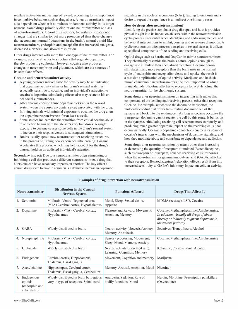
www.EliteCME.com Page 13
regulate motivation and feelings of reward, accounting for its importance in compulsive behaviors such as drug abuse. A neurotransmitter’s impact also depends on whether it stimulates or dampens activity in its target neurons. Some drugs primarily disrupt one neurotransmitter or class of neurotransmitters. Opioid drug abusers, for instance, experience changes that are similar to, yet more pronounced than those changes that accompany normal fl uctuations in the brain’s natural opioid-like neurotransmitters, endorphin and encephalin that increased analgesia, decreased alertness, and slowed respiration.
Other drugs interact with more than one type of neurotransmitter. For example, cocaine attaches to structures that regulate dopamine, thereby producing euphoria. However, cocaine also produces changes in norepinephrine and glutamate, which are the sources of its stimulant effects.
Cocaine and neurotransmitter activity ● A young person’s marked taste for novelty may be an indication
that dopamine activity in his or her brain’s reward system is especially sensitive to cocaine, and an individual’s attraction to cocaine’s dopamine-stimulating effects also may relate to his or her social circumstances.
● After chronic cocaine abuse dopamine ticks up in the reward system when the abuser encounters a cue associated with the drug.
● In living animals with minimal exposure to cocaine, the drug alters the dopamine responsiveness for at least a week.
● Some studies indicate that the transition from casual cocaine abuse to addiction begins with the abuser’s very fi rst doses. A single exposure to cocaine causes some cells in the brain’s reward system to increase their responsiveness to subsequent stimulations.
● Brains usually sprout new neurotransmitter receiving structures in the process of turning new experience into learning. Cocaine accelerates this process, which may help account for the drug’s unusual hold on an addicted individual’s attention.
Secondary impact: Due to a neurotransmitter often stimulating or inhibiting a cell that produces a different neurotransmitter, a drug that alters one can have secondary impacts on another. The key effect all abused drugs seem to have in common is a dramatic increase in dopamine
signaling in the nucleus accumbens (NAc), leading to euphoria and a desire to repeat the experience is an indirect one in many cases.
How do drugs alter neurotransmission?Identifying the precise step that a drug disrupts, and how it provides pivotal insight into its impact on abusers, within the neurotransmission cyclic process, is essential when identifying and addressing medical and behavioral interventions to inhibit, counter and or reverse disruption. A cyclic neurotransmission process transpires in several steps as it utilizes specialized components of the sending and receiving cells.
Opioid drugs such as heroin and OxyContin mimic neurotransmitters. They chemically resemble the brain’s natural opioids enough to engage and stimulate their specialized receptors. Because heroin stimulates many more receptors than the brain uses in the normal cycle of endorphin and encephalin release and uptake, the result is a massive amplifi cation of opioid activity. Marijuana and hashish mimic cannabinoid neurotransmitters, the most important of which is anandamide. Nicotine attaches to receptors for acetylcholine, the neurotransmitter for the cholinergic system.
Some drugs alter neurotransmission by interacting with molecular components of the sending and receiving process, other than receptors. Cocaine, for example, attaches to the dopamine transporter, the molecular conduit that draws free-fl oating dopamine out of the synapse and back into the sending cell. As long as cocaine occupies the transporter, dopamine cannot reenter the cell by this route. It builds up in the synapse, stimulating receiving cell receptors more copiously, and producing much greater dopamine impact on the receiving cells, than occurs naturally. Cocaine’s dopamine connections enumerates some of cocaine’s interactions with the mechanisms of dopamine signaling, and how they motivate abuse and contribute to dependence and addiction.
Some drugs alter neurotransmission by means other than increasing or decreasing the quantity of receptors stimulated. Benzodiazepines, such as diazepam or lorazepam, enhance receiving cells’ responses when the neurotransmitter gammaminobutyric acid (GABA) attaches to their receptors. Benzodiazepines’ relaxation effects result from this increased sensitivity to GABA’s inhibitory impact on cellular activity.
Examples of drug interaction with neurotransmission
Neurotransmitter Distribution in the Central Nervous System Functions Affected Drugs That Affect It
1. Serotonin Midbrain, Ventral Tegmental area (VTA) Cerebral cortex, Hypothalamus
Mood, Sleep, Sexual desire, Appetite
MDMA (ecstasy), LSD, Cocaine
2. Dopamine Midbrain, (VTA), Cerebral cortex, Hypothalamus
Pleasure and Reward, Movement, Attention, Memory
Cocaine, Methamphetamine, Amphetamine. In addition, virtually all drugs of abuse directly or indirectly augment dopamine in the reward pathway.
3. GABA Widely distributed in brain. Neuron activity (slowed), Anxiety, Memory, Anesthesia
Sedatives, Tranquilizers, Alcohol
4. Norepinephrine Midbrain, (VTA), Cerebral cortex, Hypothalamus
Sensory processing, Movement, Sleep, Mood, Memory, Anxiety
Cocaine, Methamphetamine, Amphetamine
5. Glutamate Widely distributed in brain Neuron activity (increased rate), Learning, Cognition, Memory
Ketamine, Phencyclidine, Alcohol
6. Endogenous Cerebral cortex, Hippocampus, Thalamus, Basal ganglia
Movement, Cognition and memory Marijuana
7. Acetylcholine Hippocampus, Cerebral cortex, Thalamus, Basal ganglia, Cerebellum
Memory, Arousal, Attention, Mood Nicotine
8. Endogenous opioids (endorphin and enkephalin)
Widely distributed in brain but regions vary in type of receptors, Spinal cord
Analgesia, Sedation, Rate of bodily functions, Mood
Heroin, Morphine, Prescription painkillers (Oxycodone)
Page 14 www.EliteCME.com
Chronic drug abuse and neurotransmission
During the early phase of an individual’s drug experimentation, neurotransmission normalizes, as intoxication wears off, and the substance leaves the brain. Eventually, however, drugs wreak changes in cellular structure and function that lead to long-lasting or permanent neurotransmission abnormalities. These alterations underlie drug tolerance, addiction, withdrawal, and other persistent consequences. Some longer term changes begin as adjustments to compensate for drug-induced increases in neurotransmitter signaling intensities. For example, drug tolerance typically develops because sending cells reduce the amount of neurotransmitter they produce and release, or receiving cells withdraw receptors or otherwise dampen their responsiveness. Scientists have shown that cells, for example, withdraw opioid receptors into their interiors (where they cannot be stimulated) when exposed to some opioid drugs. When exposed to morphine, however, cells appear instead to make internal adjustments that produce the same effect, which is reduced responsiveness to opiate drugs and natural opioids. Over time, this and related changes recalibrate the brain’s responsiveness to opioid stimulation downward to a level where the organ needs the extra stimulation of the drug to function normally. Therefore, without the drug, withdrawal occurs.
The drug-related mechanisms producing cumulative changes in neurotransmission sometimes are genetic in nature. While a drug cannot change a person’s genes, drugs can prompt some genes to increase their production of proteins, leading to changes in cell function, or even actual reshaping of the physical structure of cells. For example, cocaine and amphetamine stimulate genes which produce the proteins used to build dendrites, branch-like cell structures that contain neurotransmitter receptors. Brains normally sprout new dendrites as they register new learning. The accelerated dendrite formation stimulants induce may partially account for these drugs’ unusual hold of an abuser’s attention.
Some drugs are toxic to nerve cells, and the effect accumulates with repeated exposures. The club drug methylenedioxymethamphetamine (MDMA, ecstasy), for instance, damages axons that release serotonin, and the result is disruption of serotonin neurotransmission that likely underlies the long-lasting memory problems that are experienced by abusers. In addition, methamphetamine, over time, damages enough dopamine-sending cells to cause signifi cant defects in thinking and motor skills. With abstinence, dopamine function can partially recover, but it is not clear whether cognitive and motor capabilities come back as well.
Research outcomes prompt current treatment approaches for addiction
Scientifi c research and clinical practice have yielded a variety of effective approaches to treatment for addiction to certain drugs, such as heroin. Continuing research is also yielding new approaches to developing medications to treat addiction to other drugs, such as cocaine, for which no medications are currently available.
Drug abuse and addiction lead to long-term changes in the brain’s chemistry and anatomy. The changes in the brain cause drug addicts, not only to lose the ability to control their drug use, but their addiction also changes all aspects of their lives. Drug addicts often become isolated from family and friends and have trouble in school or work. In addition, the compulsive need for drugs can lead to signifi cant legal problems. While the biological foundation for drug addiction does not absolve an individual from the responsibility of his or her actions, the stigma of drug addiction needs to be lifted so individuals may receive proper medical treatment, similar to that for other chronic diseases.
The sad news is that addiction is a recurring chronic disease. No cure is available at this time, but addiction can be effectively treated. Drug addiction is often viewed as a lapse in moral character. This value judgment infl uences how society deals with the disease, both socially and medically. Unfortunately, because people, including physicians, have often viewed addiction as a self-infl icted condition, drug addicts have not always received the medical treatment common for other chronic diseases. Treating addiction requires more than a “just say no” approach.
Using pharmacological agents: Treatment for addiction is often very effective. Treatment is successful when the addict reduces or abstains from drug use, improves his or her personal health or social function, and becomes less of a threat to public health and safety.
Certain addictions, such as heroin addiction, can be treated with pharmacological agents that include:
Methadone, the most common pharmacological treatment, prevents craving and withdrawal symptoms in heroin addiction. Methadone is an opiate receptor agonist. That is, methadone binds to the opiate receptor just as heroin does. Methadone, however, does not produce the euphoria or “high” that results from heroin use.
Methadone, as dispensed at a methadone maintenance treatment facility, is a synthetic opioid that is typically administered orally, as a liquid. Methadone is the medication that is most commonly used for Opiate Agonist Pharmacotherapy of opioid dependence. Methadone maintenance treatment is also an extensively researched treatment
modality. There is strong evidence, from research and monitoring of service delivery, that Opiate Agonist Pharmacotherapy maintenance with methadone (Methadone Maintenance Treatment) is effective in reducing illicit drug use, reducing mortality, reducing the risk of spread of HIV, improving physical and mental health, improving social functioning, and reducing criminal behavior. Higher doses of methadone are generally associated with greater reductions in heroin use than either low or moderate doses.
Methadone Maintenance Treatment is associated with a low incidence of side-effects and with substantial health improvements. Around three-quarters of people who commence “Opiate Agonist Pharmacotherapy” with methadone, respond well. However, for various reasons, methadone does not suit all people with opioid dependence. For this group it is important that alternative approaches are available to encourage their retention in treatment. Some require several episodes of treatment before major progress is achieved.
A second medication prescribed for heroin addiction is naltrexone. Unlike methadone, naltrexone is an opiate receptor antagonist. Instead of competing with heroin for the opiate receptor, naltrexone prevents heroin from binding to the receptor, thereby preventing heroin from eliciting the euphoric high.
Buprenorphine is a prescribed medication with weaker opioid agonist activity than methadone. Buprenorphine is not well absorbed if taken orally, therefore the usual route of administration in treatment of opioid dependence, is sublingual. With increasing doses of buprenorphine, effects reach a plateau. Consequently buprenorphine is less likely, than either methadone or heroin to cause an opioid overdose condition, even when taken with other opioids, at the same time. The effectiveness of buprenorphine is similar to that of methadone at adequate doses, in terms of reduction of illicit opioid use and improvements in psychosocial functioning, but buprenorphine may be associated with lower rates of retention in treatment. Buprenorphine is currently more expensive than methadone.
Suboxone, (Buprenorphine and Naloxone) - Buprenorphine is acceptable to heroin users, has few side effects, and is associated with a relatively mild withdrawal syndrome. When used in “Opiate Agonist Pharmacotherapy” for pregnant women with opioid dependence, it appears to be associated with a lower incidence of neonatal withdrawal syndrome.
www.EliteCME.com Page 15
Dihydrocodeine is used in some countries for detoxifi cation and “Opiate Agonist Pharmacotherapy”. Tincture of opium (laudanum) is used in some countries in Asia for the management of opioid withdrawal and, less commonly, for “Opiate Agonist Pharmacotherapy”. The various oral preparations of morphine
formulated to provide slow release (also called sustained release, controlled release and timed release preparations) are also of potential value in the treatment of opioid dependence. However, controlled studies of the effectiveness of these preparations for Opiate Agonist Pharmacotherapy are yet to be undertaken.
Pharmacologic and behavioral treatment in combination, work for best outcomes
Pharmacological therapies, when available, are not suffi cient for effective treatment. Behavioral treatment, in combination with pharmacological treatment, is the most effective way to treat drug addiction. Recovering addicts need to address the behavioral and social consequences of their drug use and learn to cope with the social and environmental factors that contribute to their illness. Behavioral treatments can occur either individually or as a group.
Relapse is a common event for recovering drug addicts. In many ways, relapse should be thought of as a normal part of the recovery process. A recovering drug addict is more likely to experience a relapse if he or she also has other psychiatric conditions or lacks the support of family and friends. But chances of recovery grow, using both medication management and behavioral interventions such as cognitive strategies.
Cognitive strategies: Addiction challenges people to look beyond immediate gratifi cation, to the longer term consequences of their actions. Therefore, patients in drug abuse treatment are often coached to make and rehearse mental associations between situations that trigger drug
cravings and the problems that are likely to ensue from succumbing to them. “Cognitive reappraisal, through mentally changing the meaning of an event or object to lessen its emotional impact, and therefore, alter the behaviors it triggers, is a strategy that helps a variety of problems,” states researcher, Dr. Ochsner. Cognitive-behavioral therapists train patients to use this approach, among others, to cope with negative emotions, stress, and substance cravings. He continues, “People may not realize they can control cravings or emotions using cognitive strategies. For example, thinking of negative consequences, and distracting, and distancing oneself, but patients can learn these techniques and then must continue to apply them over time.” (See previously described study.)
Dr. Ochsner adds that there is broad scientifi c interest in the neurobiological mechanisms underlying cognitive control over thoughts and emotions that promote unhealthy behaviors. Such studies generally fi nd that although there is some overlap in the regions of the PFC engaged when people exert cognitive control, different areas seem to support different strategies for the regulation of emotional responses.
In summary
Despite the preconceptions and value judgments many people place on addiction, it is, in many ways, similar to other chronic diseases, such as diabetes and coronary artery disease. Genetic, environmental, and behavioral components contribute to each of these diseases. Some people may argue that drug addiction is different because it is “self-infl icted.”
The initial choice to use drugs is voluntary, but once addiction develops, drug use is compulsive—not voluntary. Moreover, voluntary choices do contribute to the onset or severity of other chronic diseases, as well. For example, a person who chooses to eat an unhealthy diet and not exercise, increases his or her risk for coronary heart disease.
Successful treatment for any chronic disease necessitates patient compliance with the prescribed treatment regimen. Adhering to a treatment plan is diffi cult for those with any chronic disease. For example, less than 50 percent of diabetics follow their routine medication plan, and only 30 percent follow their dietary guidelines. Problems adhering to a treatment plan lead to about 50 percent of diabetics needing to be treated again, within one year of diagnosis and initial treatment. Similar statistics hold true for other chronic diseases. Approximately, 40 percent of patients with hypertension, need emergency room treatment for episodes of extreme high blood pressure, and only about 30 percent of adult asthma sufferers take their medication, as prescribed.
Although treatment for drug addiction statistically is more successful than treatment for other chronic diseases, drug addicts commonly have relapses during treatment and recovery and begin using drugs again. The diffi culties in following a treatment plan and coping with the stresses of a chronic disease illustrate how diffi cult changing human behavior is. By altering neurotransmission, addictive drugs produce
effects that make people want to continue to abuse them and induce health problems that can be recurring and long-lasting, with profound consequences. The effects are drug-specifi c and each drug disrupts particular neurotransmitters in particular ways.
Scientifi c research has transformed how drug addiction is treated. Researchers seek to understand how drugs impact neurotransmission causing changes in the chemistry and function of the brain. Their discoveries have led to new medications to treat the disease of addiction. Scientists continue to work on developing medications that relieve the cravings experienced during withdrawal. In addition, scientifi c advances may reveal ways to reverse the long-term damage to the brain drugs infl ict.
Scientists must use a variety of experimental tools and methods to study drugs’ effects on neurotransmission. Utilizing both animals and people, their fi ndings enhance understanding of the experiences of drug abusers, the burden of addicts, and lead the way to new behavioral interventions, as well as medication interventions. These fi ndings provide potential bases for prevention strategies and treatment process monitoring.
As a reminder, some important effects, though, are shared by all people using drugs, and they include initial pleasurable feelings, and subsequent dependence and addiction resulting from disruption of the dopamine neurotransmitter system.
The brain is a hugely complex organ. Its complexity will prompt scientists to continue their work for many years. It is anticipated that, at some point, they will answer questions about what happens in the brain to cause addiction, which will then help them understand how to prevent the disease.
Bibliography American Psychiatric Association. (2013). Diagnostic and statistical manual of mental
disorders (5th ed.). Arlington, VA: American Psychiatric Publishing. American Psychiatric Association. (2000). Diagnostic and statistical manual of
mental disorders (4th ed., text rev.). Washington, DC: Author. Biological Psychiatry 69(7):708–711, 2011. http://www.sciencedirect.com/science/
article/pii/S0006322310007584 Overexpression of the glucocorticoid gene in the fi rst weeks after birth increased anxiety and response to cocaine in adulthood. November 29, 2012, Sharon Reynolds, NIDA Notes Contributing Writer
Brown, M.T.C., et al. Drug-driven AMPA receptor redistribution mimicked by selective dopamine neuron stimulation. PLoS One. 5:12: e15870, 2010. http://www.
ncbi.nlm.nih.gov/pmc/articles/PMC3013137/pdf/pone.0015870.pdf, Riegel, A.C., and Kalivas, P.W. Neuroscience: Lack of inhibition leads to abuse. Nature 463: 743–744, 2010. http://www.nature.com/nature/journal/v463/n7282/full/463743a.html like opioids and cannabinoids, diazepam and other benzodiazepines take the brakes off activity of dopamine-producing neurons. April 19, 2012 by NIDA Notes Staff
Kober, H., et al. Prefrontal-striatal pathway underlies cognitive regulation of craving. Proceedings of the National Academy of Sciences 107(33):14811–14816, 2010. http://www.pnas.org/content/107/33/14811.full.pdf+html
Kober, H., et al. Regulation of craving by cognitive strategies in cigarette smokers. Drug and Alcohol Dependence 106(1):52–55, 2010.
Page 16 www.EliteCME.com
Neurons and Neurotransmitters: The “Brains” of The Nervous System (Permission Granted) http://www.utexas.edu/research/asrec/neuron.html. Compiled & Edited By: D. Shrira Dated: 6 January 2007
Stowe, G.N. et al., A vaccine strategy that induces protective immunity against heroin. Journal of Medicinal Chemistry 54(14), 5195–204, 2011. http://www.ncbi.nlm.nih.gov/pubmed/21692508
The Brain: Neurotransmitters Send Chemical Messages, Understanding Addiction ( Permission Granted) http://www.utexas.edu/research/asrec/synapse_m.html, Compiled & Edited: D. Shrira Updated: 8 January 2007.
The Brain: Understanding Neurobiology Through The Study of Addiction: Neurons, Brain Chemistry and Neurotransmission Lesson 2 (NIDA, 2004)
The Brain: Understanding Neurobiology Through the Study of Addiction; Lesson 1~ The Brain: What’s Going On In There? (NIDA) 2004, Compiled and edited by: Deborah Shrira Dated: January 25, 2001, Asst. Editor: Dee Black Updated: March 16, 2012.
The Brain: Understanding Neurobiology Through The Study of Addiction Lesson 4~Drug Abuse and Addiction (NIDA 2004), Compiled and Edited By: D. Shrira Updated: 9 Jan 2007.
The Brain: Understanding Neurobiology Through The Study of Addiction Lesson 5~Drug Addiction Is A Disease So What Do We Do About It? (2) WHO/UNODC/UNAIDS position paper; Substitution Therapy In The Management of Opioid Dependence and HIV/AIDS, NIH/National Institute on Drug Abuse 2004, Compiled & Edited By: D. Shrira Dated: January 12, 2007, Asst. Editor: Dee Black Updated: March 16, 2012
Understanding Addiction (Addiction Science Research and Education, College of Pharmacy, The University of Texas ) (Permission Granted).
The Brain: Understanding Neurobiology Through The Study of Addiction: Neurons, Brain Chemistry and Neurotransmission~ Lesson 2 (NIDA 2004), Deborah Shrira, Editor 2008 August.
Wee, S., et al. Novel cocaine vaccine linked to a disrupted adenovirus gene transfer vector blocks cocaine psycho-stimulant and reinforcing effects. Neuropsychopharmacology [Epub ahead of print September 14, 2011].
Wei, Q., et al. Early-life forebrain glucocorticoid receptor overexpression increases anxiety behavior and cocaine sensitization. BiolPsychiatry 2012;71:224–231. http://www.ncbi.nlm.nih.gov/pubmed/21872848
Whitten, Lori, NIDA Notes Staff Writer April 19, 2012, Whitten, Lori, To Block the Effects of Abused Drug, NIDA Notes, September 17, 2012.
UNDERSTANDING NEUROTRANSMISSION AND THE DISEASE OF ADDICTIONFinal Examination Questions
Select the best answer for each question and proceed to www.EliteCME.com to complet your fi nal examination.
1. After taking drugs for a period of time, a person may need to take a higher dose to have the same feeling or “rush” that he or she did when fi rst taking the drug. This is called:a. Tolerance.b. Craving.c. Addiction.d. Dependence.
2. In rats and non-human primates, MDMA appears to kill neurons that produce:a. Serotonin.b. Nerepinephine.c. Neutrons.d. Seraquil.
3. While the DSM-IV utilized two separate diagnoses of “Dependence” and “Abuse”, ___________ in DSM-5 combines the DSM-IV categories of substance abuse and substance dependence into a single disorder measured on a continuum from mild to severe.a. Abuse-dependence disorder.b. Substance use disorder.c. Dependency disorder.d. Substance disorder.
4. Glial cells are categorized as:a. Fast or slow.b. Microglia or macroglia.c. With dendrites or without dendrites.d. Long or short.
5. Within a single neuron, information is conducted via:a. Electrical signaling.b. Chemical surges.c. A glial cell.d. All of the above.
6. A neurotransmitter is a(n):a. Molecule that is released from a neuron to relay information to
another cell. b. Transmission of cells to the axon. c. Electrical impulse.d. Specifi c cognitive behavior.
7. The goal of an anti-drug vaccine is to:a. Induce the immune system to block the psychoactive effects of
its target drug.b. Reduce the amount of serotonin in the pleasure center.c. Diminish the reaction within the glial cells.d. Adhere to the brain stem.
8. Because _______stimulates many more receptors than the brain uses in the normal cycle of endorphin and encephalin release and uptake, the result is a massive amplifi cation of opioid activity.a. Methadone.b. Xanax.c. Heroin.d. Dopamine.
9. Virtually all drugs of abuse directly or indirectly augment:a. Serotonin in the reward pathway.b. Dopamine in the reward pathway.c. Norepinephrine in the reward pathway.d. Neutrons in the reward pathway.
10. Buprenorphine is a prescribed medication with weaker opioid agonist activity than ____________.a. Xanax.b. Valium.c. Alcohol.d. Methadone.