Understanding multidrug-resistance: Focus on Carbapenems · Covers resistant gram-positive...
158
Nimalie D. Stone, MD,MS Dialysis and Long-term Care Team Division of Healthcare Quality Promotion GA CRE Collaborative Learning Session 1 March 20, 2014 Understanding multidrug-resistance: Focus on Carbapenems National Center for Emerging and Zoonotic Infectious Diseases Division of Healthcare Quality Promotion
Transcript of Understanding multidrug-resistance: Focus on Carbapenems · Covers resistant gram-positive...
Nimalie D. Stone, MD,MS
Dialysis and Long-term Care Team
Division of Healthcare Quality Promotion
GA CRE Collaborative
Learning Session 1
March 20, 2014
Understanding multidrug-resistance: Focus on Carbapenems
National Center for Emerging and Zoonotic Infectious Diseases
Division of Healthcare Quality Promotion
Presentation Objectives
Brief overview on microbiology and antibiotics
Describe antibiotic resistant organisms with a focus on carbapenem-resistance
Discuss how/why resistant organisms spread in healthcare settings
Identify the core prevention strategies for reducing the emergence and transmission of resistance
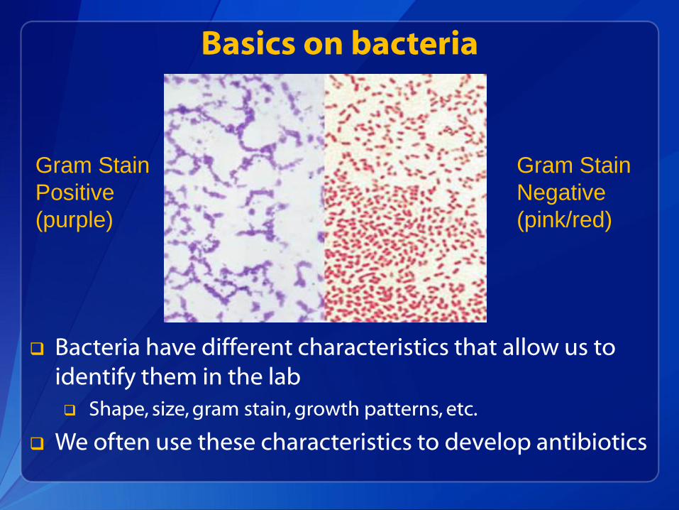
Basics on bacteria
Gram Stain
Positive
(purple)
Gram Stain
Negative
(pink/red)
Bacteria have different characteristics that allow us to identify them in the lab
Shape, size, gram stain, growth patterns, etc.
We often use these characteristics to develop antibiotics

Common bacteria in healthcare
Gram positive
Many are cocci, “round bacteria”
Examples are Streptococci, Staphylococci, Enterococci
Clostridium difficile (C. diff ) is an anaerobic, Gram positive rod
Gram negative
Most are baccili, “rod-shaped bacteria”
Examples are: E. coli, Klebsiella, Enterobacter , Proteus, Pseudomonas, Acinetobacter
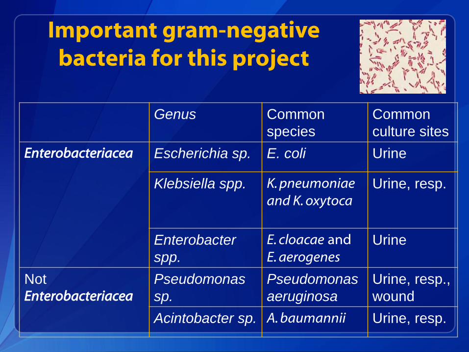
Important gram-negative bacteria for this project
Genus Common
species
Common
culture sites
Enterobacteriacea Escherichia sp. E. coli Urine
Klebsiella spp. K. pneumoniae and K. oxytoca
Urine, resp.
Enterobacter
spp.
E. cloacae and E. aerogenes
Urine
Not Enterobacteriacea
Pseudomonas
sp.
Pseudomonas
aeruginosa
Urine, resp.,
wound
Acintobacter sp. A. baumannii Urine, resp.
Antibiotics 101
Antibiotics are drugs that treat and kill bacteria
They are grouped into classes based on their structure and activity
Narrow-spectrum target a few specific bacteria
Broad-spectrum can kill a wide variety of bacteria
Antibiotic resistance = when the bacteria are no longer fully killed by the antibiotic
Bacteria with resistance can cause patients to have more severe infections which are harder and more costly to treat
Infection prevention programs track certain “bug-drug” combinations for resistance

Antibiotics: Beta Lactam classes
Penicillin and extended spectrum agents
Examples: Penicillin, amoxicillin, ampicillin, methicillin
Can be combined with a drug to help them overcome bacterial resistance
Amoxicillin + Clavulante = Augmentin;
Ampicillin + Sulbactam = Unasyn
Piperacillin + tazobactam = Zosyn
Cephalosporins
More gram positive activity: Cephalexin, Cefazolin
More gram negative activity: Ceftriaxone, Ceftazidime,
Cefepime
New broader spectrum, including MRSA: Ceftaroline

Antibiotics: Carbapenems
Extremely broad-spectrum, among the most powerful antibiotics we currently have available
Spectrum includes Streptococci, susceptible Staphylococci, Enterobactericeae, Pseudomonas, Acinetobacter sp., and anaerobic bacteria
Drug Route of Administration
Imipenem IV
Meropenem IV
Ertapenem IM, IV
Doripenem IV

Antibiotics : Gram positive agents
Vancomycin
Treats methicillin-resistant Staphylococcus aureus (MRSA)
Oral form is NOT absorbed from gut; only used to treat C difficile
IV form will get good systemic levels - used to treat all other infections
Daptomycin
Covers resistant gram-positive organisms: MRSA and Vancomycin-resistant Enterococci (VRE)
Only available as IV formula
Linezolid
Covers MRSA and VRE
Both oral and IV forms available and get good systemic levels

Antibiotics: Gram negative agents
Fluoroquinolones (oral and IV forms)
Ciprofloxacin: Mostly gram negative activity
Commonly used for UTI treatment
Levofloxacin/Moxifloxacin: Broader activity
Also used for treating UTIs and infections from gram-negative bacteria
Also covers Streptococcus pneumoniae and other respiratory bacteria
Aminoglycosides (only IV)
Examples: Gentamicin, Tobramycin, Amikacin
Excellent gram negative drugs – especially for urinary tract
Limited use because of toxicity (kidney, hearing/balance)
Antibiotics: Miscellaneous
Trimethoprim/Sulfamethoxazole (Bactrim):
Mainly given in oral form – must watch renal function
Considered narrow spectrum, but has activity against both Gram negative and Gram positive bacteria
Commonly used to treat UTIs
Also used for MRSA skin infections
Azithromycin:
Commonly given in oral dose pack called “Z-pack”
Considered narrow spectrum, used for respiratory/sinus infections
Metronidazole (Flagyl) (oral and IV form)
A primary treatment for C. difficile infections
Oral form can cause nausea and stomach upset
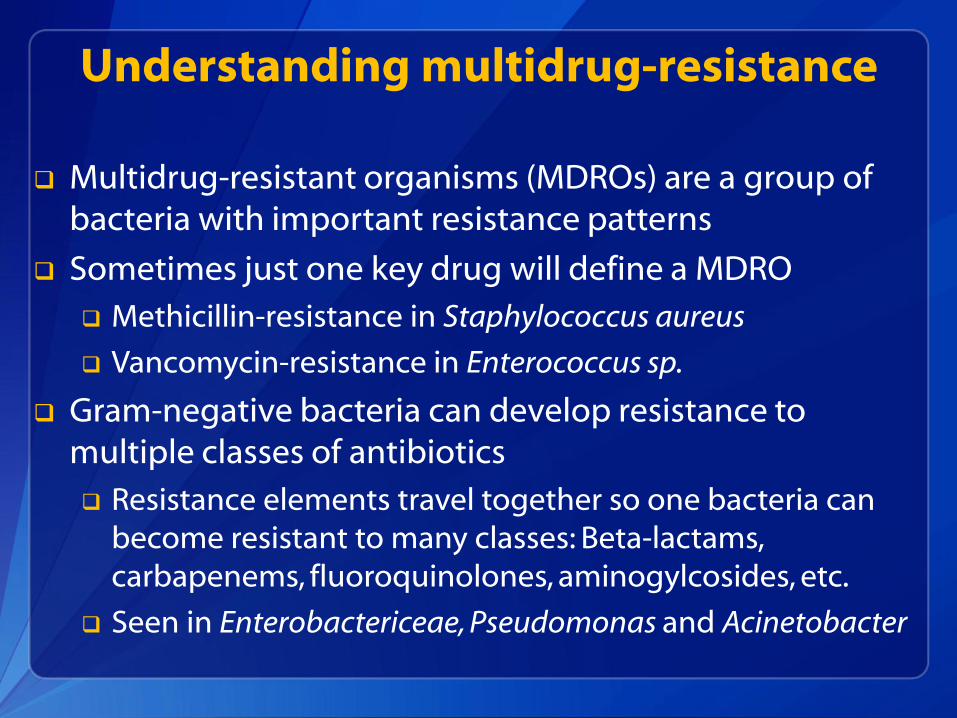
Understanding multidrug-resistance
Multidrug-resistant organisms (MDROs) are a group of bacteria with important resistance patterns
Sometimes just one key drug will define a MDRO
Methicillin-resistance in Staphylococcus aureus
Vancomycin-resistance in Enterococcus sp.
Gram-negative bacteria can develop resistance to multiple classes of antibiotics
Resistance elements travel together so one bacteria can become resistant to many classes: Beta-lactams, carbapenems, fluoroquinolones, aminogylcosides, etc.
Seen in Enterobactericeae, Pseudomonas and Acinetobacter
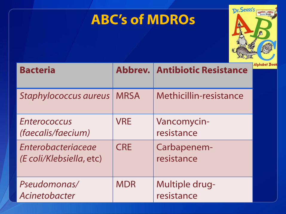
ABC’s of MDROs
Bacteria Abbrev. Antibiotic Resistance
Staphylococcus aureus MRSA Methicillin-resistance
Enterococcus (faecalis/faecium)
VRE Vancomycin-resistance
Enterobacteriaceae (E coli/Klebsiella, etc)
CRE Carbapenem-resistance
Pseudomonas/ Acinetobacter
MDR Multiple drug-resistance

Mechanisms of antibiotic resistance
Production of proteins that destroy antibiotics
Beta-lactamases
Carbapenemases
Change their cell structure so antibiotics can’t bind and block their function
Reduce their antibiotic exposure
Pump drugs out
Increase cell barriers to keep drug out http://bioinfo.bact.wisc.edu/themicrobialworld/bactresanti.html

Understanding carbapenem-resistance
There are different ways that these gram-negative bacteria become resistant to Carbapenems.
Some bacteria have to make lots of changes to become resistance. Step 1: Acquire or produce a cephalosporinase (to break down beta-
lactam antibiotics
Step 2: Lose a porin protein in the cell wall to prevent carbapenems from getting into the cell.
Step 3: Gain a pump to remove the carbapenem from the cell
Others acquire resistance by a genetic element, called a plasmid, which carries the genes for carbapenem resistance These resistance genes are called “Carbapenemases”
But, no matter HOW they became resistance, we need to stop these bacteria from spreading further


Normal bacterial colonization
People have bacteria living in and on us all the time
Some live on our skin, some in our nose and throats, others in our GI tracts (i.e., bowels)
Our bodies rely on colonizing bacteria
In the GI tract bacteria will
Aid digestion/provide nutrients
Block harmful bacteria from invading (e.g. C. difficile)
Gram-negative bacteria colonize the lower GI tract and easily spread from there to the urinary tract , and other sites
Separating colonization from infection
“Colonizing” bacteria may not be harmful, even when they are antibiotic-resistant
Example: MRSA cultured from a nasal swab may not harm the colonized person
Only when bacteria invade our bodies and cause signs/symptoms of illness do we need treatment with antibiotics
Separating colonization from infection can be difficult
Examples: Bacteriuria in an older adult; respiratory secretions from a person on a ventilator
However, both colonized and infected people can serve as a source for spreading resistant organisms
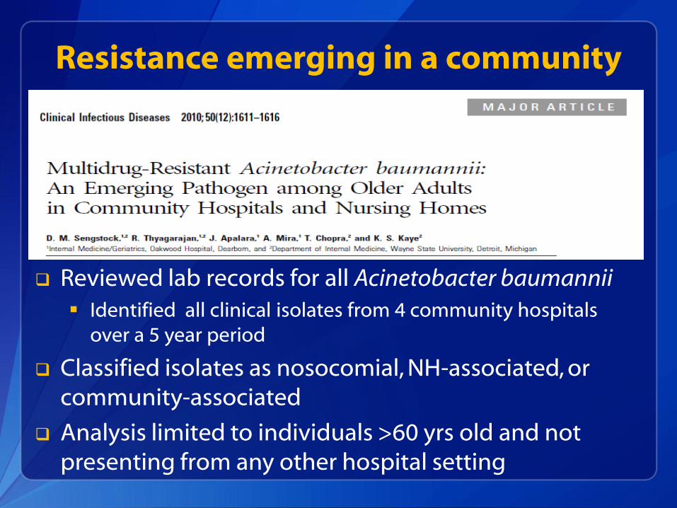
Reviewed lab records for all Acinetobacter baumannii
Identified all clinical isolates from 4 community hospitals over a 5 year period
Classified isolates as nosocomial, NH-associated, or community-associated
Analysis limited to individuals >60 yrs old and not presenting from any other hospital setting
Resistance emerging in a community
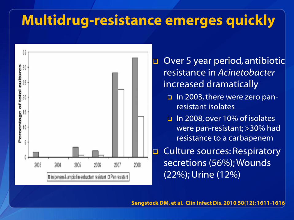
Multidrug-resistance emerges quickly
Over 5 year period, antibiotic resistance in Acinetobacter increased dramatically In 2003, there were zero pan-
resistant isolates
In 2008, over 10% of isolates were pan-resistant; >30% had resistance to a carbapenem
Culture sources: Respiratory secretions (56%); Wounds (22%); Urine (12%)
Sengstock DM, et al. Clin Infect Dis. 2010 50(12): 1611-1616
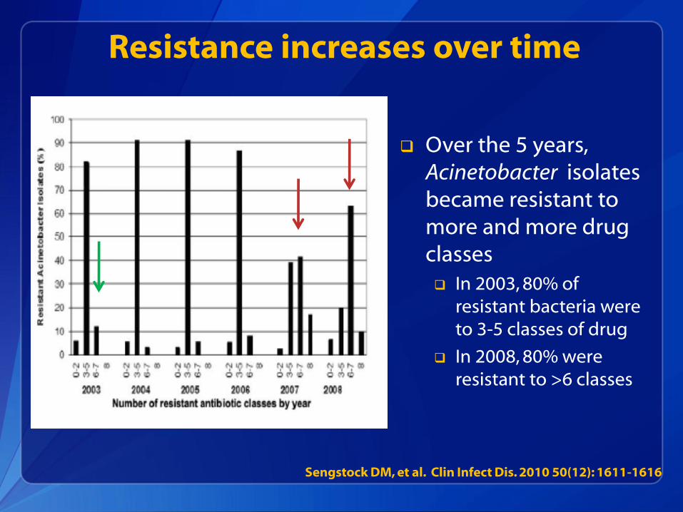
Resistance increases over time
Over the 5 years, Acinetobacter isolates became resistant to more and more drug classes In 2003, 80% of
resistant bacteria were to 3-5 classes of drug
In 2008, 80% were resistant to >6 classes
Sengstock DM, et al. Clin Infect Dis. 2010 50(12): 1611-1616
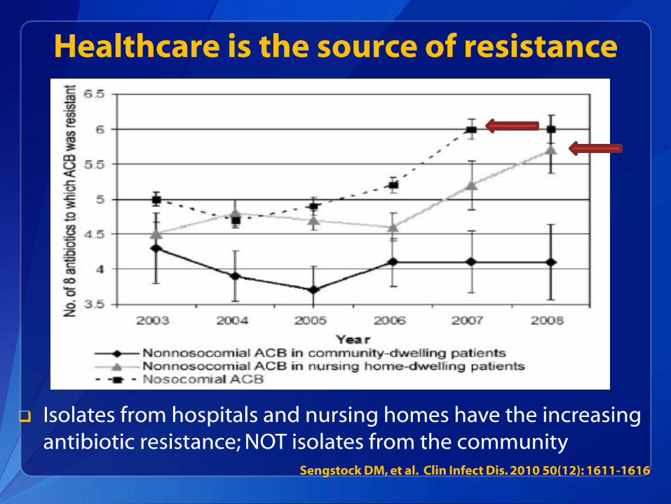
Healthcare is the source of resistance
Sengstock DM, et al. Clin Infect Dis. 2010 50(12): 1611-1616
Isolates from hospitals and nursing homes have the increasing antibiotic resistance; NOT isolates from the community
Healthcare drivers of antibiotic resistance
DEVELOPMENT
Antibiotic pressure
Risk for both acquisition and infection
Medical devices and wounds
Biofilm formation
SPREAD
Colonization pressure
Patient to patient transmission via hands of healthcare personnel
Contamination of shared environment / equipment

Resistance from antibiotic pressure
At first most of the bacteria can be killed by the drug (green)
But, once they are wiped out, the resistant bugs take over (red)
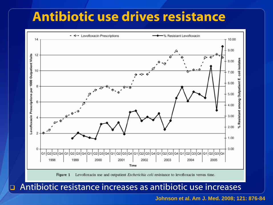
Antibiotic use drives resistance
Johnson et al. Am J. Med. 2008; 121: 876-84
Antibiotic resistance increases as antibiotic use increases
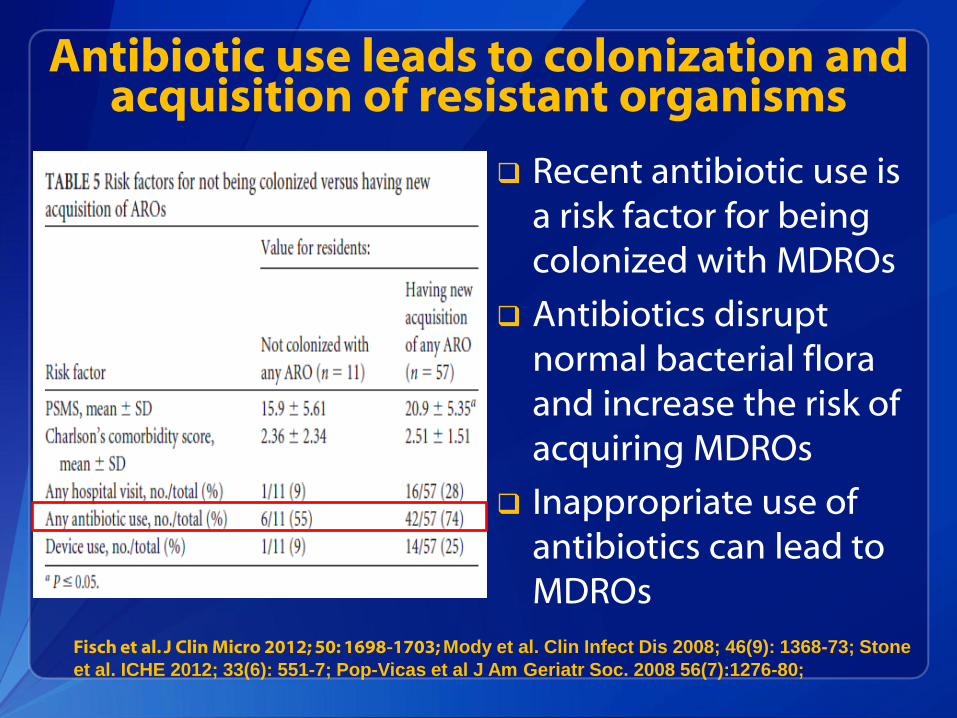
Antibiotic use leads to colonization and acquisition of resistant organisms
Recent antibiotic use is a risk factor for being colonized with MDROs
Antibiotics disrupt normal bacterial flora and increase the risk of acquiring MDROs
Inappropriate use of antibiotics can lead to MDROs
Fisch et al. J Clin Micro 2012; 50: 1698-1703; Mody et al. Clin Infect Dis 2008; 46(9): 1368-73; Stone
et al. ICHE 2012; 33(6): 551-7; Pop-Vicas et al J Am Geriatr Soc. 2008 56(7):1276-80;

Biofilm formation on device surfaces
Biofilm: An collection of bacteria within a sticky film that forms a community on the surface of a device
http://www.ul.ie/elements/Issue7/Biofilm%20Information.htm

Biofilm on an indwelling urinary catheter
Tenke, P et al. World J. Urol. 2006; 24: 13-20

Resistance develops within biofilms
Bacteria within a biofilm are grow every differently from those floating around freely These changes in their growth make our antibiotics less
effective
Antibiotics can’t penetrate the biofilm to get to the bacteria This leads to much less drug available to treat the bugs
Bacteria within the biofilm can exchange information including the traits that cause resistance Some carbapenem-resistance can be easily shared
among different bacteria Tenke, P et al. World J. Urol. 2006; 24: 13-20
Colonization pressure leading to MDRO acquisition
Colonization pressure: High burden of other MDRO carriers on a unit will increase the risk of MDRO acquisition for others
Studies have demonstrated the impact of colonization pressure on acquisition of many resistant bacteria and C. difficile
Both colonized and infected individuals act as a source for spread on a unit or within a facility
. Dubberke ER et al. Arch Intern Med. 2007 May 28;167(10):1092-7
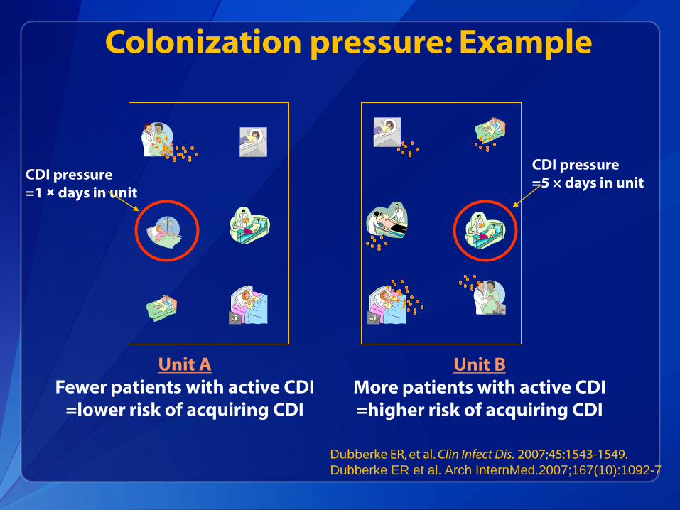
Colonization pressure: Example
Unit A Fewer patients with active CDI
=lower risk of acquiring CDI
Unit B More patients with active CDI =higher risk of acquiring CDI
CDI pressure =1 × days in unit
CDI pressure =5 × days in unit
Dubberke ER, et al. Clin Infect Dis. 2007;45:1543-1549. Dubberke ER et al. Arch InternMed.2007;167(10):1092-7
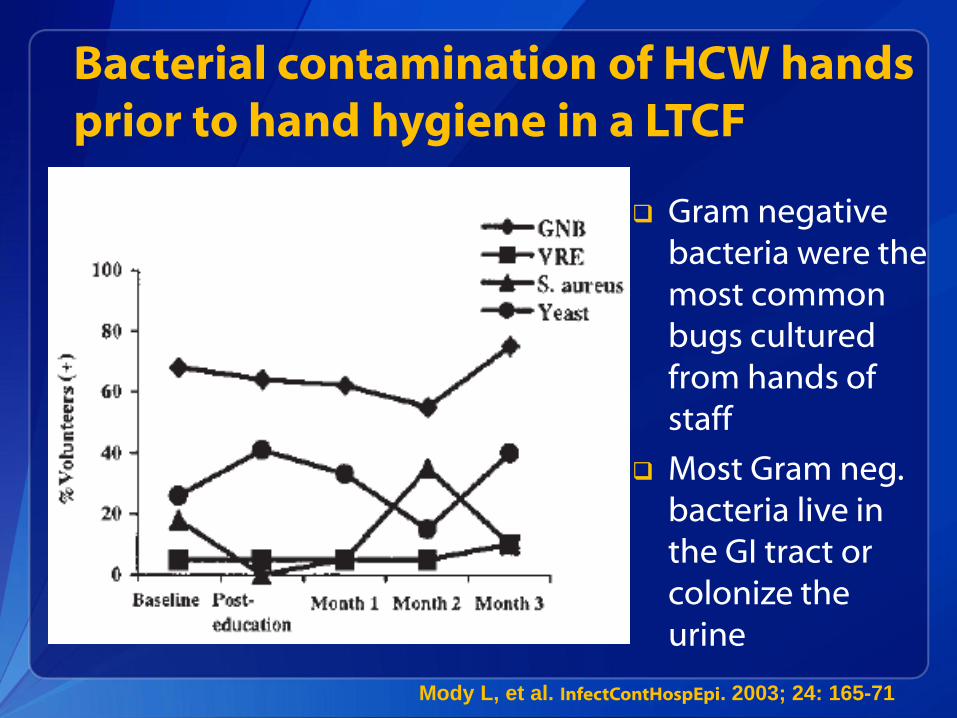
Bacterial contamination of HCW hands prior to hand hygiene in a LTCF
Mody L, et al. InfectContHospEpi. 2003; 24: 165-71
Gram negative bacteria were the most common bugs cultured from hands of staff
Most Gram neg. bacteria live in the GI tract or colonize the urine
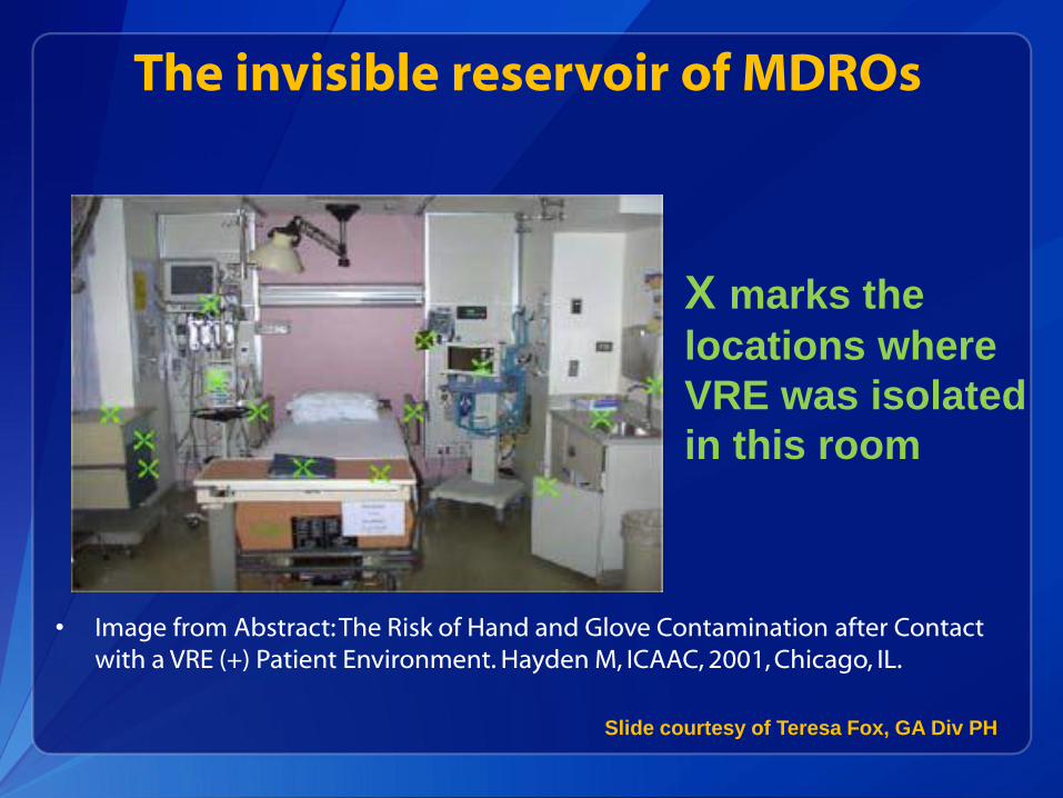
The invisible reservoir of MDROs
• Image from Abstract: The Risk of Hand and Glove Contamination after Contact with a VRE (+) Patient Environment. Hayden M, ICAAC, 2001, Chicago, IL.
X marks the
locations where
VRE was isolated
in this room
Slide courtesy of Teresa Fox, GA Div PH
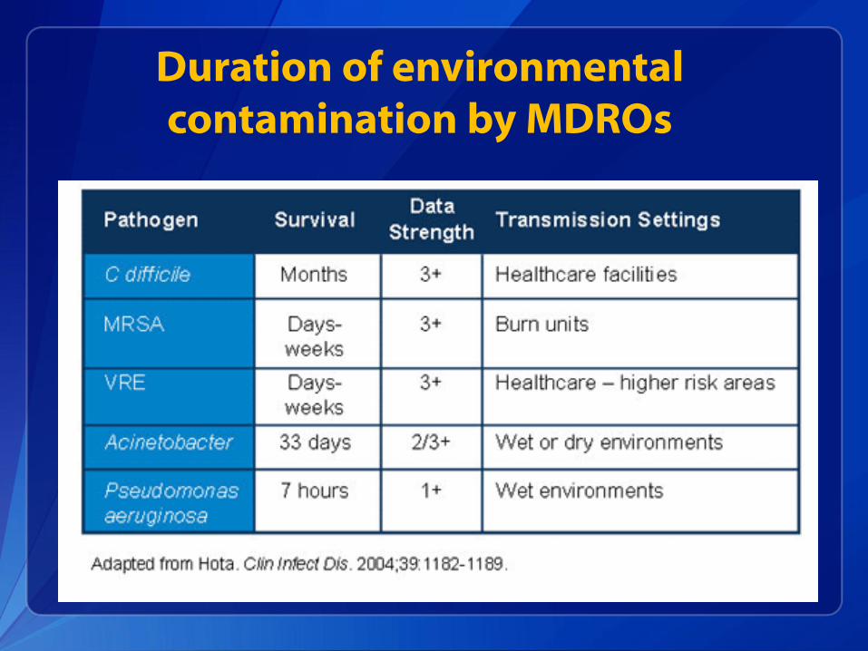
Duration of environmental contamination by MDROs
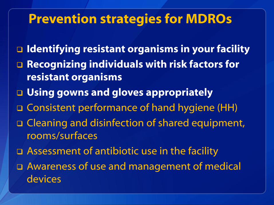
Prevention strategies for MDROs
Identifying resistant organisms in your facility
Recognizing individuals with risk factors for resistant organisms
Using gowns and gloves appropriately
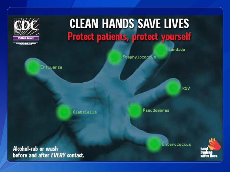
Consistent performance of hand hygiene (HH)
Cleaning and disinfection of shared equipment, rooms/surfaces
Assessment of antibiotic use in the facility
Awareness of use and management of medical devices
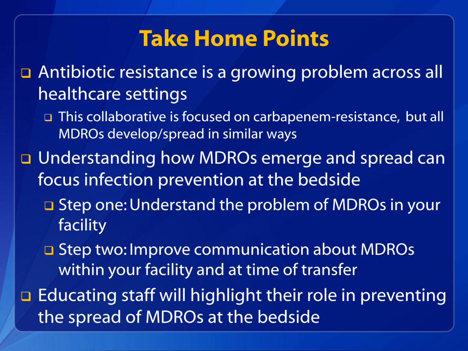
Take Home Points
Antibiotic resistance is a growing problem across all healthcare settings This collaborative is focused on carbapenem-resistance, but all
MDROs develop/spread in similar ways
Understanding how MDROs emerge and spread can focus infection prevention at the bedside
Step one: Understand the problem of MDROs in your facility
Step two: Improve communication about MDROs within your facility and at time of transfer
Educating staff will highlight their role in preventing the spread of MDROs at the bedside

For more information please contact Centers for Disease Control and Prevention
1600 Clifton Road NE, Atlanta, GA 30333 Telephone, 1-800-CDC-INFO (232-4636)/TTY: 1-888-232-6348 E-mail: [email protected] Web: www.cdc.gov
The findings and conclusions in this report are those of the authors and do not necessarily represent the official position of the Centers for Disease Control and Prevention.
Thank you!!
National Center for Emerging and Zoonotic Infectious Diseases
Division of Healthcare Quality Promotion
Email: [email protected] with questions/comments

Carbapenem-resistant Enterobacteriaceae (CRE):
A Bird’s Eye View from the US
Jesse T. Jacob, MD
Assistant Professor of Medicine
Emory University
Objectives
• Understand the context of CRE among
resistant among gram negative bacteria
• Recognize the epidemiology and natinoalal
threat of CRE
• Integrate approaches to CRE prevention

Overview of Gram Negative Bacteria
E. coli, Klebsiella, Proteus, Enterobacter, Serratia, Pseudomonas, Salmonella, Shigella, Bacteroides
Vibrio, Campylobacter jejuni, Helicobacter pylori
Neisseria meningitidis N. gonorrhoeae (gonococcus) Moraxella catarrhalis
Fusobacterium
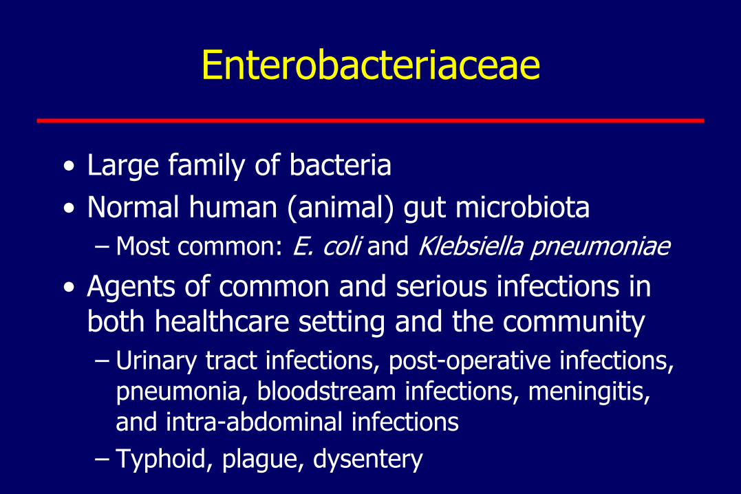
Enterobacteriaceae
• Large family of bacteria
• Normal human (animal) gut microbiota
– Most common: E. coli and Klebsiella pneumoniae
• Agents of common and serious infections in both healthcare setting and the community
– Urinary tract infections, post-operative infections, pneumonia, bloodstream infections, meningitis, and intra-abdominal infections
– Typhoid, plague, dysentery

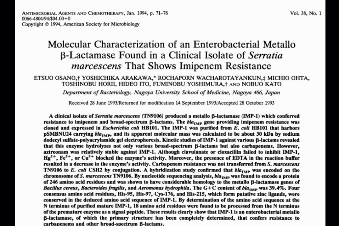
Carbapenems\Carbapenem Resistance
• Carbapenems (i.e. imipenem)
– Broadest spectrum β-lactams available
– “Antibiotics of last resort”, only given by vein
• Carbapenem resistance
– Potentially transferrable (plasmid mediated)
– Two major mechanisms
• Carbapenemase
• Porin mutation + β-lactamase

Resistance to -lactam Antibiotics

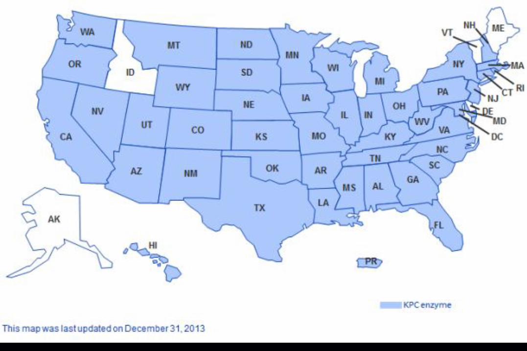
Klebsiella pneumoniae Carbapenemase
• First US isolate described in North Carolina
• Isolated 1996, reported in 2001
• Became endemic in the NE US

• Patel, Rasheed, Kitchel. 2009. Clin Micro News
• CDC, unpublished data
KPC-producing CRE in the United States
Nov 2006
DC
PR AK
HI
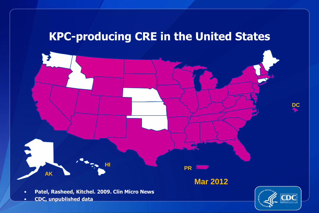
KPC-producing CRE in the United States
• Patel, Rasheed, Kitchel. 2009. Clin Micro News
• CDC, unpublished data
DC
PR AK
HI
Mar 2012
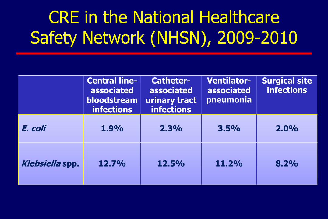
Central line-associated
bloodstream infections
Catheter-associated
urinary tract infections
Ventilator-associated pneumonia
Surgical site infections
E. coli 1.9% 2.3% 3.5% 2.0%
Klebsiella spp. 12.7% 12.5% 11.2% 8.2%
CRE in the National Healthcare Safety Network (NHSN), 2009-2010


0
10
20
30
40
50
60
Overall Mortality Attributable
Mortality
Pe
rce
nt
of
su
bje
cts
Mortality in CRE Bacteremia
P <0.001 P <0.001
Carbapenem-
resistant
Carbapenem-
susceptible
Patel G et al. Infect Control Hosp Epidemiol 2008;29:1099-1106.

International dissemination of Klebsiella pneumoniae carbapenemase (KPC)–producing Enterobacteriaceae.
Gupta N et al. Clin Infect Dis. 2011;53:60-67
• Result of clinical summit in 2012
• Developed a roadmap to tackling antibiotic resistance (not just CRE) over 5 years
– Aims to be practical, not ideal
– Medical school curriculum
– Standardize microbiology labs
– Implement hospital antimicrobial stewardship
– Regulation of over the counter antibiotics
http://www.chennaideclaration.org/
Ghafur et al. Indian J Cancer 2013

Carbapenem Use in Selected Countries
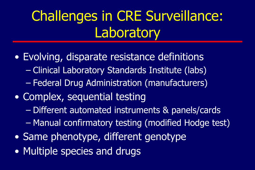
Challenges in CRE Surveillance: Laboratory
• Evolving, disparate resistance definitions
– Clinical Laboratory Standards Institute (labs)
– Federal Drug Administration (manufacturers)
• Complex, sequential testing
– Different automated instruments & panels/cards
– Manual confirmatory testing (modified Hodge test)
• Same phenotype, different genotype
• Multiple species and drugs
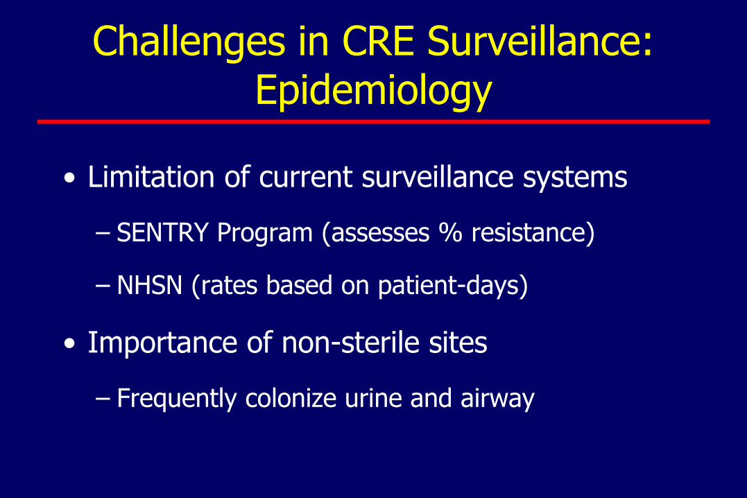
Challenges in CRE Surveillance: Epidemiology
• Limitation of current surveillance systems
– SENTRY Program (assesses % resistance)
– NHSN (rates based on patient-days)
• Importance of non-sterile sites
– Frequently colonize urine and airway
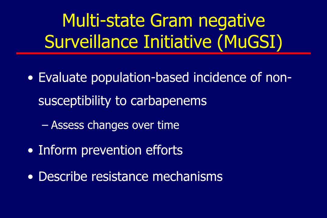
Multi-state Gram negative Surveillance Initiative (MuGSI)
• Evaluate population-based incidence of non-
susceptibility to carbapenems
– Assess changes over time
• Inform prevention efforts
• Describe resistance mechanisms
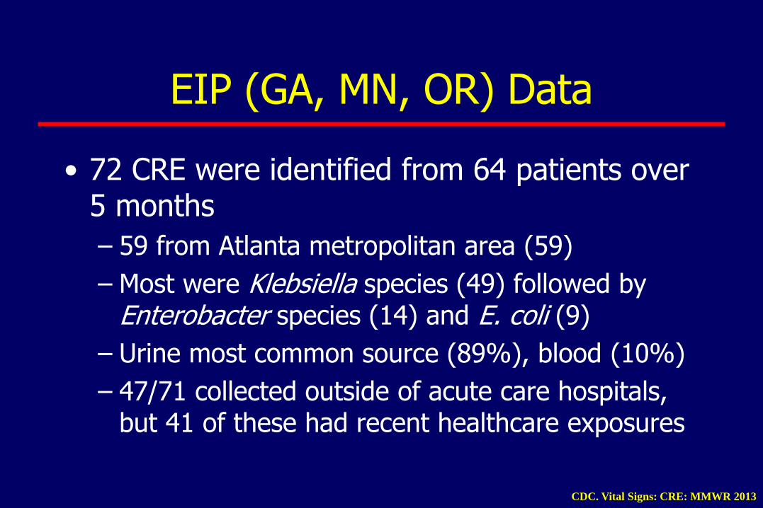
EIP (GA, MN, OR) Data
• 72 CRE were identified from 64 patients over 5 months
– 59 from Atlanta metropolitan area (59)
– Most were Klebsiella species (49) followed by Enterobacter species (14) and E. coli (9)
– Urine most common source (89%), blood (10%)
– 47/71 collected outside of acute care hospitals, but 41 of these had recent healthcare exposures
CDC. Vital Signs: CRE: MMWR 2013

Organisms
A baumannii
44%
K pneumonia
43%
E aerogenes
1% K oxytoca
1%
E coli
7%
E cloace
4%
n=81

Distribution by Body Site
Urine
76%
Blood
16%
Pleural fluid
2%
Other
2%
Peritoneal fluid
4%
n=81

CRE: A Call to Antibiotic Stewardship
• Collaboration of infection prevention & control, microbiology, pharmacy and clinicians (ID)
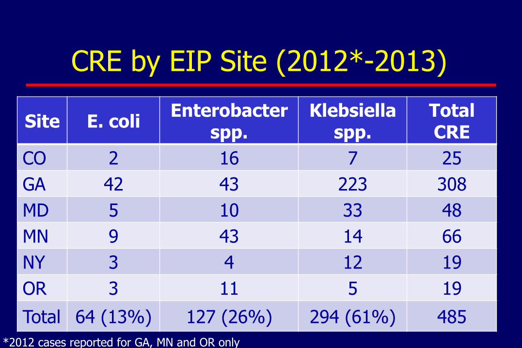
CRE by EIP Site (2012*-2013)
Site E. coli Enterobacter
spp. Klebsiella
spp. Total CRE
CO 2 16 7 25
GA 42 43 223 308
MD 5 10 33 48
MN 9 43 14 66
NY 3 4 12 19
OR 3 11 5 19
Total 64 (13%) 127 (26%) 294 (61%) 485
*2012 cases reported for GA, MN and OR only
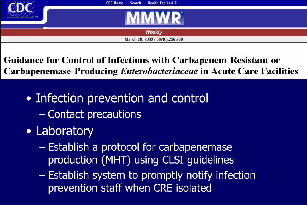
Guidance on Infection Control
• Infection prevention and control
– Contact precautions
• Laboratory
– Establish a protocol for carbapenemase production (MHT) using CLSI guidelines
– Establish system to promptly notify infection prevention staff when CRE isolated
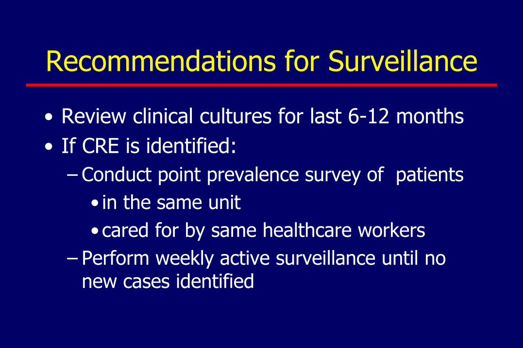
Recommendations for Surveillance
• Review clinical cultures for last 6-12 months
• If CRE is identified:
– Conduct point prevalence survey of patients
• in the same unit
• cared for by same healthcare workers
– Perform weekly active surveillance until no new cases identified
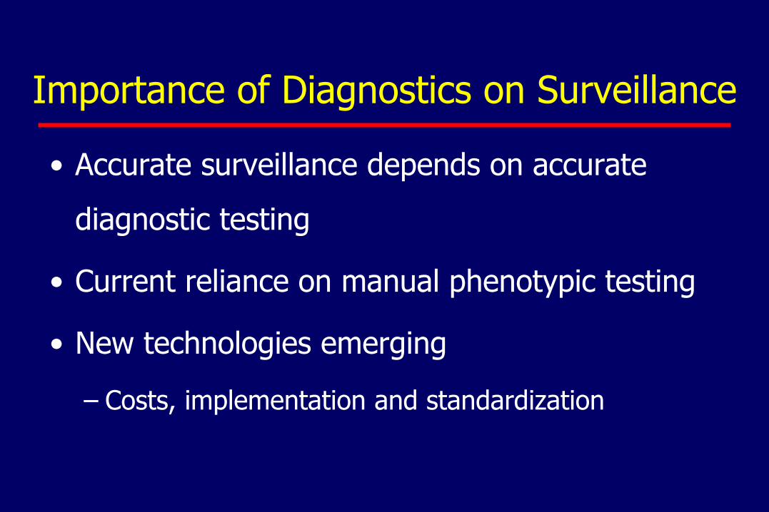
Importance of Diagnostics on Surveillance
• Accurate surveillance depends on accurate
diagnostic testing
• Current reliance on manual phenotypic testing
• New technologies emerging
– Costs, implementation and standardization

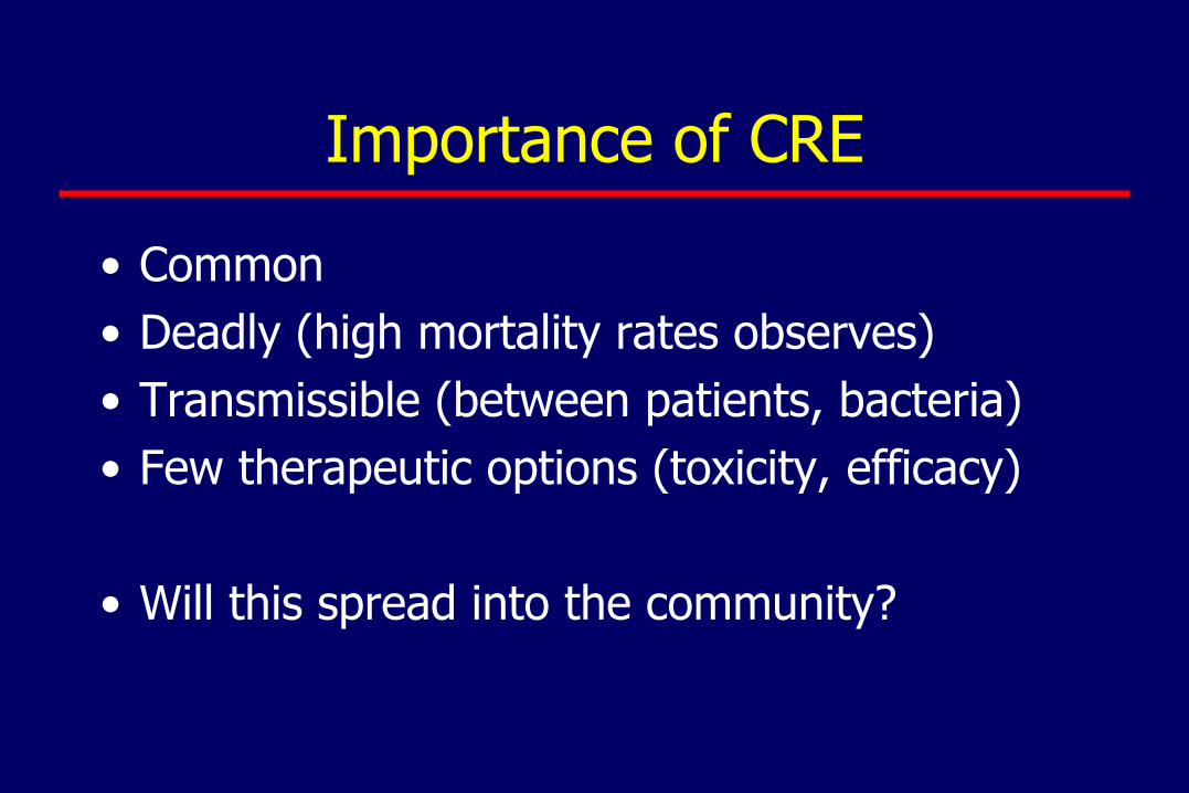
Importance of CRE
• Common
• Deadly (high mortality rates observes)
• Transmissible (between patients, bacteria)
• Few therapeutic options (toxicity, efficacy)
• Will this spread into the community?
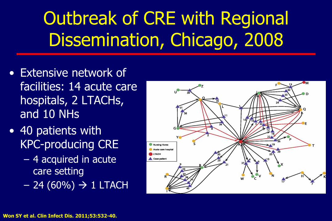
Outbreak of CRE with Regional Dissemination, Chicago, 2008
• Extensive network of facilities: 14 acute care hospitals, 2 LTACHs, and 10 NHs
• 40 patients with KPC-producing CRE
– 4 acquired in acute care setting
– 24 (60%) 1 LTACH
Won SY et al. Clin Infect Dis. 2011;53:532-40.
http://www.cdc.gov/hai/organisms/cre/cre-toolkit
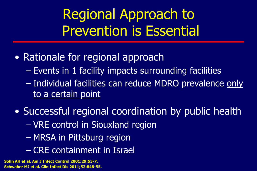
Regional Approach to Prevention is Essential
• Rationale for regional approach
– Events in 1 facility impacts surrounding facilities
– Individual facilities can reduce MDRO prevalence only to a certain point
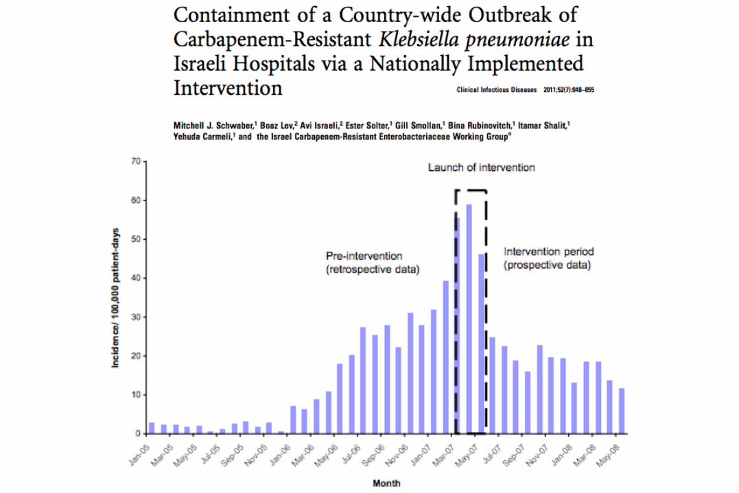
• Successful regional coordination by public health
– VRE control in Siouxland region
– MRSA in Pittsburg region
– CRE containment in Israel Sohn AH et al. Am J Infect Control 2001;29:53-7.
Schwaber MJ et al. Clin Infect Dis 2011;52:848-55.
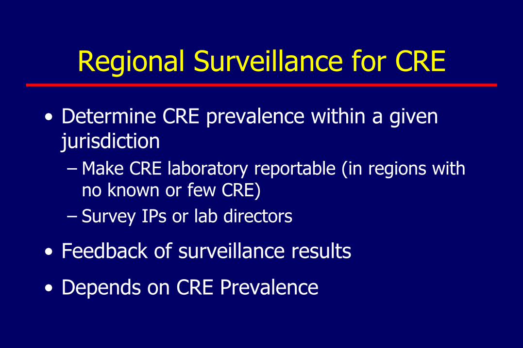
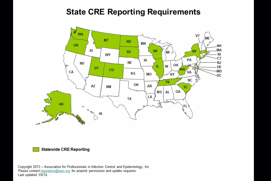
Regional Surveillance for CRE
• Determine CRE prevalence within a given jurisdiction
– Make CRE laboratory reportable (in regions with no known or few CRE)
– Survey IPs or lab directors
• Feedback of surveillance results
• Depends on CRE Prevalence
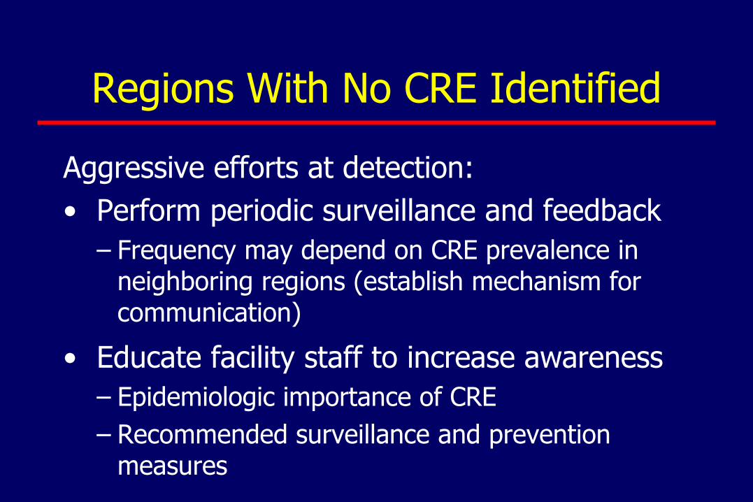
Regions With No CRE Identified
Aggressive efforts at detection:
• Perform periodic surveillance and feedback
– Frequency may depend on CRE prevalence in neighboring regions (establish mechanism for communication)
• Educate facility staff to increase awareness
– Epidemiologic importance of CRE
– Recommended surveillance and prevention measures
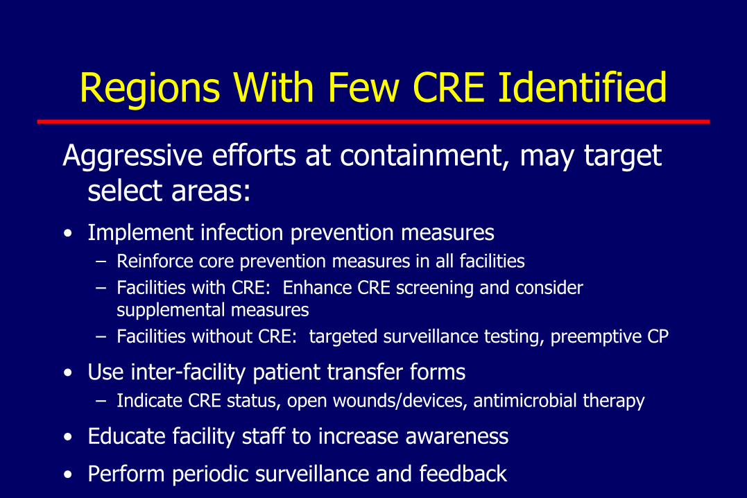
Regions With Few CRE Identified
Aggressive efforts at containment, may target select areas:
• Implement infection prevention measures
– Reinforce core prevention measures in all facilities
– Facilities with CRE: Enhance CRE screening and consider supplemental measures
– Facilities without CRE: targeted surveillance testing, preemptive CP
• Use inter-facility patient transfer forms
– Indicate CRE status, open wounds/devices, antimicrobial therapy
• Educate facility staff to increase awareness
• Perform periodic surveillance and feedback
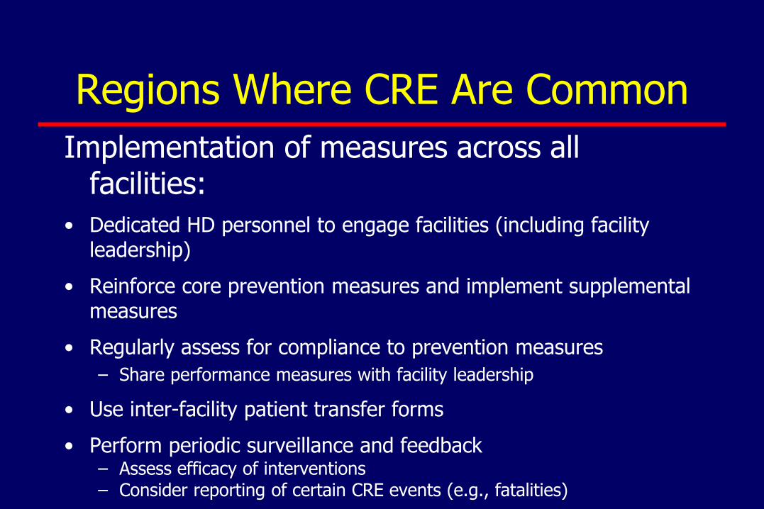
Regions Where CRE Are Common
Implementation of measures across all facilities:
• Dedicated HD personnel to engage facilities (including facility leadership)
• Reinforce core prevention measures and implement supplemental measures
• Regularly assess for compliance to prevention measures
– Share performance measures with facility leadership
• Use inter-facility patient transfer forms
• Perform periodic surveillance and feedback – Assess efficacy of interventions – Consider reporting of certain CRE events (e.g., fatalities)


Summary
• CRE are prevalent and distributed worldwide
• Prevention efforts need to be coordinated at the regional level and beyond
• Public health critical to minimizing spread
• Prevention requires collaboration
Acknowledgements
• Georgia EIP
– Jessica Reno
– Surveillance Officers
• CDC
– Brandi Limbago
– Alice Guh
– Alex Kallen
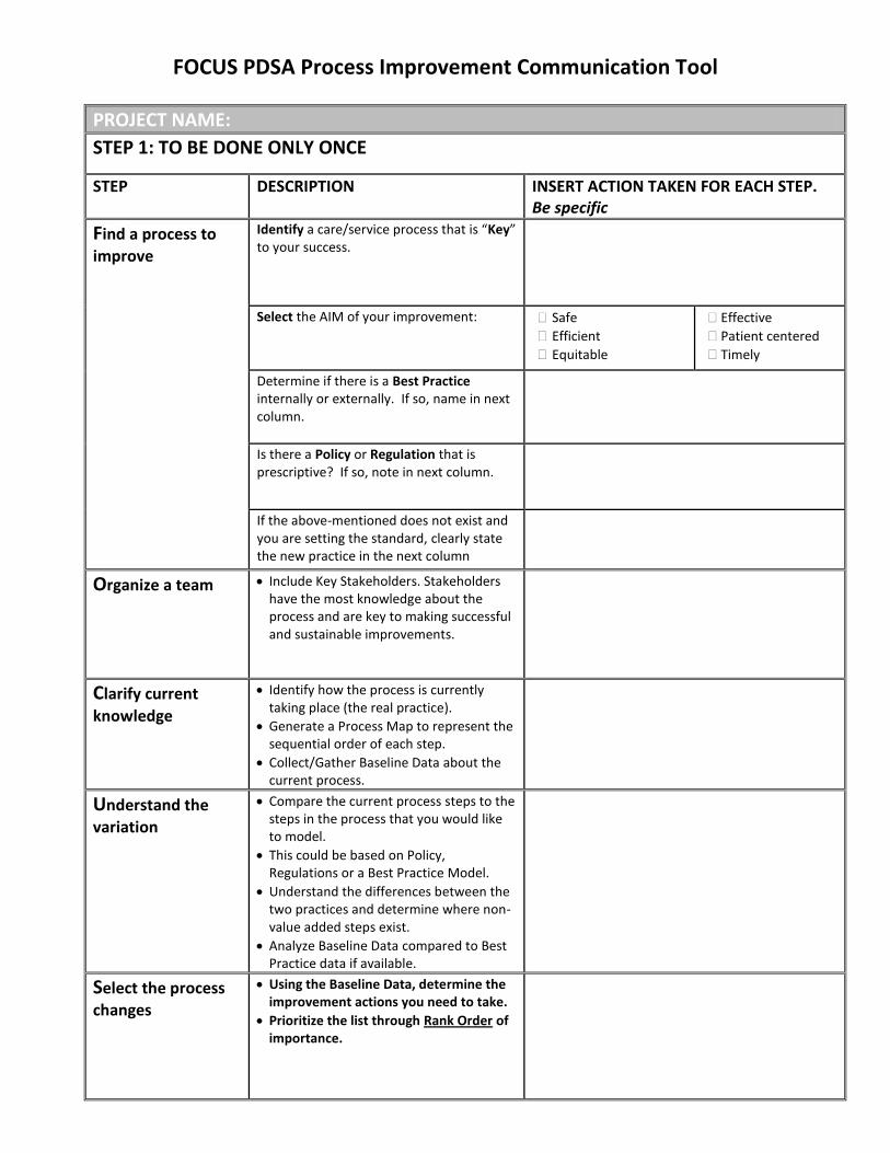
FOCUS PDSA Process Improvement Communication Tool
PROJECT NAME:
STEP 1: TO BE DONE ONLY ONCE
STEP DESCRIPTION INSERT ACTION TAKEN FOR EACH STEP. Be specific
Find a process to
improve
Identify a care/service process that is “Key” to your success.
Select the AIM of your improvement: Safe
Efficient
Equitable
Effective
Patient centered
Timely
Determine if there is a Best Practice internally or externally. If so, name in next column.
Is there a Policy or Regulation that is prescriptive? If so, note in next column.
If the above-mentioned does not exist and you are setting the standard, clearly state the new practice in the next column
Organize a team Include Key Stakeholders. Stakeholders have the most knowledge about the process and are key to making successful and sustainable improvements.
Clarify current
knowledge
Identify how the process is currently taking place (the real practice).
Generate a Process Map to represent the sequential order of each step.
Collect/Gather Baseline Data about the current process.
Understand the
variation
Compare the current process steps to the steps in the process that you would like to model.
This could be based on Policy, Regulations or a Best Practice Model.
Understand the differences between the two practices and determine where non-value added steps exist.
Analyze Baseline Data compared to Best Practice data if available.
Select the process
changes
Using the Baseline Data, determine the improvement actions you need to take.
Prioritize the list through Rank Order of importance.

FOCUS PDSA Process Improvement Communication Tool
FOCUS PDSA Process Improvement Communication Tool
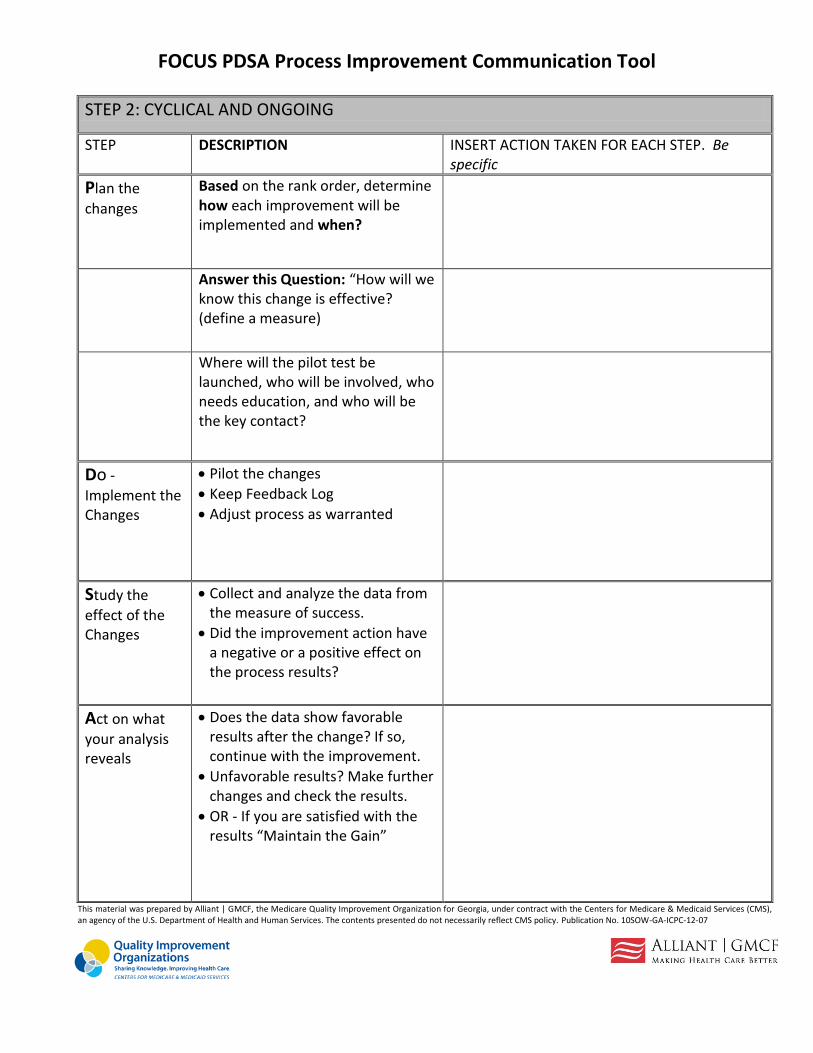
STEP 2: CYCLICAL AND ONGOING
STEP DESCRIPTION INSERT ACTION TAKEN FOR EACH STEP. Be specific
Plan the
changes
Based on the rank order, determine how each improvement will be implemented and when?
Answer this Question: “How will we know this change is effective? (define a measure)
Where will the pilot test be launched, who will be involved, who needs education, and who will be the key contact?
Do -
Implement the Changes
Pilot the changes
Keep Feedback Log
Adjust process as warranted
Study the
effect of the Changes
Collect and analyze the data from the measure of success.
Did the improvement action have a negative or a positive effect on the process results?
Act on what
your analysis reveals
Does the data show favorable results after the change? If so, continue with the improvement.
Unfavorable results? Make further changes and check the results.
OR - If you are satisfied with the results “Maintain the Gain”
This material was prepared by Alliant | GMCF, the Medicare Quality Improvement Organization for Georgia, under contract with the Centers for Medicare & Medicaid Services (CMS), an agency of the U.S. Department of Health and Human Services. The contents presented do not necessarily reflect CMS policy. Publication No. 10SOW-GA-ICPC-12-07

5/14/2014 1
PDSA Session Plan~Do~Study~Act
March 20, 2014
Presented by
Nancy Fendler, Technical Advisor, Alliant GMCF
Presented to
CRE Collaborative
Learning Objectives
At the end of this session each participant will be
able to:
► Understand the Model for Improvement
► Learn how to apply PDSA cycles
► Select measures for their improvement efforts
► Have strategies for more successful tests
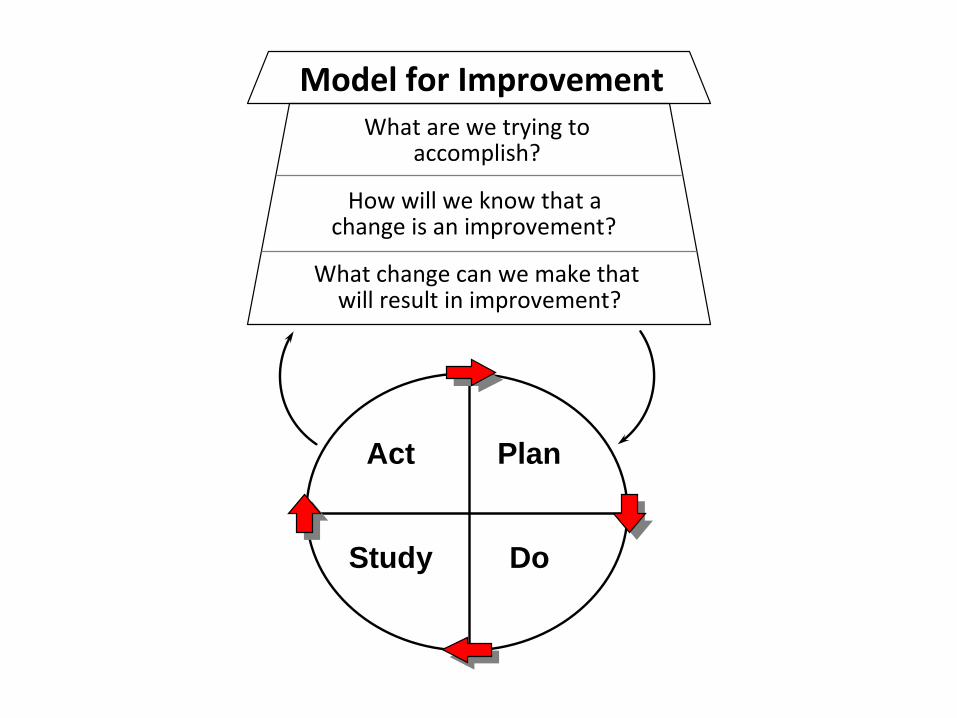
Plan, Do, Study, Act
What are we trying to accomplish?
How will we know that a change is an improvement?
What change can we make that will result in improvement?
Model for Improvement
Act Plan
Study Do
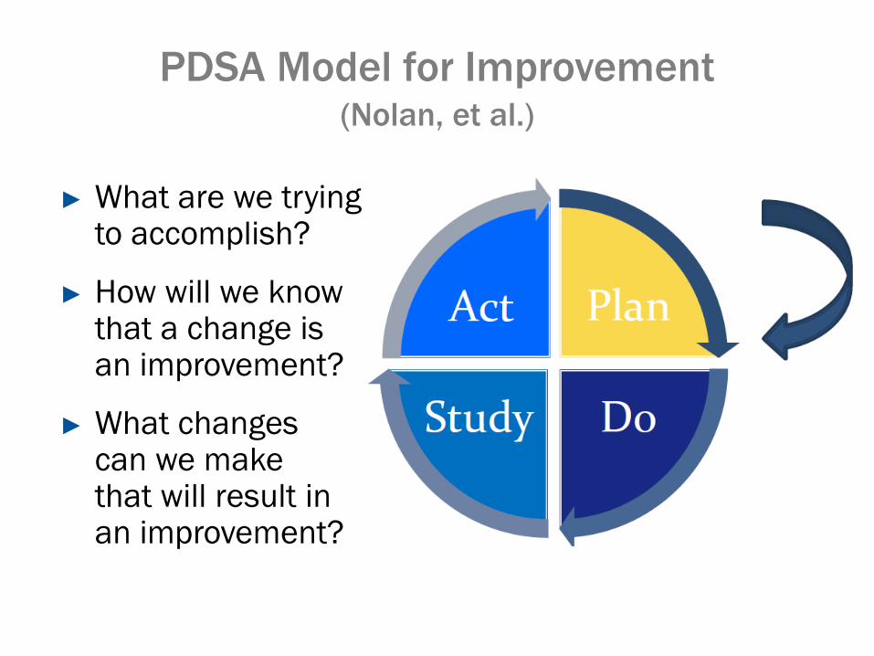
PDSA Model for Improvement (Nolan, et al.)
► What are we trying to accomplish?
► How will we know that a change is an improvement?
► What changes can we make that will result in an improvement?
PLAN~DO~STUDY~ACT
PLAN
► Need to identify an Aim or a Goal
► Who? Identify a leader
► What? A new tool? A new process?
► Where? Which areas will be impacted? Which areas will be involved?
► When? Set a date!
DO
► Carry out your change/test
► Collect data and begin analysis
► Identify the person in charge of implementation
► Keep a time frame for implementing the change
PLAN~DO~STUDY~ACT
STUDY
► Do the results agree with your predictions?
► Is it working?
► Summarize what worked and what didn’t work
PLAN~DO~STUDY~ACT
ACT
► As a result of the cycle – list your actions
► Widen your scope
► Plan for the next cycle – adapt change? Another test?
► IMPLEMENT!
PLAN~DO~STUDY~ACT

Changes that
Result in
Improvement
Hunches
Theories
Ideas
A P
D S
A P
D S DATA
Small Test of Change Worksheet
Goal: Overall goal you would like to reach
Every goal will require multiple smaller tests of change
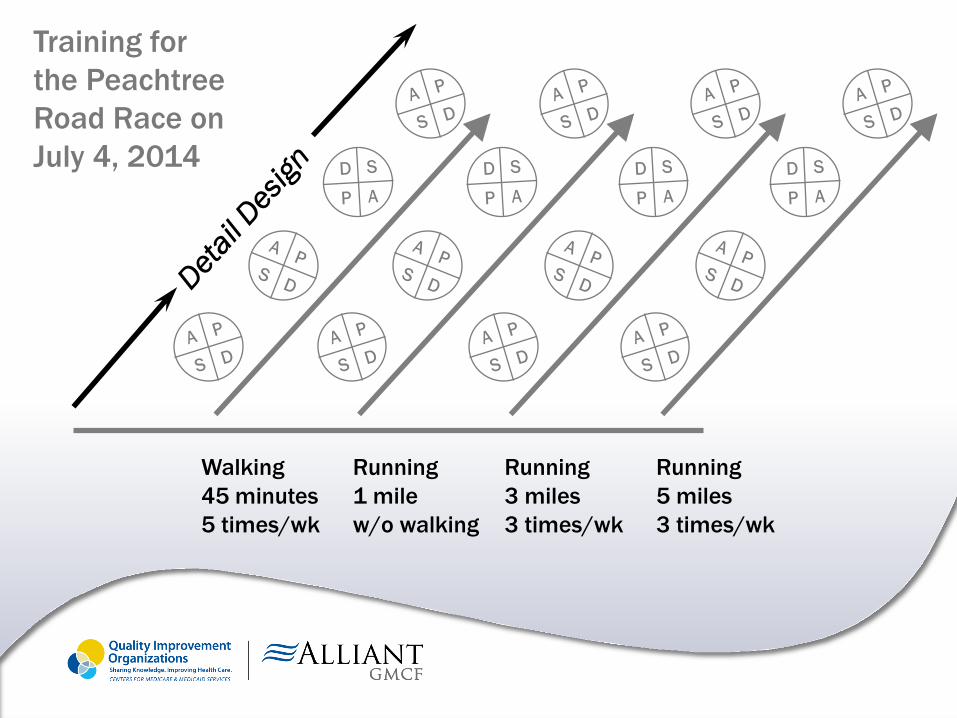
Training for
the Peachtree
Road Race on
July 4, 2014
Walking
45 minutes
5 times/wk
Running
1 mile
w/o walking
Running
3 miles
3 times/wk
Running
5 miles
3 times/wk
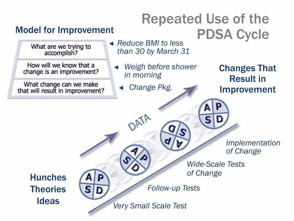
Repeated Use of the PDSA Cycle
Hunches
Theories
Ideas
Changes That Result in
Improvement
Very Small Scale Test
Follow-up Tests
Wide-Scale Tests of Change
Implementation of Change
◄ Reduce BMI to less than 30 by March 31
◄ Weigh before shower in morning
Model for Improvement
◄ Change Pkg.
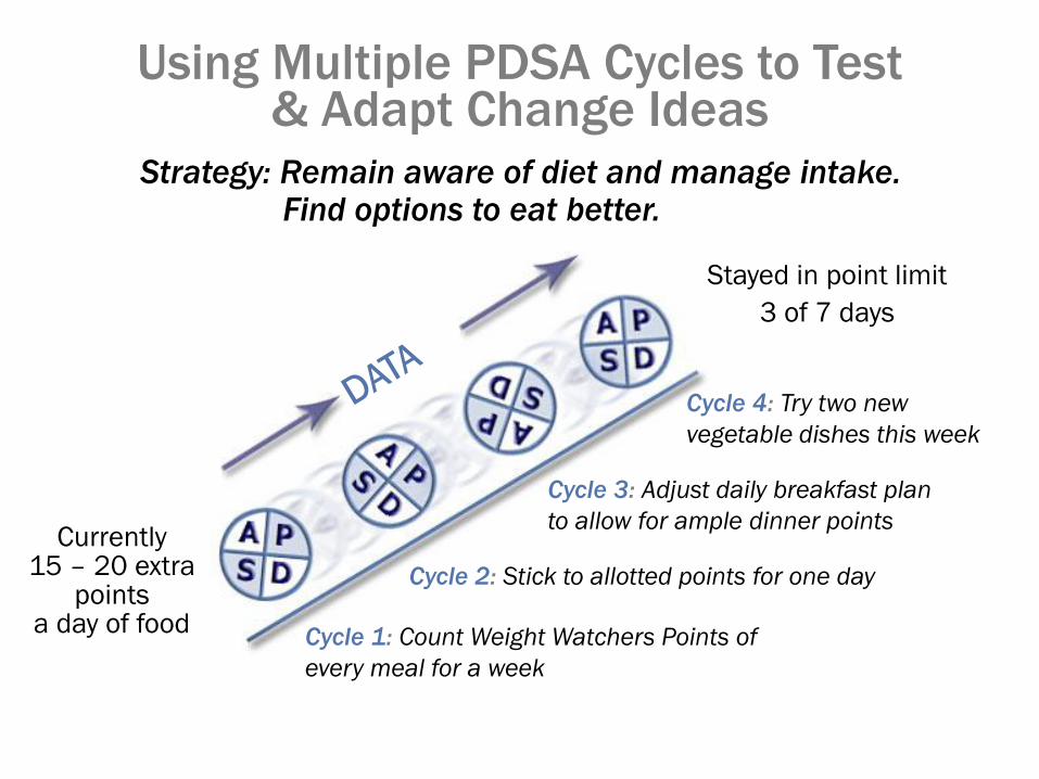
Strategy: Remain aware of diet and manage intake. Find options to eat better.
Using Multiple PDSA Cycles to Test & Adapt Change Ideas
Currently 15 – 20 extra
points a day of food
Cycle 1: Count Weight Watchers Points of
every meal for a week
Cycle 2: Stick to allotted points for one day
Cycle 3: Adjust daily breakfast plan
to allow for ample dinner points
Cycle 4: Try two new
vegetable dishes this week
Stayed in point limit
3 of 7 days
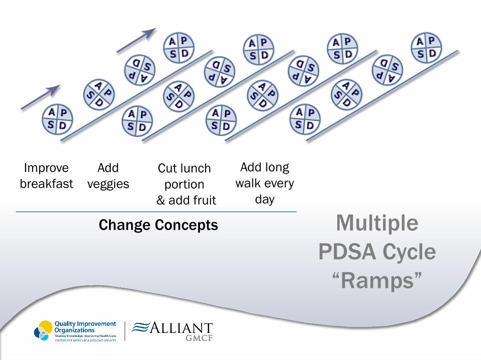
Multiple
PDSA Cycle
“Ramps”
Improve
breakfast
Add
veggies
Cut lunch
portion
& add fruit
Add long
walk every
day
Change Concepts
Before you get started… FOCUS!
Find a process to improve
Organize a team
Clarify current knowledge
Understand the variation
Select the process changes

Set a Goal ~ Think SMART
Your Goal should be SMART
Specific
Measurable
Attainable
Relevant
Time-bound
(becomes standard process)
Pilot Testing
► Gives team chance to see how to implement
a change on a small scale
► Give team early results, to see if the change
you make has any impact
► The team has a role to play in helping to
implement any change that is recommended
Pilot Testing
► Who will train staff?
► Who will update/revise/remove tool, if necessary?
► Who will monitor to see if process has changed?
► Who will team contact if they need support
implementing change?
► Who will audit outcome of process change?
Pilot Testing
► Evaluating the pilot test allows your team to
organize observations that the team has made
through the pilot test
► Evaluation also includes collecting data to
check whether the change has helped you
reach your goal
Pilot Testing
► Do we need to re-evaluate our initial goal?
► What is working well? WHY?
► What is not working? WHY?
► What can be done differently?
Pilot Testing
► Do we need to revise materials we are using
(if any)?
► How does staff feel about the change in
process?
► Are patients/residents positively affected by
the change in process?
WHY Test Change?
► Increase your belief that the change will result in improvement
► Provide an opportunity for learning from “failures” without impacting performance
► Document how much improvement can be expected from the change
WHY Test Change?
► Learn how to adapt the change to conditions in your hospital/nursing home
► Evaluate costs and side-effects of the change
► Minimize resistance upon implementation
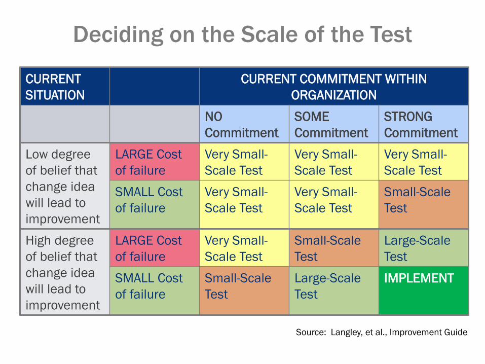
Deciding on the Scale of the Test
CURRENT
SITUATION
CURRENT COMMITMENT WITHIN
ORGANIZATION
NO
Commitment
SOME
Commitment
STRONG
Commitment
Low degree
of belief that
change idea
will lead to
improvement
LARGE Cost
of failure
Very Small-
Scale Test
Very Small-
Scale Test
Very Small-
Scale Test
SMALL Cost
of failure
Very Small-
Scale Test
Very Small-
Scale Test
Small-Scale
Test
High degree
of belief that
change idea
will lead to
improvement
LARGE Cost
of failure
Very Small-
Scale Test
Small-Scale
Test
Large-Scale
Test
SMALL Cost
of failure
Small-Scale
Test
Large-Scale
Test
IMPLEMENT
Source: Langley, et al., Improvement Guide
Successful Cycles to Test & Adapt Changes
► Plan multiple cycles (to test & adapt change)
► Think a couple of cycles ahead
► Scale down the size of the test
► Do not try to get buy-in or consensus for the test
► Be innovative to make testing feasible
► Collect USEFUL data during each test
► Eventually, test over a wide range of conditions

PDSA Cycles for IMPLEMENTATION
If you are ready to IMPLEMENT…
► The change is ready to be PERMANENT ― Develop all support processes to maintain & sustain
the change
► High expectations to see improvement ― This is the time for NO FAILURES
► Increased scope will lead to increased resistance
► Generally takes more time than tests
Test Ideas on a Small Scale ~ IT WORKS
Small Scale Test:
► Provides reduced complexity – Fewer actors, fewer items to consider
► Is the ultimate in trial-ability
► Requires observability
► Minimizes the problems with relative
advantage
Compatibility can still be a concern
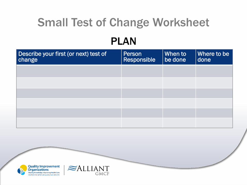
PLAN
Describe your first (or next) test of change
Person Responsible
When to be done
Where to be done
Small Test of Change Worksheet
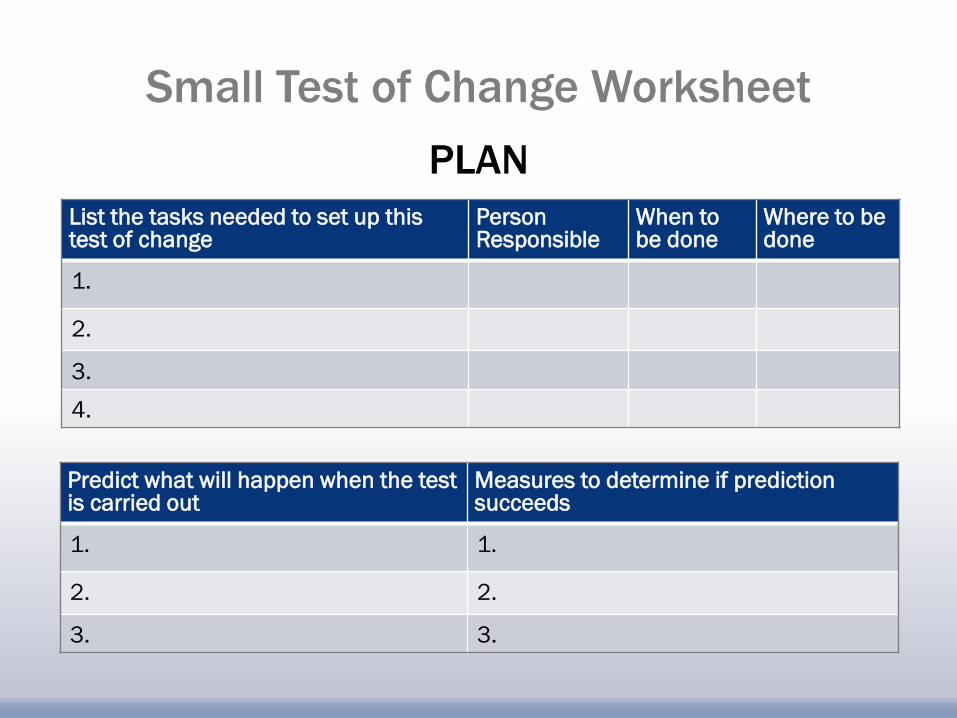
List the tasks needed to set up this test of change
Person Responsible
When to be done
Where to be done
1.
2.
3.
4.
Predict what will happen when the test is carried out
Measures to determine if prediction succeeds
1. 1.
2. 2.
3. 3.
PLAN
Small Test of Change Worksheet
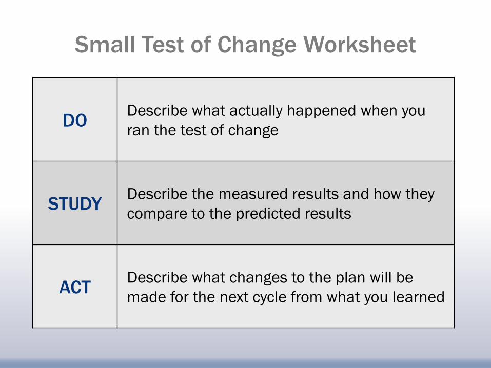
DO
Describe what actually happened when you
ran the test of change
STUDY
Describe the measured results and how they
compare to the predicted results
ACT
Describe what changes to the plan will be
made for the next cycle from what you learned
Small Test of Change Worksheet
Now it is YOUR Turn…
► Define the measures
– What will you test?
– How will you measure it?
– How will you know if you are improving?
► Identify three changes using the PDSA worksheet
► Run at least two tests of change
Complete a small test of change with a group
of eight participants

Tennis Ball PDSA Game
► Divide into teams of 8 people
► One person will need a cell
phone with a stop watch

Tennis Ball PDSA Game Debrief
► How many more PDSA cycles
would you need to complete
improvements on the time of
the process?
► What did you learn about
PDSA from this exercise?
This material was prepared by Alliant GMCF, the Medicare Quality Improvement Organization for Georgia, under contract with the Centers for Medicare & Medicaid Services
(CMS), an agency of the U.S. Department of Health and Human Services. The contents presented do not necessarily reflect CMS policy. Publication No. 10SOW-GA-IIPC-14-17
5/14/2014 0
Practical Implementation of Carbepenem-Resistant Enterobacteriaceae Prevention Practices
Date: March 20, 2014
Presented by
Cindy Prosnak, RN BSN CIC, Technical Advisor, Infection Prevention
Presented to
CRE Collaborative,
Learning Session #1
What is a QIO?
Each state has a Quality Improvement Organization,
contracted with the Centers for Medicare & Medicaid
Services (CMS) to work with Medicare beneficiaries
and providers in the state to make health care better
through use of quality initiatives. Alliant GMCF is the
Medicare QIO for Georgia.

What is a QIO?
Embracing “boundarylessness” as a prerequisite
for system-wide change, QIOs like ours are
breaking down organizational, cultural and
geographic barriers to improvement. Initiatives are
open to providers at all levels of clinical
performance that make a commitment to
improvement.

What is a QIO?
Everyone teaches and learns – Through
statewide learning and action networks, we
are accelerating the pace of change and rapidly
spreading best practices. Improvement
initiatives include collaboratives, online
interaction and peer-to-peer education.
Objectives for this presentation:
Participants will be able to:
► Recognize differences in planning and implementing standard
and transmission-based precautions in acute and long term
care settings with focus on patients and residents with
suspected or confirmed carbepenem-resistant
enterobacteriaceae
► Distinguish between appropriate practices for standard,
enhanced or modified contact precautions, and contact
precautions
► Discuss measures to apply with individual emphasis to retain
optimum patient- or resident-centered care
What is our common goal?
No matter in which health care setting we work, we all
have a common goal:
To Keep Our Patients
and Residents Safe
Health care-associated infections
► There are over 2 million cases of health care-
associated infections per year in the U.S.
► These infections cause approximately 90,000
deaths each year
► These infections add over $10 billion to the
cost of health care each year
► It has been proven that almost 40 percent of these
infections could be prevented just by improving
hand hygiene
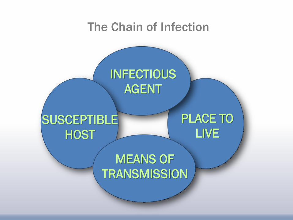
INFECTIOUS
AGENT
PLACE TO
LIVE
MEANS OF
TRANSMISSION
SUSCEPTIBLE
HOST
The Chain of Infection
What puts patients and residents at
a higher risk for infection?
► If they have been on antibiotics in the past 30 days
► If they have a chronic illness such as diabetes
or cancer
► If they have a Foley catheter or other device, such as
a central line or ventilator
► If they are on certain medications, such as steroids
► If they have impaired responses
► If they have a recent hospitalization
What makes implementing infection
prevention practices different in LTC?
► Length of stay in the facility ─ 3-5 days for hospitals stays
─ Weeks to months for rehab in LTC facility
─ Years when the LTC facility becomes a primary
residence
─ Short term treatment for patients
► Primary living facility for residents
A typical resident of your LTC facility
► Admitted to the facility 3 years ago
► Roommate has been at the facility for 5 years
► Personal property
► Routine daily and weekly activities
► Visitors
Imagine this scenario for our typical resident
► Develops signs and symptoms of a urinary tract
infection with a fever
► Physician orders an antibiotic for treatment
► Symptoms persist and resident is sent to the local
Emergency Department
► Resident is admitted to the hospital for
IV antibiotics
What happens in the hospital
► Our resident stays in the hospital for 5 days
► The resident is found to have CRE in the urine culture and
antibiotics are adjusted appropriately
► Placed on Contact Precautions while hospitalized
► Discharged back to LTC on day 5
Contact precautions in the hospital
► HCW would have used gowns and gloves each time they entered the room
► There might have been some restrictions for visitors
► Patient activities outside the room would have been restricted in some ways
► Patient probably had private room or had been cohorted with another patient with same organism/infection

Upon return to LTC our resident
still shows CRE in urine
Are Contact Precautions indicated?
Consider the following challenges:
►Asymptomatic roommate
Do we consider cohorting?
►If we decide not to change rooms,
what about toileting?
►Should long term roommates be separated
and if so, for how long?
Challenges
► The resident has always enjoyed taking meals in the
group dining area. Should this be restricted?
► What about the daily exercise program that the
resident always participates in with her friends?
► What about field trips outside the facility?
► How long should Contact Precautions for CRE
continue?
CDC Guidance and Recommendations
for CRE Prevention
2012 CRE Toolkit can be found at this link:
http://www.cdc.gov/hai/organisms/cre/cre-toolkit/ “This document contains two parts.
Part 1 contains recommendations for health care facilities and is intended to
expand upon the March 2009 “Guidance for Control of Carbapenem-Resistant or
Carbapenemase-Producing Enterobacteriaceae in Acute-Care Facilities.”
Part 2 reviews the role of public health authorities in the control of carbapenem-
resistant Enterobacteriaceae.
Unless otherwise specified, health care facilities refer to all acute care hospitals and any long-term care
facility that cares for patients who remain overnight and regularly require medical or nursing care (e.g.,
maintenance of indwelling devices, intravenous injections, wound care, etc.). This would …generally exclude
assisted living facilities and nursing homes that do not provide more than basic medical care. In addition,
this toolkit is not intended for use in ambulatory care facilities.”
Surveillance activities for CRE
► Inpatient facilities should have an awareness of
whether or not CRE have ever been cultured from
patients admitted to their facility and, if so, whether
these positive cultures were collected within 48 hours
of admission
► If CRE have been present, facilities should also
determine:
– If there is evidence of intra-facility transmission
– Which wards/units are most affected
Core measures for prevention of CRE in
all acute and long-term care facilities
1. Hand hygiene
2. Contact Precautions
3. Health Care Personnel Education
4. Use of Devices
5. Patient and Staff Cohorting
6. Laboratory Notification
7. Antimicrobial Stewardship
8. CRE Screening
There are 8 Core Measures recommended by the CDC within their 2012
CRE Toolkit (http://www.cdc.gov/hai/organisms/cre/cre-toolkit/)
In today’s presentation, we will be concentrating on the bolded items.
Others will come in later learning sessions.
Hand hygiene
These interventions are applicable in all health care
settings:
► It is not enough to have policies and procedures on hand
hygiene
► Adherence must be monitored and results must be given
back to front-line staff
► Immediate feedback should be provided when opportunities
for hand hygiene are missed during patient or resident care
► Adequate supplies and equipment for hand hygiene need to
be available at the point of care
Hand hygiene opportunities
► Before and after physical contact with a resident
► Before donning gloves and after removing gloves
► After handling soiled or contaminated items and
equipment, including linens
► Before performing an invasive procedure
► Before handling sterile or clean supplies
Hand hygiene opportunities
► When hands are visibly dirty or soiled with blood
and/or bodily fluids*
► After care of a resident with known or suspected
infectious diarrhea*
► Before and after eating or handling food*
► After personal use of bathroom*
*Situations where soap and water is preferred over
alcohol-based hand rub
Standard precautions
► Used to be called “Universal Precautions”
► Applies to EVERYBODY
► Standard Precautions is more than just using gloves
or hand hygiene
► Can include general measures such as hand
hygiene, safe injection practices, proper use of PPE,
resident placement, equipment cleaning and
disinfection
Contact precautions in acute care settings
► Patients who are colonized or infected with CRE should
be placed on Contact Precautions
► Systems should be in place to identify patients with a
history of CRE colonization or infection at admission so
that these patients can be placed on contact
precautions as soon as possible
► In addition, clinical laboratories should have an
established protocol for notifying clinical and/or infection
prevention personnel when CRE are identified from
clinical or surveillance cultures
Contact precautions in acute and long term care settings
► Involves use of gown and gloves for direct care
─ Don equipment prior to room entry
─ Remove prior to room exit
► Use of dedicated non-essential items may help
decrease transmission due to contamination
─ Blood pressure cuffs; stethoscopes; IV poles and pumps
Contact precautions in acute and long term care settings
► Private rooms or cohorting patients or residents if possible
─ Separate toileting equipment for roommates who can’t be cohorted
► Observe adherence to practices - particularly high-risk situations – and provide feedback
Tiered strategy: Consider gown/glove
use during intimate care
High risk exposures for MDRO transmission if known carrier (also high risk for acquisition if non-carrier)
► Presence of wounds (fresh/new, multiple, increased stage/size, active drainage)
► Indwelling devices (IV lines, urinary catheters, tracheostomy, PEG tubes)
► Incontinence
► Current antibiotic use
Contact Precautions in long term care settings
► Contact Precautions might be modified to fit the inherent
differences between acute and long-term care facilities
► Contact Precautions should be used for residents with CRE
who are at higher risk for transmission, including patients
– who are totally dependent upon HCP for their activities
of daily living
– are ventilator-dependent
– are incontinent of stool
– or have wounds with drainage that is difficult to control
Contact Precautions in long term care settings
(continued)
Contact Precautions might be relaxed for residents who are able to:
► perform hand hygiene
► are continent of stool
► are less dependent on staff for their activities of daily living
► are without draining wounds
However, in these situations Standard Precautions should still be observed,
including the use of gloves and/or gowns when contact with colonized/infected
sites or body fluids is possible. The caregiver must assess the individualized
nature of the resident and the care being provided at the time in order to
appropriately apply transmission-based precautions.
Transitions of Care
► The presence of CRE infection or colonization alone
should not preclude transfer of a patient from one facility
to another (e.g., acute care to long-term care)
► Communication between caregivers and facilities is the
key here – use of a common transfer form and/or
education on questions to ask and information to give at
time of report is very important
► Many times, however, this information is needed prior to
time of transfer in order to properly place resident or
patient in receiving facility
Strategic placement of residents
based on risk factors
► Focus on resident risk factors for MDRO carriage ─ High risk: Antibiotic use; presence of medical devices or
wounds; bowel/bladder incontinence; lack of mobility
► New roommate assignments based on resident characteristics and history of MDRO carriage
─ Try to avoid placing two high risk residents together
► Don’t change stable room assignments just because of a culture result unless it poses new risk
─ Roommates who’ve been together for a long time have already had opportunity to share organisms in the past (even if you only learned about it recently)
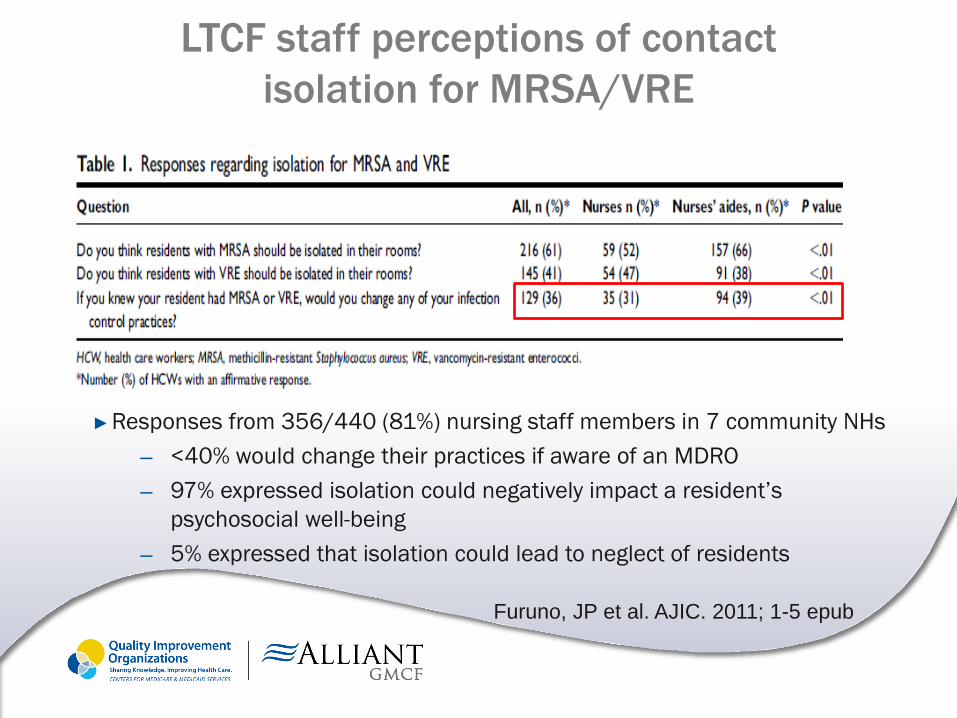
LTCF staff perceptions of contact
isolation for MRSA/VRE
► Responses from 356/440 (81%) nursing staff members in 7 community NHs
─ <40% would change their practices if aware of an MDRO
─ 97% expressed isolation could negatively impact a resident’s
psychosocial well-being
─ 5% expressed that isolation could lead to neglect of residents
Furuno, JP et al. AJIC. 2011; 1-5 epub
Challenges with contact precautions
in LTC settings
► Lack of private rooms/limited ability to move residents
─ Moving rooms is disrupting to residents and staff
─ Ability to identify carriers to cohort is limited (no active surveillance in most facilities)
► Determining duration of contact precautions
─ Unable to restrict resident mobility and participation in social events/therapy for prolonged periods
─ Unlikely to document clearance of carriage
► Large population of residents with unrecognized carriage of MDROs
─ Underestimating the sources of potential transmission
Questions?
Thank you!
Please feel free to contact me at any
time with questions:
Cindy Prosnak, RN BSN CIC
Technical Advisor, Infection Prevention
Alliant GMCF
Cell phone: 706-836-8361
This material was prepared by Alliant GMCF, the Medicare Quality Improvement Organization for Georgia, under contract with the Centers for Medicare & Medicaid Services (CMS),
an agency of the U.S. Department of Health and Human Services. The contents presented do not necessarily reflect CMS policy. Publication No. 10SOW-GA-IIPC-14-19
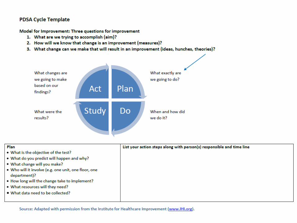
Source: Adapted with permission from the Institute for Healthcare Improvement (www.IHI.org).
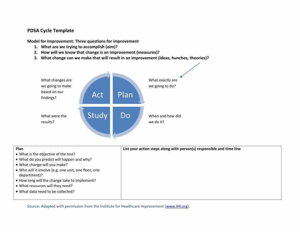
PDSA Cycle Template
Model for Improvement: Three questions for improvement 1. What are we trying to accomplish (aim)? 2. How will we know that change is an improvement (measures)? 3. What change can we make that will result in an improvement (ideas, hunches, theories)?
Plan
What is the objective of the test?
What do you predict will happen and why?
What change will you make?
Who will it involve (e.g. one unit, one floor, one department)?
How long will the change take to implement?
What resources will they need?
What data need to be collected?
List your action steps along with person(s) responsible and time line
Plan
Do Study
Act
What exactly are
we going to do?
When and how did
we do it?
What were the
results?
What changes are
we going to make
based on our
findings?
Source: Adapted with permission from the Institute for Healthcare Improvement (www.IHI.org).
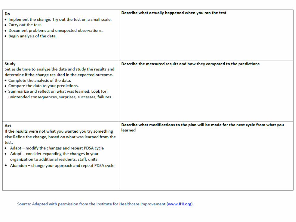
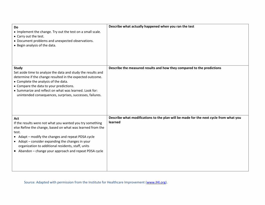
Do Implement the change. Try out the test on a small scale. Carry out the test. Document problems and unexpected observations. Begin analysis of the data.
Describe what actually happened when you ran the test
Study Set aside time to analyze the data and study the results and determine if the change resulted in the expected outcome. Complete the analysis of the data. Compare the data to your predictions. Summarize and reflect on what was learned. Look for: unintended consequences, surprises, successes, failures.
Describe the measured results and how they compared to the predictions
Act If the results were not what you wanted you try something else Refine the change, based on what was learned from the test.
Adapt – modify the changes and repeat PDSA cycle
Adopt – consider expanding the changes in your organization to additional residents, staff, units
Abandon – change your approach and repeat PDSA cycle
Describe what modifications to the plan will be made for the next cycle from what you learned