Understanding Fracture Healing Biomechanics Based … · 253/ ACTA CHIR. ORTHOP. TRAUM. ČECH., 82,...
8
253/ ACTA CHIR. ORTHOP. TRAUM. ČECH., 82, 2015, p. 253–260 CURRENT CONCEPTS REVIEW SOUBORNÝ REFERÁT Understanding Fracture Healing Biomechanics Based on the “Strain” Concept and its Clinical Applications Chápání biomechaniky hojení zlomenin postavené na koncepci „napětí“ a jeho klinické aplikace PERREN S. M. 1,2 , FERNANDEZ A. 3 , REGAZZONI P. 4 1 AO Research Institute, Davos, Switzerland 2 Trauma Research Group, Queensland University of Technology, Brisbane, Australia 3 Traumatology Hospital Brittannico, Montevideo, Uruguay 4 Traumatology University Hospital Basel, Switzerland PREAMBLE Biomechanical conditions play an obvious role in re- spect to fracture healing. Fractures can heal solidly or not heal at all depending on a variety of biomechanical conditions. What is the critical biomechanical parameter that determines and guides the mode of healing? As we compare the following cases it will become obvious that fracture mobility cannot be the main crite- rion determining the biomechanical conditions that lead to different occurrences or modes of healing. On the one hand, a highly mobile fracture bridged solidly as demonstrated by a mountain goat (Fig. 1). On the other hand, a fracture affected only slightly by mobility due to failed internal fixation did not heal in a young lady 5 (Fig. 2). We propose thinking in terms of tissue defor- 1 Prof. former director of the AO Research Institute, Davos 2 Trauma Research Group, Queensland University of Technology, Bris- bane 3 Prof. acting chief of traumatology Hospital Brittannico, Montevideo 4 Prof. former chief of traumatology University Hospital Basel 5 The two conditions differ in other respects but the main difference is biomechanical mation (strain) instead of considering only fracture mo- bility (“stability”). Strain is first and foremost a more appropriate way of understanding fracture healing and improving treatment. The clinical impact of strain will be discussed giving priority to biomechanical effects that, in our understanding, are the inductors whereby we will not address the closely related issue of the bio-
Transcript of Understanding Fracture Healing Biomechanics Based … · 253/ ACTA CHIR. ORTHOP. TRAUM. ČECH., 82,...

253/ ACTA CHIR. ORTHOP. TRAUM. ČECH., 82, 2015, p. 253–260
CURRENT CONCEPTS REVIEWSOUBORNÝ REFERÁT
Understanding Fracture Healing BiomechanicsBased on the “Strain” Concept and its ClinicalApplications
Chápání biomechaniky hojení zlomenin postavené na koncepci „napětí“ a jehoklinické aplikace
PERREN S. M.1,2, FERNANDEZ A.3, REGAZZONI P.4
1 AO Research Institute, Davos, Switzerland2 Trauma Research Group, Queensland University of Technology, Brisbane, Australia3 Traumatology Hospital Brittannico, Montevideo, Uruguay4 Traumatology University Hospital Basel, Switzerland
PREAMBLE
Biomechanical conditions play an obvious role in re-spect to fracture healing. Fractures can heal solidly ornot heal at all depending on a variety of biomechanicalconditions. What is the critical biomechanical parameterthat determines and guides the mode of healing?
As we compare the following cases it will becomeobvious that fracture mobility cannot be the main crite-rion determining the biomechanical conditions that leadto different occurrences or modes of healing. On theone hand, a highly mobile fracture bridged solidly asdemonstrated by a mountain goat (Fig. 1). On the otherhand, a fracture affected only slightly by mobility dueto failed internal fixation did not heal in a young lady 5
(Fig. 2). We propose thinking in terms of tissue defor-
1 Prof. former director of the AO Research Institute, Davos2 Trauma Research Group, Queensland University of Technology, Bris-
bane3 Prof. acting chief of traumatology Hospital Brittannico, Montevideo4 Prof. former chief of traumatology University Hospital Basel5 The two conditions differ in other respects but the main difference is
biomechanical
mation (strain) instead of considering only fracture mo-bility (“stability”). Strain is first and foremost a moreappropriate way of understanding fracture healing andimproving treatment. The clinical impact of strain willbe discussed giving priority to biomechanical effectsthat, in our understanding, are the inductors wherebywe will not address the closely related issue of the bio-
253_260_perren 7.8.15 8:55 Stránka 253

chemical sequence of events (4) that plays the role ofthe effector.
The term “stability” is widely used in internal fixation.Unfortunately “stability” is used in medical terminologyfor different, incompatible aspects. Some use it to ex-pressing strength (load at failure), others use it to ex-pressing stiffness (resistance to deformation). We use“stability” to mean stiffness but we will avoid using theterm “stability” whenever possible. The term “biologicalfracture fixation” defines a mode of fracture fixationthat comprises flexible fixation for induction of callusand minimal surgical trauma to keep soft tissues andbone healthy. The surgical trauma consists of the traumato the soft tissues during the surgical approach and thereduction of the fracture as well as of the implant contactwhich compromises the blood supply to bone. Biologicalfracture fixation aims at improved reliability of healing,improved resistance to infection and minimized risk ofrefracture. The advantages are achieved at the cost ofmore demanding procedures and implants. Precision ofreduction is weighed against biological damage and,whenever possible, the fracture should not be exposedfor inspection or even “cleaning” of soft tissues.
THE CONCEPT OF STRAIN
An important biomechanical condition for repair tissueformation and differentiation is cellular deformation.The repair cell does not “see” the amount of fragmentmovement nor the width of the gap but senses its owndeformation, called strain (1, 3, 6, 9, 10). The influenceof movement as well as the effects of gap size have beendealt with extensively (2, 7). Deformation of cells ina fracture gap depends upon gap width and amount ofrelative movement between the fracture surfaces. Suchdeformation is called strain ( which, in its practical ap-plication to fracture fixation, equals the amount of frac-ture gap movement ( L) divided by the gap width (L) or = L/L6. This means that depending on gap width very
different amounts of tissue deformation can occur forthe same amount of movement: the wider the gap theless tissue deformation and vice versa. The problem isthat if a gap is very small and is moved by a similarlysmall amount there may still be high strain conditions(e.g. 100/100 = 1/1 = 0.01/0.01). Let’s consider a verysmall fracture gap of 10 m that is about a cell width. Ifthe fracture surfaces displace only 10 m the cell size isdeformed to 10 m + 10 m which is a 100% deformation(Fig. 3). The problem is that the small gap is invisible tothe naked eye and, likewise, the displacement cannot bedetected. Still, 100% is an extreme deformation that canbe tolerated by a cell of granulation tissue but not byconnective tissue or cartilage or bone. Such conditionsare not visible but they must be understood, that is, thefunction of the surgeon’s eye is replaced by the functionof his brain.
When a tissue (or any other material) is deformed be-yond a certain limit disruption occurs. It goes withoutargument that a tissue cannot be formed when the strainexceeds its limit of elongation at rupture, in other words,conditions that would disrupt a tissue do not allow itsformation. The critical parameter is elongation at rupture,which is the upper limit of accepted strain or the limitof strain tolerance. Similarly, tissue repair or differenti-ation is not induced below a certain limit.
Successful fracture healing occurs within the band-width between strain induction and strain tolerance (ref.Perren in 6/2014 )7. Fig. 4 through Fig. 6 – illustrate themechanical properties of different tissues that play a rolein secondary fracture healing (15).
254/ ACTA CHIR. ORTHOP. TRAUM. ČECH., 82, 2015 CURRENT CONCEPTS REVIEWSOUBORNÝ REFERÁT
6 We simplify for easier understanding of the basics. In reality strainis three dimensional and depends not only on mobility but also onfluid flow as outlined early on by E. Cheal.
7 Perren, S. M.: Fracture healing: Fracture healing understood as theresult of a fascinating cascade of physical and biological interactions.Part I. An attempt to integrate observations from 30 years AO Re-search. Acta Chir. orthop. Traum. čech., 81: 355–364, 2014.
Fig. 1. Solid spontaneous bone bridging of the fracture in spiteof high mobility. The misalignment is not considered here(U. Geret).
Fig. 2. Young lady with tibia fracture treated in a way that lowmobility was not prevented (lag screw within the fracture gap.Union at 8 months).
253_260_perren 7.8.15 8:55 Stránka 254

255/ ACTA CHIR. ORTHOP. TRAUM. ČECH., 82, 2015 CURRENT CONCEPTS REVIEWSOUBORNÝ REFERÁT
STRAIN INDUCTION VS. STRAIN TOLERANCE
Aspects of strain inductionUnder conditions of spontaneous healing the limit of
strain induction is often not an issue. In contrast, underconditions of flexible internal fixation maintaining strainabove induction level is an issue. As an extreme example:when a large defect is spanned with two plates the largegap and the scarce mobility result in minimal strain thatis below induction level. The clinical aspect is charac-terized by painless function ensured by the implants thatdo not help fracture healing but replace it. We call this“prosthetic osteosynthesis” where the internal fixationprovides painless function but does not induce properhealing (12). Late bone formation filling the gap is thenobviously not induced by biomechanical conditions suchas strain. This is a type of bone formation that needsfurther attention.
Aspect of strain toleranceThe upper limit of strain, i.e. tolerance, is elongation
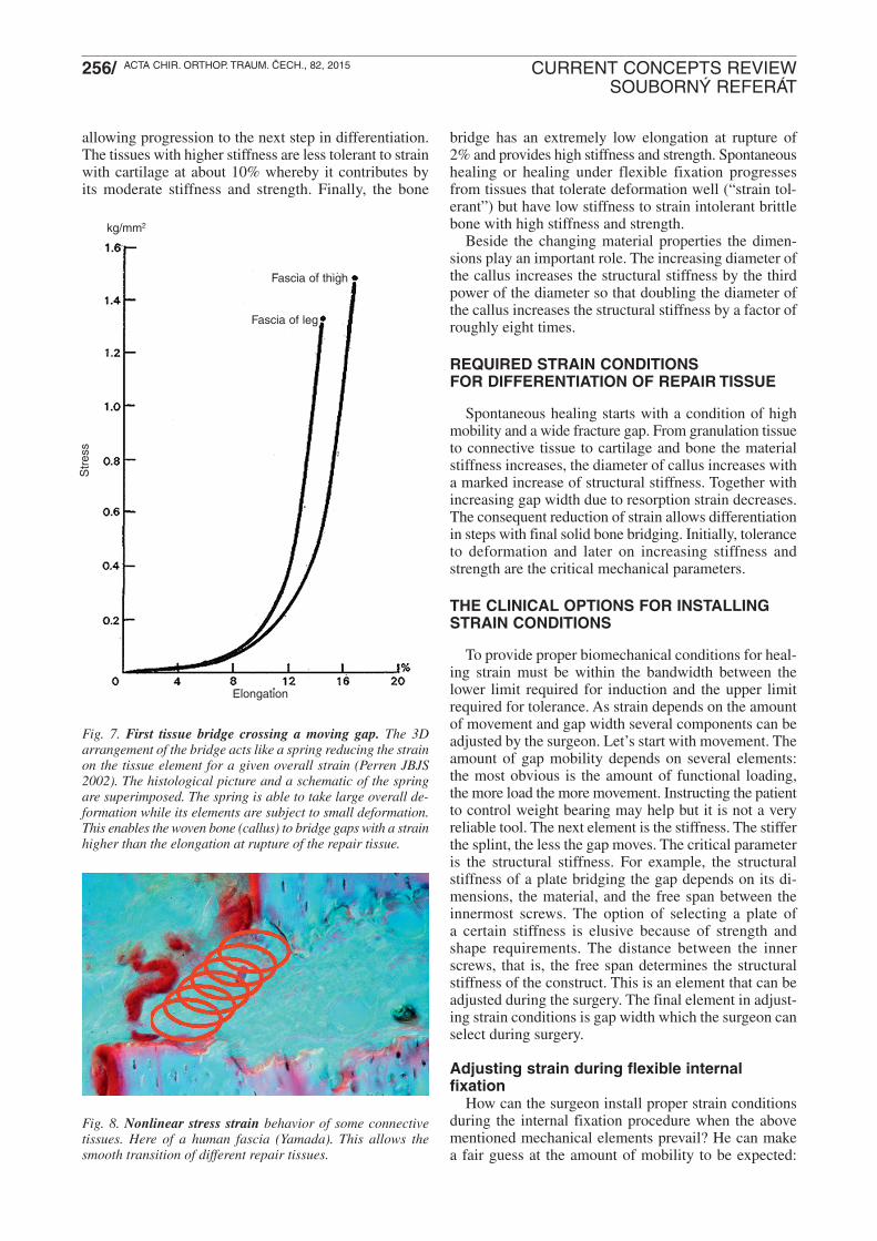
at rupture as mentioned above. The values for elongationat rupture for biological materials are listed in Yamada1970. The value for the initial repair tissue, granulationtissue, is not reported there but the closest material togranulation tissue is parenchyma with a value of roughly100%. The next stage of repair is bridging by connectivetissue and, frequently, cartilage before final bridging bycallus is rendered possible by reduction of strain due towidening of the gap, the generally increasing stiffnessof the repair tissue and a change of dimensions due tothe increasing diameter of the soft tissue cuff. An elementthat facilitates bridging is that the first bridges arearranged like a spring, thus reducing the strain withinthe spring element (Fig. 7). Furthermore, the stiffnessof connective tissues, for example, dramatically increaseswith extension above a certain limit (Fig. 8). This con-tributes through increased stiffness to reduced mobility
Fig. 3. The effect of the width of the fracture gap on tissuedeformation. Assuming a relative movement of the two fracturesurfaces of 10 μm, the cell in the smaller (10 μm) wide gap isdisrupted (100% strain) while the cells in the larger (30 μm)wide gap are less deformed (~30%) and remain intact. Thedotted line indicates the position of the fragment before dis-placement.
Fig. 4. Tolerance to deformation for three selected repair tis-sues. The data for parenchyma corresponds +/- to granulationtissue. From granulation tissue to bone the tolerated elonga-tion at rupture is decreased by 50 times. The two differentcolor shadings represent the different values. (data from Ya-mada).
Fig. 5. Stiffness for three selected repair tissues. From gran-ulation tissue to bone the stiffness increases by an enormousfactor of 2 x 10⁵. The data is plotted logarithmically. Increasingstiffness stabilizes the fracture. (data from Yamada).
Fig. 6. The strength for three selected repair tissues. Fromgranulation tissue to bone the stiffness increases by an enor-mous factor of 1400x. The data is plotted logarithmically. Thisstrength plays a role in final fracture strength (data from Ya-mada)
253_260_perren 7.8.15 8:55 Stránka 255
256/ ACTA CHIR. ORTHOP. TRAUM. ČECH., 82, 2015 CURRENT CONCEPTS REVIEWSOUBORNÝ REFERÁT
bridge has an extremely low elongation at rupture of2% and provides high stiffness and strength. Spontaneoushealing or healing under flexible fixation progressesfrom tissues that tolerate deformation well (“strain tol-erant”) but have low stiffness to strain intolerant brittlebone with high stiffness and strength.
Beside the changing material properties the dimen-sions play an important role. The increasing diameter ofthe callus increases the structural stiffness by the thirdpower of the diameter so that doubling the diameter ofthe callus increases the structural stiffness by a factor ofroughly eight times.
REQUIRED STRAIN CONDITIONS FOR DIFFERENTIATION OF REPAIR TISSUE
Spontaneous healing starts with a condition of highmobility and a wide fracture gap. From granulation tissueto connective tissue to cartilage and bone the materialstiffness increases, the diameter of callus increases witha marked increase of structural stiffness. Together withincreasing gap width due to resorption strain decreases.The consequent reduction of strain allows differentiationin steps with final solid bone bridging. Initially, toleranceto deformation and later on increasing stiffness andstrength are the critical mechanical parameters.
THE CLINICAL OPTIONS FOR INSTALLINGSTRAIN CONDITIONS
To provide proper biomechanical conditions for heal-ing strain must be within the bandwidth between thelower limit required for induction and the upper limitrequired for tolerance. As strain depends on the amountof movement and gap width several components can beadjusted by the surgeon. Let’s start with movement. Theamount of gap mobility depends on several elements:the most obvious is the amount of functional loading,the more load the more movement. Instructing the patientto control weight bearing may help but it is not a veryreliable tool. The next element is the stiffness. The stifferthe splint, the less the gap moves. The critical parameteris the structural stiffness. For example, the structuralstiffness of a plate bridging the gap depends on its di-mensions, the material, and the free span between theinnermost screws. The option of selecting a plate ofa certain stiffness is elusive because of strength andshape requirements. The distance between the innerscrews, that is, the free span determines the structuralstiffness of the construct. This is an element that can beadjusted during the surgery. The final element in adjust-ing strain conditions is gap width which the surgeon canselect during surgery.
Adjusting strain during flexible internal fixation
How can the surgeon install proper strain conditionsduring the internal fixation procedure when the abovementioned mechanical elements prevail? He can makea fair guess at the amount of mobility to be expected:
allowing progression to the next step in differentiation.The tissues with higher stiffness are less tolerant to strainwith cartilage at about 10% whereby it contributes byits moderate stiffness and strength. Finally, the bone
Fig. 7. First tissue bridge crossing a moving gap. The 3Darrangement of the bridge acts like a spring reducing the strainon the tissue element for a given overall strain (Perren JBJS2002). The histological picture and a schematic of the springare superimposed. The spring is able to take large overall de-formation while its elements are subject to small deformation.This enables the woven bone (callus) to bridge gaps with a strainhigher than the elongation at rupture of the repair tissue.
Fig. 8. Nonlinear stress strain behavior of some connectivetissues. Here of a human fascia (Yamada). This allows thesmooth transition of different repair tissues.
Elongation
Fascia of leg
kg/mm2
Fascia of thigh
Str
ess
253_260_perren 7.8.15 8:55 Stránka 256

257/ ACTA CHIR. ORTHOP. TRAUM. ČECH., 82, 2015 CURRENT CONCEPTS REVIEWSOUBORNÝ REFERÁT
weight, muscular condition and control thereof playa role. Adjusting the width of the fracture gap allowsthe surgeon to install strain within the required band-width: The greater the expected mobility, the larger therequired gap. To adequately induce and allow fracturehealing under conditions of flexible fixation for a givenamount of expected mobility of the fracture, adjustingthe gap size allows installation of proper strain but thisis a demanding option and gap width should only bevaried within a small range.
Wrong simple rules regarding strainThe surgeon needs simple rules for his procedures.
Attempts to achieve this have led to inadequate inter-pretation of strain. Keeping strain within the bandwidthdiscussed above requires adaptation of procedures.Therefore, it does not make sense to formulate a simplerule like “a small or a large gap is the goal”. Such state-ments are unfortunate and dangerous simplifications.
INTERNAL FIXATION, BIOMECHANICS AND HEALING
The priority of internal fixation is to restore the func-tion of bone, limb and patient. While the fracture pro-duces a discontinuity of bone stiffness, internal fixationreinstates continuity that should allow early restorationof painless function. Restoration of function is a prereq-uisite for keeping the tissues healthy, for example, avoid-ing reflex dystrophy, which is a consequence of pro-longed and extensive immobilization of a limb (ref.Perren in 1/2015)8
Internal fixation can install conditions where the frag-ments are kept in immobile compressed contact. Suchcontact does not allow relative displacement of the frac-ture fragments as would occur under conditions of usualfunctional load and strain is not an issue. Today our un-derstanding is that the repair tissue does not react to thepresence of a fully immobilized fracture (11). We haveobserved that osteons that cross fractured surfaces inclose permanent contact do not as a rule change speed orshape or direction when crossing. The observation thatbone necrosis due to implant contact with a damagedblood supply induces internal remodeling as kind ofcreeping substitution suggests that the internal remodelingof such an immobilized fracture is induced by bone necro-sis as a consequence of trauma. The internal remodelingis not a response to the fracture but is induced by necroticbone in the vicinity of a fracture. Therefore, we now con-sider primary healing as a mere side effect of the removalof necrotic bone tissue whereby the osteons plug thefracture like dowels and eventually, after some delay,provide strength (Fig. 9). This explains why fracturesfixed with compression need to be protected for sometime whereby the implants are generally not removed fortwo years. In contrast, Miclau et al. (8) and Tepic et al.(13) demonstrated solid reliable healing at 10 weekswhen the locally elevated (undercut) plate permitted callusformation thus preventing an avascular gap end from act-ing as a notch immediately deep to the plate. When they
Fig. 9. Osteons remodeling an area of stably fixed micro fractures. The osteons remodel the multi-fracture area as if it wereintact bone: A further hint that an immobilized fracture is not recognized in the absence of mobility. The internal remodeling re-moves dead bone which is produced by disruption of blood vessels. Fig. 9a LEFT: histology by B.A. Rahn, Fig. 9b RIGHT:schematic representation, the area within the dotted yellow lines contains the multiple micro fractures. Color coded histology.
a | b
8 Perren, S. M.: Fracture healing: Fracture healing understood as theresult of a fascinating cascade of physical and biological interactions.Part II. Acta Chir. orthop. Traum. čech., 82: 13–21, 2015.
253_260_perren 7.8.15 8:55 Stránka 257

258/ ACTA CHIR. ORTHOP. TRAUM. ČECH., 82, 2015 CURRENT CONCEPTS REVIEWSOUBORNÝ REFERÁT
subjected the fracture to bending testing after removal ofthe implants all failures were located within the fracturewith the conventional DCP whereas all fractures with-stood the load with the undercut LC-DCP and failureonly occurred at the next screw hole. The latter conditionis a prerequisite for implant removal.
Clinical aspects of strain – see also Fernandez (5)1. The amount of strain within and around the fracture
gap determines the amount of callus produced as ischaracteristic for spontaneous healing and healing un-der flexible fixation.
2. When strain between fragments exceeds the optimalbandwidth between stimulation and tolerance two dis-tinct pattern of healing problems result:
3. Low strain conditions result in a pattern that is oftenmistaken for insufficient biological activity called “atrophic nonunion”. The observation that restoringproper strain conditions frequently results in prompthealing points to the biomechanical contribution to theproblem.
4. High strain conditions result in abundant callus for-mation that cannot form a bridge and looks like hy-pertrophic nonunion where a reduction of fracturemobility and a simultaneous restoration of strainwithin optimal bandwidth results in prompt bridging(Fig. 10).
5. Dosage of optimal strain conditions depends on bal-ancing fracture mobility and gap width. This is a taskthat is demanding in the individual case (Fig. 12).
6. The fact that fracture surfaces that do not exactlycorrespond9 will experience different strain condi-tions which, consequently, increases tolerance togap width adjustments.
7. Reduction of fracture mobility10 needs to be weighedagainst additional surgical trauma to soft tissues andbone.
8. Compression fixation of a fracture prevents displace-ment of the fracture fragments in contact due to pre-loading11 and/or friction. In such a situation, strainis absent in contact areas: no irritation and thereforeno stimulation of healing.
9. Without biomechanical stimulation healing may pro-ceed through plugging of the fracture surfaces byosteons acting like dowels.
10. Osteons with their cutter heads are most likely stim-ulated to remove dead bone in the vicinity of thefracture and will remove it, as has been observed, atthe bone-to-implant interface12 (Fig. 9) .
11. In areas adjacent to compressed contacts the strainis minimal resulting only from a small deformationof the contacting bone under intermittent functionalload.
Fig. 10. The osteons crossing an osteotomy that is held inclosed contact do not appear to react to the mere presence ofthe fracture.
Fig. 11. Tear drop phenomenon. At the outer end of the com-pressed gap under functional load intermittent gaping results.The reaction is widening of the gap by resorption and with it re-ducing strain. Reduction of strain then allows filling by callus.
253_260_perren 7.8.15 8:55 Stránka 258

259/ ACTA CHIR. ORTHOP. TRAUM. ČECH., 82, 2015 CURRENT CONCEPTS REVIEWSOUBORNÝ REFERÁT
9 E.g. due to small displacement. 10 Fracture mobility is relative movement of the opposing fracture sur-
faces in relation to each other.11 Preloading results from compressing the fracture surfaces against
each other to prevent opening of the fracture gap when traction isapplied as long as preload is greater than traction.
12 Such biological activity by necessity starts in living bone adjacentto dead bone.
Fig. 12. Optimal strain: Flexible fixation of a spiral fracture.The width of the gap was properly adjusted. Solid secondaryhealing (C.Ryf).
Fig. 13. Hypertrophic non-union. Figs a–c from Weber and Cech (14). The treatment consists in reduction of mobility throughsplinting with a plate; a – abundant callus but no bridging; b – scintigraphy showing biological activity which has the potentialbut cannot bridge; c – elephant foot appearance; d – histology of a similar situation in sheep. From the fragment ends aboveand below callus advances decreasing the gap width and with it increasing strain.
a | b | c | d
12. When compression applied to a fracture surface actswithin a limited range the areas outside this rangemay undergo high strain conditions as a result ofsmall displacement combined with very small gapwidths or gaps intermittently closing. This results inthe so-called tear drop phenomenon (“gocce di cere”Fig. 11).
HOW DOES REPAIR TISSUE OVERCOMEHIGH STRAIN CONDITIONS
The initial bridging of a fracture gap is usually basedon callus. The English term of “woven” bone impliesthe understanding that callus is a 3D structure. In respectto strain such a structure acts like a spring. The spring isable to sustain large overall strain (overall deformation)while the elements of the spring sustain only very smalldeformation. The same can be observed for the first tinybridges of bone crossing a gap (Fig. 7). The histologicalpicture shows only the width of a histological cut butthe elements shown here can easily be understood asbeing a cut through a spring. Callus is therefore able tobridge a gap that is under higher strain than the straintolerance of the bone element, namely ~2% strain.
ATROPHIC DELAYED OR NON-HEALING
Formerly, mainstream opinion was that a fracturewould not heal if biology was not sufficient (atrophicpseudoarthroses). Today, based on the observation thatatrophic non unions may heal after correction of thebiomechanical conditions, the focus is more on in-stalling the proper biomechanical conditions by reduc-ing or increasing the stability of implants Fig. 13through Fig. 15.
253_260_perren 7.8.15 8:55 Stránka 259

260/ ACTA CHIR. ORTHOP. TRAUM. ČECH., 82, 2015 CURRENT CONCEPTS REVIEWSOUBORNÝ REFERÁT
CANCELLOUS BONE AND CORTEX
The tolerance of bone to elongation applies similarlyto cortical and cancellous bone as the elements of bothbone structures are similar whereas resistance to load(stress) and resistance to deformation (stiffness) is verydifferent for these two types of bone. The speed of reac-tion due to the accessibility of bone trabeculae is alsoan element to consider.
MISUNDERSTANDING STRAIN
The way we express strain in respect to clinical ap-plication is deliberately as simple as possible. Declaringit as “oversimplified” misses the point that the basicchange of thinking in terms of strain instead of stabilityis the essential and important issue. The argument thatwe show the deformation of a single cell in the fracturegap “where there are many cells” is the height of mis-understanding.
CONCLUSIONS
Biomechanical conditions play an obvious role in re-spect to fracture healing. Fractures can solidly heal ornot heal under a variety of biomechanical conditions.We propose that instead of fracture mobility considera-tion should rather be given to “tissue deformation” thatis strain. Strain depends upon mobility and, more im-portantly, upon gap width and is first and foremost a moreappropriate way of understanding the biomechanics offracture healing and of improving treatment.
References
1. CHEAL, E. J., MANSMANN, K. A., DIGIOIA, A. M. 3rd, HAYES,W. C., PERREN, S. M.: Role of interfragmentary strain in fracturehealing: ovine model of a healing osteotomy. J. Orthop. Res., 9:131–142, 1991.
2. CLAES, L., ECKERT-HÜBNER, K., AUGAT, P.: The fracturegap size influences the local vascularization and tissue differenti-ation in callus healing. Langenbecks Arch. Surg., [Epub 2003 Sep 9] 388: 316–322, 2003.
3. DIGIOIA, A. M. 3rd , CHEAL, E. J., HAYES, W. C.: Three-di-mensional strain fields in a uniform osteotomy gap. J. Biomech.Eng., 108: 273–280, 1986.
4. EINHORN, T. A.: The cell and molecular biology of fracture heal-ing. Clin. Orthop. Relat. Res., 355(Suppl.): S7-21, 1998.
5. FERNANDEZ, A., REGAZZONI, P., PERREN, S. M.: in: A realtime surgical experience at your fingertips. www.ICUC.net, 2015.
6. HENTE, R., FÜCHTMEIER, B., SCHLEGEL, U., ERNST-BERGER, A., PERREN, S. M.: The influence of cyclic compres-sion and distraction on the healing of experimental tibial fractures.J. Orthop. Res., 22: 709–715, 2004.
7. KENWRIGHT, J., GOODSHIP, A. E.: Controlled mechanical sti -mulation in the treatment of tibial fractures. Clin. Orthop. Relat.Res., 241: 36–47, 1989.
8. MICLAU, T., REMIGER, A., TEPIC, S., LINDSEY, R., MCIFF, T.:A mechanical comparison of the dynamic compression plate, limi -ted contact-dynamic compression plate, and point contact fixator.J. Orthop. Trauma, 9: 17–22, 1995.
9. PERREN, S. M., BOITZY, A.: Cellular differentiation and bonebiomechanics during the consolidation of a fracture, Anat. Clin.,1: 13–28, 1978.
10. PERREN, S. M., CORDEY, J.: The concept of interfragmentarystrain. In: UHTHOFF, H. K. (ed.): Current concepts of internalfixation of fractures, New York, Springer-Verlag, 1980, 63–77.
11. PERREN, S. M.: Evolution of the internal fixation of long bonefractures. The scientific basis of biological internal fixation: Choos-ing a new balance between stability and biology. Review. J. BoneJt Surg., 84-B: 1093–1110, 2002.
12. REGAZZONI, P., FERNANDEZ, A., PERREN, S. M.: in: A realtime surgical experience at your fingertips. www.ICUC.net, 2015.
13. TEPIC, S., REMIGER, A. R., MORIKAWA, K., PREDIERI, M.,PERREN, S. M.: Strength recovery in fractured sheep tibia treatedwith a plate or an internal fixator: an experimental study witha two-year follow-up, 11: 14–23, 1997.
14. WEBER, B. G., CECH, O.: Pseudoarthrosis: Pathology, biome-chanics, therapy, results. Berne, Hans Huber Medical Publisher1976.
15. YAMADA, H., EVANS, F. G.: Strength of biological materials.Baltimore, Williams & Wilkins 1970.
Corresponding author:Prof. Stephan M. Perren, M.D.AO Research Institute DavosClavadelerstrasse 87270 Davos, SwitzerlandE-mail: [email protected]
Fig. 14. Non-union that is biologically active but does not pro-duce callus. Large gap, low strain without biomechanical in-duction. The treatment consists in application of axial compres-sion that results in approximation and decrease of gap widthincreasing strain and enabling induction. Weber and Cech (14)
Fig. 15. Biologically inactive non-union, large gap width. Frus-trated exhaustion and/or missing biomechanical induction?Weber and Cech (14).
253_260_perren 7.8.15 8:55 Stránka 260