Umbilical Nodule with Cyclical Bleeding: A Case Report … · 2...
6
Case Report Umbilical Nodule with Cyclical Bleeding: A Case Report and Literature Review of Atypical Endometriosis Marlene Teixeira Andrade, 1 Cláudia V. Marques de Freitas, 2 Sara Filipa Camacho Câmara, 2 and José Joaquim Nunes Vieira 2 1 Centro Hospitalar de Tr´ as-Os-Montes e Alto Douro, Unidade de Vila Real, Avenida Noruega, 5000-508 Vila Real, Portugal 2 Hospital Dr. N´ elio Mendonc ¸a, Hospital Central do Funchal, Avenida Lu´ ıs de Cam˜ oes, Funchal, 9004-514 Madeira Island, Portugal Correspondence should be addressed to Marlene Teixeira Andrade; [email protected] Received 3 July 2016; Accepted 31 August 2016 Academic Editor: Stefan P. Renner Copyright © 2016 Marlene Teixeira Andrade et al. is is an open access article distributed under the Creative Commons Attribution License, which permits unrestricted use, distribution, and reproduction in any medium, provided the original work is properly cited. Endometriosis is defined as the presence of endometrial glands and stroma outside the uterus. It affects 3 to 10 percent of women of reproductive age. Umbilical endometriosis is rare, with an estimated incidence of 0.5–1.0% among all cases of endometriosis, and is usually secondary to prior laparoscopic surgery involving the umbilicus. In this report, we described a case of umbilical endometriosis treated with surgical resection and highlight the great importance of medical history compared to complementary diagnostic tests that can be sometimes inconclusive. 1. Introduction Endometriosis is a common benign disease, which is defined by the presence of endometrial tissue outside the uterus [1]. e first description of this condition was made by a German physician, Daniel Schroen, in 1690 [2]. It affects 3 to 10 percent of women of reproductive age [3, 4] and represents an important cause of infertility [5]. It occurs in 2–22% of asymptomatic patients, in 20–30% of infertil- ity cases, and in 40–60% of dysmenorrhoea cases [6]. Its etiopathogenesis is not well established yet. Endometriosis can affect any woman from premenarche [7] until post- menopause, irrespective of the race, ethnicity, or maternal status [8, 9]. It is reported that up to 70% of adolescents with chronic pelvic pain before menarche can be affected with endometriosis [7]. e most frequent locations include the ovaries, the uterosacral ligaments, the pouch of Douglas, and the other pelvic organs. Extraperitoneal locations as cervix, vagina, vulva, lungs, umbilicus, or postoperative scars are uncommon [6] and locations like nasal mucosa, brain, and eyes are very rare [10]. ere is little evidence on the real incidence and prevalence of these extrapelvic lesions. Umbilical endometriosis rarely occurs, with an estimated incidence of 0.5–1.0% among all cases of endometriosis [11, 12], usually affecting patients aſter laparoscopy or other sur- gical procedures involving the umbilicus [4] (of the reported cases of cutaneous endometriosis, over 70% are secondary to prior surgery and occur at the site of surgical scars) [13, 14]. Umbilical endometriosis can be classified as primary, named Villar’s nodule, when it appears spontaneously (any ectopic endometrium that is found superficial to the peritoneum without any history of previous surgery) or secondary, when it appears aſter surgical procedures. e term secondary endometriosis can be used even when it is not located on surgical scars, such as on the umbilicus, but only if its onset occurs within 2 years aſter the procedure [15–17]. e differential diagnosis of umbilical tumors in women comprise endometriosis in 32.2%, benign primary tumor in 29.7%, metastatic tumor in 29.7%, and malignant primary tumor in 8.4% [12, 18]. e maximal depth of penetration of the umbilical endometriosis described is up to the fascial level [19–21]. To treat umbilical endometriosis, wide resection with 2 mm margins is generally recommended [22] and there are few cases in which conservative treatment is indicated [23]. Hindawi Publishing Corporation Case Reports in Obstetrics and Gynecology Volume 2016, Article ID 7401409, 6 pages http://dx.doi.org/10.1155/2016/7401409
Transcript of Umbilical Nodule with Cyclical Bleeding: A Case Report … · 2...

Case ReportUmbilical Nodule with Cyclical Bleeding: A Case Report andLiterature Review of Atypical Endometriosis
Marlene Teixeira Andrade,1 Cláudia V. Marques de Freitas,2
Sara Filipa Camacho Câmara,2 and José Joaquim Nunes Vieira2
1Centro Hospitalar de Tras-Os-Montes e Alto Douro, Unidade de Vila Real, Avenida Noruega, 5000-508 Vila Real, Portugal2Hospital Dr. Nelio Mendonca, Hospital Central do Funchal, Avenida Luıs de Camoes, Funchal, 9004-514 Madeira Island, Portugal
Correspondence should be addressed to Marlene Teixeira Andrade; [email protected]
Received 3 July 2016; Accepted 31 August 2016
Academic Editor: Stefan P. Renner
Copyright © 2016 Marlene Teixeira Andrade et al. This is an open access article distributed under the Creative CommonsAttribution License, which permits unrestricted use, distribution, and reproduction in any medium, provided the original work isproperly cited.
Endometriosis is defined as the presence of endometrial glands and stroma outside the uterus. It affects 3 to 10 percent of womenof reproductive age. Umbilical endometriosis is rare, with an estimated incidence of 0.5–1.0% among all cases of endometriosis,and is usually secondary to prior laparoscopic surgery involving the umbilicus. In this report, we described a case of umbilicalendometriosis treated with surgical resection and highlight the great importance of medical history compared to complementarydiagnostic tests that can be sometimes inconclusive.
1. Introduction
Endometriosis is a common benign disease, which is definedby the presence of endometrial tissue outside the uterus[1]. The first description of this condition was made by aGerman physician, Daniel Schroen, in 1690 [2]. It affects3 to 10 percent of women of reproductive age [3, 4] andrepresents an important cause of infertility [5]. It occursin 2–22% of asymptomatic patients, in 20–30% of infertil-ity cases, and in 40–60% of dysmenorrhoea cases [6]. Itsetiopathogenesis is not well established yet. Endometriosiscan affect any woman from premenarche [7] until post-menopause, irrespective of the race, ethnicity, or maternalstatus [8, 9]. It is reported that up to 70% of adolescentswith chronic pelvic pain before menarche can be affectedwith endometriosis [7]. The most frequent locations includethe ovaries, the uterosacral ligaments, the pouch of Douglas,and the other pelvic organs. Extraperitoneal locations ascervix, vagina, vulva, lungs, umbilicus, or postoperative scarsare uncommon [6] and locations like nasal mucosa, brain,and eyes are very rare [10]. There is little evidence on thereal incidence and prevalence of these extrapelvic lesions.Umbilical endometriosis rarely occurs, with an estimated
incidence of 0.5–1.0% among all cases of endometriosis [11,12], usually affecting patients after laparoscopy or other sur-gical procedures involving the umbilicus [4] (of the reportedcases of cutaneous endometriosis, over 70% are secondary toprior surgery and occur at the site of surgical scars) [13, 14].Umbilical endometriosis can be classified as primary, namedVillar’s nodule, when it appears spontaneously (any ectopicendometrium that is found superficial to the peritoneumwithout any history of previous surgery) or secondary, whenit appears after surgical procedures. The term secondaryendometriosis can be used even when it is not located onsurgical scars, such as on the umbilicus, but only if itsonset occurs within 2 years after the procedure [15–17]. Thedifferential diagnosis of umbilical tumors inwomen compriseendometriosis in 32.2%, benign primary tumor in 29.7%,metastatic tumor in 29.7%, and malignant primary tumorin 8.4% [12, 18]. The maximal depth of penetration of theumbilical endometriosis described is up to the fascial level[19–21].
To treat umbilical endometriosis, wide resection with2mm margins is generally recommended [22] and there arefew cases in which conservative treatment is indicated [23].
Hindawi Publishing CorporationCase Reports in Obstetrics and GynecologyVolume 2016, Article ID 7401409, 6 pageshttp://dx.doi.org/10.1155/2016/7401409

2 Case Reports in Obstetrics and Gynecology
Figure 1: Umbilical nodule bleeding during menstrual cycle.
The authors report a case of a patient with umbilicalendometriosis associated with a previous laparoscopic inter-vention and treated by surgical excision.
2. Case Presentation
A forty-two-year-old healthy female, with menarche at theage of 13, was referred to the gynecology department bygeneral surgery, with a livid coloured nodule in the umbilicuswhich gradually increased in size over the past 3 years. Shealso presented with dysmenorrhoea (numeric rating scale ofpain (NRS): 10), dyspareunia (NRS: 10), dyschezia (NRS: 7),and tenesmus. She was medicated with an oral contraceptivewith ethinylestradiol and gestodene.The nodule was painlessand the patient mentioned cyclical umbilical bleeding syn-chronized with menstruation (Figure 1), during withdrawalbleeding. She had irregular menstruation periods and acesarean for fetal breech presentation 9 years before. Thepatient had past history of laparoscopic appendectomy fiveyears previously, with umbilical cannulation.The histopatho-logical examination of the appendix revealed endometriosis.She has no known family history of endometriosis. Atphysical examination, she had a soft swelling nodule, with twobluish-purple dots, in the umbilicuswith a diameter of 1.2 cm,with a normal skin envelope, that was irreductible (Fig-ure 2). In this first gynecological consultation, the hormonalmedication was changed to dienogest 2mg continuously.The patient stayed in amenorrhea with no bleeding of theumbilical nodule.
Abdominal examination was otherwise normal with noclinical signs of hernia. The first ultrasonography of theumbilical nodule revealed an image suggestive of dermoidcyst. Pelvic computed tomography (CT) scan revealed twocontiguous cystic images with peripheral contrast enhance-ment in the left adnexal area that were included in the differ-ential diagnosis of endometrioma, tubo-ovarian abscess, andserous cystadenoma. A second ultrasound scan of the lesionrevealed two superficial cysts measuring 2mm and anotherdeeper cystmeasuring 4mm,with nonspecific aspect, thoughcompatible with sebaceous or dermal inclusion cysts. Mag-netic resonance imaging (MRI) showed uterus measuring 92× 40 × 58mm, with multiple small nodules showing signalhypointensity on T2-weighted imaging regarded as intramu-ral leiomyomas and the presence of some endometrial glandssuggesting adenomyosis, endometriumwith 6mm(Figure 3).MRI also revealed a left septated ovarian cyst measuring
Figure 2: Umbilical nodule.
40 × 33mm and a second cyst of 28 × 18 × 24mm, containingfluid showing signal hyperintensity on T1-weighted imagingand slightly signal hyperintensity on T2-weighted imaging,suggesting hemorrhagic cyst or endometrioma (Figure 4).
The patient underwent excision of the nodule undergeneral anesthesia. The nodule was supra-aponeurotic withcentral extension (hernial ring) to the abdominal peritoneum(Figure 5). Diagnostic laparoscopy was also performed andintraoperative findings showed several typical endometrio-sis spots in the vesicouterine fold, pelvic peritoneum, andposterior fornix, increased right ovary measuring 3 cm witha simple cyst of 2.5 cm, and an endometrioma measuringless than 1 cm. Cauterization of endometriotic lesions withbipolar energy and spontaneous drainage and coagulation ofthe capsule of the endometrioma were performed. She wasdischarged from hospital after 24 hours and medicated withdesogestrel on a continuous basis.
The patient was observed at 1, 3, 6, and 12 months aftersurgery and there was no evidence of clinical recurrence13 months after that. The hormonal medication was thenchanged to dienogest plus ethinylestradiol and the patientremained asymptomatic. Histology of the lesion showedthe presence of endometrial glands and stroma; therefore adiagnosis of umbilical endometriosis was made. The clinicalevaluation for follow-up will be done annually.
3. Discussion
Endometriosis is a common disorder, defined as theextrauterine presence of endometrial glands and stroma.It is a common, estrogen-dependent inflammatory disease,affecting 3–10% of women in the reproductive age [24, 25].Endometriosis is mostly located in the pelvis but can also beencountered at nearly any organ of the body, as the lungs,bowel, ureter, brain, inguinal canal, and abdominal wall[3, 26–28]. The symptoms most frequently associated withendometriosis are pelvic pain, dysmenorrhoea, dyspareunia,and subfertility/infertility [24, 25, 29]. Its pathogenesis is notcompletely understood and multiple theories have been pro-posed to explain it: implantation of endometrial cells throughretrograde menstruation (endometrial tissue is transportedduring menstruations from the uterus through the fallopiantubes, therefore gaining access to and implanting on pelvic

Case Reports in Obstetrics and Gynecology 3
(a) (b)
Figure 3: MRI: T2-weighted imaging. (a) Coronal view. (b) Axial view: uterus with multiple small nodules regarded as leiomyomas and withsome endometrial glands suggesting adenomyosis.
(a) (b)
Figure 4: MRI: T2-weighted imaging. (a) Sagittal view. (b) Coronal view: left ovarian cyst.
Figure 5: Umbilical nodule resected.
structures (the implantation or retrograde menstruationtheory) [3]), haematogenous or lymphatic disseminationof endometrial cells (the dissemination theory), ectopicdifferentiation of pluripotent peritoneal progenitor cells toendometrial tissue (coelomic metaplasia theory) [3, 24, 25,30], production of substances to form endometriosis bysloughed endometrium (the induction theory), specific stim-ulation to aMullerian origin cell nest producing endometrio-sis (the embryonic rest theory), and proliferation of ectopic
endometrial cells produced by alterations in cell-mediatedand humoral immunity (the cellular immunity theory) [3].
The most common locations of endometriosis are theovaries (up to 88% of all cases), followed by the appendix,intestine, cervix, omentum, and skin [2]. In 70% of cuta-neous endometriosis, it is more frequently secondary, follow-ing abdominopelvic surgery, but can appear spontaneously(30%), when in the absence of prior surgery [2]. In the lattercase, it appears most commonly on the umbilicus, followedby the inguinal region [2, 11, 12], and there are cases in whichthe lateral abdominal wall is involved [3, 31, 32].
The pathogenesis of primary umbilical endometriosis isstill unclear. Possible explanations for this disorder couldbe the migration of endometrial cells to the umbilicusthrough the abdominal cavity, the lymphatic system, orthrough the embryonic remnants in the umbilical fold suchas the urachus and the umbilical vessels [12, 18], geneticpredisposition, and immunological defects [6]. The mostfeasible explanation for secondary umbilical endometriosisis the direct transplantation theory which says that this typeof endometriosis is caused by iatrogenic dissemination ofendometrial cells, for example, a laparoscopic operation [3].
4 Case Reports in Obstetrics and Gynecology
Spontaneous cutaneous endometriosis is associated withmore severe pelvic disease than scar endometriosis [12, 20].
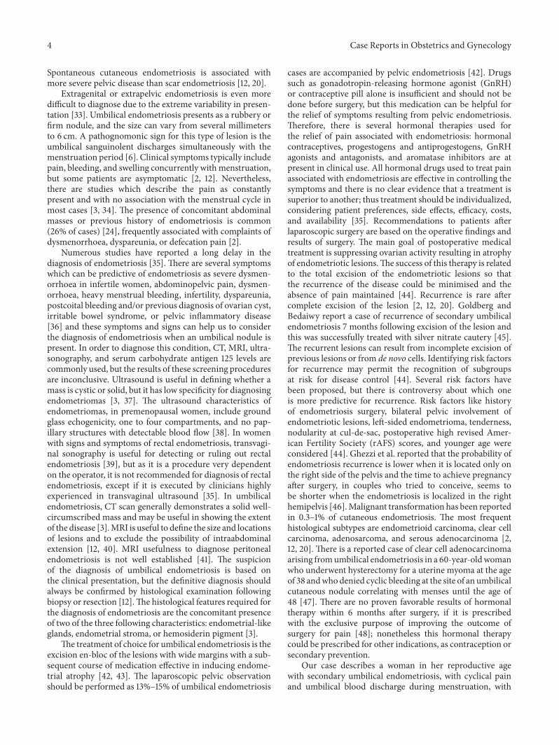
Extragenital or extrapelvic endometriosis is even moredifficult to diagnose due to the extreme variability in presen-tation [33]. Umbilical endometriosis presents as a rubbery orfirm nodule, and the size can vary from several millimetersto 6 cm. A pathognomonic sign for this type of lesion is theumbilical sanguinolent discharges simultaneously with themenstruation period [6]. Clinical symptoms typically includepain, bleeding, and swelling concurrently withmenstruation,but some patients are asymptomatic [2, 12]. Nevertheless,there are studies which describe the pain as constantlypresent and with no association with the menstrual cycle inmost cases [3, 34]. The presence of concomitant abdominalmasses or previous history of endometriosis is common(26% of cases) [24], frequently associated with complaints ofdysmenorrhoea, dyspareunia, or defecation pain [2].
Numerous studies have reported a long delay in thediagnosis of endometriosis [35]. There are several symptomswhich can be predictive of endometriosis as severe dysmen-orrhoea in infertile women, abdominopelvic pain, dysmen-orrhoea, heavy menstrual bleeding, infertility, dyspareunia,postcoital bleeding and/or previous diagnosis of ovarian cyst,irritable bowel syndrome, or pelvic inflammatory disease[36] and these symptoms and signs can help us to considerthe diagnosis of endometriosis when an umbilical nodule ispresent. In order to diagnose this condition, CT, MRI, ultra-sonography, and serum carbohydrate antigen 125 levels arecommonly used, but the results of these screening proceduresare inconclusive. Ultrasound is useful in defining whether amass is cystic or solid, but it has low specificity for diagnosingendometriomas [3, 37]. The ultrasound characteristics ofendometriomas, in premenopausal women, include groundglass echogenicity, one to four compartments, and no pap-illary structures with detectable blood flow [38]. In womenwith signs and symptoms of rectal endometriosis, transvagi-nal sonography is useful for detecting or ruling out rectalendometriosis [39], but as it is a procedure very dependenton the operator, it is not recommended for diagnosis of rectalendometriosis, except if it is executed by clinicians highlyexperienced in transvaginal ultrasound [35]. In umbilicalendometriosis, CT scan generally demonstrates a solid well-circumscribed mass and may be useful in showing the extentof the disease [3].MRI is useful to define the size and locationsof lesions and to exclude the possibility of intraabdominalextension [12, 40]. MRI usefulness to diagnose peritonealendometriosis is not well established [41]. The suspicionof the diagnosis of umbilical endometriosis is based onthe clinical presentation, but the definitive diagnosis shouldalways be confirmed by histological examination followingbiopsy or resection [12].The histological features required forthe diagnosis of endometriosis are the concomitant presenceof two of the three following characteristics: endometrial-likeglands, endometrial stroma, or hemosiderin pigment [3].
The treatment of choice for umbilical endometriosis is theexcision en-bloc of the lesions with wide margins with a sub-sequent course of medication effective in inducing endome-trial atrophy [42, 43]. The laparoscopic pelvic observationshould be performed as 13%–15% of umbilical endometriosis
cases are accompanied by pelvic endometriosis [42]. Drugssuch as gonadotropin-releasing hormone agonist (GnRH)or contraceptive pill alone is insufficient and should not bedone before surgery, but this medication can be helpful forthe relief of symptoms resulting from pelvic endometriosis.Therefore, there is several hormonal therapies used forthe relief of pain associated with endometriosis: hormonalcontraceptives, progestogens and antiprogestogens, GnRHagonists and antagonists, and aromatase inhibitors are atpresent in clinical use. All hormonal drugs used to treat painassociated with endometriosis are effective in controlling thesymptoms and there is no clear evidence that a treatment issuperior to another; thus treatment should be individualized,considering patient preferences, side effects, efficacy, costs,and availability [35]. Recommendations to patients afterlaparoscopic surgery are based on the operative findings andresults of surgery. The main goal of postoperative medicaltreatment is suppressing ovarian activity resulting in atrophyof endometriotic lesions.The success of this therapy is relatedto the total excision of the endometriotic lesions so thatthe recurrence of the disease could be minimised and theabsence of pain maintained [44]. Recurrence is rare aftercomplete excision of the lesion [2, 12, 20]. Goldberg andBedaiwy report a case of recurrence of secondary umbilicalendometriosis 7 months following excision of the lesion andthis was successfully treated with silver nitrate cautery [45].The recurrent lesions can result from incomplete excision ofprevious lesions or from de novo cells. Identifying risk factorsfor recurrence may permit the recognition of subgroupsat risk for disease control [44]. Several risk factors havebeen proposed, but there is controversy about which oneis more predictive for recurrence. Risk factors like historyof endometriosis surgery, bilateral pelvic involvement ofendometriotic lesions, left-sided endometrioma, tenderness,nodularity at cul-de-sac, postoperative high revised Amer-ican Fertility Society (rAFS) scores, and younger age wereconsidered [44]. Ghezzi et al. reported that the probability ofendometriosis recurrence is lower when it is located only onthe right side of the pelvis and the time to achieve pregnancyafter surgery, in couples who tried to conceive, seems tobe shorter when the endometriosis is localized in the righthemipelvis [46].Malignant transformation has been reportedin 0.3–1% of cutaneous endometriosis. The most frequenthistological subtypes are endometrioid carcinoma, clear cellcarcinoma, adenosarcoma, and serous adenocarcinoma [2,12, 20]. There is a reported case of clear cell adenocarcinomaarising fromumbilical endometriosis in a 60-year-oldwomanwho underwent hysterectomy for a uterine myoma at the ageof 38 andwhodenied cyclic bleeding at the site of an umbilicalcutaneous nodule correlating with menses until the age of48 [47]. There are no proven favorable results of hormonaltherapy within 6 months after surgery, if it is prescribedwith the exclusive purpose of improving the outcome ofsurgery for pain [48]; nonetheless this hormonal therapycould be prescribed for other indications, as contraception orsecondary prevention.
Our case describes a woman in her reproductive agewith secondary umbilical endometriosis, with cyclical painand umbilical blood discharge during menstruation, with

Case Reports in Obstetrics and Gynecology 5
previous diagnosis of endometriosis in the appendix anda concomitant endometrioma in the right ovary. Initially,before the patient has been referred to the gynecology depart-ment, umbilical endometriosis was misdiagnosed and wasconsidered a dermoid cyst. After laparoscopy, our patient wasfollowed up with a standard gynaecological examination, theassessment of painful symptoms, and a TV-US scan that wereperformed at 1, 3, 6, and 12 months, and this will be repeatedsubsequently on a yearly basis. The differential diagnosisof an umbilical nodule includes inflammatory disorders(abscess, folliculitis, and omphalitis), other benign lesions(lipoma, haemangioma, inclusion cyst, urachal anomalies,and pyogenic granuloma), umbilical hernia, and malignanttumors (adenocarcinoma, sarcoma, melanoma, metastaticvisceral carcinoma—Sister Mary Joseph nodule) [13, 30].
This case highlights the importance of medical historycompared to those of complementary diagnostic tests whichare sometimes inconclusive. Surgeons should always considerumbilical endometriosis in their diagnostic approach whenconfronted with umbilical nodules. The evidence of theresults of diagnosis and the different options to treat extra-genital endometriosis is limited. Clinicians may considersurgical removal of symptomatic extragenital endometriosis,and when this is not possible, medical treatment should beconsidered to relieve symptoms. More studies are necessaryto conclude about the best method of diagnosis of umbilicalendometriosis and themost appropriate recommendations totreatment and follow-upwhich include the bestmedication toprevent the recurrence of the disease.
Competing Interests
The authors declare that they have no competing interests.
References
[1] C. I. Theunissen and F. F. IJpma, “Primary umbilical endo-metriosis: a cause of a painful umbilical nodule,” Journal ofSurgical Case Reports, vol. 2015, no. 3, article rjv025, 2015.
[2] K. Kyamidis, V. Lora, and J. Kanitakis, “Spontaneous cutaneousumbilical endometriosis: report of a new case with immuno-histochemical study and literature review,” Dermatology OnlineJournal, vol. 17, no. 7, article 5, 2011.
[3] T. S. Papavramidis, K. Sapalidis, N. Michalopoulos et al.,“Spontaneous abdominal wall endometriosis: a case report,”Acta Chirurgica Belgica, vol. 109, no. 6, pp. 778–781, 2009.
[4] D. Paramythiotis, G. Stavrou, S. Panidis et al., “Concurrentappendiceal and umbilical endometriosis: a case report andreview of the literature,” Journal of Medical Case Reports, vol.8, article 258, 2014.
[5] A. Fancellu, A. Pinna, A. Manca, G. Capobianco, and A. Porcu,“Primary umbilical endometriosis. Case report and discussionon management options,” International Journal of Surgery CaseReports, vol. 4, no. 12, pp. 1145–1148, 2013.
[6] G. Pariza andC. I.Mavrodin, “Primary umbilical endometriosis(Villar’s nodule)—case study, literature revision,”Chirurgia, vol.109, no. 4, pp. 546–549, 2014.
[7] M. Gogacz, M. Sarzynski, R. Napierała, J. Sierocinska-Sawa,and A. Semczuk, “Ovarian endometrioma in an 11-year-old girl
before menarche: a case study with literature review,” Journalof Pediatric and Adolescent Gynecology, vol. 25, no. 1, pp. e5–e7,2012.
[8] N. Machairiotis, A. Stylianaki, G. Dryllis et al., “Extrapelvicendometriosis: a rare entity or an under diagnosed condition?”Diagnostic Pathology, vol. 8, no. 1, article 194, 2013.
[9] C. Simoglou, P. Zarogoulidis, N.Machairiotis et al., “Abdominalwall endometrioma mimicking an incarcerated hernia: a casereport,” International Journal of General Medicine, vol. 5, pp.569–571, 2012.
[10] B. Ata, U. Ates, T. Usta, and E. Attar, “Cervical Endometriosis,a case presenting with intractable spotting,” Medscape GeneralMedicine, vol. 7, no. 2, p. 64, 2005.
[11] J. W. Latcher, “Endometriosis of the umbilicus,” AmericanJournal of Obstetrics & Gynecology, vol. 66, no. 1, pp. 161–168,1953.
[12] M. Omori, T. Ogawa, M. Nara, A. Hashi, and S. Hirata, “Umbil-ical endometriosis with giant degenerated uterine leiomyomas:a case report,”Gynecologic Oncology Case Reports, vol. 9, pp. 18–20, 2014.
[13] T. J. Gin, A.D.Gin,D.Gin, A. Pham, and J. Cahill, “Spontaneouscutaneous endometriosis of the umbilicus,” Case Reports inDermatology, vol. 5, no. 3, pp. 368–372, 2013.
[14] M. J. Fernandez-Acenero and S. Cordova, “Cutaneousendometriosis: review of 15 cases diagnosed at a singleinstitution,” Archives of Gynecology and Obstetrics, vol. 283, no.5, pp. 1041–1044, 2011.
[15] T. J. Jaime, T. J. Jaime, P. Ormiga, F. Leal, O. M. Nogueira, andN. Rodrigues, “Umbilical endometriosis: report of a case and itsdermoscopic features,” Anais Brasileiros de Dermatologia, vol.88, no. 1, pp. 121–124, 2013.
[16] L. Attia, R. Ben Temime, J. Sidhom et al., “A case of cutaneousendometriosis developed on an abdominal scar,” Tunisie Medi-cale, vol. 88, no. 11, pp. 841–843, 2010.
[17] T. Esteves, J. Cabrita, R. Coelho, and E. Vale, “Endometriosecutanea—a proposito de um caso clınico,” Dermatology OnlineJournal, vol. 16, no. 5, article 9, 2010.
[18] M. V. Barrow, “Metastatic tumors of the umbilicus,” Journal ofChronic Diseases, vol. 19, no. 10, pp. 1113–1117, 1966.
[19] M. Stojanovic, M. Radojkovic, L. Jeremic et al., “Umbilicalendometriosis associated with large umbilical hernia. Casereport,” Chirurgia, vol. 109, no. 2, pp. 267–270, 2014.
[20] A. Agarwal and Y. F. Fong, “Cutaneous endometriosis,” Singa-pore Medical Journal, vol. 49, no. 9, pp. 704–709, 2008.
[21] R. Victory, M. P. Diamond, and D. A. Johns, “Villar’s nodule:a case report and systematic literature review of endometriosisexterna of the umbilicus,” Journal of Minimally Invasive Gyne-cology, vol. 14, no. 1, pp. 23–32, 2007.
[22] A. H. Din, L. S. Verjee, and M. A. Griffiths, “Cutaneousendometriosis: a plastic surgery perspective,” Journal of Plastic,Reconstructive and Aesthetic Surgery, vol. 66, no. 1, pp. 129–130,2013.
[23] A. Saito, K. Koga, Y. Osuga et al., “Individualized managementof umbilical endometriosis: a report of seven cases,”The Journalof Obstetrics and Gynaecology Research, vol. 40, no. 1, pp. 40–45,2014.
[24] N. Arkoulis and B. K. Chew, “An unusual case of asymptomaticspontaneous umbilical endometriosis treated with skin-sparingexcision,” Journal of Surgical Case Reports, vol. 3, pp. 1–3, 2015.
[25] S. E. Bulun, “Endometriosis,” The New England Journal ofMedicine, vol. 360, no. 3, pp. 268–279, 2009.

6 Case Reports in Obstetrics and Gynecology
[26] A. J. Dwivedi, S. N. Agrawal, and Y. J. Silva, “Abdominal wallendometriomas,” Digestive Diseases and Sciences, vol. 47, no. 2,pp. 456–461, 2002.
[27] E. Tomas, A.Martın, C. Garfia et al., “Abdominal wall endomet-riosis in absence of previous surgery,” Journal of Ultrasound inMedicine, vol. 18, no. 5, pp. 373–374, 1999.
[28] A. S. Seydel, J. Z. Sickel, E. D. Warner, and H. C. Sax,“Extrapelvic endometriosis: diagnosis and treatment,” TheAmerican Journal of Surgery, vol. 171, no. 2, pp. 239–241, 1996.
[29] C. Mowad, C. Andreychik, and T. Murphy, “Umbilicalendometriosis elucidates cause of recurrent pneumothorax,”Journal of the American Academy of Dermatology, vol. 71, no. 3,pp. e79–e80, 2014.
[30] A. D. Malebranche and K. Bush, “Umbilical endometriosis: arare diagnosis in plastic and reconstructive surgery,” CanadianJournal of Plastic Surgery, vol. 18, no. 4, pp. 147–148, 2010.
[31] S. Apostolidis, A. Michalopoulos, T. S. Papavramidis, V. N.Papadopoulos, D. Paramythiotis, and N. Harlaftis, “Inguinalendometriosis: three cases and literature review,” SouthernMedical Journal, vol. 102, no. 2, pp. 206–207, 2009.
[32] J. D. Horton, K. J. DeZee, E. P. Ahnfeldt, and M. Wagner,“Abdominal wall endometriosis: a surgeon’s perspective andreview of 445 cases,” American Journal of Surgery, vol. 196, no.2, pp. 207–212, 2008.
[33] A. Singh, “Umbilical endometriosis mimicking as papilloma togeneral surgeons: a case report,” Australasian Medical Journal,vol. 5, no. 5, pp. 272–274, 2012.
[34] J.-H. J. Hensen, A. C. Van Breda Vriesman, and J. B. C. M.Puylaert, “Abdominal wall endometriosis: clinical presentationand imaging features with emphasis on sonography,” AmericanJournal of Roentgenology, vol. 186, no. 3, pp. 616–620, 2006.
[35] G. A. J. Dunselman, N. Vermeulen, C. Becker et al., “ESHREguideline: management of women with endometriosis,”HumanReproduction, vol. 29, no. 3, pp. 400–412, 2014.
[36] K. D. Ballard, H. E. Seaman, C. S. De Vries, and J. T. Wright,“Can symptomatology help in the diagnosis of endometriosis?Findings from a national case-control study—part 1,” BJOG: AnInternational Journal of Obstetrics and Gynaecology, vol. 115, no.11, pp. 1382–1391, 2008.
[37] L. M. Vincent and C. A. Mittelstaedt, “Sonographic demon-stration of endometrioma arising in cesarean scar,” Journal ofUltrasound in Medicine, vol. 4, no. 8, pp. 437–438, 1985.
[38] C.VanHolsbeke, B. VanCalster, S. Guerriero et al., “Endometri-omas: their ultrasound characteristics,”Ultrasound in Obstetricsand Gynecology, vol. 35, no. 6, pp. 730–740, 2010.
[39] G.Hudelist, J. English, A. E.Thomas, A. Tinelli, C. F. Singer, andJ. Keckstein, “Diagnostic accuracy of transvaginal ultrasoundfor non-invasive diagnosis of bowel endometriosis: systematicreview and meta-analysis,” Ultrasound in Obstetrics and Gyne-cology, vol. 37, no. 3, pp. 257–263, 2011.
[40] C.-Y. Yu, M. Perez-Reyes, J. J. Brown, and J. A. Borrello, “Mrappearance of umbilical endometriosis,” Journal of ComputerAssisted Tomography, vol. 18, no. 2, pp. 269–271, 1994.
[41] P. Stratton, C. Winkel, A. Premkumar et al., “Diagnosticaccuracy of laparoscopy, magnetic resonance imaging, andhistopathologic examination for the detection of endometrio-sis,” Fertility and Sterility, vol. 79, no. 5, pp. 1078–1085, 2003.
[42] K. Chikazawa, J. Mitsushita, S. Netsu, and R. Konno, “Surgicalexcision of umbilical endometriotic lesions with laparoscopicpelvic observation is the way to treat umbilical endometriosis,”Asian Journal of Endoscopic Surgery, vol. 7, no. 4, pp. 320–322,2014.
[43] D. Buca, M. Leombroni, E. Falo et al., “Recurrence of primaryumbilical endometriosis: case report and review of the litera-ture,” Multidisciplinary Journal of Women’s Health, vol. 2, no. 1,pp. 6–8, 2013.
[44] I. Selcuk and G. Bozdag, “Recurrence of endometriosis; riskfactors, mechanisms and biomarkers; review of the literature,”Journal of the Turkish GermanGynecological Association, vol. 14,no. 2, pp. 98–103, 2013.
[45] J. M. Goldberg and M. A. Bedaiwy, “Recurrent umbilicalendometriosis after laparoscopic treatment of minimal pelvicendometriosis: a case report,” Journal of Reproductive Medicinefor the Obstetrician and Gynecologist, vol. 52, no. 6, pp. 551–552,2007.
[46] F. Ghezzi, P. Beretta, M. Franchi, M. Parissis, and P. Bolis,“Recurrence of ovarian endometriosis and anatomical locationof the primary lesion,” Fertility and Sterility, vol. 75, no. 1, pp.136–140, 2001.
[47] K. Obata, N. Ikoma, G. Oomura, and Y. Inoue, “Clear celladenocarcinoma arising fromumbilical endometriosis,” Journalof Obstetrics and Gynaecology Research, vol. 39, no. 1, pp. 455–461, 2013.
[48] S. Furness, C. Yap, C. Farquhar, and Y. Cheong, “Pre andpost-operative medical therapy for endometriosis surgery,”TheCochrane Database of Systematic Reviews, no. 3, Article IDCD003678, 2004.