Umbilical Cord Blood Transplantation - Hematology & Oncology
Upload
truongquynhCategory
view
220download
0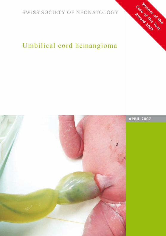
Umbilical cord hemangioma
SWISS SOCIETY OF NEONATOLOGY
APRIL 2007
Winner of the
Case of the Year
Award 2007

40
Natalucci G, Wisser J, Weil R, Stallmach T, Bucher HU,
Clinic of Neonatology (NG, BHU), Department of
Obstetrics and Gynaecology (WJ), Institute of
Pathology (ST), University Hospital, and Department
of Pediatric Surgery (WR), Children’s Hospital, Zurich,
Switzerland
© Swiss Society of Neonatology, Thomas M Berger, Webmaster
41
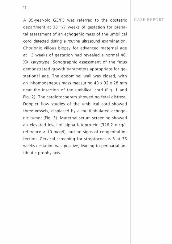
CASE REPORTA 35-year-old G3/P3 was referred to the obstetric
department at 33 1/7 weeks of gestation for prena-
tal assessment of an echogenic mass of the umbilical
cord detected during a routine ultrasound examination.
Chorionic villous biopsy for advanced maternal age
at 13 weeks of gestation had revealed a normal 46,
XX karyotype. Sonographic assessment of the fetus
demonstrated growth parameters appropriate for ge-
stational age. The abdominal wall was closed, with
an inhomogeneous mass measuring 43 x 32 x 28 mm
near the insertion of the umbilical cord (Fig. 1 and
Fig. 2). The cardiotocogram showed no fetal distress.
Doppler flow studies of the umbilical cord showed
three vessels, displaced by a multilobulated echoge-
nic tumor (Fig. 3). Maternal serum screening showed
an elevated level of alpha-fetoprotein (326.2 mcg/l,
reference < 10 mcg/l), but no signs of congenital in-
fection. Cervical screening for streptococcus B at 35
weeks gestation was positive, leading to peripartal an-
tibiotic prophylaxis.
42
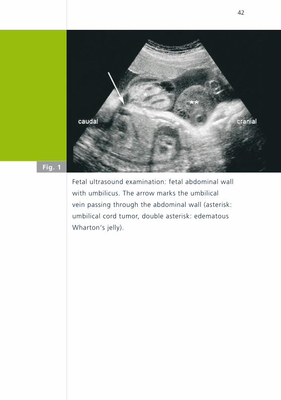
Fig. 1
Fetal ultrasound examination: fetal abdominal wall
with umbilicus. The arrow marks the umbilical
vein passing through the abdominal wall (asterisk:
umbilical cord tumor, double asterisk: edematous
Wharton‘s jelly).
43
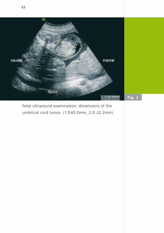
Fetal ultrasound examination: dimensions of the
umbilical cord tumor. (1:D43.0mm, 2:D 32.2mm).
Fig. 2
44
After induction of labor at 38 5/7 weeks of gestation, a
baby girl was delivered vaginally, weighing 2840 g (P
10-25), length 47 cm (P 10-25), head circumference
33 cm (P 10-25). She adapted with Apgar scores of
8 at 1 minute, 9 at 5 minutes, and 9 at 10 minutes.
Arterial cord pH was 7.31.
Surgical revis ion was undertaken for the suspected
diagnosis of a “hernia into the cord”. However,
no defect of the abdominal wal l was found. An
ord inary surg ica l resect ion of the umbil ical co rd
w i th an umb i l i cop l a s t y was pe r fo rmed . The
maximum diameter of the surgically removed umbi-
l ical cord segment was 3.6 cm. Macroscopically, the
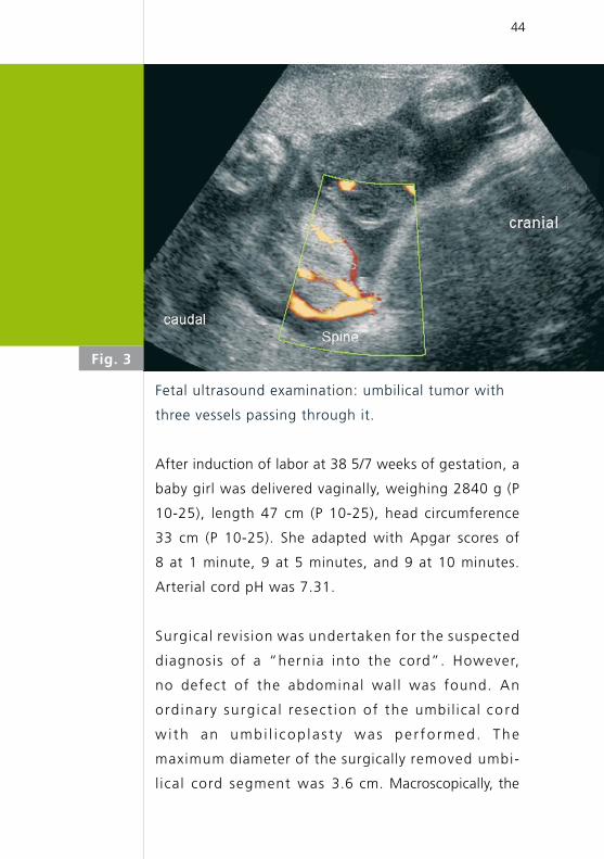
Fig. 3
Fetal ultrasound examination: umbilical tumor with
three vessels passing through it.
45
cross section revealed three vessels. Only on micros-
copic examination did it become clear that Wharton’s
jelly was mostly replaced by abundant aggregates of
thin walled capillaries (Fig. 6). The capil lary heman-
gioma showed a diffuse growth around vessels, with
focal dissection of the muscular coat of the umbilical
vein. In addition, nodular aggregates were seen, some
of which were freshly thrombosed (Fig. 7). Distal to the
lesion, the remaining umbilical cord measured 20 cm in
length and 1.2 cm in diameter and was unremarkable.
Postoperative recovery was uneventful. Subsequent-
ly, the girl developed a small supraumbilical hernia
which was successfully operated three months later.
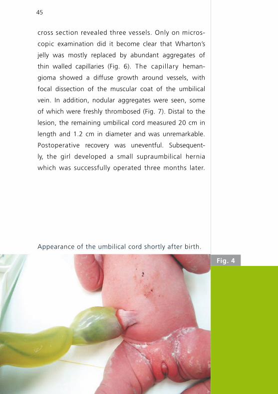
Appearance of the umbilical cord shortly after birth.
Fig. 4
46
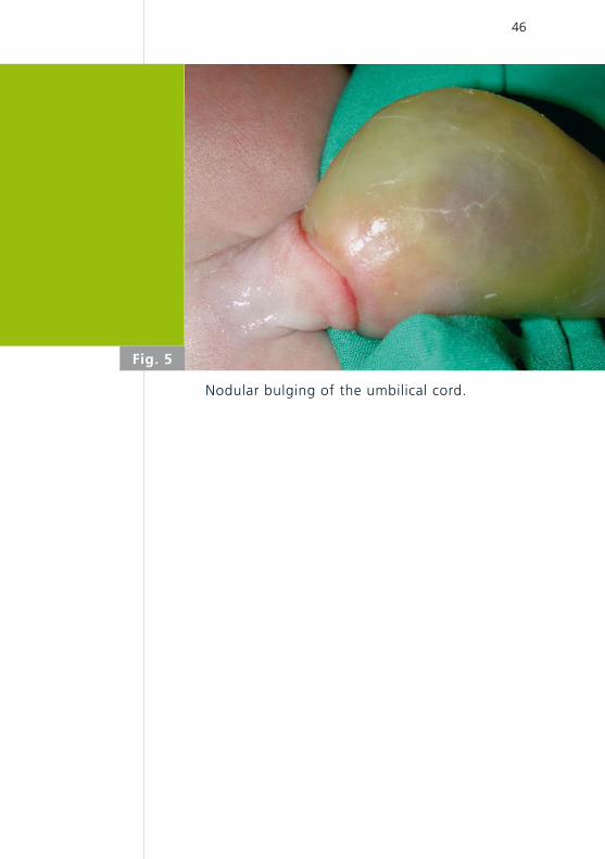
Fig. 5
Nodular bulging of the umbilical cord.
47
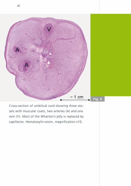
Fig. 6
Cross-section of umbilical cord showing three ves-
sels with muscular coats, two arteries (A) and one
vein (V). Most of the Wharton’s jelly is replaced by
capillaries. Hematoxylin-eosin, magnification x10.
48
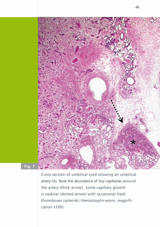
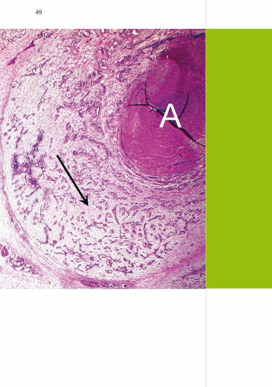
Cross-section of umbilical cord showing an umbilical
artery (A). Note the abundance of tiny capillaries around
the artery (thick arrow). Some capillary growth
is nodular (dotted arrow) with occasional fresh
thromboses (asterisk) (Hematoxylin-eosin, magnifi-
cation x100).
Fig. 7
49
50
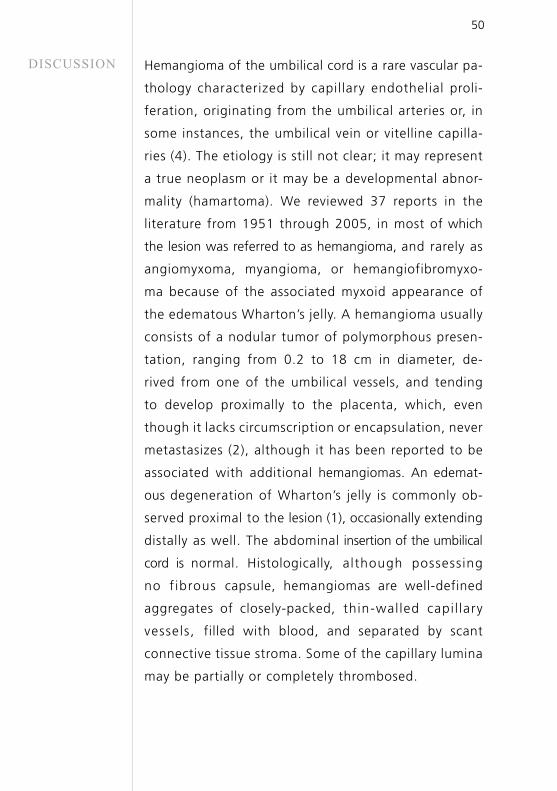
Hemangioma of the umbilical cord is a rare vascular pa-
thology characterized by capillary endothelial proli-
feration, originating from the umbilical arteries or, in
some instances, the umbilical vein or vitelline capilla-
ries (4). The etiology is still not clear; it may represent
a true neoplasm or it may be a developmental abnor-
mality (hamartoma). We reviewed 37 reports in the
literature from 1951 through 2005, in most of which
the lesion was referred to as hemangioma, and rarely as
angiomyxoma, myangioma, or hemangiofibromyxo-
ma because of the associated myxoid appearance of
the edematous Wharton’s jelly. A hemangioma usually
consists of a nodular tumor of polymorphous presen-
tation, ranging from 0.2 to 18 cm in diameter, de-
rived from one of the umbilical vessels, and tending
to develop proximally to the placenta, which, even
though it lacks circumscription or encapsulation, never
metastasizes (2), although it has been reported to be
associated with additional hemangiomas. An edemat-
ous degeneration of Wharton’s jelly is commonly ob-
served proximal to the lesion (1), occasionally extending
distally as well. The abdominal insertion of the umbilical
cord is normal. Histologically, although possessing
no f ibrous capsule, hemangiomas are well-defined
aggregates of closely-packed, thin-walled capi l lary
vessels, filled with blood, and separated by scant
connective tissue stroma. Some of the capillary lumina
may be partially or completely thrombosed.
DISCUSSION
51
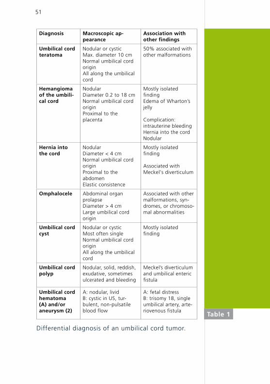
Diagnosis Macroscopic ap-pearance
Association with other findings
Umbilical cord teratoma
Nodular or cysticMax. diameter 10 cmNormal umbilical cord originAll along the umbilical cord
50% associated with other malformations
Hemangioma of the umbili-cal cord
Nodular Diameter 0.2 to 18 cmNormal umbilical cord originProximal to the placenta
Mostly isolated findingEdema of Wharton’s jelly
Complication:intrauterine bleeding Hernia into the cord Nodular
Hernia into the cord
NodularDiameter < 4 cmNormal umbilical cord originProximal to the abdomenElastic consistence
Mostly isolated finding
Associated with Meckel‘s diverticulum
Omphalocele Abdominal organ prolapse Diameter > 4 cmLarge umbilical cord origin
Associated with other malformations, syn-dromes, or chromoso-mal abnormalities
Umbilical cord cyst
Nodular or cysticMost often singleNormal umbilical cord originAll along the umbilical cord
Mostly isolated finding
Umbilical cord polyp
Nodular, solid, reddish,exudative, sometimes ulcerated and bleeding
Meckel’s diverticulum and umbilical enteric fistula
Umbilical cord hematoma (A) and/or aneurysm (2)
A: nodular, livid B: cystic in US, tur-bulent, non-pulsatile blood flow
A: fetal distress B: trisomy 18, single umbilical artery, arte-riovenous fistula
Differential diagnosis of an umbilical cord tumor.
Table 1
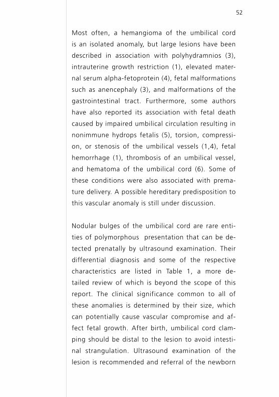
52
Most often, a hemangioma of the umbilical cord
is an isolated anomaly, but large lesions have been
described in association with polyhydramnios (3),
intrauterine growth restriction (1), elevated mater-
nal serum alpha-fetoprotein (4), fetal malformations
such as anencephaly (3), and malformations of the
gastrointestinal tract. Furthermore, some authors
have also reported its association with fetal death
caused by impaired umbilical circulation resulting in
nonimmune hydrops fetalis (5), torsion, compressi-
on, or stenosis of the umbilical vessels (1,4), fetal
hemorrhage (1), thrombosis of an umbilical vessel,
and hematoma of the umbilical cord (6). Some of
these conditions were also associated with prema-
ture delivery. A possible hereditary predisposition to
this vascular anomaly is still under discussion.
Nodular bulges of the umbilical cord are rare enti-
ties of polymorphous presentation that can be de-
tected prenatally by ultrasound examination. Their
differential diagnosis and some of the respective
characteristics are listed in Table 1, a more de-
tailed review of which is beyond the scope of this
report. The clinical significance common to all of
these anomalies is determined by their size, which
can potentially cause vascular compromise and af-
fect fetal growth. After birth, umbilical cord clam-
ping should be distal to the lesion to avoid intesti-
nal strangulation. Ultrasound examination of the
lesion is recommended and referral of the newborn
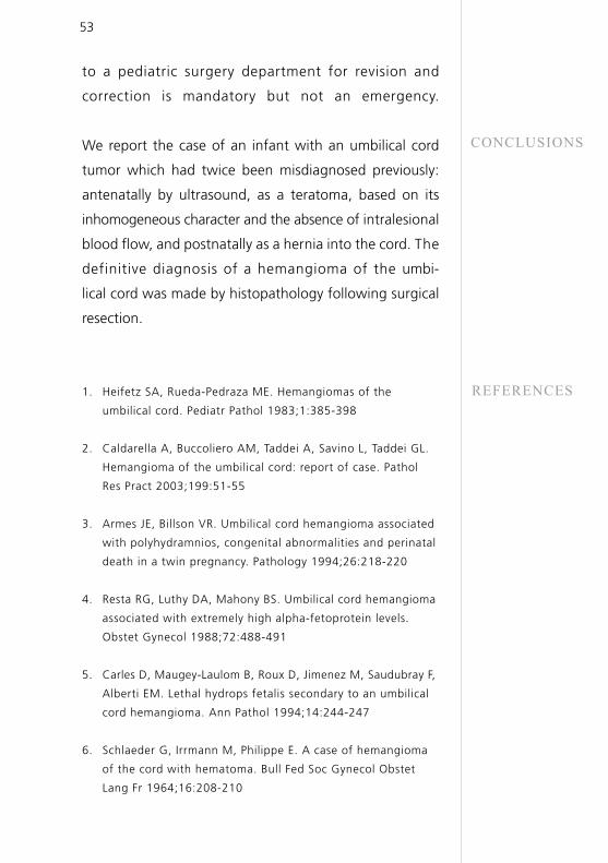
53
1. Heifetz SA, Rueda-Pedraza ME. Hemangiomas of the
umbilical cord. Pediatr Pathol 1983;1:385-398
2. Caldarella A, Buccoliero AM, Taddei A, Savino L, Taddei GL.
Hemangioma of the umbilical cord: report of case. Pathol
Res Pract 2003;199:51-55
3. Armes JE, Billson VR. Umbilical cord hemangioma associated
with polyhydramnios, congenital abnormalities and perinatal
death in a twin pregnancy. Pathology 1994;26:218-220
4. Resta RG, Luthy DA, Mahony BS. Umbilical cord hemangioma
associated with extremely high alpha-fetoprotein levels.
Obstet Gynecol 1988;72:488-491
5. Carles D, Maugey-Laulom B, Roux D, Jimenez M, Saudubray F,
Alberti EM. Lethal hydrops fetalis secondary to an umbilical
cord hemangioma. Ann Pathol 1994;14:244-247
6. Schlaeder G, Irrmann M, Philippe E. A case of hemangioma
of the cord with hematoma. Bull Fed Soc Gynecol Obstet
Lang Fr 1964;16:208-210
REFERENCES
CONCLUSIONS
to a pediatric surgery department for revision and
correction is mandatory but not an emergency.
We report the case of an infant with an umbilical cord
tumor which had twice been misdiagnosed previously:
antenatally by ultrasound, as a teratoma, based on its
inhomogeneous character and the absence of intralesional
blood flow, and postnatally as a hernia into the cord. The
definitive diagnosis of a hemangioma of the umbi-
lical cord was made by histopathology following surgical
resection.


![hernia of the umbilical cord [وضع التوافق] of the umbilical cord.pdf · Umbilical cord hernia…cont Conclusion: ¾Hernia of the umbilical cord is a rare entityy, of the](https://static.fdocuments.in/doc/165x107/5ea7ce695a148409cd011fd0/hernia-of-the-umbilical-cord-of-the-umbilical-cordpdf.jpg)