Ultraviolet fluorescence photography of the Shroud of Turin Fluorescence Miller Pellicori 1981...
15
D 8 A V. D. Miller and S. F. Pellicorl Ultraviolet fluorescence photography of the Shroud of Turin 71 Ultraviolet fluorescence phototography of the Shroud of Turin One of the nondestructive techniques used to investigate the Shroud of Turin was ultraviolet fluorescence (UV) pho- tography. This technique is able to detect organic and inorganic compounds by their integrated emission spectra and it is the complement of the more common tech- nique of reflectance photography. Pho- tographic data collection was one of many information resources designated by the Shroud of Turin Research Project 1 (STURP) team for the investigation of the body image and blood stains. The goal of the team was to determine the nature of the body image and its cause. Background The investigation in October 1978 followed the conclusion of the public ex- hibition held in celebration of the 400th anniversary of the Shroud's housing in Turin, Italy. That was the first public exhibition in 45 years and the first full- scale multidisciplinary study in the Shroud's history.2,3 4 V. D. Miller and S. F. Pellicori Members, Shroud of Turin Research Project Santa Barbara, California The Shroud is a 4.4 by 1.1 meter piece of age-yellowed linen which displays the life-sized dorsal and frontal images of a man. The appearance of the visible image and the locations of the blood stains suggest parallels with the descriptions of Christ's crucifixion in the Scriptures. For this reason, the legend associated with the Shroud of Turin is that it is the burial cloth of Jesus of Nazareth. It has also been suggested that the image was painted in the 14th century. Since the historical record is complete back only to the 1350's, the veracity of the legend cannot be di- rectly addressed. Obvious scorch and water marks can be pinpointed to a fire in 1532. Hypotheses such as artistic paint- ing, for example, can be tested for agree- ment with the observations. The team was thus charged with t he task of collecting multifaceted data of necessary and suffi- cient quality and quantity for testing various hypotheses. The United States team, an inde- pendent group of 32 scientists and assis- 10 11 12 13 © Biological Photographic Association, Inc., 1981 Journal of Biological Photography Volume 49, Number 3, July, 1981 14 15 tants, was equipped with instrumenta - tion4 capable of detecting work produced by known artistic techniques and ade- quate to provide a broadly-based foun- dation of information. The instrumenta- tion included x-ray transmission and fluorescence 5 to detect high atomic number elements expected for inorganic pigments, microscopy6 for visual and photographic examination of details, photoelectric spectrophotometry7,8 for measurements of reflectance and fluo- rescence, photography 9 through bandpass filters, infrared spectrometry 10 and UV fluorescence photography documented here for the first time. Photogra phi c pro cedu re To record the emission excited by ultraviolet, a special light source was as- sembled. For our experiment a filter- window bandpass enabled wavelengths of 335 to 375 nm to be isolated from the sources for the exciting radiation. The two sourcP.s aimed at 45-degree angles from 16 17 11 19 20 21 22
Transcript of Ultraviolet fluorescence photography of the Shroud of Turin Fluorescence Miller Pellicori 1981...
D
8
A
V. D. Miller and S. F. Pellicorl Ultraviolet f luorescence photography of the Shroud of Turin 71
Ultraviolet fluorescence phototography of the Shroud of Turin
One of the nondestructive techniques used to investigate the Shroud of Turin was ultraviolet fluorescence (UV) photography. This technique is able to detect organic and inorganic compounds by their integrated emission spectra and it is the complement of the more common technique of reflectance photography. Photographic data collection was one of many information resources designated by the Shroud of Turin Research Project1
(STURP) team for the investigation of the body image and blood stains. The goal of the team was to determine the nature of the body image and its cause.
Background The investigation in October 1978
followed the conclusion of the public exhibition held in celebration of the 400th anniversary of the Shroud's housing in Turin, Italy. That was the first public exhibition in 45 years and the first fullscale multidisciplinary study in the Shroud's history.2,3
4
V. D. Miller and S. F. Pellicori Members, Shroud of Turin Research Project
Santa Barbara, California
The Shroud is a 4.4 by 1.1 meter piece of age-yellowed linen which displays the life-sized dorsal and frontal images of a man. The appearance of the visible image and the locations of the blood stains suggest parallels with the descriptions of Christ's crucifixion in the Scriptures. For this reason, the legend associated with the Shroud of Turin is that it is the burial cloth of Jesus of Nazareth. It has also been suggested that the image was painted in the 14th century. Since the historical record is complete back only to the 1350's, the veracity of the legend cannot be directly addressed. Obvious scorch and water marks can be pinpointed to a fire in 1532. Hypotheses such as artistic painting, for example, can be tested for agreement with the observations. The team was thus charged with the task of collecting multifaceted data of necessary and sufficient quality and quantity for testing various hypotheses.
The United States team, an independent group of 32 scientists and assis-
10 11 12 13
© Biological Photographic Association, Inc., 1981
Journal of Biological Photography
Volume 49, Number 3, July, 1981
14 15
tants, was equipped with instrumentation4 capable of detecting work produced by known artistic techniques and adequate to provide a broadly-based foundation of information. The instrumentation included x-ray transmission and fluorescence5 to detect high atomic number elements expected for inorganic pigments, microscopy6 for visual and photographic examination of details, photoelectric spectrophotometry7,8 for measurements of reflectance and fluorescence, photography9 through bandpass filters, infrared spectrometry10 and UV fluorescence photography documented here for the first time.
Photographic procedure To record the emission excited by
ultraviolet, a special light source was assembled. For our experiment a filterwindow bandpass enabled wavelengths of 335 to 375 nm to be isolated from the sources for the exciting radiation. The two sourcP.s aimed at 45-degree angles from
16 17 11 19 20 21 22

72
;e !.. t-
Journal of Biological Photography
100
10
Exciter Fiiter Cell 28 Sept'78
80% Co So4 15% Cu So4 5% NI So4 3mm C7-54
I I I I I
I I
0 I
0 I
I Barrier
6 Fiiter I (Hoya
I L-42 ) I
? I I
? I I I I I
UIL......-'-~--'-~..._--'~-'-~-'-~.L----llJ.---'-~-'---'L......-'-~-'---' 320 340 360 380 400 420 440 460
~(nm)
Vol. 49, No. 3, July, 1981
Figure 1A-Transmittance curves for the ultraviolet (UV) exciting filter cell used in front of the xenon source and for the UV barrier filter used 011er the camera lens.
B-Configuration of exciter and barrier filters for ultraviolet fluorescence photography. The exciter filter consisted of a Pyrex window and a filter cell containing
a 1 cm path of the solution shown and a Corning 7-54 glass filter for absorption
of the residual red leak. The Hoya L-42 filter served as a barrier filter transmitting
only those wavelengths above 420 nm. C-Exciter filter cell attached to flash
reflector.
c
B SHROUD
// ////
//// // //
////
Liquid Solution
C?-54 Filter
Pyrex Window

v. D. Miller and S. F. Pelllcorl
~ ~
--:i
Ultraviolet fluorescence photography of the Shroud of Turin 73
14
Figure 2-Camera and Illumination pOSitioned on support rail for indexing along the length of the Shroud and thus creating a complete mosaic
of photographs according to the reference layout in Figure 3. A-Lamp heads (Norman 200 B) with liquid cell filter. B-AC power pack. C-EL Hasselblad camera with 70 mm back. 0 -Telescoping extention rod for locating rail equi-distant from Shroud frame. E-Leveling screws.
F-Centlmeter scale with indexing marked for predetermined positions. G-Adjustable sliding index pointer for starting sequence.
the camera-subject axis were 200 wattsecond xenon strobes with 15-cm reflectors. The film was to record only radiation emitted in the visible region of the spectrum, and none of the reflected, exciting UV. Consequently, the light source had to be free of visible radiation. To achieve these conditions special excitation filters were constructed to fit on the 15-cm diameter strobe sources.
The filters consisted of a 1-cm path length containing a mixture of inorganic salts of transition metals 11 to absorb radiation above about 400 nm. One window of the liquid filter cell was Corning 9863 visible-absorbing UV-transmitting glass.
This 9863 filter was required to attenuate a smaJI red leak in the exciter filter. The other window was pyrex. The isolated passband and out-of-band attenuation measured for the filter are shown in Figure 1. Since the fluorescences we sought were expected to be in the visible, the filter needed to attenuate to a level of 10- 4
since visible light from the xenon tubes would completely swamp the weak fluo rescent signals. The reasons for using a liquid filter system were that no other filters such as multilayer coatings or ultraviolet transmission glass alone are able to define the required passband while possessing adequate rejection. Further,
none of the standard filter materials could survive the heat of the xenon flash. Finally, the 15-cm size was beyond standard UV accessories.
The exciting UV energy which was partially reflected by the Shroud was prevented from reaching the film by a nonfluorescing, long-wavelength pass, UV absorbing glass placed in front of the lens (Figure 1). This barrier filter passed wavelengths greater than 410 nanometers.
The camera and light source assembly was moved along a rail parallel to the long dimension of the Shroud, which was mounted with its short dimension vertical (Figure 2). The long dimension was di-
0
8
A
74 Journal of Biological Photography
4
vided into eight 53.3-cm square areas for photography and later full-size reconstruction. The areas were intended to be coincident with the black-and-white color separation series taken at another time. The sections were numbered from left to right, beginning at the dorsal feet end of the Shroud. T he number-letter coordinate set corresponded with a master reference mosaic and is shown in Figure 3.
The camera was a Hassleblad EL with a 80 mm lens. The film used was Kodacolor 400 film, a color negative film with an exposure index of 400. Prints were then made on Kodak Ektacolor no. 78 paper.
Differences between the fluorescent record and white light photos were noted. Assisted by laboratory data, interpretation of these differences provides insight
10 11 12 13 14 15
Vol. 49, No. 3, July, 1981
into the nature and causes of the various markings on the Shroud, namely the body image, blood stains, and scourge, water and scorch marks. These interpretations can be compared with laboratory-produced simulations.
Detailed observations Some general comparisons from color
photographs of fluorescing colors and natural light colors are listed in Table I. Differences are detailed in the following pages for each section of the Shroud under discussion. Section B through E by 1 through 4, for example, defines a rectangle according to the coordinates of the reference overlay on the Shroud. (See Figure 3 and the reduced size overlay in the text.)
11 17 11 19 20 21 22
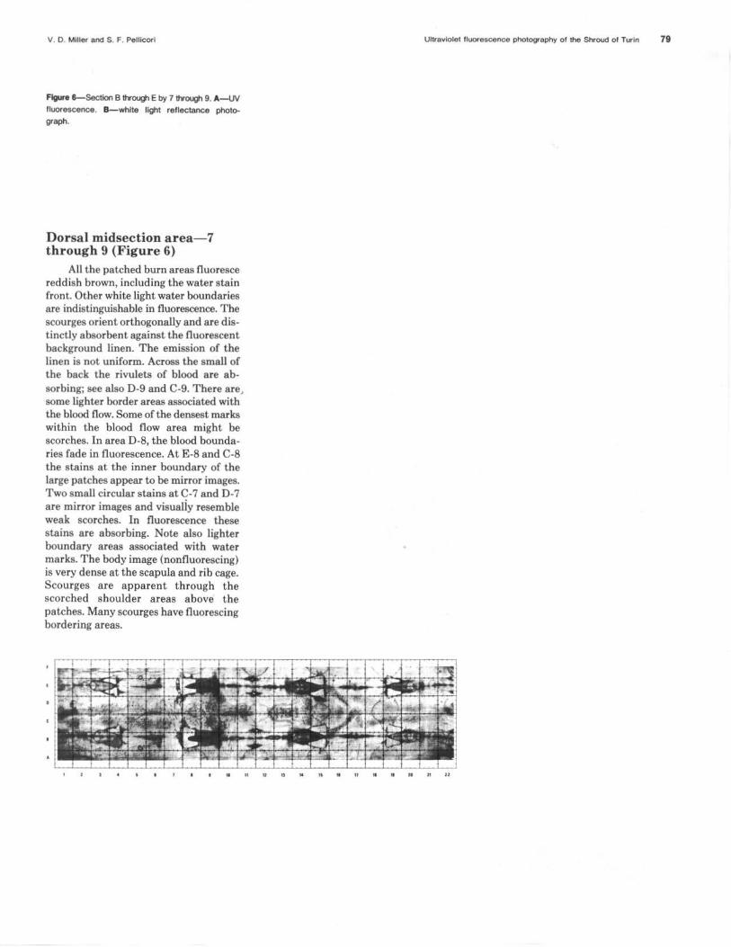
Figure 3-Reference layOU1 of the Shroud showing the numbered and lettered sections discussed in detail in the text.
V. O. Miller and S. F. Pellicori Ultraviolet fluorescence photography of the Shroud of Turin
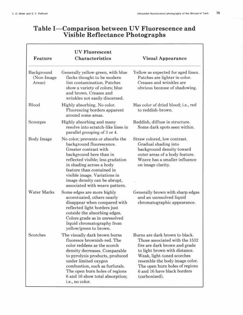
Table I-Comparison between UV Fluorescence and Visible Reflectance Photographs
Feature
Background (Non-Image Areas)
Blood
Scourges
Body Image
Water Marks
Scorches
UV Fluorescent Characteristics
Generally yellow-green, with blue flecks thought to be modern lint contamination. Patches show a variety of colors; blue and brown. Creases and wrinkles not easily discerned.
Highly absorbing. No color. Fluorescing borders apparent around some areas.
Highly absorbing and many resolve into scratch-like lines in parallel grouping of 3 or 4.
No color; prevents or absorbs the background fluorescence. Greater contrast with background here than in reflected visible; less gradation in shading across a body feature than contained in visible image. Variations in image density can be abrupt, associated with weave pattern.
Some edges are more highly accentuated, others nearly disappear when compared with reflected light borders just outside the absorbing edges. Colors grade as in unresolved liquid chromatography from yellow/green to brown.
The visually dark brown burns fluoresce brownish-red. The color reddens as the scorch density decreases. Comparable to pyrolysis products, produced under limited oxygen combustion, such as furfurals. The open burn holes of regions 6 and 16 show total absorption; i.e., no color.
Visual Appearance
Yellow as expected for aged linen. Patches are lighter in color. Creases and wrinkles are obvious because of shadowing.
Has color of dried blood; i.e. , red to reddish-brown.
Reddish, diffuse in structure. Some dark spots seen within.
Straw colored, low contrast. Gradual shading into background density toward outer areas of a body feature. Weave has a smaller influence on image clarity.
Generally brown with sharp edges and an unresolved liquid chromatographic appearance.
Burns are dark brown to black. Those associated with the 1532 fire are dark brown and grade to light brown with distance. Weak, light-toned scorches resemble the body image color. The open burn holes of regions 6 and 16 have black borders (carbonized).
75

76 Journal of Biological Photography
A
B
Vol 49. No. 3, July, 1981
Dorsal feet area- B through E by 1 through 4 (Figure 4)
Some cloth fluorescent characteristics stand out as different from the white light appearance, namely creases, thread, and shadowing are less apparent in UV. The striation in the weave pattern (warp versus weft) are enhanced in fluorescence. There is a general decrease in cloth emission at the corners. The teflon coated magnets, quite visible in white light, are nonfluorescent. This signifies the rejection level of reflected light. The visual water marks are not detectible fluorescently, whjle the scorches around the burns emit reddish fluorescence.
Left f oot-C through D by 1 through 2
Blood is red visually, but neutral to black (absorbing) fluorescently. Detailed shading in absorpt ion in the blood in center of left foot. A triangular-shaped pattern is seen at the ball of the foot. Note the very dense absorption in the blood area at the left end of the rivulet from the heel: scorching might be associated. A fluorescing border in the blood now off the body image areas is seen.
Right foot
A more distinct light border area is seen. The body image appears different from the blood in fluorescence, that is, it absorbs less intensely. The scourge marks have greater contrast in fluorescence than in white light and on the calf appear as lines rather than as dumbbell shapes as they do further up on the leg. Some scourges are not perceptible in the white light photo. The scourge Lines run parallel and diagonally from top left to bottom right. T he background cloth fluorescence is prominent between the leg images (C-D area). The absorbing water marks at 3 and B through E have Light border areas.
Figure 4-Section B through Eby 1 through 4 of the Shroud. A-UV fluorescence. B-white light reflec
tance photograph.
V 0 M iller and S F. Pellicori
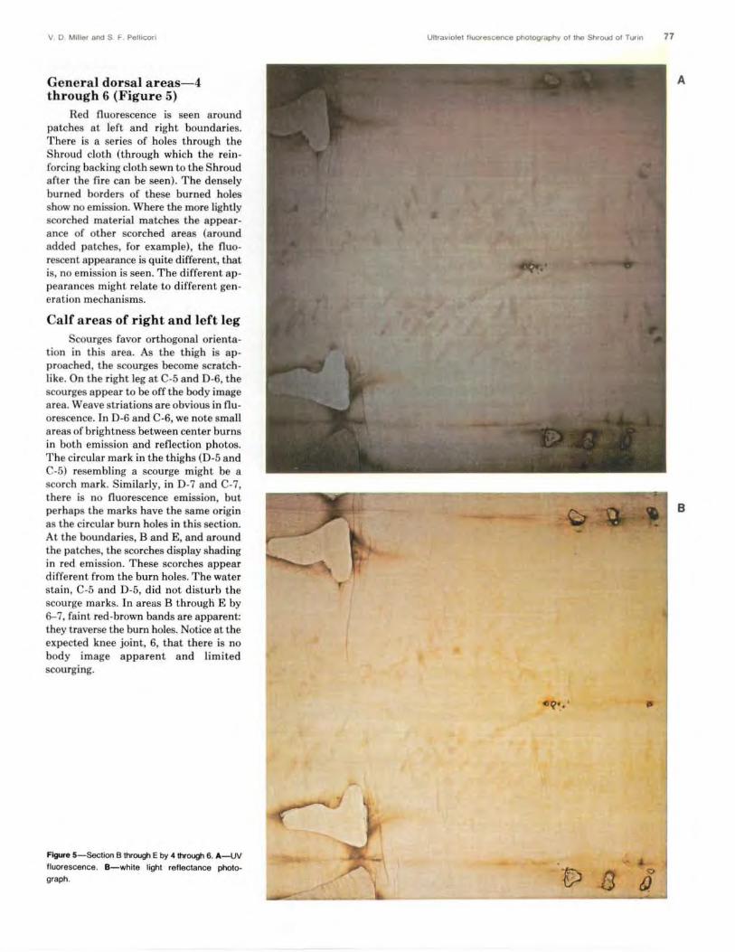
General dorsal areas- 4 through 6 (Figure 5)
Red fluorescence is seen around patches at left and right bounda ries. There is a series of holes through t he Shroud cloth (through which t he reinforcing backing cloth sewn to the Shroud after the fire can be seen). The densely burned borders of these burned holes show no emission. Where the more lightly scorched materiaJ matches the appearance of other scorched areas (around added patches, for example), the fluo rescent appearance is quite different, that is, no emission is seen. The different appea rances might relate to different generation mechanisms.
Calf areas of right and left leg Scourges favor orthogonal orienta
tion in this area. As the thigh is approached, the scourges become scratchlike. On the right leg at C-5 and D-6, the scourges appear to be off the body image area. Weave striations are obvious in flu orescence. In D-6 and C-6, we note small areas of brightness between center burns in both em ission and reflection photos. The circular mark in the thighs (D-5 and C-5) resembling a scourge might be a scorch mark. Similarly, in D-7 and C-7, there is no fluorescence emission, but perhaps the marks have the same origin as the circular burn holes in this section. At the boundaries, Band E, and a round the patches, the scorches display shading in red emission. These scorches appear different from the burn holes. The water stain, C-5 and D-5, did not disturb the scourge marks. In areas B t hrough E by 6-7, faint red-brown bands are apparent: they traverse the burn holes. Notice at the expected knee joint, 6, that there is no body image apparent and limited scourging.
Figure 5-Section B through E by 4 through 6. A-UV
fluorescence. 8 -white light reflectance photograph.
Ultra"1ole1 lluorescence photography of the Shroud o l Turin 77
A
B
78 Journal of Biological Photography Vol. 49. No. 3 , July. 1981
A
B
•
Q
fJ. ..
V. O. Millar and S. F. Pallicori
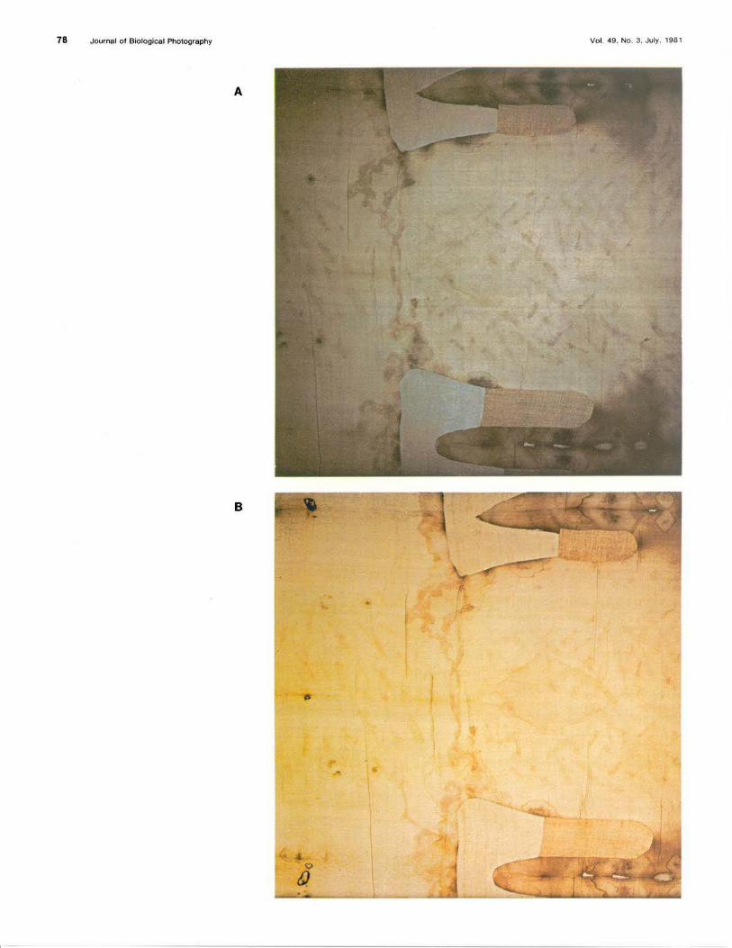
Figure 8-Section B ttrougi Eby 7 ttrougi 9. A-IN fluorescence. B-white light reflectance photograph.
Dorsal midsection a rea- 7 t hrou gh 9 (Figure 6)
All the patched burn areas fluoresce reddish brown, including the water stain front. Other white light water boundaries are indistinguishable in fluorescence. The scourges orient orthogonally and are distinctly absorbent against the fluorescent background linen. The emission of the linen is not uniform. Across the small of the back the rivulets of blood are absorbing; see also D-9 and C-9. There are, some lighter border areas associated with the blood flow. Some of the densest marks within the blood flow area might be scorches. In area D-8, the blood boundaries fade in fluorescence. At E-8 and C-8 the stains at the inner boundary of the large patches appear to be mirror images. Two small circular stains at C-7 and D-7 are mirror images and visualiy resemble weak scorches. In fluorescence these stains are absorbing. Note also lighter boundary areas associated with water marks. The body image (nonfluorescing) is very dense at the scapula and rib cage. Scourges are apparent through the scorched shoulder areas above the patches. Many scourges have fluorescing bordering areas.
"
Ultraviolet llUO<asoanoa photography of the Shroud of Tixln 79
u H M 0 • • H n 11

80 Journal of Biological Photography
Dorsal head and neck area and ventral head area- 10 through 12 by B through E (Figure 7)
At E and Band at patches 11 and 12, red fluorescing scorched areas are obvious. The brightly fluorescing yellowgreen donut-shaped areas to the right of patch 11- 12 at E and B appear to be scorches. They are mirrored elsewhere at symmetrically located fold sections. The faint water stain between the head images has light blue boundaries in fluorescence. The cloth weave striation is an apparent nonuniformity. The blood stains on the dorsal head area are bounded by brighter areas. A smudge or scourge appears to the right of the general blood flow on the dorsal head. Faint scorch pattern is visible in the light blood stains. At C-D by 11-12
A
a smudge resembling blood is visible between the head images. The dorsal body B image is more distinct than the ventral image. At the center of the dorsal head, a blue fluorescence is noted. This has a different color than the body image.
Figure 7- Section B through E by 10 through 12.
A-UV fluorescence. B-white light reflectance
photograph.
Vol. 49. No. 3. July, 1981

v. D. Miller and S. F. Pellicori Ultraviolet fluorescence photography of the Shroud of Turin 81
A
Head and chest-13 through 15 by D through C (Figure 8)
Scorches near the patches emit reddish brown fluorescence. The water stain boundary below patches at C-15 and E-15 appear to contain reddish fluorescen t materia l as from scorches, while those at C and D at 15 (abdomen) do not con tain fluorescing material. The lower left arm blood stains, B and C at 16, have ligh t border areas. Through t he scorches of B at 14 and 15, upper left arm, can be seen blood or scourges. The upper area of the left shoulder shows fa int blood , scourge and body detail. The diffusely appearing scourge marks below the central water stain, C and D at 16, become more distinct (scratch-like) near C-15 and D-15. Scourges are diagonal a nd orthogonal, wi th a scratch characteristic predominant on t he left. The face is more strongly
B bounded by the weave pat tern on its left side than on its right, C and D at 13. Going beyond this feature, no image is discernible. The blood streaks in t he hair are denser on the right side and have fluorescing boundaries, C and D at 13. In comparison wit h the general body image, t he beard and mustache are denser. ln fact, t he density is greatest on the left port ion of the beard and mustache a nd more d iffuse on t he right. A distinct boundary is present at t he lower left. The lower lip appears to have a fluorescent boundary. On the right shoulder, the blood stains are in very sharp detail, wi th the lower stain broken into dots. Compare this area with some of the scourges on the right side. Circles of yellow-green fluorescence are associated wit h t hese wounds.
Figure 8- Section B through E by 13 through 15.
A - UV fluorescence. B-white light reflectance pho
tograph.

82 Journal of B iological Photography
Ventral hands and thighsarea 16 through 19 (Figure 9)
Scorches show red fluorescence except for those associated with the sets of three holes which have heavily burned borders. Plumes of pyrolyzed materials pointing toward the feet are seen associated with the burned holes. At 19 the plume pointing toward the hands fluoresces red. Some shading from red to yellow fluorescence can be seen. The water mark above the knees at 18 has an absorbant edge with density gradations. Some fluorescing bordering can be seen also. In white light, however, this water stain is not prominent. The fluorescent color is brown as opposed to grey. The water stain situated above the series of holes to the right side has very little emission. Some of the water stains are better defined in fluorescence, others are not. The blood and body image are similar
A
in the fluorescence photos; i.e., grey and non-emitting. Notice the clear fluorescing borders around the hand wound blood stains. As is true of scourges elsewhere, the scourge wounds are more distinguishable in the fluorescence photos. As the upper thigh area is approached, B however (see 19), the scourges become more diffuse. Often scratches are contained within the diffuse areas. Some scourge marks appear only in the fluorescence photos: examples are noted between the hands and forearm areas. Around the knuckle of the left hand a slight blood flow can be seen. The body image absorption, especially arm and thigh, is greater on the left side of the body. There is a detail located where the genitals would be expected below the intersection of the hands. The dark patch below the wrist wound appears distinct from the body image. It is not understood. The fingers possess more contrast by fluorescence photography. The differences among water stain appearances might be due to differing material content, with some containing mobile pyrolysis products.
Figure 9-Section B through E by 16 through 19. A-UV fluorescence. B-whlte light reflectance
photoqraph.
Vol. 49, No. 3. July. t981
..

V. 0. Miller and S. F. Pellicori
.,
..
Ultraviolet fluorescence photography of the Shroud of Turin 83
•
A
Ventral feet, knees and thighs-19 through 22 (Figure 10)
A very faint body outline is discernible in this section, even in fl uorescence photos. The feet are not defined. The leg outl ine and scourge markings are limited by a weave line appearing blue in fl uorescent emission where the weave direction changes. This is an area of "no-print." The cent ral water stain at B and C by 18 a nd 19 is contrast-enhanced by fluorescence: it absorbs strongly against the emitting backgr<>und. While t he water stain at D-20 has fluorescing border areas, the opposing one at A-20 does not. Also, the one at D-22 has a n unusually wide absorbing boundary. As in the other scourged a reas, the scourges run d iagonally left to lower right, and both diffuse and scratch-like marks, some perpendicular to the leg, are visible. See the left leg at B-20 and 21. Some scourges appear to be bounded by the central water stain. No scourges can be found on t he ankles.
On t he feet, two blood stain areas are distinguishable on the right foot; the smaller one is denser, the la rger has a
B fluorescing border area. Curious less densely absorbing flows wit hout defini te boundary trail from the blood spots. They do not resemble the usual blood characteristics. The smal l circular mark above t he water boundary at D-22/21 has t he fluorescent colors of a scorch: red grading to a yellow border. To t he upper right a 2-inch diameter stain containing three dar k lines appears by nuorescence cont rast, but not by white light. Wax drops seen in t his area are bright. nuorescers colored yellow, or green. At the lower edge, D-22, two sharp-edged marks visible as dark brown emit brigh t white in nuorescence .
Figure 10-Section B through E by 19 through 22.
A-UV fluorescence. B-white light reflectance
photograph.

84 Journal of Biological Photography
Discussion-Laboratory experiments
•
In fluorescence photographs most of the details are visible in contrast with the Shroud linen, which itself fluoresces. With the exception of lightly scorched areas and some water stained areas, the Shroud features absorb UV energy without visible emission. Compared with modern linen, the Shroud linen fluoresces less brightly. It emits a yellow-green color. Modern linen can be artificially aged by baking at high temperatures (125°-150° C) to the point where its reflected color and fluorescent emission approach those of the Shroud.7 When foreign materials are applied to the linen, a reaction which results in locally visible darkening of the linen can be stimulated by air baking as above. These experiments7 have led to the interpretation that the body image is the result of locally accelerated dehydration/oxidation and conjugation of the cellulose molecular structure. Images produced in hours at high temperature are comparable in both reflectance and UV stimulated emittance to images produced at normal temperatures during longer time intervals (years?). Laboratory-produced images were photographed using the same equipment as was used in Turin. Exposure to sunlight of wavelengths less than 340 nm influences the rate and degree of cellulose degradation.
Laboratory data for whole blood displayed total absorption, which is in agreement with the Shroud data.
Weak scorches, which in white light have densities and colors similar to body image areas, show a significant difference with UV fluorescence photography. Scorches emit a reddish brown fluorescence while the body image is nonfluorescent. The significance of this difference is evident in relation to the suggestion that the body image was caused by con-
Vol. •9, No. 3 , July. 1981
.. u a Q a a M ~ tt
tact with a hot statue or was scorched by other means. Laboratory-produced scorches emit a bright greenish-yellow fluorescence if they were produced in air and reddish if produced under conditions of limited available oxygen. The scorches associated with the fire of 1532, during which the Shroud was involved, attest to the rapid consumption of the available oxygen. 12 Their reddish emission is probably due to furfurals, which can be produced under such conditions.
Linen lightly scorched by a soldering iron in air shows the green-yellow emission, often distributed in plumes of deposited pyrolysis products. We demonstrated in one experiment that the material of the plumes could be transported by water, but the underlying scorched cellulose retained a bright yellow-green fluorescence. This demonstration together with the observed absence of body image fluorescence is strong evidence against the cause for the body image being a scorch.
A quite frequent hypothesis is that the body image was painted. The binders used in paints as early as the 14th century would be made of proteinaceous materials, animal collagen being a favored material; egg white and gelatin are other examples. But these collagens would inherently be contaminated by fluorescing amino acids. 13 Random sampling of book bindings and illustrations from the 14th and 15th centuries were observed to emit bright fluorescence when excited by long-wave mercury vapor lamp excitation. Microchemical analyses on fibrils ret rieved from the Shroud have shown the absence of paints, pigments, stains, dyes and protein in body image areas.14
Conclusion The sharp detail revealed for the first
time, particularly in the scourges, suggests that intimate cloth-body contact occurred. The detail (and contrast) is only slightly less prominent on the front than

V. D. Miller and S. F. Pelllcorl
on the dorsal image, indicating that the large difference in weight for each side had only a minor influence on the imprinting of the scourges. This observation is contrary to what might be intuitively expected, and it might be a clue to some future understanding of the image production mechanism.
The occurrence of contact could also have transferred substances present on the skin to the cloth where they stimulated the cellulose alteration process and caused an image of the body to develop as local darkening of the linen.7
Hypotheses such as a scorch cause or paint are contradicted by the fluorescence photography results.
UV fluorescence photography has revealed some near-invisible details, many of which require explanation. For example, the pattern of distinct burn holes has characteristics unlike the burn damage attributed to the 1532 fire. Areas in the weave where the image density abruptly decreases (e.g., sides of the face) might actually contain very faint images which possibly could be retrieved by using stimulating radiation of shorter wavelengt hs. The property of the linen thread that didn't develop image density should also be discovered. The 8-cm side strip running the length of the Shroud shows weft bands that are continuous with the main body of the Shroud. Similar appearances result from backlighting and low-energy radiography. The suggestion is that this strip was not separated from the main body over its entire length. Many of these unexplained details might relate to the history of the Shroud.
Another feature requiring explanation is the lighter bordering area seen with many bloodstained a reas. The interpretation is that blood serum is present. It. might have acted to retard the image development reactions associated with the body image. Fibrils from many water stain fronts and from the area be-
Ultraviolet fluorescence photography of the Shroud of Turin 85
tween the head images contain blood particles.14 Further discussions are in preparation.15
Acknowledgments We appreciate the assistance given
by D. Devan and other members of the STURP team, and also acknowledge the cooperation extended by the church officials in Turin. J. Druzik, Conservation Center, Los Angeles County Museum of Arts, critically reviewed the manuscript.
About the authors- Vernon Miller is Head of t he Scientific and Industrial Photography Department at the Brooks Institute of Photography, Santa Barbara, California. He was the chief scientific photographer for the ST URP team in Turin, and has employed a variety of image processing techniques to the photographic data obtained.
Samuel Pellicori is an optical physicist with the Santa Barbara Research Center. He has been evaluating and simulating in the laboratory some of the spectrophotometric, fluorometric and microscopic data he collected in Turin. Address correspondence to Samuel Pellicori, Santa Barbara Research Center, 75 Coromar Drive, Goleta, California 93017,
References
1. Proceedings of the 1977 U.S. Conference of Research on the Shroud of Turin, Holy Shroud Guild, 294 E. 150 St., Bronx, NY 10451.
2. Kenneth F. Weaver, "The Mystery of the Shroud," National Geographic 157, 730 (1980).
3. B. J. Culliton, "Science Investigates the Shroud of Turin," Science 201, 235 (1978).
4. E. J. Jumper and R. W. Mottern, "Scientific Investigation of the Shroud of Turin," Appl. Opt. 19, 1909 (1980).
5. R. A. Morris, L. A. Schwalbe and J. R. London, "X-Ray Fluorescence l nvesti-
gation of the Shroud of Turin," X-Ray Spectrometry 9, No. 2, 40 (1980).
6. Samuel Pellicori and Mark S. Evans, "The Shroud of Turin Through the Microscope," Archaeology 34, 34, Jan/Feb 1981.
7. S. F. Pellicori, "Spectral Properties of the Shroud of Turin," Applied Optics 19, 1913 (1980).
8. Roger Gilbert, Jr., and Marion M. Gilbert, "Ultraviolet-Visible Reflectance and Fluorescence Spectra of the Shroud of Turin," Applied Optics 19, 1930 (1980).
9. V. Miller and D. Lynn, "Photography of the Turin Shroud," Science and Technology, Feb. 1981 (in Dutch).
10. J. S. Accetta and J. S. Baumgart, "Infrared Reflectance Spectroscopy and Thermographic Investigations of the Shroud of Turin," Appl. Opt. 19, 1921 (1980).
11. S. F. Pellicori, "Transmittances of Some Optical Materials for Use Between 1900 and 3400 A," Appl. Opt. 3, 361 (1964).
12. R. N. Rogers in Reference 1, p. 133. 13. Eric J. Jumper, John P. Jackson, John H.
Heller, Alan D. Adler, Samuel F. Pellicori and Raymond N. Rogers, "A Comprehensive Examination of the Various Stains and Images on the Shroud of Turin," American Chem. Soc. Proc. on Archaeological Chem. (1982). in preparation.
14. J. H. Heller and A. D. Adler, "A Chemical Investigation of the Shroud of Turin," submitted to Journal of Forensic Sciences.
15. L.A. Schwalbe and R. N. Rogers, "Physics and Chemistry of the Shroud of Turin: Summary of the 1978 Investigation," in preparation.