ULTRASTRUCTURE OF HUMAN LEUKOCYTES AFTER SIMULTANEOUS FIXATION
13
ULTRASTRUCTURE OF HUMAN LEUKOCYTES AFTER SIMULTANEOUS FIXATION WITH GLUTARALDEHYDE AND OSMIUM TETROXIDE AND "POSTFIXATION" IN URANYL ACETATE JAMES G. HIRSCH and MARTHA E. FEDORKO From The Rockefeller University, New York 10021 ABSTRACT Human leukocytes in suspension or in monolayer cultures have been processed for electron microscopy by fixation in a freshly made cold mixture of glutaraldehyde and osmium tetrox- ide and by "postfixation" in uranyl acetate. Simultaneous exposure to glutaraldehyde and osmium tetroxide eliminates many of the shortcomings seen when either of these agents is used alone as the initial fixative. Specimens are processed to the stage of dehydration as single cell suspensions or as very small clumps to assure rapid penetration of fixatives and efficient washing. The technique is rapid and reproducible. Electron micrographs presented in this report illustrate the ultrastructural features of human white cells prepared by this method. Neutrophilic polymorphonuclear leukocytes are commonly not well preserved by standard meth- ods of fixation for electron microscopy. Cold osmium tetroxide produces in these cells variable destruction in their cytoplasmic matrix and other damage probably attributable to oxidative effects or to inadequate fixation of proteins. Neutrophils fixed with glutaraldehyde followed by osmium tetrox- ide frequently show mottled granules, occasional myelin figures, and poorly defined membranes, findings which may reflect some degree of autolysis or lipid extraction during the aldehyde exposure. We report here a method for obtaining improved fixation of neutrophils and of other white blood cells. Essential features of the method are the fol- lowing: (a) the cells are fixed and processed to the stage of dehydration as single cells in suspension or as very small clumps; (b) initial fixation is ac- complished with a freshly made mixture of glu- taraldehyde and osmium tetroxide, similar to that employed by Trump and Bulger (1); (c) the cells are postfixed' by suspension in uranyl acetate so- Postfixation in uranyl acetate may be incorrect terminology since uranyl ions are probably acting lution modified from Kellenberger (2). This "mixed fixative," as we call it, has been applied, with good results, to several other types of cells in suspension or in monolayer cultures, some of which were illustrated in separate reports (3, 4). MATERIALS AND METHOD Collection of Leukocytes Venous blood from healthy adults was collected with minimal trauma in a plastic syringe and imme- diately mixed with heparin (Connaught Medical Research Lab., Toronto, Canada) to give a final con- centration of 0.1 mg/ml. The heparinized blood was mixed with an equal volume of similarly heparinized 2% clinical dextran (Abbott Laboratories, North Chicago, 11.) in physiologic saline. The tube was slanted at 45 ° and kept at room temperature for 45 min to allow red cell sedimentation. The opalescent cell- and platelet-rich supernatant plasma layer was then collected and centrifuged at room temperature primarily as a stain rather than as a fixative. The term will nevertheless be used since it signifies clearly the technical procedure and avoids confusion with the separate later step of staining of the sections with uranyl acetate. 615 on April 5, 2019 jcb.rupress.org Downloaded from http://doi.org/10.1083/jcb.38.3.615 Published Online: 1 September, 1968 | Supp Info:
Transcript of ULTRASTRUCTURE OF HUMAN LEUKOCYTES AFTER SIMULTANEOUS FIXATION
ULTRASTRUCTURE OF HUMAN LEUKOCYTES
AFTER SIMULTANEOUS FIXATION WITH
GLUTARALDEHYDE AND OSMIUM TETROXIDE
AND "POSTFIXATION" IN URANYL ACETATE
JAMES G. HIRSCH and MARTHA E. FEDORKO
From The Rockefeller University, New York 10021
ABSTRACT
Human leukocytes in suspension or in monolayer cultures have been processed for electronmicroscopy by fixation in a freshly made cold mixture of glutaraldehyde and osmium tetrox-ide and by "postfixation" in uranyl acetate. Simultaneous exposure to glutaraldehyde andosmium tetroxide eliminates many of the shortcomings seen when either of these agents isused alone as the initial fixative. Specimens are processed to the stage of dehydration as singlecell suspensions or as very small clumps to assure rapid penetration of fixatives and efficientwashing. The technique is rapid and reproducible. Electron micrographs presented in thisreport illustrate the ultrastructural features of human white cells prepared by this method.
Neutrophilic polymorphonuclear leukocytes arecommonly not well preserved by standard meth-ods of fixation for electron microscopy. Coldosmium tetroxide produces in these cells variabledestruction in their cytoplasmic matrix and otherdamage probably attributable to oxidative effects orto inadequate fixation of proteins. Neutrophils fixedwith glutaraldehyde followed by osmium tetrox-ide frequently show mottled granules, occasionalmyelin figures, and poorly defined membranes,findings which may reflect some degree of autolysisor lipid extraction during the aldehyde exposure.
We report here a method for obtaining improvedfixation of neutrophils and of other white bloodcells. Essential features of the method are the fol-lowing: (a) the cells are fixed and processed to thestage of dehydration as single cells in suspensionor as very small clumps; (b) initial fixation is ac-complished with a freshly made mixture of glu-taraldehyde and osmium tetroxide, similar to thatemployed by Trump and Bulger (1); (c) the cellsare postfixed' by suspension in uranyl acetate so-
Postfixation in uranyl acetate may be incorrectterminology since uranyl ions are probably acting
lution modified from Kellenberger (2). This"mixed fixative," as we call it, has been applied,with good results, to several other types of cells insuspension or in monolayer cultures, some ofwhich were illustrated in separate reports (3, 4).
MATERIALS AND METHOD
Collection of LeukocytesVenous blood from healthy adults was collected
with minimal trauma in a plastic syringe and imme-diately mixed with heparin (Connaught MedicalResearch Lab., Toronto, Canada) to give a final con-centration of 0.1 mg/ml. The heparinized blood wasmixed with an equal volume of similarly heparinized2% clinical dextran (Abbott Laboratories, NorthChicago, 11.) in physiologic saline. The tube wasslanted at 45° and kept at room temperature for 45min to allow red cell sedimentation. The opalescentcell- and platelet-rich supernatant plasma layer wasthen collected and centrifuged at room temperature
primarily as a stain rather than as a fixative. The termwill nevertheless be used since it signifies clearly thetechnical procedure and avoids confusion with theseparate later step of staining of the sections withuranyl acetate.
615
on April 5, 2019jcb.rupress.org Downloaded from http://doi.org/10.1083/jcb.38.3.615Published Online: 1 September, 1968 | Supp Info:
for 5 min at 200 g to deposit the white cells whileleaving most of the platelets in suspension. Sedi-mented leukocytes were fixed directly or were washedonce in heparinized saline and suspended in 10-20%autologous serum-Hanks' solution for short term(1-2 hr) culture in plastic Petri dishes or glass Tflasks.
The Fixation, Staining, and
Washing Solutions
Stock solutions of 2.5% glutaraldehyde (FischerScientific Co., Fairlawn, New Jersey) in 0.1 M caco-dylate pH 7.4 and of 1%7 osmium tetroxide in 0.1 Mcacodylate pH 7.4 were maintained separately at 40C.These were brought to 0°C in an ice bath and mixed,one part glutaraldehyde plus two parts osmium tetrox-ide, within an hour of use. Under these conditionssuch a mixture remained clear and colorless or faintlytan during the fixation.
Ice cold physiologic saline was used for washing.Uranyl acetate (0.25% in 0.1 M acetate buffer at
pH 6.3) was employed for postfixation of the cells insuspension.
The Fixation and Processing Procedure
The sedimented leukocytes at room temperaturefrom 1 ml (approximately 5 X 10⁶ cells or 5-mm3
packed cells) of the dextran supernatant cell-richplasma were suspended in 2 ml of the cold, mixed fix-ative. After approximately 2 min, the suspension wastransferred to a 3 ml conical centrifuge tube and spunin a tabletop clinical centrifuge (International Equip-ment Co. Needham Heights, Mass.) at 300 g for 1min. The supernatant fluid was decanted and aspiratedwith a Pasteur pipette. The pellet was chilled brieflyon ice, was resuspended in another 2 ml of mixed fix-ative, and was allowed to stand on ice for 10-30 min.Centrifugation (as above) was followed by two washesin cold saline; each involved gentle suspension of thecells with a Pasteur pipette and then prompt sedimen-tation in the centrifuge. The pellet was next suspendedin uranyl acetate solution and allowed to stand at0°C for 15-30 min. Two saline washes (as above)followed. The cell pellet was warmed in a 50°C waterbath for 5 min and then suspended in a few drops of2% Noble agar which had been boiled or autoclavedand allowed to cool to 50°C in a water bath. Carehad to be taken to transfer the agar with a warmPasteur pipette to avoid solidification. The cell sus-pension in fluid agar was centrifuged at 750 g for 2min in a carrier half-filled with hot tap water. (Ifsolidification of the agar had been avoided, the cellsformed a firm pellet at the bottom of the tube; other-wise, they were distributed in a fuzzy zone. In thelatter event the tube could be heated in a steam bathuntil the agar had melted, and then it could be re-centrifuged. Such steaming of fixed cells had little or
no discernible effect on their ultrastructure.) The tubewas cooled in ice to solidify the agar. After addition of70%fC alcohol and further standing on ice for an houror more, the agar filling the conical tip of the tubecould then be displaced by careful pipetting, and theblack cell pellet was trimmed to small blocks for de-hydration and embedding by standard methods (5).
Cells cultured in vitro on glass or plastic surfaceswere fixed by draining off the fluid culture mediumand flooding the cell layer with a freshly made, coldmixture of osmium tetroxide and glutaraldehyde.The cells were then gently scraped from the surfacewith a plastic policeman. This cell suspension wastransferred to a 3 mi conical tube and centrifuged at300 g for 1 min. The supernatant was thoroughly re-moved, and the pellet was chilled on ice, suspended ina fresh aliquot of mixed fixative, and kept on ice for15-30 min. Washing, postfixation in uranyl acetate,and subsequent processing were as those describedabove for blood leukocytes.
Thin sections were cut with diamond knives on aPorter-Blum microtome and picked up on 200-meshgrids coated with thin layers of Formvar and carbon.Sections were stained with uranyl acetate and leadcitrate (6) and were examined and photographed in aSiemens Elmiskop I at 80 kv with a 50 A[ objectiveaperture.
RESULTS
In our early attempts to employ a mixture of
glutaraldehyde and osmium tetroxide as a fixative,
we observed, as have others (1), that the two fixa-
tives interacted to produce a brownish purple
product; this reaction was more rapid and exten-sive at higher temperatures and apparently was
accelerated by the presence of organic material in
the specimen. Although in many instances well-
preserved cells were obtained after exposure to
these discolored fixative mixtures, the results werevariable. We found that cold stock solutions of
glutaraldehyde and osmium tetroxide, both in
cacodylate buffer at pH 7.4, could be mixed andkept at 0° C for 1 hr with little or no discolorationand no precipitation, and that this mixture gaverapid, reproducible fixation of various cell types.
In order to avoid possible change in the shape
of the cells or damage to them by chilling or wash-ing prior to fixation, the cell pellet or culture, atroom or incubator temperature, was suspended incold fixative; after the suspension had stood for ap-proximately 2 min, the cells were collected as apellet, the supernatant was discarded, and thepellet was chilled thoroughly and then suspendedin a fresh aliquot of the mixed fixative and heldat 0° C. Studies were made on the relative concen-
616 THE JOUIRNAL OF CELL BIOLOGY VOLUME 38, 1968
trations of osmium tetroxide and glutaraldehydein the mixture and on the optimal duration of fixa-tion. These concentrations and times were foundnot to be critical. Similar fixation was obtainedwith mixtures ranging from 0.5 to 2.5% of eachof the components, and no differences were ap-parent after fixation times ranging from 10 minto 1 hr. Care was required not to use too large aquantity of cells for the volume of mixed fixative.Overloading with cells led to discoloration of thesolution during fixation and to variability in re-sults. It was essential to suspend the cells in pre-mixed fixative solution; cells that were exposedfirst for only 1 min to one of the reagents and thento the other reagent by its addition to the solutionexhibited features of sequential rather thansimultaneous fixation.
Early experience with the mixed fixative estab-lished that its preservative qualities were good, butthat membranes were poorly defined. Applicationof a modified Kellenberger technique for stainingof the fixed cells by suspension in uranyl acetatesolution resulted in striking improvement in mem-brane definition. Postfixation in uranyl acetate forperiods ranging from 10 to 30 min gave essentiallyidentical results. The usefulness of uranyl acetatepostfixation was related, in some unknown man-ner, to the initial treatment of the cells with mixedfixative; exposure to uranyl acetate of cells se-quentially fixed with glutaraldehyde and osmiumtetroxide did not significantly improve the poormembrane definition.
This mixed fixation procedure thus far has beenapplied with success to human white blood cells,to peritoneal cells of mice, and to tissue culturemonolayers of mouse macrophages and L cells.Figs. 1-11 illustrate general and detailed featuresof the ultrastructure of human white blood cellsprocessed by this method.
A low power view of a blood monocyte is shownin Fig. 1. The monocyte nucleus is commonlyhoreshoe-shaped, with extensive, light-staining,central nucleoplasm and dense, granular, pe-ripheral chromatin. Nucleoli are rarely seen.Perinuclear filament bundles (Figs. 1 and 3) areoften present in monocytes. In transverse section(not shown here) these bundles are clearly com-posed of filaments, not tubules. The Golgi appa-ratus is well developed and often multicentric.Small vesicles (50-200 mg in diameter) arescattered throughout the cytoplasm but are es-pecially numerous in the Golgi region. Thesevesicles can be classified into three general types:
(a) a few showing surface spikes typical of coated
vesicles; (b) some larger vesicles with an electron-
lucent content, probably of pinocytic origin; and
(c) many vesicles with a smooth surface and a
matrix density similar to that of cytoplasmicground substance, most likely representing ele-
ments derived from the Golgi apparatus or fromsmooth endoplasmic reticulum (ER). Monocyte
mitochondria (Figs. 1 and 2), commonly located
in the peripheral cytoplasm, are round or elon-
gated with a dense matrix and well-defined cris-
tae. Variable numbers of small (100-200 m), oval
or rod-shaped, dense bodies are seen in the peri-nuclear region of most monocytes. Usually only a
few short strips of slightly dilated, partly rough andpartly smooth ER are present in the peripheral
cytoplasm. The cytoplasmic ground substance is
moderately dense and has a microgranular appear-
ance. The plasma and nuclear membranes (Figs. 1
and 2) are well defined and generally smooth. Insome instances a faint layer of amorphous material
appears to be attached to the outer surface of the
cell limiting membrane. At high magnification the
cell surface membrane shows a typical trilaminarunit structure (Fig. 2).
Human blood lymphocytes processed by the
mixed fixation technique are illustrated at low
magnification in Fig. 4. Lymphocyte nuclei are
oval with indentations or kidney-shaped and show
a chromatin distribution of approximately equal
amounts of centrally located light-staining ma-
terial and of peripheral dense-staining granular
elements. Some sections of lymphocytes showprominent nucleoli (Fig. 5) which usually are
composed of three zones: an outer zone consisting
of loosely packed granules identical in appearanceto aggregated nuclear chromatin; a central core
of tightly packed smaller granules (nucleolargranular area); and electron-opaque, nongranular
material (fibrillar zone) coursing around and
through the central zone. Also demonstrated inFig. 5 is the good preservation of nuclear mem-branes and pores. The cytoplasm of lymphocytesis relatively sparse. Centrioles are frequently seen
and appear to be well fixed. Several oval or rod-
shaped mitochondria are typically grouped in arosette pattern about the centrosomal region in thelymphocyte. The Golgi apparatus is poorly de-
veloped or not seen, and only small numbers ofcytoplasmic vesicles are present. A common note-worthy feature, however, is the occurrence oflarge (300-400 m in diameter) multivesicularbodies (Figs. 4 and 6) distributed among the mito-
J. G. HIRSCH AND M. E. FEDORKO Human Leukocyte Ultrastructure 617
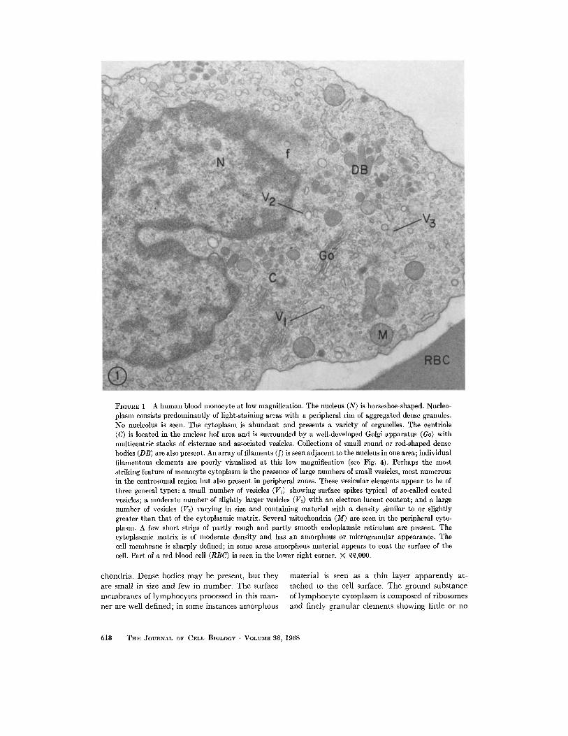
FIGURE 1 A human blood monocyte at low magnification. The nucleus (N) is horseshoe-shaped. Nucleo-plasm consists predominantly of light-staining areas with a peripheral rim of aggregated dense granules.No nucleolus is seen. The cytoplasm is abundant and presents a variety of organelles. The centriole(C) is located in the nuclear hof area and is surrounded by a well-developed Golgi apparatus (Go) withmulticentric stacks of cisternae and associated vesicles. Collections of small round or rod-shaped densebodies (DB) are also present. An array of filaments (f) is seen adjacent to the nucleus in one area; individualfilamentous elements are poorly visualized at this low magnification (see Fig. 4). Perhaps the moststriking feature of monocyte cytoplasm is the presence of large numbers of small vesicles, most numerousin the centrosomal region but also present in peripheral zones. These vesicular elements appear to be ofthree general types: a small number of vesicles (VI) showing surface spikes typical of so-called coatedvesicles; a moderate number of slightly larger vesicles (V2) with an electron-lucent content; and a largenumber of vesicles (V3) varying in size and containing material with a density similar to or slightlygreater than that of the cytoplasmic matrix. Several mitochondria (M) are seen in the peripheral cyto-plasm. A few short strips of partly rough and partly smooth endoplasmic reticulum are present. Thecytoplasmic matrix is of moderate density and has an amorphous or microgranular appearance. Thecell membrane is sharply defined; in some areas amorphous material appears to coat the surface of thecell. Part of a red blood cell (RBC) is seen in the lower right corner. X ,000.
chondria. Dense bodies may be present, but they material is seen as a thin layer apparently at-
are small in size and few in number. The surface tached to the cell surface. The ground substance
membranes of lymphocytes processed in this man- of lymphocyte cytoplasm is composed of ribosomes
ner are well defined; in some instances amorphous and finely granular elements showing little or no
618 THE JOURNAL OF CELL BIOLOGY · VOLUME 38, 1968
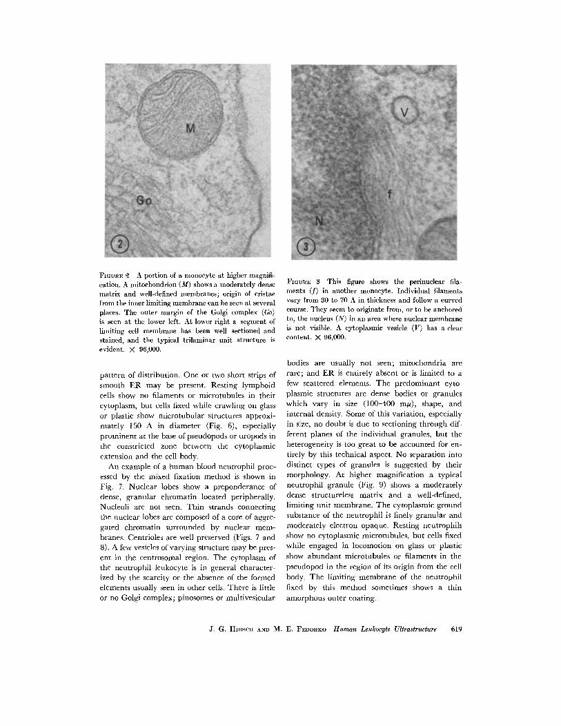
FIGURE A portion of a monocyte at higher magnifi-cation. A mitochondrion (M) shows a moderately densematrix and well-defined membranes; origin of cristaefrom the inner limiting membrane can be seen at severalplaces. The outer margin of the Golgi complex (Go)is seen at the lower left. At lower right a segment oflimiting cell membrane has been well sectioned andstained, and the typical trilaminar unit structure isevident. X 96,000.
pattern of distribution. One or two short strips of
smooth ER may be present. Resting lymphoidcells show no filaments or microtubules in their
cytoplasm, but cells fixed while crawling on glassor plastic show microtubular structures approxi-mately 150 A in diameter (Fig. 6), especiallyprominent at the base of pseudopods or uropods in
the constricted zone between the cytoplasmicextension and the cell body.
An example of a human blood neutrophil proc-essed by the mixed fixation method is shown in
Fig. 7. Nuclear lobes show a preponderance ofdense, granular chromatin located peripherally.
Nucleoli are not seen. Thin strands connectingthe nuclear lobes are composed of a core of aggre-gated chromatin surrounded by nuclear mem-
branes. Centrioles are well preserved (Figs. 7 and8). A few vesicles of varying structure may be pres-
ent in the centrosomal region. The cytoplasm ofthe neutrophil leukocyte is in general character-ized by the scarcity or the absence of the formedelements usually seen in other cells. There is littleor no Golgi complex; pinosomes or multivesicular
FImGRE 3 This figure shows the perinuclear fila-ments (f) in another monocyte. Individual filamentsvary from 30 to 70 A in thickness and follow a curvedcourse. They seem to originate from, or to be anchoredto, the nucleus (N) in an area where nuclear membraneis not visible. A cytoplasmic vesicle (V) has a clearcontent. X 96,000.
bodies are usually not seen; mitochondria arerare; and ER is entirely absent or is limited to afew scattered elements. The predominant cyto-
plasmic structures are dense bodies or granuleswhich vary in size (100-400 m), shape, andinternal density. Some of this variation, especiallyin size, no doubt is due to sectioning through dif-ferent planes of the individual granules, but theheterogeneity is too great to be accounted for en-
tirely by this technical aspect. No separation into
distinct types of granules is suggested by theirmorphology. At higher magnification a typicalneutrophil granule (Fig. 9) shows a moderatelydense structureless matrix and a well-defined,limiting unit membrane. The cytoplasmic groundsubstance of the neutrophil is finely granular and
moderately electron opaque. Resting neutrophilsshow no cytoplasmic microtubules, but cells fixed
while engaged in locomotion on glass or plastic
show abundant microtubules or filaments in the
pseudopod in the region of its origin from the cell
body. The limiting membrane of the neutrophil
fixed by this method sometimes shows a thin
amorphous outer coating.
J. G. HIRSCH AND M. E. FEDORKO Human Leukocyte Ultrastructure 619
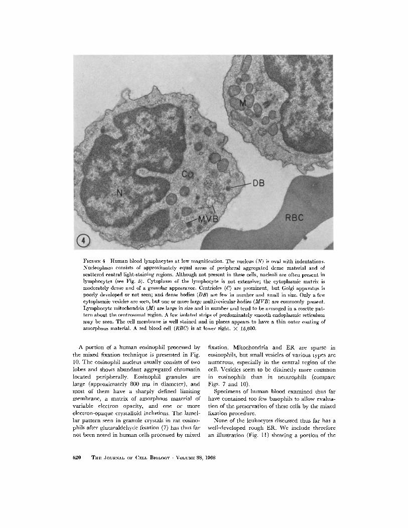
FIGURE 4 Human blood lymphocytes at low magnification. The nucleus (N) is oval with indentations.Nucleoplasm consists of approximately equal areas of peripheral aggregated dense material and ofscattered central light-staining regions. Although not present in these cells, nucleoli are often present inlymphocytes (see Fig. 5). Cytoplasm of the lymphocyte is not extensive; the cytoplasmic matrix ismoderately dense and of a granular appearance. Centrioles (C) are prominent, but Golgi apparatus ispoorly developed or not seen; and dense bodies (DB) are few in number and small in size. Only a fewcytoplasmic vesicles are seen, but one or more large multivesicular bodies (MVB) are commonly present.Lymphocyte mitochondria (M) are large in size and in number and tend to be arranged in a rosette pat-tern about the centrosomal region. A few isolated strips of predominately smooth endoplasmic reticulummay be seen. The cell membrane is well stained and in places appears to have a thin outer coating ofamorphous material. A red blood cell (RBC) is at lower right. X 15,600.
A portion of a human eosinophil processed by
the mixed fixation technique is presented in Fig.10. The eosinophil nucleus usually consists of twolobes and shows abundant aggregated chromatinlocated peripherally. Eosinophil granules arelarge (approximately 800 m/y in diameter), andmost of them have a sharply defined limitingmembrane, a matrix of amorphous material ofvariable electron opacity, and one or moreelectron-opaque crystalloid inclusions. The lamel-lar pattern seen in granule crystals in rat eosino-phils after glutaraldehyde fixation (7) has thus farnot been noted in human cells processed by mixed
fixation. Mitochondria and ER are sparse ineosinophils, but small vesicles of various types arenumerous, especially in the central region of thecell. Vesicles seem to be distinctly more commonin eosinophils than in neutrophils (compareFigs. 7 and 10).
Specimens of human blood examined thus farhave contained too few basophils to allow evalua-tion of the preservation of these cells by the mixedfixation procedure.
None of the leukocytes discussed thus far has awell-developed rough ER. We include thereforean illustration (Fig. 11) showing a portion of the
620 THE JOURNAL OF CELL BIOLOGY VOLUME 38, 1968
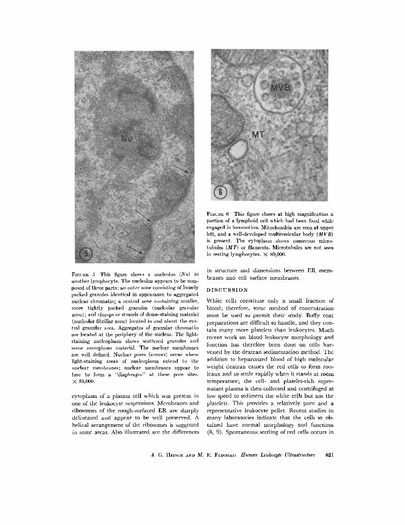
FIGURE 6 This figure shows at high magnification aportion of a lymphoid cell which had been fixed whileengaged in locomotion. Mitochondria are seen at upperleft, and a well-developed multivesicular body (MVB)is present. The cytoplasm shows numerous micro-tubules (MT) or filaments. Microtubules are not seenin resting lymphocytes. X 80,000.
FIGuRE 5 This figure shows a nucleolus (Nu) inanother lymphocyte. The nucleolus appears to be com-posed of three parts: an outer zone consisting of looselypacked granules identical in appearance to aggregatednuclear chromatin; a central zone containing smaller,more tightly packed granules (nucleolar granularzone); and clumps or strands of dense-staining material(nucleolar fibrillar zone) located in and about the cen-tral granular area. Aggregates of granular chromatinare located at the periphery of the nucleus. The light-staining nucleoplasm shows scattered granules andsome amorphous material. The nuclear membranesare well defined. Nuclear pores (arrows) occur wherelight-staining areas of nucleoplasm extend to thenuclear membranes; nuclear membranes appear tofuse to form a "diaphragm" at these pore sites.X 35,000.
cytoplasm of a plasma cell which was present in
one of the leukocyte suspensions. Membranes andribosomes of the rough-surfaced ER are sharplydelineated and appear to be well preserved. Ahelical arrangement of the ribosomes is suggestedin some areas. Also illustrated are the differences
in structure and dimensions between ER mem-
branes and cell surface membranes.
DISCUSSION
White cells constitute only a small fraction ofblood; therefore, some method of concentration
must be used to permit their study. Buffy coatpreparations are difficult to handle, and they con-tain many more platelets than leukocytes. Much
recent work on blood leukocyte morphology andfunction has therefore been done on cells har-
vested by the dextran sedimentation method. The
addition to heparinized blood of high molecularweight dextran causes the red cells to form rou-
leaux and to settle rapidly when it stands at room
temperature; the cell- and platelet-rich super-natant plasma is then collected and centrifuged at
low speed to sediment the white cells but not theplatelets. This provides a relatively pure and arepresentative leukocyte pellet. Recent studies inmany laboratories indicate that the cells so ob-
tained have normal morphology and functions(8, 9). Spontaneous settling of red cells occurs in
J. G. HIRSCH AND M. E. FEDORKO Human Leukocyte Ultrastructure 621
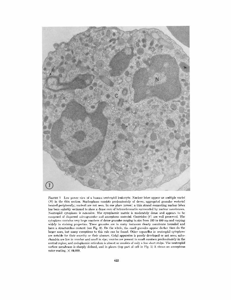
FIGURE 7 Low power view of a human neutrophil leukocyte. Nuclear lobes appear as multiple nuclei(N) in the thin section. Nucleoplasm consists predominately of dense, aggregated granular materiallocated peripherally; nucleoli are not seen. In one place (arrow) a thin strand connecting nuclear lobeshas been suitably sectioned to show a dense core of heterochromatin surrounded by nuclear membranes.Neutrophil cytoplasm is extensive. The cytoplasmic matrix is moderately dense and appears to becomposed of dispersed ilicrogranular and amorphous material. Centrioles (C) are well preserved. Thecytoplasm contains very large numbers of dense granules ranging in size from 100 to 400 my and varyingwidely in staining properties. These granules are in many instances clearly membrane bounded andhave a structureless content (see Fig. 9). On the whole, the small granules appear darker than do thelarger ones, but many exceptions to this rule can be found. Other organelles in neutrophil cytoplasmare notable for their scarcity or their absence. Golgi apparatus is poorly developed or not seen; mito-chondria are few in number and small in size; vesicles are present in small numbers predominately in thecentral region; and endoplasmic reticulum is absent or consists of only a few short strips. The neutrophilsurface membrane is sharply defined, and in places (top part of cell in Fig. 7) it shows an amorphousouter coating. X 4,000.
622
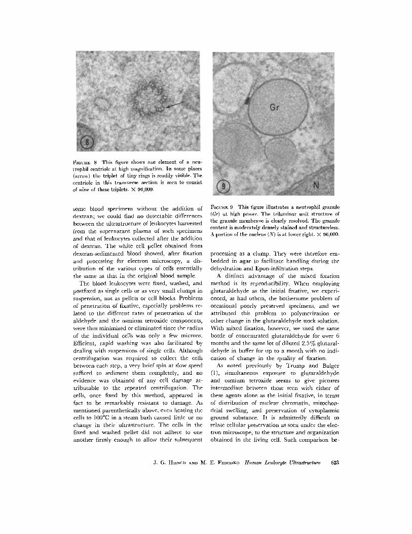
FIGURE 8 This figure shows one element of a neu-trophil centriole at high magnification. In some places(arrow) the triplet of tiny rings is readily visible. Thecentriole in this transverse section is seen to consistof nine of these triplets. X 96,000.
some blood specimens without the addition ofdextran; we could find no detectable differences
between the ultrastructure of leukocytes harvestedfrom the supernatant plasma of such specimens
and that of leukocytes collected after the addition
of dextran. The white cell pellet obtained from
dextran-sedimented blood showed, after fixation
and processing for electron microscopy, a dis-
tribution of the various types of cells essentially
the same as that in the original blood sample.The blood leukocytes were fixed, washed, and
postfixed as single cells or as very small clumps in
suspension, not as pellets or cell blocks. Problems
of penetration of fixative, especially problems re-
lated to the different rates of penetration of the
aldehyde and the osmium tetroxide components,were thus minimized or eliminated since the radius
of the individual cells was only a few microns.
Efficient, rapid washing was also facilitated by
dealing with suspensions of single cells. Although
centrifugation was required to collect the cellsbetween each step, a very brief spin at slow speed
sufficed to sediment them completely, and no
evidence was obtained of any cell damage at-
tributable to the repeated centrifugation. Thecells, once fixed by this method, appeared in
fact to be remarkably resistant to damage. Asmentioned parenthetically above, even heating thecells to 1000 C in a steam bath caused little or no
change in their ultrastructure. The cells in thefixed and washed pellet did not adhere to one
another firmly enough to allow their subsequent
FIGURE 9 This figure illustrates a neutrophil granule(Gr) at high power. The trilaminar unit structure ofthe granule membrane is clearly resolved. The granulecontent is moderately densely stained and structureless.A portion of the nucleus (N) is at lower right. X 96,000.
processing as a clump. They were therefore em-bedded in agar to facilitate handling during thedehydration and Epon-infiltration steps.
A distinct advantage of the mixed fixationmethod is its reproducibility. When employingglutaraldehyde as the initial fixative, we experi-enced, as had others, the bothersome problem of
occasional poorly preserved specimens, and we
attributed this problem to polymerization or
other change in the glutaraldehyde stock solution.With mixed fixation, however, we used the same
bottle of concentrated glutaraldehyde for over 6months and the same lot of diluted 2.5% glutaral-
dehyde in buffer for up to a month with no indi-cation of change in the quality of fixation.
As noted previously by Trump and Bulger(1), simultaneous exposure to glutaraldehydeand osmium tetroxide seems to give pictures
intermediate between those seen with either ofthese agents alone as the initial fixative, in termsof distribution of nuclear chromatin, mitochon-drial swelling, and preservation of cytoplasmicground substance. It is admittedly difficult torelate cellular preservation as seen under the elec-tron microscope, to the structure and organizationobtained in the living cell. Such comparison be-
J. G. HIRsCH AND M. E. FEDORKO Human Leukocyte Ultrastructure 623
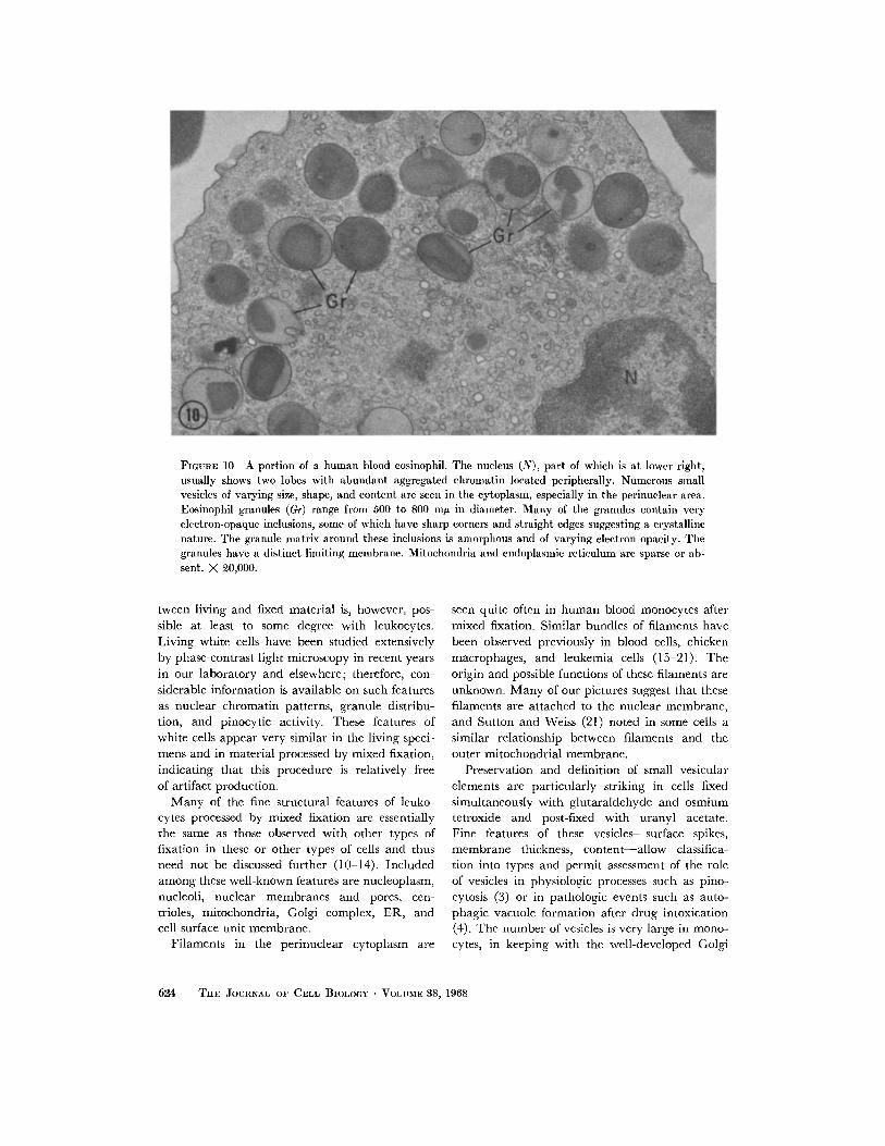
FIGuRE 10 A portion of a human blood eosinophil. The nucleus (N), part of which is at lower right,usually shows two lobes with abundant aggregated chromatin located peripherally. Numerous smallvesicles of varying size, shape, and content are seen in the cytoplasm, especially in the perinuclear area.Eosinophil granules (Gr) range from 500 to 800 mjt in diameter. Many of the granules contain veryelectron-opaque inclusions, some of which have sharp corners and straight edges suggesting a crystallinenature. The granule matrix around these inclusions is amorphous and of varying electron opacity. Thegranules have a distinct limiting membrane. Mitochondria and endoplasmic reticulum are sparse or ab-sent. X 20,000.
tween living and fixed material is, however, pos-sible at least to some degree with leukocytes.
Living white cells have been studied extensivelyby phase-contrast light microscopy in recent years
in our laboratory and elsewhere; therefore, con-siderable information is available on such featuresas nuclear chromatin patterns, granule distribu-tion, and pinocytic activity. These features ofwhite cells appear very similar in the living speci-mens and in material processed by mixed fixation,indicating that this procedure is relatively freeof artifact production.
Many of the fine structural features of leuko-cytes processed by mixed fixation are essentiallythe same as those observed with other types offixation in these or other types of cells and thusneed not be discussed further (10-14). Includedamong these well-known features are nucleoplasm,nucleoli, nuclear membranes and pores, cen-
trioles, mitochondria, Golgi complex, ER, andcell surface unit membrane.
Filaments in the perinuclear cytoplasm are
seen quite often in human blood monocytes aftermixed fixation. Similar bundles of filaments havebeen observed previously in blood cells, chickenmacrophages, and leukemia cells (15-21). Theorigin and possible functions of these filaments areunknown. Many of our pictures suggest that thesefilaments are attached to the nuclear membrane,and Sutton and Weiss (21) noted in some cells asimilar relationship between filaments and theouter mitochondrial membrane.
Preservation and definition of small vesicularelements are particularly striking in cells fixedsimultaneously with glutaraldehyde and osmiumtetroxide and post-fixed with uranyl acetate.Fine features of these vesicles--surface spikes,membrane thickness, content-allow classifica-tion into types and permit assessment of the roleof vesicles in physiologic processes such as pino-cytosis (3) or in pathologic events such as auto-phagic vacuole formation after drug intoxication(4). The number of vesicles is very large in mono-cytes, in keeping with the well-developed Golgi
624 THE JOURNAL OF CELL BIOLOGY VOLUME 38, 1968
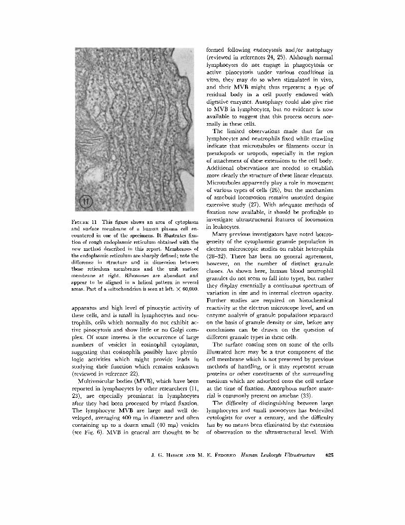
FIGURE 11 This figure shows an area of cytoplasmand surface membrane of a human plasma cell en-countered in one of the specimens. It illustrates fixa-tion of rough endoplasmic reticulum obtained with thenew method described in this report. Membranes ofthe endoplasmic reticulum are sharply defined; note thedifference in structure and in dimension betweenthese reticulum membranes and the unit surfacemembrane at right. Ribosomes are abundant andappear to be aligned in a helical pattern in severalareas. Part of a mitochondrion is seen at left. X 60,000.
apparatus and high level of pinocytic activity ofthese cells, and is small in lymphocytes and neu-trophils, cells which normally do not exhibit ac-tive pinocytosis and show little or no Golgi com-plex. Of some interest is the occurrence of largenumbers of vesicles in eosinophil cytoplasm,
suggesting that eosinophils possibly have physio-logic activities which might provide leads instudying their function which remains unknown
(reviewed in reference 22).Multivesicular bodies (MVB), which have been
reported in lymphocytes by other researchers (11,
23), are especially prominent in lymphocytesafter they had been processed by mixed fixation.The lymphocyte MVB are large and well de-veloped, averaging 400 my in diameter and oftencontaining up to a dozen small (40 m/) vesicles(see Fig. 6). MVB in general are thought to be
formed following endocytosis and/or autophagy(reviewed in references 24, 25). Although normallymphocytes do not engage in phagocytosis oractive pinocytosis under various conditions invitro, they may do so when stimulated in vivo,and their MVB might thus represent a type ofresidual body in a cell poorly endowed withdigestive enzymes. Autophagy could also give riseto MVB in lymphocytes, but no evidence is nowavailable to suggest that this process occurs nor-mally in these cells.
The limited observations made thus far onlymphocytes and neutrophils fixed while crawlingindicate that microtubules or filaments occur inpseudopods or uropods, especially in the regionof attachment of these extensions to the cell body.Additional observations are needed to establishmore clearly the structure of these linear elements.Microtubules apparently play a role in movementof various types of cells (26), but the mechanismof ameboid locomotion remains unsettled despiteextensive study (27). With adequate methods offixation now available, it should be profitable toinvestigate ultrastructural features of locomotionin leukocytes.
Many previous investigators have noted hetero-geneity of the cytoplasmic granule population inelectron microscopic studies on rabbit heterophils(28-32). There has been no general agreement,however, on the number of distinct granuleclasses. As shown here, human blood neutrophilgranules do not seem to fall into types, but ratherthey display essentially a continuous spectrum ofvariation in size and in internal electron opacity.Further studies are required on histochemicalreactivity at the electron microscope level, and onenzyme analysis of granule populations separatedon the basis of granule density or size, before anyconclusions can be drawn on the question ofdifferent granule types in these cells.
The surface coating seen on some of the cellsillustrated here may be a true component of thecell membrane which is not preserved by previousmethods of handling, or it may represent serumproteins or other constituents of the surroundingmedium which are adsorbed onto the cell surfaceat the time of fixation. Amorphous surface mate-rial is commonly present on amebae (33).
The difficulty of distinguishing between largelymphocytes and small monocytes has bedeviledcytologists for over a century, and the difficultyhas by no means been eliminated by the extensionof observation to the ultrastructural level. With
J. G. HIRSCH AND M. E. FEDORKO Human Leukocyte Ultrastructure 625
the present method of fixation there remain rarecells which defy classification, but, on the whole,distinctions between monocytes and lymphocytesare clear. Among the features, some old and wellknown and others new, helpful in classifying thesemononuclear cells are the following. The mono-cyte is a larger cell and has more abundant cyto-plasm than does the lymphocyte. Monocyte nucleitend to be horseshoe-shaped or even multiple inthin sections, whereas the lymphocyte nucleus istypically oval with indentations. Monocyte nucleishow a higher proportion of light-staining areasthan do those of lymphocytes. Perinuclear fila-ments are often present in monocytes, but theyhave not been observed in resting lymphocytes.Lymphocyte mitochondria are elongated struc-tures usually gathered in a rosette pattern aboutthe centrosomal area, whereas monocyte mito-chondria tend to be oval or round and are scat-tered in the peripheral cytoplasm. Monocyteshave as a rule many more cytoplasmic vesiclesand small dense bodies than do lymphocytes, butmultivesicular bodies are more common in cir-culating lymphocytes than in monocytes. Mono-cytes usually exhibit a prominent Golgi apparatus,whereas lymphocytes have a poorly developedGolgi apparatus. When the distinguishing be-tween lymphocytes and monocytes is of particularimportance, we have found it useful to incubatethe cells with colloidal gold or with heat-killed,washed Staphylococcus albus; monocytes displayactive phagocytosis and pinocytosis, and gold orbacteria are thus seen in digestive vacuoles oreventually in dense bodies. Lymphocytes exhibitneither pinocytic nor phagocytic activity demon-strable by these methods under various in vitroconditions studied thus far in our laboratory.
From the over-all view point, the mixed fixa-tion procedure leads to distinctly better and morereproducible preservation of white blood cellsthan is possible with osmium tetroxide alone,with glutaraldehyde followed by osmium tetrox-ide, or with glutaraldehyde followed by osmiumtetroxide and uranyl acetate (compare illustra-tions in references 10-13 and those presentedhere). Especially noteworthy features of cellsprocessed by the mixed fixation method are thesharp membrane definition and the good preserva-tion of granules and vesicles. Other methods offixation may, of course, be preferable for specialpurposes; for example, glycogen deposits in neu-trophil cytoplasm appear to be poorly definedafter mixed fixation, which perhaps reflectsextraction or modification in their staining prop-erties. Our experience with the mixed fixativehas been limited to leukocytes and to mousemacrophages and L cells in culture. Trump andBulger (1) subjected blocks of rat and flounderrenal tissue to simultaneous fixation with glutaral-dehyde and osmium tetroxide and obtained gener-ally good results. Whether our procedure will alsoserve for satisfactory fixation of various other celltypes in suspension or in tissue blocks remains tobe investigated. Suffice it to conclude, for thepresent, that the method described here is con-venient and reliable, and that it enables studies,previously difficult or impossible, on normal andon pathologic features of leukocytes and macro-phages.
This work was supported by United States PublicHealth Service grant No. AI 01831.
Received for publication 14 March 1968, and in revised form29 April 1968.
REFERENCES
1. TRUMP, B., and R. BULGER. 1966. New ultra-structural characteristics of cells fixed in aglutaraldehyde-osmium tetroxide mixture. Lab.Invest. 15:368.
2. KELLENBERGER, E., A. RYTER, and J. SCHAUD.1958. Electron microscope study of DNA-containing plasms. J. Biophys. Biochem. Cytol.4:671.
3. HIRSCH, J. G., M. E. FEDORKO, and Z. A. COHN.
1968. Vesicle fusion and formation at the sur-face of pinocytic vacuoles in macrophages. J.Cell Biol. 38:629.
4. FEDORKO, M. E., J. G. HIRscH, and Z. A. COHN.
1968. Autophagic vacuoles produced in vitro.J. Cell Biol. 38:377.
5. LUFT, J. H. 1961. Improvements in epoxy resinembedding methods. J. Biophys. Biochem. Cytol.
9:409.6. VENABLE, J. H., and R. COGGESHALL. 1965. A
simplified lead citrate stain for use in electronmicroscopy. J. Cell Biol. 25:407.
7. MILLER, F., E. DE HARVEN, and G. E. PALADE.
1966. The structure of eosinophil leukocyte
granules in rodents and in man. J. Cell Biol.
31:349.8. HIRSCH, J. G., and A. B. CHURCH. 1960. Studies
of phagocytosis of group A streptococci bypolymorphonuclear leucocytes in vitro. J. Exptl.Med. 111:309.
9. RABINOWITZ, Y. 1964. Separation of lympho-
626 THE JOURNAL OF CELL BIOLOGY VOLUME 8, 1968
cytes, polymorphonuclear leukocytes andmonocytes on glass columns, including tissueculture observations. Blood. 23:811.
10. I.ow, F. N., and J. A. FREEMAN. 1958. ElectronMicroscopic Atlas of Normal and LeukemicHuman Blood. McGraw-Hill Book, Company,New York.
11. BESSIS, M., and J. P. THIERY. 1961. Electronmicroscopy of white blood cells and their stemcells. Intern. Rev. Cytol. 12:199.
12. FAWCETT, D. W. 1966. An Atlas of Fine Struc-ture: The Cell, its Organelles, and Inclusions.W. B. Saunders Co., Philadelphia.
13. ANDERSON, D. R. 1966. Ultrastructure of normaland leukemic leukocytes in human peripheralblood. J. Ultrastruct. Res. Suppl. 9:1.
14. WATANABE, I., S. DONAHUE, and N. HOGGATT.
1967. Method for electron microscopic studiesof circulating human leukocytes and observa-tions on their fine structure. J. Ultrastruct. Res.20:366.
15. BESSIS, M., and J. BRETON-BoRIus. 1957. Examen
au microscope electronique des cellules desleucbmies mybloides. Bull. Microscop. Appl. 5:9.
16. FREEMAN, J. A., and M. S. SAMUELS. 1958. Theultrastructure of a "fibrillar formation" ofleukemic human blood. Blood. 13:725.
17. ACKERMAN, G. A., J. A. GRASSO, and R. A.KNOUFF. 1960. Morphological and histochemi-cal studies of the leukemic cells from a patientwith atypical myeloblastic leukemia with specialreference to intracytoplasmic mucopolysaccha-ride vacuoles and fibrillar formation. Blood. 16:1253.
18. DE PETRIS, S., G. KARLSBAD, and B. PERNIS. 1962.
Filamentous structures in the cytoplasm ofnormal mononuclear phagocytes. J. Ultra-struct. Res. 7:39.
19. TANAKA, Y. 1964. Fibrillar structures in the cells
of blood forming organs. J. Natl. Cancer Inst.33:467.
20. DE THE, G. 1964. Cytoplasmic microtubules indifferent animal cells. J. Cell Biol. 23:265.
21. SUTTON, J. S., and L. WEIsS. 1966. Transforma-tion of monocytes in tissue culture into macro-
phages, epithelioid cells and multinucleatedgiant cells. J. Cell Biol. 28:303.
22. HIRSCH, J. G. 1965. Neutrophil and eosinophilleucocytes. In The Inflammatory Process.Academic Press Inc., New York. 245.
23. ZUCKER-FRANKLIN, D. 1963. The ultrastructure
of cells in human thoracic duct lymph. J.Ultrastruct. Res. 9:325.
24. NOVIKOFF, A. B., E. ESSNER, and N. QUINTANA.
1964. Golgi apparatus and lysosomes. Federa-tion Proc. 23:1010.
25. DE DUVE, C., and R. WATTIAUX. 1966. Functionsof lysosomes. Ann. Rev. Physiol. 28:435.
26. PORTER, K. R. 1966. Cytoplasmic microtubules
and their functions. In Ciba Foundation Sym-posium on Principles of Biomolecular Organi-zation. G. E. W. Wolstenholme and M.O'Connor, editors. Little, Brown and Com-pany, Boston. 308.
27. ALLEN, R. D. 1961. Ameboid movement. InThe Cell. J. Brachet and A. E. Mirsky, editors.Academic Press Inc., New York. 2:135.
28. FLOREY, H. W., and L. H. GRANT. 1961. Leuco-
cyte migration from small blood vessels stimu-lated with ultraviolet light: An electron-microscope study. J. Pathol. Bacteriol. 82:13.
29. LOCKWOOD, W. R., and F. ALLISON. 1963. Elec-tromicrographic studies of phagocytic cells. IMorphological changes of the cytoplasm andgranules of rabbit granulocytes associated withingestion of rough pneumococcus. Brit. J.Exptl. Pathol. 44:593.
30. ZUCKER-FRANKLIN, D., and J. G. HIRSCH. 1964.
Electron microscope studies on the degranula-tion of rabbit peritoneal leukocytes duringphagocytosis. J. Exptl. Med. 120:569.
31. BAINTON, D. F., and M. FARQUHAR. 1966. Origin
of granules in polymorphonuclear leucocytes.J. Cell Biol. 28:277.
32. WETZEL, B. K., R. G. HORN, and S. S. SPICER.
1967. Fine structural studies on the develop-ment of heterophil, eosinophil and basophilgranulocytes in rabbits. Lab. Invest. 16:349.
33. MERCER, E. H. 1959. An electron microscopic
study of Amoeba proteus. Proc. Roy. Soc. Biol. 150:216.
J. G. HIRSCH AND M. E. FEDORKO Human Leukocyte Ultrastructure 627