ULTRASTRUCTURAL CHANGES IN RAT … · ULTRASTRUCTURAL CHANGES IN RAT TESTICULAR TISSUE AFTER ......
9
JOURNAL OF INTERNATIONAL ACADEMIC RESEARCH FOR MULTIDISCIPLINARY Impact Factor 1.393, ISSN: 2320-5083, Volume 2, Issue 1, February 2014 518 www.jiarm.com ULTRASTRUCTURAL CHANGES IN RAT TESTICULAR TISSUE AFTER WHOLE BODY EXPOSURE TO ELECTROMAGNETIC RADIATION EMITTED FROM MOBILE PHONES PRADEEP KUMAR* DR.VINEETA SHUKLA** *Ph.D, Scholar, Senior Research Fellow, Dept. of Zoology, M.D. University Rohtak, Haryana, India **Professor, Dept. of Zoology, M.D. University Rohtak, Haryana, India ABSTRACT The possible adverse reproductive effects resulting from exposure to electromagnetic fields (EMF) are currently of great public concern. The objective of the present study is to reveal possible effects of electromagnetic fields emitted from a CDMA mobile phone on the ultrastructural aspects of the testicular cells. Approximately 6 weeks old Swiss albino rats were procured from LLRUVAS, Hisar, Haryana. Rats were acclimatized in plastic cages in a room maintained at 24 ± 1 o C and 50 ± 5 % humidity with an alternating 12 h light-darkness cycle. After one week, rats were exposed under electromagnetic radiation emitted from a CDMA mobile phone with 3 hrs exposure followed by 30 minutes rest and then again 3 hrs exposure per day for five months. One sham group was kept away from the source of radiation and was used as control for the experimental groups. Immediately after the last irradiation, rats were sacrificed and their testes were analyzed using Electron Microscopic techniques. Studies revealed pycnotic nuclei in germ cells, vacuolization in spermatogenic cells and detachment of spermatogonia and sertoli cells from basal lamina. Shrinkage was induced on the surface of the seminiferous epithelium due to exposure. Residual cytoplasm and debris of degenerating cells were also observed in the seminiferous tubules. KEYWORDS: Radio Frequency Electromagnetic Fields (RF-EMF), Mobile Phone, Swiss Albino Rats, Testes, Seminiferous Tubules. INTRODUCTION Mobile phones and cell towers are amongst the most common sources of electromagnetic radiations. Increasing number of devices emitting such radiations raised the valid question concerning their safety and the potential risk of human exposure and its limits (Valberg, 1996). The possible adverse reproductive effects resulting from exposure to electromagnetic fields are currently of great public concern. Some investigations have suggested that one of the environmental factors potentially involved in the etiology of DNA damage in human spermatozoa is an increased exposure to radio frequency electromagnetic
-
Upload
vuongquynh -
Category
Documents
-
view
218 -
download
0
Transcript of ULTRASTRUCTURAL CHANGES IN RAT … · ULTRASTRUCTURAL CHANGES IN RAT TESTICULAR TISSUE AFTER ......
JOURNAL OF INTERNATIONAL ACADEMIC RESEARCH FOR MULTIDISCIPLINARY Impact Factor 1.393, ISSN: 2320-5083, Volume 2, Issue 1, February 2014
518 www.jiarm.com
ULTRASTRUCTURAL CHANGES IN RAT TESTICULAR TISSUE AFTER WHOLE BODY EXPOSURE TO ELECTROMAGNETIC RADIATION EMITTED
FROM MOBILE PHONES
PRADEEP KUMAR* DR.VINEETA SHUKLA**
*Ph.D, Scholar, Senior Research Fellow, Dept. of Zoology, M.D. University Rohtak, Haryana, India
**Professor, Dept. of Zoology, M.D. University Rohtak, Haryana, India
ABSTRACT
The possible adverse reproductive effects resulting from exposure to electromagnetic
fields (EMF) are currently of great public concern. The objective of the present study is to
reveal possible effects of electromagnetic fields emitted from a CDMA mobile phone on the
ultrastructural aspects of the testicular cells. Approximately 6 weeks old Swiss albino rats
were procured from LLRUVAS, Hisar, Haryana. Rats were acclimatized in plastic cages in a
room maintained at 24 ± 1o C and 50 ± 5 % humidity with an alternating 12 h light-darkness
cycle. After one week, rats were exposed under electromagnetic radiation emitted from a
CDMA mobile phone with 3 hrs exposure followed by 30 minutes rest and then again 3 hrs
exposure per day for five months. One sham group was kept away from the source of
radiation and was used as control for the experimental groups. Immediately after the last
irradiation, rats were sacrificed and their testes were analyzed using Electron Microscopic
techniques. Studies revealed pycnotic nuclei in germ cells, vacuolization in spermatogenic
cells and detachment of spermatogonia and sertoli cells from basal lamina. Shrinkage was
induced on the surface of the seminiferous epithelium due to exposure. Residual cytoplasm
and debris of degenerating cells were also observed in the seminiferous tubules.
KEYWORDS: Radio Frequency Electromagnetic Fields (RF-EMF), Mobile Phone, Swiss Albino Rats, Testes, Seminiferous Tubules.
INTRODUCTION
Mobile phones and cell towers are amongst the most common sources of
electromagnetic radiations. Increasing number of devices emitting such radiations raised the
valid question concerning their safety and the potential risk of human exposure and its limits
(Valberg, 1996). The possible adverse reproductive effects resulting from exposure to
electromagnetic fields are currently of great public concern. Some investigations have
suggested that one of the environmental factors potentially involved in the etiology of DNA
damage in human spermatozoa is an increased exposure to radio frequency electromagnetic
JOURNAL OF INTERNATIONAL ACADEMIC RESEARCH FOR MULTIDISCIPLINARY Impact Factor 1.393, ISSN: 2320-5083, Volume 2, Issue 1, February 2014
519 www.jiarm.com
radiation (RF-EMR) emitted from mobile phones. Initial studies revealed negative correlation
between mobile phone usage reproductive toxicity (Fejes et al., 2005). Large doses of
radiofrequency (RF)-EMF have been shown in previous studies to be related to genetic
defects, such as changes in the integrity of epididymal mitochondrial DNA (Aitken et al.,
2005), increased micronuclei for mutations (Tice et al., 2002), increased chromosomal
instability (Sykes et al., 2001; Mashevich et al., 2003), altered proto-oncogene c-fos
(Goswami et al., 1999) and changes in morphology and gene expression (Pacini et al., 2002) .
RF-EMF of the commercially available cell phones may affect the fertilizing potential of
spermatozoa and this can explain the RF-EMF related infertility cases observed in numerous
studies (Wdowiak et al., 2007). Experimental studies specifically designed to evaluate
testicular damage caused by low intensity RF show conflicting results (Saunders and
Kowalczuk, 1981; Dasdag et al., 1999,2003; Ozguner et al., 2005; Ribeiro et al., 2007; Yan et
al., 2007). Tissues with higher hydration as testes were more sensitive to magnetic fields
(Arutiunian et al., 1998), yet many controversies regarding the biological effects on the
organs were encountered. Some investigators reported affection of testicular germ cells (Lee
et al., 2004) while others denied any magnetic field exposure related histopathological
alteration in testicular tissue (Forgaces et al., 2004).
In the light of such consideration, present study was conducted to analyze the ultrastructural
consequences of chronic exposure of RF-EMR emitted form domestic mobile phone on rat
testes . To avoid any secondary thermal effect, temperature of the room was kept at 240 C
where the animals were placed throughout the experiment.
MATERIALS AND METHOD
I. Experimental animals
After the clearance from local Institutional Animal Ethical Committee (IAEC),
approximately 6 weeks old male Swiss albino rats, weighing 50-60 g were kept in steady-
state micro-environmental conditions (24 ± 1o C and 50 ± 5 % humidity), housed in
plastic cases with 6 per cage with an alternating 12 h light-darkness cycle. The cages were
built to provide proper ventilation to keep the animals aerated and dimensions prevent the
free movement of the animals away from the mobile phone. All animals were maintained
at an animal care facility according to the guidelines for the use and care of laboratory
animals and food and water were available ad libitum. Cleaning, changing water and food
was provided to all animals, daily.
JOURNAL OF INTERNATIONAL ACADEMIC RESEARCH FOR MULTIDISCIPLINARY Impact Factor 1.393, ISSN: 2320-5083, Volume 2, Issue 1, February 2014
520 www.jiarm.com
II. Experimental Design
After one week of acclimatization and quarantine, 24 male rats were divided at random
into two groups of 12 animals i.e. one experimental and other control group. Rats in the
experimental group were exposed under electromagnetic radiation emitted from a Code
Division Multiple Access (CDMA) mobile phone with 3 hrs exposure, followed by 30
minutes rest and again 3 hrs exposure per day for five months. The sham controls were
handled in the same manner as the treated ones, but were not irradiated at any point
III. Histopathological examination
A. Extraction of the testes
Immediately after the last irradiation, the rats were sacrificed by overdose of ether. Testes
were dissected out and decapsulated, put in buffered glutaraldehyde 2.5% for one hour
and then cubes of 1mm in dimension were cut for transmission electron microscopy and
cubes of 1cm for scanning electron microscopy by a sharp razor from the outer layer of
the testes with careful manipulation. All the samples were then transferred in a fixative (
2.5% glutaraldehyde and 2% paraformaldehyde in 0.1M phosphate buffer of ph 7.4) for
12 hr at 40 C (Aisha et al., 2006).
B. Processing for transmission electron microscopy
Washing was given to the samples by phosphate buffer (ph 7.4) three timesfor 10 minutes
each. The slices were then fixed in 1% osmium tetroxide for 1 hr followed by another
washing. Dehydration of the samples was performed by gradually increasing
concentration of ethyl alcohol for 30 minutes each and then in absolute alcohol for 1 hr.
After treating with propylene oxide, samples were embedded in spur resin to form gelatin
blocks. Blocks were trimmed and ultra thin sections (300 A0) were cut and picked up on
copper grids. Uranyl acetate and lead citrate were used to stain the sections which were
examined and photographed by transmission electron microscopy.
C. Processing for scanning electron microscopy
Samples were fixed at room temperature and rinsed three times using the same buffer
used for the fixative for five minutes each rinse. After passing through 1% osmium
tetroxide for 1 hr, samples were again rinsed three times using the same buffer for 5
minutes each rinse. Then samples were dehydrated using gradually increasing
concentration of ethyl alcohol for 10 minutes each and then in absolute alcohol for 1 hr.
JOURNAL OF INTERNATIONAL ACADEMIC RESEARCH FOR MULTIDISCIPLINARY Impact Factor 1.393, ISSN: 2320-5083, Volume 2, Issue 1, February 2014
521 www.jiarm.com
Finally samples were passed through the step of critical point dry followed by mounting
and coating with conductive material.
RESULTS
1. Transmission electron microscopy
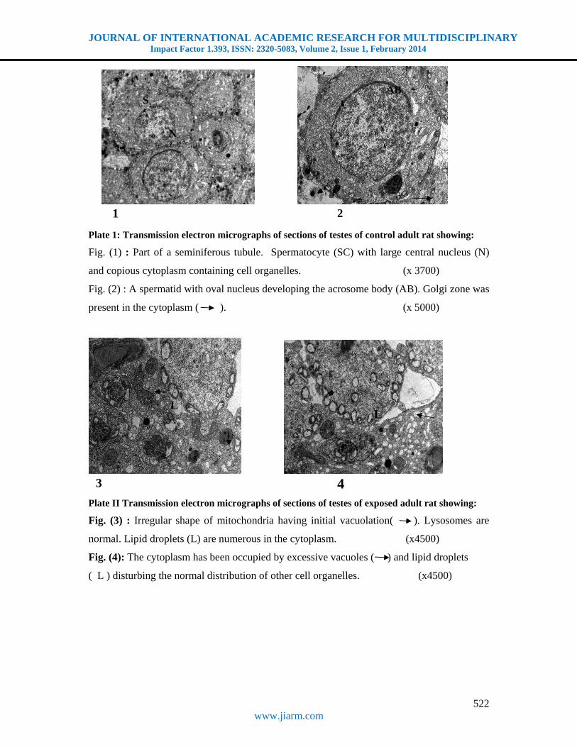
Electron microscopic examination of the testes of the control adult rats illustrated the
normal secondary spermatocytes and few early spermatids with acrosome cap formation
(fig. 1). Seminiferous tubules were consisting of spermatogenic cells at different stages of
differentiation together with supporting sertoli cells. Sertoli cells were present in close
proximity to the basement membrane with adjacent spermatogonia situated towards the
lumen of the seminiferous tubules. Primary young spermatids were present as large
rounded cells with oval nucleus, containing different cell organelles. Mitochondria were
present with ill defined cristae. Acrosome formation at earlier stage was noticed (fig. 2).
Myoid cells were present outside the basal lamina which encircled the seminiferous
tubules.
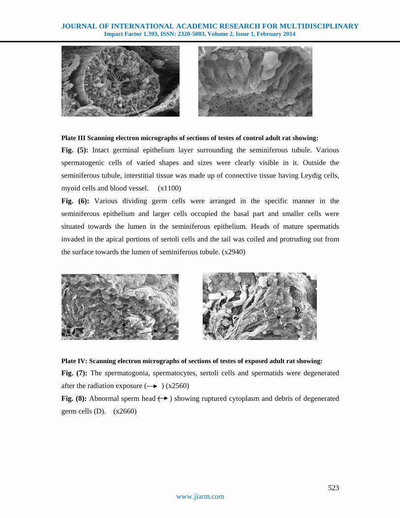
The exposed testes to Radio Frequency Electromagnetic Fields (RF-EMF) showed
variable degenerative changes in the spermatogenic cells. Some cells showed electron
dense areas and vacuolation within the cytoplasm. (fig. 3). Irregular shaped and multiple
vacuolated mitochondria were also observed. (fig. 4).
2. Scanning electron microscopy
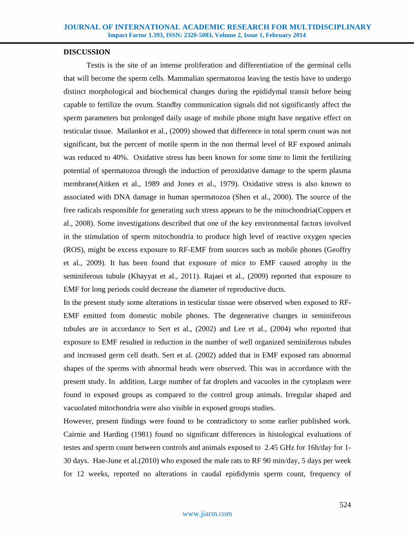
Scanning electron micrograph of control testicular tissue illustrates the cross section of
seminiferous tubules. Furrow like depression was observed running longitudinally on the
outer surface of it. Cells of varied shapes and sizes were clearly visible in it. Outside the
seminiferous tubule, interstitial tissue was made up of connective tissue having Leydig
cells, myoid cells and blood vessel (fig. 5). Various dividing germ cells were arranged in
the specific manner in the seminiferous epithelium. Larger cells occupied the basal part
and smaller cells were situated towards the lumen and the surface of spermatogonia,
spermatocytes and spermatids was smooth (fig. 6).
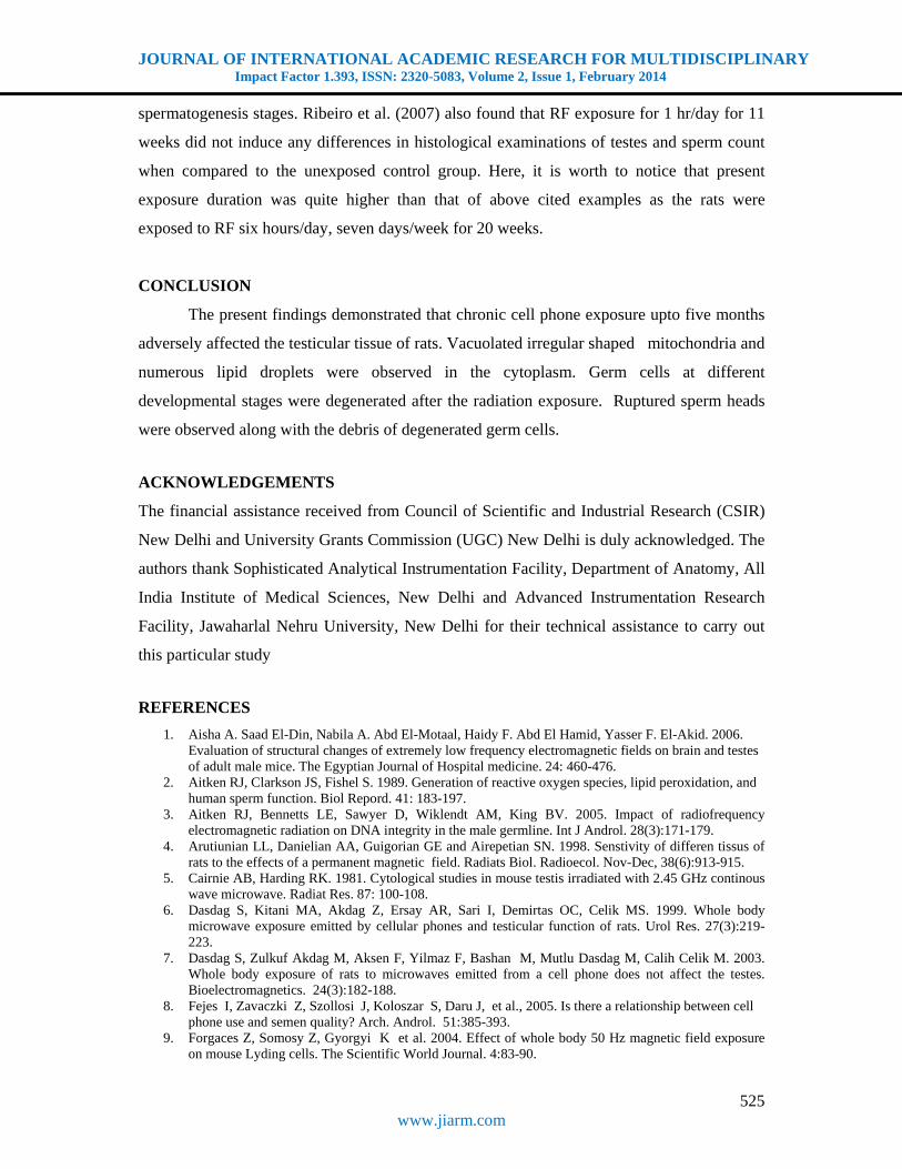
Scanning electron micrograph of testicular tissue exposed under electromagnetic
radiations showing sharp edge craters and shrinkage induced on the surface of
degenerating cells in the seminiferous epithelium due to exposure(fig. 7). The residual
cytoplasm and debris of degenerating cells in the epithelium were clearly visible.
Ruptured sperm head and distorted tail were also observed (fig. 8).
JOURNAL OF INTERNATIONAL ACADEMIC RESEARCH FOR MULTIDISCIPLINARY Impact Factor 1.393, ISSN: 2320-5083, Volume 2, Issue 1, February 2014
522 www.jiarm.com
Plate 1: Transmission electron micrographs of sections of testes of control adult rat showing:
Fig. (1) : Part of a seminiferous tubule. Spermatocyte (SC) with large central nucleus (N)
and copious cytoplasm containing cell organelles. (x 3700)
Fig. (2) : A spermatid with oval nucleus developing the acrosome body (AB). Golgi zone was
present in the cytoplasm ( ). (x 5000)
Plate II Transmission electron micrographs of sections of testes of exposed adult rat showing:
Fig. (3) : Irregular shape of mitochondria having initial vacuolation( ). Lysosomes are
normal. Lipid droplets (L) are numerous in the cytoplasm. (x4500)
Fig. (4): The cytoplasm has been occupied by excessive vacuoles ( ) and lipid droplets
( L ) disturbing the normal distribution of other cell organelles. (x4500)
1 2
N
SC
AB
3 4
L L
JOURNAL OF INTERNATIONAL ACADEMIC RESEARCH FOR MULTIDISCIPLINARY Impact Factor 1.393, ISSN: 2320-5083, Volume 2, Issue 1, February 2014
523 www.jiarm.com
Plate III Scanning electron micrographs of sections of testes of control adult rat showing:
Fig. (5): Intact germinal epithelium layer surrounding the seminiferous tubule. Various
spermatogenic cells of varied shapes and sizes were clearly visible in it. Outside the
seminiferous tubule, interstitial tissue was made up of connective tissue having Leydig cells,
myoid cells and blood vessel. (x1100)
Fig. (6): Various dividing germ cells were arranged in the specific manner in the
seminiferous epithelium and larger cells occupied the basal part and smaller cells were
situated towards the lumen in the seminiferous epithelium. Heads of mature spermatids
invaded in the apical portions of sertoli cells and the tail was coiled and protruding out from
the surface towards the lumen of seminiferous tubule. (x2940)
Plate IV: Scanning electron micrographs of sections of testes of exposed adult rat showing:
Fig. (7): The spermatogonia, spermatocytes, sertoli cells and spermatids were degenerated
after the radiation exposure ( ) (x2560)
Fig. (8): Abnormal sperm head ( ) showing ruptured cytoplasm and debris of degenerated
germ cells (D). (x2660)
D
JOURNAL OF INTERNATIONAL ACADEMIC RESEARCH FOR MULTIDISCIPLINARY Impact Factor 1.393, ISSN: 2320-5083, Volume 2, Issue 1, February 2014
524 www.jiarm.com
DISCUSSION
Testis is the site of an intense proliferation and differentiation of the germinal cells
that will become the sperm cells. Mammalian spermatozoa leaving the testis have to undergo
distinct morphological and biochemical changes during the epididymal transit before being
capable to fertilize the ovum. Standby communication signals did not significantly affect the
sperm parameters but prolonged daily usage of mobile phone might have negative effect on
testicular tissue. Mailankot et al., (2009) showed that difference in total sperm count was not
significant, but the percent of motile sperm in the non thermal level of RF exposed animals
was reduced to 40%. Oxidative stress has been known for some time to limit the fertilizing
potential of spermatozoa through the induction of peroxidative damage to the sperm plasma
membrane(Aitken et al., 1989 and Jones et al., 1979). Oxidative stress is also known to
associated with DNA damage in human spermatozoa (Shen et al., 2000). The source of the
free radicals responsible for generating such stress appears to be the mitochondria(Coppers et
al., 2008). Some investigations described that one of the key environmental factors involved
in the stimulation of sperm mitochondria to produce high level of reactive oxygen species
(ROS), might be excess exposure to RF-EMF from sources such as mobile phones (Geoffry
et al., 2009). It has been found that exposure of mice to EMF caused atrophy in the
seminiferous tubule (Khayyat et al., 2011). Rajaei et al., (2009) reported that exposure to
EMF for long periods could decrease the diameter of reproductive ducts.
In the present study some alterations in testicular tissue were observed when exposed to RF-
EMF emitted from domestic mobile phones. The degenerative changes in seminiferous
tubules are in accordance to Sert et al., (2002) and Lee et al., (2004) who reported that
exposure to EMF resulted in reduction in the number of well organized seminiferous tubules
and increased germ cell death. Sert et al. (2002) added that in EMF exposed rats abnormal
shapes of the sperms with abnormal heads were observed. This was in accordance with the
present study. In addition, Large number of fat droplets and vacuoles in the cytoplasm were
found in exposed groups as compared to the control group animals. Irregular shaped and
vacuolated mitochondria were also visible in exposed groups studies.
However, present findings were found to be contradictory to some earlier published work.
Cairnie and Harding (1981) found no significant differences in histological evaluations of
testes and sperm count between controls and animals exposed to 2.45 GHz for 16h/day for 1-
30 days. Hae-June et al.(2010) who exposed the male rats to RF 90 min/day, 5 days per week
for 12 weeks, reported no alterations in caudal epididymis sperm count, frequency of
JOURNAL OF INTERNATIONAL ACADEMIC RESEARCH FOR MULTIDISCIPLINARY Impact Factor 1.393, ISSN: 2320-5083, Volume 2, Issue 1, February 2014
525 www.jiarm.com
spermatogenesis stages. Ribeiro et al. (2007) also found that RF exposure for 1 hr/day for 11
weeks did not induce any differences in histological examinations of testes and sperm count
when compared to the unexposed control group. Here, it is worth to notice that present
exposure duration was quite higher than that of above cited examples as the rats were
exposed to RF six hours/day, seven days/week for 20 weeks.
CONCLUSION
The present findings demonstrated that chronic cell phone exposure upto five months
adversely affected the testicular tissue of rats. Vacuolated irregular shaped mitochondria and
numerous lipid droplets were observed in the cytoplasm. Germ cells at different
developmental stages were degenerated after the radiation exposure. Ruptured sperm heads
were observed along with the debris of degenerated germ cells.
ACKNOWLEDGEMENTS
The financial assistance received from Council of Scientific and Industrial Research (CSIR)
New Delhi and University Grants Commission (UGC) New Delhi is duly acknowledged. The
authors thank Sophisticated Analytical Instrumentation Facility, Department of Anatomy, All
India Institute of Medical Sciences, New Delhi and Advanced Instrumentation Research
Facility, Jawaharlal Nehru University, New Delhi for their technical assistance to carry out
this particular study
REFERENCES
1. Aisha A. Saad El-Din, Nabila A. Abd El-Motaal, Haidy F. Abd El Hamid, Yasser F. El-Akid. 2006. Evaluation of structural changes of extremely low frequency electromagnetic fields on brain and testes of adult male mice. The Egyptian Journal of Hospital medicine. 24: 460-476.
2. Aitken RJ, Clarkson JS, Fishel S. 1989. Generation of reactive oxygen species, lipid peroxidation, and human sperm function. Biol Repord. 41: 183-197.
3. Aitken RJ, Bennetts LE, Sawyer D, Wiklendt AM, King BV. 2005. Impact of radiofrequency electromagnetic radiation on DNA integrity in the male germline. Int J Androl. 28(3):171-179.
4. Arutiunian LL, Danielian AA, Guigorian GE and Airepetian SN. 1998. Senstivity of differen tissus of rats to the effects of a permanent magnetic field. Radiats Biol. Radioecol. Nov-Dec, 38(6):913-915.
5. Cairnie AB, Harding RK. 1981. Cytological studies in mouse testis irradiated with 2.45 GHz continous wave microwave. Radiat Res. 87: 100-108.
6. Dasdag S, Kitani MA, Akdag Z, Ersay AR, Sari I, Demirtas OC, Celik MS. 1999. Whole body microwave exposure emitted by cellular phones and testicular function of rats. Urol Res. 27(3):219-223.
7. Dasdag S, Zulkuf Akdag M, Aksen F, Yilmaz F, Bashan M, Mutlu Dasdag M, Calih Celik M. 2003. Whole body exposure of rats to microwaves emitted from a cell phone does not affect the testes. Bioelectromagnetics. 24(3):182-188.
8. Fejes I, Zavaczki Z, Szollosi J, Koloszar S, Daru J, et al., 2005. Is there a relationship between cell phone use and semen quality? Arch. Androl. 51:385-393.
9. Forgaces Z, Somosy Z, Gyorgyi K et al. 2004. Effect of whole body 50 Hz magnetic field exposure on mouse Lyding cells. The Scientific World Journal. 4:83-90.
JOURNAL OF INTERNATIONAL ACADEMIC RESEARCH FOR MULTIDISCIPLINARY Impact Factor 1.393, ISSN: 2320-5083, Volume 2, Issue 1, February 2014
526 www.jiarm.com
10. Geoffry N. De luliis, Rhiannon J. Newey, Bruce V. king, R. John Aitken. 2009. Mobile phone radiation induces reactive oxygen species production and DNA damage in human spermatozoa in vivo. Plos one. 4(7):1-9.
11. Goswami PC, Albee LD, Parsian AJ, Baty JD, Moros EG, Pickard WF, Roti Roti JL, Hunt CR. 1999. Proto-oncogene mRNA levels and activities of multiple transcription factors in C3H 10T ½ murine embryonic fibroblasts exposed to 835.62 and 847.74 MHz cellular phone communication frequency radiation. Radiat. Res. 151(3):300-309.
12. Hae-June Lee, Jeong-Ki Pack, Tae-Hong Kim, Nam Kim, Soo Yong Choi, Lae-Seon Lee, Sung-Ho Kim, and Yun-Sil Lee. 2010. The lack of histological changes of CDMA cellular phone-based radio frequency on rat testes. Bioelectromagnetics. 31:528-534.
13. Jones R, Mann T, Sherins RJ. 1979. Peroxidative breakdown of phospholipids in human spermatozoa: spermicidal effects of fatty acid peroxides and protective action of seminal plasma. Fertil. Steril. 31: 531-537.
14. Khayyat, L.I. 2011. The histopathological effects of an electromagnetic field on the kidney and testis of mice. Eurasia J. Biosci. 5:103-109.
15. Kopper AJ, De Iuliis GN, Finnie JM, McLaughlin EA, Aitken RJ. 2008. Significance of mitochondrial reactive oxygen species in the generation of oxidative stress in spermatozoa. J Clin Endocrinol Metab. 93:3199-3207.
16. Lee JS, Ahn SS, Jung KC, Kin YW and Lee SK. 2004. Effects of 60 Hz EMF exposure on testicular germ cell apoptosis in mice. Asian J Androl., Mar. 6(1):29-34
17. Mashevich M, Folkman D, Kesar A, Barbul A, Korenstein R, Jerby E, Avivi L. 2003. Exposure of human peripheral blood lymphocytes to electromagnetic fields associated with cellular phones leads to chromosomal instability. Bioelectromegnetics. 24(2): 82-90.
18. Mailankot M, Kunnath AP, Jayalekshmi H, Koduru B. and Valsalan, R. 2009. Radio frequency electromagnetic radiation (RF-EMR)from GSM (0.9/1.8GHZ) mobile phones induces oxidatives stress and reduces sperm motility in rats. Clinics, 64:561-565.
19. Ozguner M, Koyu A, Cesur G, Urtal M, Ozguner F, Gokcimen A, Delibas N. 2005. Biological and morphological effects on the reproductive organs of the rats after exposure to electromagnetic fields. Saudi Med J. 26(3):405-410.
20. Pacini S, Ruggiero M, Sardi I, Aterini S, Gulisano F, Gulisano M. 2002. Exposure to global system of mobile communication (GSM) cellular phone radiofrequency alters gene expression, proliferarion and morpho0logy of human skin fibroblast. Oncol. Res. 13(1):19-24.
21. Rajaei, F., F. Mahdi, N. Ghasemi, M. Sarreshtehdari, N.A. Gheybi and M.S. Saraeisahneh. 2009. Effects of electromagnetic field on mice epididymis and vas defern: A morphometric study. J. Gorgan Univ.Med.Sci. 11:1-7.
22. Ribeiro EP, Rhoden EL, Lima LP, Toniolo L. 2007. Effects of subchronic exposure to radiofrequency from a conventional cellular telephone on testicular function in adult rats. J Urol. 177(I)395-399.
23. Saunders RD, Kowalczuk CL. 1981. Effects of 2.45 GHz microwave radiation and heat on mouse spermatogenic epithelium. Int J Radiot Biol Relat Stud Phya Chem Med. 40(6):623-632.
24. Sert C, Akdag MZ, Bashan M, Buyukbayram H, AND Dasdag S. 2002. ELF magnetic field effects on fatty acid composition of phospholipid fraction and reproduction of rats testes. Electromagnetic Biology and Medicine. 21:19-29.
25. Shen H, Ong C. 2000. Detection of oxidative DNA damage in human sperm and its association with sperm function and male infertility. Free Radic Biol Med 28: 529-536.
26. Sykes PJ, McCallum BD, Bangay MJ, Hooker AM, Morley AA. 2001. Effect of exposure to 900 MHz radiofrequency radiation on intrachromosomal recombination in pKZ I mice. Radiat Res. 156(5 Pt I):495-502.
27. Tice RR, Hook GG, Donner M, McRee DI, Guy AW. 2002. Genotoxocity of radiofrequency signals. I. Investigation of DNA damage and micronuclei induction in cultured human blood cells. Bioelectromagnetics. 23(2):113-126.
28. Valberg PA. 1996. Can low level 50/60 Hz electric and magnetic fields cause biological effects. Med. Res. 148:2-21.
29. Valberg PA, van Deventer E, Rapacholi MH. 2007. Workgroup report:Base stations and wireless networks-Radiofrequency (RF) exposures and health consequences. Environmental Health Perspectives. 115:416-424.
30. Wdowiak A, Wdowiak L, Wiktor H. 2007. Evaluation of the effect of using mobile phones on male fertility. Ann Agric Environ Med. 14:169-172.
31. Yan JG, Agresti M, Bruce T, Yan YH, Granlund A, Matloub HS. 2007. Effect of cellular phone emissions on sperm mortility in rarts. Fertil. Steril. 88(4):957-964.