Ultrastructural Changes Associated With Dexamethasone...
12
Physiology and Pharmacology Ultrastructural Changes Associated With Dexamethasone- Induced Ocular Hypertension in Mice Darryl R. Overby, 1 Jacques Bertrand, 1 Ozan-Y¨ uksel Tektas,* ,2 Alexandra Boussommier-Calleja, 1 Martin Schicht, 2 C. Ross Ethier, 1,3 David F. Woodward, 4 W. Daniel Stamer, 5 and Elke L¨ utjen-Drecoll 2 1 Department of Bioengineering, Imperial College London, London, United Kingdom 2 Department of Anatomy II, University of Erlangen-N¨ urnberg, Erlangen, Germany 3 Department of Biomedical Engineering, Georgia Institute of Technology, Atlanta, Georgia, United States 4 Department of Biological Sciences, Allergan, Inc., Irvine, California, United States 5 Department of Ophthalmology, Duke University Medical Center, Durham, North Carolina, United States Correspondence: Darryl R. Overby, Department of Bioengineering, Im- perial College London, London SW7 2AZ, UK; [email protected]. Current affiliation: *Department of Psychiatry, Friedrick-Alexander Uni- versity of Erlangen, Erlangen, Germany. Submitted: March 23, 2014 Accepted: June 29, 2014 Citation: Overby DR, Bertrand J, Tek- tas O-Y, et al. Ultrastructural changes associated with dexamethasone-in- duced ocular hypertension in mice. Invest Ophthalmol Vis Sci. 2014;55:4922–4933. DOI:10.1167/ iovs.14-14429 PURPOSE. To determine whether dexamethasone (DEX)-induced ocular hypertension (OHT) in mice mimics the hallmarks of steroid-induced glaucoma (SIG) in humans, including reduced conventional outflow facility (C), increased extracellular matrix (ECM), and myofibroblasts within the outflow pathway. METHODS. Osmotic mini-pumps were implanted subcutaneously into C57BL/6J mice for systemic delivery of DEX (3–4 mg/kg/d, n ¼ 31 mice) or vehicle (n ¼ 28). IOP was measured weekly by rebound tonometry. After 3 to 4 weeks, mice were euthanized and eyes enucleated for ex vivo perfusion to measure C, for electron microscopy to examine the trabecular meshwork (TM) and Schlemm’s canal (SC), or for immunohistochemistry to examine type IV collagen and a-smooth muscle actin. The length of basement membrane material (BMM) was measured along the anterior-posterior extent of SC by electron microscopy. Ultrastructural changes in BMM of DEX-treated mice were compared against archived human SIG specimens. RESULTS. Dexamethasone increased IOP by 2.6 6 1.6 mm Hg (mean 6 SD) over 3 to 4 weeks and decreased C by 52% 6 17% versus controls. Intraocular pressure elevation correlated with decreased C. Dexamethasone treatment led to increased fibrillar material in the TM, plaque-like sheath material surrounding elastic fibers, and myofibroblasts along SC outer wall. The length of BMM underlying SC was significantly increased in mice with DEX and in humans with SIG, and in mice decreased C correlated with increased BMM. CONCLUSIONS. Dexamethasone-induced OHT in mice mimics hallmarks of human SIG within 4 weeks of DEX treatment. The correlation between reduced C and newly formed ECM motivates further study using DEX-treated mice to investigate the pathogenesis of conventional outflow obstruction in glaucoma. Keywords: steroid glaucoma, trabecular meshwork, basement membrane I n susceptible individuals, sustained corticosteroid therapy causes ocular hypertension (OHT), developing to steroid- induced glaucoma (SIG) and eventual vision loss if not discontinued. 1 Ocular hypertension induced by corticosteroids mimics aspects of primary open-angle glaucoma (POAG) in that both conditions present with open chamber angles and decreased conventional outflow facility 2–4 ; furthermore, most patients with POAG experience elevated IOP when treated with corticosteroids. 5,6 Common mechanisms may therefore underlie the pathogenesis of POAG and SIG, motivating further investigation of corticosteroid-induced OHT as an inducible model of conventional outflow dysfunction that may improve our understanding of POAG. 7 Despite many laboratory studies of corticosteroid-induced OHT in cows, 8–10 cats, 11,12 rabbits, 13 sheep, 14–16 rats, 17 mice, 18–21 monkeys, 22 and humans, 23–26 relatively few stud- ies 9,23,27,28 have examined the morphological basis for cortico- steroid-induced outflow obstruction within the trabecular meshwork (TM). In human eyes treated with corticosteroids, fine fibrillar material was observed in the juxtacanalicular connective tissue (JCT) underlying the inner wall of Schlemm’s canal (SC), 28 and ‘‘fingerprint’’ like basement membrane material (BMM) was observed in the TM. 23,27 Sheath-derived ‘‘plaques’’ similar to those seen in human eyes with POAG 9 were observed in bovine eyes with corticosteroid-induced OHT. These studies 9,23,27,28 suggest that extracellular matrix (ECM) alterations contribute to conventional outflow obstruc- tion within the TM or JCT, but no studies have yet correlated corticosteroid-induced changes in ECM to outflow facility so as to more precisely identify the ultrastructural basis of cortico- steroid-induced OHT. The trabecular outflow pathway of mice is structurally and functionally similar to that of primates. Like primates, mice have a continuous SC and a lamellated TM, 29 and mice have an elastic fiber net in the TM that tethers the ciliary muscle to the inner wall of SC. 30 Mice exhibit a similar IOP 31 and aqueous humor turnover time 32 to humans and, like human eyes, mouse eyes do not appear to exhibit ‘‘wash-out.’’ 33 The pharmacology Copyright 2014 The Association for Research in Vision and Ophthalmology, Inc. www.iovs.org j ISSN: 1552-5783 4922 Downloaded From: http://iovs.arvojournals.org/pdfaccess.ashx?url=/data/journals/iovs/932996/ on 06/29/2018
Transcript of Ultrastructural Changes Associated With Dexamethasone...

Physiology and Pharmacology
Ultrastructural Changes Associated With Dexamethasone-Induced Ocular Hypertension in Mice
Darryl R. Overby,1 Jacques Bertrand,1 Ozan-Yuksel Tektas,*,2 Alexandra Boussommier-Calleja,1
Martin Schicht,2 C. Ross Ethier,1,3 David F. Woodward,4 W. Daniel Stamer,5 andElke Lutjen-Drecoll2
1Department of Bioengineering, Imperial College London, London, United Kingdom2Department of Anatomy II, University of Erlangen-Nurnberg, Erlangen, Germany3Department of Biomedical Engineering, Georgia Institute of Technology, Atlanta, Georgia, United States4Department of Biological Sciences, Allergan, Inc., Irvine, California, United States5Department of Ophthalmology, Duke University Medical Center, Durham, North Carolina, United States
Correspondence: Darryl R. Overby,Department of Bioengineering, Im-perial College London, London SW72AZ, UK;[email protected].
Current affiliation: *Department ofPsychiatry, Friedrick-Alexander Uni-versity of Erlangen, Erlangen,Germany.
Submitted: March 23, 2014Accepted: June 29, 2014
Citation: Overby DR, Bertrand J, Tek-tas O-Y, et al. Ultrastructural changesassociated with dexamethasone-in-duced ocular hypertension in mice.Invest Ophthalmol Vis Sci.2014;55:4922–4933. DOI:10.1167/iovs.14-14429
PURPOSE. To determine whether dexamethasone (DEX)-induced ocular hypertension (OHT) inmice mimics the hallmarks of steroid-induced glaucoma (SIG) in humans, including reducedconventional outflow facility (C), increased extracellular matrix (ECM), and myofibroblastswithin the outflow pathway.
METHODS. Osmotic mini-pumps were implanted subcutaneously into C57BL/6J mice forsystemic delivery of DEX (3–4 mg/kg/d, n ¼ 31 mice) or vehicle (n ¼ 28). IOP was measuredweekly by rebound tonometry. After 3 to 4 weeks, mice were euthanized and eyes enucleatedfor ex vivo perfusion to measure C, for electron microscopy to examine the trabecularmeshwork (TM) and Schlemm’s canal (SC), or for immunohistochemistry to examine type IVcollagen and a-smooth muscle actin. The length of basement membrane material (BMM) wasmeasured along the anterior-posterior extent of SC by electron microscopy. Ultrastructuralchanges in BMM of DEX-treated mice were compared against archived human SIG specimens.
RESULTS. Dexamethasone increased IOP by 2.6 6 1.6 mm Hg (mean 6 SD) over 3 to 4 weeksand decreased C by 52% 6 17% versus controls. Intraocular pressure elevation correlatedwith decreased C. Dexamethasone treatment led to increased fibrillar material in the TM,plaque-like sheath material surrounding elastic fibers, and myofibroblasts along SC outer wall.The length of BMM underlying SC was significantly increased in mice with DEX and inhumans with SIG, and in mice decreased C correlated with increased BMM.
CONCLUSIONS. Dexamethasone-induced OHT in mice mimics hallmarks of human SIG within 4weeks of DEX treatment. The correlation between reduced C and newly formed ECMmotivates further study using DEX-treated mice to investigate the pathogenesis ofconventional outflow obstruction in glaucoma.
Keywords: steroid glaucoma, trabecular meshwork, basement membrane
In susceptible individuals, sustained corticosteroid therapycauses ocular hypertension (OHT), developing to steroid-
induced glaucoma (SIG) and eventual vision loss if notdiscontinued.1 Ocular hypertension induced by corticosteroidsmimics aspects of primary open-angle glaucoma (POAG) in thatboth conditions present with open chamber angles anddecreased conventional outflow facility2–4; furthermore, mostpatients with POAG experience elevated IOP when treatedwith corticosteroids.5,6 Common mechanisms may thereforeunderlie the pathogenesis of POAG and SIG, motivating furtherinvestigation of corticosteroid-induced OHT as an induciblemodel of conventional outflow dysfunction that may improveour understanding of POAG.7
Despite many laboratory studies of corticosteroid-inducedOHT in cows,8–10 cats,11,12 rabbits,13 sheep,14–16 rats,17
mice,18–21 monkeys,22 and humans,23–26 relatively few stud-ies9,23,27,28 have examined the morphological basis for cortico-steroid-induced outflow obstruction within the trabecularmeshwork (TM). In human eyes treated with corticosteroids,
fine fibrillar material was observed in the juxtacanalicularconnective tissue (JCT) underlying the inner wall of Schlemm’scanal (SC),28 and ‘‘fingerprint’’ like basement membranematerial (BMM) was observed in the TM.23,27 Sheath-derived‘‘plaques’’ similar to those seen in human eyes with POAG9
were observed in bovine eyes with corticosteroid-inducedOHT. These studies9,23,27,28 suggest that extracellular matrix(ECM) alterations contribute to conventional outflow obstruc-tion within the TM or JCT, but no studies have yet correlatedcorticosteroid-induced changes in ECM to outflow facility so asto more precisely identify the ultrastructural basis of cortico-steroid-induced OHT.
The trabecular outflow pathway of mice is structurally andfunctionally similar to that of primates. Like primates, micehave a continuous SC and a lamellated TM,29 and mice have anelastic fiber net in the TM that tethers the ciliary muscle to theinner wall of SC.30 Mice exhibit a similar IOP31 and aqueoushumor turnover time32 to humans and, like human eyes, mouseeyes do not appear to exhibit ‘‘wash-out.’’33 The pharmacology
Copyright 2014 The Association for Research in Vision and Ophthalmology, Inc.
www.iovs.org j ISSN: 1552-5783 4922
Downloaded From: http://iovs.arvojournals.org/pdfaccess.ashx?url=/data/journals/iovs/932996/ on 06/29/2018

of outflow facility regulation in mice also mimics thatoccurring in humans, with a similar facility response to G-protein–coupled receptor agonists34 and pilocarpine.30,35 Onaccount of these similarities, mice provide an appropriatemodel to study TM function and IOP regulation as relevant forthe pathogenesis of human glaucoma. Recently, Whitlock etal.18 and Zode et al.21 developed mouse models of glucocor-ticoid-induced OHT in response to systemic dexamethasone(DEX) and topical ocular DEX, respectively, and Kumar etal.19,20 showed that triamcinolone acetonide delivered subcon-junctivally decreases outflow facility in living mice. The goal ofthe current study was to determine whether DEX-inducedOHT in mice coincides with decreased conventional outflowfacility, and whether changes in outflow facility correlate withaccumulation of ECM in the JCT, thought to be an importantlocus of outflow resistance generation.
METHODS
Experimental Design
All procedures on living mice were done in compliance withthe ARVO Statement for the Use of Animals in Ophthalmic andVision Research under UK Home Office Project License 70/7306.
Steroid OHT was induced by systemic delivery of DEX over3 to 4 weeks via a subcutaneous osmotic mini-pump implantedinto the lateral back of C57BL/6J mice (Jackson Laboratories,supplied through Charles River Ltd., Margate, UK), followingthe procedure outlined by Whitlock et al.18 Intraocularpressure was measured weekly by rebound tonometry. At theend of the 3- to 4-week period, mice were euthanized and theireyes were enucleated for ex vivo perfusion to measureconventional outflow facility. Typically one eye was used forperfusion, while the contralateral eye was prepared forelectron microscopy or immunohistochemistry to examinethe morphology of the conventional outflow pathway. Resultsobtained from DEX-treated mice were compared against resultsfrom sham-treated control mice that were implanted withosmotic mini-pumps containing vehicle without DEX.
This study used 59 C57BL/6J mice (Jackson Laboratories,Bar Harbour, ME, USA) between 10 and 14 weeks of age at thetime of surgery, including 31 DEX-treated and 28 sham-treatedcontrol mice separated into three cohorts (19 or 20 percohort). All mice were male. The osmotic mini-pumps (Alzet,model 1004; DURECT Corp., Cupertino, CA, USA) each hold avolume of 100 lL with a nominal delivery rate of 0.11 lL/h,and the DEX concentration loaded into the mini-pumps wasadjusted (30–40 mg/mL) depending on the average body massof that cohort to achieve a nominal systemic dosage between 3and 4 mg/kg/d to match the dosage used by Whitlock et al.18
The DEX was water-soluble (D2915; Sigma-Aldrich Corp., St.Louis, MO, USA), contained cyclodextrin (1.36 g per 100 mgDEX), and was solubilized in PBS. Cyclodextrin was included inthe sham-treated control group of cohort 3, but not in controlsfor cohorts 1 and 2. All solutions were first sterile-filteredthrough a 0.22-lm inline syringe filter, and the mini-pumpreservoirs were filled and prepared within a Class 2 laminarflow bio-safety cabinet and stored in individual sterile tubes.
Animal Husbandry and Surgery
The mice were housed in individually ventilated cages with a12-hour light/12-hour dark cycle. All mice had access to foodand water ad libitum. Following arrival from the commercialsupplier, mice were allowed to acclimatize for at least 1 weekbefore surgery. Surgery was performed under general anesthe-
sia induced by 5-minute exposure to 3% to 4% isoflurane and1.0 to 1.5 L/min oxygen in an anesthetic chamber. Followingloss of consciousness, the mouse was transferred to a Baincoaxial circuit (Able Scientific, Perth, Australia) with 3% to 4%isoflurane and 1 L/min oxygen. The surgical site was shavedand sanitized with chlorhexidine alcohol (Krusse, Langeskov,Denmark). The mice were subcutaneously injected near thesurgical site with 5 mg/kg carprofen nonsteroidal analgesic(Rimadyl; Pfizer, New York, NY, USA) and 5 mg/kg enrofloxacinantimicrobial (Baytril; Bayer Healthcare, Leverkusen, Ger-many). In cohort 3, carprofen was replaced by 0.05 mg/kgintramuscular buprenorphine (Vetergesic; Reckitt BenckiserHealthcare, Hull, UK).
A dorsal incision approximately 1 cm long was made alongthe midline from the base of the scapula, and a subcutaneouspocket was created along the side of the animal using bluntdissection with sterilized surgical scissors. The pump wasgently inserted into the pocket with the flow moderatordirected posteriorly away from the incision. The pocket wasdeepened until the wound could be closed completely withthe implant in place. Surgical tissue adhesive (Tissue Bond; 3M,St. Paul, MN, USA) was then used to seal the wound margins.Mice were removed from the Bain coaxial circuit and placed ina warm 368C chamber for recovery. Mice were housed singlyand closely monitored for the remainder of the study. Mice thatlost significant weight (1–5 g, typical for DEX-treated mice)were given access to a high-caloric dietary supplement(Complan; H. J. Heinz Co., Ltd., Middlesex, UK) or weretreated following recommendations by the staff veterinarian.Mice that lost more than 20% of initial body mass weredropped from the study. A second dose of analgesic (carprofenor buprenorphine, as described above) was delivered approx-imately 12 hours after surgery.
Measurement of Serum DEX Concentration
Serum DEX concentration was measured by high-performanceliquid chromatography and tandem mass spectrometry (LC/MS-MS). Whole-blood samples were taken from the tail vein ofliving mice (20 lL) or from the retro-orbital sinus soon afterdeath (100 lL). Serum was separated from whole blood viacentrifugation (12,000g for 5 minutes) in Microvettes (Model100Z; Sarstedt, Numbrecht, Germany) containing clottingactivator. Serum samples were stored at �208C and shippedovernight on dry ice to a commercial LC/MS-MS serviceprovider (Intertek Pharmaceutical Services, San Francisco,CA, USA) for analysis. Samples were analyzed from 11 DEX-treated and 8 control mice from cohort 2, with samples takenat 3, 10, and 28 days after mini-pump implantation. The serviceprovider performed the calibration using known concentra-tions of DEX in mouse serum between 2 and 200 ng/mL. Thelower limit of quantification was 10 ng/mL for 20-lL samples(days 3 and 10) and 2 ng/mL for 100-lL samples (day 28).
Measurement of IOP
Based on a pilot study that had shown that IOP elevation inresponse to DEX was similar between left and right eyes, IOPwas measured in the right eye of each mouse between 8:00 AMand 12:00 noon by using a commercial rebound tonometer(TonoLab; Icare, Helsinki, Finland) with the mouse undergeneral isoflurane anesthesia. For cohorts 1 and 2, baseline IOPwas measured immediately before surgery, and IOP wasmeasured at weekly intervals (typically 7 6 1 day) for 4 weeksafter surgery. For cohort 3, baseline IOP was measured 5 to 8days before surgery, and IOP was measured at weekly intervalsfor 3 weeks after surgery. Each IOP measurement was definedas the average of three consecutive tonometer readings taken
DEX-Induced Ocular Hypertension in Mice IOVS j August 2014 j Vol. 55 j No. 8 j 4923
Downloaded From: http://iovs.arvojournals.org/pdfaccess.ashx?url=/data/journals/iovs/932996/ on 06/29/2018

directly from the instrument display panel, with eachtonometer reading based on six consecutive rebound events,following manufacturer’s instructions. The tonometer-reportedIOP was then corrected by the calibration curve describedbelow. Occasionally, IOP was measured twice within a weeklyinterval, and for these cases the average between the two IOPmeasurements was used for data analysis. For IOP measure-ments, mice were anesthetized by 2-minute exposure to 3% to4% isoflurane and 1.0 to 1.5 L/min oxygen in an anestheticchamber. Mice were then placed in a soft clay mold supportedon an adjustable laboratory jack stand using a Bain coaxialcircuit to maintain anesthesia with 4% isoflurane. For cohort 1,the tonometer was held by hand during the measurement. Forcohorts 2 and 3, the tonometer was mounted on amicromanipulator supported by a ring stand with the tippositioned approximately 2 mm from the central cornea alongthe optical axis. Attempts to measure IOP on nonanesthetizedmice were prevented by difficulties restraining and calming themice with the implant in place. Because anesthesia decreasesIOP in mice,36–39 the tonometer will likely underestimate thetrue IOP by 1 to 2 mm Hg, but we did not correct for this effectin any of the cohorts.
The tonometer was calibrated using cadaveric eyes, withIOP controlled manometrically using an adjustable-heightreservoir to give a pressure between 8 and 30 mm Hg.Calibration was performed using two eyes from a single DEX-treated mouse and one eye from a single sham-treated mouse,both from cohort 3. The eyes were maintained in situ withoutenucleation, and the eyes were connected to the reservoirthrough a 33-G intracameral needle placed near the limbus.The calibration trend line describing the relationship betweentonometric and manometric IOP was indistinguishable be-tween DEX- and sham-treated control mice (P ¼ 0.76, linearregression, SPSS [IBM, Armonk, NY, USA]; Supplementary Fig.S1). Calibration data from DEX- and sham-treated mice weretherefore lumped together to produce a single regression thatwas used to correct all tonometer measurements (IOPt) in thisstudy according to
IOP ¼ 1:2 IOPt � 1:7 mm Hg: ð1Þ
Measurement of Conventional Outflow Facility
Typically, one eye (OD) of each mouse was used for ex vivoocular perfusion to measure conventional outflow facility,while the contralateral (OS) eye was immersion fixed and usedfor ultrastructural or immunohistochemical analysis (seebelow). Mice from cohort 1 were euthanized by cervicaldislocation; however, this led to an immediate loss of tensionand partial collapse of the globe that we attributed to a suddendecrease in episcleral venous pressure following rupture of thecervical vessels. To avoid this effect, mice from cohorts 2 and 3were euthanized by intraperitoneal pentobarbital (~5 mg).Eyes were enucleated immediately after death and placed inDulbecco’s modified Eagle’s medium at room temperature toawait perfusion, typically within 30 minutes.
Ex vivo perfusion of enucleated mouse eyes followedpreviously described techniques.33,34,39,40 Briefly, eyes weremounted on a plastic post using cyanoacrylate glue and werecannulated by a 33-G needle with the tip positioned in theanterior chamber. The intracameral needle was connected to apressure transducer (142PC01; Honeywell, Morristown, NJ,USA), and a syringe pump (PHD ULTRA; Harvard Apparatus,Holliston, MA, USA) under the control of a computerprogrammed41 using LabVIEW (National Instruments, Austin,TX, USA). Each eye was perfused at successive pressure stepsof 4, 8, 15, and 25 mm Hg, with at least 10 minutes of stable
perfusion tracing at each pressure step. The average stableflow rate at each step was plotted versus perfusion pressure,and linear regression analysis was used to calculate the slope ofthe flow versus pressure relationship that represents theconventional outflow facility for that eye.33 Eyes that achievedstable flow rate tracings for at least three pressure steps wereincluded in the statistical analysis, giving 31 valid perfusions of45 total attempts. The perfusion solution was Dulbecco’s PBS(Life Technologies Ltd., Paisley, UK) with divalent cations and5.5 mM glucose (referred to as ‘‘DBG’’) that was sterile filtered(0.22 lm) before perfusion. For cohort 1, the eye was perfusedat room temperature and covered with tissue paper that waskept moist by regular drops of isotonic saline. For cohorts 2and 3, the eye was submerged to the limbus in a heated bath(358C) of isotonic saline with the cornea covered with moisttissue paper. Although we present perfusion data from cohort1, we focus mainly on data from cohorts 2 and 3 because thesewere perfused under more physiologic conditions.
Immunohistochemistry and UltrastructuralAnalysis
Unperfused mouse eyes were immersion fixed immediatelyafter enucleation (n¼50) and within minutes of death by using4% paraformaldehyde for immunohistochemistry or Ito’sfixative42 for ultramicroscopy. A small incision was made inthe cornea or posterior sclera to facilitate fixative entry into theeye. Eyes were shipped overnight in fixative from London toErlangen.
Immunohistochemistry was performed to label type IVcollagen or a-smooth muscle actin (a-SMA) on paraffin sectionsof three DEX-treated, two sham-treated, and four naıve C57BL/6 mice from a separate colony. Following embedding, 5-lmsagittal sections were cut, deparaffinized in xylol, and washedin PBS. Sections were incubated for 1 hour in blocking buffer(BLOTTO; Santa Cruz Biotechnology; Heidelberg, Germany) atroom temperature to prevent nonspecific binding. Sectionswere incubated overnight at 48C with primary rabbit mono-clonal antibody against a-SMA (Clone EPR5368; Epitomics-Abcam, Burlingame, CA, USA) at a dilution of 1:500 or primaryrabbit polyclonal antibody against type IV collagen (AB756P;Chemicon-Millipore, Temecula, CA, USA) at a dilution of 1:100.Primary antibodies were diluted in PBS containing 2% bovineserum albumin (Merck, Darmstadt, Germany) and 0.2% TritonX-100. Sections were then rinsed three times with PBS for 10minutes each, and incubated for 2 hours with affinity-purifiedanti-rabbit secondary monoclonal antibody Alexa 488 or 555(MobiTec, Gottingen, Germany) at a dilution of 1:500. Sectionswere rinsed in PBS and mounted in Kaiser’s glycerin jelly(Merck KGaA, Darmstadt, Germany). Specimens were exam-ined and photographed using a Biorevo BZ-9000 microscope(Keyence; Higashi-Nakajima, Osaka, Japan). Negative controlswere treated identically except that the primary antibodieswere omitted.
For electron microscopy, 41 eyes (22 DEX-treated and 19sham-treated controls) were postfixed in OsO4 and dehydrated.Whole eyes were embedded in Epon resin and 1 lm semithin orultrathin sections were cut using an ultramicrotome (Ultracut E;Reichert Jung, Vienna, Austria). Semithin sections were stainedwith toluidine blue, and ultrathin sections were stained withuranyl acetate and lead citrate. Analysis included sagittal sectionsthrough the entire anterior-posterior extension of the globe andtangential sections parallel to the plane of the inner wall. Toobtain the tangential sections, consecutive semithin sectionswere taken through the cornea and sclera in a plane parallel tothe outer surface of the limbus. Once the lumen of SC wasreached, consecutive ultrathin sections were taken and exam-ined until the inner wall of SC, then the JCT and finally the
DEX-Induced Ocular Hypertension in Mice IOVS j August 2014 j Vol. 55 j No. 8 j 4924
Downloaded From: http://iovs.arvojournals.org/pdfaccess.ashx?url=/data/journals/iovs/932996/ on 06/29/2018

lamellated TM was reached. Semithin sections were viewed witha Biorevo BZ-9000 microscope (Keyence, Mechelen, Belgium),and ultrathin sections were viewed with an electron microscope(EM 902; Zeiss, Oberkochen, Germany).
Quantitative Measurement of Basement MembraneMaterial
To quantify the changes in BMM in response to DEX, thefractions of inner wall length exhibiting continuous BMM weremeasured in seven DEX-treated and five sham-treated controleyes from cohorts 2 and 3. Continuous basement membranewas identified as a thin line of basal lamina-like structureformed by fine fibrillar and granular material underlying theinner wall endothelium of SC as seen at 312,000 magnification.If fibrillar material was lacking, and only a line of granularmaterial was observed underlying the inner wall endothelium,then this region was not considered to be BMM and was notincluded in the measurement of basement membrane length(cf. Fig. 4A). The basement membrane length was measured incontiguous sagittal sections (312,000) spanning the entireanterior-to-posterior length of SC. For this purpose, slot gridscoated with Pioloform (Plano, Wetzlar, Germany) were used.Typically, two or three anterior-to-posterior sections (31sections in total), with 1- to 2-mm separation between sections,could be obtained and were included in the analysis for eacheye (one control eye had a single section). The length of eachcontinuous portion of basement membrane was measured andsummed to calculate the total basement membrane length forthat section, and the total basement membrane length wasdivided by the total inner wall length for that section. Thisquotient was averaged over the one to three anterior-to-posterior sections to calculate the percent inner wall lengthwith underlying BMM for each eye. Measurements wereperformed directly on the microscope using commercialsoftware (analySIS software, ver. 3.1; Olympus Soft ImagingSolutions GmbH, Munster, Germany).
The length of basement membrane underlying the innerwall of SC was also measured in human eyes with long-termcorticosteroid treatment and the diagnosis of glaucoma (n¼ 7)archived from a prior study.23 These were compared againstostensibly normal eyes without corticosteroid treatment (n ¼5) also taken from the same study.23 All eyes were from 60- to80-year-old donors and were immersion fixed after enucleation.Eyes were processed and sectioned for electron microscopy,but because the full anterior-posterior extent of the inner wallwas too long to fit within a single section, only regions fromthe middle portion of the inner wall were used for basementmembrane measurements in human eyes. The basementmembrane length was measured as described above for mice,and divided by the length of inner wall included within thesections. Sections were included from three to four quadrantsper eye.
Statistical Analysis
A repeated-measures Generalized Linear Model (GLM) wasused to test the effects of DEX on IOP and body mass (SPSSAdvanced Statistics module, version 20; IBM). Within-subjectfactors were defined to be IOP or body mass, whereasbetween-subject factors were defined as treatment (DEX orcontrol) and cohort. The GLM model allowed a full factorialdesign with polynomial contrasts to account for dependenceon time (with baseline defined as time 0). Pairwise compar-isons were performed using a post hoc Bonferroni test.Because the GLM algorithm ignores data from any individualcontaining at least one missing data point, we performed theGLM analysis between baseline and week 3 so as to include
data from cohort 3 that was missing week 4 IOP data. A total of21 DEX-treated and 28 sham-treated control mice across all 3cohorts had complete IOP data between 0 and 3 weeks andwere included in the GLM analysis (10 DEX-treated micedropped out before 3 weeks). There were no outliers in theIOP and body mass data sets.
Statistical tests on outflow facility and basement membranelength were performed using a GLM univariate ANOVA (SPSS),with a full factorial design where the fixed factor was definedas the treatment group and the random factor defined as thecohort. Outliers for the facility analysis were identified basedon Dixon’s Q-test with a 95% confidence threshold.43 Based onthis criterion, the conventional outflow facility from one DEX-treated mouse was found to be an outlier and excluded fromstatistical analysis. Linear regression analysis was performedusing SPSS, with outliers for the linear regression defined asthose points having a Cook’s distance larger than unity. Basedon this criterion, one data point was excluded from thecorrelation between outflow facility and basement membranelength. The significance threshold was defined to be 0.05.
RESULTS
Intraocular pressure and C Changes in Response toDEX
Systemic DEX-treatment resulted in sustained OHT of 2 to 3 mmHg in all three cohorts of mice (P¼ 2 3 10�10, 95% confidenceinterval: 2.0–3.3 mm Hg, n ¼ 21 DEX and 28 control miceexcluding drop-outs; Fig. 1A, Supplementary Fig. S2). Intraocularpressure elevation was rapid and typically occurred within 1week following the onset of DEX delivery, increasing from 15.46 1.9 to 18.6 6 3.5 mm Hg (mean 6 SD; n ¼ 30) betweenbaseline (BL) and week 1. Ocular hypertension was sustainedthroughout the 28-day operational life of the mini-pump, suchthat at 3 to 4 weeks after surgery, IOP was 18.9 6 3.0 mm Hg inDEX-treated mice (n ¼ 21) versus 14.1 6 1.8 mm Hg in sham-treated mice (n¼28). A histogram of the IOP values measured 3to 4 weeks after surgery (Supplementary Fig. S3) revealed thatsome DEX-treated mice have IOP values within the range of thesham-treated controls, but no evidence for a bimodal distribu-tion to suggest a subpopulation of steroid ‘‘nonresponders.’’ Incontrast, IOP remained near BL levels in sham-treated mice, withthe exception of a transient increase of nearly 2 mm Hgbetween BL and week 1 (15.0 6 2.9 to 16.7 6 2.5 mm Hg; n¼28).
As measured by perfusion of enucleated eyes, systemic DEXtreatment led to a decrease in the perfusion flow rate at eachperfusion pressure compared with sham-treated control eyes(Fig. 1B, Supplementary Fig. S3). The slope of the flow versuspressure relationship represents C and was 0.018 6 0.005 lL/min/mm Hg in control mice (n ¼ 11) and 0.008 6 0.001 lL/min/mm Hg in DEX-treated mice (n¼9, excluding one outlier),representing a 56% 6 17% reduction in C caused by DEX (P¼1 3 10�5; GLM ANOVA; Fig. 1C). The zero pressure intercept ofthe flow versus pressure relationship, often assumed torepresent unconventional outflow,33,39 was nearly zero inDEX-treated mice (�0.003 6 0.019 lL/min; n ¼ 10), butsignificantly more negative in sham-treated control mice(�0.024 6 0.024 lL/min, n ¼ 11; P ¼ 0.005; Fig. 1C). Therewas a statistically significant relationship between IOPmeasured immediately before death and outflow resistance(1/C) measured in the same mouse after enucleation (P ¼0.007, R2 ¼ 0.33, n ¼ 10 DEX and 11 control mice; Fig. 1D),suggesting that elevated IOP caused by DEX was partlyattributable to reduced C. Cohort 1 was excluded from thefacility analysis above because perfusion of that cohort was
DEX-Induced Ocular Hypertension in Mice IOVS j August 2014 j Vol. 55 j No. 8 j 4925
Downloaded From: http://iovs.arvojournals.org/pdfaccess.ashx?url=/data/journals/iovs/932996/ on 06/29/2018

done under different experimental conditions than for cohorts2 and 3 (see Methods). Repeating the analysis including allthree cohorts (including outliers) had little effect on theresults, and the facility reduction caused by DEX remainedhighly significant (50% 6 27%, n ¼ 15 DEX and 16 controlmice, P ¼ 4 3 10�5; GLM ANOVA), as did the relationshipbetween IOP and 1/C (P ¼ 0.002, R2 ¼ 0.30, n ¼ 31).
Serum DEX Concentration and Body Mass
The serum DEX concentration (n ¼ 11 mice from cohort 2)was similar between day 3 and day 10 (105 6 49 ng/mLlumped mean), but decreased to 47 6 31 ng/mL by 28 days(Supplementary Fig. S4A). In contrast, the serum DEX
concentration in the control group (n ¼ 8 mice) remained
below the quantitative detection limit (�10 ng/mL) for all mice
at all time points examined (day 3, 10, and 28). In the treated
group, there was a large coefficient of variation (CV, ratio of SD
to mean) in serum DEX concentration within any given
individual over time (CV ¼ 64% 6 40%) and between
individuals at any given time point (CV ¼ 61% 6 4%). DEX-
treated mice lost more body mass compared with sham-treated
mice (P ¼ 3 3 10�8, n ¼ 21 DEX and 28 control mice;
Supplementary Fig. S4B), despite the DEX-treated mice being
put on high-calorie chow. The loss in body mass contributed to
a dropout rate reaching 40% by 3 to 4 weeks in DEX-treated
mice (Supplementary Fig. S4C).
FIGURE 1. Systemic DEX increases IOP and decreases conventional outflow facility in mice. (A) Intraocular pressure as a function of time aftersurgery for DEX (filled circles) and sham-treated control mice (open circles) averaged over all three cohorts (P¼ 2 3 10�10; N¼ 31 DEX-treated and28 sham-treated control mice at baseline; n¼ 21 DEX and 28 controls at week 3). For cohorts 1 and 2, BL was taken immediately before surgery,whereas for cohort 3, BL was taken 5 to 8 days before surgery. Cohort 3 was terminated after 3 weeks and was not included in the 4-week timepoint. Bars are SD. (B) The perfusion flow rate (mean 6 SD) as a function of perfusion pressure in enucleated eyes from DEX-treated (filled circles;n ¼ 9) and sham-treated control mice (open circles; n ¼ 11). The trend lines represent the slope and intercept of the fitted flow rate (Q) versuspressure (P) relationship averaged over all perfusions for each group (excluding one outlier in the DEX-treated group). Data are from cohorts 2 and 3only, where perfusions were done at physiological temperature and hydration. (C) Box and whisker plots for the slope, representing conventionaloutflow facility (C), and zero pressure intercept of the flow rate versus pressure relationship for DEX-treated and sham-treated control mice. Thedecrease in C with DEX is statistically significant (P ¼ 1 3 10�5). The zero pressure intercept is often assumed to represent the unconventionaloutflow rate. Whiskers represent the range, box edges represent the first and third quartiles, and the centerline represents the median, excludingone outlier (asterisk). (D) Intraocular pressure measured immediately before death plotted as a function of conventional outflow resistance (1/C)measured in the same mouse after enucleation for DEX-treated (filled circles, n ¼ 10) and sham-treated control mice (open circles, n ¼ 11) fromcohorts 2 and 3. The gray-shaded data point represents the outlier from the DEX group identified in (C). The slope of the line of best fit isstatistically significant (P¼ 0.007; R2¼ 0.33, including outlier), suggesting that elevated IOP following DEX is due in large part to obstruction of theconventional outflow pathway. Supplementary Figures S2 and S3 show IOP and C data from each cohort separately.
DEX-Induced Ocular Hypertension in Mice IOVS j August 2014 j Vol. 55 j No. 8 j 4926
Downloaded From: http://iovs.arvojournals.org/pdfaccess.ashx?url=/data/journals/iovs/932996/ on 06/29/2018
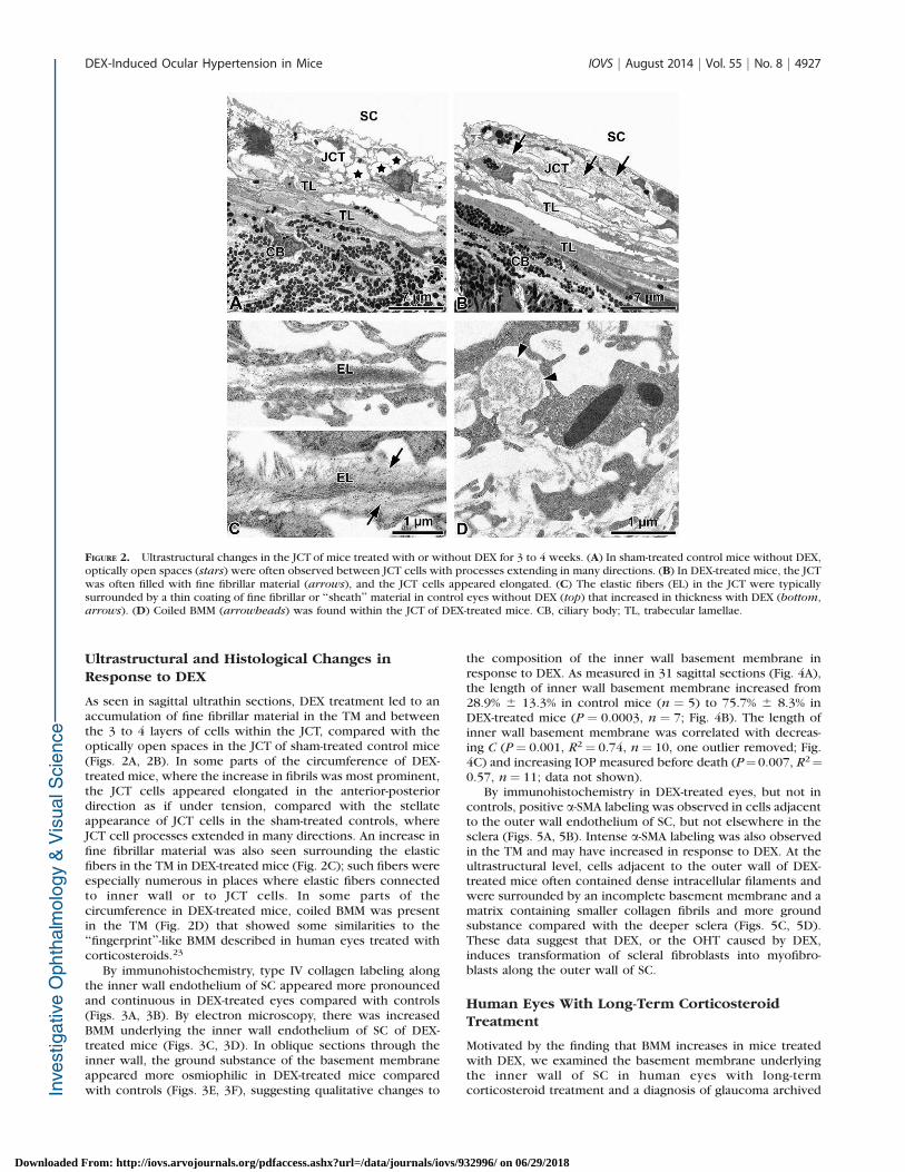
Ultrastructural and Histological Changes inResponse to DEX
As seen in sagittal ultrathin sections, DEX treatment led to anaccumulation of fine fibrillar material in the TM and betweenthe 3 to 4 layers of cells within the JCT, compared with theoptically open spaces in the JCT of sham-treated control mice(Figs. 2A, 2B). In some parts of the circumference of DEX-treated mice, where the increase in fibrils was most prominent,the JCT cells appeared elongated in the anterior-posteriordirection as if under tension, compared with the stellateappearance of JCT cells in the sham-treated controls, whereJCT cell processes extended in many directions. An increase infine fibrillar material was also seen surrounding the elasticfibers in the TM in DEX-treated mice (Fig. 2C); such fibers wereespecially numerous in places where elastic fibers connectedto inner wall or to JCT cells. In some parts of thecircumference in DEX-treated mice, coiled BMM was presentin the TM (Fig. 2D) that showed some similarities to the‘‘fingerprint’’-like BMM described in human eyes treated withcorticosteroids.23
By immunohistochemistry, type IV collagen labeling alongthe inner wall endothelium of SC appeared more pronouncedand continuous in DEX-treated eyes compared with controls(Figs. 3A, 3B). By electron microscopy, there was increasedBMM underlying the inner wall endothelium of SC of DEX-treated mice (Figs. 3C, 3D). In oblique sections through theinner wall, the ground substance of the basement membraneappeared more osmiophilic in DEX-treated mice comparedwith controls (Figs. 3E, 3F), suggesting qualitative changes to
the composition of the inner wall basement membrane inresponse to DEX. As measured in 31 sagittal sections (Fig. 4A),the length of inner wall basement membrane increased from28.9% 6 13.3% in control mice (n ¼ 5) to 75.7% 6 8.3% inDEX-treated mice (P ¼ 0.0003, n ¼ 7; Fig. 4B). The length ofinner wall basement membrane was correlated with decreas-ing C (P¼ 0.001, R2¼ 0.74, n¼ 10, one outlier removed; Fig.4C) and increasing IOP measured before death (P¼0.007, R2¼0.57, n¼ 11; data not shown).
By immunohistochemistry in DEX-treated eyes, but not incontrols, positive a-SMA labeling was observed in cells adjacentto the outer wall endothelium of SC, but not elsewhere in thesclera (Figs. 5A, 5B). Intense a-SMA labeling was also observedin the TM and may have increased in response to DEX. At theultrastructural level, cells adjacent to the outer wall of DEX-treated mice often contained dense intracellular filaments andwere surrounded by an incomplete basement membrane and amatrix containing smaller collagen fibrils and more groundsubstance compared with the deeper sclera (Figs. 5C, 5D).These data suggest that DEX, or the OHT caused by DEX,induces transformation of scleral fibroblasts into myofibro-blasts along the outer wall of SC.
Human Eyes With Long-Term CorticosteroidTreatment
Motivated by the finding that BMM increases in mice treatedwith DEX, we examined the basement membrane underlyingthe inner wall of SC in human eyes with long-termcorticosteroid treatment and a diagnosis of glaucoma archived
FIGURE 2. Ultrastructural changes in the JCT of mice treated with or without DEX for 3 to 4 weeks. (A) In sham-treated control mice without DEX,optically open spaces (stars) were often observed between JCT cells with processes extending in many directions. (B) In DEX-treated mice, the JCTwas often filled with fine fibrillar material (arrows), and the JCT cells appeared elongated. (C) The elastic fibers (EL) in the JCT were typicallysurrounded by a thin coating of fine fibrillar or ‘‘sheath’’ material in control eyes without DEX (top) that increased in thickness with DEX (bottom,arrows). (D) Coiled BMM (arrowheads) was found within the JCT of DEX-treated mice. CB, ciliary body; TL, trabecular lamellae.
DEX-Induced Ocular Hypertension in Mice IOVS j August 2014 j Vol. 55 j No. 8 j 4927
Downloaded From: http://iovs.arvojournals.org/pdfaccess.ashx?url=/data/journals/iovs/932996/ on 06/29/2018

from a previous study.23 As observed in DEX-treated mice, SCbasement membrane was more continuous in corticosteroid-treated human eyes, appearing as a dense band of ECMapproximately 0.1-lm thick immediately underlying the SCcells (Fig. 6). Quantifying the length of SC basement membranerevealed a 5- to 6-fold increase between human eyes withoutcorticosteroids (9.9% 6 4.1%) and age-matched human eyeswith SIG (55.5% 6 22.6%; P¼ 0.001, Student’s t-test; Fig. 6C).These data suggest that ultrastructural changes in the innerwall basement membrane may contribute to outflow obstruc-tion in human SIG.
DISCUSSION
This project investigated DEX-induced OHT in mice. Within 1week of systemic DEX, IOP became elevated by 2 to 3 mm Hg,and IOP elevation was sustained throughout the entire 3- to 4-
week duration of the study. Conventional outflow facility wasreduced by 52%, and there was a correlation between elevatedIOP and decreased C. These data suggest that increasedaqueous humor outflow resistance contributed to DEX-induced OHT in mice, as in other species. In mice, our resultswere consistent with previous studies of corticosteroid-induced OHT, including Whitlock et al.18 who first describedIOP elevation by systemic DEX, Zode et al.21 who reported IOPelevation in response to topical ocular DEX, and Kumar etal.19,20 who reported decreased C by subconjunctival triam-cinolone acetonide (but without IOP elevation). Ultrastructuralanalyses revealed that DEX increased ECM in the JCT, includingfine fibrillar material and BMM immediately underlying theinner wall endothelium of SC. Because the bulk of outflowresistance is likely generated near the JCT and inner wall, atleast in primates,44,45 accumulation of ECM in these tissuescould have contributed to conventional outflow obstructionfollowing DEX. Indeed, our studies revealed a correlation
FIGURE 3. The morphology of the basement membrane underlying the inner wall endothelium of SC with or without DEX treatment. (A) Byimmunofluorescence microscopy in sagittal sections, staining type IV collagen (green) was present in the TM and along the inner and outer walls ofSC (arrowheads) in sham-treated control mice, but the distribution tended to be patchy and discontinuous. (B) Dexamethasone-treated mice, incontrast, showed a more continuous labeling of type IV collagen along the inner wall of SC (arrowheads). Arrowheads indicate the inner wall of SCin (A) and (B). (C) By electron microscopy, sham-treated control mice show a patchy and discontinuous basement membrane underlying the innerwall endothelium of SC with frequent open spaces immediately underneath inner wall SC cells (stars, sagittal section). (D) Dexamethasone-treatedmice show a more continuous and dense basement membrane (arrows) underlying the inner wall of SC (sagittal section). (E) Oblique sectionsthrough the inner wall of SC in sham-treated control mice show that the basement membrane, where present, consists of a loosely arranged netfilled with granular material (arrows) interspersed between SC cell processes and open spaces. (F) Oblique sections through the inner wall of SC inDEX-treated mice reveal a net that is filled with electron-dense BMM (arrows), indicating that the inner wall basement membrane is qualitativelydifferent compared with that in sham-treated control mice shown in (E).
DEX-Induced Ocular Hypertension in Mice IOVS j August 2014 j Vol. 55 j No. 8 j 4928
Downloaded From: http://iovs.arvojournals.org/pdfaccess.ashx?url=/data/journals/iovs/932996/ on 06/29/2018

between C and length of basement membrane underlying theinner wall of SC.
The ultrastructural changes in the TM of DEX-treated miceclosely resembled the changes previously described incorticosteroid-treated human eyes. Namely, in DEX-treatedmice there was an accumulation of fine fibrillar material andcoiled BMM in the JCT that shows some similarities to the‘‘fingerprint’’-like material described in living human eyestreated with corticosteroids23,27 and in organ-cultured humaneyes perfused with DEX.28 As in corticosteroid-treated humaneyes,23 there was a pronounced increase in myofibroblastsalong the outer wall of SC in DEX-treated mice, and the fibrillarECM surrounding these myofibroblasts differed from thethicker collagen fibers in the deeper sclera. Althoughcorticosteroid-induced alterations to the inner wall basementmembrane have not been described extensively, our findings inmice motivated a reexamination of archived human SIGspecimens23 and revealed a similar increase in the basementmembrane length along SC compared with age-matchedhuman specimens that did not receive corticosteroids. Thesedata demonstrate that DEX-treated mice mimic several
hallmarks of SIG in humans, including IOP elevation, conven-tional outflow obstruction, ECM accumulation, and myofibro-blast transformation within the outflow pathway. Therefore,DEX-treated mice may provide an excellent model system forinvestigating the pathogenesis of SIG as occurs in humans.
Following DEX, there was increased thickness of thefibrillar material surrounding elastic fibers in the JCT thatpartially resembled the ‘‘sheath-derived’’ plaques described inhuman TM.46 Sheath-derived plaques become thicker with ageand with glaucoma,47 and sheath-derived–like plaque materialhas been described in the JCT of bovine eyes treated withcorticosteroids.9 Although the hydrodynamic consequences ofsheath-derived plaques remain unclear,48,49 it is generallybelieved that sheath-derived plaques play a role in TMdysfunction in POAG.50 In DEX-treated mice, however, thefine fibrillar material surrounding the elastic fibers did notexhibit the typical 50-nm banding periodicity observed forsheath-derived plaques in human TM,51 and therefore mice didnot form ‘‘true’’ sheath-derived plaques in response to DEX.The details of sheath-derived plaque development are largelyunknown, but the initial thickening of the fibrillar sheath
FIGURE 4. The basement membrane underlying the inner wall endothelium of SC becomes more continuous following systemic DEX treatment. (A)Continuous sections of the inner wall basement membrane (red brackets) were identified based on the presence of a thin basal lamina–likestructure immediately underlying the inner wall endothelium. The length of basement membrane was then divided by the total length of the innerwall endothelium (blue brackets) to calculate the basement membrane length expressed as a percentage of the total inner wall length. (B) Thebasement membrane (BM) length in DEX-treated (n¼ 7 eyes) and sham-treated control mice (n¼ 5) measured along the entire anterior-posteriorlength of SC in three independent sagittal sections per eye. The increase in BM length with DEX is statistically significant (P¼ 0.0003). Bars are SD.(C) Conventional outflow facility (C) plotted versus BM length measured in contralateral eyes of the same mouse from DEX-treated (black circles)and sham-treated control mice (white circles). The relationship was statistically significant (P¼ 0.001, R2¼ 0.74). The data point indicated by thegray circle was found to be an outlier (Cook’s distance ¼ 1.3) and was excluded from the regression analysis, but the relationship remainedsignificant even if the outlier was included (P ¼ 0.009, R2¼ 0.55). Data in (B) and (C) are from cohorts 2 and 3 only.
DEX-Induced Ocular Hypertension in Mice IOVS j August 2014 j Vol. 55 j No. 8 j 4929
Downloaded From: http://iovs.arvojournals.org/pdfaccess.ashx?url=/data/journals/iovs/932996/ on 06/29/2018

suggests that DEX-treated mice may provide a promising modelto investigate the early-stage development of sheath-derivedplaques.
Ultrastructural analysis revealed a more pronounced andcontinuous basement membrane underlying the inner wallendothelium of SC following DEX, and increased length ofinner wall basement membrane was correlated with decreasedC. These data suggested that accumulation of BMM along the
inner wall of SC might contribute to outflow obstruction inresponse to DEX. However, the resistance generated by theinner wall basement membrane on its own could not accountfor more than a small fraction of the total outflow resistance onaccount of its many discontinuities52 that are present evenwithin DEX-treated mice. It is tempting to speculate, however,that the inner wall basement membrane may channel or‘‘funnel’’ outflow through these discontinuities so as to
FIGURE 5. Myofibroblasts along the outer wall of SC in mice treated with systemic DEX. (A) In sham-treated control mice, there is dense labeling ofa-SMA in the ciliary muscle (CM), and many cells within the TM express a-SMA. (B) Following DEX, there is an increase in a-SMA labeling along theouter wall of SC (arrowheads) and possibly in the TM. (C) In sham-treated control mice, as seen by transmission electron microscopy (TEM), theouter wall of SC contains few cells between the endothelium and the deeper sclera. (D) In DEX-treated mice, as seen by TEM, the outer wall of SCreveals the presence of myofibroblasts (MF), indicated by dense cytoplasmic filaments and incomplete basement membranes (arrowheads). TheECM surrounding the MF is loosely arranged with smaller fibers (stars) relative to the deeper sclera.
FIGURE 6. Ultrastructure of the basement membrane underlying the inner wall of SC in human eyes treated with or without corticosteroidsarchived from a previous study.23 (A) Electron micrograph of SC inner wall showing a small region of basement membrane (arrows) typical fornormal human eyes without steroid treatment (65-year-old female). The amorphous material on the left side was not considered to be basementmembrane. (B) The basement membrane (arrows) underlying the inner wall endothelium of SC appears as a continuous dark band of ECM in ahuman eye treated with corticosteroids (63-year-old female). (C) The length of the inner wall basement membrane (BM) is greater in human eyesdiagnosed with SIG (n¼ 7) compared with normal human eyes without corticosteroid treatment (n¼ 5; P¼ 0.001; Student’s t-test).
DEX-Induced Ocular Hypertension in Mice IOVS j August 2014 j Vol. 55 j No. 8 j 4930
Downloaded From: http://iovs.arvojournals.org/pdfaccess.ashx?url=/data/journals/iovs/932996/ on 06/29/2018

modulate or augment resistance generation in the JCT aspreviously proposed53 as a modification to the conventionalfunneling hypothesis.54 DEX, by causing the basementmembrane to become more continuous, may have increasedthe effective outflow resistance generated in the JCT bymodulating the funneling effect. Consistent with this idea, DEXis known to stimulate basement membrane synthesis55 andturnover56–58 that may have contributed to a progressiveaccumulation of ECM in DEX-treated mice. Alternatively, DEXreduces the permeability of endothelial barriers and tightensintercellular junctions in SC cells,59,60 and changes to the innerwall basement membrane may in fact be secondary to changesin the properties of SC cells themselves. Future studiesexamining the ultrastructural changes to SC cells and basementmembrane over time following DEX may provide insight intothe mechanisms of outflow resistance elevation in corticoste-roid-induced OHT.
Previous studies of corticosteroid-induced OHT typicallydescribe a subpopulation of ‘‘nonresponders’’ who fail toexhibit IOP elevation despite prolonged steroid treatment. Inmice of mixed genetic background, Whitlock et al.18 reportedthat approximately one-third of those receiving DEX viasubcutaneous mini-pump failed to exhibit an IOP elevationexceeding 2 SDs from the baseline mean. Zode et al.,21 incontrast, reported IOP elevation in 90% or more of inbred micereceiving topical ocular DEX. Similarly, our current study usinginbred mice showed no clear evidence for a subpopulation ofsteroid nonresponders, despite some DEX-treated mice havingIOP values that fell within the range of the control population.These data suggest that steroid responsiveness may beassociated with genetic background in mice. However, withoutclear evidence for bimodality in IOP histogram, it is difficult todetermine whether the DEX-treated mice with low IOPs aretrue ‘‘nonresponders’’ or simply those individuals that fall nearthe low end of the IOP distribution where some overlap withthe baseline distribution may be expected.18
A disadvantage of the implantable mini-pumps is theinability to regulate or target DEX delivery. For instance, theserum DEX concentration varies by nearly 60% betweenindividuals despite identical mini-pump loading conditions,and it is uncertain how variability in serum DEX levels mayinfluence the IOP response or the IOP distribution in thetreated population. Furthermore, with systemic DEX treat-ment, mice experience adverse effects, including weight loss,and it would therefore be desirable to reduce the DEX dosageafter implantation so as to minimize distress and dropout.These limitations may be overcome by using alternativemethods that provide more consistent, tunable, or targetedDEX delivery to achieve sufficient IOP elevation with minimalsystemic effects.
Relationship Between IOP and C
It is useful to examine whether the reduction in C in responseto DEX was sufficient to explain the elevation in IOP.According to Goldmann’s equation, IOP may be written interms of conventional outflow resistance (1/C) as follows:
IOP ¼ 1
CðFin � FuÞ þ EVP ð2Þ
where Fin is the aqueous production rate, Fu is theunconventional outflow rate, and EVP is episcleral venouspressure. Assuming that the conventional outflow rate (Fc¼Fin
� Fu) and EVP remain constant, then Equation 2 predicts alinear relationship between IOP and 1/C across any arbitrarypopulation of mice (or any species for that matter). As shownin Figure 1D, despite DEX-treated mice having larger values of
IOP and 1/C, the entire population was well described by alinear relationship between IOP and 1/C, consistent withEquation 2. Deviations from the line of best fit were likelyattributable to measurement errors in IOP or 1/C or tofluctuations in EVP or Fc between individuals. Regardless, thestatistical significance of the correlation suggests that elevatedIOP following DEX is due to an increase in 1/C and an effective‘‘translation’’ of the population to the right (to higher values of1/C) along the regression shown in Figure 1D. According toEquation 2, the slope of the line of best fit between IOP and 1/C estimates Fc, whereas the intercept (at 1/C ¼ 0) estimatesEVP,39 yielding Fc ¼ 0.037 6 0.012 lL/min and EVP ¼ 11.3 61.3 mm Hg (mean 6 SEM). These predictions are indirect anddiffer from direct measurements of Fc (0.062 lL/min) and EVP(6.5 mm Hg) measured in the same strain of mice.61
Alternatively, one may examine the population statistics todetermine whether the mean decrease in C is sufficient toexplain the mean increase in IOP. Because Goldmann’sequation is strictly true for individual eyes and not populationsof eyes, we may take the expected value of Equation 2,neglecting any covariance between Fc and 1/C to obtain39:
IOP ¼ Fc Rþ EVP ð3Þ
where overbars indicate population averages and R representsthe average value of 1/C. Assuming that Fc and EVP areconstants and unaffected by DEX, then we may expressEquation 3 as
Rd
Rs
¼ IOPd � EVP
IOPs � EVPð4Þ
where the subscripts ‘‘d’’ and ‘‘s’’ refer to DEX-treated andsham-treated controls, respectively. Limiting the analysis toonly those data from cohorts 2 and 3 where C and IOP weremeasured in the same individuals, yields IOPd ¼17.0 6 2.3 mmHg (mean 6 SD) and Rd ¼ 130 6 39 mm Hg/lL/min for DEX-treated mice (n¼ 10), and IOPs ¼ 12.8 6 1.9 mm Hg and Rs ¼60 6 17 mm Hg/lL/min for sham-treated control mice (n ¼11). The right-hand side of Equation 4 depends on the value ofEVP, which is unknown, but if EVP ¼ 9.2 mm Hg, then therelative change in conventional outflow resistance can fullyaccount for the change in IOP with DEX. If EVP is less than 9.2mm Hg, which seems possible based on the single study thatdirectly measured EVP to be 6.5 6 0.3 mm Hg in C57BL/6mice,61 then the right-hand side of Equation 4 becomes smallerthan the left-hand side, meaning that the change in outflowresistance with DEX is larger than necessary to fully accountfor the IOP elevation. This conclusion holds even if we allowEVP to increase with DEX (motivated by the observation thatDEX increases systolic blood pressure18) and let EVP¼ 6.5 mmHg in the sham-treated control mice. This population-basedanalysis suggests that within the limitations of our measure-ment accuracy and assumptions (i.e., constant Fc), the increasein conventional outflow resistance is sufficient to explain themajority, if not all, of the elevation in IOP following systemicDEX in mice.
In summary, this project examined the mechanism for DEX-induced OHT in mice, showing that IOP elevation can belargely attributed to decreased conventional outflow facility.Obstruction of the conventional outflow pathway appears tobe related to accumulation of fine fibrillar ECM in the JCT,increased thickness of sheath material surrounding elasticfibers in the TM, and/or increased length of the basementmembrane underlying the inner wall endothelium of SC.Myofibroblasts in the conventional outflow pathway also arepresent following DEX. These data demonstrate that systemicDEX in mice causes phenotypic changes in the conventional
DEX-Induced Ocular Hypertension in Mice IOVS j August 2014 j Vol. 55 j No. 8 j 4931
Downloaded From: http://iovs.arvojournals.org/pdfaccess.ashx?url=/data/journals/iovs/932996/ on 06/29/2018

outflow pathway that mimic the hallmarks of SIG in humans,providing an animal model to study the pathogenesis of TMdysfunction and OHT related to steroid glaucoma.
Acknowledgments
Supported by an unrestricted research gift from Allergan, Inc.Additional support provided by National Glaucoma Research, aProgram of the BrightFocus Foundation (formerly as the AmericanHealth Assistance Foundation), the National Eye Institute GrantEY022359, and the Dr. Valentin Aplas-Stiftung Foundation.
Disclosure: D.R. Overby, Allergan (F); J. Bertrand, None; O.-Y.Tektas, None; A. Boussommier-Calleja, None; M. Schicht,None; C.R. Ethier, None; D.F. Woodward, Allergan (E); W.D.Stamer, None; E. Lutjen-Drecoll, Allergan (F)
References
1. Goldmann H. Cortisone glaucoma. Arch Ophthalmol. 1962;68:621–626.
2. Bernstein HN, Schwartz B. Effects of long-term systemicsteroids on ocular pressure and tonographic values. Arch
Ophthalmol. 1962;68:742–753.
3. Armaly MF. Effect of corticosteroids on intraocular pressureand fluid dynamics. Arch Ophthalmol. 1963;70:482–491.
4. Armaly MF. Statistical attributes of the steroid hypertensiveresponse in the clinically normal eye. I. the demonstration ofthree levels of response. Invest Ophthalmol. 1965;4:187–197.
5. Becker B. Intraocular pressure response to topical corticoste-roids. Invest Ophthalmol. 1965;4:198–205.
6. Becker B, Hahn KA. Topical corticosteroids and heredity inprimary open-angle glaucoma. Am J Ophthalmol. 1964;57:543–551.
7. Clark AF, Wordinger RJ. The role of steroids in outflowresistance. Exp Eye Res. 2009;88:752–759.
8. Gerometta R, Podos SM, Candia OA, et al. Steroid-inducedocular hypertension in normal cattle. Arch Ophthalmol. 2004;122:1492–1497.
9. Tektas O-Y, Hammer CM, Danias J, et al. Morphologic changesin the outflow pathways of bovine eyes treated withcorticosteroids. Invest Ophthalmol Vis Sci. 2010;51:4060–4066.
10. Mao W, Tovar-Vidales T, Yorio T, Wordinger RJ, Clark AF.Perfusion-cultured bovine anterior segments as an ex vivomodel for studying glucocorticoid-induced ocular hyperten-sion and glaucoma. Invest Ophthalmol Vis Sci. 2011;52:8068–8075.
11. Zhan GL, Miranda OC, Bito LZ. Steroid glaucoma: corticoste-roid-induced ocular hypertension in cats. Exp Eye Res. 1992;54:211–218.
12. Bhattacherjee P, Paterson CA, Spellman JM, Graff G, Yanni JM.Pharmacological validation of a feline model of steroid-induced ocular hypertension. Arch Ophthalmol. 1999;117:361–364.
13. Ticho U, Lahav M, Berkowitz S, Yoffe P. Ocular changes inrabbits with corticosteroid-induced ocular hypertension. Br J
Ophthalmol. 1979;63:646–650.
14. Candia OA, Gerometta R, Millar JC, Podos SM. Suppression ofcorticosteroid-induced ocular hypertension in sheep byanecortave. Arch Ophthalmol. 2010;128:338–343.
15. Gerometta R, Spiga M-G, Borras T, Candia OA. Treatment ofsheep steroid–induced ocular hypertension with a glucocor-ticoid-inducible MMP1 gene therapy virus. Invest Ophthalmol
Vis Sci. 2010;51:3042–3048.
16. Gerometta R, Podos SM, Danias J, Candia OA. Steroid-inducedocular hypertension in normal sheep. Invest Ophthalmol Vis
Sci. 2008;50:669–673.
17. Miyara N, Shinzato M, Yamashiro Y, Iwamatsu A, Kariya K-I,Sawaguchi S. Proteomic analysis of rat retina in a steroid-induced ocular hypertension model: potential vulnerability tooxidative stress. Jpn J Ophthalmol. 2008;52:84–90.
18. Whitlock NA, McKnight B, Corcoran KN, Rodriguez LA, RiceDS. Increased intraocular pressure in mice treated withdexamethasone. Invest Ophthalmol Vis Sci. 2010;51:6496–6503.
19. Kumar S, Shah S, Tang HM, Smith M, Borras T, Danias J. Tissueplasminogen activator in trabecular meshwork attenuatessteroid induced outflow resistance in mice. PLoS One. 2013;8:e72447.
20. Kumar S, Shah S, Deutsch ER, Tang HM, Danias J. Triamcin-olone acetonide decreases outflow facility in C57BL/6 mouseeyes. Invest Ophthalmol Vis Sci. 2013;54:1280–1287.
21. Zode GS, Sharma AB, Lin X, et al. Ocular-specific ER stressreduction rescues glaucoma in murine glucocorticoid-inducedglaucoma. J Clin Invest. 2014;124:1956–1965.
22. Fingert JH, Clark AF, Craig JE, et al. Evaluation of the myocilin(MYOC) glaucoma gene in monkey and human steroid-induced ocular hypertension. Invest Ophthalmol Vis Sci.2001;42:145–152.
23. Johnson D, Gottanka J, Flugel C, Hoffmann F, Futa R, Lutjen-Drecoll E. Ultrastructural changes in the trabecular meshworkof human eyes treated with corticosteroids. Arch Ophthalmol.1997;115:375–383.
24. Clark AF, Steely HT, Dickerson JE, et al. Glucocorticoidinduction of the glaucoma gene MYOC in human and monkeytrabecular meshwork cells and tissues. Invest Ophthalmol Vis
Sci. 2001;42:1769–1780.
25. Johnson DH, Knepper PA. Microscale analysis of the glycos-aminoglycans of human trabecular meshwork: a study inperfusion cultured eyes. J Glaucoma. 1994;3:58–69.
26. Johnson DH, Bradley JM, Acott TS. The effect of dexameth-asone on glycosaminoglycans of human trabecular meshworkin perfusion organ culture. Invest Ophthalmol Vis Sci. 1990;31:2568–2571.
27. Rohen JW, Linner E, Witmer R. Electron microscopic studieson the trabecular meshwork in two cases of corticosteroid-glaucoma. Exp Eye Res. 1973;17:19–31.
28. Clark AF, Wilson K, De Kater AW, Allingham RR, McCartneyMD. Dexamethasone-induced ocular hypertension in perfu-sion-cultured human eyes. Invest Ophthalmol Vis Sci. 1995;36:478–489.
29. Smith RS, John SWM, Nishina PM, Sundberg JP. Systematic
Evaluation of the Mouse Eye. Boca Raton, FL: CRC Press;2001.
30. Overby DR, Bertrand J, Schicht M, Paulsen F, Stamer WD,Lutjen-Drecoll E. The structure of the trabecular meshwork, itsconnections to the ciliary muscle and the effect of pilocarpineon outflow facility in mice. Invest Ophthalmol Vis Sci. 2014;55:3727–3736.
31. Savinova OV, Sugiyama F, Martin JE, et al. Intraocular pressurein genetically distinct mice: an update and strain survey. BMC
Genet. 2001;2:12.
32. Aihara M, Lindsey JD, Weinreb RN. Aqueous humor dynamicsin mice. Invest Ophthalmol Vis Sci. 2003;44:5168–5173.
33. Lei Y, Overby DR, Boussommier-Calleja A, Stamer WD, EthierCR. Outflow physiology of the mouse eye: pressure depen-dence and washout. Invest Ophthalmol Vis Sci. 2011;52:1865–1871.
34. Boussommier-Calleja A, Bertrand J, Woodward DF, Ethier CR,Stamer WD, Overby DR. Pharmacologic manipulation ofconventional outflow facility in ex vivo mouse eyes. Invest
Ophthalmol Vis Sci. 2012;53:5838–5845.
35. Li G, Farsiu S, Chiu SJ, et al. Pilocarpine-induced dilation ofSchlemm’s canal and prevention of lumen collapse at elevated
DEX-Induced Ocular Hypertension in Mice IOVS j August 2014 j Vol. 55 j No. 8 j 4932
Downloaded From: http://iovs.arvojournals.org/pdfaccess.ashx?url=/data/journals/iovs/932996/ on 06/29/2018

intraocular pressures in living mice visualized by OCT. Invest
Ophthalmol Vis Sci. 2014;55:3737–3746.
36. Wang WH, Millar JC, Pang I-H, Wax MB, Clark AF. Noninvasivemeasurement of rodent intraocular pressure with a reboundtonometer. Invest Ophthalmol Vis Sci. 2005;46:4617–4621.
37. Johnson TV, Fan S, Toris CB. Rebound tonometry in conscious,conditioned mice avoids the acute and profound effects ofanesthesia on intraocular pressure. J Ocul Pharmacol Ther.2008;24:175–185.
38. Camras LJ, Sufficool KE, Camras CB, Fan S, Liu H, Toris CB.Duration of anesthesia affects intraocular pressure, but notoutflow facility in mice. Curr Eye Res. 2010;35:819–827.
39. Boussommier-Calleja A, Overby DR. The influence of geneticbackground on conventional outflow facility in mice. Invest
Ophthalmol Vis Sci. 2013;54:8251–8258.
40. Stamer WD, Lei Y, Boussommier-Calleja A, Overby DR, EthierCR. eNOS, a pressure-dependent regulator of intraocularpressure. Invest Ophthalmol Vis Sci. 2011;52:9438–9444.
41. Overby D, Gong H, Qiu G, Freddo TF, Johnson M. Themechanism of increasing outflow facility during washout inthe bovine eye. Invest Ophthalmol Vis Sci. 2002;43:3455–3464.
42. Ito S, Karnovsky MJ. Formaldehyde-glutaraldehyde fixativescontaining trinitro compounds. J Cell Biol. 1968;39:168A–169A.
43. Dean RB, Dixon WJ. Simplified statistics for small numbers ofobservations. Anal Chem. 1951;23:636–638.
44. Lutjen-Drecoll E. Structural factors influencing outflow facilityand its changeability under drugs. A study in Macaca
arctoides. Invest Ophthalmol. 1973;12:280–294.
45. Maepea O, Bill A. Pressures in the juxtacanalicular tissue andSchlemm’s canal in monkeys. Exp Eye Res. 1992;54:879–883.
46. Rohen JW, Witmer R. Electron microscopic studies on thetrabecular meshwork in glaucoma simplex. Albrecht Von
Graefes Arch Klin Exp Ophthalmol. 1972;183:251–266.
47. Lutjen-Drecoll E, Shimizu T, Rohrbach M, Rohen JW. Quanti-tative analysis of ‘‘plaque material’’ in the inner and outer wallof Schlemm’s canal in normal and glaucomatous eyes. Exp Eye
Res. 1986;42:443–455.
48. Alvarado JA, Yun AJ, Murphy CG. Juxtacanalicular tissue inprimary open angle glaucoma and in nonglaucomatousnormals. Arch Ophthalmol. 1986;104:1517–1528.
49. Murphy CG, Johnson M, Alvarado JA. Juxtacanalicular tissue inpigmentary and primary open angle glaucoma. The hydrody-
namic role of pigment and other constituents. Arch Oph-
thalmol. 1992;110:1779–1785.
50. Tektas O-Y, Lutjen-Drecoll E. Structural changes of thetrabecular meshwork in different kinds of glaucoma. Exp
Eye Res. 2009;88:769–775.
51. Lutjen-Drecoll E, Futa R, Rohen JW. Ultrahistochemical studieson tangential sections of the trabecular meshwork in normaland glaucomatous eyes. Invest Ophthalmol Vis Sci. 1981;21:563–573.
52. Johnson M. What controls aqueous humour outflow resis-tance? Exp Eye Res. 2006;82:545–557.
53. Overby DR, Stamer WD, Johnson M. The changing paradigm ofoutflow resistance generation: Towards synergistic models ofthe JCT and inner wall endothelium. Exp Eye Res. 2009;88:656–670.
54. Johnson M, Shapiro A, Ethier CR, Kamm RD. Modulation ofoutflow resistance by the pores of the inner wall endothelium.Invest Ophthalmol Vis Sci. 1992;33:1670–1675.
55. Dickerson JE, Steely HT, English-Wright SL, Clark AF. The effectof dexamethasone on integrin and laminin expression incultured human trabecular meshwork cells. Exp Eye Res.1998;66:731–738.
56. Snyder RW, Stamer WD, Kramer TR, Seftor RE. Corticosteroidtreatment and trabecular meshwork proteases in cell andorgan culture supernatants. Exp Eye Res. 1993;57:461–468.
57. Samples JR, Alexander JP, Acott TS. Regulation of the levels ofhuman trabecular matrix metalloproteinases and inhibitor byinterleukin-1 and dexamethasone. Invest Ophthalmol Vis Sci.1993;34:3386–3395.
58. Flugel-Koch C, Ohlmann A, Fuchshofer R, Welge-Lussen U,Tamm ER. Thrombospondin-1 in the trabecular meshwork:localization in normal and glaucomatous eyes, and inductionby TGF-beta1 and dexamethasone in vitro. Exp Eye Res. 2004;79:649–663.
59. Underwood JL, Murphy CG, Chen J, et al. Glucocorticoidsregulate transendothelial fluid flow resistance and formationof intercellular junctions. Am J Physiol. 1999;277:C330–C342.
60. Fujimoto T, Inoue T, Kameda T, et al. Involvement of rhoA/rho-associated kinase signal transduction pathway in dexametha-sone-induced alterations in aqueous outflow. Invest Ophthal-
mol Vis Sci. 2012;53:7097–7108.
61. Lee YS, Tresguerres M, Hess K, et al. Regulation of anteriorchamber drainage by bicarbonate-sensitive soluble adenylylcyclase in the ciliary body. J Biol Chem. 2011;286:41353–41358.
DEX-Induced Ocular Hypertension in Mice IOVS j August 2014 j Vol. 55 j No. 8 j 4933
Downloaded From: http://iovs.arvojournals.org/pdfaccess.ashx?url=/data/journals/iovs/932996/ on 06/29/2018