Ultrasound in the 3rd trimester · Routine scan versus serendipitous finding ??? •The detection...
108
Ultrasound in the 3 rd trimester Interesting cases
Transcript of Ultrasound in the 3rd trimester · Routine scan versus serendipitous finding ??? •The detection...

Ultrasound in the 3rd trimester
Interesting cases

Routine scan versus serendipitous finding ???
• The detection of fetal structural abnormalities is a routine part of antenatal care.
• An additional ultrasound for fetal structural anomalies in the 3rd trimester
seems important for many reasons ????? • Some abnormalities develop or first become apparent later in pregnancy. • A 1998 to 2008 study scanned 5044 fetuses between 28 and 32 weeks and
they reported an additional 44 (15 %) structural abnormalities were found in the 3rd trimester.

Prospective study
• 1st group structural abnormalities of the urogenital system (n = 18),
– mainly cases of hydronephrosis
– Unilateral renal agenesis (2),
– dysplastic kidneys (3),
– pelvic kidney (1)
• 2nd group abnormalities of the heart (n = 9),
– mainly small, muscular ventricular septal defects (n = 7)
– Infantile arterial calcification
– Pulmonary stenosis.

Prospective study • 3rd group abnormal findings pertaining to the gastrointestinal system (n = 6).
– Pyloric stenosis (1),
– functional diaphragmatic hernia (1),
– splenic cyst (1),
– neuroblastoma (1),
– Meconium peritonitis (1),
– intraabdominal cyst (1)
• 4th group abnormalities involving the central nervous system (4)
– hydrocephalus (n = 2),
– spina bifida (n = 1)
– corpus callosum agenesis (n = 1).

Prospective study
• 5th miscellaneous
– achondroplasia
– late onset hydrothorax in combination with trisomy 21 without any other associated anomalies and without an increased risk according to first trimester screening and NT measurement
– ovarian cysts (n = 2)
– hemangiomas (n = 1)
– late hydrops (n = 2)

Do you feel that this is a reasonable snap shot??????

My perusal of our database
• No “new” diagnosis of a renal abnormality – Progression of known defects – Development of “typical features” of
suspected or known familial cystic disease
• 2 late T18 • Fetal ovarian cysts • Anaemia ascites and hydrops • Progression of suspected cardiac defects • Achondroplasia • Recto vesical fistula • Hypospadius • Hydrometrocolpos
• Vaginal atresia • McKusick Kaufman • Oesophageal atresia • Missed BWS • Duodenal atresia • Bowel obstruction • Intracranial haemorrhage • Hydrocephalus
– Progression – New diagnosis
• ACC complications • Connatal cysts • Fetal tumour

Progression of known renal anomalies
Duplex Kidneys and PUJ obstruction
• Obstruction of the upper pole moiety of a duplex system
• Upper pole moiety ureter is the one that will have the ureterocoele
• Ureterocoeles may be missed
• Ureterocoeles may prolapse and cause bladder outlet obstruction
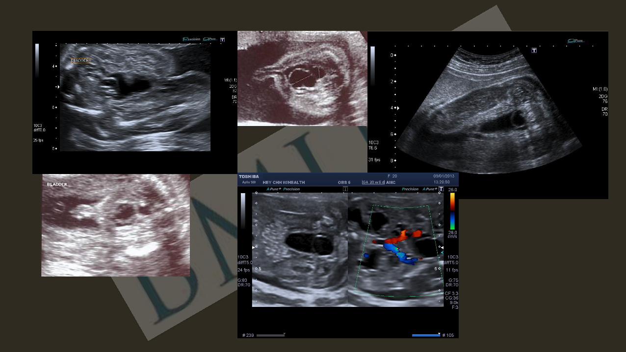

Duplex Kidney: hydro ureter upper pole moiety
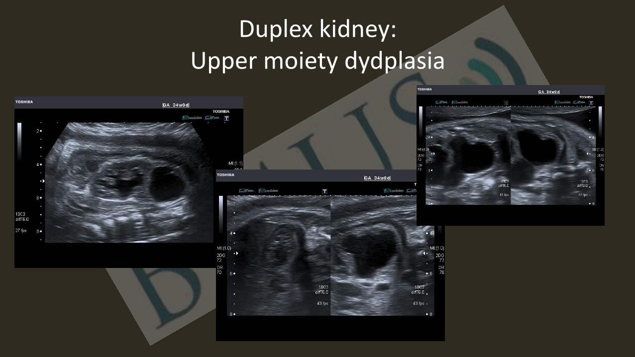
Duplex kidney: Upper moiety dydplasia
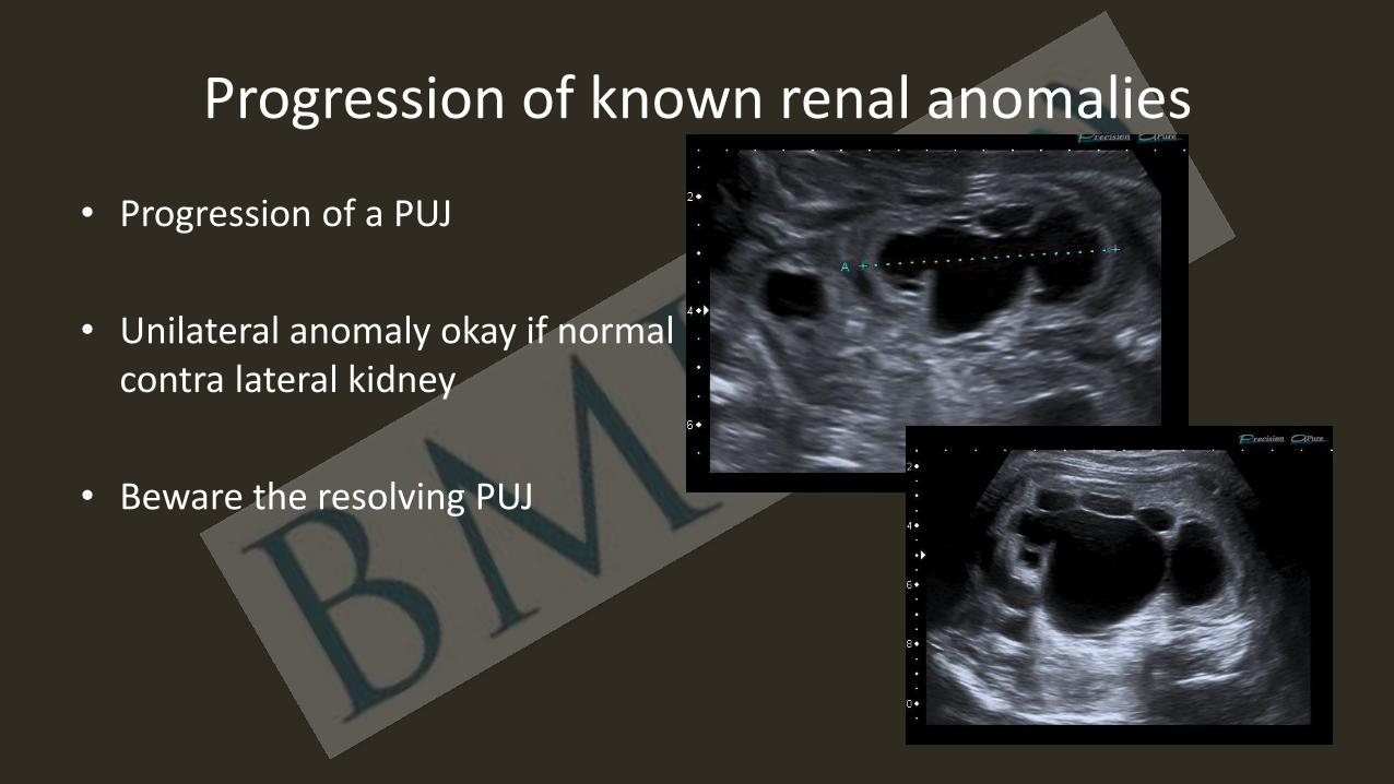
Progression of known renal anomalies
• Progression of a PUJ
• Unilateral anomaly okay if normal contra lateral kidney
• Beware the resolving PUJ

Marked hydronephrosis

Progression of known renal anomalies
Development of a urinoma
• Severe obstruction: usually PUV or PUJ
• Usually a damaged kidney
• Complex mechanism worsening the renal dysplasia

Progression of known renal anomalies
Development of renal dysplasia
– Lack of normal corticomedullary differentiation
– Development of cortical cysts
– Small echogenic kidney
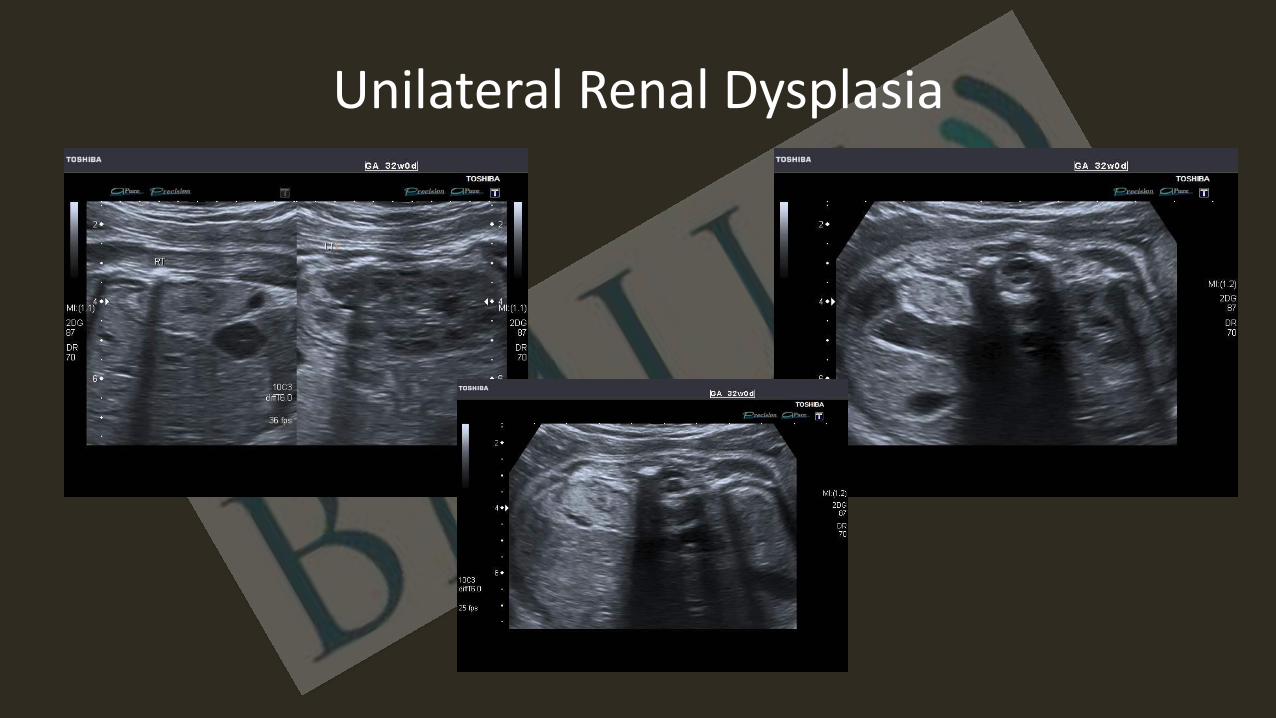
Unilateral Renal Dysplasia

Progression of known renal anomalies
• Fully blown picture of
– ADPDK
– ARPDK

Progression of known renal anomalies
• Fully blown picture of
– ADPDK
– ARPDK

Cessation of function of ectopic kidneys

Bilateral renal ectopia

Bilateral renal ectopia

Bilateral renal ectopia

Missed T18
Polyhydramnios
• Multiplicity of causes
• The greater the degree the more likely that there is an underlying structural abnormality
Reduced fetal movement
• Perception issue due to polyhydramnios
• True arthrogryposis will be associated with polyhydramnios
• Two lethal causes of talipes both associated with polhydramnios – T18
– FADS

Late T18

Late T18

Fetal ovarian cysts
• Ovarian cysts are the most frequent, prenatally diagnosed intra-abdominal cysts.
• The most widely accepted aetiological hypothesis being that the ovary produces them under the influence of various hormones
– fetal gonadotrophins,
– maternal oestrogen
– placental human chorionic gonadotrophin.

Fetal ovarian cysts • These cysts have their origin for the
most part in the follicle epithelium; they can also be theca-lutein cysts, corpus luteum cysts or simple cysts of unknown origin
• The association of fetal ovarian cysts with maternal diabetes or hypothyroidism has also been described.
• In the majority of cases of ovarian cysts (> 50%) the natural course is spontaneous resolution either pre- or postnatally
• A normal ovarian cyst has a smooth border SINGLE wall and is without an internal structure.

Fetal ovarian cysts
• When complicated by bleeding or torsion, the cyst acquires a heterogeneous structure
• Bleeding within an ovarian cyst usually arises in connection with torsion
• A possible further sign of torsion is the emergence of fetal tachycardia due to peritoneal irritation.
• Torsion is also the most serious complication of a fetal ovarian cyst, as it usually requires postnatal ovariectomy.

Complicated Ovarian Cyst

Development of fetal anaemia, ascites and hydrops
• Rhesus disease
– Known and routine follow up
– anti Kell
• Parvovirus
– Cardiomegaly
– Big placenta
– Ascites
• Polyhydramnios
• Reduced fetal movement

Ascites
• A very thin rim presenting as a thin black line on the inner aspect of the abdomen is a normal appearance which can be mistaken for ascites.
• Otherwise any free fluid in the abdominal cavity is abnormal. • The presence of persistent isolated ascites is most commonly associated
with intra abdominal pathology, although this is uncommon. • The two most common associations are urinary ascites secondary to
obstructive uropathy and meconium peritonitis. • It may also rarely be associated with liver disease and metabolic storage
disease. • Abdominal ascites may be an early manifestation of hydrops fetalis and
hence cardiac, other fetal structural and karyotypical abnormalities should be excluded.

Ascites
• Isolated ascites
– Early manifestation of fetal hydrops especially when anaemia is the underlying cause
– Parvovirus B19

Fetal anaemia Subtle ascites

Ascites Anaemia

Fetal anaemia

Splenomegaly

Ascites
Persistent isolated ascites
Intra abdominal pathology
Obstructive uropathy
Meconium peritonitis

Urinary Ascites

Progression of known fetal cardiac defects
• Suspected coarctation
• Suspected cardiomyopathy
• Development of pulmonary stenosis
• Development of an additional anomaly in Di George

Idiopathic Infantile Arterial Calcification

20 weeks RVD
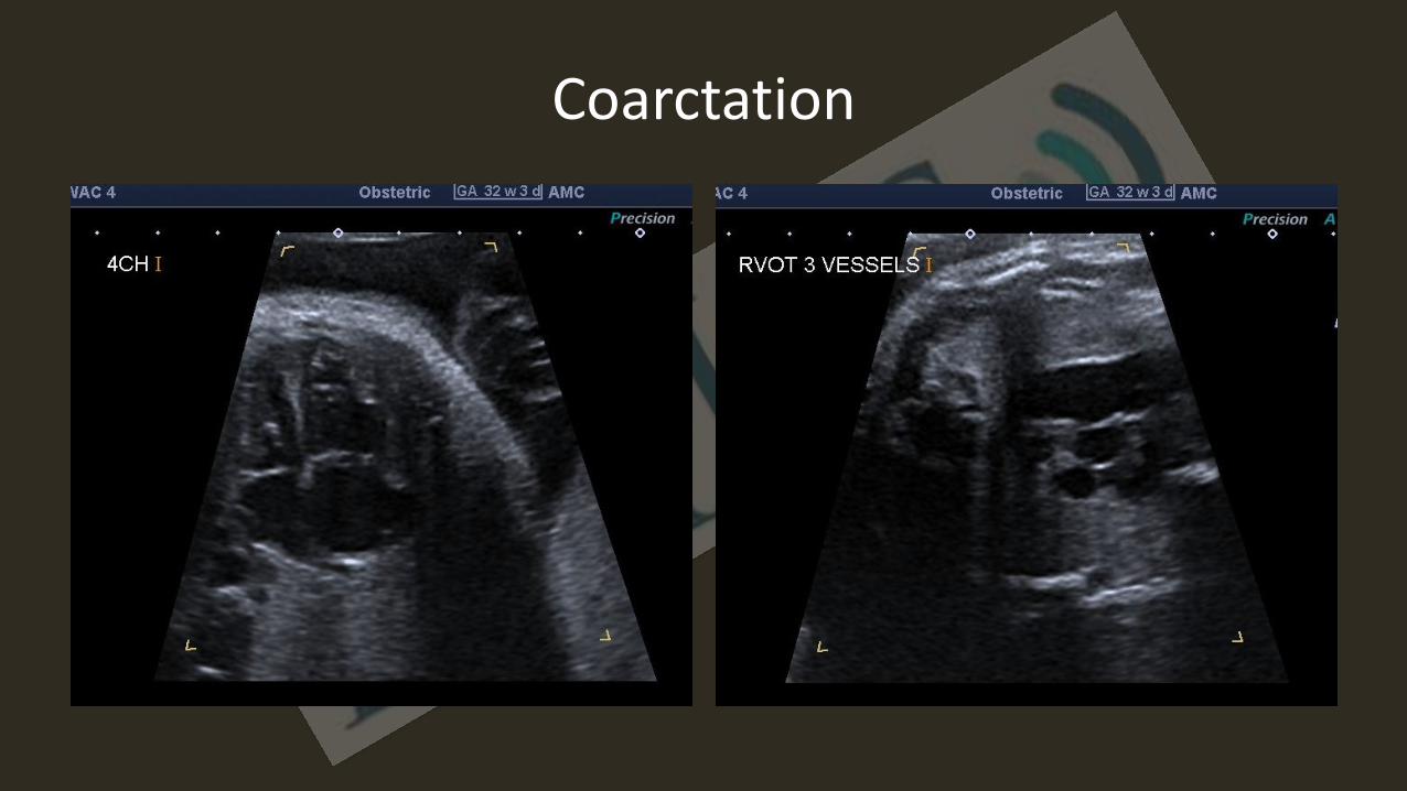
Coarctation

Something not quite right !!!!

Cardiac enlargement
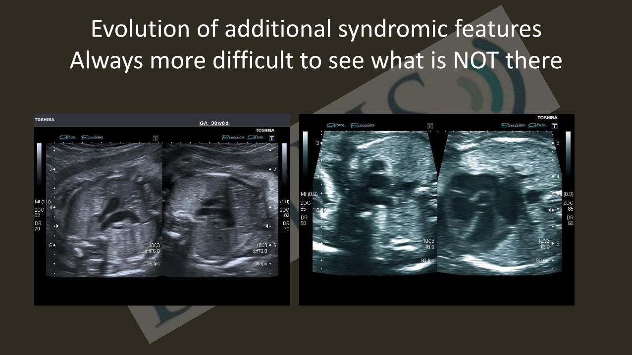
Evolution of additional syndromic features Always more difficult to see what is NOT there

Evolution of additional syndromic features

Evolution of additional syndromic features

Achondroplasia
• Normal 20 and often 24 week scan
• 3rd trimester polyhydramnios

Achondroplasia
• Normal 20 and often 24 week scan
• 3rd trimester polyhydramnios

Achondroplasia

Achondroplasia

Fetal bowel calcification
• Intraluminal calcification of meconium is rare and is a result of mixing of meconium and stagnant alkaline fetal urine. It is the pathognomonic sign of a fistula between the bowel and the fetal renal tract.
• Enterolithiasis is seen in association with the following bowel pathologies once a fistula has been established with the fetal urinary tract
imperforate anus,
gastrointestinal atresia/stenosis,
functional ileal obstruction
total colonic Hirschsprung’s disease.

Enterolithiasis

Hypospadius
• Hypospadias is an anomaly of the male urogenital tract with an incidence of between 0.2 and 4.1 per 1000 live births.
• The term is derived from the Greek language and refers to a rent (spadon) on the ventrum of the penis.
• The anomaly occurs as a result of failure of complete development of the anterior urethra.
• The urethra may terminate just proximal to the glans (glandular hypospadias), at some point along the penile shaft (penile hypospadias), at the anterior margin of the scrotum (penoscrotal hypospadias), or in the perineum (perineal hypospadias)
Hypospadius
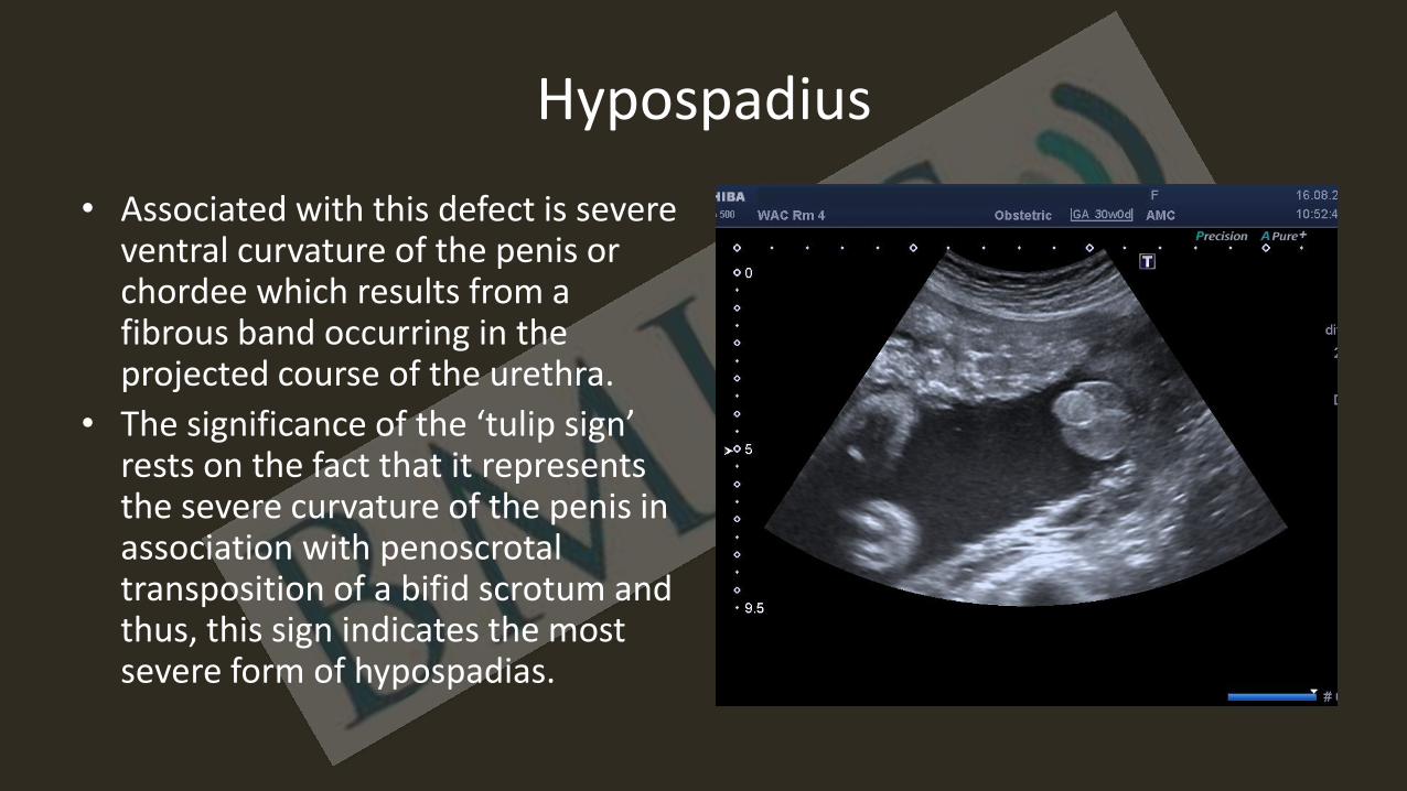
• Associated with this defect is severe ventral curvature of the penis or chordee which results from a fibrous band occurring in the projected course of the urethra.
• The significance of the ‘tulip sign’ rests on the fact that it represents the severe curvature of the penis in association with penoscrotal transposition of a bifid scrotum and thus, this sign indicates the most severe form of hypospadias.

Beware late diagnosis

Vaginal atresia
Hydrocolpos involves the dilatation of the vagina due to obstruction of the genital tract, leading to accumulation of secretions. This condition may be associated with distension of the uterus (hydrometrocolpos). The presence of prenatally detected hydro(metro)colpos warrants a systematic evaluation of fetal and neonatal anatomy to rule out a large variety of possibly associated malformations or syndromes

Hydrometrocolpos
• Hydrometrocolpos is a rare anomaly, but accounts for 15% of abdominal masses in female infants, surpassed in frequency only by hydronephrosis
• Vaginal outflow obstruction leading to accumulation of secretions and subsequent hydro(metro)colpos might be secondary to the presence of a transverse vaginal septum, vaginal atresia or imperforate hymen, often as a result of Müllerian duct failure

Hydrometrocolpos
• Since the primitive cloacal membrane differentiates into the sinus urogenitalis and the anorectal canal, the very close relation of the two regions is obvious
• This is supported by the frequent association of hydrometrocolpos with imperforate anus
• During pregnancy hydrometrocolpos is frequently accompanied by urinary tract obstruction, causing hydronephrosis and oligohydramnios

Hydrometrocolpos
It requires a systematic evaluation of the fetus, as a high number of these females may present with associated malformations and syndromes, such as
• McKusick–Kaufman,
• Ellis–van Creveld
• Bardet–Biedl syndromes

McKusick Kaufman
Introduction This is an autosomal recessive syndrome with variable expression within the same family, different families and different sex. Heterozygous are asymptomatic. There is a known mutation in 20p12 in the MKKS gene. The syndrome is more frequent in females, In males, the findings are limited to finger abnormalities and sometimes genital abnormalities such as hypospadias or micropenis. The final diagnosis cannot be made until at least age five years.

McKusick Kaufman
Findings in affected females include • hydrometrocolpos, • ascites • postaxial polydactyly • occasionally cardiac defects. Hydrometrocolpos can be caused by failure of the distal third of vagina to develop, a transverse vaginal membrane or imperforate hymen. Cardiovascular malformations include atrial and ventricular septal defect, AV canal, small aorta and hypoplastic left ventricle, tetralogy of Fallot and patent ductus arteriosus. Retrograde flow of the secretion collected in the vagina into the peritoneal cavity can lead to fetal ascites. The increased vaginal secretion is caused by an high level of maternal estrogen.

McKusick Kaufman
Differential Diagnosis Bardet Biedl syndrome, phenotypic differences between these two syndromes become evident later in life, usually after the age of 4-5 years. McKusick-Kaufman syndrome tends to be benign when not associated with cardiac defects. The close relationship between Bardet Biedl syndrome and McKusick-Kaufman syndrome has been further complicated by alterations in the MKKS gene in both McKusick-Kaufman syndrome and in 4-6 % of individuals with Bardet Biedl syndrome. Highlighting this diagnostic limitation at the time of prenatal diagnosis is crucial during the counseling.

McKusick Kaufman

McKusick Kaufman

McKusick Kaufman

Oesophageal atresia
The prenatal diagnosis of oesophageal atresia is classically made on the basis of failure to visualise the fetal stomach on serial ultrasound scans usually in the presence of polyhydramnios. A small stomach may be seen even in complete oesophageal atresia due to distension by gastric secretions. The diagnosis must however be considered if a small stomach is seen in the presence of polyhydramnios in the 2nd or 3rd trimesters after exclusion of other causes of absent or small fetal stomach such as • 1. congenital diaphragmatic hernia, • 2. impaired fetal swallowing associated with
congenital myopathy or neurologic disorder.

Tracheo oesophageal atresia/ fistula
• The prenatal detection rates of oesophageal atresia are poor: 9-24%.
• The combination of a small stomach and increased liquor has a positive predictive value of 56% for oesophageal atresia.
• The finding of an oesophageal pouch has a high specificity for atresia this sign may not be present until after 26 weeks.

Tracheo oesophageal atresia/ fistula
In 60% of cases there will be other defects, – 25% cardiac – 14% skeletal – 10% will have VACTERL.
The incidence of aneuploidy is 10-20%
– T13 – T21.
Growth scans will be necessary to monitor growth [20% of fetuses will be small for gestational age]. Liquor will need to be monitored also.
Small fetal stomach
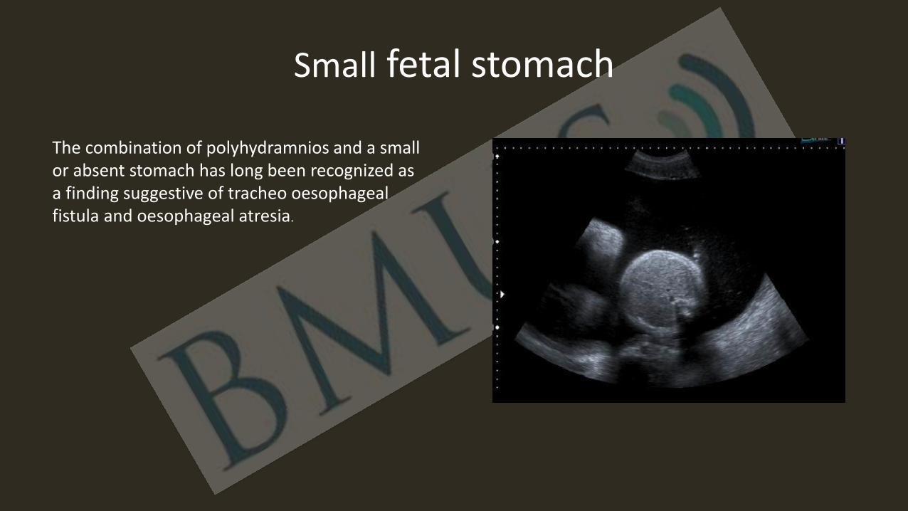
The combination of polyhydramnios and a small or absent stomach has long been recognized as a finding suggestive of tracheo oesophageal fistula and oesophageal atresia.

Oesophageal atresia
• In cases of trachea-oesophageal fistula, the diagnosis (if it is made at all) is not made until the development of polyhydramnios in the third trimester as fluid can pass via the trachea through the fistula into the distal oesophagus and lead to a normal stomach appearance.
• Studies have assessed the ability to detect oesophageal atresia on ultrasound by visualisation of a small or absent stomach and the presence of polyhydramnios.
• The ultrasound appearances have a positive predictive value of 56%. The sensitivity of prenatal ultrasound to detect the abnormality is 42%.
• In some cases it may be possible to make the diagnosis by visualisation of the upper oesophageal “pouch” on fetal swallowing which has been reported as being visualised in some cases as early as 23 weeks.

Omphalocoele
• Beckwith Wiedemann Syndrome BWS – Macrosomia – Renal enlargement and increased renal echogenicity – macroglossia – Placental mesenchymal dysplasia.
• Meckel Gruber, Pentalogy of Cantrell and Cloacal abnormalities were excluded
• Chromosomal abnormalities such as T18 and T13 • Conotruncal cardiac anomaly which can be seen in association with
non syndromic and non chromosomal omphalocoeles

Beckwith Wiedemann Syndrome BWS
2 major diagnostic criteria or a combination of 2 minor and 1 major criteria at the time of diagnosis
Major Minor
• Macroglossia Aneuploidy abnormal loci
• Macrosomia (>90th percentile) Polyhydramnios
• Abdominal wall defect Nephromegaly Dysgenesis adrenal enlargement

Beckwith Wiedemann Syndrome BWS,
The 2 most consistent findings on prenatal ultrasound are • 2nd trimester polyhydramnios • Macrosomia > 90th centile, whilst 87% of affected neonates with be
macrocosmic, macrosomia is only seen in 50% of cases prenatally • The exact onset of the macrosomia is unknown but tit may
manifest as accelerated growth at any time form 25 to 30 weeks but may only exceed the 90th centile after 36 weeks of gestation
• An omphalocoele complicates 50% of cases of BWS but actually < 3% of all cases of fetuses with an omphalocoele will be diagnosed as BWS

Beckwith Wiedemann Syndrome BWS

Beckwith Wiedemann Syndrome BWS

Beckwith Wiedemann Syndrome BWS

Beckwith Wiedemann Syndrome BWS

Duodenal atresia
• Double bubble sonographically
• NB normal gall bladder!!
• Late 2nd /3rd trimester diagnosis
– Isolated
– T21 30%
– VATER

Duodenal atresia
• Atresia
• Duodenal web
• Ladd’s bands
• Prognosis generally good provided no other anomaly


Jejunal and Ileal atresia
• Small bowel atresia is visualised prenatally in 1:5000 births and with duodenal atresia make up the most common causes of intestinal obstruction in live born neonates.
• The most common site for the lesion is the proximal jejunum or distal ileum.
• Small bowel atresia is thought to result from intrauterine vascular “accidents” such as volvulus, intussusception or vascular constriction/hypoperfusion.
• Atresia may be seen in association with other bowel anomalies such as malrotation, gastroschisis, duplication and meconium ileus.
• Additional non-intestinal structural abnormalities and karyoptypic anomalies are uncommon
Jejunal and Ileal atresia
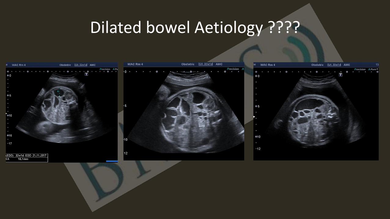
• The diagnosis is made by seeing persistently dilated fluid filled bowel loops proximal to the obstruction.
• Classically jejunal atresia has a triple bubble appearance and can be associated with polyhydramnios (more common the more proximal the atresia) and ileal atresia has a honeycomb appearance.
• The diagnosis is not usually made until late in the second or third trimester. There is no evidence to support early delivery. In isolated cases overall survival is good and the degree of morbidity tends to relate to the length of gut remaining post surgery.
• The neonate requires initial nasogastric decompression and intravenous fluids while imagining studies are performed to confirm the diagnosis and exclude malrotation or volvulus.

Large bowel atresia
Colonic atresia is rare accounting for less than 10-15% of all intestinal atresias and is estimated to occur in 1:20,000 live births. The underlying pathogenesis is thought to be the same as for small bowel atresia. 50% of cases will be due to obstruction proximal to and 50% of cases will be due to obstruction distal to the splenic flexure. Colonic atresia may be an isolated condition but will be seen in association with gastroschisis , imperforate anus and Hirschsprung’s disease. The sonographic features are simply those of large bowel dilatation. Usually isolated large bowel obstruction does not affect the on-going pregnancy

Large bowel atresia
Sonographic features which facilitate the differentiation of small versus large bowel obstruction • Jejunal atresia
– dilated loops should be traced down from the stomach
– fluid within the loops is usually quite hypo echoic – more likely to have polyhydramnios
• Large bowel obstruction – peripherally located dilated loops are traced down
to the rectum – haustral pattern may be seen – fluid within is more echogenic due to meconium
+/- enterolithiasis – the liquor volume is usually normal – ascites and meconium peritonitis are more likely

Dilated bowel Aetiology Unknown
Dilated bowel Aetiology ????

Intra cranial haemorrhage
• Symptomatic intracranial haematoma
in a full-term neonate is considered a fairly unusual event especially in a well baby with a normal clotting mechanism. The incidence in the fetal population is unknown but it is a reported phenomenom
• The ultrasound appearance of an intracranial haematoma will be variable depending on the amount of haemorrhage that has occurred and the age of the haemorrhage relative to the timing of the US examination.
Intra cranial haemorrhage
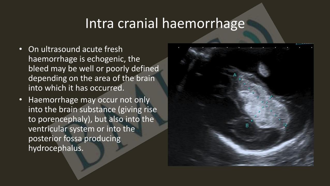
• On ultrasound acute fresh haemorrhage is echogenic, the bleed may be well or poorly defined depending on the area of the brain into which it has occurred.
• Haemorrhage may occur not only into the brain substance (giving rise to porencephaly), but also into the ventricular system or into the posterior fossa producing hydrocephalus.

Intra cranial haemorrhage
• The prognosis is determined by site and extent and pooled data from several series show a poor outcome in 68% of all cases.
• The recognition of porencephaly or haemorrhage in utero should prompt a search for the underlying aetiology.
• In the absence of apparent other causes, haematological, infectious and traumatic causes should be sought.

Hydrocephalus
• Progression of known ventriculomegaly
• True 3rd trimester diagnosis

ACC

Complicated ACC

Connatal cysts
• These cysts are a well known entity to neonatologists as they are reported to occur in 0.7% of low birth weight preterm infants. The exact cause of these cysts is not known, they are believed to be a normal variant due to approximation of the walls of the frontal horns of the lateral ventricles but regardless they usually resolve without sequelae.
• They are being detected more frequently prenatally and it is very important to get the diagnosis correct so as to reassure the parents and then to ignore the finding!!!!
• Connatal cysts are seen adjacent to the superolateral margins of the body and frontal horns of the lateral ventricles. These cysts are located at or just below the superolateral angles of the frontal horns/ body of the lateral ventricles anterior to the foramina of Monro.
• It is the symmetrical nature and the location of the cysts that enable the correct diagnosis to be made

Connatal cysts
Fetal tumours
A fetal tumour can have a myriad of possible effects and features which will vary according to the tumour type and in part the tumour location but there are sonographic features that will make recognition easier.
General features common to most tumour types
Most tumours irrespective of where they are will have a similar sonographic appearance i.e. that of a mass distorting normal anatomy.
The mass may develop within weeks of a normal ultrasound examination.

Fetal tumours
The sonographic diagnosis of fetal tumours is based on recognition of these signs of alteration of normal fetal anatomy. Any of the following sonographic features should raise the suspicion of an underlying fetal tumour.
1. Failure to see a normal structure on ultrasound because its contour or shape or architecture have been disrupted,
2. Presence of an abnormal structure/mass 3. Abnormal fetal biometry, which is usually an increasing size of the affected part
of the body due to the presence of a mass such as increasing head and abdominal circumference due either to primary growth of the tumour itself or the evolution of hydrocephalus or bowel obstruction .
4. Polyhydramnios (very important). 5. Hydrops fetalis

Fetal tumours
Organ specific signs • Organ specific signs are rare but some sonographic appearances are highly
suspicious of an underlying tumour diagnosis i.e. a large solid or cystic mass occupying the entire heart is suggestive of an intrapericardial teratoma.
Tumour specific signs • This refers to the development of a specific change within the tumour mass
itself. A tumour may have calcifications, undergo liquefaction and therefore appear cystic, produce oedema or haemorrhage in the tissue of origin and also undergo rapid changes in size. Tumour metastases are a very good example of a tumour specific sign.

Fetal tumours
Of the ultrasound features discussed in relation to the signs of a fetal tumour polyhydramnios is undoubtedly one of the most frequent and important.
Fetal presentation in the 3rd trimester with polyhydramnios provides an opportunity for careful scrutiny of the entire fetus for all causes which must include a consideration of the possibility of a fetal tumour
Fetal tumours
The underlying cause of polyhydramnios includes – Tumours of the gastro intestinal tract will produce mechanical
obstruction to swallowing i.e. epignathus and lymphangioma
– Polyhydramnios may develop as a consequence of depression of the central nervous system swallowing mechanisms and indeed polyhydramnios occurs in approximately 50% of intracranial tumours.
– Excessive production of liquor which is unusual but may be the mechanism with a sacrococcygeal teratoma,
– Decreased resorption of amniotic fluid by lung tissue due to a tumour such as a CCAM blocking the airways.

Aneurysm of the Vein of Galen
• The term is actually a misnomer as it is actually the result of an arteriovenous malformation that has formed between primitive choroidal vessels and the median prosencephalic vein of Markowski and it is the median prosencephalic vein that is dilated.
• It is a midline abnormality located supra tentorially in the midline in the quadrigeminal plate cistern which represents 1% of all vascular brain malformations.

Aneurysm of the Vein of Galen
• Sonographically the vein of Galen malformation is an anechoic vascular tubular structure situated supratentorially in the midline above the cerebellum

Aneurysm of the Vein of Galen
• The diagnosis of an aneursym of the vein of Galen is rarely made before the third trimester because this is a dynamic abnormality whose appearance will alter with increasing flow and shunting as gestation advances.
• As the lesion increases in size and the vascular shunt becomes haemodynamically significant, the fetus can develop high output cardiac failure (94% of newborns)
• This is manifest by cardiomegaly and cardiac failure with hepatic venous congestion and finally hydrops.

Aneurysm of the Vein of Galen
• In the past due to rapidly developing hydrops and uncontrollable heart failure the prognosis, even with prenatal diagnosis, has been dismal with very few survivors.
• Poor prognostic lesions prenatally are those with hydrops, abnormal texture of the brain (hyperechoic owing to oedema) and retrograde flow in the descending aorta during diastole.

Aneurysm of the Vein of Galen

3rd Trimester
• Routine scan versus Serendipitous findings
• GAP: opportunities to review
• 2nd trimester Aim to get it right
• Even more IMPORTANT POLYHYDRAMNIOS

And remember !!!!!!