Ultrasound imaging - SOCIEDADE PORTUGUESA DE RADIOLOGIA E MEDICINA
16
INSTITUTE OF PHYSICS PUBLISHING PHYSICS IN MEDICINE AND BIOLOGY Phys. Med. Biol. 51 (2006) R83–R98 doi:10.1088/0031-9155/51/13/R06 REVIEW Ultrasound imaging P N T Wells Institute of Medical Engineering and Medical Physics, School of Engineering, Cardiff University, Queen’s Buildings, The Parade, Cardiff CF24 3AA, UK E-mail: [email protected] Received 2 February 2006, in final form 26 February 2006 Published 20 June 2006 Online at stacks.iop.org/PMB/51/R83 Abstract Ultrasound imaging is now in very widespread clinical use. The most important underpinning technologies include transducers, beam forming, pulse compression, tissue harmonic imaging, contrast agents, techniques for measuring blood flow and tissue motion, and three-dimensional imaging. Specialized and emerging technologies include tissue characterization and image segmentation, microscanning and intravascular scanning, elasticity imaging, reflex transmission imaging, computed tomography, Doppler tomography, photoacoustics and thermoacoustics. Phantoms and quality assurance are necessary to maintain imaging performance. Contemporary ultrasonic imaging procedures seem to be safe but studies of bioeffects are continuing. It is concluded that advances in ultrasonic imaging have primarily been pushed by the application of physics and innovations in engineering, rather than being pulled by the identification of specific clinical objectives in need of scientific solutions. Moreover, the opportunities for innovation to continue into the future are both challenging and exciting. 1. Introduction A review of the state of the art of ultrasonics in medicine and biology was published in Physics in Medicine and Biology about 30 years ago (Wells 1977). At that time, the propagation of ultrasound in tissue was considered to be linear, there was a reasonable grasp of the processes of attenuation and scattering, and real-time imaging was a possibility, particularly with the then recent introduction of electronically controlled transducer arrays. Blood flow was being measured using the ultrasonic Doppler effect. The foundations of the understanding of ultrasonic bioeffects were in place but the safety of ultrasonic imaging was a controversial issue. The next 20 years were a period of considerable activity. By about 7 years ago (Wells 1999), the significance of nonlinear propagation and tissue inhomogeneity had been recognized. Improved piezoelectric transducer materials had become available and real-time 0031-9155/06/130083+16$30.00 © 2006 IOP Publishing Ltd Printed in the UK R83
Transcript of Ultrasound imaging - SOCIEDADE PORTUGUESA DE RADIOLOGIA E MEDICINA

INSTITUTE OF PHYSICS PUBLISHING PHYSICS IN MEDICINE AND BIOLOGY
Phys. Med. Biol. 51 (2006) R83–R98 doi:10.1088/0031-9155/51/13/R06
REVIEW
Ultrasound imaging
P N T Wells
Institute of Medical Engineering and Medical Physics, School of Engineering, Cardiff University,Queen’s Buildings, The Parade, Cardiff CF24 3AA, UK
E-mail: [email protected]
Received 2 February 2006, in final form 26 February 2006Published 20 June 2006Online at stacks.iop.org/PMB/51/R83
AbstractUltrasound imaging is now in very widespread clinical use. The mostimportant underpinning technologies include transducers, beam forming,pulse compression, tissue harmonic imaging, contrast agents, techniques formeasuring blood flow and tissue motion, and three-dimensional imaging.Specialized and emerging technologies include tissue characterization andimage segmentation, microscanning and intravascular scanning, elasticityimaging, reflex transmission imaging, computed tomography, Dopplertomography, photoacoustics and thermoacoustics. Phantoms and qualityassurance are necessary to maintain imaging performance. Contemporaryultrasonic imaging procedures seem to be safe but studies of bioeffects arecontinuing. It is concluded that advances in ultrasonic imaging have primarilybeen pushed by the application of physics and innovations in engineering, ratherthan being pulled by the identification of specific clinical objectives in needof scientific solutions. Moreover, the opportunities for innovation to continueinto the future are both challenging and exciting.
1. Introduction
A review of the state of the art of ultrasonics in medicine and biology was published in Physicsin Medicine and Biology about 30 years ago (Wells 1977). At that time, the propagationof ultrasound in tissue was considered to be linear, there was a reasonable grasp of theprocesses of attenuation and scattering, and real-time imaging was a possibility, particularlywith the then recent introduction of electronically controlled transducer arrays. Blood flowwas being measured using the ultrasonic Doppler effect. The foundations of the understandingof ultrasonic bioeffects were in place but the safety of ultrasonic imaging was a controversialissue.
The next 20 years were a period of considerable activity. By about 7 years ago(Wells 1999), the significance of nonlinear propagation and tissue inhomogeneity had beenrecognized. Improved piezoelectric transducer materials had become available and real-time
0031-9155/06/130083+16$30.00 © 2006 IOP Publishing Ltd Printed in the UK R83

R84 Review
transducer arrays had largely replaced other scanning techniques in clinical practice. Theimportance of image speckle had been appreciated, leading to a better understanding ofthe scattering of ultrasound, particularly by blood. Colour flow imaging was commonplace.Three-dimensional imaging was becoming clinically relevant with the development of morepowerful computers. Specialized imaging methods, including endoluminal scanning, syntheticaperture imaging, computed tomography, elasticity imaging, microscanning, contrast agentsand tissue harmonic imaging, were beginning to emerge.
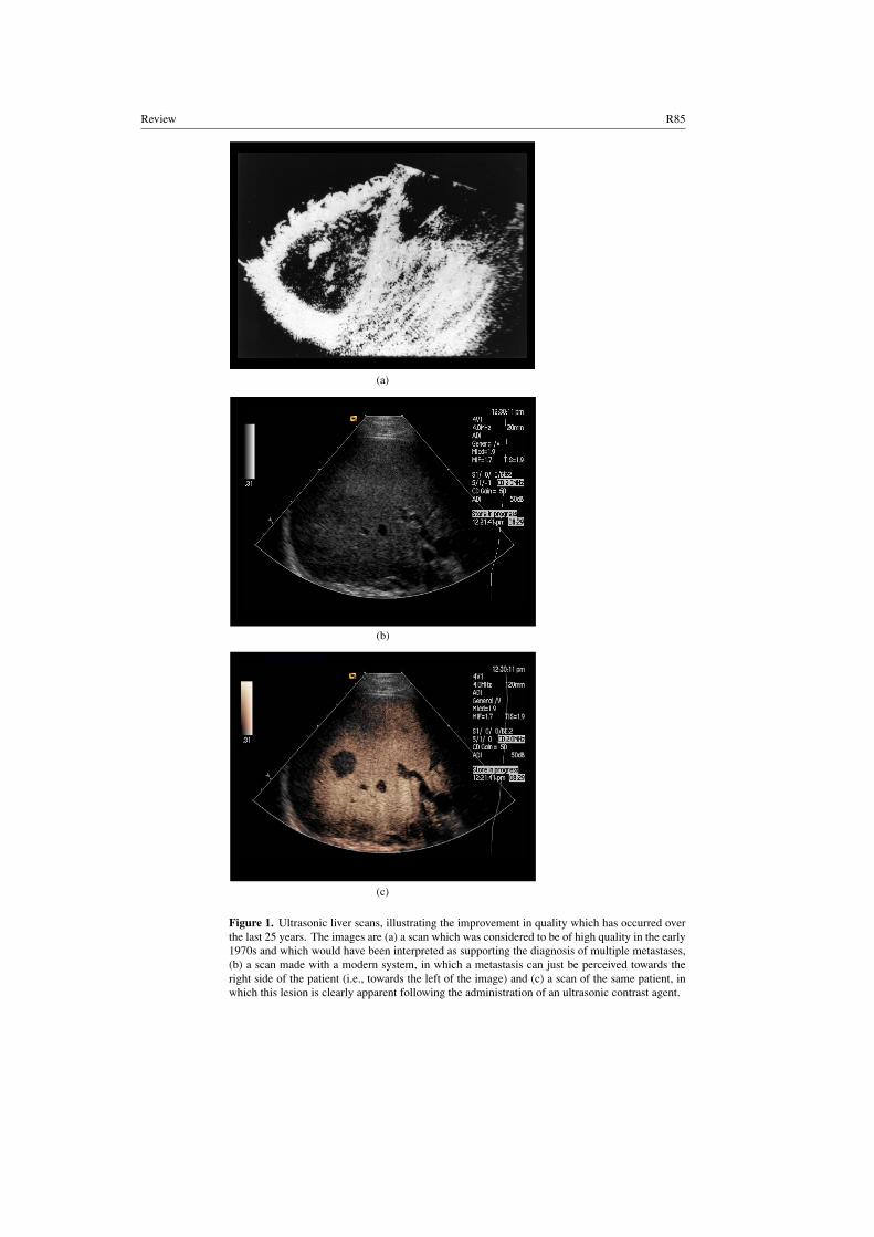
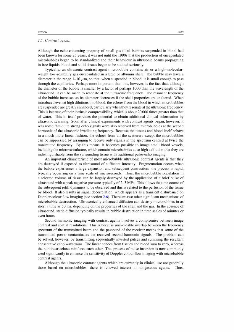
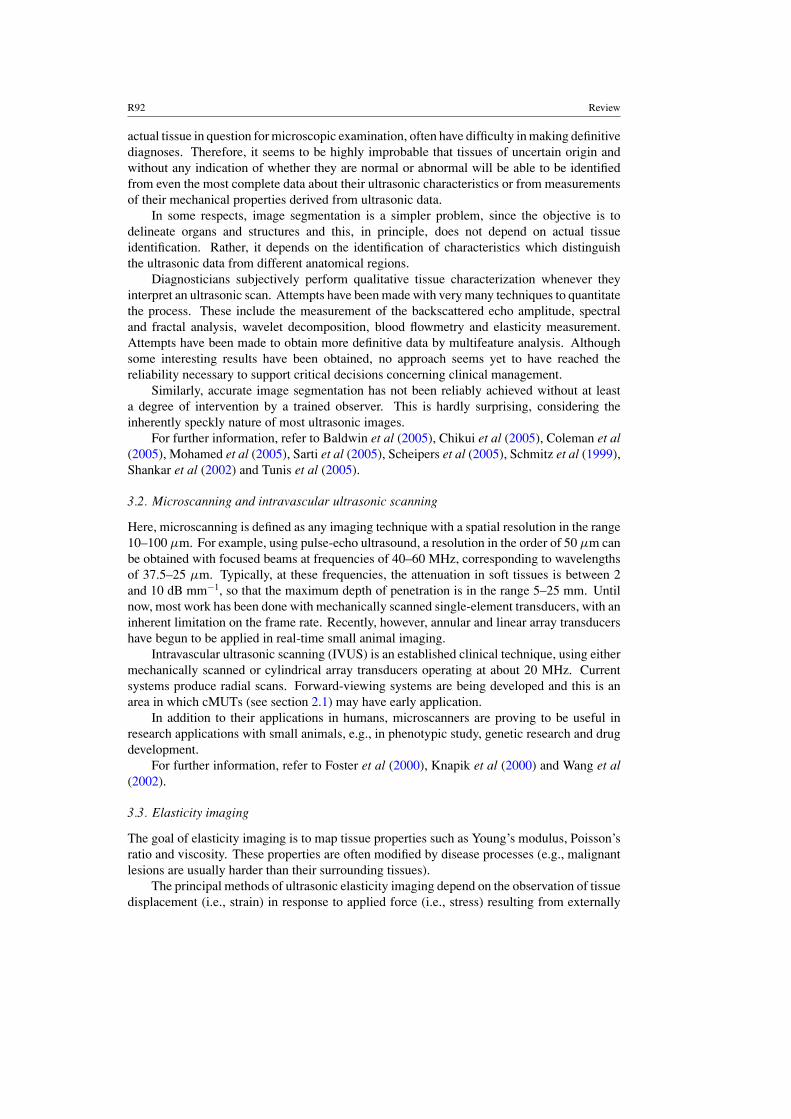
Ultrasonic imaging is now a mature technology, to the extent that it has a well-establishedplace in clinical practice, as confirmed by the fact that it currently accounts for about one infour of all imaging procedures worldwide. This does not mean, however, that the pace ofdevelopment, either of the understanding of the physics of the interactions between ultrasoundand tissue or of innovation in techniques and instrumentation, has slowed down. Indeed, theopposite is true. In this review, an attempt is made briefly to describe most of the significantadvances that have taken place in the last 10 years, from the perspective of the physics and,to some extent, the engineering involved. Because the subject is vast and the space availableis limited, the review takes the form of a series of short vignettes, each of which ends with alist of representative publications to which the reader interested in pursuing the topic may findit helpful to refer. Even so, significant topics have idiosyncratically had to be omitted. Also,there is no space for figures to illustrate all the principles involved and it would obviouslybe inappropriate to make a random, sparse selection. Nevertheless, to whet the appetite ofthe reader, figure 1 presents three ultrasonic images, two contemporary and one from about25 years ago, to illustrate something of the progress which has been made in the quality andclinical usefulness of ultrasonic scanning over the last quarter of a century.
2. Mainstream technologies
In this section, the state of the art in the mainstream technologies which underpin thecontemporary clinical applications of ultrasonic imaging is reviewed. The range of thesetechnologies is now so great that it has been necessary to be selective in choosingthose technologies which seem to be most important; it is hoped that this selection willnot be thought to be too biased.
2.1. Transducers
In some respects, the transducer is the most critical component in any ultrasonic imagingsystem. In other words, such is the state of the art in systems such as electronic circuitry anddisplay technology that it is the performance of the transducer which determines how closelythe limits imposed by the characteristics of the tissues themselves can be approached.
Nowadays, the transducers which are in clinical use almost exclusively use a piezoelectricmaterial, of which the artificial ferroelectric ceramic, lead zirconate titanate (PZT), is the mostcommon. The ideal transducer for ultrasonic imaging would have a characteristic acousticimpedance perfectly matched to that of the (human) body, have high efficiency as a transmitterand high sensitivity as a receiver, a wide dynamic range and a wide frequency response forpulse operation. PZT has a much higher characteristic impedance than that of water but it canbe made to perform quite well by the judicious use of matching layers consisting of materialswith intermediate characteristic impedances. Even better performance can be obtained byembedding small particles or shaped structures of PZT in a plastic to form a compositematerial: this has a lower characteristic impedance than that of PZT alone, although it hassimilar ferroelectric properties.
Review R85
(a)
(b)
(c)
Figure 1. Ultrasonic liver scans, illustrating the improvement in quality which has occurred overthe last 25 years. The images are (a) a scan which was considered to be of high quality in the early1970s and which would have been interpreted as supporting the diagnosis of multiple metastases,(b) a scan made with a modern system, in which a metastasis can just be perceived towards theright side of the patient (i.e., towards the left of the image) and (c) a scan of the same patient, inwhich this lesion is clearly apparent following the administration of an ultrasonic contrast agent.

R86 Review
Polyvinylidene difluoride (PVDF) is a plastic which can be polarized so that it haspiezoelectric properties. The piezoelectric effect can be enhanced by the addition of smallquantities of appropriate chemicals. The advantages of this material are that it has a relativelylow characteristic impedance and broad frequency bandwidth; it is fairly sensitive as a receiverbut rather inefficient as a transmitter.
Piezoelectric transducers are normally operated over a band of frequencies centred attheir resonant frequency. The resonant frequency of a transducer occurs when it is half awavelength in thickness. Typically, a PZT transducer resonant at a frequency of, say, 3 MHzis about 650 µm thick and this means that it is sufficiently mechanically robust for simple,even manual, fabrication techniques to be employed in probe construction. Higher frequencytransducers are proportionally thinner and, consequently, more fragile.
The potential of capacitive micromachined ultrasonic transducers (cMUTs) at leastpartially to replace PZT and PVDF devices in ultrasonic imaging is the subject of currentresearch. A cMUT consists of a micromachined capacitor, typically mounted on a siliconsubstrate and with a thin electroded membrane as the other plate of the capacitor: this acts asthe active surface of the transducer. A dc voltage is applied between the plates of the device;the application of an ac voltage causes the membrane to transmit a corresponding oscillatoryforce, while a received wave causes a corresponding change in the spacing between the plates,thus generating an electrical signal. cMUTs are adequately sensitive as receivers, but needhigh voltages to be effective transmitters. Some of the potential advantages of these devicesare that they can be fabricated into arrays with integrated electronics and, if manufactured inlarge quantities, could be relatively inexpensive.
Although some simple probes contain single-element transducers (e.g., one element fortransmitting and one for receiving, in a continuous-wave Doppler system), most modernimaging systems use arrays of transducer elements for beam forming (see section 2.2).
For further information, refer to Davidsen and Smith (1998), Foster et al (2000),Hunt et al (1983), Jin et al (1998) and Seyed-Boloforosh (1996).
2.2. Beam forming
In ultrasonic imaging, the beam may be scanned through the tissue either by mechanicalmovement of a single element or an annular array transducer, or by electronic control of atransducer array consisting of a number of small elements. For two-dimensional scanning,the array typically consists of 256 elements. The simplest arrangement is a linear array,within which an aperture is formed from, say, 16 contiguous elements and which is steppedalong the array element by element to acquire an image with, in this example, 241 lines.The same number of lines in a sector format can be acquired by curving the array into asegment of a cylinder. A sector scan can also be acquired by controlling the phases of thesignals associated with each of the elements in the aperture. Whatever the arrangement, theapplication of distinct time delays to excite each element focuses the transmitted beam at aparticular range. By transmitting several beams in the same position but with different foci, asharply focused transmitted beam can be synthesized. On reception, the focus can be sweptalong the beam by dynamically changing the time delays associated with the active transducerelements, so that its position coincides continuously with that of the instantaneous origin ofthe echoes. Both when transmitting and receiving, the amplitudes of the signals associatedwith the active elements can be weighted to minimize the amplitudes of the beam side lobes,which are critical in determining the image contrast resolution. Also, the number of elementsin the active aperture can be dynamically increased with increasing depth of penetration to

Review R87
maintain a constant f-number, within the limit imposed by the total length of the array andoptimized to minimize the effect of tissue inhomogeneity.
For three-dimensional imaging, the two-dimensional scan plane produced by a one-dimensional linear, curved or phased array can be swept mechanically, either linearly in theorthogonal direction or through a sector. Recently, two-dimensional transducer arrays havebeen developed. Because of the very large number of transducer elements in these arrays, beamforming in three dimensions can be achieved more economically but with some degradationin performance by sparsely populating the array. For real-time three-dimensional scanning,several transmitting beams can be synthesized simultaneously; a single receiving beam canbe associated with each transmitting beam, or, by increasing the width of each transmittingbeam, several sharply focused receiving beams can be accommodated simultaneously in eachtransmitted beam.
The beam-forming time delays and aperture apodization functions are digitally controlled.The sampling frequency has to be at least twice the highest ultrasonic frequency, in order toavoid aliasing. Further improvement can be obtained by applying a finite impulse responsedigital filter or by demodulating the radio-frequency signals to baseband to obtain quadraturesignals so that the associated time delays can be finely adjusted by phase rotation.
A very powerful software tool, Field II, has been developed to simulate ultrasonic beamand image formation. The current program assumes linear acoustics and it is freely availableunder certain restrictions.
An intriguing development in high-speed imaging has recently been brought about bythe application of limited diffraction beams. A limited diffraction beam can be produced byappropriate excitation of a transducer array. Following the transmission of a single planewave pulse, the received signals are weighted with limited diffraction beams simultaneouslyto produce multiple A-lines which can be used to create a complete two-dimensional image.
For further information, refer to Brown and Lockwood (2005), Jensen (2002), Li andHuang (2002), Lu (1998) and Ranagathan and Walker (2003a, 2003b).
2.3. Pulse compression
Ultrasonic pulse-echo imaging traditionally involves the transmission of as brief a pulseof ultrasound as is consistent with the frequency of ultrasound determined by the requiredpenetration into the tissue. Considerations of the safety of such brief high-powered pulses,the nonlinear properties of tissue and the destruction of contrast agent microbubbles revealthat this approach may often not be optimal. Coded transmission pulses have the potentialto increase the penetration depth, to improve the signal-to-noise ratio and/or to increase theimage frame rate. The principle is simple. The transmitted pulse has a relatively long durationand low amplitude, in comparison with the traditional brief pulse, and the frequency of thepulse typically is either swept so that it is a chirp, or it is modulated with, for example, abinary code. The signals received as echoes from within the tissues are then correlated with asignal corresponding to that which was transmitted, by means of a matched filter. This has theeffect of compressing the received signals so that they correspond to a traditional pulse-echowavetrain, which can be further processed to provide the image information.
Pulse compression schemes of this kind have long been used in radar, sonar and mobilecommunications systems. The situation in ultrasonic imaging, however, is rather morechallenging. In radar and sonar, the purpose is usually to detect isolated targets, whereas,in ultrasonic imaging, virtually the whole of the tissue is occupied by reflectors and scatterers.In ultrasonic imaging, there is strong frequency-dependent attenuation, which is much less ofa problem in radar and sonar. Added to this, the bandwidth of ultrasonic transducers is limited.

R88 Review
Finally, ultrasonic echoes tend to be dominated by speckle, whereas the echoes in radar andsonar are much less spatially variable. The effects of these processes on pulse compressionschemes for ultrasonic imaging are to introduce echo range ambiguities resulting from thetemporal side lobes which are produced in the cross-correlation process.
System optimization with a transducer array operating with coded pulses also needs to takeinto account the ultrasonic beam axial side lobes. In comparison with traditional brief pulsetransmission, however, current pulse compression techniques can result in a signal-to-noiseratio improvement approaching 20 dB, with temporal side lobes of below 60 dB, and the imageframe rate limitation imposed by cross-talk between spatially separated beams simultaneouslyacquiring image information can be reduced by up to a factor of as much as 25.
For further information, refer to Misaridis and Jensen (2005a, 2005b, 2005c).
2.4. Tissue harmonic imaging
It was noted, perhaps by accident during experiments with ultrasonic contrast agents (seesection 2.5), that the echoes from deeper parts of tissues being imaged by pulse-echo techniquesactually contain quite significant components at the second harmonic of the transmittedfrequency. These harmonic components arise because a finite-amplitude transmitted pulseis distorted in its propagation through inherently nonlinear tissue. Moreover, by selectingthe second harmonic frequency signals for imaging in the absence of contrast agents,improvements in spatial resolution and other aspects of the image were apparent, particularlywhen examining ‘technically difficult’ patients, such as those with obesity and/or densemuscle structure. The reasons for this are that, because the harmonics develop slowly,the low-frequency transmitted beam suffers relatively little distortion in passing through thesuperficial tissues, most of the second harmonics develop in the main beam rather than in thelower amplitude side lobes and the side lobes of the transmitting and the receiving beams havedifferent angular distributions, whereas the main beams are coincident.
Although tissue harmonic imaging at the second harmonic frequency is apparently betterthan imaging at the fundamental frequency, there is some controversy over what aspects of theimaging are ‘better’ and the process certainly has some difficulties. Thus, the bandwidth ofthe transducer necessarily has to accommodate the frequency spectra of both the transmittingand receiving beams, and so it is inevitable that some of the high-frequency components of thespectrum of the short transmitted pulse leak into the spectrum of the received signals. Also,the useful dynamic range of the received signals is reduced, because their absolute amplitudeis closer to that of the system noise.
An improvement in tissue harmonic imaging can be obtained, at the expense ofimage frame rate, by transmitting pulses with sequentially reversed polarities and addingconsecutively received echo wavetrains. This pulse inversion technique effectively removessignals with odd harmonic frequencies (including echoes at the fundamental transmittedfrequency) and doubles the amplitudes of those at the second harmonic and higher evenfrequencies.
Of course, harmonics higher than the second one are also produced as a result of thenonlinear propagation of the transmitted pulse. Although they are relatively weak, a usablesignal can be obtained by summing the third, fourth and even the fifth harmonics to constitutewhat has been called the superharmonic component of the received echoes. For this, acomposite broadband transducer is required. Superharmonic imaging is characterized by highspatial and temporal resolutions and the relative absence of clutter.
For further information, refer to Bouakaz and de Jong (2003), Browne et al (2005), Maet al (2005) and Tranquart et al (1999).
Review R89
2.5. Contrast agents
Although the echo-enhancing property of small gas-filled bubbles suspended in blood hadbeen known for some 25 years, it was not until the 1990s that the production of encapsulatedmicrobubbles began to be standardized and their behaviour in ultrasonic beams propagatingin free liquids, blood and solid tissues began to be studied seriously.
Typically, an ultrasonic contrast agent microbubble contains air or a high-molecular-weight low-solubility gas encapsulated in a lipid or albumin shell. The bubble may have adiameter in the range 1–10 µm, so that, when suspended in blood, it is small enough to passthrough the capillaries. Perhaps more important than this, however, is the fact that, althoughthe diameter of the bubble is smaller by a factor of perhaps 1000 than the wavelength of theultrasound, it can be made to resonate at the ultrasonic frequency. The resonant frequencyof the bubble increases as its diameter decreases if the shell properties are unaltered. Whenintroduced even at high dilutions into blood, the echoes from the blood in which microbubblesare suspended are greatly enhanced, particularly when they resonate at the ultrasonic frequency.This is because of their intrinsic compressibility, which is about 20 000 times greater than thatof water. This in itself provides the potential to obtain additional clinical information byultrasonic scanning. Soon after clinical experiments with contrast agents began, however, itwas noted that quite strong echo signals were also received from microbubbles at the secondharmonic of the ultrasonic irradiating frequency. Because the tissues and blood itself behavein a much more linear fashion, the echoes from all the scatterers except the microbubblescan be suppressed by arranging to receive only signals in the spectrum centred at twice thetransmitted frequency. By this means, it becomes possible to image small blood vessels,including the microvasculature, which contain microbubbles at so high a dilution that they areindistinguishable from the surrounding tissue with traditional pulse-echo imaging.
An important characteristic of most microbubble ultrasonic contrast agents is that theyare destroyed if exposed to ultrasound of sufficient intensity. Fragmentation occurs whenthe bubble experiences a large expansion and subsequent contraction: the process is rapid,typically occurring on a time scale of microseconds. Thus, the microbubble population ina selected volume of tissue can be largely destroyed by the application of a brief pulse ofultrasound with a peak negative pressure typically of 2–3 MPa. This allows the time course ofthe subsequent refill dynamics to be observed and this is related to the perfusion of the tissueby blood. It also results in signal decorrelation, which appears as a transient disturbance onDoppler colour flow imaging (see section 2.6). There are two other significant mechanisms ofmicrobubble destruction. Ultrasonically enhanced diffusion can destroy microbubbles in asshort a time as 50 ms, depending on the properties of the shell and the gas. In the absence ofultrasound, static diffusion typically results in bubble destruction in time scales of minutes oreven hours.
Second harmonic imaging with contrast agents involves a compromise between imagecontrast and spatial resolutions. This is because unavoidable overlap between the frequencyspectrum of the transmitted beam and the passband of the receiver means that some of thetransmitted power contaminates the received second harmonic signals. The problem canbe solved, however, by transmitting sequentially inverted pulses and summing the resultantconsecutive echo wavetrains. The linear echoes from tissues and blood sum to zero, whereasthe nonlinear echoes reinforce each other. This process of pulse inversion is now commonlyused significantly to enhance the sensitivity of Doppler colour flow imaging with microbubblecontrast agents.
Although the ultrasonic contrast agents which are currently in clinical use are generallythose based on microbubbles, there is renewed interest in nongaseous agents. Thus,

R90 Review
perfluorocarbon nanoparticles and gold-bound colloid microtubules seem to be quitepromising, particularly as they may be immunologically targeted to specific anatomical orpathological sites. Antibodies can be conjugated with the microtubules and can provide echoenhancement which approaches that of microbubbles.
The clinical potential of targeted ultrasonic contrast agents is considerable, both fordiagnosis and for drug and gene delivery. Eventually, they might even allow ultrasoundto replace much of contemporary and future radionuclide imaging for functional studies,thus avoiding the problems associated with radioactivity, and with relatively high spatial andtemporal resolutions.
For further information, refer to Bekeredian et al (2002), Chomas et al (2001), HopeSimpson et al (1999, 2001), Hu et al (2004), Hughes et al (2003), Kvikliene et al (2004) andStride and Saffari (2004).
2.6. Flow and motion
Historically, the earliest ultrasonic studies of physiological motion were made using the pulse-echo technique in M-mode echocardiography. Subsequently, emphasis has largely shifted tothe Doppler effect, although direct measurement of target or ensemble movement is also used,with the increase in system sensitivity, partly because it avoids the aliasing limitation inherentin the pulsed Doppler approach.
In principle, the direct measurement of the velocity profile of blood flowing in a vesselprovides a more accurate basis for the estimation of the blood flow volume rate than doesa single measurement of, say, the maximum blood flow velocity. There have been twoapproaches to the measurement of the velocity profile. First, a multigated (or infinite gate)pulsed Doppler system has been used. Second, the motion of the speckle, observed by pulse-echo ultrasound to arise from the blood, is tracked over a sequence of consecutive pulse-echowavetrains. (It is interesting to note that it is the motion of this same speckle that actuallyenables the pulsed Doppler approach to result in a usable signal.) Having obtained the flowvelocity profile and its variation over time (e.g., as the result of cardiac pulsation), the bloodflow volume rate may be calculated by assuming the profile to be circularly symmetrical.
Colour flow imaging, which was introduced in the 1980s, similarly may be based onfrequency or phase domain (i.e., Doppler) processing or on target motion tracking approaches.The early Doppler processors were simple autocorrelators, with displays colour coded eitheraccording to the mean flow velocity or to the amplitude (or power) of the Doppler-shiftedsignals. More recently, improved velocity estimators, such as the maximum likelihoodestimator, have been introduced: they can handle a larger velocity search range which extendsto lower values of velocity, and with smaller errors.
For the study of blood flow, it is the relatively low amplitude signals from the blood itselfwhich are relevant. The magnitude of these signals can be increased by means of contrastagents and the echoes from solid tissues can be suppressed by second harmonic imaging(see section 2.4). When the greatest possible sensitivity is required (e.g., when imaging themicrovasculature), the power of the Doppler signals may be used to control the brightness ofthe image, in distinction from colour coding the image according to the velocity of the flow.
In the study of blood flow, the accompanying motion of neighbouring solid tissues isa source of difficulty because it gives rise to relatively large amplitude Doppler signals.Various cancelling schemes are used, more or less effectively to suppress these echoes. It isusually also necessary to use a high-pass filter to remove remaining solid tissue echoes which,fortunately, move more slowly than most of the blood and so give rise to Doppler signals oflower frequencies.

Review R91
Quite a long time after the introduction of colour flow imaging for the study of blood flow,it was realized that colour-coded images of moving solid tissue could also be of clinical utility.For example, some cardiac lesions are associated with abnormal motion of the myocardium.It turns out to be relatively easy to produce such images, simply by eliminating the solid tissueecho canceller and reducing the cut-off frequency of the passband filter. The process is knownas tissue Doppler imaging.
For further information, refer to Bohs et al (1998), Dunmire et al (2000), Fan et al (2001),Jansson et al (2003) and Schlaikjer and Jensen (2004).
2.7. Three-dimensional imaging
Most contemporary ultrasonic scanning systems have hand-held probes which produce two-dimensional images in real time. A skilled operator moves the probe over the surface ofthe patient to explore the internal anatomy and thus to build up, in his or her mind’s eye, athree-dimensional picture.
There are two approaches to display the scanned anatomy in three dimensions and thus,in principle, at least to some extent to deskill the process of image interpretation. First, thetwo-dimensional scanning probe may be moved mechanically in a predetermined trajectoryor the beam may be scanned in three dimensions by means of an electrically controlled two-dimensional array so that position and orientation are directly measured and these data areused to assemble a three-dimensional image volume from the set of two-dimensional images.Second, the position and orientation of the two-dimensional scanning probe, whilst being freelymoved by the operator over the surface of the patient, may be measured by sensors with respectto a frame of reference which is fixed in relation to the patient. By appropriate calibration ofthe spatial relationships in the system, a three-dimensional image can be assembled.
Because a three-dimensional image is assembled from a set of two-dimensional scans,scanning is proportionately slower than it is for two-dimensional imaging, unless a scheme toaccelerate the process is adopted. For example, the use of separate simultaneously transmittedbeams and/or the use of wide transmitted beams accommodating several narrow receivingbeams is mentioned in section 2.2. As with other medical imaging modalities, the display ofthree-dimensional data is challenging. Since it is usually difficult automatically to segmentultrasonic images (see section 3.1), three-dimensional rendering is problematic. Currently,the easiest way to explore a three-dimensional ultrasonic image volume is usually to enablethe observer rapidly to display two-dimensional images in any desired orientation within thescanned volume. Of course, this is not really ideal, especially if four-dimensional imaging(i.e., three time-varying spatial dimensions) is actually what is needed.
For further information, refer to Fenster et al (2001), Mercier et al (2005), Nelson andPretorius (1998) and Shipley et al (2005).
3. Specialized and emerging technologies
In this section, some of the newer technologies which are not yet in routine clinical use arereviewed. At this stage in their development, it is inappropriate to attempt to predict whichof them will prove to be the most significant. Again, the selection of which technologies areincluded is idiosyncratic, but they all have some elements of significant novelty.
3.1. Tissue characterization and image segmentation
Before discussing recent advances in ultrasonic tissue characterization, it is timely briefly toplace the problem in its clinical context. Even histopathologists, who have specimens of the
R92 Review
actual tissue in question for microscopic examination, often have difficulty in making definitivediagnoses. Therefore, it seems to be highly improbable that tissues of uncertain origin andwithout any indication of whether they are normal or abnormal will be able to be identifiedfrom even the most complete data about their ultrasonic characteristics or from measurementsof their mechanical properties derived from ultrasonic data.
In some respects, image segmentation is a simpler problem, since the objective is todelineate organs and structures and this, in principle, does not depend on actual tissueidentification. Rather, it depends on the identification of characteristics which distinguishthe ultrasonic data from different anatomical regions.
Diagnosticians subjectively perform qualitative tissue characterization whenever theyinterpret an ultrasonic scan. Attempts have been made with very many techniques to quantitatethe process. These include the measurement of the backscattered echo amplitude, spectraland fractal analysis, wavelet decomposition, blood flowmetry and elasticity measurement.Attempts have been made to obtain more definitive data by multifeature analysis. Althoughsome interesting results have been obtained, no approach seems yet to have reached thereliability necessary to support critical decisions concerning clinical management.
Similarly, accurate image segmentation has not been reliably achieved without at leasta degree of intervention by a trained observer. This is hardly surprising, considering theinherently speckly nature of most ultrasonic images.
For further information, refer to Baldwin et al (2005), Chikui et al (2005), Coleman et al(2005), Mohamed et al (2005), Sarti et al (2005), Scheipers et al (2005), Schmitz et al (1999),Shankar et al (2002) and Tunis et al (2005).
3.2. Microscanning and intravascular ultrasonic scanning
Here, microscanning is defined as any imaging technique with a spatial resolution in the range10–100 µm. For example, using pulse-echo ultrasound, a resolution in the order of 50 µm canbe obtained with focused beams at frequencies of 40–60 MHz, corresponding to wavelengthsof 37.5–25 µm. Typically, at these frequencies, the attenuation in soft tissues is between 2and 10 dB mm−1, so that the maximum depth of penetration is in the range 5–25 mm. Untilnow, most work has been done with mechanically scanned single-element transducers, with aninherent limitation on the frame rate. Recently, however, annular and linear array transducershave begun to be applied in real-time small animal imaging.
Intravascular ultrasonic scanning (IVUS) is an established clinical technique, using eithermechanically scanned or cylindrical array transducers operating at about 20 MHz. Currentsystems produce radial scans. Forward-viewing systems are being developed and this is anarea in which cMUTs (see section 2.1) may have early application.
In addition to their applications in humans, microscanners are proving to be useful inresearch applications with small animals, e.g., in phenotypic study, genetic research and drugdevelopment.
For further information, refer to Foster et al (2000), Knapik et al (2000) and Wang et al(2002).
3.3. Elasticity imaging
The goal of elasticity imaging is to map tissue properties such as Young’s modulus, Poisson’sratio and viscosity. These properties are often modified by disease processes (e.g., malignantlesions are usually harder than their surrounding tissues).
The principal methods of ultrasonic elasticity imaging depend on the observation of tissuedisplacement (i.e., strain) in response to applied force (i.e., stress) resulting from externally

Review R93
applied static or vibrating pressure (including cardiac pulsations in arteries) or from internallygenerated acoustic radiation force. The change in tissue stiffness with time can also be imaged:this is related to the viscoelastic properties of the tissue.
The spatial resolution of elasticity imaging is at least comparable with that of traditionalpulse-echo B-scanning. It is a speckle-free technique. Seeing that manual palpation is one ofthe mainstays of clinical examination, elasticity imaging is already proving to be of significantpractical importance.
For further information, refer to Doyley et al (2005), Gao et al (1996), Konofagou et al(2001), Nightingale et al (2002) and Righetti et al (2005).
3.4. Reflex transmission imaging
Reflex transmission imaging is the technique in which backscattered echoes gated from atissue volume situated beyond the focal region of a strongly focused ultrasonic beam areintegrated and displayed while the focal region is scanned through the tissue to be imaged.The amplitude of the received signal is dominated by the attenuation of the tissue in thefocal volume. The technique works quite well when there is a suitably uniform volume oftissue beyond the image plane and when the intervening tissue is relatively homogeneous.Since it was originally demonstrated nearly 15 years ago, the technique has been largelyunexplored but, recently, interest has been revived and its potential is now being more activelyinvestigated, particularly when combined with other approaches, the performance of whichmay be enhanced by being able to compensate for attenuation.
For further information, refer to Green et al (1991).
3.5. Computed tomography
Ultrasonic computed tomography produces images by backprojection reconstruction fromprofiles of attenuation or speed acquired at multiple angles by scanning the tissue in atwo-dimensional plane. The female breast and the long bones are obvious candidates tobe imaged by this technique. Unfortunately, however, an ultrasonic beam is deviated byrefraction and distorted by inhomogeneities in tissue. Consequently, the results which haveso far been obtained have been rather disappointing. Like reflex transmission imaging (seesection 3.4), however, ultrasonic CT could potentially produce data which might be used tocorrect aberrations in traditional B-scans.
For further information, refer to Duric et al (2005) and Lasaygues et al (2005).
3.6. Doppler tomography
In Doppler tomography, a wide (but thin) ultrasonic beam is rotated around the tissue to beimaged. The reflectors and scatterers within the tissue return echoes which are correspondinglyDoppler shifted in frequency. The Doppler frequency spectra acquired at a set of angles aroundthe tissue are equivalent to a set of cross-range profiles of the corresponding radial positionsof the reflectors and scatterers and a two-dimensional image can consequently be producedby backprojection reconstruction. Doppler tomography uses continuous wave ultrasound andso it is a technique with narrow bandwidth and high sensitivity. Encouraging results havebeen obtained by amplitude-only reconstruction. It is anticipated that, by including phasedata in the reconstruction, it should be possible to image, e.g., the female breast with a spatialresolution which significantly improves upon that which can be obtained by B-scanning at thesame frequency.
R94 Review
For further information, refer to Liang et al (2001), Mensa et al (1983) and Wade et al(1978).
3.7. Photoacoustics and thermoacoustics
It has been demonstrated that thermally induced ultrasound can be produced by absorption ofa pulse of light or of microwave electromagnetic radiation. In the case of tissue, the absorptionof the pulse causes a rapid increase in temperature which results in localized expansion,according to the intensity of the pulse and the corresponding absorption coefficient. Thephenomenon can be used for measuring the depth distribution of thermally induced ultrasonicsignals from their times of arrival or, more generally, to produce three-dimensional images byreconstruction from the signals acquired by an array of ultrasonic sensors positioned aroundthe tissue. Some encouraging results have been obtained with breast imaging.
For further information, refer to Feng et al (2001), Kruger (1994) and Manohar et al(2005).
4. Phantoms and quality assurance
Modern ultrasonic scanners employ solid-state electronic circuitry and are manufactured tomeet tight performance specifications. Apart from the possibility of accidental damage to therelatively fragile ultrasonic probes, they are reliable and stable. Consequently, it might besupposed that there would be no need for periodic checks of imaging performance. This is notthe case, however, for the very reason that imaging performance tends to deteriorate slowlyand imperceptibly, usually as a result of minor damage to the probe (such as the failure of asingle element in a transducer array with a large number of elements), or to slow drift in thesettings of the electronics.
Pragmatically, the simplest and most direct method of checking the performance of anultrasonic scanner is to display and examine the images which it produces from physicalphantoms made up of tissue-mimicking materials of differing geometries, scattering andabsorbing characteristics.
The simplest phantoms can be used to check the spatial resolution of the imaging system.More sophisticated phantoms are designed to test the contrast resolution. Elasticity imaging isamongst the other characteristics which can be assessed; flow and motion are other possibilities.
Except for the simplest of phantoms, the phantoms themselves do not have characteristicswhich can be depended upon not to change with time. Materials which closely mimicthe ultrasonic properties of tissues are usually prepared by mixing, e.g., agar, gelatine andother substances which are themselves biological in origin and which are, consequently,themselves liable to become infected by bacteria or otherwise to deteriorate. The art ofphantom manufacture is indeed akin to the art of preparing gourmet food.
Other aspects of quality assurance include the measurement of ultrasonic power, beamshape and intensity (spatial and temporal peaks and averages), as well as the relevant aspectsof electrical, thermal and mechanical safety, infection control and so on.
For further information, refer to Browne et al (2003), Dandekar et al (2005), Goodsittet al (1998), Poepping et al (2004) and Spirou et al (2005).
5. Safety considerations
Under certain exposure conditions, ultrasound may cause harmful bioeffects. Because ofthis, there is a hypothetical possibility that ultrasonic imaging may not be completely safe.Consequently, both regulatory authorities and prudent clinicians are concerned to balance
Review R95
the likely benefits of the information which imaging provides against the risk of damage.Although this is the appropriate response to the question of the risk of adverse bioeffects dueto ultrasonic exposure, the reality is that it is the consequences of misdiagnosis that are likelyto be the greatest risk to the patient.
Regulatory agencies in some countries now oblige manufacturers to provide on-screendisplays of quantities designated as the ‘thermal index’ (which relates to the risk of causingthermal damage) and the ‘mechanical index’ (which helps primarily to assess the likelihoodof damage due to cavitation). These indices are for the guidance of clinicians in assessingrisks and benefits and they provide reminders of the importance of applying the ‘as lowas reasonably achievable’ (ALARA) principle in the context of justification for individualdiagnostic investigations.
Although ultrasonic diagnosis has an impeccable record of safety in relation to the lackof adverse bioeffects, research into thermal, nonthermal and other mechanisms of interactionbetween ultrasound and biological systems continues to be an area of considerable activity.For example, with both the tendency for the ultrasonic exposure levels used by commerciallyavailable scanners steadily to be increased in order to obtain more information and theincreasing use of microbubble contrast agents, there is fresh anxiety that safety may yetbecome a real concern.
For further information, refer to Abbott (1999), Fowlkes and Holland (2000), ter Haarand Duck (2000) and Nyborg (2002).
6. Conclusions
Looking back over the entire history of medical ultrasonic imaging, it is obvious that the trulymajor advances which have taken place have resulted from innovations in the applicationsof physics and engineering, rather than having been in response to the expressed aspirationsof clinicians. In reality, what has happened is that physicists and engineers—who often areindistinguishable—have created new capabilities and clinicians, often sceptical and reluctant,have eventually embraced those technologies which have been perceived to be useful. To beeven more controversial, it could be added that the utilities of new imaging technologies havehardly ever been scientifically proved before both clinicians and patients have demanded thatthey should be made available, so obvious to them have been their benefits.
Ultrasonic imaging stands alongside x-ray computed tomography and magnetic resonanceimaging as one of the most disruptive of the medical applications of physics and engineering,having profoundly changed the practice of medicine and having brought immense benefits tomankind in the last half century. Speaking for ultrasound, but certainly not excluding otherimaging techniques, the future is stimulating, exciting and challenging, and, for health andhealth care, the prospects are indeed brighter than ever before.
Acknowledgments
Figures 1(b) and (c) were kindly provided by Professor David Cosgrove, Imperial CollegeLondon, and were processed by software supplied on an experimental basis by Toshiba MedicalSystems.
References
Abbott J G 1999 Rationale and derivation of MI and TI—a review Ultrasound Med. Biol. 25 431–41Baldwin S L, Marutyan K R, Yang M, Wallace K D, Holland M R and Miller J G 2005 Estimating myocardial
attenuation from M-mode ultrasonic backscatter Ultrasound Med. Biol. 31 477–84

R96 Review
Bekeredian R, Behrens S, Ruef J, Dinjus E, Unger E, Baum M and Kuecherer H F 2002 Potential of gold-boundmicrotubules as a new ultrasound contrast agent Ultrasound Med. Biol. 28 691–5
Bohs L N, Geiman B J, Anderson M E, Breit S M and Trahey G E 1998 Ensemble tracking for 2D vectorvelocity measurement: experimental and initial clinical results IEEE Trans. Ultrason. Ferroelectr. Freq.Control 45 912–24
Bouakaz A and de Jong N 2003 Native tissue imaging at superharmonic frequencies IEEE Trans. Ultrason. Ferroelectr.Freq. Control 50 496–506
Brown J A and Lockwood G R 2005 A digital beamformer for high-frequency annular arrays IEEE Trans. Ultrason.Ferroelectr. Freq. Control 52 1262–9
Browne J E, Ramnarine K V, Watson A J and Hoskins P R 2003 Assessment of the acoustic properties of commontissue-mimicking test phantoms Ultrasound Med. Biol. 29 1053–60
Browne J E, Watson A J, Hoskins P R and Elliott A T 2005 Investigation of the effect of subcutaneous fat on imagequality performance of 2D conventional imaging and tissue harmonic imaging Ultrasound Med. Biol. 31 957–64
Burns P N, Powers J E and Fritsch T 1992 Harmonic imaging: new imaging and Doppler method for contrastenhanced US Radiology 185P 142
Chikui T, Tokumori K, Yoshiura K, Oobu K, Nakamura S and Nakamura K 2005 Sonographic texture characterizationof salivary gland tumors by fractal analysis Ultrasound Med. Biol. 31 1297–304
Chomas J E, Dayton P, Allen J, Morgan K and Ferrara K 2001 Mechanisms of contrast agent destruction IEEE Trans.Ultrason. Ferroelectr. Freq. Control 48 232–48
Coleman D P, Rakebrandt F, Pugh N D, Crawford D C and Woodcock J P 2005 Development and validation of anin vivo analysis tool to identify changes in carotid plaque tissue types in serial 3D ultrasound scans UltrasoundMed. Biol. 31 329–35
Dandekar S, Li Y, Molloy J and Hossack J 2005 A phantom with reduced complexity for spatial 3D ultrasoundcalibration Ultrasound Med. Biol. 31 1083–93
Davidsen R E and Smith S W 1998 Two-dimensional arrays for medical ultrasound using multi-layer flexible circuitinterconnection IEEE Trans. Ultrason. Ferroelectr. Freq. Control 45 338–48
Doyley M M, Srinivasan S, Prendergrass S A, Wu Z and Ophir J 2005 Comparative evaluation of strain-based andmodel-based modulus elastography Ultrasound Med. Biol. 31 787–802
Dunmire B, Beach K W, Labs K-H, Plett M and Strandness D E 2000 Cross-beam vector Doppler ultrasound forangle-independent velocity measurements Ultrasound Med. Biol. 26 1213–35
Duric N et al 2005 Development of ultrasound tomography for breast imaging: technical assessment Med. Phys.32 1375–86
Fan L, Evans D E and Naylor A R 2001 Automated embolus identification using a rule-based expert system UltrasoundMed. Biol. 27 1065–77
Feng D, Xu Y, Ku G and Wang L 2001 Microwave-induced thermoacoustic tomography: reconstruction by syntheticaperture Med. Phys. 28 2427–31
Fenster A, Downey D B and Cardinal H N 2001 Three-dimensional ultrasound imaging Phys. Med. Biol.46 R67–99
Foster F S, Harasiewicz K A and Sherar M D 2000 A history of medical and biological imaging with polyvinylidenefluoride (PVDF) transducers IEEE Trans. Ultrason. Ferroelectr. Freq. Control 47 1363–71
Foster F S, Pavlin C J, Harasiewicz K A, Christopher D A and Turnbull D H 2000 Advances in ultrasoundbiomicroscopy Ultrasound Med. Biol. 26 1–27
Fowlkes J B and Holland C K (ed) 2000 Mechanical bioeffects from diagnostic ultrasound: AIUM consensusstatements J. Ultrasound Med. 19 67–168
Gao L, Parker K J, Lerner R M and Levinson S F 1996 Imaging of the elastic properties of tissue—a review UltrasoundMed. Biol. 22 959–77
Goodsitt M M, Carson P L, Witt S, Hykes D L and Kofler J M 1998 Real-time B-mode ultrasound quality control testprocedures. Report of AAPM Ultrasound Task Group no 1 Med. Phys. 25 1385–406
Green P S, Ostrem J S and Whitehurst T K 1991 Combined reflection and transmission ultrasound imaging UltrasoundMed. Biol. 17 283–9
Hope Simpson D, Burns P N and Averkion M A 2001 Techniques for perfusion imaging with microbubble contrastagents IEEE Trans. Ultrason. Ferroelectr. Freq. Control 48 1483–94
Hope Simpson D, Chin C T and Burns P N 1999 Pulse inversion Doppler: a new method for detecting nonlinearechoes from microbubble contrast agents IEEE Trans. Ultrason. Ferroelectr. Freq. Control 46 372–82
Hu Y, Qin S and Jiang Q 2004 Characteristics of acoustic scattering from a double-layered micro shell for encapsulateddrug delivery IEEE Trans. Ultrason. Ferroelectr. Freq. Control 51 808–20
Hughes M S, Lanza G M, Marsh J N and Wickline S A 2003 Targeted ultrasonic contrast agents for molecular imagingand therapy: a brief review Medicamundi 47 66–73

Review R97
Hunt J W, Arditi M and Foster F S 1983 Ultrasound transducers for pulse-echo medical imaging IEEE Trans. Biomed.Eng. 30 453–81
Jansson T, Hernandez-Andrade E, Lingman G and Marsal K 2003 Estimation of fractional moving blood volume infetal lung using power Doppler ultrasound: methodological aspects Ultrasound Med. Biol. 29 1551–9
Jensen J A 2002 Field II Ultrasound Simulation Program http://www.es.oersted.dtu.dk/staff/jaj/field/index.htmlJin X C, Ladabaum I and Khuri-Yakub B T 1998 The microfabrication of capacitive ultrasonic transducers IEEE J.
Microelectromech. Syst. 7 295–302Knapik D A, Starkoski B, Parkin C J and Foster F S 2000 A 100–200 MHz ultrasound biomicroscope IEEE Trans.
Ultrason. Ferroelectr. Freq. Control 47 1540–9Konofagou E A, Harrigan T P, Ophir J and Krouskop T A 2001 Poroelastography: imaging the poroelastic properties
of tissues Ultrasound Med. Biol. 27 1387–97Kruger R A 1994 Photoacoustic ultrasound Med. Phys. 21 127–31Kvikliene A, Jarkonis R, Ressner M, Hoff L, Jansson T, Janerot-Sjoberg B, Lukosevicius A and Ask P 2004
Modelling of nonlinear effects and the response of ultrasound contrast micro bubbles: simulation and experimentUltrasonics 42 301–7
Lasaygues P, Ouedraogo E, Lefebvre J-P, Gindre M, Talmant M and Laugier P 2005 Progress towards in vitroquantitative imaging of human femur using compound quantitative ultrasonic tomography Phys. Med.Biol. 50 2633–49
Li P-C and Huang J-J 2002 Efficient dynamic focus control for three-dimensional imaging using two-dimensionalarrays IEEE Trans. Ultrason. Ferroelectr. Freq. Control 49 1191–202
Liang H-D, Halliwell M and Wells P N T 2001 Continuous wave ultrasonic tomography IEEE Trans. Ultrason.Ferroelectr. Freq. Control 48 285–92
Lu J-Y 1998 Experimental study of high frame rate imaging with limited diffraction beams IEEE Trans. Ultrason.Ferroelectr. Freq. Control 45 84–97
Ma Q, Ma Y, Gong X and Zhang D 2005 Improvement of tissue harmonic imaging using the pulse-inversion techniqueUltrasound Med. Biol. 31 889–94
Manohar S, Kharine A, van Hespen J C G, Steenbergen W and van Leeuwen T G 2005 The Twente photoacousticmammoscope: system overview and performance Phys. Med. Biol. 50 2543–57
Mensa D L, Halevy S and Wade G 1983 Coherent Doppler tomography for microwave imaging Proc. IEEE 71254–61
Mercier L, Lango T, Lindseth F and Collins D L 2005 A review of calibration techniques for freehand 3D ultrasoundsystems Ultrasound Med. Biol. 31 449–71
Misaridis T and Jensen J A 2005a Use of modulated excitation signals in medical ultrasound: part I. Basic conceptsand expected benefits IEEE Trans. Ultrason. Ferroelectr. Freq. Control 52 177–91
Misaridis T and Jensen J A 2005b Use of modulated excitation signals in medical ultrasound: part II. Design andperformance for medical imaging IEEE Trans. Ultrason. Ferroelectr. Freq. Control 52 192–207
Misaridis T and Jensen J A 2005c Use of modulated excitation signals in medical ultrasound: part III. High framerate imaging IEEE Trans. Ultrason. Ferroelectr. Freq. Control 52 208–19
Mohamed S S, Salama M M A, Kamel M, El-Sandary E F, Rizkalla K and Chin J 2005 Prostate cancer multi-featureanalysis using trans-rectal ultrasound images Phys. Med. Biol. 50 N175–85
Nelson T R and Pretorius D H 1998 Three-dimensional ultrasound imaging Ultrasound Med. Biol. 24 1243–70Nightingale K, Soo M S, Nightingale R and Trahey G 2002 Acoustic radiation force impulse imaging: in vivo
demonstration of clinical feasibility Ultrasound Med. Biol. 28 227–35Nyborg W L 2002 Safety of medical diagnostic ultrasound Semin. Ultrasound CT MRI 23 377–86Poepping T L, Nikolov H N, Thorne M L and Holdsworth D W 2004 A thin-walled carotid vessel phantom for
Doppler ultrasound flow studies Ultrasound Med. Biol. 30 1067–78Ranagathan K and Walker W F 2003a A novel beamformer design method for medical ultrasound: part I. Theory
IEEE Trans. Ultrason. Ferroelectr. Freq. Control 50 15–24Ranagathan K and Walker W F 2003b A novel beamformer design method for medical ultrasound: part II. Simulation
results IEEE Trans. Ultrason. Ferroelectr. Freq. Control 50 25–39Righetti R, Ophir J and Krouskop T A 2005 A method for generating permeability elastograms and Poisson’s ratio
time-constant elastograms Ultrasound Med. Biol. 31 803–16Sarti A, Corsi C, Mazzini E and Lamberti C 2005 Maximum likelihood segmentation of ultrasound images with
Rayleigh distribution IEEE Trans. Ultrason. Ferroelectr. Freq. Control 52 947–60Scheipers U, Siebers S, Gottwald F, Ashfaq M, Bozzato A, Zenk J, Iro H and Ermert H 2005 Sonohistology for the
computerized differentiation of parotid gland tumors Ultrasound Med. Biol. 31 1287–96Schlaikjer M and Jensen J A 2004 Maximum likelihood blood velocity estimator incorporating properties of flow
physics IEEE Trans. Ultrason. Ferroelectr. Freq. Control 51 80–92
R98 Review
Schmitz G, Ermert H and Senge T 1999 Tissue-characterization of the prostate using radio frequency ultrasonicsignals IEEE Trans. Ultrason. Ferroelectr. Freq. Control 46 126–38
Seyed-Boloforosh M S 1996 Integrated impedance matching layer Ultrasonics 34 135–8Shankar P M, Dumane V A, Piccoli C W, Reid J M, Forsberg F and Goldberg B B 2002 Classification of breast masses
in ultrasonic B-mode images using a compounding technique in the Nakagami distribution domain UltrasoundMed. Biol. 28 1295–300
Shipley J A, Duck F A, Goddard D A, Hillman M R, Halliwell M, Jones M G and Thomas B T 2005 Automatedquantitative volumetric breast ultrasound data-acquisition system Ultrasound Med. Biol. 31 905–17
Spirou G M, Oraevsky A A, Vitkin J A and Whelan W M 2005 Optical and acoustic properties at 1064 nm of polyvinylchloride-plastisol for use as a tissue phantom in biomedical optoacoustics Phys. Med. Biol. 50 N141–53
Stride E and Saffari N 2004 Theoretical and experimental investigation of the behaviour of ultrasound contrast agentparticles in whole blood Ultrasound Med. Biol. 30 1495–509
ter Haar G and Duck F A (ed) 2000 The Safe Use of Ultrasound in Medical Diagnosis (London: British MedicalUltrasound Society/British Institute of Radiology)
Tranquart F, Grenier N, Eder V and Pourcelot L 1999 Clinical use of ultrasound tissue harmonic imaging UltrasoundMed. Biol. 25 889–94
Tunis A S, Czarnota G J, Giles A, Sherar M D, Hunt J and Kolios M C 2005 Monitoring structural changes in cellswith high-frequency ultrasound signal statistics Ultrasound Med. Biol. 31 1041–9
Wade G, Elliott S, Khogeer I, Flesher G, Eisler J, Mensa D, Ramesh N S and Heidbreder G 1978 Acoustic echocomputer tomography Acoustic Imaging vol 8 ed A F Metherell (London: Plenum) pp 567–76
Wang Y, Stephens D N and O’Donnell M 2002 Optimizing the beam pattern of a forward-viewing ring-annularultrasound array for intravascular imaging IEEE Trans. Ultrason. Ferroelectr. Freq. Control 49 1652–64
Wells P N T 1977 Ultrasonics in medicine and biology Phys. Med. Biol. 22 629–69Wells P N T 1999 Ultrasonic imaging of the human body Rep. Prog. Phys. 62 671–722
Biography
Peter Wells (FRS, FREng, FMedSci) trained in electrical engineering, physics and zoology.Following a student apprenticeship with the General Electric Company in Coventry, his researchcareer has been focused on the applications of ultrasound in medicine. For 25 years, he was Head ofthe Department of Medical Physics and Bioengineering at the United Bristol Healthcare NHS Trustand its predecessors, having previously been Professor of Medical Physics at what is now CardiffUniversity School of Medicine. He is now Distinguished Research Professor at Cardiff University,Visiting Professor at Imperial College London and Emeritus Professor at Bristol University.