Ultrasound diagnosis of endometriosis - SFOG · The role of transvaginal ultrasound in the...
42
Ultrasound diagnosis of endometriosis Lil Valentin Department of Obstetrics and Gynecology Malmö University Hospital Malmö, Sweden
Transcript of Ultrasound diagnosis of endometriosis - SFOG · The role of transvaginal ultrasound in the...

Ultrasound diagnosis of
endometriosis
Lil Valentin Department of Obstetrics and Gynecology
Malmö University Hospital
Malmö, Sweden

The role of transvaginal ultrasound in the
diagnosis and management of endometriosis
• Discriminate endometriomas from other lesions
• Other sites than the ovaries affected (DIE)?
• Provide information for choice of treatment
– Medical or surgical treatment
– Laparosopic surgery or laparotomy
• surgeon with special skills?
– bowel resection?
• ureteral stents?

Typical endometrioma
51% (416/713) of 713 endometriomas had this appearance
Ultrasound Obstet Gynecol. 2010 Jun;35(6):730-40

Typical endometrioma
Wall nodularity Wall nodularity

Atypical endometriomas

Atypical endometrioma
- atypical internal echogenicity
King´s College King´s College

Endometrioma with fluid level
Hyperechoic contents inferiorly
From Asch and Levine JUM 2007; 26: 993 Dermoid Endometrioma

Atypical endometrioma
- retracted blood clots

Atypical endometrioma
- blood clots?
Leuven

Atypical endometrioma
– bi- or multi-locular

Atypical endometrioma
– calcified

Atypical endometrioma
15 x 9 x 8 cm
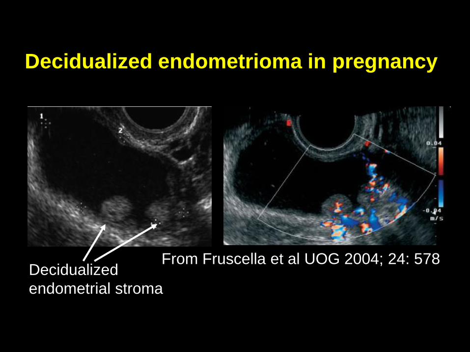
Decidualized endometrioma in pregnancy
From Fruscella et al UOG 2004; 24: 578 Decidualized
endometrial stroma

Unusual case Endometrioma with endometroid and
clear cell cancer
Malignancy in 0.3 – 0.8% of endometriotic lesions

Summary
Typical endometrioma
– Unilocular, ground glass, +/- nodule
Atypical endometrioma
– NOT ground glass
– Bi- or multi-locular
– Retracted blood clots
– Papillary projections, vascularized
– Calcified lesions
– Completely atypical

Malignization Decidualization
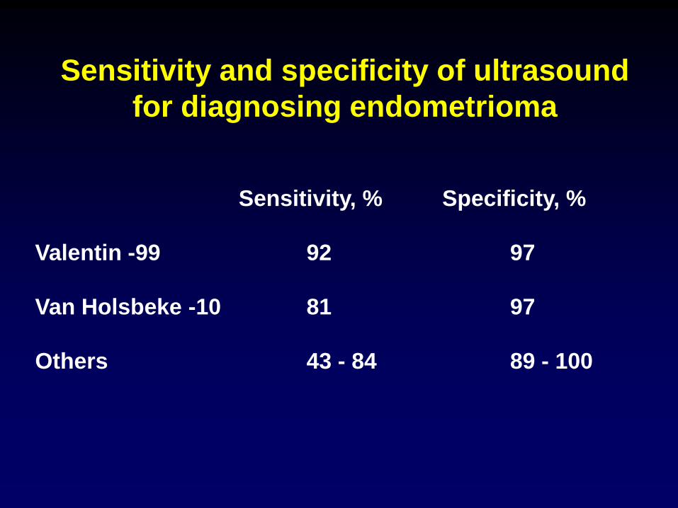
Sensitivity and specificity of ultrasound
for diagnosing endometrioma
Sensitivity, % Specificity, %
Valentin -99 92 97
Van Holsbeke -10 81 97
Others 43 - 84 89 - 100

When an endometrioma is found…
• Fixed or freely movable?
• Signs of extraovarian endometriosis (deep infiltrating endometriosis, DIE)? – Difficult surgery?
• 93% of women with DIE in one location have DIE also in other locations
• Adenomyosis?

Adenomyosis
Enlarged uterus
”Hypoechoic linear
striations”
”Rain in the forest”
Cysts in the myometrium

Adenomyosis
Enlarged uterus
”Rain in the forest”

Adenomyosis
Poorly defined endometrium

Adenomyosis
Best criterion?
cystic spaces
AND
hypoechoic linear striations
Sensitivity 0.90
Specificity 1.00
LR+ -
LR - 0.1
Bazot et al 01, 02
Conclusive
diagnosis

Sites typically involved in extraovarian
endometriosis
2
1 1
3
3
4
4
5 5

What do endometriotic nodules look like?
• Hypoechoic
• Diffuse borders
• Firm and tender when pushed upon
• Poorly vascularized
• Bowel endometriosis = Indian head sign
• Bladder endometriosis
– Dome or base
– Round or comma shaped

Endometriosis with
adhesions to bowel
Kissing ovaries Adherent bowel


Bowel endometriosis
Indian head sign

Endometriosis in bowel wall

Endometriosis in bowel wall

Endometriosis in the bowel wall
Case 3, courtesy of
Dr Luca Savelli, Bologna

Bladder endometriosis
Bladder endometriosis

Bladder endometriosis

Bladder endometriosis

Endometriosis in
the sacrouterine ligaments
Savelli UOG 2009:33;497

Endometriosis in the abdominal wall
Case 1, courtesy of Dr Luca Savelli, Bologna

Ultrasound examination
should be guided by
• Patient´s symptoms
• Speculum examination
• Gynecological palpation
How to scan for deep
infiltrating endometriosis?

Association between endometriotic
lesions and symptoms
Fauconnier, Fertil Steril, 2002
Type of pain Type of lesion
Dysmenorhea Adenomyosis, adhesions
Dyspareunia Sacrouterine ligaments
Pain at defecation Vagina, rectum
Chronic pelvic pain Bowel
Dysuria Bladder

Deep infiltrating endometriosis - scan technique
• Introduce probe scrutinizing the vaginal walls
• Uterus - adenomyosis
• Ovaries – endometriomas/freely movable?
• Sacrouterine ligaments
• Posterior compartment - rectovaginal septum, rectum, rectosigmoid junction
• Anterior compartment - bladder
• Abdominal scan - hydronephrosis
• Are organs freely movable - sliding?

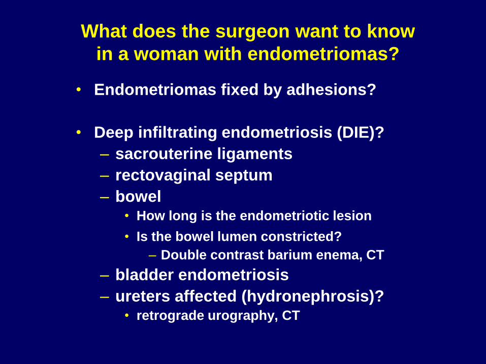
What does the surgeon want to know
in a woman with endometriomas?
• Endometriomas fixed by adhesions?
• Deep infiltrating endometriosis (DIE)?
– sacrouterine ligaments
– rectovaginal septum
– bowel • How long is the endometriotic lesion
• Is the bowel lumen constricted? – Double contrast barium enema, CT
– bladder endometriosis
– ureters affected (hydronephrosis)? • retrograde urography, CT

Summary
• Be aware of deep infiltrating endometriosis
• Let the patient´s symptoms guide your scan
• Let findings at speculum and vaginal examination guide your scan
• Nodules are hypoechoic, irregular borders, firm, tender, poorly vascularized
• Assess mobility!
• Check for hydronephrosis

THANK you Tisdag 28 augusti C-hallen 14.00-15.30, fritt föredrag
Jan-Henrik Stjerndahl
Laparoskopisk kirurgi ger goda resultat vid
endometrios med tarmengagemang
Tisdag 28 augusti C-hallen 17.00-17.30
Per Boström
Endometrios, en resurskrävande sjukdom både för
patienten och för samhället.