Ultrasound biomicroscopy as a diagnostic method of corneal ...
55
Ultrasound biomicroscopy as a diagnostic method of corneal degeneration and inflammation Ph.D. Thesis Ákos Skribek M.D. Department of Ophthalmology Faculty of Medicine University of Szeged Szeged, Hungary 2013
Transcript of Ultrasound biomicroscopy as a diagnostic method of corneal ...
Ultrasound biomicroscopy as a diagnostic
method of corneal degeneration and
inflammation
Ph.D. Thesis
Ákos Skribek M.D.
Department of Ophthalmology
Faculty of Medicine
University of Szeged
Szeged, Hungary
2013
PUBLICATIONS
List of full papers directly related to the subjects of the Thesis:
I. Sohar N, Skribek A, Fulop Z, Kolozsvari L. The success of treating keratoconus:
visual acuity and follow-up with ultrasound biomicroscopy. Spectrum der
Augenheilkunde 2012, 26:(3) pp. 159-164.
IF: 0.274
II. Skribek A, Sohar N, Nogradi A, Kolozsvari L. Amniotic membrane
transplantation in cases of corneal calcification - follow up with ultrasound
biomicroscopy. Spectrum der Augenheilkunde 2011, 25:(3) pp. 210-214.
IF: 0.274
III. Skribek A, Sohar N, Gyetvai T, Nogradi A, Kolozsvari L. Role of ultrasound
biomicroscopy in diagnosis and treatment of Terrien disease. Cornea 2008, 27:(4)
pp. 427-433.
IF: 1.853
IV. Skribek Á, Gyetvai T, Kolozsvári L. Ultrahang-biomikroszkópos vizsgálat
jelentősége a Terrien-betegség követésében. Szemészet 2006, 143: pp. 50-52.
List of abstracts directly related to the subjects of the Thesis:
1. Skribek Á, Sohár N, Facskó A: Elülső szegmentum képalkotó eljárások pellucid
marginális degeneráció eseteiben. Magyar Szemorvostársaság Kongresszusa,
Siófok, 2012.06.07-2012.06.09. p. 65.
2. Skribek Á, Sohár N, Kolozsvári L: Az amnionmembrán-transzplantáció szerepe a
szaruhártya mészképződéssel járó eseteiben. Magyar Szemorvostársaság
Kongresszusa, Pécs, 2008.05.29-2008.05.31. p. 85.
3. Sohár N, Skribek A, Fülöp Zs, Kolozsvári L: Retrospective study of patients with
keratoconus. In: The Joint Congress of the European Society of Ophthalmology
and American Academy of Ophthalmology: SOE/AAO. Bécs, Ausztria,
2007.06.09-2007.06.12. p. 170.
4. Skribek Á.: UH szerepe az uveitisek diagnosztikájában. Kötelező szemészeti
szintentartó tanfolyam, Szeged 2007. október 27. 29. 30. 31.
5. Skribek Á.: Az amnion szerepe a szaruhártya betegségek kezelésében Kötelező
szemészeti szintentartó tanfolyam, Szeged 2007. október 27. 29. 30. 31.
6. Skribek Á, Gyetvai T, Hári Kovács A, Sohár N, Kolozsvári L: The role of
ultrasound biomicroscopy in the diagnosis and treatment of marginal corneal
thinning. In: The Joint Congress of the European Society of Ophthalmology and
American Academy of Ophthalmology: SOE/AAO. Bécs, Ausztria, 2007.06.09-
2007.06.12. p. 171.
7. Skribek Á, Sohár N, Gyetvai T, Kolozsvári L: Keratoconus súlyosságának
megítélése ultrahang biomikroszkópos vizsgálattal. In: Magyar Műlencse
Implantációs és Refraktív Sebészeti Társaság Kongresszusa Keszthely,
Magyarország, 2007.03.30-2007.03.31. p. 71.
8. Skribek Á, Sohár N, Gyetvai T, Hári Kovács A, Kolozsvári L: Az ultrahang-
biomikroszkóp szerepe a szaruhártya perifériás elvékonyodása diagnosztikájában.
In: Magyar Szemorvostársaság 2007. évi Kongresszusa: Szemészet 2007; 144.
Suppl., Debrecen, Magyarország, 2007.06.21-2007.06.23. p. 78.
9. Hári Kovács A, Skribek Á, Tóth-Molnár E, Kolozsvári L: Three cases of ocular
filariasis in Hungary. In: The Joint Congress of the European Society of
Ophthalmology and American Academy of Ophthalmology: SOE/AAO. Bécs,
Ausztria, 2007.06.09-2007.06.12. p. 213.
10. Skribek Á, Kolozsvári L: Keratoconus miatt végzett perforáló keratoplasztika
műtétek postoperatív gyógyulása és szövődményei. In: Magyar Műlencse
Implantációs és Refraktív Sebészeti Társaság Kongresszusa Keszthely,
Magyarország, 2006.03.30-2006.04.01. p. 87.
11. Skribek Á, Sohár N, Fülöp Zs, Kolozsvári L: Keratoconus miatt gondozott
betegeink követése. In: Magyar Szemorvostársaság 2006. évi Kongresszusa,
Alpok-Adria Nemzetközi Szemorvostársaság Kongresszusa. Sopron,
Magyarország, 2006.06.15-2006.06.17. p. 96.
12. A Skribek, T Gyetvai, A, Hári Kovács, L Kolozsvári: The role of ultrasound
biomicroscopy (UBM) in the diagnosis and treatment of Terrien’s disease. In:
János Németh, Béla Csákány, György Barcsay (szerk.) Ophthalmic Echography
Konference Budapest, Magyarország: 2006. pp. 13-16.(ISBN:963-85636-5-6).
13. Gyetvai T, Skribek Á, Kolozsvári L: A cornea és a sclera vastagságának
változása különböző kórképekben. In: Magyar Szemorvostársaság 2006. évi
Kongresszusa, Alpok-Adria Nemzetközi Szemorvostársaság Kongresszusa.
Sopron, Magyarország, 2006.06.15-2006.06.17. p. 46.
14. T Gyetvai, Z Horóczi, Á Skribek, A Hári Kovács, L Kolozsvári: Ultrasound
biomicroscopical examination of the corneal incision after cataract surgery. In:
János Németh, Béla Csákány, György Barcsay (szerk.) Ophthalmic Echography
Budapest, Magyarország, 2006 Budapest:2006. pp. 43-46. (ISBN:963-85636-5-6)
15. Skribek Á, Gyetvai T, Hári Kovács A, Kolozsvári L: The role of ultrasound
biomicroscope in the diagnosis and treatment of marginal corneal thinning. In:
15th SOE Congress and 103rd DOG Congress. Berlin, Németország, 2005.09.25-
2005.09.29. p. 235.
16. Hári Kovács A, Gyetvai T, Skribek Á, Kolozsvári L: Ultrasound biomikroscopic
findings in corneal astigmatism. In: 15th SOE Congress and 103rd DOG
Congress, Berlin, Németország, 2005.09.25-2005.09.29. p. 236.
17. Gyetvai T, Horóczi Z, Skribek Á, Hári Kovács A, Kolozsvári L: Ultrasound
biomicroscopical examination of the corneal incision after cataract surgery. In:
15th SOE Congress and 103rd DOG Congress. Berlin, Németország, 2005.09.25-
2005.09.29. p. 234.
18. Gyetvai T, Horóczi Z, Skribek Á, Hári Kovács A, Kolozsvári L: A cornealis
sebzés vizsgálata ultrahang biomikroszkóppal katarakta műtét után. In: Magyar
Műlencse Implantációs és Refraktív Sebészeti Társaság Kongresszusa Keszthely,
Magyarország, 2005.03.31-2005.04.02. p. 92.
19. Skribek Á, Gyetvai T, Hári-Kovács A, Kolozsvári L: The role of ultrasound
biomicroscopy (UBM) in the diagnosis and treatment of Terrien’s disease. In:
SIDUO XX Congress of the International Society for Ophthalmic Ultrasound,
Budapest, Magyarország, 2004.09.12-2004.09.16. p. 29.
20. T Gyetvai, Z Horóczi, Á Skribek, A Hári Kovács, L Kolozsvári: Ultrasound
biomicroscopical examination of the corneal incision after cataract surgery. In:
SIDUO XX Congress of the International Society for Ophthalmic Ultrasound,
Budapest, Magyarország, 2004.09.12-2004.09.16. p. 33.
21. Skribek Á, Kolozsvári L: A Terrien-betegségekről három eset kapcsán. In:
Magyar Szemorvostársaság Kongresszusa: Szemészet, 140 (Suppl I.). Budapest,
Magyarország, 2003.08.28-2003.08.30. p. 73.
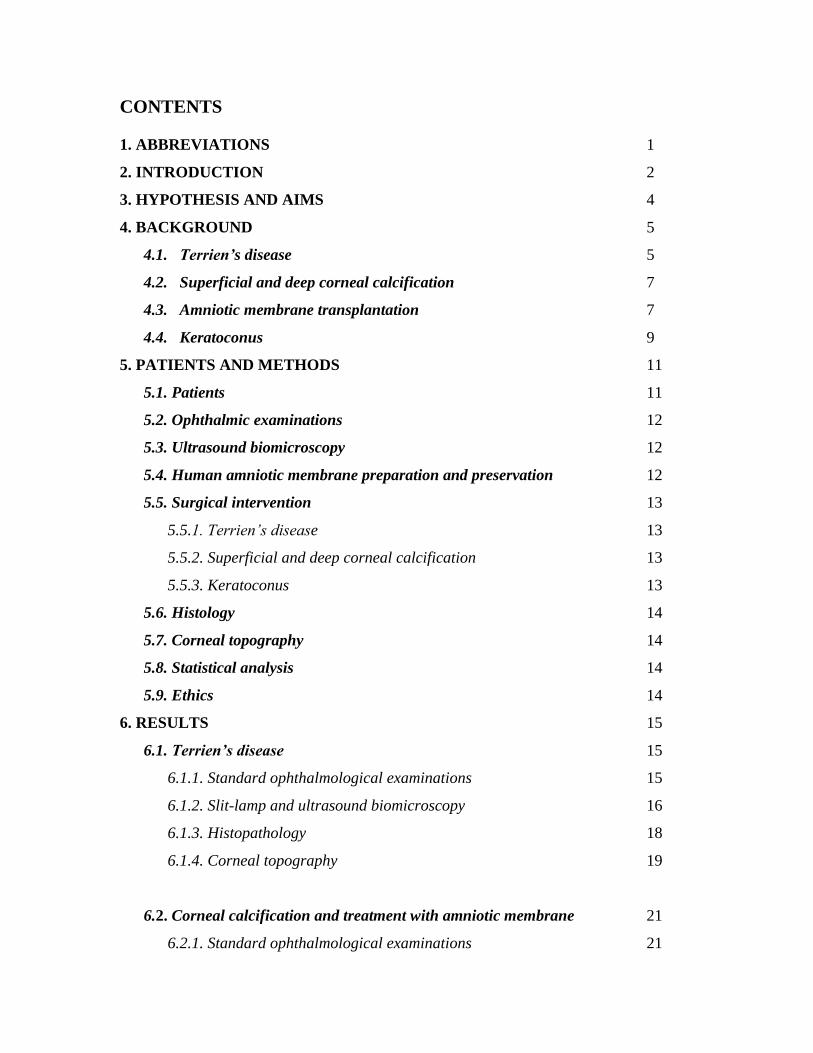
CONTENTS
1. ABBREVIATIONS 1
2. INTRODUCTION 2
3. HYPOTHESIS AND AIMS 4
4. BACKGROUND 5
4.1. Terrien’s disease 5
4.2. Superficial and deep corneal calcification 7
4.3. Amniotic membrane transplantation 7
4.4. Keratoconus 9
5. PATIENTS AND METHODS 11
5.1. Patients 11
5.2. Ophthalmic examinations 12
5.3. Ultrasound biomicroscopy 12
5.4. Human amniotic membrane preparation and preservation 12
5.5. Surgical intervention 13
5.5.1. Terrien’s disease 13
5.5.2. Superficial and deep corneal calcification 13
5.5.3. Keratoconus 13
5.6. Histology 14
5.7. Corneal topography 14
5.8. Statistical analysis 14
5.9. Ethics 14
6. RESULTS 15
6.1. Terrien’s disease 15
6.1.1. Standard ophthalmological examinations 15
6.1.2. Slit-lamp and ultrasound biomicroscopy 16
6.1.3. Histopathology 18
6.1.4. Corneal topography 19
6.2. Corneal calcification and treatment with amniotic membrane 21
6.2.1. Standard ophthalmological examinations 21
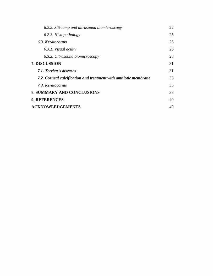
6.2.2. Slit-lamp and ultrasound biomicroscopy 22
6.2.3. Histopathology 25
6.3. Keratoconus 26
6.3.1. Visual acuity 26
6.3.2. Ultrasound biomicroscopy 28
7. DISCUSSION 31
7.1. Terrien’s diseases 31
7.2. Corneal calcification and treatment with amniotic membrane 33
7.3. Keratoconus 35
8. SUMMARY AND CONCLUSIONS 38
9. REFERENCES 40
ACKNOWLEDGEMENTS 49
1
1. ABBREVIATIONS
AM amniotic membrane
AMT amniotic membrane transplantation
BCVA best corrected visual acuity
BCCVA best corrected visual acuity with contact lenses
BCSVA best corrected visual acuity with spectacles
D diopter
DMSO dimethyl sulfoxide
HSV-1 herpes simplex virus 1
IOP intraocular pressure
KC keratoconus
MHz megahertz
OCT optical coherence tomography
PKP penetrating keratoplasty
TMS topographic modeling system
UBM ultrasound biomicroscopy
UCVA uncorrected visual acuity
2
2. INTRODUCTION
The demand for noninvasive diagnostic techniques in ophthalmology yielded the
development of special noninvasive tools that can function as diagnostic indicators.
Imaging techniques of the anterior segment of the eye provide important information
for detecting and managing the pathology, pathophysiology, prognosis and treatment of
disorders of the cornea, limbus, anterior chamber, iris, and lens (1).
Anterior segment imaging has significantly altered the diagnosis and evaluation of eye
diseases and become a rapidly advencing field of ophthalmology. It could be regarded as
more complex method than retinal imaging due to the depth of structures and surfaces of
interest being obscured by other anatomical features. Several techniques have been developed
over the last years to image objectively the anterior segment of the eye (1-4).
Ultrasonography is an ultrasound-based diagnostic imaging technique, a measurement
tool, and a device used for visualizing and characterising ocular tissues. The used ultrasound
frequency causes limited resolution (5).
In early 1990’s, Pavlin et al. created a new ultrasound instrument for visualising the
anterior segment structures (6,7). High-frequency ultrasound biomicroscopy (UBM) makes a
more detailed image and more accurate measurement due to the greater resolution than
regular ultrasound, but at the expense of decreased tissue penetration (6,7). High-energy
sound waves are bounced off the inside of the eye and the echo patterns are shown on the
screen of an ultrasound machine. In contradiction to ultrasonography, UBM provides high-
resolution in vivo imaging of the anterior segment of the eye in a noninvasive manner (8) and
is the most established anterior segment imaging device offering objective, high-resolution
images of angle structure. The B-scan mode UBM has a high frequency transducer (35-100
MHz) which limits sound waves through ocular tissues and detects their reflection from tissue
interfaces (8). Pathologic changes, involving anterior segment structures can be evaluated
qualitatively and quantitatively by this method (8).
UBM has numerous potential uses in clinical situations. Although the anterior segment
of the eye is partially visible by direct observation with slit lamp, there are several surfaces
not easily accessible by this device, such as the posterior surface of the iris and the region of
posterior chamber - ciliary body. Angle depth can be determined by UBM without the
3
requirement of a clear cornea for gonioscopy. In addition, corneal opacification precluding a
view of the anterior chamber would allow an image of the areas behind the opacity with high
frequency resolution. Lesions of the iris and the ciliary body are difficult to diagnose with
usual ultrasound techniques, but it is easy to determine these alterations with UBM (6-11).
Clinically, it is very important to measure the central and peripheral thickness of the
cornea. These parameters could be determined by using a number of examination methods.
Optical pachymetry was the gold standard in the past. Later, it has been replaced by ultrasonic
pachymetry because of it’s easy use (1,12-14). Anterior segment imaging with UBM has been
shown accurate measuring of the corneal thickness and curvature (8). Optical coherence
tomography (OCT) is an emerging technology for performing high-resolution cross-sectional
imaging. OCT is analogous to ultrasound imaging, except that it uses light instead of sound.
OCT can provide cross-sectional images of tissue structures within the cornea (13,15). UBM
is another device that can be used to object and follow the progression of the central and
peripheral thickness or thinning of the cornea in different corneal diseases (13,16). Each of
the above techniques has certain advantages.
After studying the very advantageous method of UBM, we decided to use this device
to find exact diagnosis and to follow the courses of the corneal degenerative and
inflammatory diseases. UBM would be more precise than the other methods we used
previously, even in following of the accuracy of the treatment we applied. We also carried out
the new idea of our group to replace with amniotic membrane (AM) the surface of the
artificial corneal defect that was formed when the calcified corneal part was cut out.
4
3. HYPOTHESISES AND AIMS
Based on previous studies, we hypothetized that
- UBM could be useful device in investigating and following the courses of corneal
degenerative and inflammatory diseases, and
- AM transplantation might be helpful in the healing process of epithelial defects in
corneal degenerative diseases.
In order to get answer for the hypothesises above, patients with Terrien’s disease, corneal
calcification, and keratoconus were chosen for our investigations.
The aims of this study were:
- to find new information about the diagnosis of corneal diseases with using UBM,
- to estimate the progression of the corneal diseases,
- to replace the surface of the artificial corneal defect with AM,
- to verify and follow the results of the management and treatment of the corneal
diseases that were treated by our team,
- to find out whether the histopathological examinations confirm the structural
changes diagnosed by UBM.

5
4. BACKGROUND
4.1. Terrien’s disease
Terrien’s disease is a rare form of bilateral asymmetrical corneal degeneration,
characterised by a chronic, slow, and progressive thinning of the peripheral part of the cornea
(16,17). Its occurance is confined primarily to men (3:1) mostly between twenty and fourty
years of age. A variant form of Terrien’s disease with prominent inflammatory signs occuring
in young population has also been observed (16,18-22).
Because of the slowly progressive and painless property of the disease, when patients
visit the hospital the degeneration often has reached an advanced stage at which the risk of
spontaneous or traumatic corneal rupture becomes high (16,17,21,22). The first symptom of
Terrien’s disease is poor visual acuity caused by irregular astigmatism. In the early stages, the
upper part of the peripheral cornea is vascularized superficially, producing a semilunar fold.
This part of the cornea slowly narrows, then dilates, and becomes ectatic. The narrowed and
regular corneal parts are delimited by a sharp yellowish-white border that contains lipid
deposits (16,21,23,24).
In the development and progression of the disease, five stages can be distinguished (25-27):
1. Gerontoxon like marginal opacification with peripheral vascularisation.
2. An indentation appears parallel to the limbus indicating the initiation of corneal thinning
in addition to the changes noted in the initial stage.
3. Thinning of the cornea progresses, but it does not reach the central part of the cornea.
Ectasia of the thinned part of the cornea begins. Spontaneous or traumatic perforation of
the thinned cornea and prolapse of the iris may occur.
4. The ectasia reaches the central part of the cornea, its pattern is similar to that of
keratoconus.
5. Opacification of the central part of the cornea. The perforation of the cornea usually
occurs before their stage.
There have been two main theories concerning the nature of this disease: an
inflammatorical and a degenerational theory (28). Many investigators supported the
6
degenerational theory, partly based on the earlier histopathologic studies which showed little
or no signs of inflammation. Other studies dealing with light, electron, and confocal
microscopic investigations have reported inflammatory signs as well (25,28).
In the study of Süveges et al. was reported a histochemical and electron microscopic
method for finding the pathophysiology of the disease. They demonstrated that in stage 3 of
the disease, the phagocytation of the substantia propria by histiocyta-like cells that penetrated
the cornea along the vessels led to marked thinning of the cornea (25).
Srinivasan et al. described in their study a nineteen years old female with unilateral
Terrien’s disease with spontaneous corneal perforation. They followed the patient by Orbscan
and suggested that their patient had the inflammatory subtype of Terrien’ s disease, and the
associated inflammation and subclinical epithelial changes may led to spontaneous corneal
perforation (29).
Ferrari and coworkers investigated in vivo corneal changes in Terrien’s disease with
corneal confocal microscopy that allowed real-time visualization of fine corneal structures.
They described microstructural abnormalities in a presurgical stage of the progression of the
disease, when pathology specimens would not be available. They found an amorphous
hyperreflective material corresponding to the lipid deposition and an irregular Bowman layer
with direct examination of the peripheral lesion. They have found with anterior OCT
examination, that the central cornea may allow initial signs of thinning within the central 10
mm zone close to the peripheral lesion even in cases when the clinical inspections seemed to
be normal (30). The group of Ceresara reported an in vivo confocal microscopy study in
Terrien’ s disease. In their studies, they observed inflammatory cell infiltration at the level of
paralimbar conjunctiva and in the upper peripheral basal epithelium of both eyes (31).
Penetrating keratoplasty (PKP), crescentic lamellar keratoplasty, and C shaped
lamellar keratoplasty have been employed in the treatment of marginal corneal degeneration
earlier, but some are difficult to perform and carry high risks for the disease in the advanced
stage due to very thin corneal stroma (32-35).
7
4.2. Superficial and deep corneal calcification
There is a wide spectrum of calcium deposition in the cornea with different types
ranging from superficial changes to full thickness calcification. Two types of corneal
calcification come into question (36):
a) The superficial type appears as a band shaped keratopathy and refers to calcium
deposition in the Bowman layer and superficial structures (36-38). The causes of
superficial calcification of the cornea are unknown.
b) Deeper calcification of the cornea is described as calcareous degeneration and
represents the other type where the calcium deposition founded in the deeper
stroma including Descemet membrane. This type may exist with band-shaped
keratopathy (36-38).
The etiology of corneal calcification has been classified as metastatic or dystrophic processes,
although the mechanism is still unclear (38).
Metastatic calcification occurs, when there is elevation of the calcium phosphate
product like in chronic renal failure, sarcoidosis, hyperparathyreoidism, hypercalcaemia,
hyperphosphataemia, and hypervitaminosis D. In these cases, the calcification is typically
mild and restricted to the perilimbal cornea in the interpalpebral zone. The tear pH may be
more alkaline as a result of evaporation and desiccation (38,39). Dystrophic calcium
deposition can occure in case of superficial inflammation or tissue injury (38-44).
There are several methods to treat corneal calcifications such as mechanical removal
of the calcified part of the cornea, the use of freshly prepared chelating agent combined with
vigorous and frequent massage of residual deposits to completely remove dense local
concentrations of calcium and the use of phototherapic keratectomy (45).
4.3. Amniotic membrane transplantation
Intact corneal epithelium is one of the most important factors in maintaining ocular
surface health. Corneal epithelial defects usually heal without any complications by rapid
proliferisation of the epithelial cells. Incomplete wound healing would lead to persistent
epithelial defect, that can be decreased by several treatments such as artificial tear drops,
8
lubricants, fibronectin, and growth factors. In cases with persistent defects even after the
application of expensive treatments, surgical interventions such as tissue adhesive glue,
tarsorraphy or conjunctival flap were often tried (46-49).
The newest alternative for the management of the corneal epithelial defect or ulcer is
the reconstruction of the surface with using amniotic membrane transplantation (AMT). This
method has become well established as a treatment for chronic epithelial defects and
conjunctival reconstruction (42).
Amniotic membrane (AM) is the innermost layer of the placenta. It consist of a single
layer of ectodermally derived amnion cells, thick basement membrane, and an avascular
stromal matrix. AM contains a high concentration of basic fibroblast growth factor, basement
membrane components, and unknown trophic factors. These factors might be related to the
expression of different anti-inflammatory proteins, inhibition of proteinase activity, exclusion
of polymorphonuclear cells by subsequent apoptosis, and decrease of lipid peptidation (50-
52). AM can be used as a substrate to replace damaged mucosal surfaces and has recently
been used successfully for reconstructing corneal surfaces. AMT may facilitate epithelization
and reduce inflammation, vascularisation, and scarring (46-54).
There are several types of AMT using one or more layers of AM as a patch, graft, or
sandwich to cover corneal epithelial defects (46,51,52,55-58). In each cases the AM is used
epithelial side up.
- When the corneal diseases with epithelial defects have no or only shallow stromal
defects, patch onlay can be used. The AM is sutured from limbus to limbus over
the peripheral epithelial remnants and the centrally denuded stroma. The local
epithelium is expected to grow under the AM and the epithelial defect should close
(55,59,60).
- The other type of AM is the single–layer graft inlay that can be used for shallow
stromal defects. In this case the graft is fixed in this superficial defect with
interrupted 10-0 nylon sutures in the border of the corneal ulcer. In this type of
AMT, the epithelium is expected to grow over the AM, providing a new basement
membrane (55,59,60).
- For deep stromal defects, multilayer–graft inlay can be used. In case of this corneal
epithelial defect, smaller portions of the AM could be placed layer by layer into the
9
ground of the ulcer, which is filled without sutures before a superficial graft is
sutured to the periphery of the ulcer. In this case, the epithelium is expected to
grow over the uppermost layer of this multilayer graft.
- The sandwich is a special combination of the previous techniques. It consist of one
or more grafts and a patch on the top. The epithelium is expected to grow under the
patch but over the uppermost graft (55,59,60).
AMT may be beneficial in the treatment of persistent epithelial defects after
penetrating keratoplasty, especially when applying the sandwich technique (61,62). AM can
be used as a trigger in cases of Mooren’ s ulcer which do not heal with intensive
immunosuppressive regimens alone (63), for intraoperative conjunctival repair during
trabeculectomy (64), in children with symblepharon and pannus (65), and for symptomatic
bullous keratopathy as well (66).
4.4. Keratoconus
Keratoconus (KC) is still an enigmatic disease that remains an era of wide-ranging,
dynamic, international research although it was first described more than 150 years ago (67).
KC is a noninflammatory, progressive, corneal degeneration. The main characteristic of KC is
a bilateral thinning of the cornea without neovascularisation. The development of a
corresponding protrusion with an apex located centrally or in an inferior exentric position
could be seen (68,69). This disorder impresses both eyes, although only one eye may be
affected initially. The high degree of irregular astigmatism, corneal thinning, and scarring that
can occur with KC may result in severe visual impairment (70,71). The incidence is
approximately 1/2000 in general population (68,71).
The pathology of KC remains unclear. Predisposition to developing KC is related to
genetic, constitutional and environmental factors, mechanical and surgical eye rubbing, or
other metabolic inbalances (69,72).
The diagnosis of KC is based on the detection of changes in the corneal curvature and
corneal thickness, which is thinner than usual (69,73).
10
Colin (70) and Krumeich and coauthors (74) have proposed a clinical classification of
four stages of KC based on astigmatism, corneal power, corneal transparency, and corneal
thickness:
1. Excentric corneal steepening, induced myopia and/or astigmatism < 5.0D, corneal
radii < 48.0D, Vogt’s striae, no scars.
2. Induced myopia and/or astigmatism 5.0D - 8.0D, corneal radii < 53.0D, no central
scars, corneal thickness > 400µm.
3. Induced myopia and/or astigmatism 8.0D - 10.0D, corneal radii > 53.0D, no central
scars, corneal thickness 200-400µm.
4. Refraction not measurable, corneal radii > 55.0D, central scars, perforation, corneal
thickness 200 µm.
The symptoms are widely variable and depend on the stage of the progression of the
disorder. In the early stage of the disease there may have been no symptoms at all. In the
advanced stage of the disease, the shown contribution of the stromal thinning, conical
protrusion, Fleischer’s ring, and Vogt’s striae may be detectible by slit-lamp. Munson’s sign
and Rizzuti’s sign could be the external signs in a severe form of KC (69,75).
PKP is the most commonly used surgical option for advanced cases of KC which can
not be successfully managed with contact lenses (69,73,76).
11
5. PATIENTS AND METHODS
5.1. Patients
Three groups of patients were investigated with corneal diseases, like Terrien’s
disease, corneal calcification, and keratoconus.
Two patients (one female and one male) were diagnosed with Terrien’s disease during
the last ten years at our Department with a mean age of 43 years. Patient No.1. was 21 years
old and she didn’t have any systemic diseases. Decreased visual acuity was observed as her
first symptom when she was 10 years old. Before her first visit to our Departement, she had
conjunctivitis and keratoconjunctivitis several times. Patient No. 2 was a 64-year-old man. He
was healthy. His ophthalmological problem started with recurrent keratoconjunctivitis in both
eyes at the age of 15 years. At his first visit to our Department, he had an acute
keratoconjunctivitis on his right eye.
In addition to the above introduced cases, we treated three patients (three females)
with persistent, non healing corneal ulcer and calcification with a mean age of 70 years. In
this group, patient No.1. was a 80-year-old woman. Cataract extraction with
phacoemulsification and posterior chamber lens implantation were carried out on her left eye
before development of corneal ulcer that was caused by HSV1. Patient No.2 was a 77-year-
old woman with herpes zooster infection causing non healing corneal ulcer. The patient was
treated with antibiotic and antiinflammatory eyedrops that contained phosphate buffer for
more than a half year.
The keratoconus study consisted of 147 patients with KC. Sixty-six were excluded
from the investigation because they were pleased with their spectacle corrections. Among the
remaining 81 patients, 65 patients (42 males and 23 females) received contact lenses for their
95 eyes and 16 patients had penetrating keratoplasty (PKP). Thirty subjects wore contact
lenses on both eyes and 35 patients only on one eye. The mean age of the patients in the
contact lense group was 29 years. Two patients needed repeated PKP because of
complications. The mean age of the patients in the keratoplasty group was 24 years. The mean
follow-up time was 38 and 36 months for patients with contact lenses and with KC,
respectively.

12
5.2. Ophthalmic examinations
Patients with Terrien’s disease, corneal calcification, and keratoconus underwent basic
ophthalmic examinations, including visual acuity tests using Kettesy’s decimal visual chart,
intraocular pressure with Goldmann applanation tonometry, slit-lamp biomicroscopic
examination of the anterior segment of the eye using HOYA H-100 (7900, Japan) and Haag
Streit (Liebefeld-Bern, Switzerland) slit lamps, and ophthalmoscopic examination with
Welch-Allyn 11620 direct ophthalmoscope (Shamateles Falls, NY, USA) and 90 D aspheric
ocular lense (060123, Bellevue, WA, USA).
5.3. Ultrasound biomicroscopy
The corneal findings of all three investigated goups were evaluated with UBM. We
used two models in these studies: the Zeiss Humphrey, UBM Model 840 and the Sonomed,
VuMax 35-50 MHz.
The technology of UBM is based on the 35 to 100 MHz transducer incorporated into a
B-mode clinical scanner (8). Higher frequency transducers provide finer resolution of the
more superficial stuctures, lower frequency transducers provide greater depth of penetration
with less resolution. The commercially available unit operates at 50 MHz and provides lateral
and axial physical resolution of approximately 50 and 25 µm. Higher frequencies, while
potentially offering even finer resolution, are more affected by absorption in ocular tissues.
Tissue penetration is at least 5 mm. The scanner produces a 5x5 mm field with 256 vertical
image lines at a scan rate of 8 frames /s. The real time image is developed on a video monitor
(2,8).
Before the ultrasound examination, the examiner applies topical anesthetic drops to the
examined eye and inserts an eye-cup under the eyelid. After that, the eye is covered with an
eye-cup filled half with coupling solution. The operator slowly moves the probe from one end
of the scleral cup to the other, acquiring longitudinal and transversal scans.
5.4. Human amniotic membrane preparation and preservation
For AM preparation, the human placenta was obtained after elective caesarian
delivery. The female patients at the Department of Gynaecology and Obstetrics gave written
consent regarding their approval for this procedure.
13
Serological tests were performed to exclude human immunodeficiency virus and
hepatitis virus type B and C. Under laminar air hood, the placenta was cleared of blood clots
with sterile Hank’s balanced salt solution containing fungison, cefuroxim, cefamandol,
clindamycin and vancomycin. The amnion was separated from the rest of the chorion by blunt
dissection through the potential spaces between these two tissues and flattened onto a
nitrocellulose paper with epithelial membrane surface up. The paper with the amniotic
membrane was cut into 5.0 x 5.0 centrimeter size, then put into Hank’s solution and dimethyl
sulfoxide (DMSO) 1:1 mixture. After this preparation, the amniotic membrane was stored at
– 80oC. Before using, we had to wait for 30 minutes to defreeze.
5.5. Surgical intervention
5.5.1. Terrien’s disease
In these patients, the peripheral full thickness keratectomy using the method of
Alberth and Süveges was performed with retrobulbar anesthesia (21). The upper semilunar
thinned and narrowed part of the cornea was cut out, and basal iridectomy was performed. We
sutured the corneal wound by 8/0 nylon running sutures.
5.5.2. Superficial and deep corneal calcification
Before the operation the patients were anesthetized by retrobulbar injection of 2 ml of
2% xylocaine and 3 ml of 0.5% bupivacain. The calcified part of the cornea was cut out. In
cases of superficial corneal defects, a single layer membrane was used to cover the corneal
gap by interrupted 10/0 nylon sutures. In deep calcification, sandwich technique AMT was
used (46,48,49). Three pieces of the AM were trimmed to fit the shortage of the cornea, and
the final layer was applied to cover the ulcer bed. 10/0 interrupted nylon sutures were placed
to anchor the AM grafts to the cornea.
5.5.3 Keratoconus
Penetrating keratoplasty (PKP) surgeries were performed with retrobulbar injection of
2 ml of 2% xylocaine and 3 ml of 0.5% bupivacain or general anesthesia. All of the recipient
and donor corneas were trephined with hand hold trephines by one surgeon. Graft diameters
ranged from 7.0 to 7.1 mm, the host diameter was 7.0 to 7.1 mm. The graft was sutured by a
single line 10/0 nylon running sutures.
14
5.6. Histology
The affected corneal parts were cut out with diamond knife and immersion-fixed in
2.5% phosphate-buffered glutaraldehyde (pH=7.4). The corneal pieces were embedded in
Durcupan® (Merck Ltd) and semithin (0.5 µm thick) as well as ultrathin sections were cut on
a Leica UltraCut-R ultramicrotome (Leica GmbH). The semithin sections were stained with
methylene blue and fuchsin or with Alizarin Red after deosmication and removal of resin.
Ultrathin sections were stained with lead nitrate, contrasted with uranyl citrate and
investigated in a JEOL JEM 1010 electron microscope (JEOL Ltd, Tokyo, Japan).
5.7. Corneal topography
In this study we used the TMS 1 Topographic Modelling System. This projects
illuminated concentric rings which provide corneal reflections at approximately 180 micron
intervals from a central dot at the apex. The patient should be comfortably positioned on the
adjustable chin-rest and asked to look into the cone and fixate at the blinking light. Pressing
the joystich button by the operator, we can see a video from the patient’s cornea. This image
is then analyzed by a program (MSWIN 4.1) which voluntifies the location of circumference
points around each ring reflection.
5.8. Statistical analysis
Statistical significance was assessed by the Student t test. The results were considered
significant, if p < 0.05.
5.9. Ethics
This study was conducted in accordance with the Declaration of Helsinki. This
medical research was subject to ethical standards that promote respect for all human beings
and protect their health and rights. It conformed to generally accepted scientific principles
based on a thorough knowledge of scientific literature, other relevant sources of information.
The experimental protocol was approved by the ethical review committee of the University of
Szeged. The right of research subjects to safeguard their integrity was always respected.
15
6. RESULTS
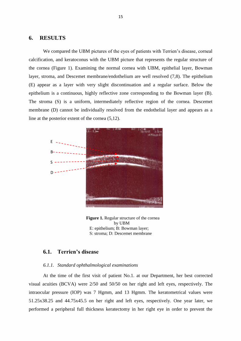
We compared the UBM pictures of the eyes of patients with Terrien’s disease, corneal
calcification, and keratoconus with the UBM picture that represents the regular structure of
the cornea (Figure 1). Examining the normal cornea with UBM, epithelial layer, Bowman
layer, stroma, and Descemet membrane/endothelium are well resolved (7,8). The epithelium
(E) appear as a layer with very slight discontinuation and a regular surface. Below the
epithelium is a continuous, highly reflective zone corresponding to the Bowman layer (B).
The stroma (S) is a uniform, intermediately reflective region of the cornea. Descemet
membrane (D) cannot be individually resolved from the endothelial layer and appears as a
line at the posterior extent of the cornea (5,12).
Figure 1. Regular structure of the cornea
by UBM
E: epithelium; B: Bowman layer;
S: stroma; D: Descemet membrane
6.1. Terrien’s disease
6.1.1. Standard ophthalmological examinations
At the time of the first visit of patient No.1. at our Department, her best corrected
visual acuities (BCVA) were 2/50 and 50/50 on her right and left eyes, respectively. The
intraocular pressure (IOP) was 7 Hgmm, and 13 Hgmm. The keratometrical values were
51.25x38.25 and 44.75x45.5 on her right and left eyes, respectively. One year later, we
performed a peripheral full thickness keratectomy in her right eye in order to prevent the
E
B
S
D
16
additional thinning of her peripheral cornea and the increase of against-the-rule astigmatism.
The postoperative period was without any complications and any signs of inflammation or
infiltration. Two months after her operation, the BCVA was 30/50 on her right eye. Her
follow up study was not possible because she did not show up at our Department since then.
At the time of the first visit of patient No.2. at our Department, his BCVA was 8/50
and 45/50, his IOP was 13 Hgmm and 14 Hgmm, his keratometrical values were
61.5x143/45.5x53 and 41.75x100/49.5x10 on his right and left eyes, respectively. Thinned
and ectatic part of the cornea was delimited by a sharp yellowish-white border. Because of the
danger of a sudden perforation, we performed a full thickness keratectomy and peripheral
iridectomy on his right eye. The postoperative period was without any complications.
Two months after his operation the BCVA was 40/50 on his right eye, and his IOP
was 12 mmHg.
Fourteen months after the operation, the BCVA was 20/50. The sutures were then
removed and his BCVA improved to 40/50, and his IOP was 11 mmHg. The right eyebulb
displayed no alteration by slit-lamp examination.
His last visit was 18 months after his operation. We observed a distinct decrease in
transparency towards the central part of the cornea.
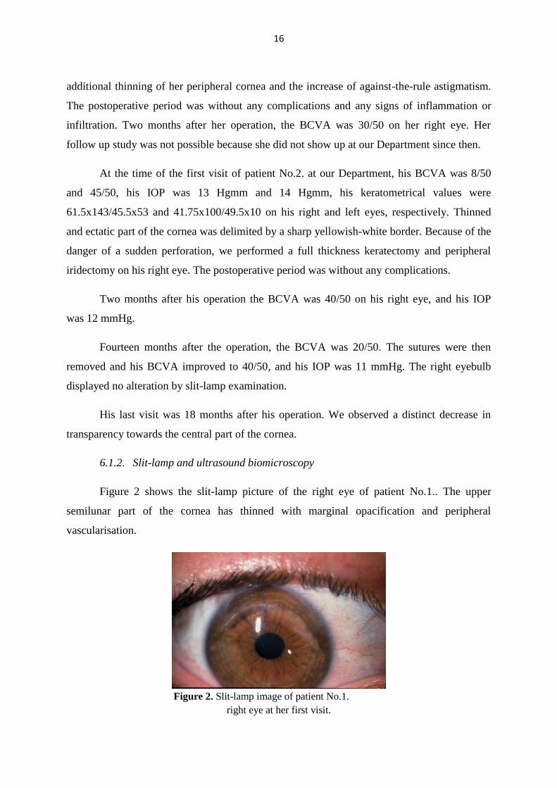
6.1.2. Slit-lamp and ultrasound biomicroscopy
Figure 2 shows the slit-lamp picture of the right eye of patient No.1.. The upper
semilunar part of the cornea has thinned with marginal opacification and peripheral
vascularisation.
Figure 2. Slit-lamp image of patient No.1.
right eye at her first visit.
17
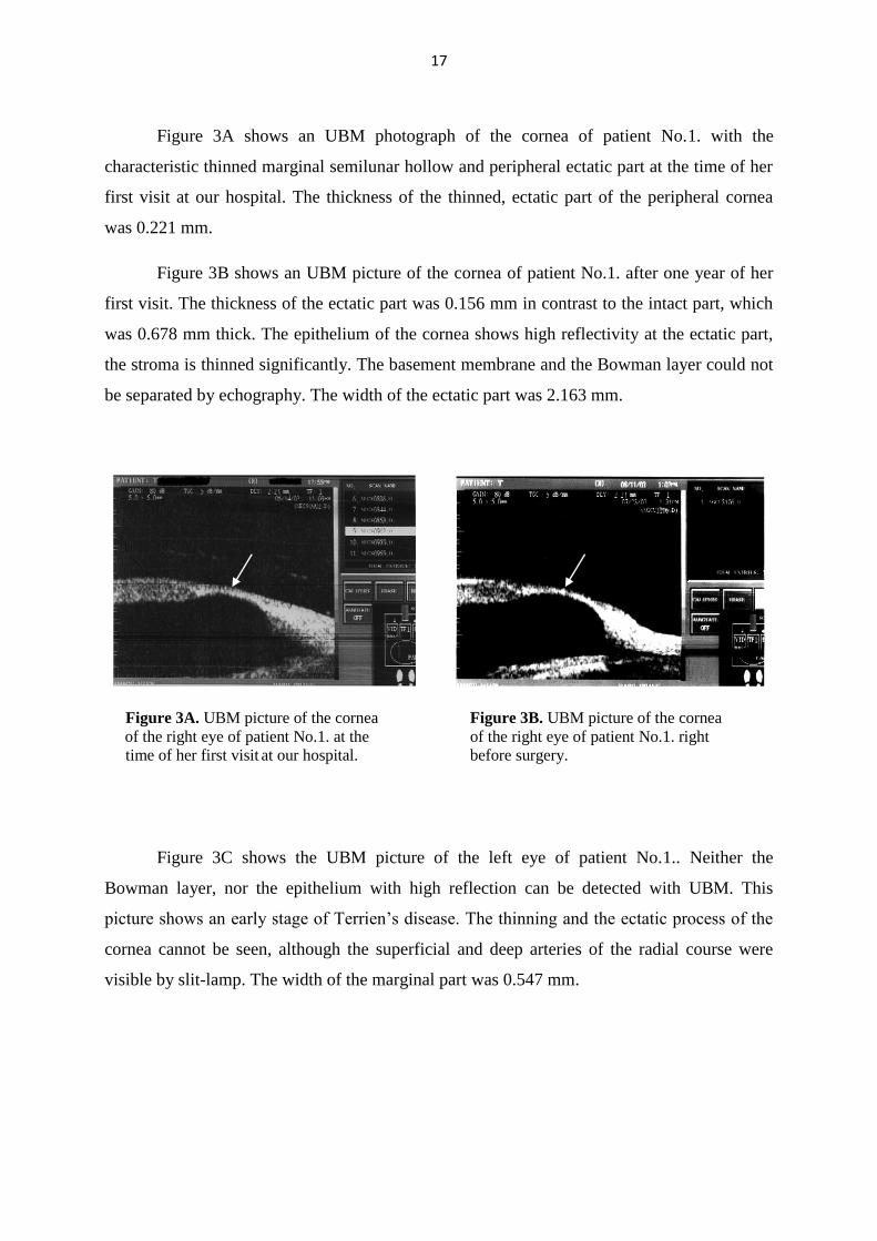
Figure 3A shows an UBM photograph of the cornea of patient No.1. with the
characteristic thinned marginal semilunar hollow and peripheral ectatic part at the time of her
first visit at our hospital. The thickness of the thinned, ectatic part of the peripheral cornea
was 0.221 mm.
Figure 3B shows an UBM picture of the cornea of patient No.1. after one year of her
first visit. The thickness of the ectatic part was 0.156 mm in contrast to the intact part, which
was 0.678 mm thick. The epithelium of the cornea shows high reflectivity at the ectatic part,
the stroma is thinned significantly. The basement membrane and the Bowman layer could not
be separated by echography. The width of the ectatic part was 2.163 mm.
Figure 3A. UBM picture of the cornea Figure 3B. UBM picture of the cornea
of the right eye of patient No.1. at the of the right eye of patient No.1. right
time of her first visit at our hospital. before surgery.
Figure 3C shows the UBM picture of the left eye of patient No.1.. Neither the
Bowman layer, nor the epithelium with high reflection can be detected with UBM. This
picture shows an early stage of Terrien’s disease. The thinning and the ectatic process of the
cornea cannot be seen, although the superficial and deep arteries of the radial course were
visible by slit-lamp. The width of the marginal part was 0.547 mm.
18
Figure 3C. UBM picture of the peripheral cornea
of the left eye of patient No.1. at the time of the
first visit.
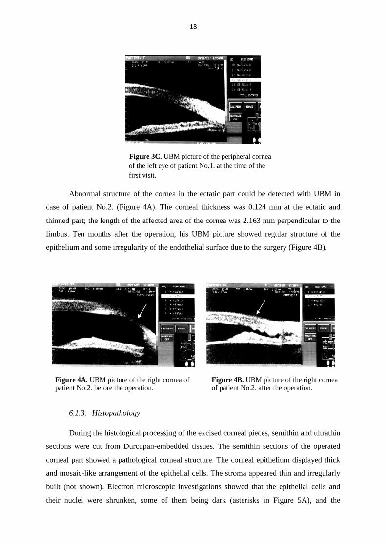
Abnormal structure of the cornea in the ectatic part could be detected with UBM in
case of patient No.2. (Figure 4A). The corneal thickness was 0.124 mm at the ectatic and
thinned part; the length of the affected area of the cornea was 2.163 mm perpendicular to the
limbus. Ten months after the operation, his UBM picture showed regular structure of the
epithelium and some irregularity of the endothelial surface due to the surgery (Figure 4B).
Figure 4A. UBM picture of the right cornea of Figure 4B. UBM picture of the right cornea
patient No.2. before the operation. of patient No.2. after the operation.
6.1.3. Histopathology
During the histological processing of the excised corneal pieces, semithin and ultrathin
sections were cut from Durcupan-embedded tissues. The semithin sections of the operated
corneal part showed a pathological corneal structure. The corneal epithelium displayed thick
and mosaic-like arrangement of the epithelial cells. The stroma appeared thin and irregularly
built (not shown). Electron microscopic investigations showed that the epithelial cells and
their nuclei were shrunken, some of them being dark (asterisks in Figure 5A), and the
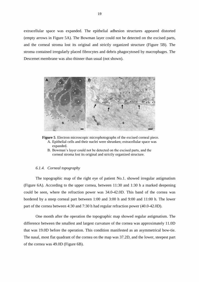
19
extracellular space was expanded. The epithelial adhesion structures appeared distorted
(empty arrows in Figure 5A). The Bowman layer could not be detected on the excised parts,
and the corneal stroma lost its original and strictly organized structure (Figure 5B). The
stroma contained irregularly placed fibrocytes and debris phagocytosed by macrophages. The
Descemet membrane was also thinner than usual (not shown).
Figure 5. Electron microscopic microphotographs of the excised corneal piece.
A. Epithelial cells and their nuclei were shrunken; extracellular space was
expanded.
B. Bowman’s layer could not be detected on the excised parts, and the
corneal stroma lost its original and strictly organized structure.
6.1.4. Corneal topography
The topographic map of the right eye of patient No.1. showed irregular astigmatism
(Figure 6A). According to the upper cornea, between 11:30 and 1:30 h a marked deepening
could be seen, where the refraction power was 34.0-42.0D. This band of the cornea was
bordered by a steep corneal part between 1:00 and 3:00 h and 9:00 and 11:00 h. The lower
part of the cornea between 4:30 and 7:30 h had regular refraction power (40.0-42.0D).
One month after the operation the topographic map showed regular astigmatism. The
difference between the smallest and largest curvature of the cornea was approximately 11.0D
that was 19.0D before the operation. This condition manifested as an asymmetrical bow-tie.
The nasal, most flat quadrant of the cornea on the map was 37.2D, and the lower, steepest part
of the cornea was 49.0D (Figure 6B).
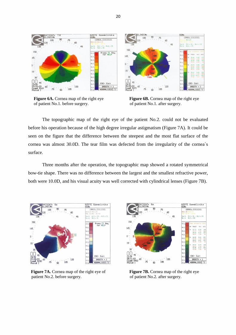
20
Figure 6A. Cornea map of the right eye Figure 6B. Cornea map of the right eye
of patient No.1. before surgery. of patient No.1. after surgery.
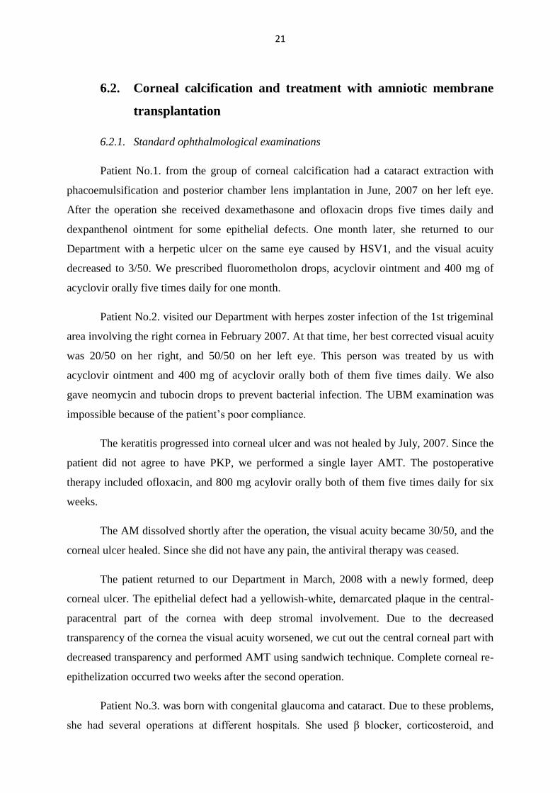
The topographic map of the right eye of the patient No.2. could not be evaluated
before his operation because of the high degree irregular astigmatism (Figure 7A). It could be
seen on the figure that the difference between the steepest and the most flat surface of the
cornea was almost 30.0D. The tear film was defected from the irregularity of the cornea`s
surface.
Three months after the operation, the topographic map showed a rotated symmetrical
bow-tie shape. There was no difference between the largest and the smallest refractive power,
both were 10.0D, and his visual acuity was well corrected with cylindrical lenses (Figure 7B).
Figure 7A. Cornea map of the right eye of Figure 7B. Cornea map of the right eye
patient No.2. before surgery. of patient No.2. after surgery.
21
6.2. Corneal calcification and treatment with amniotic membrane
transplantation
6.2.1. Standard ophthalmological examinations
Patient No.1. from the group of corneal calcification had a cataract extraction with
phacoemulsification and posterior chamber lens implantation in June, 2007 on her left eye.
After the operation she received dexamethasone and ofloxacin drops five times daily and
dexpanthenol ointment for some epithelial defects. One month later, she returned to our
Department with a herpetic ulcer on the same eye caused by HSV1, and the visual acuity
decreased to 3/50. We prescribed fluorometholon drops, acyclovir ointment and 400 mg of
acyclovir orally five times daily for one month.
Patient No.2. visited our Department with herpes zoster infection of the 1st trigeminal
area involving the right cornea in February 2007. At that time, her best corrected visual acuity
was 20/50 on her right, and 50/50 on her left eye. This person was treated by us with
acyclovir ointment and 400 mg of acyclovir orally both of them five times daily. We also
gave neomycin and tubocin drops to prevent bacterial infection. The UBM examination was
impossible because of the patient’s poor compliance.
The keratitis progressed into corneal ulcer and was not healed by July, 2007. Since the
patient did not agree to have PKP, we performed a single layer AMT. The postoperative
therapy included ofloxacin, and 800 mg acylovir orally both of them five times daily for six
weeks.
The AM dissolved shortly after the operation, the visual acuity became 30/50, and the
corneal ulcer healed. Since she did not have any pain, the antiviral therapy was ceased.
The patient returned to our Department in March, 2008 with a newly formed, deep
corneal ulcer. The epithelial defect had a yellowish-white, demarcated plaque in the central-
paracentral part of the cornea with deep stromal involvement. Due to the decreased
transparency of the cornea the visual acuity worsened, we cut out the central corneal part with
decreased transparency and performed AMT using sandwich technique. Complete corneal re-
epithelization occurred two weeks after the second operation.
Patient No.3. was born with congenital glaucoma and cataract. Due to these problems,
she had several operations at different hospitals. She used β blocker, corticosteroid, and
22
lubricant drops for more than 20 years. The best corrected visual acuity was 1/50 on her left
eye during the last ten years.
Patient No.3. showed up at our Department in December, 2009, as she had decreased
transparency of the left cornea. The corneal picture showed a demarcated whitish plaque in
the center of the cornea. Since the plaque was very disturbing for her, she wanted to have an
operation. We decided to perform AMT using single layer graft inlay after cutting out the
demarcated corneal part. The patients’ data are summarized in Table 1.
Patients Age Sex Cause Therapy Follow-up
time (month)
Type of operation Histology
1. 80 female
cataract and dry
eye, herpes
simplex keratitis
dexamethasone, ofloxacine,
neomycin, acyclovir, tubocin 24
AMT using
sandwich
technique
stromal
calcification
2. 77 female herpes simplex
keratitis
acyclovir, dexamethasone,
ofloxacine 40
AMT using
sandwich
technique
stromal
calcification
3. 54 female congenital
glaucoma
timolol, dexamethasone,
lubricants 8
AMT single layer
graft inlay
stromal
calcification
6.2.2. Slit-lamp and ultrasound biomicroscopy
Figure 8 shows the slit-lamp and the UBM pictures of patient No.1. before the
operation. The calcified part of the paracentral cornea can be seen in Figure 8A and has
yellowish-whitish color on the picture. Figure 8B presents the UBM picture of this state,
where the calcified cornea can be seen on the transverse biomicroscopic picture. The central
width of the cornea is 565 μm. There is high reflectivity on the paracentral cornea that it is
well demarcated from the regular part of the cornea. The extension of this part is 2.68 x 3.45
mm, and the deepness is 248 μm. The UBM structure of the cornea cannot be recognized in
the calcified part, only homogeneous, highly reflective echos can be seen. She returned to our
Department one month later with a non-healing ulcer with supposed calcification. It caused
visual impairment, so we cut the calcificated part out and performed a sandwich technique
AMT. The patient was followed up daily after the AMT procedure for a week. Thereafter she
Table 1. Clinical data and treatment of patients diagnosed with corneal calcification.

23
did not come to our Department because of her Alzheimer disease. At her last visit, the visual
acuity was 10/50 on the left eye.
Figure 8. Slit-lamp and UBM pictures before surgery of patient No.1.with
corneal calcification. A. Calcificated part of the cornea by slit- lamp.
B. UBM picture of the calcificated cornea.
Patient No.2. had pain in her right eye and poor compliance before her first operation,
that is why we could not examine her with UBM at that time. One week after her reoperation
the AM still could be seen on the top of the corneal excised part on the slit-lamp picture
(Figure 9A). On the UBM picture of the transverse segment of the cornea, the transplanted
AM was parallel, foliated, and had a rough surface and higher reflectivity than the other part
of the cornea (Figure 9B).
Figure 9. Slit-lamp and UBM pictures of patient No.2. with corneal calcification one week
after surgery. A. Surface of the cornea by slit-lamp. The edges of the AM are attached
by10/0 sutures. B. UBM picture of the cornea with AM attached to it.
A B
A
A B
24
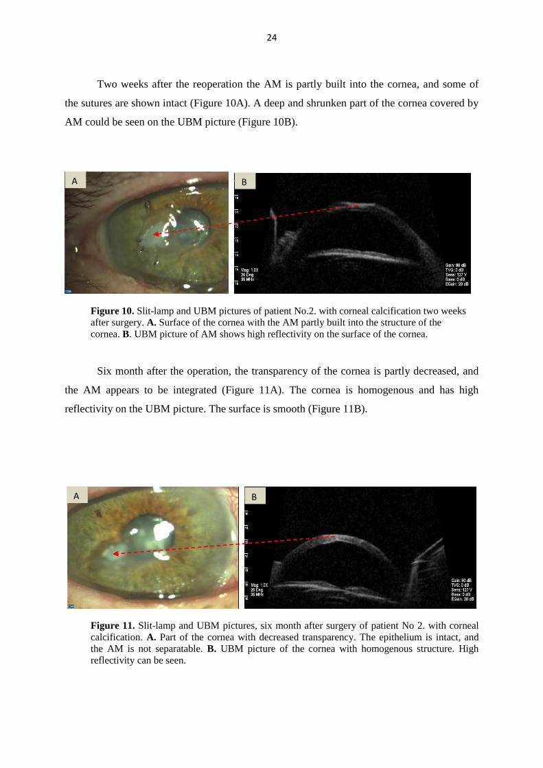
Two weeks after the reoperation the AM is partly built into the cornea, and some of
the sutures are shown intact (Figure 10A). A deep and shrunken part of the cornea covered by
AM could be seen on the UBM picture (Figure 10B).
Figure 10. Slit-lamp and UBM pictures of patient No.2. with corneal calcification two weeks
after surgery. A. Surface of the cornea with the AM partly built into the structure of the
cornea. B. UBM picture of AM shows high reflectivity on the surface of the cornea.
Six month after the operation, the transparency of the cornea is partly decreased, and
the AM appears to be integrated (Figure 11A). The cornea is homogenous and has high
reflectivity on the UBM picture. The surface is smooth (Figure 11B).
Figure 11. Slit-lamp and UBM pictures, six month after surgery of patient No 2. with corneal
calcification. A. Part of the cornea with decreased transparency. The epithelium is intact, and
the AM is not separatable. B. UBM picture of the cornea with homogenous structure. High
reflectivity can be seen.
A
A
B
A
A
A
B
A
25
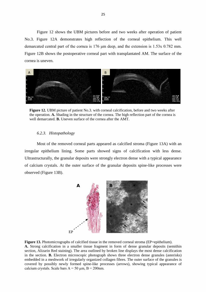
Figure 12 shows the UBM pictures before and two weeks after operation of patient
No.3. Figure 12A demonstrates high reflection of the corneal epithelium. This well
demarcated central part of the cornea is 176 μm deep, and the extension is 1.53x 0.782 mm.
Figure 12B shows the postoperative corneal part with transplantated AM. The surface of the
cornea is uneven.
Figure 12. UBM picture of patient No.3. with corneal calcification, before and two weeks after
the operation. A. Shading in the structure of the cornea. The high reflection part of the cornea is
well demarcated. B. Uneven surface of the cornea after the AMT.
6.2.3. Histopathology
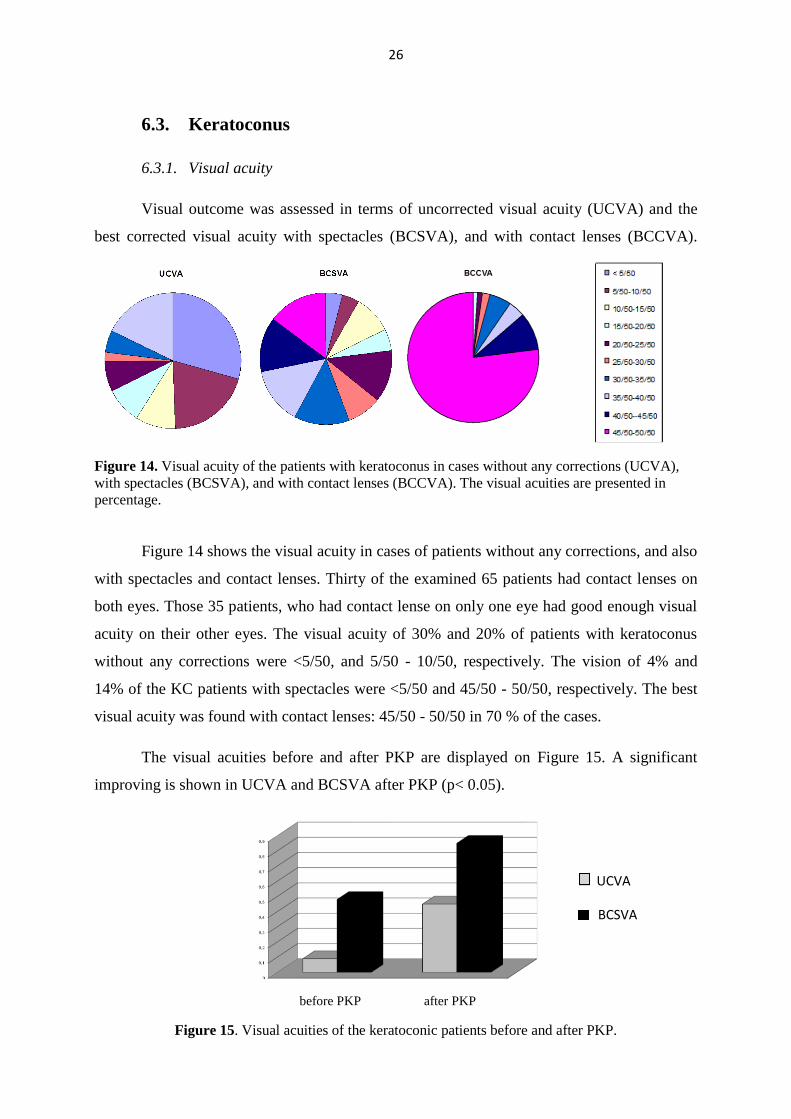
Most of the removed corneal parts appeared as calcified stroma (Figure 13A) with an
irregular epithelium lining. Some parts showed signs of calcification with less dense.
Ultrastructurally, the granular deposits were strongly electron dense with a typical appearance
of calcium crystals. At the outer surface of the granular deposits spine-like processes were
observed (Figure 13B).
EP
Figure 13. Photomicrographs of calcified tissue in the removed corneal stroma (EP=epithelium).
A. Strong calcification in a smaller tissue fragment in form of dense granular deposits (semithin
section, Alizarin Red staining). The area outlined by broken line displays the most dense calcification
in the section. B. Electron microscopic photograph shows three electron dense granules (asterisks)
embedded in a meshwork of irregularly organized collagen fibres. The outer surface of the granules is
covered by possibly newly formed spine-like processes (arrows), showing typical appearance of
calcium crystals. Scale bars A = 50 µm, B = 200nm.
A
A
B
A
26
6.3. Keratoconus
6.3.1. Visual acuity
Visual outcome was assessed in terms of uncorrected visual acuity (UCVA) and the
best corrected visual acuity with spectacles (BCSVA), and with contact lenses (BCCVA).
before and after PKP.
Figure 14. Visual acuity of the patients with keratoconus in cases without any corrections (UCVA),
with spectacles (BCSVA), and with contact lenses (BCCVA). The visual acuities are presented in
percentage.
Figure 14 shows the visual acuity in cases of patients without any corrections, and also
with spectacles and contact lenses. Thirty of the examined 65 patients had contact lenses on
both eyes. Those 35 patients, who had contact lense on only one eye had good enough visual
acuity on their other eyes. The visual acuity of 30% and 20% of patients with keratoconus
without any corrections were <5/50, and 5/50 - 10/50, respectively. The vision of 4% and
14% of the KC patients with spectacles were <5/50 and 45/50 - 50/50, respectively. The best
visual acuity was found with contact lenses: 45/50 - 50/50 in 70 % of the cases.
The visual acuities before and after PKP are displayed on Figure 15. A significant
improving is shown in UCVA and BCSVA after PKP (p< 0.05).
before PKP after PKP
Figure 15. Visual acuities of the keratoconic patients before and after PKP.
UCVA
BCSVA
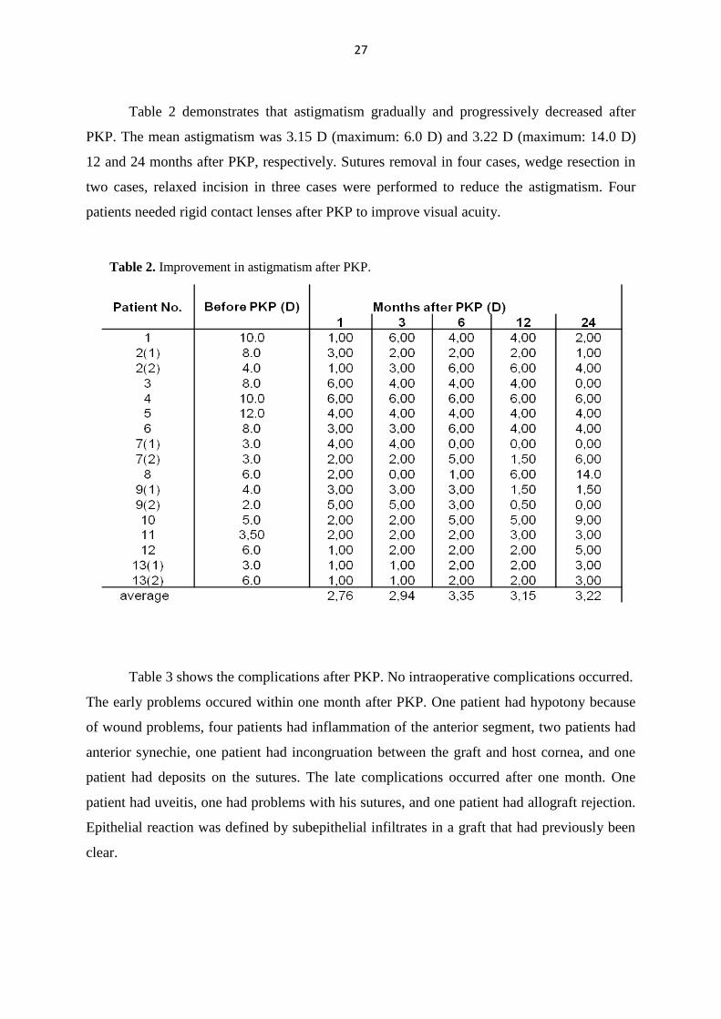
27
Table 2 demonstrates that astigmatism gradually and progressively decreased after
PKP. The mean astigmatism was 3.15 D (maximum: 6.0 D) and 3.22 D (maximum: 14.0 D)
12 and 24 months after PKP, respectively. Sutures removal in four cases, wedge resection in
two cases, relaxed incision in three cases were performed to reduce the astigmatism. Four
patients needed rigid contact lenses after PKP to improve visual acuity.
Table 3 shows the complications after PKP. No intraoperative complications occurred.
The early problems occured within one month after PKP. One patient had hypotony because
of wound problems, four patients had inflammation of the anterior segment, two patients had
anterior synechie, one patient had incongruation between the graft and host cornea, and one
patient had deposits on the sutures. The late complications occurred after one month. One
patient had uveitis, one had problems with his sutures, and one patient had allograft rejection.
Epithelial reaction was defined by subepithelial infiltrates in a graft that had previously been
clear.
Table 2. Improvement in astigmatism after PKP.
28
Appearance of
complications after
PKP
Signs Number of patients
Early hypotony
iridocyclitis
anterior synechia
incongruation of the graft
deposits on the sutures
1
4
2
1
1
Late uveitis
rejection
suture problems
1
1
1
6.3.2. Ultrasound biomicroscopy
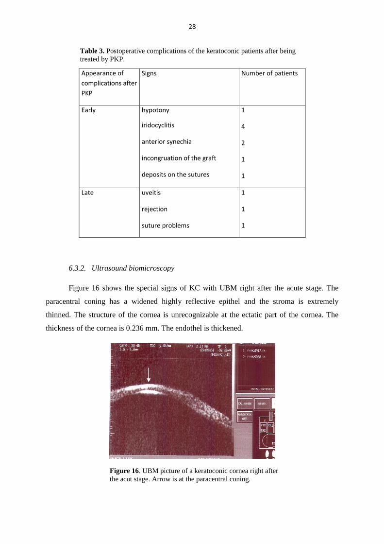
Figure 16 shows the special signs of KC with UBM right after the acute stage. The
paracentral coning has a widened highly reflective epithel and the stroma is extremely
thinned. The structure of the cornea is unrecognizable at the ectatic part of the cornea. The
thickness of the cornea is 0.236 mm. The endothel is thickened.
Figure 16. UBM picture of a keratoconic cornea right after
the acut stage. Arrow is at the paracentral coning.
Table 3. Postoperative complications of the keratoconic patients after being
treated by PKP.
29
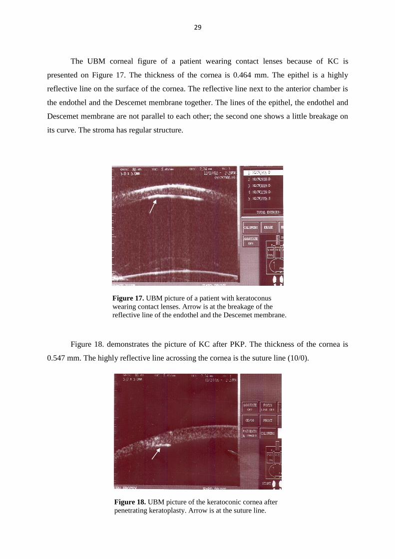
The UBM corneal figure of a patient wearing contact lenses because of KC is
presented on Figure 17. The thickness of the cornea is 0.464 mm. The epithel is a highly
reflective line on the surface of the cornea. The reflective line next to the anterior chamber is
the endothel and the Descemet membrane together. The lines of the epithel, the endothel and
Descemet membrane are not parallel to each other; the second one shows a little breakage on
its curve. The stroma has regular structure.
Figure 17. UBM picture of a patient with keratoconus
wearing contact lenses. Arrow is at the breakage of the
reflective line of the endothel and the Descemet membrane.
Figure 18. demonstrates the picture of KC after PKP. The thickness of the cornea is
0.547 mm. The highly reflective line acrossing the cornea is the suture line (10/0).
Figure 18. UBM picture of the keratoconic cornea after
penetrating keratoplasty. Arrow is at the suture line.
30
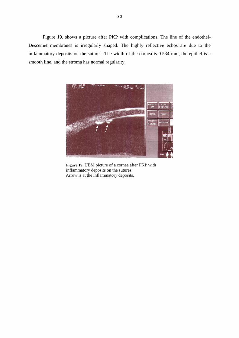
Figure 19. shows a picture after PKP with complications. The line of the endothel-
Descemet membranes is irregularly shaped. The highly reflective echos are due to the
inflammatory deposits on the sutures. The width of the cornea is 0.534 mm, the epithel is a
smooth line, and the stroma has normal regularity.
Figure 19. UBM picture of a cornea after PKP with
inflammatory deposits on the sutures.
Arrow is at the inflammatory deposits.
31
7. DISCUSSION
High resolution UBM is an excellent echographic method for the examination of the
anterior part of the eye. This examination is noninvasive, repeatable, and suitable for
diagnostic and morphometric examinations (1,5-7,9,16).
Using slit-lamp we could demonstrate the thinning status of the cornea, but this
method is not able to measure the exact corneal thickness. The thickness of the cornea can be
evaluated quantitatively by number of methods including ultrasonic pachymetry (11,14)
optical slit-lamp pachymetry (12), specular microscopy (12), confocal microscopy, OCT
(9,14,77), high frequency UBM (9,16), laser Doppler interferometry, Placido disc based
corneal topography, and pentacam (78-81). Dada et al. compared OCT and UBM pictures of
the anterior segment of the eye and found, that both methods can be used for the
measurements of the anterior segment and yielded comparable results (82).
Earlier studies (71,79) showed the ability of UBM in estimating corneal thickness,
both in patients with early stage keratoconus and in normal subjects. The 50 MHz probe is an
accurate and reproducible method for determining corneal thickness in clinical practice.
The aims of our studies were to investigate and present the characteristics of several
corneal diseases, to measure the corneal thinning in Terrien’s disease and KC, to follow the
corneal reconstruction after the removal of the calcified part and amniotic membrane
transplantation by UBM.
7.1. Terrien’s disease
Terrien’s degeneration is a rare peripheral corneal disorder of unknown etiology,
which produces marginal corneal thinning and subsequently a significant amount of
astigmatism. Several recent investigations deal with clinical and histological research and
treatments of patients with Terrien’s disease (29-31,83,84).
Iwamoto and coworkers in their electron microscopic studies found hyperemic blood
vessels with increased inflammatory cell infiltrates in the superficial corneal layers in cases of
inflammatory Terrien’ s disease (28). Our electron microscopic investigations of patients with
Terrien’s disease showed, that the epithelial cells and their nuclei were shrunken in the
32
thickened epithelium and the extracellular space was expanded. The Bowman membrane
could not be detected, and the stroma lost its original structure and contained irregularly
placed fibrocytes and debris phagocytosed by macrophages.
Lampé et al. summarized the typical morphological changes in case of Terrien’s
disease by using corneal topography (85). The postoperative surface was described as bowtie
or nipple shaped. The postoperative follow-up examinations showed the decrease of the
regular, indirect, and high degree of astigmatism, which can be corrected with cylindrical
lenses (85). The topographic map of our patients showed irregular astigmatism before
operation. After surgeries, their conditions manifested as an asymmetric-symmetric bowtie.
UBM is a useful tool with high magnification power for examining the anterior part of
the eyebulb, finding the right diagnosis, and carrying out morphometry of the anterior
segment. Previously, two studies used UBM as a diagnostic device to define the presence of
hydrops in Terrien ’s disease (86,87). In our studies UBM was used to observe the structure
and thickness of the degenerative corneal area.
Our study was the first one that presented the echographic changes of the stages in
Terrien’s disease. We measured the thickness and the width of the thinned, ectatic part of the
peripheral cornea. In the second and third stages of the disease, the thickness and the width of
the thinned peripheral part of the cornea, the progression of the illness, and the best time for
the operation can be determined. The UBM findings correlated with and justified our
histologic examinations and corneal topography. The histologic samples showed that the
Bowman layer could not be detected on the excised parts; the corneal stroma lost its original
and strictly organized structure; and the Descemet membrane was also thinner than the usual
ones.
We found that UBM provides reliable information for the diagnosis of Terrien’s
disease and allows us to follow its course. We proved that it is an excellent diagnostic method
for determining the extension, the thickness, and the structure of the disordered, ectatic part of
the peripheral cornea. The actual size of the protruded corneal part could also be determined
by using UBM which information is important and helpful for planning the operation.
33
7.2. Corneal calcification and treatment with amniotic membrane
transplantation
The causes of deep calcification of the cornea are still not clearly understood. Intact
epithelium and absence of Bowman layer in the presence of high level of phosphate ions plus
an inflammatory response seem to provide the necessary environment for the formation of
calcium salts deep within the stroma of the cornea (37,45). Reports have implicated the role
of sodium hyaluronate artificial tears, preservatives of eyedrops with phosphate buffers,
timolol, retinoic acid and dexamethasone in calcium salt depositions (36,41,42). Calcium salt
deposition can also occur in cases of older patients, decreased tear production, and history
with previous herpes virus infection (37,45). The high phosphate concentration leads to a
critical increase of the calcium phosphate which together with the breakdown of the tissue
barrier, results in the formation of hydroxyapatite deposits (36).
In our studies we described three patients with different etiology of corneal
calcification. Although each of our cases had a known risk factor for corneal calcification
formation (herpes simplex keratitis, dry eye, persistent epithelial defect, congenital
glaucoma), it is possible that corneal calcification is a multifactorial process as was mentioned
in the study of Ebrahimi (88). We investigated these patients with UBM. We found that this
method provides excess information to establish the diagnosis of corneal calcification, makes
possible to determine the extent of the part to be excised, and may be used to estimate the
territory that should be operated.
Previous studies showed that AMT promised the restoration of ocular surface
integrity, reduction of stromal infiltration, and improvement of vision in ulcerative and
necrotising herpetic keratitis. Gabric et al. effectively cured forty patients with persistent
corneal epithelial defects (recurrent pterygia, corneal graft, impending perforation) resistant to
other treatments covering with AM. There was complete epithelization of the corneal defects
of 67% of their patients during the first three postoperative weeks (46). Comparing these data
with our investigations, our patient No. 2. had perfect re-epithelisation two weeks after her
AMT.
In the management of acute ulcerative herpes simplex keratitis the appropriate
antiviral and antimicrobial therapy should be instituted at least several days before AMT. In
these cases AMT might be considered and installated after these conservative therapy (47).
34
We treated our patients before the surgical procedure with acyclovir, neomycin,
dexamethason, and lubricant drops, too.
Kim and Spelsberg suggested, that AMT may be an initial surgical option in selected
cases with deep ulcers from herpetic viruses (49,89). Shi used the AM in the management of
necrotising stromal keratitis of fifteen patients. The adjunctive use of antiviral and steroid
therapy with AMT should be recommended in the treatment of patients having corneal
classification in order to increase the safety and efficacy of AMT (54).
Seitz and Resch (55), Prabhasawat (56), and Müller (90) published articles dealing
with patients treated because of different ocular surface pathologies and histories. Each of
these patients required a different technique of AMT. They suggested, that the AM graft
served as a new basement membrane for corneal epithelial cells. A differentiated
microsurgical technique results in different integration patterns of AM into the human cornea
(55,56,88,91). The above studies confirmed, that AM may be integrated fully into the normal
corneal surface and preserved there for months, and AM could not notice later.
In our investigations, patient No.1. had phacoemulsification before the formation of
the non-healing epithelial defect of the cornea. Corneal calcification was developed after
using topical treatment for three months. Deep corneal calcification formed after treating
patient No.2. with antiviral, antibiotic, and corticosteroid drops containing phosphate buffer
because of her herpes zoster keratitis for more than a half year. Patient No.3. had several
operations and used different eyedrops for years in order to treat congenital glaucoma. The
anti-glaucomatous eyedrops contained timolol and other phosphate buffers. Using these drugs
for ten years, superficial corneal degeneration was formed.
Our results showed, that frequent and long use of several eyedrops containing
phosphate buffer may considerably increase the risk of calcium deposition, especially in
patients with persistent epithelial defect. The applied eye drops contained phosphate buffers,
ofloxacin, neomycin, acyclovir, and dexamethasone. In order to treat these patients, we
transplanted AM using either single layer graft inlay or sandwich techniques. Since our
histological examinations demonstrated strong calcification in the affected stroma, we had to
cut out the calcified part of the cornea before AMT.
In concordance with previous reports, we recommend that predisposed patients who
are using eye drops containing phosphate buffer be followed up carefully for the potential

35
complications of corneal stromal calcification. If possible, nonphosphated corticosteroid eye
drops should be used.
Several methods are known to treat corneal calcifications, but no one has used AMT to
treat corneal calcification so far. We concluded from our results of imaging studies, that AMT
could provide good anatomical and functional information in cases of persistent corneal
epithelial defect and ulcer with calcium deposition.
In order to follow and prove the good therapeutic effect of AM, we used UBM. This
device was helpful in the follow up of the treatment of patients with AM who had corneal
calcification.
7.3. Keratoconus
Keratoconus (KC) is a continuing clinical problem and a leading indication for PKP.
Major advances in the treatment of KC include improved technology in rigid gas permeable
and hybrid contact lenses and corneal transplantation (72,73,92-94). PKP offers good long-
term visual rehabilitation for KC (79,94-98). High postoperative corneal astigmatism,
sometimes seen after PKP, could prevent good visual rehabilitation even when a graft is clear
and otherwise surgically successful. Some factors influencing postkeratoplasty astigmatism
are independent on the surgeon, like wound healing and recipient corneal pathology (99-101).
Several surgical techniques like incisional procedures such as astigmatic keratotomy or
ablative procedures such as laser in situ keratomileusis are used to treat astigmatism after
PKP. In cases of progressive KC, wedge resection is considered. The resection is centered on
the negative axis and a wedge of tissue is removed from both the host and the donor, using a
nomogram (99). Repeating PKP leads to intraoperative and postoperative complications,
therefore this should be only considered when all other options have failed (98, 101).
We compared the visual acuity among patients wearing contact lenses and among
those subjects who underwent corneal transplantation. We also investigated the complications
of these methods to evaluate recommendations to patients with KC. In our investigations, we
used UBM as a proved to be good method to diagnose KC and follow its course. Using UBM
we could in vivo diagnose KC, detecting conal shape of the cornea, and corneal thinning.
36
We found that after PKP, UCVA and BCVA significantly improved and astigmatism
gradually and progressively decreased. Wearing contact lenses slows down the formation of
KC. The curvature of the endothel showed a little breakage on the UBM figure, but the
thickness of the cornea was almost regular (0.464 mm). The different complications of PKP
could also be seen in keratoconic patients with UBM that proved to be a safe and reliable
device to follow the course of patients with KC.
The visual acuity of the patients with KC without any corrections was mainly <5/50
(30%). The vision of the KC patients with spectacles were <5/50 in 4%, and 45/50-50/50 in
14%. The visual acuity with contact lense was the best, 45/50-50/50 in 70% postoperatively,
the average corrected visual acuity was 40/50.
Our investigations showed that astigmatism gradually and progressively improved
after PKP. Other studies revealed that factors like sex, donor age, age of the patients when
keratoplasty was performed, diameters of host and donor cornea trephinations, suture
technique, preoperative astigmatism, and time of suture removal after keratoplasty were not
any significant predictors of postgraft astigmatism. Postgraft astigmatism could be caused by
the different method of trephination and asymmetry of suture forces (96,97,100). To reduce
the postoperative astigmatism, we performed wedge resection in two cases and relaxed
incision in three cases.
PKP for KC is associated with a low graft failure rate in most series (79,95,101). In
our study, only one patient had rejection amounting to graft failure. One of our patients had
incongruation of the sutures and one had hypotony after PKP because of loose sutures. These
two patients needed resuturing because these occured less than 6 months after keratoplasty.
Suture problems started at one of our patients later than 6 months. In this case, the allograft
rejection episodes were circumvented with promt removal of the sutures and prophylactic use
of topical corticosteroids.
Four of our patients had inflammation after PKP, although prophylactic use of
corticosteroids and antibiotics prevented rejection in these cases. Synechiolysis were done
with two of our patients who had anterior synechiae.
In conclusion, half of the patients with KC had stable visual acuity with using rigid gas
permeable and hybrid contact lenses. We can say that the progression of the KC could be
delayed by initiating the wear of contact lenses as soon as possible, even in early stages of

37
KC. PKP for KC is associated with an excellent graft survival rate (95% success in 36
months) and good visual acuity (the mean BCVA after PKP was 40/50).
Both wearing contact lenses and performing PKP could result in a very good visual
acuity for a longer time and good anatomical status is obtained by PKP.
More research remains to be done, and numerous clinical studies must be performed to
determine the possible role of UBM in other clinical situations in ophthalmology. However,
our results (in cases of Terrien’s disease, corneal calcification, and keratoconus) and the
unique capabilities of UBM imaging suggest that it has the potential to have significant
impact on the diagnosis and clinical management of many eye diseases.
38
8. SUMMARY AND CONCLUSIONS
- In cases of Terrien’s disease we found that the thickness of the ectatic part was
significantly thinner than the intact part. The epithelium of the cornea showed high
reflectivity at the ectatic part, and the stroma was thinned significantly. The
basement membrane and the Bowman layer could not be separated by echography.
- UBM provided reliable information for the diagnosis of Terrien’s disease and
allowed us to follow its course. It proved to be an useful diagnostic tool for
determining the extension of the ectatic part, the thickness and the structure of the
cornea.
- We published first the echographic changes of the stages of Terrien’s disease.
- The UBM pictures of corneal calcification showed that the central width of the
cornea was thicker than the normal structure. There was high reflectivity on the
paracentral cornea that is well demarcated from the regular part of the cornea.
- UBM was easy to use and provided excess information to establish the diagnosis
of corneal calcification, made possible to determine the extent of the calcified part,
and might be used to estimate the territory that should be operated. It was also
helpful to determine the cornea’s pathological processes which could not be seen
with slit lamp.
- On the UBM picture of the transverse segment of the cornea, the transplanted AM
was built into the cornea. It was parallel, foliated, and had a rough surface and
higher reflectivity than the other part of the cornea The cornea covered by AM was
homogenous and had high reflectivity on the UBM picture, and the surface was
smooth. AMT could be a proper method to heal epithelial defects in cases of
superficial and deep corneal calcification.
- No one has used AMT to treat corneal calcification before.
- We can conclude from our results that UBM is helpful in the follow up and in the
improvement of the therapeutic benefits of AMT in cases of corneal calcification.
AMT provided good anatomical and functional formation in cases of persistent
corneal epithelial defect and ulcer with calcium deposition.
39
- Examination of patients with keratoconus revealed that the paracentral coning had
a widened highly reflective epithel and extremely thinned stroma, and the endothel
was thickened. The structure of the cornea was unrecognizable at the ectatic part.
The thickness of the cornea was significantly thinner than the normal one. The
endothel was thickened.
- Both wearing contact lenses and performing PKP could result in a very good visual
acuity for a longer period of time. In addition, good anatomical status has been
obtained by PKP.
- UBM was suitable for the examination of the shape, the curvature, and the
superficial and innermost irregularities of the cornea.
- The UBM findings correlated with the results of our histologic examinations.
- Since UBM is relatively cheap and easy to use as compared to other examination
methods, we suggest the use of UBM in order to get a diagnosis and to follow the
course of different inflammatorical and degenerational corneal diseases. This
method could also be helpful in the estimation of the progression of the
investigated corneal diseases.
40
9. REFERENCES
1. Wolffsohn JS, Davies LN. Advances in anterior segment imaging. Curr Opin
Ophthalmol 2007; 18: 32-38.
2. Konstantopoulos A, Hassain P, Anderson DF. Recent advances in ophthalmic anterior
segment imaging: a new era for ophthalmic diagnosis? Br J Ophthalmol 2007; 91:
551-557.
3. Ishikawa H, Liebmann JM, Ritch R. Quantitative assessment of the anterior segment
using ultrasound biomicroscopy. Curr Opin Ophthalmol 2000; 11: 133-139.
4. Ishikawa H, Inazumi K, Liebmann JM, et al. Inadvertent corneal indentation may
cause artifactitious widening of the iridocorneal angle on ultrasound biomicroscopy.
Ophthal Surg Lasers 2000; 11: 133-139.
5. Lizzi FL, Coleman J. History of ophthalmic ultrasound. J Ultrasound Med 2004;
23:1255–1266.
6. Pavlin CJ, Harasiewicz K, Eng P, et al. Ultrasound biomicroscopy of anterior segment
structures in normal and glaucomatous eyes. Am J Ophthalmol 1992; 113: 381-389.
7. Pavlin CJ, Foster FS. Ultrasound Biomicroscopy of the Eye. New York: Springer
Verlag; 1994. Pavlin CJ, Harasiewicz K, Sherar M, et al. Clinical use of ultrasound
biomicroscopy. Am J Ophthalmol. 1992; 99: 287-295.
8. Ishikawa H, Schuman J. Anterior segment imaging: Ultrasound biomicroscopy.
Ophthalmol Clin North Am 2004; 17: 7-20.
9. Nolan W. Anterior segment imaging: ultrasound biomicroscopy and anterior segment
optical coherence tomography. Curr Opin Ophthalmol 2008; 19: 115-121.
10. Zhang M, Chen J, Liang I, Laties AM, Liu Z. Ultrasound biomicroscopy of Chinese
eyes with iridocorneal endothelial syndrome. Br J Ophthalmol 2006; 90: 64-69.
11. Kim HY, Budenz DI, Lee PS, Feuer WJ, Barton K. Comparison of central corneal
thickness using anterior segment optical coherence tomography vs ultrasound
pachymetry. Am J Ophthalmol 2008; 145: 228-232.
41
12. Swartz T, Marten L, Wang M. Measuring the cornea: the latest developments in
corneal topography. Curr Opin Ophthalmol 2007; 18: 325-333.
13. Morris D, Somner J, Scott K, et al. Corneal thickness at high altitude. Cornea 2007;
26: 308-311.
14. Li E, Mohamed S, Leung C, et al. Agreement among 3 methods to measure corneal
thickness: Ultrasound pachymetry, Orbscan II, and Visante Anterior Segment Optical
Coherence Tomography. Ophthalmol 2007; 114: 1842-1847.
15. Cosar C, Sener B. Orbscan Corneal topography system in evaluating the anterior
structures of the human eye. Cornea 2003; 22: 118-121.
16. Skribek Á, Sohár N, Gyetvai T, et al. Role of ultrasound biomicroscopy in diagnosis
and treatment of Terrien disease. Cornea 2008; 27: 427-433.
17. Terrien F. Dystrophie marginale symetrique des deux cornée. Arch Opht 1900; 20: 12-
21.
18. Austin P, Brown S. Inflammatory Terrien’s marginal corneal disease. Am J
Ophthalmol 1981; 92: 189-192.
19. Grayson M. Disease of the cornea. St. Louis: CV Mosby Co 1979: 189-191.
20. Duke-Elder S. System of Ophthalmology. Vol 8. part 2, London: H. Kimpton, 1965:
909-914.
21. Alberth B, Süveges I. Die Formen und die Behandlung der Terrienschen Erkrankung.
Klin Monatsbl Augenheilkd 1970; 157: 419-424.
22. Guyer DR, Barraquer J, McDonnell PJ, et al.: Terrien’s marginal degeneration:
clinicopathologic case reports. Graefe’s Arch Clin Exp Ophthalmol 1987; 25: 19-27.
23. Doggart JH. Marginal degeneration of the cornea. Br J Ophthalmol 1930; 14: 510-
516.
24. Krachmer JH. Pellucid marginal corneal degeneration. Arch Ophthalmol 1978; 96:
1217-1221.
42
25. Süveges I, Levai G, Alberth B. Pathology of Terrien’s disease. Am J Ophthalmol
1972; 74: 1191-1200.
26. Francois J: La degenerescence marginale de la cornee. Arch Ophthalmol (Paris) 1936;
53: 616.
27. Stucchi CA. La maladie de Terrien (degenerescence marginale de la cornée).
Keratoplastie transfixiante: Histologie. Ann Ocul (Paris) 1968; 201: 720-731.
28. Iwamoto T, DeVoe AG, Farris RL. Electron microscopy in cases of marginal
degeneration of the cornea. Invest Ophthalmol 1972; 11: 241-257.
29. Srinivasan S, Murphy CC, Fisher AC, et al. Terrien marginal degeneration
presentingwith spontaneous corneal perforation. Cornea 2006; 25: 977-980.
30. Ferrari G, Tedesco S, Delfini E, Macaluso C. Laser scanning in vivo confocal
microscopy in a case of Terrien marginal degeneration. Cornea 2010; 29: 471-475.
31. Ceresara G, Migliavacca L, Orzalesi N, Rossetti L. In vivo confocal microscopy in
Terrien marginal corneal degeneration: a case report. Cornea 2011; 30: 820-824.
32. Varley GA, Macsai MS, Krachmer JH. The results of penetrating keratoplasty for
pellucid marginal corneal degeneration. Am J Ophthalmol 1990; 110: 149-152.
33. Cameron JA. Results of lamellar crescentic resection for pellucid marginal corneal
degeneration. Am J Ophthalmol 1992; 113: 296-302.
34. Dubroff S. Pellucid marginal degeneration: report on corrective surgery. J Cataract
Refract Surg 1989; 15: 89-93.
35. Cheng CL, Theng JT, Tan DT. Compressive C shaped lamellar keratoplasty: a
surgical alternative for the management of severe astigmatiswm from peripheral
corneal degeneration. Opthatmology 2005; 112: 425-530.
36. Bernauer W, Thiel MA, Kurrer M, et al. Corneal calcification following intensified
treatment with sodium hyaluronate artificial tears. Br J Ophthalmol 2006; 90: 285-
288.
37. Lake D, Tarn A, Ayliffe W. Deep corneal calcification associated with preservative-
free eye drops and persistent epithelial defects. Cornea 2008; 27: 292-296.
43
38. Daly M, Tuft ST, Munro PMG. Acute corneal calcification following chemical injury.
Cornea 2005; 24: 761-765.
39. Messmer EM, Hoops JP, Kampik A. Bilateral recurrent calcareous degeneration of the
cornea. Cornea 2005; 24: 498-502.
40. Kompa S, Redbrake C, Dunkel B, et al. Corneal calcification after chemical eye burns
caused by eye drops containing phosphate buffer. Burns 2006; 32: 744-747.
41. Schlötzer-Schrehardt U, Zagórski Z, Holbach LM, et al. Corneal stromal calcification
after steroid-phosphate therapy. Arch Ophthalmol 1999; 117: 1414-1418.
42. Anderson SB, Ferreira de Souza R, Hofmann-Rummelt C, et al. Corneal calcification
after amniotic membrane transplantation. Br J Ophthalmol 2003; 87: 587-591.
43. Yeh PT, Hou YC, Lin WC, et al. Recurrent corneal perforation and acute calcareous
corneal degeneration in chronic graft-versus- host disease. J Formos Med Assoc 2006;
105: 334-339.
44. Ehlers N, Hjordal J, Nielsen K, et al. Riboflavin-UVA treatment in the management of
edema and nonhealing ulcers of the cornea. J Refract Surg 2009; 25: S803-806.
45. Skribek Á, Sohár N, Nógrádi A, et al. Amniotic membrane transplantation in cases of
corneal calcification – follow up with ultrasound biomicroscopy. Spectrum
Augenheilkd 2011; 25: 210-214.
46. Gabric N, Mravicic I, Dekaris I, et al. Human amniotic membrane in the
reconstruction of the ocular surface. Doc Ophthalmol 1999; 98: 273-283.
47. Heiligenhaus A, Li H, Galindo EE, et al. Management of acute ulcerative and
necrotising herpes simplex and zoster keratitis with amniotic membrane. Br J
Ophthalmol 2003; 87: 1215-1219.
48. Duke-Elder S. System of ophthalmology. Vol 14 part 2 , St. Louis: Mosby, 1972:
1252-1254.
49. Kim JS, Kim JC, Hahn TW, et al. Amniotic membrane transplantation in infectious
corneal ulcer. Cornea 2001; 20: 720-726.
44
50. Heinz C, Eckstein A, Steuhl K-P, et al. Amniotic membrane transplantation for
reconstruction of corneal ulcer in Graves ophthalmopathy. Cornea 2004; 23: 524-526.
51. Maharajan VS, Shanmuganathan V, Currie A. Amniotic membrane transplantation for
ocular surface reconstruction: indications and outcomes. Clin and Exp Ophthalmol
2007; 35: 140-147.
52. Módis L, Tóth E, Berta A. A szemfelszín betegségeinek sebészi kezelése. Orvosi
Hetilap 2009; 150 (34): 1599-1606.
53. Pires RT, Tseng SC, Prabhasawat P, et al. Amniotic membrane transplantation for
symptomatic bullous keratopathy. Arch Ophthalmol 1999; 117: 1291-1297.
54. Shi W, Chen M, Xie L. Amniotic membrane transplantation combined with antiviral
and steroid therapy for herpes necrotizing stromal keratitis. Ophthalmol 2007; 114:
1476-1481.
55. Seitz B, Resch MD, Schlötzer-Schrehardt U, et al. Histopathology and ultrastructure
of human corneas after amniotic membrane transplantation. Arch Ophthalmol 2006;
124: 1487-1490.
56. Prabhasawat P, Tesavibul N, Komolsuradej W. Single and multilayer amniotic
membrane transplantation for persistent corneal epithelial defect with and without
stromal thinning and perforation. Br J Ophthalmol 2001; 85: 1455-1463.
57. Kerényi Á, Nagy Á, Asztalos A és mtsai. Amnion transzplantáció a szaruhártya
betegségeink kezelésében. Szemészet 2003; 140: 177-181.
58. Módis L, Steiber Z, Komár T és mtsai. Amnion membrán transzplantációval kezelt
cornea betegségek. Szemészet 2005; 142: 153-159.
59. Bhat P, Foster CS. Amniotic membrane grafting. Contemp Ophthalmol 2008; 7: 1-9.
60. Azuara- Blanco A, Pillai CT, Dua HS. Amniotic membrane transplantation for ocular
surface reconstruction. Br J Ophthalmol 1999; 83: 399-402.
61. Seitz B, Das S, Sauer R, et al. Amniotic membrane transplantation for persistent
corneal epithelial defects in eyes after penetrating keratoplasty. Eye 2009; 23: 840-
848.
45
62. Dietrich T, Sauer R, Hoffmann-Rummelt C, et al. Simultaneous amniotic membrane
transplantation in emergency penetrating keratoplasty: a therapeutic option for
severecorneal ulcerations and melting disorders. Br J Ophthalmol 2010; 95: 1034-
1035.
63. Spelsberg H, Sundmacher R. Amniotic membrane transplantation and high-dose
systemic cyclosporin A (Sandimmunoptoral) for Mooren’s ulcer. Klin Monatsbl
Augenheilkd 2007; 224: 135-139.
64. Li G, O’Hearn T, Yiu S, Francis BA. Amniotic membrane transplantation for
intraoperative conjunctival repair during trabeculectomy with mitomycin C. J
Glaucoma 2007; 16: 521-526.
65. Goyal R, Jones S, Espinosa M, et al. Amniotic membrane transplantation in children
with symblepharon and massive panus. Arch Ophthalmol 2006; 124: 1435-1440.
66. Georgiadis N, Ziakas N, Boboridis K, et al. Cryopreserved amniotic membrane
transplantation for the management of symptomatic bullous keratopathy. Clin Exp
Ophthalmol 2008; 36: 130-135.
67. Notthingham G. Practical Observations on Conical Cornea. London, 1854.
68. Bourges J-L, Savoldelli M, Dighiero P, et al.: Rekurrence of keratoconus
characteristic. Ophthalmol 2003; 110: 1920-1925.
69. Rabinowitz YS: Keratoconus. Surv Ophthalmol 1998; 42: 297-319.
70. Colin J, Velou S: Current surgical options for keratoconus. J Cataract Refract Surg
2003; 29: 379-386.
71. Avitabile T, Franco L, Ortisti E: Keratoconus staging. A computer assisted
ultrabiomicroscopic method compared with videokeratographic analysis. Cornea
2004; 23: 655-660.
72. Sugar J, Macsai MS. What causes keratoconus? Cornea 2012; 31: 716-719.
73. Romero-Jimenez M, Santodomingo-Rubido J, Wolffsohn JS. Keratoconus: A review.
Contact Lens & Anterior Eye 2010; 33: 157-166.
46
74. Krumeich JH, Daniel J, Knülle A. Live- epikeratophakia for keratoconus. J Cataract
Refract Surg 1998; 24: 456-463.
75. Krachmer JH, Fered RS, Belin MW. Keratoconus and related noninflammatory
corneal thinning disorders. Surv Ophthalmology 1984; 28: 293-322.
76. Reeves SW, Stinnett S, Adelman RA, et al.: Risk factors for progression to penetrating
keratoplasty in patients with keratoconus. Am J Ophthalmol 2005; 140: 607-611.
77. Li Y, Meisler DM, Tang M, et al. Keratoconus diagnosis with optical cherence
tomography pachymetry mapping. Ophthalmology 2008; 115: 2159-2166.
78. Quisling S, Sjoberg S, Zimmerman B, et al. Comparison of pentacam and Orbscan Ilz
on posterior curvature topography measurements in keratoconus eyes.
Ophthalmolology 2006; 113: 1629-1632.
79. Thompson RW, Price MO, Bowers PJ, et al. Long term graft survival after penetrating
keratoplasty. Ophthalmology 2003; 110: 1396-1402.
80. Sohár N, Skribek Á, Fülöp Zs, et al. The success of treating keratoconus: visual acuity
and follow-up with ultrasound biomicroscopy. Spectrum Augenheilkd 2012; 26: 159-
164.
81. Nakagawa T, Maeda N, Higashiura B, et al. Corneal topographic analysis in patients
with keratoconus using 3-dimensional anterioir segment optical coherence
tomography. J Cataract Refract Surg 2011; 37: 1871-1878.
82. Dada T, Sihota R, Gadia R, et al. Comparison of anterior segment optical coherence
tomography and ultrasound biomicroscopy for assessment of the anterior segment. J
Cataract Surg 2007; 33: 837-840.
83. Lopez JS, Price FW, Whitchup SM, et al. Immunohistochemistry of Terrien’s and
Mooren’s corneal degeneration. Arch Ophthalmol 1991; 109: 988-992.
84. Wang T, Shi W, Ding G, et al. Ring-shaped corneoscleral lamellar keratoplasty guided
by high-definition optical coherence tomography and Scheimpflug imaging for severe
Terrien’s marginal corneal degeneration. Graefe’s Arch Clin Exp Ophthalmol 2012;
250: 1795-1801.
47
85. Lampé Zs, Békési L, Csutak A, Berta A. Die postoperativen hornhauttopographischen
Charakteristika der Terrienschen Krankheit. Klin Monatsbl Augenheilkd 2003; 220:
404-410.
86. Ashenhurst M, Slomovic A. Corneal hydrops in Terrien’s marginal degeneration: an
unusual complication. Can J Opthalmol 1987; 22: 328-330.
87. Berrochal AM, Chen PC, Soukiasian SH. Ultrasound biomicroscopy of corneal
hydrops in Terrien’s marginal degeneration. Ophthalmic Surg Lasers 2002; 33: 228-
230.
88. Ebrahimi KB, Oster SF, Green WR, et al. Calcareous degeneration of host-donor
interface after descemet membrane stripping with automated endothelial keratoplasty.
Cornea 2009; 28: 342-344.
89. Spelsberg H, Reichelt J A. Amnoitic membrane transplantation in proven ulcerative
herpetic keratitis: successful anti-inflammatory treatment in time. Klin Monbl
Augenheilkd 2008; 225: 75-79.
90. Müller M, Meltendorf C, Mirshahi A, Kohnen T. Use of multilayer amniotic
membrane as first therapy for penetrating corneal ulcers. Klin Monatbl Augenheilkd
2009; 226: 640-644.
91. Seitz B. Amniotic membrane transplantation. An indispensable therapy option for
persistent corneal epithelial defects. Ophthalmologe 2007; 104: 1075-1079.
92. Patel D, McGhee C. Understanding keratoconus: what have we learned from the New
Zealand perspective? Clin Exp 2012; Epub ahead of print.
93. Gore DM, Shortt AJ, Allan BD. New clinical pathways for keratoconus. Eye 2012;
Epub ahead of print.
94. Jhanji V, Sharma N, Vajpayee RB. Management of keratoconus: current scenario. Br J
Ophthalmol 2011; 95: 1044-1050.
95. Olson RJ, Pingree M, Ridges R, et al. Penetrating keratoplasty for keratoconus: a
long-term review of results and complications. J Cataract Refract Surg 2000; 26: 987-
991.

48
96. Pramanik S, Musch DC, Sutphin JE, Farjo AA. Extended long-term outcomes of
penetrating keratoplasty for keratoconus. Ophthalmology 2006; 113: 1633-1638.
97. Langenbucher A, Seitz B: Changes in corneal power and refraction due to sequentia
suture removal following nonmechanical penetrating keratoplasty in eyes with
keratoconus. Am J Ophthalmol 2006; 141: 287-293.
98. Szentmáry N, Seitz B, Langenbucher A, Naumann GOH: Repeat keratoplasty for
correction of high or irregular postkeratoplasty astigmatism in clear corneal grafts. Am
J Ophthalmol 2005; 139: 826-830.
99. Ilari L, Daya SM. Corneal wedge resection to treat progressive keratoconus in the host
cornea after penetrating keratoplasty. J Cataract Refract Surg 2003; 29: 396-401.
100. Dolorico AMT, Tayyani R, Ong HV, et al. Shortterm and longterm visual and
astigmatic results o fan opposing 10-0 nylon double running suture technique for
penetrating keratoplasty. J Am Coll Surg 2003; 197: 991-999.
101. Lim L, Pesudovs K, Coster DJ. Penetrating keratoplasty for keratoconus: visual
outcome and success. Ophthalmology 2000; 107: 1125-113

49
ACKNOWLEDGEMENTS
I greatly aknowledge to Professor Andrea Facskó, the head of the Department of
Ophthalmology at the University of Szeged, for providing me with the possibility of working
at the Department of Ophthalmology and to complete my investigations, as well as for
inspiring me to finish my thesis.
I would like to thank to Professor Lajos Kolozsvári, the former head of the
Department of Ophthalmology for his invitation to work at University of Szeged and for
being my tutor and encouraging me to investigate patients with corneal degeneration and
inflammation by ultrasound biomicroscopy.
I am grateful to my co-author Dr. Nicolette Sohár for her valuable help and
inspiration to write the articles and this study.
I am thankful to Dr. Antal Nógrádi for the histological examinations and instructions
in our study.
I would like to thank Mrs Ilona Kovács and Mrs Erzsébet Katona for their technical
assistance in the laboratory work, Ms Emilia Orosz for the assistance in the ultrasound
laboratory.
I am indebted to Ms Enikő Szabó for help in editing the graphs and tables.
I am grateful to my family for their endless support and patience during this work and
their encourage to accomplish my thesis.