ukmss-27828
16
Lister strain vaccinia virus, a potential therapeutic vector targeting hypoxic tumou rs Crispi n T Hiley, MD 1 , Ming Yuang , MD, PhD 1 , Nick R Lemo ine, MD, PhD 1,2 , and Yaohe Wang, MD, PhD 1,2 1 Centre for Molecular Oncology & Imaging, Institute of Cancer, Barts and the London School of Medicine and Dentistry, Queen Mary University of London, London EC1M 6BQ, UK 2 Sino-British Research Center for Molecular Oncology, Zhengzhou University, Zhengzhou 450052, China Ab st r act Hypoxia contributes to the aggressive and treatment-resistant phenotype of pancreatic ductal adenocarcinoma (PDAC). Oncolytic vaccinia virus has potential as an anti-tumour agent but the ability to lyse hypoxic tumour cells is vital for clinical efficacy. We hypothesised that unique aspects of the poxvirus lifecycle would protect it from attenuation in hypoxic conditions. We characterised and compared the viral protein production, viral replication, cytotoxicity and transgene expression of Lister strain vaccinia virus in a panel of pancreatic cancer cell lines after exposure to normoxic or hypoxic conditions. Viral protein production was not affected by hypoxia, and high viral titres were produced in both normoxic and hypoxic conditions. Interestingly there was a five-fold (P<0.001) and 10-fold (P<0.0001) increase in viral cytotoxicity for CFPac1 and MiaPaca2 cell lines respectively in hypoxic conditions. Cytotoxicity was equivalent in the remaining cell lines. Levels of transgene expression (luciferase reporter gene) from the vaccinia viral vector were comparable regardless of the ambient oxygen concentration. The present study suggests that vaccinia virus is a promising vector for targeting pancreatic cancer and potentially other hypoxic tumour types. Keywords human pancreatic cancer; vaccinia virus; hypoxia Introduction Solid tumours are characterized by regions of hypoxia that are inherently resistant to both radiotherapy and chemotherapy1. Many studies have shown that a wide variety of tumour types exhibit hypoxia-related resistance mechanisms resulting in a worse prognosis2,3. Pancreatic ductal adenocarcinoma (PDAC) remains a disease with a dismal prognosis. The majority of patients present with metastatic disease and attempts to alter the natural history with conventional chemotherapy have shown limited benefit. Response rates of only 5-20% are seen and median survival for those with advanced disease remains less than 6 months4. PDACs contain significant areas of hypoxia that have been measured intraoperatively in a clinical setting5. In addition, hypoxia is associated with a poor prognosis in PDAC6 and is implicated in the resistance to gemcitabine, the current standard of care7. Therefore, Correspondence: Professor Nick Lemoine ([email protected]ul.ac.uk) and Yaohe Wang ([email protected]) and Tel: 44-2078823596, Fax: 44-2078823884. Europe PMC Funders Group Au th or Manus cr ip t Gene Ther . Author manuscript; available in PMC 2010 August 01. Published in final edited form as: Gene Ther . 2010 February ; 17(2): 281–287. doi:10.1038/gt.2009.132. E u r o p e P M C F u n d e r s A u t h o r M a u s c r i p t s E u r o p e P M C F u d e r s A u t h o M a n u s c r i p t s
-
Upload
susasuresh -
Category
Documents
-
view
220 -
download
0
Transcript of ukmss-27828

8/12/2019 ukmss-27828
http://slidepdf.com/reader/full/ukmss-27828 1/15
Lister strain vaccinia virus, a potential therapeutic vector
targeting hypoxic tumours
Crispin T Hiley, MD1, Ming Yuang, MD, PhD1, Nick R Lemoine, MD, PhD1,2, and Yaohe
Wang, MD, PhD1,2
1Centre for Molecular Oncology & Imaging, Institute of Cancer, Barts and the London School of
Medicine and Dentistry, Queen Mary University of London, London EC1M 6BQ, UK
2Sino-British Research Center for Molecular Oncology, Zhengzhou University, Zhengzhou
450052, China
Abstract
Hypoxia contributes to the aggressive and treatment-resistant phenotype of pancreatic ductal
adenocarcinoma (PDAC). Oncolytic vaccinia virus has potential as an anti-tumour agent but theability to lyse hypoxic tumour cells is vital for clinical efficacy. We hypothesised that unique
aspects of the poxvirus lifecycle would protect it from attenuation in hypoxic conditions. We
characterised and compared the viral protein production, viral replication, cytotoxicity and
transgene expression of Lister strain vaccinia virus in a panel of pancreatic cancer cell lines after
exposure to normoxic or hypoxic conditions. Viral protein production was not affected by
hypoxia, and high viral titres were produced in both normoxic and hypoxic conditions.
Interestingly there was a five-fold (P<0.001) and 10-fold (P<0.0001) increase in viral cytotoxicity
for CFPac1 and MiaPaca2 cell lines respectively in hypoxic conditions. Cytotoxicity was
equivalent in the remaining cell lines. Levels of transgene expression (luciferase reporter gene)
from the vaccinia viral vector were comparable regardless of the ambient oxygen concentration.
The present study suggests that vaccinia virus is a promising vector for targeting pancreatic cancer
and potentially other hypoxic tumour types.
Keywords
human pancreatic cancer; vaccinia virus; hypoxia
Introduction
Solid tumours are characterized by regions of hypoxia that are inherently resistant to both
radiotherapy and chemotherapy1. Many studies have shown that a wide variety of tumour
types exhibit hypoxia-related resistance mechanisms resulting in a worse prognosis2,3.
Pancreatic ductal adenocarcinoma (PDAC) remains a disease with a dismal prognosis. The
majority of patients present with metastatic disease and attempts to alter the natural history
with conventional chemotherapy have shown limited benefit. Response rates of only 5-20%
are seen and median survival for those with advanced disease remains less than 6 months4.
PDACs contain significant areas of hypoxia that have been measured intraoperatively in a
clinical setting5. In addition, hypoxia is associated with a poor prognosis in PDAC6 and is
implicated in the resistance to gemcitabine, the current standard of care7. Therefore,
Correspondence: Professor Nick Lemoine ([email protected]) and Yaohe Wang ([email protected]) and Tel:44-2078823596, Fax: 44-2078823884.
Europe PMC Funders Group Author Manuscr iptGene Ther . Author manuscript; available in PMC 2010 August 01.
Published in final edited form as:
Gene Ther . 2010 February ; 17(2): 281–287. doi:10.1038/gt.2009.132.
E ur ope P MC F und e r s Aut h or Ma n
us c r i pt s
E ur op
e P MC F und e r s Aut h or Ma nus c
r i pt s

8/12/2019 ukmss-27828
http://slidepdf.com/reader/full/ukmss-27828 2/15
development of novel therapeutics to conquer this obstacle is pivotal to improving the
survival of this lethal disease.
Replicating oncolytic viruses have a natural tropism for tumour cells. Further modification
of viruses enables selective tumour targeting and offers the possibility of treating cancers
that are resistant to conventional therapies. The oncolytic viruses are not subject to the same
resistance mechanisms as conventional cytotoxic therapies and are effective even if
apoptosis is blocked8. Despite encouraging laboratory data, clinical trials using oncolyticviral therapy for pancreatic cancer have demonstrated safety but limited efficacy. A
replication-selective oncolytic adenovirus, Onyx 015 (dl 1520), has been administered by
intratumoral (IT) injection into patients with locally advanced pancreatic tumors in phase I/
II trials. Although treatments were well tolerated, no objective responses were seen in
patients after virus alone, and only two of 21 patients showed objective responses when
gemcitabine was used in combination9,10.
One major hurdle affecting oncolytic adenovirus potency is the tumour environment which
can affect different stages of the viral life cycle. Recent studies show that replication of
Adenovirus serotype 5 (Ad5), the most commonly used oncolytic viral vector, is attenuated
in hypoxic conditions. Expression of cell surface receptors for adenovirus, Coxsackie/
Adenovirus receptor (CAR) and αv integrins, is unaffected by hypoxia as is the mRNA
expression of critical viral genes such as E1A and Hexon. However, translation of viralmRNA to protein is reduced, resulting in a 10-100 fold reduction in the yield of infectious
virus particles 11,12. In addition, the group B adenoviruses, serotype 3 and 11, are
attenuated in hypoxia with both reduced lytic potential and production of virus particles
independent of viral receptor status or viral gene expression13. Consequently adenoviruses
may not be the ideal vectors for tumours with significant hypoxic fractions such as PDAC.
Vaccinia virus is an alternative oncolytic virus and has some potential advantages over other
viral vectors. Vaccinia is a DNA virus with an extensive safety profile in humans as the
virus has been used in millions of people for the World Health Organisation smallpox
eradication programme14. In comparison to adenoviral vectors the virion particle size and
DNA organisation of vaccinia virus allows insertion of multiple transgenes with less
deleterious effects on subsequent viral DNA replication, virion packaging and dissemination
15. As an Orthopoxvirus the life cycle of vaccinia virus is entirely located in the cytoplasmof infected host cells, in contrast to other viral vectors which replicate in the nucleus and
rely on host transcription factors for DNA replication16. In addition the infectious virion is
packaged with pre-transcribed early viral gene mRNA and ATP so consequently viral
replication is initiated early after infection and the life cycle of vaccinia is shorter than other
oncolytic viruses 17.
We hypothesised that, unlike adenovirus, the unique features of vaccinia virus would mean
that its inherent oncolytic potential would not be deleteriously affected under hypoxic
conditions. This hypothesis was further supported by two recent reports. First, it was
demonstrated that the entry of mature vaccinia virions to host cells is accelerated by brief
low-pH via an endosomal pathway18. Second, the tumour microenviroment is known to be
hypoxic and genes involved in regulating intracellular pH are upregulated by hypoxia
inducible factor (Hif-1α)19. Given that pancreatic cancer has been shown to be one of themost hypoxic tumours5, we therefore investigated the effect of hypoxia on the life cycle of
vaccinia virus using pancreatic cancer as a model and characterised vaccinia virus as an
alternative vector targeting hypoxic tumour cells.
Hiley et al. Page 2
Gene Ther . Author manuscript; available in PMC 2010 August 01.
E
ur ope P MC F und e r s Aut h or Ma nus c r i pt s
E ur ope P MC F und e r s Aut h or Ma nu
s c r i pt s

8/12/2019 ukmss-27828
http://slidepdf.com/reader/full/ukmss-27828 3/15
Materials and Methods
Cells and tissue culture cond itions
All cell lines were obtained from Cancer Research UK Central Cell Services and maintained
in DMEM containing 0.06μg/l penicillin and 0.1μg/l streptomycin with 10% foetal calf
serum (FCS). Cell lines were cultured at 37°C with 5% CO2 unless otherwise specified.
Similarly low passage numbers were used for each experiment.
Viral Stocks and Viral Infection
The Lister vaccine strain of vaccinia virus (VVLister) and the recombinant luciferase-
expressing vaccinia viruses (VVL15) were constructed and kindly provided by Professor
Istvan Fodor (Loma Linda University Campus, CA, USA). VVL15 was constructed by the
insertion of the firefly luciferase and the lacZ reporter genes into the thymidine kinase (TK)
region of VVLister downstream of the early–late vaccinia p7.5 promoter20. Cells were
trypsinised, replated and exposed to either normoxia or hypoxia for 16 hours prior to any
viral infection. Infections were performed using a multiplicity of infection (MOI) = 1 plaque
forming unit (pfu) per cell unless otherwise specified.
Hypoxia
For this study, hypoxia is defined as 1% oxygen, which is an oxygen concentration of approximately 7mmHg. This was achieved using a hypoxic incubator maintained at 94%
nitrogen, 5% CO2 and 1% oxygen (Heto-Holten Cell Chamber 170, Surrey, United
Kingdom).
Immunoblotting
Nuclear extracts were isolated using the NE-PER Nuclear and Cytoplasmic Extraction
Reagents (Pierce, Rockford, IL, USA) according to the manufacturer’s instructions. Whole
cell lysates were prepared by removing adherent cells using a cell scraper, washing cells in
1ml of PBS at 4°C and centrifuging at 2000rpm for 5 mins at 4°C. Following the removal of
PBS, cells were resuspended in 50μl of NP40 cell lysis buffer (50mM Tris pH7.4, 150mM
NaCl, 10mM Ca2+, protease inhibitor cocktail (Roche Applied Science, Mannheim,
Germany) and 1% Nonidet P40 (Sigma Chemicals Co., Poole, UK)) prior to storage at
−80°C. Total protein concentration was determined using the BCA protein assay (Pierce,
Rockford, IL, USA), and equal amounts of protein were electrophoresed on a denaturing
10% polyacrylamide gel. Proteins were transferred by electroblotting to a PVDF membrane
(Immobilon-P, Millipore, Bedford, MA, USA). Non-specific binding was blocked in 5%
bovine serum albumin (BSA) in PBS-t (0.1% Tween-20). Antibodies were incubated in 3%
BSA in PBS-t. The murine monoclonal Hif-1α antibody (AbCam Plc, Cambridge, UK) was
used at a dilution of 1:750. The rabbit polyclonal Vaccinia virus coat protein antibody
(MorphoSys UK Ltd, Bath, UK) was used at a dilution of 1:1000. The PCNA antibody
(Santa Cruz Biotech Inc, California, USA) was used at a dilution of 1:1000. The β actin
antibody was used at a dilution of 1:3000 (AbCam Plc, Cambridge, UK). The appropriate
anti-mouse or anti-rabbit secondary antibody (Santa Cruz Biotech Inc, California, USA) was
used at a dilution of 1:1000. Chemiluminescent detection was performed using ECL
Detection reagent (GE Healthcare, Buckinghamshire, UK) according to the manufacturer’sinstructions.
Vaccinia Virus replication Assay
Cells were seeded in triplicate at a density of 2×10⁵ cells. All plates were incubated
overnight in normoxia or hypoxia. Plates were infected 16 hours later with VVLister at a
MOI= 1 pfu/cell. Cells and supernatant were harvested using a cell scraper. Samples were
Hiley et al. Page 3
Gene Ther . Author manuscript; available in PMC 2010 August 01.
E
ur ope P MC F und e r s Aut h or Ma nus c r i pt s
E ur ope P MC F und e r s Aut h or Ma nu
s c r i pt s

8/12/2019 ukmss-27828
http://slidepdf.com/reader/full/ukmss-27828 4/15
freeze-thawed three times then centrifuged. The viral titre in each sample was determined by
measuring the 50% tissue culture infective dose (TCID50) on indicator CV1 green monkey
kidney cells. The cytopathic effect (CPE) on CV1 was determined by light microscopy 10
days after infection. The Reed-Meunch accumulate method was used to calculate the
TCID50 value for each sample21. Triplicates were used for each time point and each
replicate had CPE assayed twice. Viral burst titres were converted to PFU/cell based on the
number of cells present at viral infection.
MTS Assay
Cells were seeded in medium supplemented with 5% FCS in 96- well plates, and maintained
under hypoxic or normoxic conditions for 16 h prior to infection. On the day of infection
medium containing serial dilutions of vaccinia virus was added to each well. Cell viability
was measured at 6 days by the MTS (3-(4,5-dimethylthiazol-2-yl)-5-(3-
carboxymethoxyphenyl)-2-(4-sulfophenyl)-2H-tetrazolium) assay to assess vaccinia virus
cytotoxicity. The MTS assays were carried out by adding 20 μl of the CellTiter96 Aqueous
Nonradioactive cell proliferation assay kit (Promega, Madison, WI, USA) to each well and
the plates were incubated at 37°C with 5% CO2 for 2–3 h. Hypoxic plates were always
incubated in the hypoxic chamber for the duration of the experiment. Cell viability was
determined by measuring the absorbance or optical density (OD) at 490nm using an Opsys
MR 96-well plate absorbance reader (Dynex, VA, USA), and a dose-response curve created
by non-linear regression using Prism® (GraphPad Software, CA, USA) allowing calculation
of the concentration of virus required to kill 50% of cells (EC50) for each cell line and
oxygen condition. The cell viability was measured as a percentage of viable cells remaining
in the infected wells against viable cells remaining in the non-infected wells. Each assay
contained six replicates and each assay was repeated four times.
Reporter gene expression detected by IVIS camera in vitro
Cells were infected with VVL15 for 24, 48 or 72 hours and the luciferase activity measured
using an IVIS camera (In Vivo Imaging System; Xenogen Corp., CA, USA). Between
5×10⁴ cells were seeded in 0.5ml of media with 10% FCS in 24-well plates. Cells were
incubated in normoxia or hypoxia as indicated for 16 hours. Cells were harvested from
control plates and the mean number of cells per well used to calculate the amount of virus
required for infection. Cells were infected with a multiplicity of infection (MOI) of 1 PFU/ cell of VVL15 in DMEM with 5% FCS. At 24, 48 and 72 hours after infection, luciferase
expression was determined as per the manufacturer’s instructions. Media was replaced with
150μg/ml D-Luciferin (Xenogen Corp., CA, USA) in serum-free medium at 37°C and
luminescence measured after two minutes. Light emission was quantified as the sum of all
detected photon counts within uniform-sized regions of interest (ROI) with each well
manually defined during post-data acquisition image analysis. This was measured in photons
per second per cm2 (p/s/cm2) using Living Image software (Xenogen Corp., CA, USA). The
mean light emission per cell (p/sec/cm2/cell) was calculated using the number of cells
infected at time 0 and compared for each MOI at 24,48 and 72 hours.
Statistical Analysis
The unpaired students T-test was used for all statistical analysis unless otherwise specified.
Results
Hypoxia stabilises Hif-1α and induces nuclear translocation
Hif-1α is the key protein mediating the response of cells to a hypoxic microenvironment. In
the presence of oxygen Hif-1α is hydroxylated at specific proline residues which results in
Hiley et al. Page 4
Gene Ther . Author manuscript; available in PMC 2010 August 01.
E
ur ope P MC F und e r s Aut h or Ma nus c r i pt s
E ur ope P MC F und e r s Aut h or Ma nu
s c r i pt s
8/12/2019 ukmss-27828
http://slidepdf.com/reader/full/ukmss-27828 5/15
its interaction with the Von Hippel–Lindau gene product and subsequent ubiquitination and
degradation22. In the absence of ambient oxygen this degradation does not occur and
subsequent nuclear localisation results in the transcription of Hif-1α target genes and
cellular adaptation to hypoxia. Vaccinia Virus is becoming an increasingly common vector
for viral gene and oncolytic therapy. However, its ability to replicate in hypoxia has not been
reported to date. In the present study hypoxic conditions were simulated with the use of a
hypoxic incubator maintaining the ambient oxygen concentration at 1% pO2.
Immunoblotting for Hif-1α in nuclear extracts of three pancreatic cancer cell lines, shown inFig.1, demonstrated that nuclear localisation of this protein was observed only when cells
were exposed to 1% pO2 and validates the use of this equipment in subsequent experiments.
This suggests the environment used in the present study is suitably hypoxic.
Vaccinia Virus Protein Expression is not affected by hypoxic conditions
Given the fact that viral protein expression is the direct indicator of initiation of the viral life
cycle and hypoxia has been shown to limit the total amount of protein synthesis23, we first
investigated if production of viral proteins in pancreatic cancer cell lines exposed to hypoxia
would be altered. CFPac1 and Miapaca2 cell lines were infected with VVLister at an MOI=1
and cell lysates were harvested at 24h, 48h and 72h post-infection. Similar levels of vaccinia
virus protein were present at 72 hours when exposed to normoxia or hypoxia as shown in
lanes 6 & 7 of Fig. 2 A. Immunoblotting for stabalisation and nuclear translocation of
Hif-1α from MiaPaca2 lysates was performed to confirm that exposure to hypoxia had been
adequate during the experiment (Fig. 2 B). This result confirms that critical viral proteins are
translated efficiently in hypoxic conditions. This also implies that the steps of the vaccinia
virus life cycle before viral gene expression such as attachment and internalisation etc may
be not affected by hypoxia.
Vaccinia Virus replication under hypoxic conditi ons
The ability of replication-competent viruses to infect, multiply, lyse and then subsequently
infect neighbouring cells is crucial in order for them to spread throughout a tumour. There
has been concern that hypoxia may present a barrier to this24. We investigated the
replication of VVLister in pancreatic cancer cell lines Suit-2, MiaPaca2 and CFPac1 when
exposed to normoxia or hypoxia prior to and post-viral infection. Cells and supernatant were
collected at 24, 48, 72 and 96 hours post-infection. The number of pfu/cell produced foreach cell line in different conditions was determined using a TCID50 assay as described in
the Materials and Methods section. The levels of viral replication in MiaPaca-2 and CFPac1
cells are unaffected at any point by hypoxic conditions (Fig. 3). Suit2 cells show a similar
pattern at 24h and 48h, producing a high titre of infectious viral particles in both hypoxia
and normoxia. At later time points, even higher titres are achieved when replication occurs
in ambient oxygen concentrations, however, there was no significant difference compared to
those in hypoxia. In summary, high viral titres of VVLister (approximately more than
100pfu/cell) were achievable in all pancreatic cancer cell lines tested in both normoxia and
hypoxia.
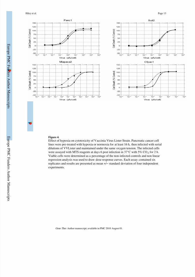
Enhanced Cytotoxicity of Vaccinia Virus in Hypoxia
Effective lysis of infected tumour cells is the ultimate aim of oncolytic therapy. We used theMTS assay to determine the EC50 (dose of virus required to kill 50% of cells) for four
pancreatic cancer cell lines. Cells were infected and maintained in the indicated oxygen
conditions for the duration of the experiment and cell viability was analysed at 6 days post-
infection. Dose response curves and EC50 values were calculated and the results were shown
in Fig.4 and Table 1. Cytotoxicity of vaccinia virus in Suit-2 and Panc1 cell lines was
maintained irrespective of a reduction in ambient oxygen concentration. Interestingly, for
MiaPaca2 and CFPac1 cell lines there were a statistically significant increase in vaccinia
Hiley et al. Page 5
Gene Ther . Author manuscript; available in PMC 2010 August 01.
E
ur ope P MC F und e r s Aut h or Ma nus c r i pt s
E ur ope P MC F und e r s Aut h or Ma nu
s c r i pt s

8/12/2019 ukmss-27828
http://slidepdf.com/reader/full/ukmss-27828 6/15
virus cytotoxicity in hypoxia with an approximately 10-fold (P < 0.001) and 5-fold (P <
0.01) reduction in EC50 respectively. These data suggest that Lister strain Vaccinia Virus is
a potential agent for oncolytic virotherapy where hypoxia occurs in the tumour
microenvironment.
Transgene expression in vaccinia virus vector is not affected by hypox ic condit ions
Many replicating viruses used for oncolytic therapy have additional therapeutic transgenes
inserted into the viral genome to increase their therapeutic effect. Examples include pro-apoptotic proteins, prodrug-converting enzymes and cytokines 25. One of the attractions of
vaccinia virus over alternative viral vectors is its large capacity for transgene insertion17.
Hypoxia will modulate the gene expression of any host cell so verifying the effect of
hypoxia on transgene expression from replication-competent vaccinia virus is important if
this vector is to be of clinical use23. We used VVL15 (a recombinant vaccinia virus derived
from the Lister vaccine strain) in which the firefly luciferase reporter gene was inserted into
the thymidine kinase (TK) region downstream of the early–late vaccinia p7.5 promoter to
assess transgene expression. The levels of luciferase activity after infection of four
pancreatic cancer cell lines were detected by the Live Imaging System IVS200 (Xenogen,
USA) at multiple time points as opposed to isolated readings or early time points as used in
other studies to produce more representative data on gene expression26,27. As shown in Fig.
5, luciferase expression was largely unaffected by hypoxia. Only two of the four cell lines
tested showed a significant difference between normoxia and hypoxia at two isolated time
points. There was a decrease at 24 hours and increase at 48 hours in luciferase expression for
CFPac1 and Panc1 respectively. However, this difference was not sustained at later time
points. This result suggests that hypoxia does not compromise transgene expression from
replication-competent vaccinia virus.
Discussion
Wild-type vaccinia virus has been well characterized and much data on the molecular
biology, genome sequence, viral life cycle and immunology have been reported. Vaccinia
virus is an appealing candidate agent for oncolytic virotherapy because of these inherent
properties. Besides several other attractive qualities (such as fast and efficient replication
with rapid cell-to-cell spread; natural tropism for tumours; strong lytic ability; large cloning
capacity; well-defined molecular biology; safety in human beings and good stability28-31),
a defining feature that we demonstrated from the present study, is that hypoxia does not
significantly affect viral gene expression, viral replication, cytotoxicity and even enhances
the tumour-killing activity in some tumour cell lines.
We have demonstrated here that Lister strain vaccinia virus shows comparable efficacy in
infection, replication and transgene expression regardless of the ambient oxygen
concentration. It is important to highlight that hypoxic cells used in these experiments had
been exposed to reduced oxygen concentration for at least 16 hours prior to infection which
is more likely to reflect cellular adaptation to hypoxia and model clinical vaccinia virus
infection than shorter exposure times that have been used for the study of other oncolytic
viruses12,26. In addition, we found that effective tumour cell lysis was maintained after
infection of hypoxic PDAC cell lines and in half of the cell lines tested there was astatistically significant improvement in viral cytotoxicty. This is an important result given
that tumour lysis is the ultimate goal of oncolytic therapy.
Many groups have tried to target hypoxic fractions of tumours using hypoxia-specific
promoters often containing hypoxia-response elements (HRE) that facilitate HIF-1α binding
and downstream gene transcription. Unfortunately, such promoters are invariably less
powerful drivers of gene expression than constitutive viral promoters and result in lower
Hiley et al. Page 6
Gene Ther . Author manuscript; available in PMC 2010 August 01.
E
ur ope P MC F und e r s Aut h or Ma nus c r i pt s
E ur ope P MC F und e r s Aut h or Ma nu
s c r i pt s
8/12/2019 ukmss-27828
http://slidepdf.com/reader/full/ukmss-27828 7/15
levels of gene expression and viral replication relative to wild-type viruses32,33. Our results
show that vaccinia virus has the capacity to infect and replicate in hypoxic tumour cells
without the need for such approaches. A recent report from Conner et al . showed that
Oncolytic Vesicular Stomatitis Virus (VSV) has comparable viral replication in normoxic
HeLa cells versus those exposed to 1% O2 for 2 hours prior to infection and only a slight
reduction of viral induced CPE on semi-quantitative analysis26. The availability of other
oncolytic viruses that are not significantly attenuated in hypoxia in comparison to adenoviral
strains is a welcome finding. However, our results suggest a several-fold, statisticallysignificant improvement in the oncolytic potential of vaccinia virus in some pancreatic cell
lines exposed to hypoxic conditions. The underlying mechanisms are not clear, which
warrants further investigation. Conner et al conclude that their results suggest an advantage
of RNA viruses over DNA viruses in targeting hypoxic tumour cells because of a greater
reduction of DNA synthesis in hypoxic cells26. Vaccinia virus is a double-stranded DNA
virus and like VSV replicates in the cytoplasm, encodes its own polymerases and is
consequently less dependent on host gene and protein expression. Our alternative conclusion
is that it is a cytoplasmic life cycle, with a reduced dependence on host gene and protein
expression, rather the nucleic acid construction of the viral genome that dictates the efficacy
of oncolytic viruses in hypoxia.
In summary we report the comparable efficacy of oncolytic vaccinia virus (including direct
cancer cell killing and transgene expression) in both normoxic and hypoxic pancreatictumour cells. These results suggest that vaccinia virus may be a potent therapeutic vector for
targeting pancreatic cancer and potentially other hypoxic tumour types.
Acknowledgments
This project is supported by Cancer Research UK (C633-A6253/A6251), and Barts and The London Research
Advisory Board. We are very grateful to Professor Istvan Fodor of Loma Linda University, Loma Linda, CA, USA,
for providing the viruses.
Financial disclosures: None of the authors have any financial arrangement nor involvement with commercial
organizations producing competing products.
References
1. Harris AL. Hypoxia--a key regulatory factor in tumour growth. Nat Rev Cancer. 2002; 2:38–47.
[PubMed: 11902584]
2. Hiley CT, Green MML, Shanks JH, Bottomley IC, West CML, Cowan RA, et al. Expression of
vascular endothelial growth factor (VEGF) in locally invasive prostate cancer is prognostic for
radiotherapy outcome. International journal of radiation oncology, biology, physics. 2007; 67:84–
90.
3. Hutchison GJ, Valentine HR, Loncaster JA, Davidson SE, Hunter RD, Roberts SA, et al. Hypoxia-
inducible factor 1alpha expression as an intrinsic marker of hypoxia: correlation with tumor oxygen,
pimonidazole measurements, and outcome in locally advanced carcinoma of the cervix. Clinical
cancer research : an official journal of the American Association for Cancer Research. 2004;
10:8405–8412. [PubMed: 15623619]
4. Ghaneh P, Costello E, Neoptolemos JP. Biology and management of pancreatic cancer. Postgrad
Med J. 2008; 84:478–497. [PubMed: 18940950]
5. Koong AC, Mehta VK, Le QT, Fisher GA, Terris DJ, Brown JM, et al. Pancreatic tumors show high
levels of hypoxia. International journal of radiation oncology, biology, physics. 2000; 48:919–922.
6. Sun HC, Qiu ZJ, Liu J, Sun J, Jiang T, Huang KJ, et al. Expression of hypoxia-inducible factor-1
alpha and associated proteins in pancreatic ductal adenocarcinoma and their impact on prognosis.
Int J Oncol. 2007; 30:1359–1367. [PubMed: 17487356]
7. Yokoi K, Fidler IJ. Hypoxia increases resistance of human pancreatic cancer cells to apoptosis
induced by gemcitabine. Clin Cancer Res. 2004; 10:2299–2306. [PubMed: 15073105]
Hiley et al. Page 7
Gene Ther . Author manuscript; available in PMC 2010 August 01.
E
ur ope P MC F und e r s Aut h or Ma nus c r i pt s
E ur ope P MC F und e r s Aut h or Ma nu
s c r i pt s

8/12/2019 ukmss-27828
http://slidepdf.com/reader/full/ukmss-27828 8/15
8. Hawkins LK, Lemoine NR, Kirn D. Oncolytic biotherapy: a novel therapeutic platform. Lancet
Oncol. 2002; 3:17–26. [PubMed: 11905600]
9. Mulvihill S, Warren R, Venook A, Adler A, Randlev B, Heise C, et al. Safety and feasibility of
injection with an E1B-55 kDa gene-deleted, replication-selective adenovirus (ONYX-015) into
primary carcinomas of the pancreas: a phase I trial. Gene therapy. 2001; 8:308–315. [PubMed:
11313805]
10. Hecht JR, Bedford R, Abbruzzese JL, Lahoti S, Reid TR, Soetikno RM, et al. A phase I/II trial of
intratumoral endoscopic ultrasound injection of ONYX-015 with intravenous gemcitabine in
unresectable pancreatic carcinoma. Clinical cancer research : an official journal of the American
Association for Cancer Research. 2003; 9:555–561. [PubMed: 12576418]
11. Pipiya T, Sauthoff H, Huang YQ, Chang B, Cheng J, Heitner S, et al. Hypoxia reduces adenoviral
replication in cancer cells by downregulation of viral protein expression. Gene Ther. 2005;
12:911–917. [PubMed: 15690061]
12. Shen B, Hermiston T. Effect of hypoxia on Ad5 infection, transgene expression and replication.
Gene Ther. 2005; 12:902–910. [PubMed: 15690062]
13. Shen BH, Bauzon M, Hermiston TW. The effect of hypoxia on the uptake, replication and lytic
potential of group B adenovirus type 3 (Ad3) and type 11p (Ad11p). Gene therapy. 2006; 13:986–
990. [PubMed: 16525485]
14. Fenner, F. Smallpox and Its Eradication (History of International Public Health, No. 6). World
Health Organization; Geneva: 1988.
15. Smith GL, Moss B. Infectious poxvirus vectors have capacity for at least 25 000 base pairs of
foreign DNA. Gene. 1983; 25:21–28. [PubMed: 6229451]
16. Schramm B, Locker J. Cytoplasmic organization of POXvirus DNA replication. Traffic
(Copenhagen, Denmark). 2005; 6:839–846.
17. Thorne SH. Oncolytic vaccinia virus: from bedside to benchtop and back. Curr Opin Mol Ther.
2008; 10:387–392. [PubMed: 18683104]
18. Townsley A, Weisberg A, Wagenaar T, Moss B. Vaccinia Virus Entry into Cells via a Low-pH-
Dependent Endosomal Pathway. The Journal of Virology. 2006; 80:8899–8908.
19. Swietach P, Vaughan-Jones RD, Harris AL. Regulation of tumor pH and the role of carbonic
anhydrase 9. Cancer metastasis reviews. 2007; 26:299–310. [PubMed: 17415526]
20. Hung CF, Tsai YC, He L, Coukos G, Fodor I, Qin L, et al. Vaccinia virus preferentially infects and
controls human and murine ovarian tumors in mice. Gene Ther. 2007; 14:20–29. [PubMed:
16915291]
21. Reed LJ, Muench H. A simple method of estimating fifty percent endpoints. The American Journalof Hygiene. 1938; 27:493–497.
22. Masson N, Ratcliffe PJ. HIF prolyl and asparaginyl hydroxylases in the biological response to
intracellular O(2) levels. J Cell Sci. 2003; 116:3041–3049. [PubMed: 12829734]
23. Kraggerud SM, Sandvik JA, Pettersen EO. Regulation of protein synthesis in human cells exposed
to extreme hypoxia. Anticancer Res. 1995; 15:683–686. [PubMed: 7645943]
24. Hay JG. The potential impact of hypoxia on the success of oncolytic virotherapy. Curr Opin Mol
Ther. 2005; 7:353–358. [PubMed: 16121701]
25. Bhattacharyya M, Lemoine NR. Gene therapy developments for pancreatic cancer. Best practice &
research Clinical gastroenterology. 2006; 20:285–298. [PubMed: 16549328]
26. Connor JH, Naczki C, Koumenis C, Lyles DS. Replication and cytopathic effect of oncolytic
vesicular stomatitis virus in hypoxic tumor cells in vitro and in vivo. J Virol. 2004; 78:8960–8970.
[PubMed: 15308693]
27. Shen B, Bauzon M, Hermiston T. The effect of hypoxia on the uptake, replication and lyticpotential of group B adenovirus type 3 (Ad3) and type 11p (Ad11p). Gene Ther. 2006; 13:986–
990. [PubMed: 16525485]
28. Poland GA, Grabenstein JD, Neff JM. The US smallpox vaccination program: a review of a large
modern era smallpox vaccination implementation program. Vaccine. 2005; 23:2078–2081.
[PubMed: 15755574]
29. Roenigk HH Jr. Deodhar S, Jacques R, Burdick K. Immunotherapy of malignant melanoma with
vaccinia virus. Arch Dermatol. 1974; 109:668–673. [PubMed: 4828533]
Hiley et al. Page 8
Gene Ther . Author manuscript; available in PMC 2010 August 01.
E
ur ope P MC F und e r s Aut h or Ma nus c r i pt s
E ur ope P MC F und e r s Aut h or Ma nu
s c r i pt s

8/12/2019 ukmss-27828
http://slidepdf.com/reader/full/ukmss-27828 9/15
30. Kim JH, Oh JY, Park BH, Lee DE, Kim JS, Park HE, et al. Systemic armed oncolytic and
immunologic therapy for cancer with JX-594, a targeted poxvirus expressing GM-CSF. Mol Ther.
2006; 14:361–370. [PubMed: 16905462]
31. Park BH, Hwang T, Liu TC, Sze DY, Kim JS, Kwon HC, et al. Use of a targeted oncolytic
poxvirus, JX-594, in patients with refractory primary or metastatic liver cancer: a phase I trial.
Lancet Oncol. 2008; 9:533–542. [PubMed: 18495536]
32. Binley K, Iqball S, Kingsman A, Kingsman S, Naylor S. An adenoviral vector regulated by
hypoxia for the treatment of ischaemic disease and cancer. Gene therapy. 1999; 6:1721–1727.
[PubMed: 10516721]
33. Binley K, Askham Z, Martin L, Spearman H, Day D, Kingsman S, et al. Hypoxia-mediated tumour
targeting. Gene therapy. 2003; 10:540–549. [PubMed: 12646859]
Hiley et al. Page 9
Gene Ther . Author manuscript; available in PMC 2010 August 01.
E
ur ope P MC F und e r s Aut h or Ma nus c r i pt s
E ur ope P MC F und e r s Aut h or Ma nu
s c r i pt s

8/12/2019 ukmss-27828
http://slidepdf.com/reader/full/ukmss-27828 10/15

8/12/2019 ukmss-27828
http://slidepdf.com/reader/full/ukmss-27828 11/15
Figure 2.
Viral gene expression of vaccinia virus in human pancreatic cancer after viral infection, and
Hif-1α stabilisation and nuclear translocation in normoxia or hypoxia. A, Hypoxia does not
affect viral gene expression of vaccinia virus. Cells were maintained in normoxia or hypoxia
prior to and after viral infection. Cells were infected with VVLister at an MOI=1. Vaccinia
virus protein was measured using an anti-vaccinia polyclonal antibody. Human PCNA was
used as a loading control; B, Hif-1α stabilisation and nuclear translocation was
demonstrated on nuclear lysates from MiaPaca2 cells in normoxia (20% pO2) or hypoxia
(1% pO2) over a time course. β-actin was used as a loading control.
Hiley et al. Page 11
Gene Ther . Author manuscript; available in PMC 2010 August 01.
E
ur ope P MC F und e r s Aut h or Ma nus c r i pt s
E ur ope P MC F und e r s Aut h or Ma nu
s c r i pt s
8/12/2019 ukmss-27828
http://slidepdf.com/reader/full/ukmss-27828 12/15
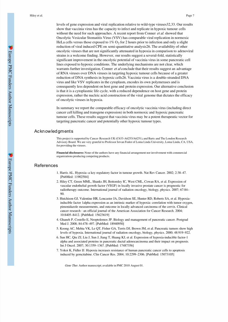
Figure 3.
Viral replication of VVLister in normoxia (solid line) and hypoxia (dashed line) measured
by TCID50 assay of viral burst assays. Cell lines were exposed to normoxia or hypoxia prior
to and post infection with an MOI=1 of VVLister. Burst assay samples were collected at 24,
48, 72 and 96 hours post infection. TCID50 assays were performed on CV1 green monkey
kidney cells. Experiments were performed in triplicate for each cell line, time point and
condition. Results are presented as mean +/− standard deviation.
Hiley et al. Page 12
Gene Ther . Author manuscript; available in PMC 2010 August 01.
E
ur ope P MC F und e r s Aut h or Ma nus c r i pt s
E ur ope P MC F und e r s Aut h or Ma nu
s c r i pt s
8/12/2019 ukmss-27828
http://slidepdf.com/reader/full/ukmss-27828 13/15

8/12/2019 ukmss-27828
http://slidepdf.com/reader/full/ukmss-27828 14/15
Figure 5.
The effect of hypoxia on transgene expression from VVL15. Cells were infected with 1pfu/ cell of VVL15 and luciferase activity measured at the time points indicated. All experiments
were performed in triplicate and results represent the data from three separate experiments.
Results are presented as mean +/− s.d. (Solid line & triangle = 20% pO2, Dashed line &
triangle = 1% pO2, Solid line and square & mock infection. Light units = photons/second/
cm2. * = P<0.05).
Hiley et al. Page 14
Gene Ther . Author manuscript; available in PMC 2010 August 01.
E
ur ope P MC F und e r s Aut h or Ma nus c r i pt s
E ur ope P MC F und e r s Aut h or Ma nu
s c r i pt s

8/12/2019 ukmss-27828
http://slidepdf.com/reader/full/ukmss-27828 15/15
E ur ope P MC F und e r s Aut h or Ma nu
s c r i pt s
E ur ope
P MC F und e r s Aut h or Ma nus c r i pt s
Hiley et al. Page 15
T a b l e
1
C o m p a r i s o n o f c y
t o t o x i c i t y o f v a c c i n i a v i r u s i n n o r m o x
i a a n d h y p o x i a
C e l l L i n e s
E C 5 0 ( 9 5 % C
I )
P V a l u e
H i l l S l o p e
2 0 % O x y g e n
1 % O x y g e n
2 0 %
O x y g e n
1 %
O x y g e n
P a n c 1
1 . 2 1
( 1 . 0 3 t o 1 . 4 1 )
0 . 8 2
( 0 . 7 1 t o 0 . 9 5 )
= 0 . 0 7
1 . 0 7 5
0 . 9 6 6 2
S u i t 2
1 . 5 0
( 1 . 2 4 t o 1 . 8 1 )
1 . 8 8
( 1 . 3 7 t o 2 . 5 9 )
= 0 . 4 8
0 . 9 2 4
1 . 1 2
M i a P a c a 2
1 8 7 . 0 0
( 1 5 4 . 3 9 t o
2 2 6 . 3 7 )
9 . 1 5
( 7 . 2 4 t o
1 1 . 5 8 )
< 0 . 0 0 0 0 4
0 . 9 5 5 5
0 . 6 0 6 5
C F P a c 1
3 . 8 9
( 3 . 5 9 t o 4 . 2 0 )
1 . 1 0
( 1 . 0 4 t o 1 . 1 6 )
< 0 . 0 0
2
1 . 6 6
1 . 1 2 2
T h e E C 5 0 v a l u e s o f V a c c i n i a V i r u s i n t h e f o u r d i f f e r e n t p a n c r e a t i c c a n c e r
c e l l l i n e s i s p r e s e n t e d w i t h 9 5 % c o n f i d e n c e i n t e r v a l s a n d P v a l u e s r e p r e s e n t a n y s i g n i f i c a n t d i f f e r e n c e b e t w e e n i n f e c t i o n i n
n o r m o x i a v e r s u s h y p o x
i a . H i l l s l o p e v a l u e s f o r t h e d o s e r e s p o n s e c u r v e s a
r e a l s o p r e s e n t e d .
Gene Ther . Author manuscript; available in PMC 2010 August 01.