Ueda2016 a1 c uses& limitations-fareed fawzy
36
1 Glycated Hemoglobin: Uses & Limitations By FARID FAWZY, MD,PhD Professor of Medicine, Zagazig University, EGYPT
-
Upload
ueda2015 -
Category
Health & Medicine
-
view
194 -
download
3
Transcript of Ueda2016 a1 c uses& limitations-fareed fawzy
1
Glycated Hemoglobin:
Uses & Limitations
By
FARID FAWZY, MD,PhD
Professor of Medicine, Zagazig University, EGYPT
2
Disclosure
• I have honoraria and sponsored conferences with Novartis, Novo-
Nordisk, Eli-Lilly, Astra-Zeneca, MSD, Inspire, HSO and Global-Napi
• No potential conflict of interest relevant to this presentation
3
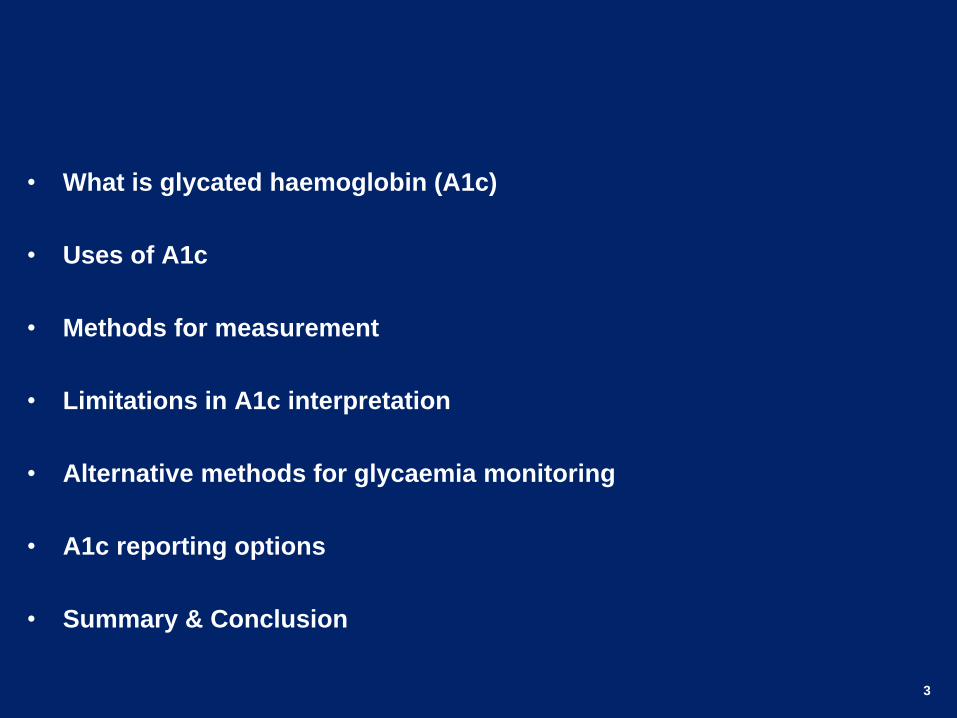
• What is glycated haemoglobin (A1c)
• Uses of A1c
• Methods for measurement
• Limitations in A1c interpretation
• Alternative methods for glycaemia monitoring
• A1c reporting options
• Summary & Conclusion
4
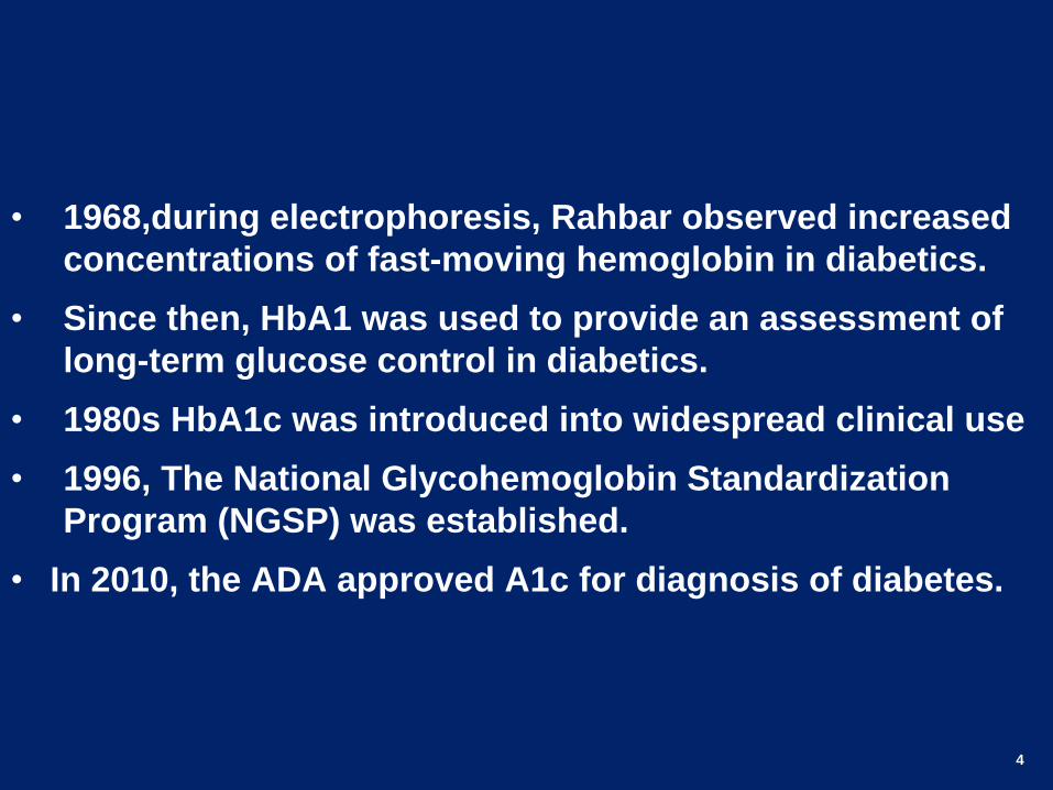
• 1968,during electrophoresis, Rahbar observed increased
concentrations of fast-moving hemoglobin in diabetics.
• Since then, HbA1 was used to provide an assessment of
long-term glucose control in diabetics.
• 1980s HbA1c was introduced into widespread clinical use
• 1996, The National Glycohemoglobin Standardization
Program (NGSP) was established.
• In 2010, the ADA approved A1c for diagnosis of diabetes.
5
What is A1c?
• A1c (HbA1c) is the measurement of glycated hemoglobin.
• Glycation is an irreversible, non-enzymatic binding of glucose to the N-
terminus of the beta chain of the hemoglobin.
• The rate of glycation is proportional to the glucose level in blood
• Glycation increases with the age of the red blood cell.
• A1C reflects the average blood glucose in the past 2 to 3 months
• 50% of A1C is determined by blood glucose in the preceding 1 month.
6
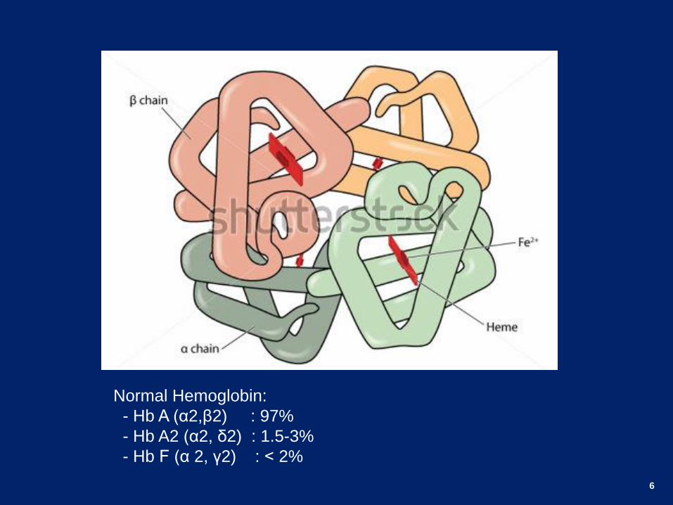
Normal Hemoglobin:
- Hb A (α2,β2) : 97%
- Hb A2 (α2, δ2) : 1.5-3%
- Hb F (α 2, γ2) : > 2%
7
A1c Uses
• Timely diagnosis of diabetes and Pre-diabetes.
• Assessment of glycemic control.
• Decision making in diabetes management.
• Evaluation for quality of care in diabetes.
• Reliable research tool.
• Basis for pay-for-performance systems.
• Confirm the accuracy of the SMBG and the adequacy of testing
schedule.
8
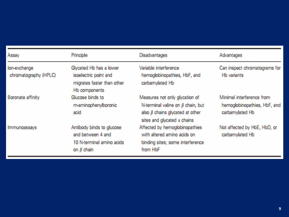
Methods for A1c Measurement
• Methods utilizing the charge difference between glycated
(HbA1c) & non-glycated hemoglobin (HbA0):
- Ion-exchange chromatography.
- Agar electrophoresis.
- High Performance Liquid Chromatography (HPLC).
• Methods utilizing structural difference:
- Affinity chromatography.
- Enzyme immunoassay (EIA).
9
10
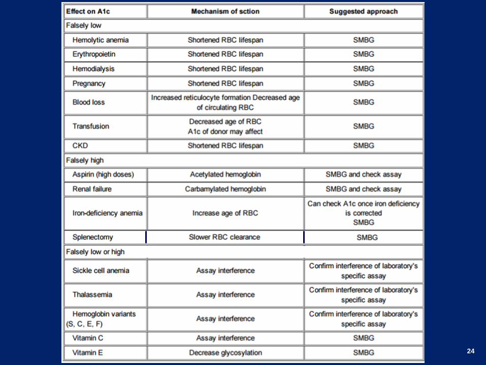
Conditions That Affect the A1C
11
12
13
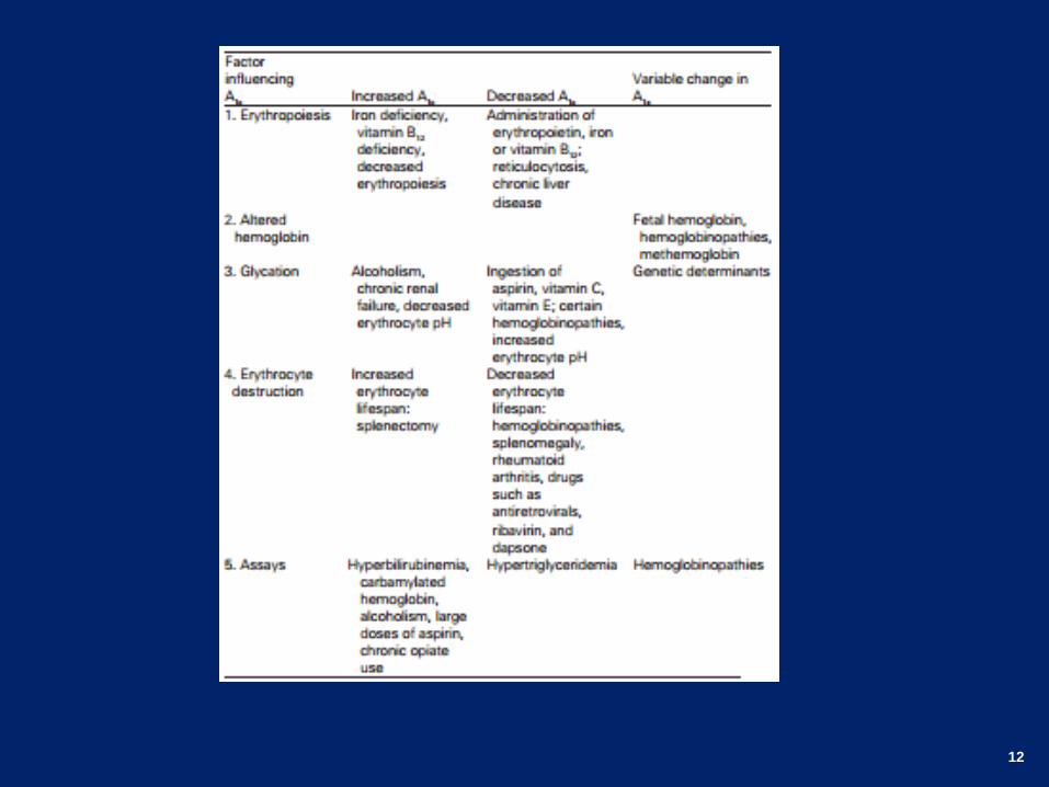
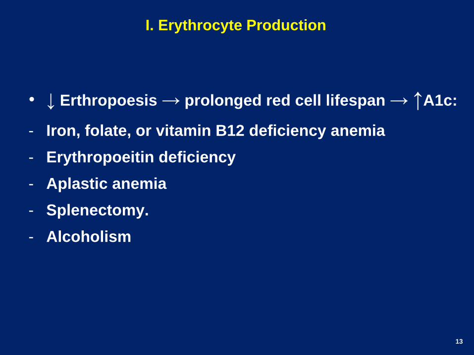
I. Erythrocyte Production
• ↓ Erthropoesis → prolonged red cell lifespan → ↑A1c:
- Iron, folate, or vitamin B12 deficiency anemia
- Erythropoeitin deficiency
- Aplastic anemia
- Splenectomy.
- Alcoholism
14
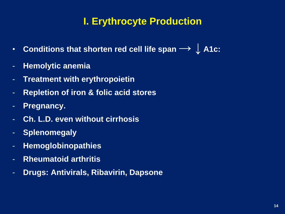
I. Erythrocyte Production
• Conditions that shorten red cell life span → ↓ A1c:
- Hemolytic anemia
- Treatment with erythropoietin
- Repletion of iron & folic acid stores
- Pregnancy.
- Ch. L.D. even without cirrhosis
- Splenomegaly
- Hemoglobinopathies
- Rheumatoid arthritis
- Drugs: Antivirals, Ribavirin, Dapsone
15
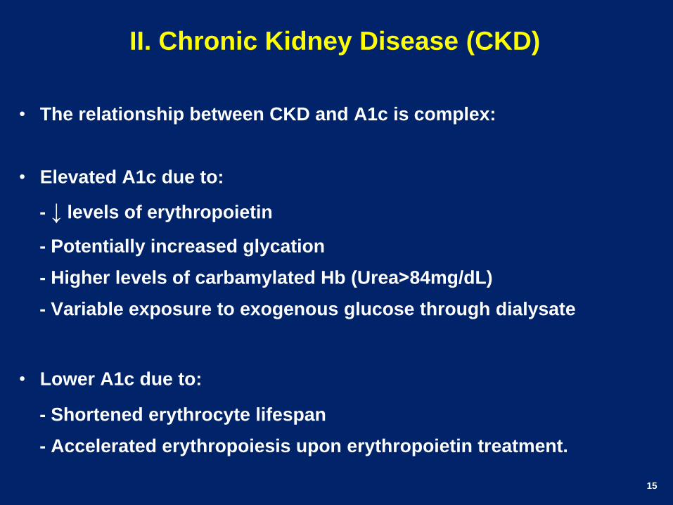
II. Chronic Kidney Disease (CKD)
• The relationship between CKD and A1c is complex:
• Elevated A1c due to:
- ↓ levels of erythropoietin
- Potentially increased glycation
- Higher levels of carbamylated Hb (Urea<84mg/dL)
- Variable exposure to exogenous glucose through dialysate
• Lower A1c due to:
- Shortened erythrocyte lifespan
- Accelerated erythropoiesis upon erythropoietin treatment.
16
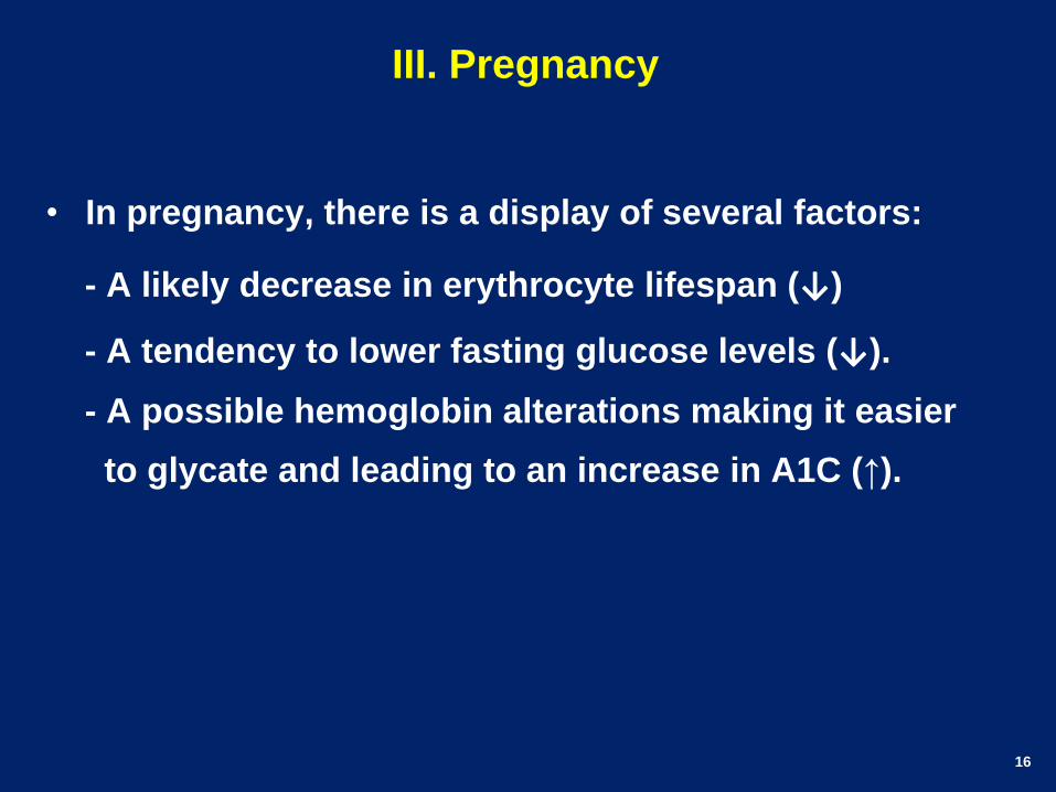
III. Pregnancy
• In pregnancy, there is a display of several factors:
- A likely decrease in erythrocyte lifespan (↓)
- A tendency to lower fasting glucose levels (↓).
- A possible hemoglobin alterations making it easier
to glycate and leading to an increase in A1C (↑).
17
IV. Blood loss and transfusion
• Active bleeding causes a decrease in the average age
of red cells leading to lower A1C.
• Older cells with glycated haemoglobin are replaced
with newly formed cells not exposed to blood glucose
• In patients with recent transfusion, A1C values may be
skewed depending on A1C of the donor.
• Blood transfusions are commonly required in surgical
patients, ICU, and patients with CKD.
18
V. Ethnicity
• A1c may differ between races despite similar blood glucose levels.
• The racial difference in A1c widens as the A1c increased.
• Diabetic complications also may differ between races with the same A1c
• There is a need to determine whether A1c should be adjusted for
ethnicity.
• The molecular mechanism of racial A1c difference may include:
- Differences in rates of glucose uptake into erythrocytes
- Rates of intra-erythrocytic glucose metabolism
- Rates of glucose attachment to or release from hemoglobin
- Erythrocyte life span.
• Racial variations in A1C concentrations are relatively small (≤0.4%)
19
VI. Hemoglobin Variants
• Americans of African, Asian, and Mediterranean descent are at
increased risk for hemoglobin variants.
• Africans, east India and Mediterraneans may have HbSC
• Asian Americans may have HbE & Indians may have HbD
• Hemoglobin F also may be present in up to 12% of patients
• Patients with β-thalassemia, sickle cell disease, pregnancy,
anemia and certain leukemias have higher HbF
• Drugs like Hydroxyurea may increase the percentage of HbF
• HbF > 5% does not affect A1c assay.
• In Egypt Thalassemia and G6PD deficiency deserves special
attention.
20
• The presence of Abnormal Hb Affects A1c in different ways:
- Some Hb variants can affect the net charge of the hemoglobin
and/or the recognition of the glycated Nterminus.
- Hemoglobinopathies can alter the normal process of glycation
- Abnormal Hb causes an abnormal peak on chromatography, making
estimation unreliable
- Abnormal Hb can make the red blood cell more prone to hemolysis.
• A hemoglobinopathy should be suspected when the A1c
results do not correlate with SMBG or if the A1c value
changed following a change in laboratory A1c methods.
• A1c values<15% are suspicious.
21
VII. Chemically Modified Hemoglobin
• Acetylated or carbamylated hemoglobin may affect A1c assay.
• High dose aspirin may cause acetylation of the hemoglobin.
• Alcohol ingestion forms acetaldehyde causing also Hb acetylation.
• Charge changes make it difficult for the ion-exchange to distinguish
acetylated from glycated hemoglobin, subsequently measuring both.
• Renal failure can result in elevated urea, which dissociates into
cyanate that binds to hemoglobin forming carbamylated Hb.
• Carbamylated hemoglobin has a similar charge to that of glycated
hemoglobin and may interfere with ion-exchange assay.
• Significant interference occurs when serum urea exceed 84 mg/dL.
22
VIII. Altered Rate of Glycation
• Inter-individual variations in Hb glycation may exist.
• Variance in glycation is reported to be related to variations in the
degree of glucose entry into the erythrocyte as well as to 2,3 DPG
and pH levels within the erythrocyte.
• Other influences on glycation have yet to be determined.
• Large doses of vitamins C, E and other antioxidants are believed to
reduce glycation of hemoglobin and can affect (↓) A1c values.
• On the other hand vitamin C binding to hemoglobin changes the
charge of the hemoglobin similarly to the binding of glucose,
causing an increase in A1c when measured by electrophoresis.
23
IX. Aging
• The effect of age on A1c may be more a function of age related
changes in glucose regulation.
• Increased A1c with age may be due to a decrease in physical
activity, increased insulin resistance, decreased insulin
secretion and mitochondrial dysfunction.
• Although the ADA Standards of Care have recognized this,
NO age specific values have been recommended.
24
25
Alternative Markers for A1c
• The 2015 ADA Standards of Care state that discrepancies
between A1c and SMBG levels should warrant further
exploration.
• Considerations should include testing method as well as
patient conditions that may interfere with the assay.
• If A1c is unreliable, alternative strategies for assessment of
glycemic control should include more frequent SMBG and
CMG.
• In addition, clinicians can consider alternative methods of
glycemic control: fructosamine, glycated albumin, and
1,5AG.
26
• Lack of guidelines recommending their use
• Lack of standardization
• Limited outcome data
• Lack of consensus target values for glycemic control.
27
1. Fructosamine & Glycated Albumin
• Fructosamine is formed when fructose binds to serum proteins, mostly albumin.
• Glycated albumin is formed by the glycation of serum albumin.
• The half life of albumin is 2 to 3 weeks.
• As such, fructosamine and glycated albumin reflect the average glucose in the last 2 to 3 w.
• Therefor both are useful in shorter follow up.
28
• Some studies suggest GA could be more reliable than A1c in CKD 4/5.
• Pitfalls in fructosamine and GA may be due to:
- Hypoalbuminemia as in CLD, nephrotic syndrome, and protein-losing enteropathy.
- Conditions with increased protein metabolism as in hyperuricemia, hyperthyroidism,
and hypertriglyceridemia.
- Conditions with decreased protein metabolism, such as hypothyroidism and mild
cirrhosis, can lead to falsely high levels.
• Some studies have looked at correcting for hypoalbuminemia as is done with
calcium, but there is no consensus for this.
29
1,5 Anhydroglucitol (1,5 AG)
• 1,5 AG is a dietary polyol, structurally similar to glucose
• 1,5 AG is normally filtered and completely reabsorbed by the kidneys.
• Glucose is a competitive inhibitor of 1,5AG for renal reabsorption.
• When glucose levels exceed 180 mg/dL, 1,5 AG levels will decrease.
• 1,5AG does not reflect glycemic variability or hypoglycemia and is less
useful to assess overall glycemic control.
• Patients with impaired renal function may have falsely low levels of 1,5-
AG making the test unhelpful in that situation.
30
Alternative methods for A1c reporting
• The standard method is to express glycated hemoglobin as a percentage of total hemoglobin
• The second is to express the A1c after the IFCC as mmol⁄ mol Hb, with a normal range of (25–42 mmol/mol).
• A third suggested approach is to abandon the term A1c and to instead report an estimated average glucose or A1c-derived average glucose (ADAG).
31
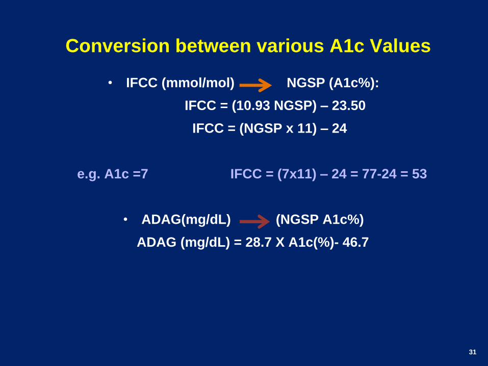
Conversion between various A1c Values
• IFCC (mmol/mol) NGSP (A1c%):
IFCC = (10.93 NGSP) – 23.50
IFCC = (NGSP x 11) – 24
e.g. A1c =7 IFCC = (7x11) – 24 = 77-24 = 53
• ADAG(mg/dL) (NGSP A1c%)
ADAG (mg/dL) = 28.7 X A1c(%)- 46.7
32
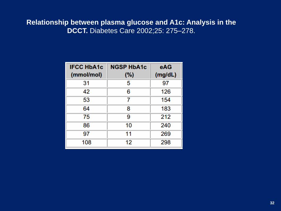
Relationship between plasma glucose and A1c: Analysis in the
DCCT. Diabetes Care 2002;25: 275–278.
33
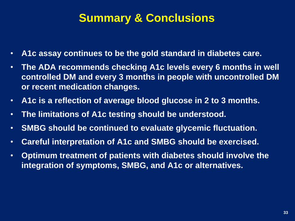
Summary & Conclusions
• A1c assay continues to be the gold standard in diabetes care.
• The ADA recommends checking A1c levels every 6 months in well
controlled DM and every 3 months in people with uncontrolled DM
or recent medication changes.
• A1c is a reflection of average blood glucose in 2 to 3 months.
• The limitations of A1c testing should be understood.
• SMBG should be continued to evaluate glycemic fluctuation.
• Careful interpretation of A1c and SMBG should be exercised.
• Optimum treatment of patients with diabetes should involve the
integration of symptoms, SMBG, and A1c or alternatives.

34
Areas of Uncertainty
• A1c in children and the older population.
• A1c in different ethnic groups.
• Genetic influences on A1c variance.
• The effects of glucose fluctuations.
• The significance of glycating sites other than the N-terminal
valine of the βchain.
• Mechanisms of A1c changes with pregnancy, chronic liver
disease, renal disease, and HIV.

35

Thank You