U Nordic Forum - Trauma & Emergency Radiology U...
10
U Acute Abdominal Bleeding: Detection with MDCT Nordic Forum - Trauma & Emergency Radiology Borut Marincek Institute of Diagnostic Radiology University Hospital Zurich, Switzerland U • To review the role of MDCT in the assessment of acute bleeding from solid abdominal organs after blunt or penetrating trauma • To discuss the MDCT imaging features of acute bleeding after rupture of an abdominal neoplasm • To illustrate the value of MDCT in defining the site and cause of acute gastrointestinal bleeding Lecture Objectives U • Evidence of CM pooling caused by vascular or visceral organ pathology • Attenuation of CM pooling - similar aorta / major adjacent arteries - greater than surrounding parenchyma • Types of CM pooling: - focal, diffuse or „jet“ of extravasated CM surrounded by hematoma Diagnostic Criteria of Acute Bleeding on CE-MDCT U Blunt Trauma: Spleen Snowboard accident: multiple lacerations, extravasated CM & surrounding hematoma, hemoperitoneum U Blunt Trauma: Spleen Snowboard accident: multiple intrasplenic pseudoaneurysms, extravasated CM; coiling of proximal SA “hypotensive hemostasis”, reperfusion of SA via gastroepiploic art. & short gastric art. U Blunt Trauma: Kidney Multiple kidney lacerations, extravasated CM & surrounding hematoma, retro-hemoperitoneum Construction worker, fall from 16 m: injuries spleen, kidney, thoracic aorta, pelvic rim
Transcript of U Nordic Forum - Trauma & Emergency Radiology U...

1
U
Acute Abdominal Bleeding: Detection with MDCT
Nordic Forum - Trauma & Emergency Radiology
Borut MarincekInstitute of Diagnostic Radiology
University Hospital Zurich, Switzerland
U
• To review the role of MDCT in the assessment of acute bleeding from solid abdominal organs after blunt or penetrating trauma
• To discuss the MDCT imaging features of acute bleeding after rupture of an abdominal neoplasm
• To illustrate the value of MDCT in defining the site and cause of acute gastrointestinal bleeding
Lecture Objectives
U
• Evidence of CM pooling caused by vascular
or visceral organ pathology
• Attenuation of CM pooling
- similar aorta / major adjacent arteries
- greater than surrounding parenchyma
• Types of CM pooling:
- focal, diffuse or „jet“ of extravasated CM
surrounded by hematoma
Diagnostic Criteria of Acute Bleeding on CE-MDCT U Blunt Trauma: Spleen
Snowboard accident: multiple lacerations, extravasated CM & surrounding hematoma, hemoperitoneum
U Blunt Trauma: Spleen
Snowboard accident: multiple intrasplenic pseudoaneurysms,
extravasated CM; coiling of proximal SA “hypotensive hemostasis”,
reperfusion of SA via gastroepiploic art. & short gastric art.
U Blunt Trauma: Kidney
Multiple kidney lacerations, extravasated CM & surrounding
hematoma, retro-hemoperitoneum
Construction worker, fall from 16 m:
injuries spleen, kidney, thoracic aorta,
pelvic rim
2
U
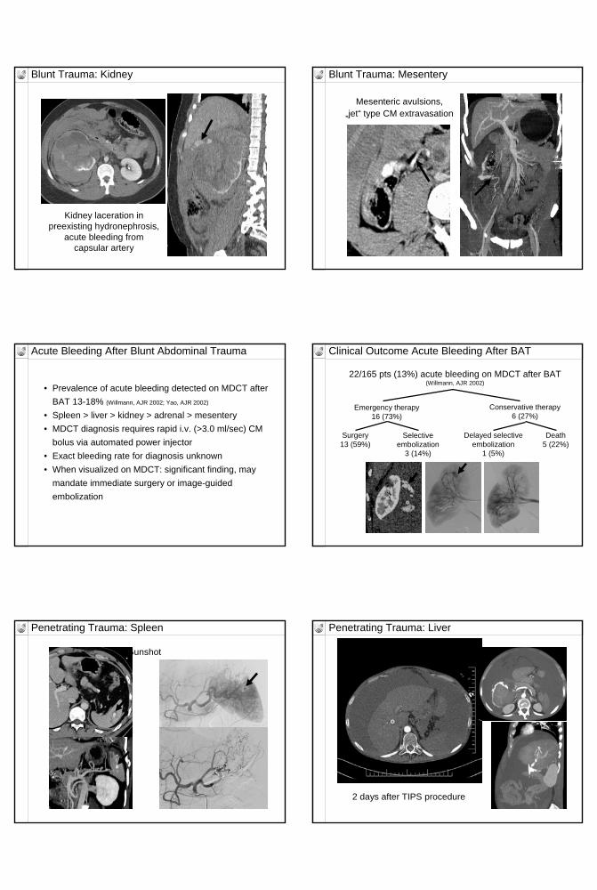
Kidney laceration in preexisting hydronephrosis,
acute bleeding fromcapsular artery
Blunt Trauma: Kidney U Blunt Trauma: Mesentery
Mesenteric avulsions, „jet“ type CM extravasation
U
• Prevalence of acute bleeding detected on MDCT after
BAT 13-18% (Willmann, AJR 2002; Yao, AJR 2002)
• Spleen > liver > kidney > adrenal > mesentery
• MDCT diagnosis requires rapid i.v. (>3.0 ml/sec) CM
bolus via automated power injector
• Exact bleeding rate for diagnosis unknown
• When visualized on MDCT: significant finding, may
mandate immediate surgery or image-guided
embolization
Acute Bleeding After Blunt Abdominal Trauma U
22/165 pts (13%) acute bleeding on MDCT after BAT(Willmann, AJR 2002)
Surgery13 (59%)
Selectiveembolization
3 (14%)
Emergency therapy16 (73%)
Conservative therapy6 (27%)
Delayed selectiveembolization
1 (5%)
Death5 (22%)
Clinical Outcome Acute Bleeding After BAT
U Penetrating Trauma: Spleen
Gunshot
U Penetrating Trauma: Liver
2 days after TIPS procedure

3
U
Replaced righthepatic artery
Penetrating Trauma: Liver U
Leriche syndrome, 1 day after laparotomy and
extraanatomicaorto-bifemoral Y-Graft
Bleeding from falciform ligament, perihepatic & perisplenic hematoma
Penetrating Trauma: Liver
U
2 days after ESWL for right-sided kidney stones
Penetrating Trauma: Liver U
Bleeding incidence after ESWL 0.28% (Collado, Scand J UrolNephrol 1999)
Penetrating Trauma: Kidney
A few hours after ESWL for left-sided kidney stones
U Spontaneous Rupture HCC
Incidence spontaneous rupture HCC 3-15% (Lai, Arch Surg 2006)
U Spontaneous Rupture Liver Metastasis
Undifferentiatedpancreatic carcinoma+ oral anticoagulation

4
U Spontaneous Rupture Renal Arteriopathy
Polyarteritis nodosa
U Spontaneous Rupture Sporadic Renal AML
Angiomyolipoma >4 cmBleeding in 50-60%
U Spontaneous Rupture TS Associated Renal AML
20 yo, f: multiple & bilateral tumors
U
Mean age
TuberousSclerosis
Associated AML
30
Sporadic AML
52
Tumor diameter 9 cm 5 cm
Multiple tumors 97% 13%
Symptomaticacute hemorrhage
44% 14%
(Nelson, J of Urol 2002)
Characteristics of Renal AML
U
• Right tube: ectopic pregnancy• Left tube: hydrosalpinx• ß-HCG 15’034
Spontaneous Rupture Ectopic Pregnancy U
Contrast-enhanced MDCT
• Rapid, noninvasive, and accurate in localizing
acute bleeding from solid organs in traumatic and
in non-traumatic conditions
• Can be used as a guidance for subsequent
angiographic intervention
Acute Abdominal Bleeding: Summary (1)

5
U
• Upper GI bleeding / lower GI bleeding:
proximal / distal ligament of Treitz
• Acute/active/massive: clinically stable - unstable
• Obscure GI bleeding: clinically stable
Persisting or recurring bleeding of unknown origin
after negative endoscopy of upper and lower GI tract
- Overt: see blood (hematemesis, hematochezia,
melena)
- Occult: + fecal occult blood testing, do not see blood
Gastrointestinal (GI) Bleeding: Definitions
(American Gastroenterological Association, Gastroenterology 2007;133:1697-1717)
U
• Small bowel barium examination
• Enteroclysis
• Tc-labeled RBC scintigraphy
• Catheter angiography
• Wireless capsule endoscopy
• CT enteroclysis
• Catheter-directed CT angiography
• MDCT angiography
GI Bleeding: Diagnostic Imaging Modalities
U
14-56%5,7
40-90%4
Catheterangiography
40-70%624%5Obscure GIB
93%391-92%1,2Acute GIB
RBC scintigraphy
MDCT angiography
GI Bleeding: Sensitivity Imaging Modalities
1. Yoon, Radiology 2006;239:160-1672. Jaeckle, Eur Radiol 2008;18:1406-14133. Zuckier, Sem Nuc Med 2003;33:297-3114. Laing, RadioGraphics 2007;27:1055-10705. Saperas, Am J Gastroenterol 2007;102:731-7376. Lin, Gastroenterol Clin N Am 2005;34.679-6957. Neu, Am J Gastroenterol 2005;100:1736-1742
U
best
better
worst
Locali-zation
Up to 1 min
Up to 1.5 min
Up to 48 hrs
Imagingtime
No0.3YesMDCT angiography
Yes
No
InvasiveCantreat
0.5-1.0YesCatheterangiography
0.1-0.2NoRBCscintigraphy
Bleedingrate
detectedml/min
Iodinatediv
contrast
GI Bleeding: Comparison of Imaging Modalities
U Obscure GI Bleeding: Etiologies
Commonly overlooked lesions
Upper GI lesions Lower GI lesions
Cameron’s erosions in Angiectasia
large hiatal hernias Neoplasms
Fundic varices
Peptic ulcer
Angiectasia
Dieulafoy’s lesion
Gastric antral vascular ectasia
(American Gastroenterological Association, Gastroenterology 2007;133:1697-1717)
U Obscure GI Bleeding: Etiologies
Small intestinal bleeding
<40 yo >40 yo
Tumors Angiectasia
lymphoma NSAID enteropathy
carcinoid Celiac disease
adenocarcinoma
Meckel diverticulum Uncommon
Dieulafoy’s lesion Hemobilia
Crohn disease Hemosuccus pancreaticus
Celiac disease Aortoenteric fistula
(American Gastroenterological Association, Gastroenterology 2007;133:1697-1717)

6
U Acute Upper & Lower GI Bleeding
• Acute = hematemesis, melena or hemochezia within 24 hours before MDCT
• Severe = hemodynamic instability (systol. pressure <100 / pulse rate >100); mild = no hemodynamic instability
• MDCT (pts) January 2001 - May 2006 :4-row (6), 16-row (11), 64-row (1)
• MDCT protocol: iv CM, arterial & portal venous phase(additional unenhanced scans in 9 pts), no oral CM
U
Melana since 3 weeks, aorta-bifemoral Y-graft 11 years ago
Aortoduodenal Fistula
U
coilingcystojejunal anastomosis8. Mild / pseudoaneursym lienal art.
embolizationduodenum10. Severe / ischemic anastomotic ulcer
stentgraftduodenum4. Severe / aortoenteric fistula
-duodenum9. Severe / arteriobiliary fistula
coilingbiliodigest. anastomosis5. Severe / pseudoaneurysm hepatic art.
coilingduodenum7. Mild / pseudoaneurysm gastroduod. art.
coilingbiliodigest. anastomosis6. Mild / pseudoaneurysm hepatic art.
stentgraftduodenum3. Severe / aortoenteric fistula
stentgraftduodenum2. Severe / aortoenteric fistula
excisionduodenum1. Severe / aortoenteric fistula
TreatmentBleeding sourceBlood loss / Pathology
Acute Upper GI Bleeding: MDCT Detection U Aortoduodenal Fistula
U Pseudoaneurysm Hepatic Artery
7 weeks after laparotomy & biliodigestive anastomosis forpancreatic carcinoma
U Pseudoaneurysm Hepatic Artery
Hypovolemic shock non-occlusive mesentericischemia & pneumatosis

7
U Pseudoaneurysm Hepatic Artery
Coil-embolization,stent-grafting
U Bleeding Complications after Pancreatic Surgery
Postoperative arterial bleeding 3-4% (Sohn, J Gastrointest Surg 2003)
University Hospital Zurich 1998-2004 (Pfammatter, CIRSE 2005)
N = 11, average delay surgery – bleeding = 54 days (range 10-250 days)Type of surgery:
Whipple‘s procedure 6Partial pancreatic resection 3Pancreatic head mobilization 1Hepaticojejunostomy 1
Presentation of acute bleeding:Upper GI 2Lower GI 3Intraabdominal 6
Initial diagnosis of bleeding source:MDCT 7DSA 3Scintigraphy 1
U Pseudoaneurysm Gastroduodenal Artery
Chronic tuberculous ulcerationof duodenum
U Pseudoaneurysm Gastroduodenal Artery
U Arterio-biliary Fistula
Liver cirrhosis & portal hypertension, 1 week after transjugular liver biopsy
U Arterio-biliary Fistula
Liver cirrhosis & portal hypertension, 1 week after transjugular liver biopsy

8
U Penetrating Subacute Duodenal Ulcer
Arrosion gastroduodenal art.
U
embolizationtransverse colon6. Severe / ischemic ulcer
endoscopical clippingcecum5. Severe / nonocclusive ischemic ulcer
embolizationsigmoid7. Severe / diverticulum
excisionileum3. Mild / neuroendocrine carcinoma
TIPSrectosigmoid8. Mild / varices
embolizationcecum4. Mild / nonocclusive ischemic ulcer
coilingjejunum1. Severe / mucositis
excisionileum2. Mild / stromal tumor
TreatmentBleeding sourceBlood loss / Pathology
Acute Lower GI Bleeding: MDCT Detection
U Ischemic Mucositis Jejunum U Ischemic Mucositis Jejunum
U Stromal Tumor Ileum U Nonocclusive Ischemic Ulcer Cecum
Anticoagulation, 10 d after myocardial infarction: sepsis, acute abdomen
art
ven
embolization

9
U Nonocclusive Ischemic Ulcer Cecum
Cardiogenic shock14 d after
myocardial infarction
art pv
U Ischemic Ulcer Transverse Colon
Nonsteroidal anti-inflammatorydrugs (NSAID) since 5 weeks
U Ischemic Ulcer Transverse Colon U
Abdominal aortic aneurysm, Plavix loading dose aftercoronary stent, diverticulosis of sigmoid colon
Diverticulum Sigmoid Colon
U
Coil-embolization
Sigmoid Diverticulum U Ectopic Varices Cecum

10
U
University Hospital Zurich (N=18)
• MDCT identification of bleeding source:
prospectively 15/18 (83%)
retrospectively 3/18 (17%)
• CM extravsation:
11/11 pts with severe bleeding
1/7 pts with mild bleeding
• Identification of underlying pathology:
15/18 (83%)
Acute Upper & Lower GI-Bleeding U
Contrast-enhanced MDCT
• Accurate localization of acute upper GI or lower GI
bleeding
• Plays a complementary role to endoscopy for
localization of obscure GI bleeding
• Can be used as a guidance for subsequent
angiographic intervention
Acute Abdominal Bleeding: Summary (2)