Typical chest pain with normal ECG - Human … chest pain with normal ECG F. Mut, C. Bentancourt, M....
19
Typical chest pain with normal ECG F. Mut, C. Bentancourt, M. Beretta Nuclear Medicine Service, Asociacion Española Montevideo, Uruguay
Transcript of Typical chest pain with normal ECG - Human … chest pain with normal ECG F. Mut, C. Bentancourt, M....

Typical chest pain with normal ECG
F. Mut, C. Bentancourt, M. Beretta
Nuclear Medicine Service, Asociacion Española
Montevideo, Uruguay

Clinical history
• Male 41 y.o.
• Overweight, hypertension, high cholesterol, stress.
• Typical chest pain.
• Echocardiogram: mild LVH, normal LV function.
• Exercise test: chest pain witn no ECG changes
• Myocardial perfusion study (MPS) with exerecise.

2-D echocardiogram
• Mild concentric LVH, normal systolic function, LA dilation, normal valves.

Stress ECG
Rest
Recov
Worse
ST
Stress

Myocardial perfusion study

a) Ischemia in the RCA territory.
b) Ischemia in the LCx territory.
c) Ischemia in the LAD territory.
d) Ischemia in the RCA + LAD territories.
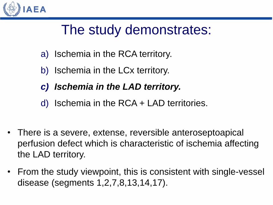
The study demonstrates:
a) Ischemia in the RCA territory.
b) Ischemia in the LCx territory.
c) Ischemia in the LAD territory.
d) Ischemia in the RCA + LAD territories.
The study demonstrates:
• There is a severe, extense, reversible anteroseptoapical
perfusion defect which is characteristic of ischemia affecting
the LAD territory.
• From the study viewpoint, this is consistent with single-vessel
disease (segments 1,2,7,8,13,14,17).

Vascular territories (17-segment LV model)

Quantitative results
Stress LVEF
Rest LVEF
Stress WMA Rest WMA

a) Normal perfusion.
b) Mildly abnormal perfusion.
c) Moderately abnormal perfusion.
d) Severely abnormal perfusion.
The stress perfusion score (SSS)
in this case indicates:

<4 = Normal
4-8 = Mildly abnormal
9-13 = Moderately abnormal
>13 = Severely abnormal
The stress perfusion score (SSS)
in this case indicates:
a) Normal perfusion.
b) Mildly abnormal perfusion.
c) Moderately abnormal perfusion.
d) Severely abnormal perfusion (18).
SSS

a) Possible myocardial scarring/fibrosis.
b) Possible myocardial stunning.
c) Possible hibernated myocardium.
d) Possible dilated cardiomyopathy.
The LV function parameters indicate:

a) Possible myocardial scarring/fibrosis.
b) Possible myocardial stunning.
c) Possible hibernated myocardium.
d) Possible dilated cardiomyopathy.
The LV function parameters indicate:
• There is a drop in post-stress LVEF (67% rest vs. 52% post-
stress) and development of regional hypokinesis in wall
motion analysis (WMA).
• Both are typical findings of post-ischemic regional ventricular
dysfunction or myocardial stunning.
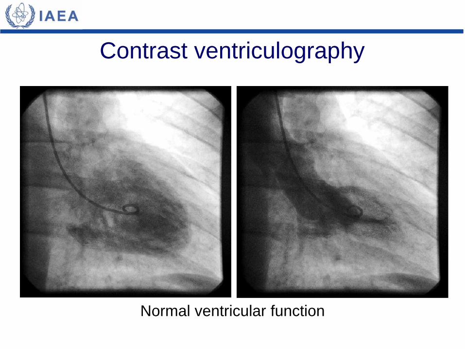
Contrast ventriculography
Normal ventricular function

Coronary angiography
LAD stenosis (>90%, arrow)
Normal LCx
Normal RCA
left right

PTCA procedure
Pre-procedure Balloon inflation Post-procedure

Follow-up • 4 months after successful PTCA, the patient is
asymptomatic and controlling risk factors with medication
and life-style modifications.
• Exercise test with no symptoms and normal ECG (below).

• In patients with no known coronary artery disease and at overall low-to-
intermediate risk, myocardial perfusion SPECT adds prognostic information
and risk-stratifies patients beyond clinical and exercise data.
• Semiquantitative information obtained by gated SPECT provides important
measurements of disease extent and severity.
• Perfusion scores are useful tools in clinical decision making and have been
shown to have independent risk-stratification value.
• Post-ischemic stunning, as assessed by gated SPECT, is a marker for poor
prognosis, particularly for ischemic cardiac events.
• Patients with high risk results should be managed aggressively - with
revascularization procedures if possible.
Teaching points
Bibliography
• Johnson LL, Verdesca SA, Aude WY, et al. Postischemic stunning can affect left
ventricular ejection fraction and regional wall motion on post-stress gated
sestamibi tomograms. J Am Coll Cardiol 1997; 30:1641-8.
• Hachamovitch R, Berman DS, Shaw LJ, et al. Incremental prognostic value of
myocardial perfusion single photon emission computed tomography for the
prediction of cardiac death: differential stratification for risk of cardiac death and
myocardial infarction. Circulation 1998; 97:535-43.
• Cerqueira MD, Weissman NJ, Dilsizian V, et al. Standardized myocardial
segmentation and nomenclature for tomographic imaging of the heart: a
statement for healthcare professionals from the Cardiac Imaging Committee of
the Council on Clinical Cardiology of the American Heart Association. Circulation
2002; 105:539-42.
• Usui Y, Chikamori T, Nakajima K, et al. J-ACCESS Investigators. Prognostic value
of post-ischemic stunning as assessed by gated myocardial perfusion single-
photon emission computed tomography: a subanalysis of the J-ACCESS study.
Circ J 2010; 74:1591-9.