Typical absence seizures in adults: clinical, EEG, video-EEG ...
7
12ournal of Neurology, Neurosurgery, and Psychiatry 1992;55:1002-1008 Typical absence seizures in adults: clinical, EEG, video-EEG findings and diagnostic/syndromic considerations C P Panayiotopoulos, E Chroni, C Daskalopoulos, A Baker, S Rowlinson, P Walsh Abstract Eighteen women and five men had typical absences. These included 10% of a consec- utive hospital series of 200 adult patients with epileptic disorders. The absences began between the ages of seven and 46 years and varied in type and severity. Twenty patients also had generalised tonic-clonic seizures, ranging in fre- quency from one in a lifetime to one per month. Myoclonic jerks of the limbs occurred in 11 patients but were not associated with the absence attacks. Eye- lid myoclonus consistently occurred with absence attacks in four patients and peri- oral myoclonus in two patients. Absence status occurred in five patients. Absence seizures were frequently unrecognised or misdiagnosed as complex partial seizures. Satisfactory control was achieved with sodium valproate. Electroencephalogra- phy, particularly video-electroencepha- lography, was invaluable in the diagnosis, but focal abnormalities in seven patients might have been erroneously interpreted as indicating partial seizures. This series showed that clinical and EEG manifesta- tions are often syndrome-related and that there are more epileptic syndromes with typical absences than those presently recognised. (7 Neurol Neurosurg Psychiatry 1992;55: 1002-1008) Department of Clinical Neurophysiology and Epilepsy, St Thomas' Hospital, London C P Panayiotopoulos E Chroni C Daskalopoulos A Baker S Rowlinson P Walsh Correspondence to: Dr Panayiotopoulos, Department of Clinical Neurophysiology and Epilepsy, St Thomas' Hospital, London SEI 7EH, England. Received 30 September 1991 and in revised forms 21 January 1992 and 14 February 1992. Accepted 5 March 1992 Though often considered rare in adults, typical absences with onset in childhood and puberty persist into adult life in 7-81% of cases.1-19 Loss or severe impairment of consciousness is considered to be the clinical hallmark of absences.2021 EEG manifestations of typical absences, however, are frequently associated with mild, inconspicuous cognitive impair- ment.22.24 Furthermore, typical absences have been studied in a uniform fashion' 19 25 26 despite existing evidence that their clinical and EEG features are syndrome-related.24 Thus prognosis has been based on a symptom (typical absences)20 rather than a syndrome/ disease diagnosis.21 This is the first clinical; EEG and video- EEG study of typical absence seizures and syndromes in adults. Methods and patients Twenty patients with typical absences, as defined by the Commission on the Classifica- tion and Terminology of the International League Against Epilepsy (ILAE),20 were iden- tified from the first 200 consecutive clinical referrals between April 1989 and April 1991 to a new epilepsy clinic in St Thomas' Hospital. Three additional patients, referred for the purpose of this study, were included. Patients were referred from accident and emergency departments, general practitioners, physicians and other neurologists within St Thomas' Hospital and the south-east Thames region. Eighteen of these 23 patients had been seen by at least one consultant neurologist and they all had had one or more EEGs. Clinical and EEG evaluation of all patients was made by one of us (CPP). Diagnosis was based on the proposal of ILEA;2` when not possible, a seizure-symptom categorisation20 and a differential syndromic diagnosis was made. Early clinical records and EEG reports were obtained and scrutinised. Routine EEG and video-EEG (34 channel- Telefactor) recordings were carried out using previously reported techniques.23 24 Patients were asked to count their breaths out loud during hyperventilation, which allowed us to evaluate impairment of consciousness and speech during generalised spike-wave dischar- ges." 2 Twelve patients were studied with video-EEG and 83 recorded absences were analysed. Results Prevalence, sex, age Ten per cent of the 200 unselected patients had typical absences. There was a threefold female predominance of the total group (18 women, 5 men). All patients were over 20 years of age with normal neurological and mental status. Table 1 shows the age at referral, onset of seizures and duration of the disease. Absences All patients had absences documented with EEG or video-EEG or both. The age at onset of absences is shown in table 1. Late-onset absences, occurring after the second decade of life, were reported by six patients. Onset of absences could not be estimated by six patients and another one (case 23) remained unaware of them despite video-EEG confirmation. The absences were subjectively perceived by the patients in a stereotyped way and were described as brief "flashes of blackouts", "like in a daze", "getting into a trance" (table 2). The degree of impairment of consciousness from mild "momentary lack of concentration" 1002
Transcript of Typical absence seizures in adults: clinical, EEG, video-EEG ...
12ournal of Neurology, Neurosurgery, and Psychiatry 1992;55:1002-1008
Typical absence seizures in adults: clinical, EEG,video-EEG findings and diagnostic/syndromicconsiderations
C P Panayiotopoulos, E Chroni, C Daskalopoulos, A Baker, S Rowlinson, P Walsh
AbstractEighteen women and five men had typicalabsences. These included 10% of a consec-utive hospital series of 200 adult patientswith epileptic disorders. The absencesbegan between the ages of seven and 46years and varied in type and severity.Twenty patients also had generalisedtonic-clonic seizures, ranging in fre-quency from one in a lifetime to one permonth. Myoclonic jerks of the limbsoccurred in 11 patients but were notassociated with the absence attacks. Eye-lid myoclonus consistently occurred withabsence attacks in four patients and peri-oral myoclonus in two patients. Absencestatus occurred in five patients. Absenceseizures were frequently unrecognised ormisdiagnosed as complex partial seizures.Satisfactory control was achieved withsodium valproate. Electroencephalogra-phy, particularly video-electroencepha-lography, was invaluable in the diagnosis,but focal abnormalities in seven patientsmight have been erroneously interpretedas indicating partial seizures. This seriesshowed that clinical and EEG manifesta-tions are often syndrome-related and thatthere are more epileptic syndromes withtypical absences than those presentlyrecognised.
(7 Neurol Neurosurg Psychiatry 1992;55: 1002-1008)
Department ofClinicalNeurophysiology andEpilepsy, St Thomas'Hospital, LondonC P PanayiotopoulosE ChroniC DaskalopoulosA BakerS RowlinsonP WalshCorrespondence to:Dr Panayiotopoulos,Department of ClinicalNeurophysiology andEpilepsy, St Thomas'Hospital, London SEI 7EH,England.Received 30 September1991 and in revised forms21 January 1992 and14 February 1992.Accepted 5 March 1992
Though often considered rare in adults, typicalabsences with onset in childhood and pubertypersist into adult life in 7-81% of cases.1-19Loss or severe impairment of consciousness isconsidered to be the clinical hallmark ofabsences.2021 EEG manifestations of typicalabsences, however, are frequently associatedwith mild, inconspicuous cognitive impair-ment.22.24 Furthermore, typical absences havebeen studied in a uniform fashion' 19 25 26despite existing evidence that their clinical andEEG features are syndrome-related.24 Thusprognosis has been based on a symptom(typical absences)20 rather than a syndrome/disease diagnosis.21This is the first clinical; EEG and video-
EEG study of typical absence seizures andsyndromes in adults.
Methods and patientsTwenty patients with typical absences, asdefined by the Commission on the Classifica-
tion and Terminology of the InternationalLeague Against Epilepsy (ILAE),20 were iden-tified from the first 200 consecutive clinicalreferrals between April 1989 and April 1991 toa new epilepsy clinic in St Thomas' Hospital.Three additional patients, referred for thepurpose of this study, were included.
Patients were referred from accident andemergency departments, general practitioners,physicians and other neurologists within StThomas' Hospital and the south-east Thamesregion. Eighteen of these 23 patients had beenseen by at least one consultant neurologist andthey all had had one or more EEGs.
Clinical and EEG evaluation of all patientswas made by one of us (CPP). Diagnosis wasbased on the proposal of ILEA;2` when notpossible, a seizure-symptom categorisation20and a differential syndromic diagnosis wasmade. Early clinical records and EEG reportswere obtained and scrutinised.
Routine EEG and video-EEG (34 channel-Telefactor) recordings were carried out usingpreviously reported techniques.23 24 Patientswere asked to count their breaths out loudduring hyperventilation, which allowed us toevaluate impairment of consciousness andspeech during generalised spike-wave dischar-ges." 2 Twelve patients were studied withvideo-EEG and 83 recorded absences wereanalysed.
ResultsPrevalence, sex, ageTen per cent of the 200 unselected patients hadtypical absences. There was a threefold femalepredominance of the total group (18 women, 5men). All patients were over 20 years of agewith normal neurological and mental status.Table 1 shows the age at referral, onset ofseizures and duration of the disease.
AbsencesAll patients had absences documented withEEG or video-EEG or both. The age at onsetof absences is shown in table 1. Late-onsetabsences, occurring after the second decade oflife, were reported by six patients. Onset ofabsences could not be estimated by six patientsand another one (case 23) remained unawareof them despite video-EEG confirmation.The absences were subjectively perceived by
the patients in a stereotyped way and weredescribed as brief "flashes of blackouts", "likein a daze", "getting into a trance" (table 2).The degree of impairment of consciousnessfrom mild "momentary lack of concentration"
1002

Typical absence seizures in adults: clinical, EEG, video-EEG findings and diagnosticlsyndromic considerations
Table 1 Chronological dqta, frequency of seizures and syndromic classification
Onset (years) of Frequency of
Case No Sex Age Absences Myoclonic jerks GTCS GTCS Absences Classification
1 F 22 Not known 13 None Occasional JME2 M 29 Not known 13 24 3/life Frequent JME3 F 25 Not known 16 20 /life Occasional JME4 F 40 7 10 12 I/month 2/day JME5 F 25 Not known 13 13 4/life Occasional JME6 F 41 Not known 16 22 4/year 3/day JME7 F 26 11 13 14 Not available Occasional, evoked JME8 F 42 18 35 I/life 3-4/day Eye-lid myoclonia with absences9 M 43 30 35 9 4/year 1-5/day Eye-lid myoclonia with absences10 F 38 20 11 1-3/year 6/month Eye-lid myoclonia with absences11 M 26 12 12 I/month Several/day JAE12 F 44 11 12 1-2/year 1-2/year JAE13 M 38 12 None 1-2/month JAE14 F 39 8 None Many/day JAE15 F 46 46 46 2/life 2-4/life Absence status16 F 34 24 12 TV induced Frequent, evoked Photopattern sensitive epilepsy17 F 30 28 28 2/life 1-2/month Symptomatic absence epilepsy18 F 35 15 18 25 1-2/year Infrequent Atypical JME? JAE?19 M 60 Not known 35 9 1-2/year 2/month Atypical JME? JAE?20 F 21 7 7 1/year 5-6/day Perioral myoclonus with absences21 F 27 13 22 3/Life Frequent Perioral myoclonus with absences22 F 67 30 30 1/year 1/month Late-onset absences/absence status?23 F 27 Unaware 1-2/life Unaware Late-onset absences with GTCS?
Mean 35.8 18.3 18.2 19.1SD 11.3 10.5 8.7 10.1
GTCS = generalised tonic-clonic seizures; JME = juvenile myoclonic epilepsy; JAE = juvenile absence epilepsy.
Table 2 Patients' own description of the absences
Case no Description
234567891011121314151617181920212223
Like in a distance, mind becomes blank, thoughts jumbled.Flashes of loss of concentration, tiny/little blackouts.Momentary lack of concentration.Momentary lack of concentration, blanks, dream, deja-vu, strange and terrifying feelings.Brief episodes of absent mindedness.Blank, going to a distance, lack of awareness, you are there and not there.Momentarily loss of concentration.Loss of concentration,unresponsive.Feels away from things and unreal.Gets into a trance, like day-dreaming, not responding to commands.Loss of concentration-like being hit on the back of the head-like in a daze.Like in a trance.Discontinuation of what one thinks or says, a very quick black-out.Blank spells, unresponsive.Becomes slow, not herself.Momentary loss of contact, freeze in front of the kitchen carpet.Mentally as if I slipped a cog.The world around me seems to have moved without me, lost the thread of what they had said.In and out of a state of consciousness, disorientated behaviour, some jerking.Blank episodes, blurring as if in a dream, unresponsive.Vacancies, distracted, unable to understand basic commands.Strange episodes of inappropriate behaviour, confused.Momentary loss of track of things.
Fp2 F4
F4 - C4 vv5M
C4 - P4
P4 -02
Fpl - F3
F3 - C3
C3 - P3
P3 1 -
10O0pv EYES CLOSED
Ist PERIORALAtUTOMATMS
5 t HAND RECALLS5PASSIVELYMOVED
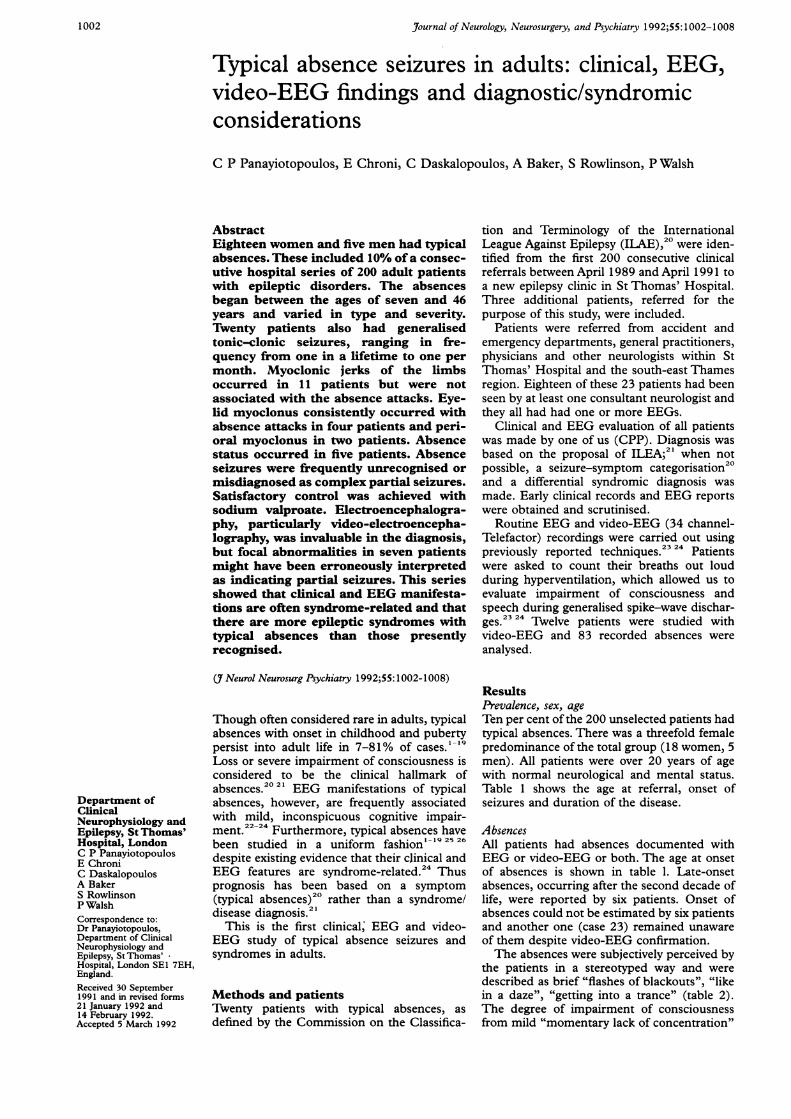
Figure 1 Video-EEG of a 39 year old woman (case 14) who had severe and frequentabsences since age 8 years without myoclonic jerks or generalised tonic-clonic seizures. Theabsence seizure illustrated was recorded during breath-counting in hyperventilation. Hereyes remained closed, she stopped overbreathing and counting but, with some hesitation,pronounced correctly the subsequent number (56) after the onset of the discharge. Ictaloral automatisms were observed. She could remember the number five spoken to herimmediately before the end of the ictus. The seizure was terminated by passively movingher hand.
to severe "the world has moved around mewithout me" was often reflected in the patients'own descriptions.
De-realisation and fear were experienced by2 patients (cases 4 and 9). Automatisms werenever reported by witnesses or by patients butwere recognised in video-EEG studies in onepatient (fig 1, case 14). Facial myoclonuslocalised in the eyelids (fig 2, cases 8-10, 17)or perioral muscles (cases 20-21), ofwhich thepatients were unaware, was demonstrated dur-ing the absences with video-EEG or describedby witnesses. Severity and frequency of absen-ces varied from causing little disturbance toimposing a serious social and professionalhandicap to the patient. Absence status wasdocumented with EEG or video-EEG, or both,in four patients (cases 15, 19, 21, 22, fig 3) andsuspected in a fifth (case 12). Episodes com-prised protracted periods (5-12 hours) of mildto moderate confusion. In four cases absencestatus was unrecognised by medical and nurs-ing personnel for many years.
1003

Panayiotopoulos, Chroni, Daskalopoulos, Baker, Rowlinson, Walsh
F4*)tCI9 \t ; +
as '-PsT K W4 'r i
P4 1.
Ps -asv ^ f w l N h _
Ps _0t'I8
Figure 2 EEG of a 43 year old patient (case 9) who had onseyears. He became aware of absences at age 30 years. Eyelid myassociated with the ictus. Discontinuation of breath counting watrace) but not another (lower trace) of the multiple-spike waveThese discharges were consistently associated with eyelid myoclotcognitive impairment showed a considerable variation, probablylevel of alertness.
Fp2 - F4
F4 - C4 .e4lC4 -P4r(VrC4DP4 -02
Fpl - F3.
F3 -C3M,VHAW/C3 -P3
P3 -0 WA100 ItV
1 sec ABSENCE STATU
Figure 3 Absence status recorded by video-EEG of a 67 year Iwas treated with primidone and sulthiame for 37 years. Her GIstate of mild confusion, including "strange episodes of inapproprputting on her trousers over her pyjamas" which were interpretedseizures. This video-EEG recording, seven hours after onset of s)absence status. Clinically she was slow in her answers and modewas terminated with diazepam iv and sodium valproate iv Notwave activity which was continuous.
Myoclonic jerks
- ;yj ,I
varied in frequency from one in a lifetime (for9,S,,,,\,^,_, example, case 8) to several per month (case 4).
The age at onset ranged from 7-46 yearstel,*' '''th'si's'' (mean 19 1 (SD 10-1)). Although absences
occurred first, it was often a GTCS that, r i, r precipitated medical attention.
EEG and video-EEG studiesAbsences were recorded in all patients. Pre-vious clinical records and EEG reports_~ revealed that some patients had spike-slowwave discharges long before typical absenceswere clinically recognised, such as in case 18.The duration of the discharges varied from
~ 1-2 to 20 s but lasted for hours in absencestatus. Discharges were generalised, of higheramplitude in the anterior regions, with domi-nant intradischarge spike-wave frequency of
et of GTCS at age 10 3-4 Hz (complexes of 2-6 Hz were also seen).oclonus was consistently In some patients, such as cases 13 and 14 (figis observed in one (upper 1), the discharges consisted of repetitive spike-zus but the severity of slow wave complexes with steady fall in fre-depending on his pre-ictal quency from the onset to the termination of
the paroxysms. In others (cases 1-10 mainly,figs 2, 4) there were frequent multiple spikecomponents and discharge fragmentations.2324The paroxysms of patients 1-7 were oftenirregular, with a variation of the intradischarge
rv>$~M;r' frequency (fig 4).The discharges were associated with impair-
ment of consciousness which greatly varied in,, severity. Some patients had clinical manifesta-
tions during spike-wave paroxysms as short as15-2-5 s (fig 5). In others, or even the samepatient, however, there were no discerniblemental changes in much longer EEG dis-charges (fig 2), although in one patient impair-ment of consciousness was often proportionalto the length of the paroxysm. Automatismswere recorded in one, and facial myoclonus infour patients. A discharge often could beinterrupted by auditory or somatosensory
Is stimuli or both.Focal paroxysmal abnormalities were recor-
old woman (case 22) who ded in seven patients.These were short tran-rcs were preceded by aiate behaviour like [sic] sients of localised slow, sharp waves or spikes,d as complex partial or both (fig 4). Focal spikes were eitherymptoms, demonstrated independent or associated with the generalised,rately confused. Statusfast 3-4 Hz spike-slow paroxysms (preceding, following or inter-
spersed). The same patient could have spikes inmultiple locations in the same or a previousEEG. These focal abnormalities had oftenbeen interpreted previously as evidence ofpartial seizures with secondary generalisation.
A clear distinction between limb myoclonicjerks and localised facial (eyelid or perioral)myoclonus was apparent. Limb myoclonus didnot occur during the absence ictus, exceptduring myoclonic absence status. Conversely,facial myoclonus alone (eyelid in four patientsand perioral in two) was always associated withthe absence ictus. Myoclonic jerks of the limbsoccurred in 11 patients: on awakening in seven(cases 1-7), random, diurnal and usually mildin three (cases 9, 18, 19) and induced bytelevision in one (case 10).
Generalised tonic-clonic seizuresGeneralised tonic-clonic seizures (GTCS)were reported by 20 patients (table 1) and
EEG and precipitating factorsHyperventilation facilitated the spike-wavedischarges in all patients. Photoconvulsiveresponses were recorded in eight (34-8%)patients (fig 6). One patient (case 7) hadabsences on rubbing her face, particularlyaround the nose, was photosensitive (fig 6) andhad myoclonic jerks and GTCS on awakening.Another patient (case 16) with clinical andEEG photosensitivity from 12 years of age,developed late-onset absences precipitated byviewing complex patterns (carpets) confirmedby appropriate EEG recordings.The background EEG was normal except in
chronic and heavily medicated patients where
1 004
--T --w-f V, 'K-T -'-v N -1 FT -r -V T ?.'NovvwAwvocLom1fw
I1_

Typical absence seizures in adults: clinical, EEG, video-EEG findings and diagnosticlsyndromic considerations
Fp2 - F4iR >
- C4
C4 P4- A-- ' t4J
P402%A
Fpl F3
F3 -C3Vs3 -t><!A
C3 - P3 tP3 - 01 ~. f
Fp2 - F8S
FS - T4 _ _ * ,
T4 - T6 __ __-_ __W
T6 02
Fpl - F7
F7 - T3
T3 - T5
T5 - 01 -^ _
I sec
Figure 4 EEG recording of a 40 year old woman (case 4) with juvenile myoclonicepilepsy (JME) (absences at 7 years, myoclonic jerks at 10 years and GTCS at 12years). Initially diagnosed as suffering from complex partial and secondary generalisedseizures, she had one or two GTCS per month and one to three absences per day while oncarbamazepine and phenytoin. Absences and myoclonic jerks stopped after carbamazepinewas replaced by sodium valproate. She had five nocturnal GTCS in the last two years offolUow up. CT of the brain was normaL The EEG (upper trace) showed the typical JMEpattern with Ws, discharge fragmentations, inconsistent spike andlor multiple spike-slowwave relation and intradischarge frequency variations. Focal spikes, independently right ormore frequently left, were seen either at the onset or within the discharges. The restingEEG (lower trace) had also transients of slow waves localised either on the left or rightmid-temporal electrodes.
an excess of diffuse theta activity was found(fig 4).
Syndromic classification of the patientsTable 1 shows the syndromic classification ofthe 23 patients.
Seven patients (cases 1-7) had juvenilemyoclonic epilepsy (jME).21 23 All had typicalabsences, myoclonic jerks on awakening andGTCS (except case 1 who did not haveGTCS). Clinical and EEG manifestations oftypical absences (figs 4 and 6) were as pre-viously described in adolescent patients withJME.2'
Eyelid myoclonus with absences27 28 wasdiagnosed in three patients (cases 8-10).Rhythmic eyelid myoclonus was documentedby EEG and video-EEG during the absences.Photosensitivity was found in one of the three(case 10). Typical absences varied in durationfrom 3-12 s. Impairment of consciousness wasmore severe than in JME but not as intense asin juvenile absence epilepsy (fig 2).
Juvenile absence epilepsy (JAE)2 2429 wasthe likely diagnosis in four patients (cases11-14). Two patients (cases 11 and 12) hadtypical absences with onset at puberty. Theyalso had GTCS and occasional diurnal mildmyoclonic jerks. One patient (case 13) had
pyknolepsy from age 12, clinical remission atage 20 and a relapse at age 30 with mild andinfrequent absences. He had no other type ofseizures. Another (case 14) had onset ofintractable pyknoleptic absences at age 8 yearswhich continue to date with the same fre-quency and severity; she has never had anyother type of seizures. Typical absences in thesepatients showed more severe impairment ofconsciousness than in any other epilepticsyndrome in this study (fig 1).
Late-onset absence status with GTCS30(case 15), photo- and pattern-sensitive epi-lepsy3" (case 16) and symptomatic late-onsetabsences32 (case 17, fig 5) were also classifiedwith relative confidence.The remaining six patients had uncertain
syndromic classification (see table 1 and dis-cussion).
Diagnosis on referral and treatmentNone of the 23 patients had a syndromicdiagnosis on referral. Furthermore, absenceswere not recognised in 14 patients and in eightof them, the working diagnosis was of complexpartial seizures.
Seizures in the majority of patients were notcontrolled on referral, despite often multipleanti-epileptic drugs. Carbamazepine was themost widely used drug (nine patients) followedby sodium valproate (eight), phenytoin (six),phenobarbitone (four), sulthiame (one), viga-batrin (one), ethosuximide (one). None of thepatients were on clonazepam.The follow up period was short (maximum
two years) for quantitative conclusions regard-ing treatment, mainly with sodium valproate,and slow withdrawal of other anti-epilepticmedication. Four patients were free of seizures,a 60-80% reduction of all types of seizure wasseen in 10 and there was no change in threepatients. Data for the other three of the 20unselected patients were incomplete.
DiscussionAll patients-adults with normal neurologicaland mental status-had typical absences asdefined by the Commission of the ILAE.20This report refers to patients with overt clinicalabsences documented by EEG or video-EEGrecordings, or both, and not to patients withEEG epileptiform discharges unaccompaniedby clinical changes (subclinical, larval or elec-trical seizures).22We have documented that typical absences
in adults show a considerable syndrome-related variation in their combined clinicalpresentation and EEG features. Impairment ofconsciousness varies from severe to mild and isfrequently difficult to document objectivelywith conventional means. This is in agreementwith our previous study on children andadolescents.23 24
Clinically, the absences were perceived bythe patients as transient sensations of"momentary lack of concentration", "flashesof blackouts" which could be misinterpreted asnormal sensations or drug-induced in patientswho were often overmedicated. We, the clini-
,OA wt..A%w-w-v W- .
NV PI k7 lj
1005

Panayiotopoulos, Chroni, Daskalopoulos, Baker, Rowlinson, Walsh
FP2- F4
F4 -04C''-
C4- P4
P4 -02
Fpl - F3 C V,,,/i
F3 - C3 'j1I
C3 - P3 iojvlw,, A,,
P3 -O01
100 ,uV1 sec EYE LID FLICKERING
Fp2 F4
F4 - C4
C4 - P4 ,
P4 - 02
Fp1 F3
F3 - C376 77 77 78
C3 P33w
P3 -1
100 iVL
1 sec
Figure 5 Symptomatic absences of a 30 year old female with late onset absences andGTCS. Her initial EEG (left) demonstrated absences with eyelid flickering. Impairmentof breath-counting was documented during brief generalised discharges in subsequentvideo-EEG recordings (right). CT scan demonstrated a left frontal lesion, probably a lowgrade glioma.
cians, have been trained to identify absences intheir classic form,20 21 that is, of a child withtransient episodes of loss of consciousness. Weare not familiar with absences in an adultwhose level of awareness is not severelyimpaired during the absence ictus. We havedemonstrated in this study that absences inadults may escape recognition because they are
often mild. This significant observation hasbeen made in only one other clinical report:".6... in the patients who continued to havepetit mal, the duration of the attacks was
usually one second or less, and they were oftennot noticeable to family, friends, or school-mates".6 Extensive studies have demonstratedthat the generalised spike-wave dischargesdescribed as "subclinical, larval or electricalseizures" are frequently associated with cogni-tive impairment (see references 22 and 33 forreview).
Furthermore, absences may escape clinicaldetection, even during conventional EEG.Current practice is to test, in an all-or-nonemanner, the ability of a patient to recall a
phrase or number administered during a gen-eralised spike-wave discharge. It has beenshown in this study that this is often notadequate. We have frequently reviewed video-EEG of patients with spike-wave discharges in
whom clinical manifestations (such as slowingdown of breath-counting, repetition and mis-takes, or stuttering) became apparent onlyafter careful video-EEG viewing.For practical purposes we propose that
patients with spike wave paroxysms should beasked to count their breaths during hyper-ventilation; this may show slowing, disconti-nuity, errors, repetition and other signs ofimpairment of consciousness and speechwhich otherwise would escape recognition withthe current practice established in EEGdepartments.23 24 This method is sensitive(involves concentration, memory, recollectionof learned experience, expressive speech andother cognitive functions), practical (easy toperform by the patient and easy to evaluate bythe observer) and is clinically relevant (reflectsimpairment of day-life performance).The frequency of 10% of typical absences
found in adults in this study is the highest everreported.1 19 Absences were undiagnosed inmore than half of our patients on referral, aproportion comparable to that of misdiag-nosed absence status despite its long duration(hours or even days) and the associated severemanifestations of confusion and impairedbehaviour. 3
Underestimation of absences may be evenmore extensive than in this study; impairmentof consciousness is often manifested in patientswith series of repetitive myoclonic seizures ormyoclonic status, or both, as illustrated byanother of our more recent patients (fig 7).
In one third of our patients the absenceswere misinterpreted as complex partial seiz-ures. Typical absences in adults are brief,lasting only for seconds with, usually, mildimpairment of consciousness. It is because ofthis mild impairment that automatisms were sorare in our patients compared with the fre-quent automatisms occurring in childhood andjuvenile absences.24 Conversely, complex par-tial seizures are of longer duration (minutes)and are associated with prominent emotionaland sensory perceptions of the patients withfrequent automatisms.
Despite these differences, the diagnosis ofcomplex partial seizures was maintained, evenafter EEG data were available, either becausethe physician was reluctant to change his or herclinical impression because of an EEG reportshowing "generalised discharges without asso-ciated clinical manifestations", or becausemild, occasionally persistent, focal EEGabnormalities were misinterpreted as indicat-ing partial seizures. Focal EEG abnormalitieshave been reported and are accepted in child-hood absence epilepsy33 but are not generallyknown in other idiopathic generalised epi-lepsies, such as JME, despite their documenta-tion in this and previous reports.23 34 3
Therapeutic decisions were influenced bythe diagnostic problems. Sodium valproate,which is the drug of choice in idiopathicgeneralised epilepsies" was prescribed lessoften than carbamazepine, which is not appro-priate treatment for absences or myoclonicepilepsy37 (its effect in GTCS of idiopathicgeneralised epilepsies has not been clarified).
1006

Typical absence seizures in adults: clinical, EEG, video-EEG findings and diagnosticlsyndromic considerations
gopm
Mf
Figure 6 EEG of a 26-year old nurse with JME. Generalised spike and occasionalmultiple spike and slow wave discharges are recorded on eye-closure and are evoked bysomatosensory stimuli (rubbing her nose) and intermittent photic stimulation.
Fp2-Fjee! ,rm virI, Ie & j lFp2- F3 - #Wj '4I% +
R -C4 ^ l f t $
F3 - C3
C4-P4
C3 P3-4PA3
P4 -02 _
P3 - 0 - N7, ^,,
Fp2- F8 q Ag
Fpl - FT 4A/.PU#
FS -T4 < 4 b g
F7 T3 r^> r
TX - T6G , f ;, ; ! 9
T3 - T5 r'- ' ' - -
TS - 02 ~_+
TS -cm r .
Figure 7 EEG of myoclonic status of a 36-year-old woman with JrME while oncarbamazepine and phenytoin. Myoclonic jerks and GTCS, but not absences, wereelicited on history. Typical EEG features of myoclonic jerks (right) and absences (left)were recorded. In addition, eyelid myoclonus was noted by the technician during thismyoclonic-absence status.
Ethosuximide38 and clonazepam39 were rarelyused, despite their importance as adjuncttherapy in patients with refractory absencesyndromes. Our patients showed considerableimprovement in frequency of all seizure types(absences, myoclonic jerks and GTCS) onsodium valproate. Longer follow up is needed,however, to confirm these results, as well as theefficacy of ethosuximide and clonazepam as acombined treatment with sodium valproate.The significant predominance of women in
this study is similar to that found in childhoodabsence2' and photosensitive epilepsy3' butunlike the male predominance of juvenileabsence epilepsy29 and the equal sex23" "
distribution ofJME. It may be attributed to thefinding that remission of typical absences ismore likely in boys than in girls.'3We attempted an epilepsy disease/syndrome
diagnosis of the 23 patients with absences, inaccordance with the proposal of ILEA.2' It isbeyond the scope of this report to discusslimitations and drawbacks of this proposal(such as inadequately defined and overlappingsyndromes, bias towards criteria supported byone but not other authors, complex anddifficult terminology) which are well illustratedin this study. This should not be used as an
argument against a syndromic classification ofepilepsies. Common syndromes such as JME
are well defined2' 23 and a confident diagnosiscould be made for the majority of the patientsreported here. The following discussion aimsto promote the use of a medical disease/syndrome diagnosis of patients with seizuresbecause this is probably the only way out of themonolectic diagnosis "epilepsy" which ourpatients are often labelled with for life.
Juvenile myoclonic epilepsy with the triad ofage-related idiopathic generalised seizures,myoclonic jerks on awakening and the charac-teristic clinical and EEG manifestations oftypical absences," 2 was easy to recognise inseven of our 23 patients. This study confirmsour previous reports 24 that typical absencesin JME have distinct combined clinical andEEG features.
Ictal eyelid myoclonus with absence seizureswas consistently recorded in three patients; itwas also occasionally seen in one patient withsymptomatic absences (case 17) and may alsobe observed during myoclonic status (fig 7).Only one of the three patients would meet thecriteria of the syndrome described by Jea-vons,27 that is, photosensitive patients withabsences that are resistant to treatment andshow marked jerking of the eyelids associatedwith spike wave-discharges on eye-closure.Although not representing a monosympto-matic criterion for this classification, ictaleyelid myoclonus with absences may be used asa basis for a syndrome as yet ill-defined, notrecognised in the ILAE classification2' andprobably wider28 than that involving photo-sensitive patients only.Four patients were classified as having juven-
ile absence epilepsy20 23 25 on the basis of age atonset, combined ictal clinical and EEG mani-festations24, frequency of seizures and exclu-sion criteria (lack of myoclonic jerks onawakening, lack of facial or limb myoclonusduring the absences or early-onset GTCS).
Juvenile and childhood absence epilepsy aresometimes difficult to differentiate.2025 TheILAE proposal2l uses criteria of age at onset,frequency of the seizures and the retropulsivemovements of the eyes and head (a svmptomconsistently found in eyelid myoclonus withabsences but rarely in other forms of childhoodabsences). Combination of ictal clinical andEEG features of absences together with someof the ILEA criteria may provide a moresatisfactory syndromic classification. Exclusioncriteria, for example myoclonic jerks on awak-ening and eyelid myoclonus, may be as impor-tant as inclusion criteria.
Classification of three patients with lateonset absence status30 (case 15), photosensi-tive and pattern sensitive epilepsy with absen-ces31 (case 16) and symptomatic epilepsy withtypical absences32 (case 17) was straightfor-ward.The remaining six patients could not be
classified with confidence. Case 18 had absen-ces with EEG but without clinical documenta-tion, as early as five years of age. JME was apossible diagnosis but myoclonic jerks weremild and did not show the characteristicclustering on awakening. Juvenile absence epi-lepsy was also likely, in view of age at onset of
1 007

Panayiotopoulos, Chroni, Daskalopoulos, Baker, Rowlinson, Walsh
clinical manifestations of absences, associatedsymptoms and other seizure-types. Childhoodabsence epilepsy was unlikely, in view of mildimpairment of consciousness.24
Case 19 had the seizure triad of JME butmyoclonic jerks appeared very late (50 years ofage) and did not cluster on awakening. Age atonset of absences and absence status with mildmyoclonic jerks could not be determined.
Cases 20 and 21 had severe absences withconsistent perioral myoclonus and late onsetGTCS. Absence status with perioral myoclo-nus occurred in case 21. Childhood (case 21)or juvenile (case 22) absence epilepsy would bethe diagnosis dictated by the present ILAEclassification.2" Consistent perioral myoclonusin absences may, like absences with eyelidmyoclonus, be a feature of a separate epilepticsyndrome which is resistant to treatment andpersists in adult life. This is supported byanother three patients with absences and peri-oral myoclonus that we have studied recently.Perional myoclonus consists of rhythmic pro-
fusion of the lips, such as, the mouthingmovement of goldfish.
Case 22 may be a case of late-onset absencestatus, absences and GTCS. The remainingpatient (case 23) with late-onset GTCS, was
unaware of absences from which she may havesuffered for years. A mild form of juvenileabsence epilepsy was a possible diagnosis.
Irrespective of arguments for or against theproposed classification of the patients, theclinical and EEG manifestations describedmay help in the recognition, differential diag-nosis, prognosis and treatment of adultabsence syndromes, which are not unusual.Furthermore, this study may encourage revi-sion of the classification of epileptic syndromeswith typical absences based on prospectiveclinical and video-EEG studies.
We thank British Telecom Charitable Organisation and theSpecial Trustees of StThomas' Hospital for their grants to CPPfor the study of the classification of epilepsies.
1 Adie WJ. Pyknolepsy: a form of epilepsy occurring inchildren with a good prognosis. Brain 1924;47:96-102.
2 Owen JW, Berlinrood L. A study of diagnosis and prognosisof petit mal. Pediatr 1941;19:762-77.
3 Lennox WG, Lennox MA. Epilepsy and Related Disorders.Boston, MA:Little, Brown and Company, 1960.
4 Lees F, Liversedge LA. The prognosis of "petit mal" andminor epilepsy. Lancet 1962;ii:797-9.
5 Hertoft P.The clinical, electroencephalographic and socialprognosis in petit mal epilepsy. Epilepsia 1963;4:298-314.
6 Currier RD, Kooi KA, Saidman J. Prognosis of pure petitmal: a follow-up study. Neurology 1963;13:959-67.
7 Gordon N.The natural history of petit mal epilepsy. DevMed Child Neurol 1965;7:537-42.
8 Livingston S, Torres J, Pauli LL, Rider RV. Petit malepilepsy: Results of a prolonged follow-up study of 117patients. JAMA 1965;194:227-32.
9 Gibberd FB. The prognosis of petit mal. Brain 1966;89:531-8.
10 Gibberd FB. The prognosis of petit mal in adults. Epilepsia1972;13:171-5.
11 Charlton MH, Yahr MD. Long term follow-up of patientswith petit mal. Arch Neurol 1967;16:595-8.
12 Barnhart DA, Newsom TD, Crawley JW, et al. Long termprognosis of petit mal epilepsy. Electroenceph Clin Neu-rophysiol 1969;27:549-50.
13 Dalby MA. Epilepsy and 3 per second spike and waverhythms. A clinical, electroencephalographic and prog-nostic analysis of 346 patients. Acta Neurol Scand,1969;Suppl 40:45.
14 Lugaresi E, Pazzaglia PP, Franck L, et al. Evolution andprognosis of primary generalised epilepsy of the petit malabsence type. In: Lugaresi E, Pazzaglia PP, Tassinari CA,eds. Evolution and Prognosis of Epilepsy. Bologna, Italy:Auto Gaggi, 1973:3-22.
15 Sato S, Dreifuss FE, Penry JK, Kirby DD, Palesh Y Longterm follow-up of absence seizures. Neurology1983;33: 1590-5.
16 Loiseau P, Pestre M, Dartigues JF, Commenges D, Barber-ger-Gateau C, Cohadon S. Long term prognosis in twoforms of childhood epilepsy: typical absence seizures andepilepsy with rolandic (centrotemporal) EEG foci. AnnNeurol 1983;13:642-8.
17 Wolf P, InoueY Therapeutic response of absence seizures inpatients of an epilepsy clinic for adolescents and adults.J Neurol 1984;231:225-9.
18 Dieterich E, Baier WK, Doose H, Tuxhorn I, Fichsel H.Long term follow-up of childhood epilepsy with absences.I. Epilepsy with absences at outset. Neuropediatrics 1985;16:149-54.
19 Gastaut H, Zifkin BG, Mariani E, Puig JS. The long-termcourse of primary generalized epilepsy with persistingabsences. Neurology 1986;36:1021-8.
20 Commission on Classification and Terminology of theInternational League Against Epilepsy. Proposed revi-sions of clinical and electroencephalographic classifica-tion of epileptic seizures. Epilepsia 1981;22:480-501.
21 Commission on Classification and Terminology of theInternational League Against Epilepsy. Proposal forclassification of epilepsies and epileptic syndromes. Epi-lepsia 1989;30:389-99.
22 Aarts HR, Binnie CD, Smit AM, Wilkins AJ. Selectivecognitive impairment during focal and generalized epi-leptiform EEG activity. Brain 1984;107:293-308.
23 Panayiotopoulos CP, Obeid T, Waheed G. Absences injuvenile myoclonic epilepsy: A clinical and video-electro-encephalographic study. Ann Neurol 1989;25:391-7.
24 Panayiotopoulos CP, Obeid T, Waheed G. Differentiation oftypical absences in epileptic syndromes. A video EEGstudy of 224 seizures in 20 patients. Brain 1989;112:1039-56.
25 Holmes GL, Mckeever M, Adamson M. Absence seizures inchildren: clinical and electroencephalographic features.Ann Neurol 1987;21:268-73.
26 Berkovic SF, Andermann F, Andermann E, Gloor P.Concepts of absence epilepsies: discrete syndromes orbiological continuum? Neurology 1987;37:993-1000.
27 Jeavons PM. Nosological problems of myoclonic epilepsiesin childhood and adolescence. Dev Med Child Neurol1977;19:3-8.
28 Panayiotopoulos CP. Fixation-off sensitive epilepsy in eyelidmyoclonia with absence seizures. Ann Neurol 1987;22:87-9.
29 Wolf P. Juvenile absence epilepsy. In: Roger J, Dravet P,Bureau M, et al, eds. Epileptic Syndromes in Infancy,Childhood and Adolescence. London: John Libbey, Euro-text, 1985:242-6.
30 Roger J, Lob H, Tassinari CA. Generalised status epilepti-cus expressed as a confusional state (petit mal status orabsence status epilepticus) In: Vinken PL, Bruyn GW,eds. Handbook of Clinical Neurology. Amsterdam: NorthHolland Publishing Co. 1974;15:167-96.
31 Jeavons PM, Harding GFA. Photosensitive epilepsy. ClinDev Med No 56. Spastics International Medical BooksPublications. London: William Heinemann Books Ltd.1975;10:65.
32 Niedermeyer E, Lopes da Silva F. Electroencephalography:basic principles, clinical applications and related fields.Baltimore,MA: Urban & Schwarzenberg, 1982.
33 Gomez MR, Westmoreland BF. Absence seizures. In:Luders H, Lesser RP, eds. Epilepsy. Electroclinical Syn-dromes. Berlin: Springer-Verlag, 1987:105-28.
34 Obeid T, Panayiotopoulos CP. Juvenile myoclonic epilepsy:a study in Saudi Arabia. Epilepsia 1988;29:280-2.
35 Panayiotopoulos CP, Obeid T.Juvenile myoclonic epilepsyI. Clinical aspects II. Precipitating factors, genetics, EEGfindings and treatment JME (Abstract). J Neurol Neu-rosurg Psychiat 1991;54:752.
36 Editorial. Sodium Valproate. Lancet 19.88;ii:1229-31.37 Editorial. Carbamazepine update. Lancet 1989;ii:595-7.38 Rowan AJ, Meijer JWA, Beer-1awlikowski N, Van der Geest
P, Meinardi H.Valproate-ethosuximide combination ther-apy for refractory absence seizures. Arch Neurol 1983;40:797-802.
39 Obeid T, Panayiotopoulos CP. Clonazepam in juvenilemyoclonic epilepsy. Epilepsia 1989;30:603-6.
1 008