Types of circulatory systems · 10.09.2018 · •Circulatory system •Lymphatic system Functions...
70
Transcript of Types of circulatory systems · 10.09.2018 · •Circulatory system •Lymphatic system Functions...

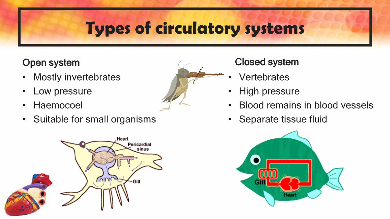
Types of circulatory systems
Open system
• Mostly invertebrates
• Low pressure
• Haemocoel
• Suitable for small organisms
Closed system
• Vertebrates
• High pressure
• Blood remains in blood vessels
• Separate tissue fluid

Closed circulatory system
• Vertebrates
• High pressure
• Blood remains in blood vessels
• Separate tissue fluid
• Heart – arteries – arterioles – capillaries – venules – veins – heart
• Exchange occurs between capillaries and body tissues

Component Structure function
Heart Muscular organ Acts as a pump that keeps the blood flowing
through the body in one direction
Blood vessels Network of tubular
structures
Contains the blood as it circulates throughout the
body
Blood Type of connective
tissue, consisting of red
blood cells, white blood
cells and platelets in a
matrix of blood plasma
Transports:
• O2 and CO2 to and from the lungs
• Absorbed nutrients from small intestine to liver
• Hormones from the endocrine glands to the
target organs
• Waste products like urea from liver to kidney
• Heat from the liver and muscles to the rest of
the body
Lymphatic vessels Network of tubular
structures
• Contains the lymph as it circulates throughout
the body
• Acts as an additional drainage system that
transports lymph to the immune organs and
into the bloodstream
Lymph Colourless fluid derived
from tissue fluid
• Contains white blood cells to fight infections
• Transports products of fat digestion from small
intestines into the blood stream

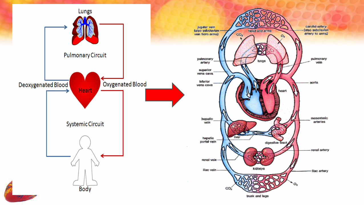
Double circulatory system
Advantages:
• Pumped to lungs
constantly to
be oxygenated
• Creates a high
pressure –
oxygen supplied
and wastes
removed

• Protection of the heart

External view of the heart
pulmonary artery
pulmonary vein
coronaryartery
left ventricle
right ventricle
inferior vena cava
right atrium
pulmonary vein
aorta
superiorvena cava

Internal structure of the heart

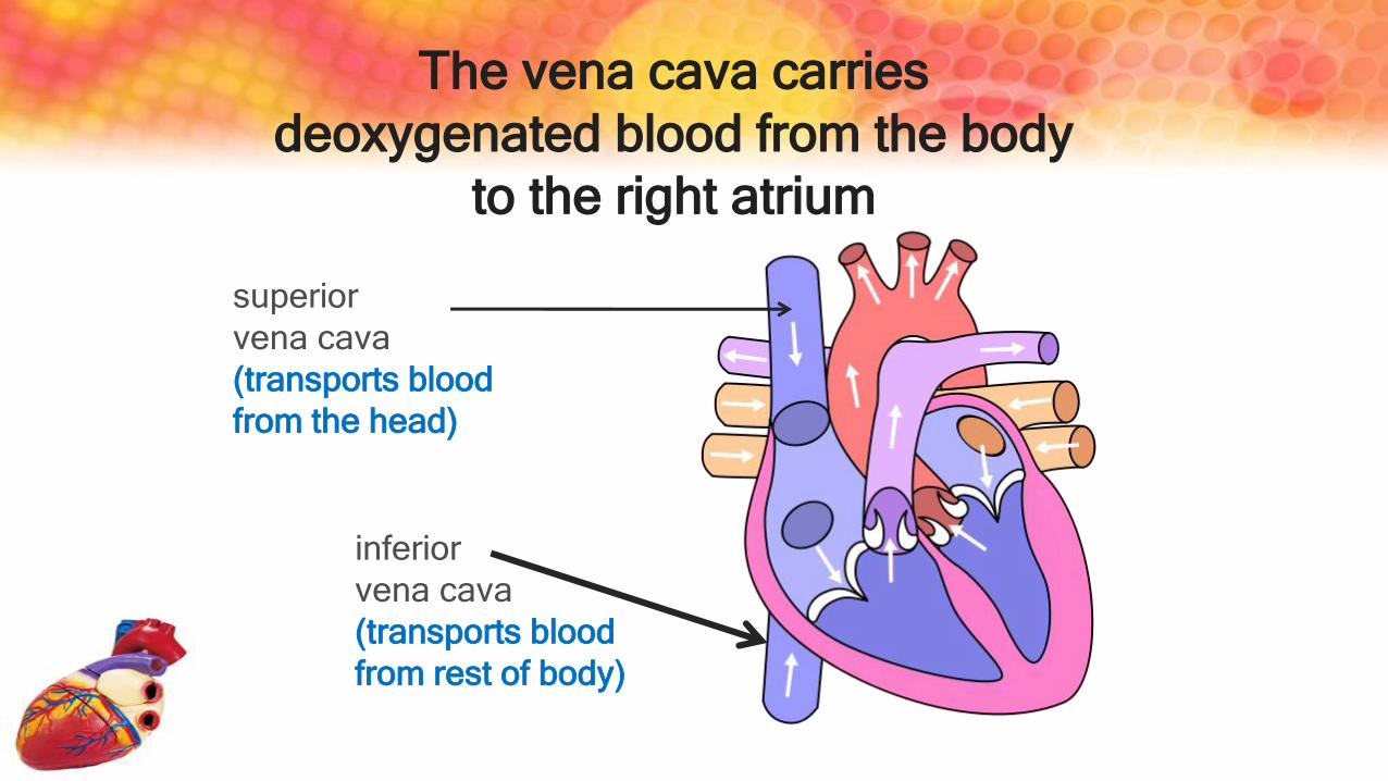
superior
vena cava
(transports blood
from the head)
inferior
vena cava
(transports blood
from rest of body)
The vena cava carries
deoxygenated blood from the body
to the right atrium
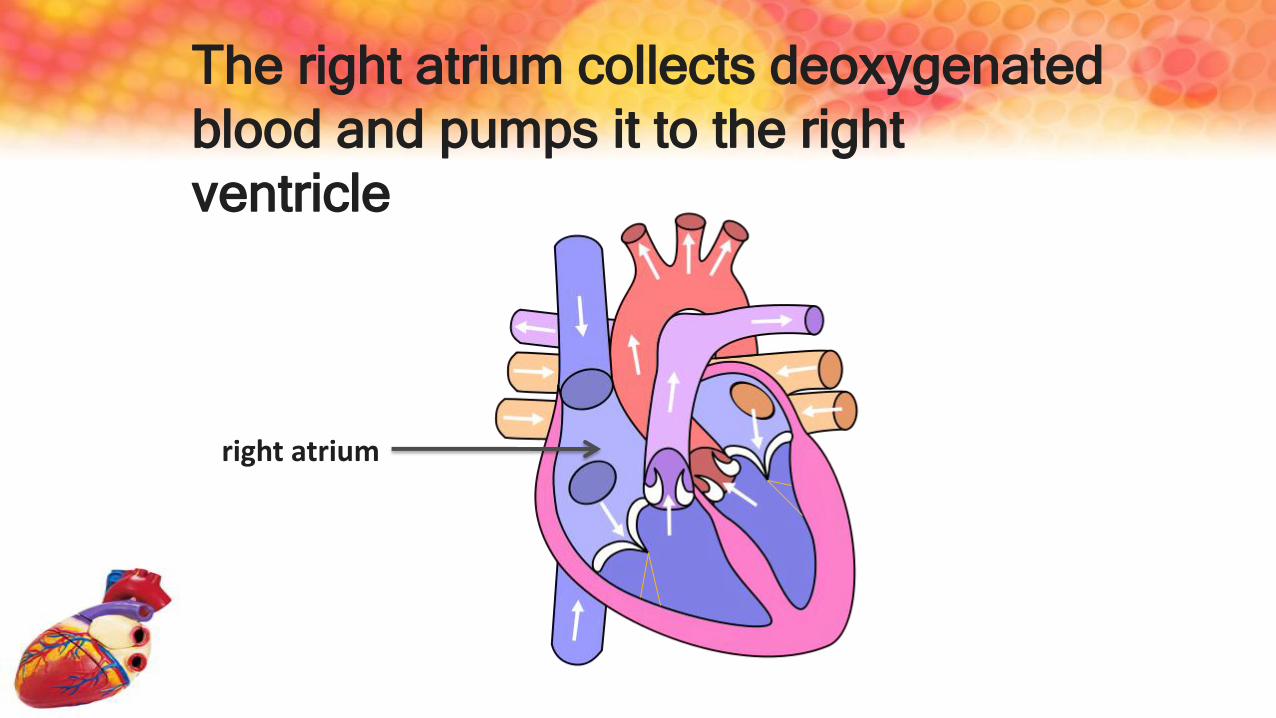
The right atrium collects deoxygenated
blood and pumps it to the right
ventricle
right atrium
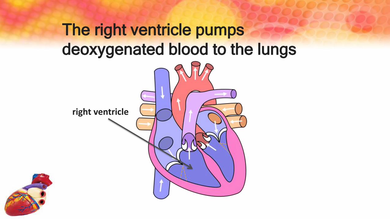
The right ventricle pumps
deoxygenated blood to the lungs
right ventricle

The pulmonary artery carries
deoxygenated blood from the right
ventricle to the lungs
Pulmonary artery

The septum separates the left and
right sides of the heart
septum

The pulmonary veins carry
oxygenated blood from the lungs to
the left atrium
Pulmonary veins

The left atrium collects the
oxygenated blood and pumps it to the
left ventricle
Left atrium

The left ventricle pumps oxygenated
blood to the body via the aorta
Left ventricle

The aorta carries the oxygenated from
the left ventricle to the rest of the body
Aorta
Aortic arch

Atrio-ventricular valves prevent
backflow of blood into the atria when
ventricles contract
Bicuspid valve
(mitral valve)
Tricuspid valves
Tendonous cords

The semi-lunar valves prevent
backflow of blood from the arteries into
the ventricles
Aortic semi-lunar valve
Pulmonarysemi-lunar valve


FUNCTION OF THE HEART
To pump blood around the body so that all living cells
can receive oxygen and other nutrients and release
excretory products to be transported away


Transport systems in Humans
• Circulatory system
• Lymphatic system
Functions of the
transport system of humans
Human body cells
•Require food and oxygen to
release energy
•Waste products get produced
Lungs
Digestive
system
Kidneys
Sweat
Oxygen
Carbon
dioxide
food
Urine
Sweat

HOW IS THE HEART RATE
CONTROLLED?

How does the heart beat
1 Sinoatrial node (Pacemaker) – SA node
2 Atrioventricular node
3 Atrioventricular Bundle (Bundle of His)
4 Left & Right Bundle branches
5 Bundle Branches / Purkinje fibres
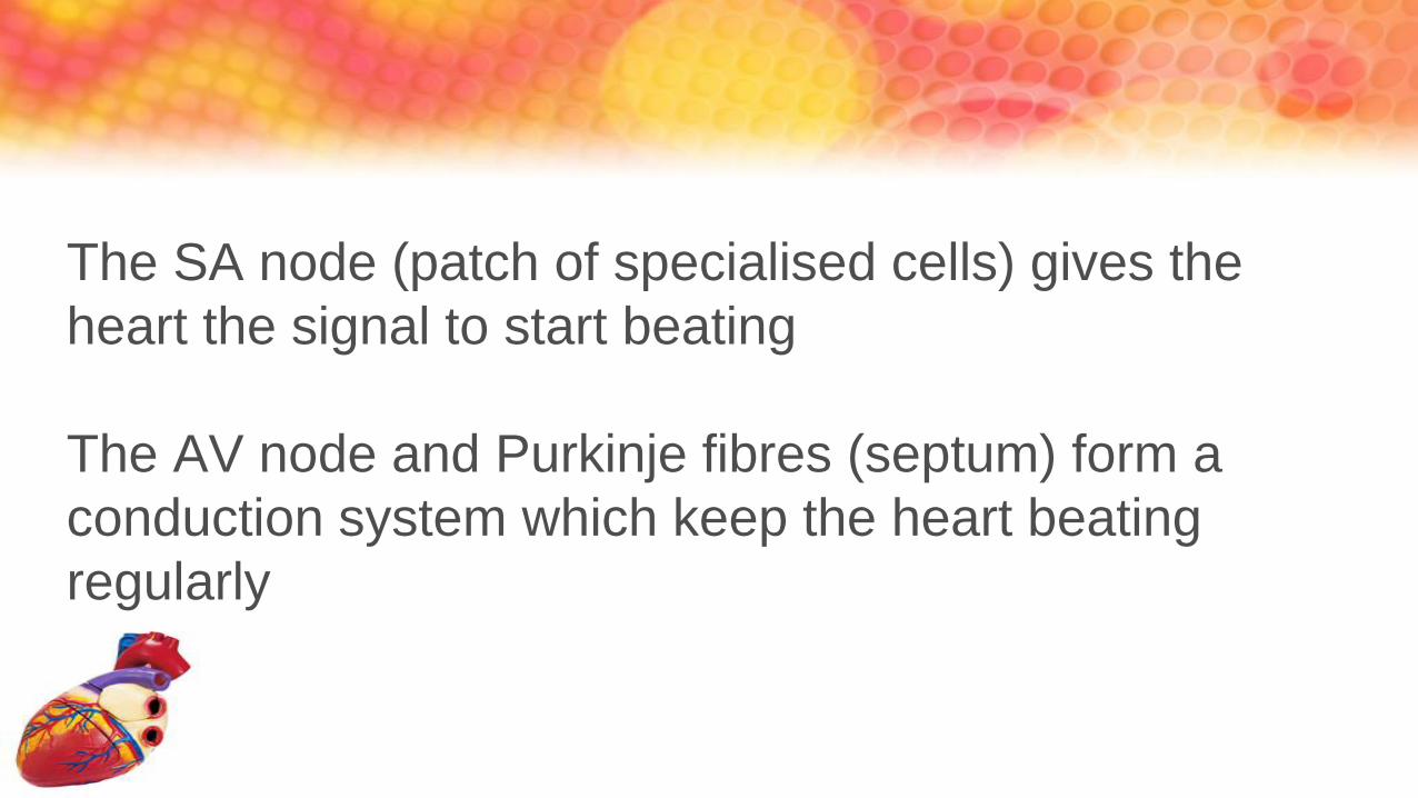
The SA node (patch of specialised cells) gives the
heart the signal to start beating
The AV node and Purkinje fibres (septum) form a
conduction system which keep the heart beating
regularly

• Electrical signal starts from SA node and spreads through the walls
of the atria
• Signal spreads to the AV node
• Once the atria relax the signal travels down the Purkinje fibres
• This causes the ventricles to contract from the bottom up, squeezing
the blood up into the arteries
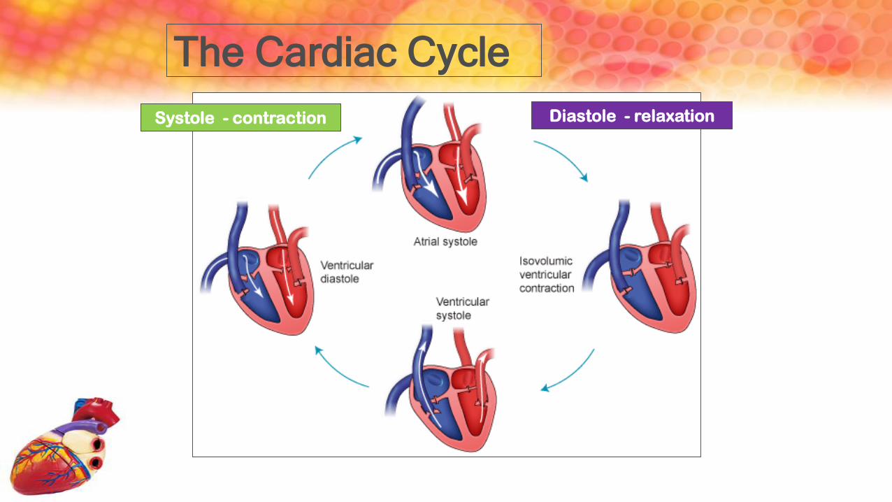
The Cardiac Cycle
Systole - contraction Diastole - relaxation
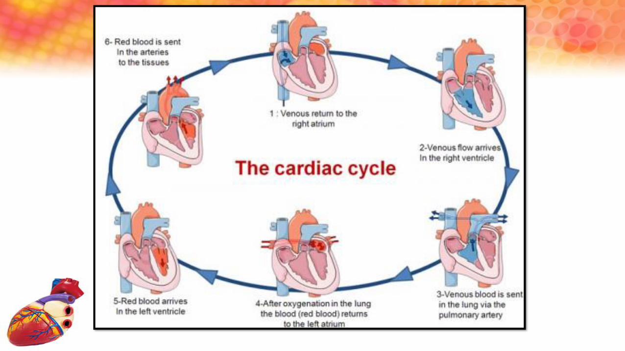

• The sino-atrial node (SA node)sends electrical impulses to the muscle fibres of both the left and right atria.
• The two atria contract at the same time.
• The tricuspid and bicuspid valves open.
• The blood flows into the two ventricles which are relaxed.
• The SA node acts as a pacemaker causing the heart to beat at a slower or faster rate depending on the body’s needs.

• The two ventricles contract at same
time.
• The blood is forced into the aorta and
pulmonary artery.
• The tricuspid and bicuspid valves close.
• This prevents blood from flowing back
into the atria.

• Both the atria and ventricles relax.
• The semi-lunar valves at the base of
the aorta and pulmonary artery close,
preventing blood from flowing back into
the ventricles from the aorta or
pulmonary artery.
• Blood enters the atria through the
superior and inferior vena cavae and
pulmonary veins.


PulseThe pulse is the
regular expansion and
contraction of an
artery
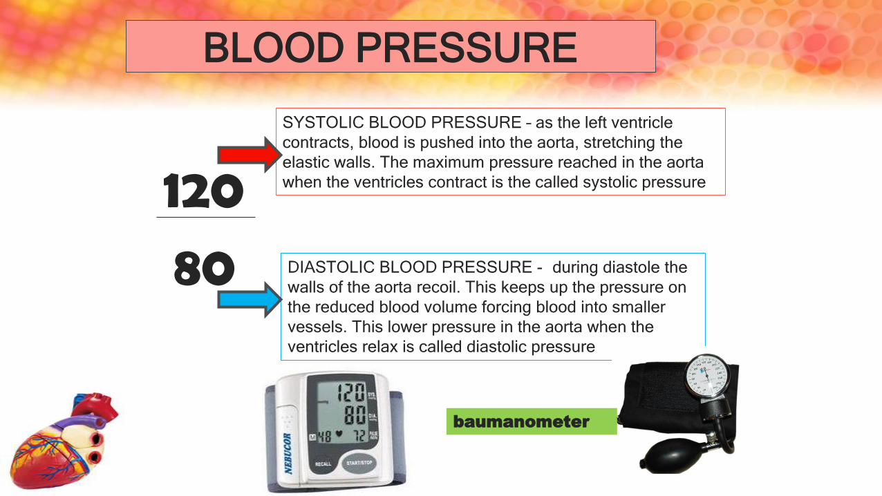
BLOOD PRESSURE
12080
SYSTOLIC BLOOD PRESSURE – as the left ventricle
contracts, blood is pushed into the aorta, stretching the
elastic walls. The maximum pressure reached in the aorta
when the ventricles contract is the called systolic pressure
DIASTOLIC BLOOD PRESSURE - during diastole the
walls of the aorta recoil. This keeps up the pressure on
the reduced blood volume forcing blood into smaller
vessels. This lower pressure in the aorta when the
ventricles relax is called diastolic pressure
baumanometer

BLOOD VESSELS

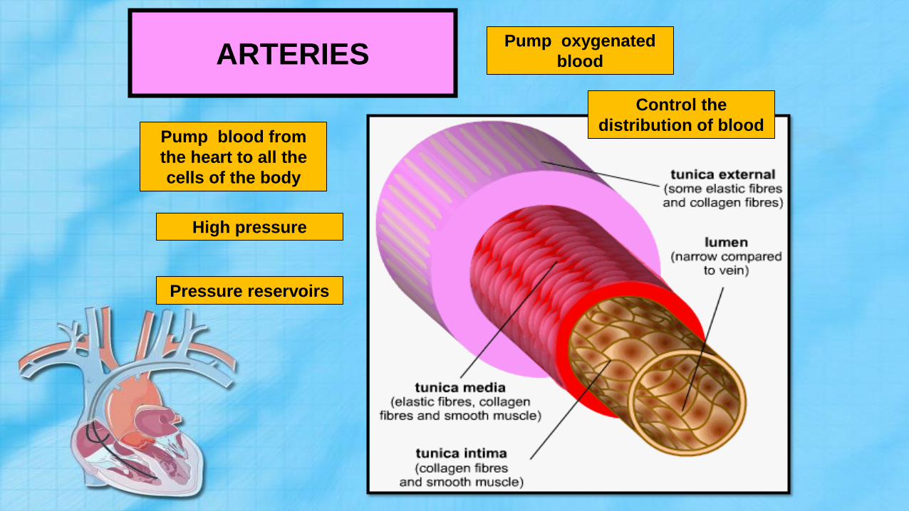
ARTERIES
Pump blood from
the heart to all the
cells of the body
Pump oxygenated
blood
High pressure
Pressure reservoirs
Control the
distribution of blood

VEINS
Pump blood from
the cells to the heart
Low pressure
Have valves to
prevent backflow
Pump deoxygenated
blood

Capillaries
Easy diffusion of
substances between
blood and cells
Allow phagocytes
(white blood cell) to
move in and out
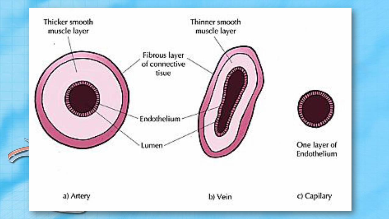
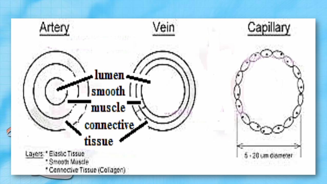


Artery Vein Capillary
Blood is under a high
pressure
Blood is not under a high
pressure
Blood is not under a high pressure
Does not have valves Does have semi-lunar valves Does not have valves
Has thick muscular walls Has thin muscular walls Has a single layer of cells in the
walls for easy diffusion
Has a smaller lumen then
veins
Has a large lumen Has a very narrow lumen
Carries blood away from the
heart
Carries blood to the heart Produces tissue fluid at the capillary
beds
Oxygenated blood except
pulmonary arteries
Deoxygenated blood except
pulmonary veins
Exchanges O2 & CO2 between
plasma & tissue fluid
Leads to capillaries Leads from capillaries Branches of arteries and join up to
form veins

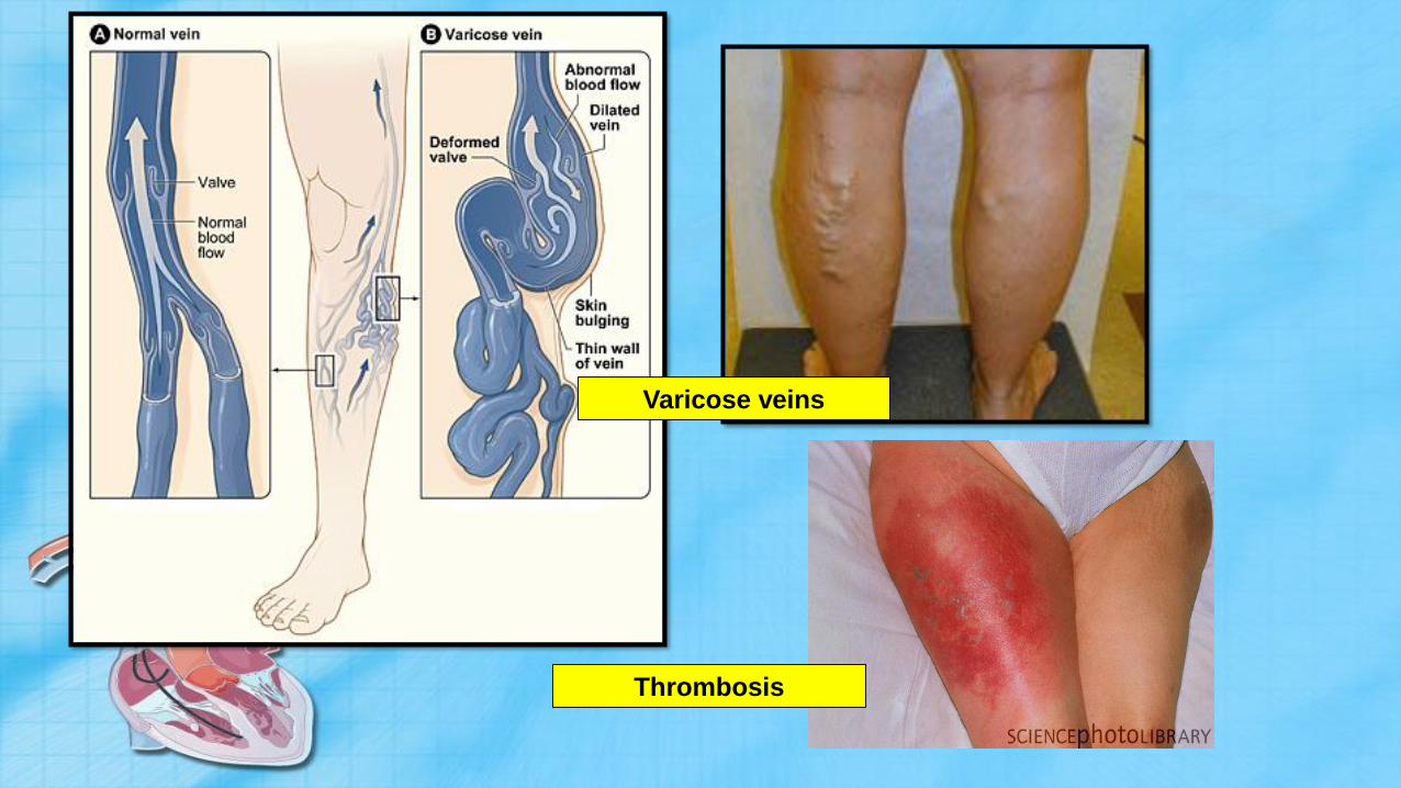
Varicose veins
Thrombosis

Effect of exercise on heart rate
• When you exercise, cellular respiration increases so more
CO2 is produced
• This lowers the pH of the blood
• Impulse is sent via sensory neurons to the medulla
oblongata
• Responds by telling the SA node to beat quicker.
• Why?
THE LYMPHATIC SYSTEM

The lymphatic system is a circulatory system that transports lymph
throughout the body. Unlike the blood circulatory system, the
lymphatic system has no pump. It is a drainage system.


Lymph Is the body fluid containing white blood cells, chief
lymphocytes, that is drained from the tissue spaces by the
lymphatic vessels


• Blood plasma and white blood cells leak out of the capillaries and return to the
blood circulation system
• In between body tissues are blood capillaries and lymphatic capillaries
• Blood plasma that leaks out of the capillaries and surrounds body tissues it is called
tissue fluid
• Tissue fluid that drains into the lymph vessels is called lymph.
• Lymph capillaries join to form lymph vessels
• Lymph vessels flow through lymph nodes
• Lymph nodes contain many white blood cells – immunity
• Lymph nodes: neck, armpits, groin
• Lymph is returned to the circulatory system via a vein in the neck
• Flow is uni-directional because of valves

Constituents of lymph
• Water
• Solutes – proteins, salts, glucose,
urea
• Fat
• White blood cells – lymphocytes and
macrophages
• Lymphocytes – B and T
• B – produce antibodies that enter
blood to fight germs
• T – variety of functions
• Macrophages – trap and digest
foreign matter and harmful
bacteria by phagocytosis
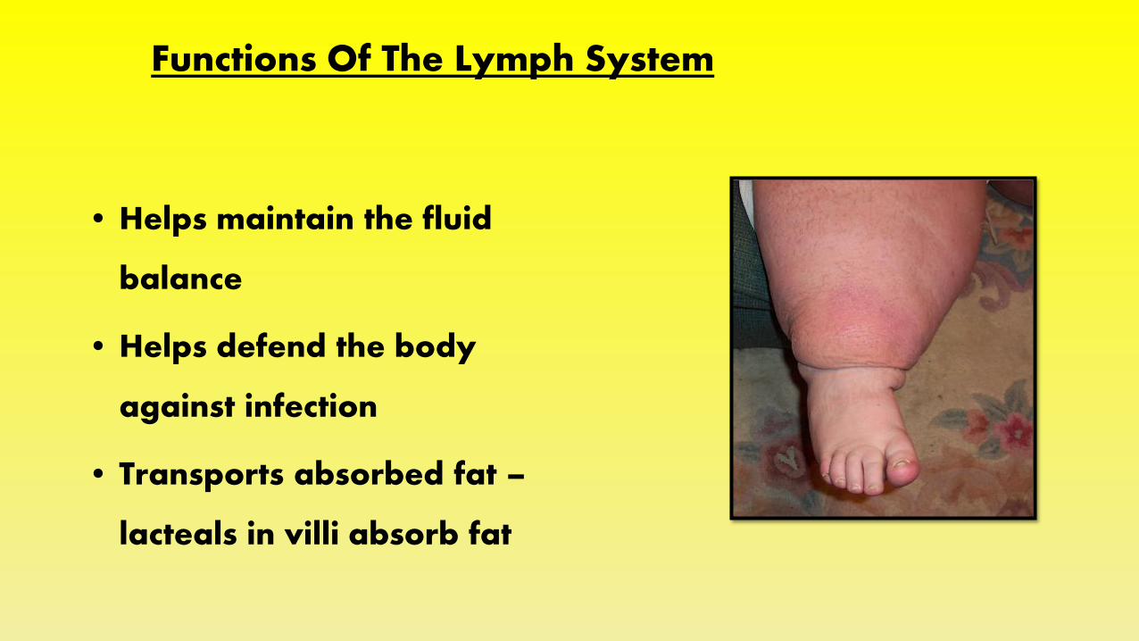
Functions Of The Lymph System
• Helps maintain the fluid
balance
• Helps defend the body
against infection
• Transports absorbed fat –
lacteals in villi absorb fat

CIRCULATORY DISEASES

Diseases and disorders
Diseases of the cardiovascular system can be serious and are a major cause of death, seeing as blood is the manner in which all your cells in the body receive useful substances and get rid of waste products.
Sometimes causes are genetic, but lifestyle is also a major contribution to diseases and disorders. These are smoking, drinking, lack of exercise, obesity etc.

Examples of diseases and disorders
• Anaemia
When a person has too few red blood cells due to a lack of iron. The person appears pale with no energy.
• Leukaemia
A type of blood cancer that causes uncontrolled division of the leucocytes (white blood cells).
• Blood pressure
Hypertension is high blood pressure and hypotension is low blood pressure. In high blood pressure, the heart has to work harder, which can lead to stroke, heart attack or kidney disease. In low blood pressure, a person feels dizzy and has fainting spells.

• Angina, arteriosclerosis
Angina is chest pain associated with too little oxygen being delivered to the heart muscles. This can be associated with arteriosclerosis which is a narrowing of the arteries due to arteriosclerotic plaque.
• Heart attack and strokes
In a heart attack, the coronary arteries, supplying oxygen to the heart, become blocked or they spasm. The heart no longer has enough oxygen to work and stops pumping.
In a stroke, a clot may form and block one of the arteries to the brain, starving it of oxygen, or a blood vessel may burst as a result of a blockage or hypertension and weakening of the artery walls.

Coronary artery disease
• Heart attack – coronary artery is
blocked or cut off – heart is starved of
oxygen and the tissue dies
• High risk factors:
– High blood pressure, smoker, obesity,
high blood cholesterol, diabetes, stress,
sedentary lifestyle, diet high in sugar
and fat and low in fruit and vegetables
– Sex- male, middle-age, high achiever
personality, genetic predisposition

Atherosclerosis
• Hardening of the
arteries, also called
atherosclerosis, is a
common disorder. It
occurs when fat,
cholesterol, and other substances build up
in the walls of arteries
and form hard
structures called
plaques.


Treatment • Coronary artery bypass graft
• Coronary angioplasty
• Atherslerosis treated with:
– Balloon angioplasty (with or without stent)
– Laser angioplasty (breaks up thrombus)
– Coronary by-pass
– Heart transplant
• Irregular heartbeat treated with:
– Pacemaker (regulates heartbeat if too slow or too fast)

• Valve replacements (repair or replace valves)
• Heart transplant (receive heart from recently
deceased person – commonly brain dead)

• Thrombosis in a brain artery – brain
cells get cut off form oxygenated
blood
• Symptoms
– paralysis on one side of the body
– Difficulties speaking/swallowing
– Problem seeing properly
– Unconsciousness
Stroke

• Sudden rupture of blood vessel in the
brain – sudden death, same
symptoms as above
• Rupture of blood vessel inside skull
but outside brain – pressure – severe
headache – brain cells could die