Pharmacology of Interferon. Interferon Natural Interferons Man Made Interferons (Recombinant)
Upload
trinhtuyenCategory
view
218download
3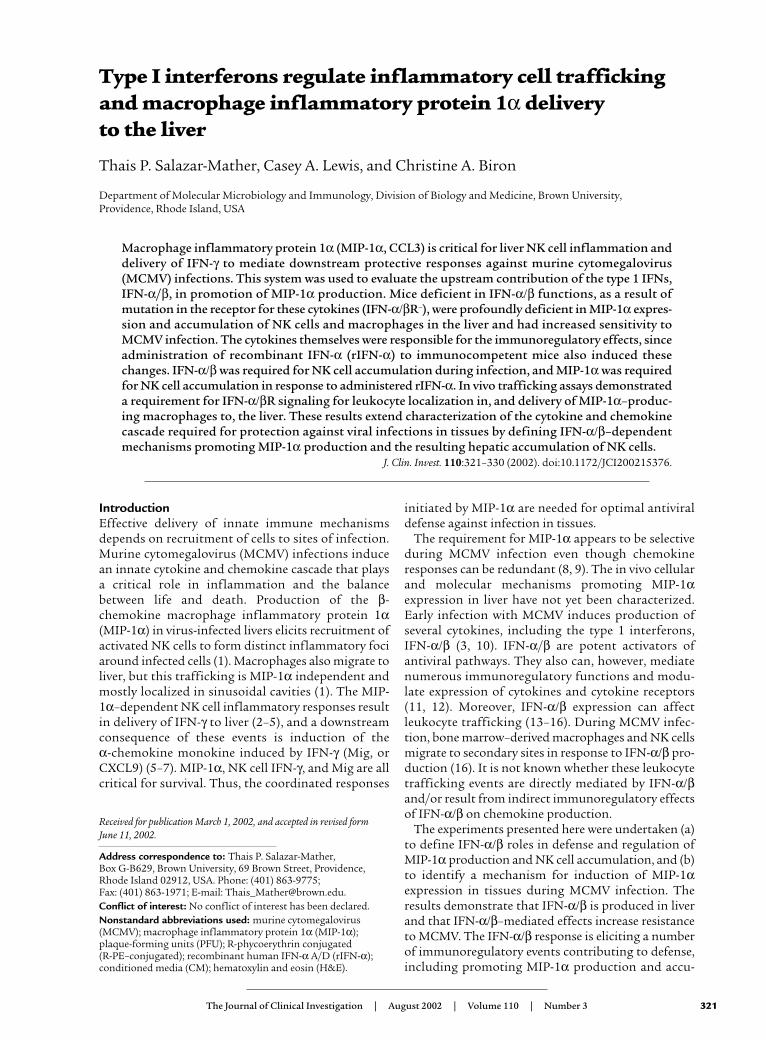
IntroductionEffective delivery of innate immune mechanismsdepends on recruitment of cells to sites of infection.Murine cytomegalovirus (MCMV) infections inducean innate cytokine and chemokine cascade that playsa critical role in inflammation and the balancebetween life and death. Production of the β-chemokine macrophage inflammatory protein 1α(MIP-1α) in virus-infected livers elicits recruitment ofactivated NK cells to form distinct inflammatory fociaround infected cells (1). Macrophages also migrate toliver, but this trafficking is MIP-1α independent andmostly localized in sinusoidal cavities (1). The MIP-1α–dependent NK cell inflammatory responses resultin delivery of IFN-γ to liver (2–5), and a downstreamconsequence of these events is induction of the α-chemokine monokine induced by IFN-γ (Mig, orCXCL9) (5–7). MIP-1α, NK cell IFN-γ, and Mig are allcritical for survival. Thus, the coordinated responses
initiated by MIP-1α are needed for optimal antiviraldefense against infection in tissues.
The requirement for MIP-1α appears to be selectiveduring MCMV infection even though chemokineresponses can be redundant (8, 9). The in vivo cellularand molecular mechanisms promoting MIP-1αexpression in liver have not yet been characterized.Early infection with MCMV induces production ofseveral cytokines, including the type 1 interferons,IFN-α/β (3, 10). IFN-α/β are potent activators ofantiviral pathways. They also can, however, mediatenumerous immunoregulatory functions and modu-late expression of cytokines and cytokine receptors(11, 12). Moreover, IFN-α/β expression can affectleukocyte trafficking (13–16). During MCMV infec-tion, bone marrow–derived macrophages and NK cellsmigrate to secondary sites in response to IFN-α/β pro-duction (16). It is not known whether these leukocytetrafficking events are directly mediated by IFN-α/βand/or result from indirect immunoregulatory effectsof IFN-α/β on chemokine production.
The experiments presented here were undertaken (a)to define IFN-α/β roles in defense and regulation ofMIP-1α production and NK cell accumulation, and (b)to identify a mechanism for induction of MIP-1αexpression in tissues during MCMV infection. Theresults demonstrate that IFN-α/β is produced in liverand that IFN-α/β–mediated effects increase resistanceto MCMV. The IFN-α/β response is eliciting a numberof immunoregulatory events contributing to defense,including promoting MIP-1α production and accu-
The Journal of Clinical Investigation | August 2002 | Volume 110 | Number 3 321
Type I interferons regulate inflammatory cell traffickingand macrophage inflammatory protein 1α delivery to the liver
Thais P. Salazar-Mather, Casey A. Lewis, and Christine A. Biron
Department of Molecular Microbiology and Immunology, Division of Biology and Medicine, Brown University, Providence, Rhode Island, USA
Macrophage inflammatory protein 1α (MIP-1α, CCL3) is critical for liver NK cell inflammation anddelivery of IFN-γ to mediate downstream protective responses against murine cytomegalovirus(MCMV) infections. This system was used to evaluate the upstream contribution of the type 1 IFNs,IFN-α/β, in promotion of MIP-1α production. Mice deficient in IFN-α/β functions, as a result ofmutation in the receptor for these cytokines (IFN-α/βR–), were profoundly deficient in MIP-1α expres-sion and accumulation of NK cells and macrophages in the liver and had increased sensitivity toMCMV infection. The cytokines themselves were responsible for the immunoregulatory effects, sinceadministration of recombinant IFN-α (rIFN-α) to immunocompetent mice also induced thesechanges. IFN-α/β was required for NK cell accumulation during infection, and MIP-1α was requiredfor NK cell accumulation in response to administered rIFN-α. In vivo trafficking assays demonstrateda requirement for IFN-α/βR signaling for leukocyte localization in, and delivery of MIP-1α–produc-ing macrophages to, the liver. These results extend characterization of the cytokine and chemokinecascade required for protection against viral infections in tissues by defining IFN-α/β–dependentmechanisms promoting MIP-1α production and the resulting hepatic accumulation of NK cells.
J. Clin. Invest. 110:321–330 (2002). doi:10.1172/JCI200215376.
Received for publication March 1, 2002, and accepted in revised form June 11, 2002.
Address correspondence to: Thais P. Salazar-Mather, Box G-B629, Brown University, 69 Brown Street, Providence,Rhode Island 02912, USA. Phone: (401) 863-9775; Fax: (401) 863-1971; E-mail: [email protected] of interest: No conflict of interest has been declared.Nonstandard abbreviations used: murine cytomegalovirus(MCMV); macrophage inflammatory protein 1α (MIP-1α);plaque-forming units (PFU); R-phycoerythrin conjugated (R-PE–conjugated); recombinant human IFN-α A/D (rIFN-α);conditioned media (CM); hematoxylin and eosin (H&E).
mulation of NK cells and macrophages in liver. TheIFN-α/β effects on NK cell accumulation are MIP-1αdependent, and on MIP-1α expression are dependenton recruiting MIP-1α–producing macrophages intoliver. Taken together, these results provide new insightsinto IFN-α/β functions and demonstrate a uniquepathway in regulation of chemokine responsesrequired for localizing innate antiviral defenses.
MethodsMice. Specific pathogen–free C57BL/6 and 129/Sv micewere purchased from The Jackson Laboratory (Bar Har-bor, Maine, USA) or Taconic Laboratory Animals andServices (Germantown, New York, USA), respectively.Breeding pairs of homozygous MIP-1α mutants,C57BL/6-MIP-1α– (17), from The Jackson Laboratorywere used to establish colonies at Brown University.Mice functionally deficient in IFN-α/β as a result ofmutation of the receptor, 129-IFN-α/βR– (18), wereoriginally obtained from B&K Universal Ltd. (NorthHumberside, United Kingdom) but also bred andmaintained at Brown University. Male and female micewere used between 5 and 6 weeks of age. Mouse han-dling and experimental procedures were conducted inaccordance with institutional guidelines.
Virus, infections, and in vivo treatment protocols. Stocksof Smith-strain MCMV salivary gland extracts wereprepared (2). Infections were initiated on day 0 (unin-fected) with 5 × 10⁴ (moderate dose) plaque-formingunits (PFU) of MCMV, or when indicated with 1 × 10⁴
(low dose) PFU of MCMV, via the intraperitonealroute. In survival experiments, mice were assessedtwice daily. Animals treated in vivo with recombinanthuman IFN-α A/D (rIFN-α) (Pestka Biomedical Labo-ratories, New Brunswick, New Jersey, USA), biologi-cally active in mice, were given three intraperitonealdaily injections of 1 × 10⁵ units in PBS containing 0.1%BSA. Control animals were given vehicle injections.
Preparation of leukocytes, tissue homogenates, and condi-tioned media. Liver leukocytes and homogenates wereisolated and prepared as previously described (5, 19).For generation of leukocyte–conditioned media (leuko-cyte-CM), cells were suspended in RPMI media (LifeTechnologies Inc., Rockville, Maryland, USA) contain-ing 5% FCS, and incubated at 37°C for 24 hours. Cell-free supernatants were collected and used in cytokineanalyses. Production levels are expressed as ng/g liverin homogenates or as pg/million cells in CM.
Flow cytometric analyses. Proportions and numbers ofcells were evaluated as reported (5). For NK cell identi-fication in C57BL/6 mice, the following antibodieswere used: R-phycoerythrin conjugated (R-PE–conju-gated) anti-NK1.1 mAb clone PK136 and CyChrome-conjugated anti–TCR-β chain mAb clone H57-597 (BDPharMingen, San Diego, California, USA). As mice ona 129 background do not express NK1.1, NK cells werealso identified with the following antibodies: biotin-conjugated anti–pan-NK mAb clone DX5 followed bystreptavidin-allophycocyanin (APC), CyC-conjugated
anti–TCR-β chain mAb clone H57-597, and R-PE–con-jugated anti-CD122 (IL-2 and IL-15 β chain receptor)mAb clone TMβ1 (BD PharMingen). In the studies pre-sented, cells identified as DX5+ (20) were 74–90% posi-tive for CD122. As NK cells constitutively expressCD122, this marker helped to identify the DX5+TCR-β–
NK cells. To distinguish migrating from resident mac-rophages (21), the following antibodies were used: R-PE–conjugated anti–F4/80 (Serotec Ltd., Kidlington,Oxford, United Kingdom) and allophycocyanin-conju-gated anti-CD11b mAb clone M1/70 (BD PharMin-gen), or biotin-conjugated anti–F4/80 (Serotec) fol-lowed by streptavidin-allophycocyanin and FITC-conjugated anti-CD11b mAb clone M1/70 (BDPharMingen). The latter antibodies were used to iden-tify macrophages labeled with red fluorescent PKH26(Sigma-Aldrich, St. Louis, Missouri, USA). Controlantibodies not recognizing specific murine determi-nants (BD PharMingen) were used to correct for back-ground fluorescence and set analyses gates. Totals of atleast 20,000–100,000 events per sample were acquiredand analyzed using a FACSCalibur (BD Biosciences,Palo Alto, California, USA) and the CellQuest (BD Bio-sciences) version 3.1 software package (5) or FCSPress(Cambridge, United Kingdom).
Enrichment of macrophage populations using magnetic beads.Liver leukocytes were pooled, suspended at ≤1 × 10⁷
cells/ml, and blocked with 2.4G2 antibody. To specifi-cally enrich for migrating macrophages (21), cells werestained with FITC-labeled CD11b (BD PharMingen)and PE-F4/80 (Serotec). The CD11b+ cells were enrichedusing MultiSort anti-FITC microbeads and positiveselection columns followed by MultiSort ReleaseReagent as described by the manufacturer (MiltenyiBiotec GmbH, Bergisch Gladbach, Germany). After sep-aration, positive selection for PE-F4/80+ cells was per-formed using anti-PE magnetic beads and columns(Miltenyi Biotec GmbH). F4/80+CD11b+ cells wereenriched by more than 90%.
Cytokine analyses. Samples of liver homogenates andliver leukocyte-CM were tested for MIP-1α or IFN-α byELISA. MIP-1α was detected using a commercial sand-wich ELISA kit as recommended by the manufacturer(R&D Systems Inc., Minneapolis, Minnesota, USA).IFN-α was detected using a rat anti–mouse IFN-α mAb(F-18; HyCult biotechnology b.v., Uden, The Nether-lands) as described (22).
Histology and immunohistochemistry. Liver sectionswere prepared, stained with hematoxylin and eosin(H&E), and analyzed microscopically (1). Forimmunohistochemical analyses, sections wereblocked, and IFN-α/β protein was detected asdescribed (22). Images were collected digitally with aRoper Scientific Photometrics CoolSNAP CameraSystem (Roper Scientific Inc., Trenton, New Jersey,USA) and processed using Adobe Photoshop (AdobeSystems Inc., Mountain View, California, USA).
In vivo trafficking assays. Bone marrow leukocytes wereisolated and labeled with the red fluorescent dye
322 The Journal of Clinical Investigation | August 2002 | Volume 110 | Number 3
PKH26 (Sigma-Aldrich) (1, 16). Labeled cells were deliv-ered intravenously and localized in livers of recipientmice 24 hours after transfer. Liver sections were pre-pared as described (1), mounted with VECTASHIELD(Vector Laboratories Inc., Burlingame, California, USA)and observed using a Microphot-SA microscope(Nikon, Tokyo, Japan) (1, 16). Cells in liver sinusoidswere quantitated by counting fluorescent cells indefined areas of representative liver tissues at ×100magnification. Images were collected digitally with aSpot RT slider camera from Diagnostic InstrumentsInc. (Sterling Heights, Michigan, USA) and processedusing Adobe Photoshop.
Statistical analyses. Statistical significance of experi-mental results was analyzed by two-tailed Student’s t test where indicated.
ResultsLiver NK cell accumulation during MCMV infection. NKcells accumulate and localize in liver inflammatory fociduring MCMV infections of C57BL/6 mice (1, 23). Tocharacterize NK cell accumulation in another strain ofmice, H&E-stained liver sections were prepared fromC57BL/6 and 129 mice that were uninfected or infect-ed with MCMV for 48 hours. The focal clustering ofnucleated cells between portal areas and central veinstypical of NK cell inflammation was observed follow-ing infections in both strains (Figure 1, a and b). Toquantitate the NK cell yields (NK1.1+TCR-β– orDX5+TCR-β–) in this compartment, leukocytes wereprepared, and NK cell proportions and numbers weredetermined using flow cytometric and total cell recov-ery. The liver leukocyte yields increased following infec-tion to 4 × 10⁶ ± 3 × 10⁵ from the uninfected values of2 × 106 ± 2 × 10⁵ in C57BL/6 mice, and to 3 × 10⁶ ± 4 ×10⁵ from the uninfected values of 1 × 10⁶ ± 1 × 10⁵ in129 mice. Although liver NK cell percentages and yieldswere higher in C57BL/6 mice, both C57BL/6 and 129
mice demonstrated twofold increases in frequencies(Figure 1, c and e) and fourfold increases in absolutenumbers (Figure 1, d and f). Thus, both strains exhibitsimilar liver histological characteristics and inducedaccumulation of NK cells during infection.
Induction of IFN-α/β protein in liver. As IFN-α/β canhave profound effects on cell trafficking (13–16) andon cytokines and chemokines (11, 12), liver IFN-α/βexpression was examined by immunohistochemistry.Tissue sections were prepared from uninfected orMCMV-infected C57BL/6 (Figure 2, a–c) or 129 (Fig-ure 2, d–f) mice. IFN-α/β was not detected in unin-fected mice (Figure 2, a and d). However, at 36 and 48hours after challenge, production was dramaticallyinduced in both strains. Positive cells were detected in sinusoids and as scattered populations withinparenchyma (Figure 2, b, c, e, and f). To quantitate type 1 IFN proteins, ELISA assays measuring IFN-αwere performed on liver homogenates. At 36 hours,C57BL/6 mice had IFN-α values of 66 ± 3 and 129mice had values of 67 ± 17 ng/g liver. At 48 hours afterinfection, C57BL/6 mice had 44 ± 12 and 129 mice had64 ± 5 ng/g liver. IFN-α was below the limit of detec-tion (<7 ng/g liver) in uninfected mice. Taken togeth-er, these results demonstrate that both strains produceIFN-α/β in liver during MCMV infection.
IFN-α/β deficiency and resistance to MCMV infection. Todetermine the effects of IFN-α/β responses for antiviraldefense, H&E-stained liver sections were prepared frommice deficient in IFN-α/β–mediated functions (IFN-α/βR–) and infected with MCMV for 48 or 72 hours. Incontrast to immunocompetent (IFN-α/βR+) mice (Fig-ure 1b), there was a profound inhibition of inflamma-tory foci in liver from IFN-α/βR– mice at both 48 (Figure3a) and 72 (Figure 3b) hours after infection. Cells havingthe morphological characteristics of MCMV infection,i.e., cytomegalic inclusion bodies, were frequently seen at48 hours and were readily visible, along with necrotic
The Journal of Clinical Investigation | August 2002 | Volume 110 | Number 3 323
Figure 1NK cell accumulation in MCMV-infectedC57BL/6 and 129 livers. Livers were harvestedand H&E-stained tissue sections were preparedfrom C57BL/6 (a) or 129 (b) mice infectedwith MCMV for 48 hours as described in Meth-ods. Arrows in a and b denote inflammatoryfoci. Images were digitally captured at the orig-inal magnifications of ×10. Scale bar = 100µm. (c–f) Liver leukocytes were prepared fromC57BL/6 (c and d) or 129 (e and f) mice thatwere uninfected (0 hours) or infected withMCMV for 48 hours. Leukocytes were analyzedby flow cytometry as described in Methods.Both the percentage (c and e) and number (dand f) of NK1.1+TCR-β– or DX5+TCR-β– cellsper g liver are shown. Data are the means ± SE(n = 3–6). Differences between uninfected andinfected mice are significant at *P ≤ 0.03 and**P < 0.001.

foci, at 72 hours after infection (Figure 3, a and b). Thus,virus-induced liver pathology was elevated in the absenceof IFN-α/β–mediated functions. To further examine theimportance of IFN-α/β for antiviral defense, IFN-α/βR+
and IFN-α/βR– mice were infected with a lower dose (1 × 10⁴ PFU) of MCMV and monitored for survival. Allof the IFN-α/βR–, but none of the IFN-α/βR+, mice suc-cumbed to infection by day 5 (Figure 3c). Therefore,under both moderate- and low-dose conditions of infec-tion the absence of IFN-α/β–mediated functions pro-foundly increases susceptibility to MCMV.
IFN-α/β effects on NK cell infiltrates and MIP-1α induc-tion in response to infection. NK cell inflammation isimportant in promoting defense against MCMV in liv-
ers, and our previous studies have shown the criticalrole of MIP-1α for this response (1, 5). To evaluate theeffects of IFN-α/β on NK cell accumulation, liverleukocytes were prepared and analyzed from IFN-α/βR+ and IFN-α/βR– mice that were uninfected orinfected with MCMV for 48 hours. Although liver NKcell frequencies were equivalent in both IFN-α/βR+ andIFN-α/βR– mice (data not shown), significant differ-ences in NK cell numbers were evident under bothmoderate-dose (Figure 4a) and low-dose (Figure 4c)conditions of infection. As total liver leukocyte yieldsincreased, from the uninfected values of 1 to 2 × 10⁶
per g liver to 3.5 × 10⁶ per g liver after moderate-doseand to 4.5 × 10⁶ per g liver after low-dose MCMV infec-
324 The Journal of Clinical Investigation | August 2002 | Volume 110 | Number 3
Figure 2Induction of IFN-α/β protein expression in MCMV-infected livers. Organs were harvested from C57BL/6 and 129 mice that were uninfect-ed (0 hours) or infected with MCMV for 36 or 48 hours. Tissue sections were prepared, immunostained, and counterstained with methylgreen as described in Methods. Results shown are from uninfected C57BL/6 (a) and 129 (d) mice, C57BL/6 mice after 36 (b) or 48 (c) hours’infection, and 129 mice after 36 (e) or 48 (f) hours of infection. Insets represent positive cells at a higher magnification. Images were digi-tally captured at the original magnifications of ×10 and ×40. Scale bars = 100 µm.
Figure 3Effects of IFN-α/β on susceptibility to MCMV. Livers were harvested and H&E-stained sections were prepared from 129-IFN-α/βR– miceinfected with 5 × 10⁴ PFU MCMV for 48 (a) or 72 (b) hours. Arrows denote a liver area with cytomegalic inclusion bodies, and the arrow-head in b represents an area of tissue necrosis. The inset shows cytomegalic inclusion bodies at a higher magnification. Images were digi-tally captured at the original magnifications of ×10 and ×40. Scale bars = 100 µm. (c) The 129-IFN-α/βR+ and 129-IFN-α/βR– mice wereuninfected or infected with 1 × 10⁴ PFU MCMV and monitored twice daily for survival (n = 6).

tion, IFN-α/βR+ mice had twofold elevations in NK cellnumbers induced by challenge (Figure 4, a and c). Incontrast, liver leukocyte yields from IFN-α/βR– micewere only 1 × 10⁶ per g liver after moderate-dose and 9 × 10⁵ per g liver after low-dose infection, comparedwith the uninfected values of 2 × 10⁶ per g liver. As aresult, there were twofold reductions in liver NK cellnumbers at 48 hours MCMV infection of IFN-α/βR–
mice (Figure 4, a and c). Although there were decreas-es in NK cell numbers in other compartments duringinfections of the IFN-α/βR– mice at the moderate dose,these were not observed during infections at the lowerdose (data not shown). Therefore, the reductions inNK cells infiltrating the liver were not a consequenceof generalized NK cell deficiencies. Hence, IFN-α/βfunctions can promote accumulation of NK cells inthe liver during infection.
To determine whether IFN-α/β modified inductionof MIP-1α, ELISA assays were performed with liverhomogenates prepared from IFN-α/βR+ or IFN-α/βR–
mice that were uninfected or infected with MCMV for48 hours. Under the conditions of both moderate-dose(Figure 4b) and low-dose (Figure 4d) infection, MIP-1αproduction was induced in livers of both groups ofmice. The responses observed in IFN-α/βR–, however,were dramatically reduced compared with those ininfected IFN-α/βR+ mice (Figure 4, b and d). By com-parison to IFN-α/βR+, IFN-α/βR– mice had three- tofourfold reductions in the levels of liver MIP-1α proteinunder both conditions of moderate- and low-doseinfections. Thus, IFN-α/β is necessary for initiation ofMIP-1α expression in liver during MCMV infection.
Induction of MIP-1α and accumulation of NK cells fol-lowing rIFN-α administration. As demonstrated above,IFN-α/β–mediated functions are necessary to effec-tively control virus (Figure 3) as well as to promoteMIP-1α induction and NK cell accumulation duringinfection (Figure 4). To evaluate the immunoregula-tory effects of IFN-α/β in the absence of secondaryeffects resulting from increased virus-induced disease,the consequences of treating uninfected mice withrIFN-α were examined. MIP-1α protein was measuredin liver homogenates prepared from vehicle- or rIFN-α–treated C57BL/6 and 129 mice. The levels of MIP-1α were low (0.08 ng/g liver) in vehicle-treatedC57BL/6 and 129 mice (Figure 5, a and c). In contrast,MIP-1α protein was dramatically induced to values of0.5 ng/g liver in C57BL/6 and 0.2 ng/g liver in 129mice after rIFN-α administration (Figure 5, a and c).Thus, rIFN-α exposure result in greater than two- tosixfold inductions of MIP-1α in livers.
To examine the contribution of rIFN-α to NK cellaccumulation, liver leukocytes were prepared and ana-lyzed. Mice receiving rIFN-α had two- to threefoldincreases in the proportions of NK cells when comparedwith mice receiving vehicle treatments (data not shown).In both C57BL/6 and 129 mice, the NK cell numbersincreased from the vehicle-treated values of 7 × 10⁴ perg liver to rIFN-α–treated values of 2 × 10⁵ per g liver
(Figure 5, b and d). Therefore, rIFN-α elicited a three-fold amplification of NK cell yields in liver. Takentogether, these results show that rIFN-α treatment pro-motes both MIP-1α production and accumulation ofliver NK cells even in the absence of virus infection.
Requirement for MIP-1α in the rIFN-α induction of NK cellaccumulation. To demonstrate the role of rIFN-α induc-tion of MIP-1α in NK cell liver accumulation, theresponse was evaluated in MIP-1α+ and MIP-1α– micetreated with vehicle or rIFN-α. Compared with vehicle-treated mice, MIP-1α+ mice demonstrated twofoldincreases in total cell yields (3 × 10⁶ to 7 × 10⁶ per g liver)and proportions of NK cells (11% to 19%) after rIFN-αtreatment. In contrast, these parameters were not signif-icantly affected after rIFN-α treatment of MIP-1α– mice.Consequently, NK cell numbers were profoundly elevat-ed in MIP-1α+ mice from 3 × 10⁵ per g liver after vehicletreatment to 1 × 10⁶ per g liver after rIFN-α administra-
The Journal of Clinical Investigation | August 2002 | Volume 110 | Number 3 325
Figure 4Effects of IFN-α/β functions on MIP-1α production and NK cell accu-mulation during MCMV infection. Samples were prepared from 129-IFN-α/βR+ (black bars) and 129-IFN-α/βR– (gray bars) mice that wereuninfected or infected with 5 × 10⁴ PFU (moderate dose) (a and b) or1 × 104 PFU (low dose) (c and d) MCMV for 48 hours. Liver leukocyteswere harvested and analyzed by flow cytometry as described in Meth-ods. Numbers of DX5+TCR-β– NK cells per g liver are shown (a and c).Data represent the means ± SE (n = 3–6). Liver homogenates were pre-pared from the IFN-α/βR+ or IFN-α/βR– mice that were uninfected orinfected with moderate-dose (b) or low-dose (d) MCMV for 48 hours.MIP-1α protein was measured by ELISA. The levels of detection were0.06–0.08 ng/g liver. Means ± SE are shown (n = 3–6 mice tested indi-vidually). Differences between control IFN-α/βR+ and IFN-α/βR– aresignificant at *P ≤ 0.03, **P ≤ 0.01, and ***P < 0.0001.
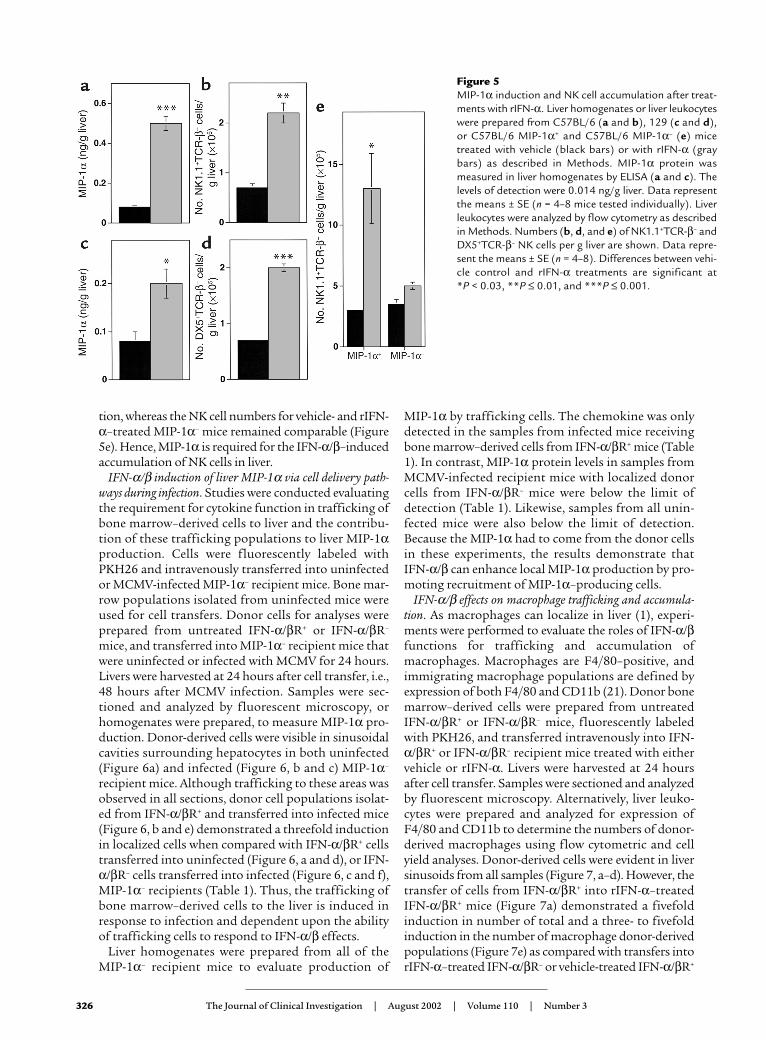
tion, whereas the NK cell numbers for vehicle- and rIFN-α–treated MIP-1α– mice remained comparable (Figure5e). Hence, MIP-1α is required for the IFN-α/β–inducedaccumulation of NK cells in liver.
IFN-α/β induction of liver MIP-1α via cell delivery path-ways during infection. Studies were conducted evaluatingthe requirement for cytokine function in trafficking ofbone marrow–derived cells to liver and the contribu-tion of these trafficking populations to liver MIP-1αproduction. Cells were fluorescently labeled withPKH26 and intravenously transferred into uninfectedor MCMV-infected MIP-1α– recipient mice. Bone mar-row populations isolated from uninfected mice wereused for cell transfers. Donor cells for analyses wereprepared from untreated IFN-α/βR+ or IFN-α/βR–
mice, and transferred into MIP-1α– recipient mice thatwere uninfected or infected with MCMV for 24 hours.Livers were harvested at 24 hours after cell transfer, i.e.,48 hours after MCMV infection. Samples were sec-tioned and analyzed by fluorescent microscopy, orhomogenates were prepared, to measure MIP-1α pro-duction. Donor-derived cells were visible in sinusoidalcavities surrounding hepatocytes in both uninfected(Figure 6a) and infected (Figure 6, b and c) MIP-1α–
recipient mice. Although trafficking to these areas wasobserved in all sections, donor cell populations isolat-ed from IFN-α/βR+ and transferred into infected mice(Figure 6, b and e) demonstrated a threefold inductionin localized cells when compared with IFN-α/βR+ cellstransferred into uninfected (Figure 6, a and d), or IFN-α/βR– cells transferred into infected (Figure 6, c and f),MIP-1α– recipients (Table 1). Thus, the trafficking ofbone marrow–derived cells to the liver is induced inresponse to infection and dependent upon the abilityof trafficking cells to respond to IFN-α/β effects.
Liver homogenates were prepared from all of the MIP-1α– recipient mice to evaluate production of
MIP-1α by trafficking cells. The chemokine was onlydetected in the samples from infected mice receivingbone marrow–derived cells from IFN-α/βR+ mice (Table1). In contrast, MIP-1α protein levels in samples fromMCMV-infected recipient mice with localized donorcells from IFN-α/βR– mice were below the limit ofdetection (Table 1). Likewise, samples from all unin-fected mice were also below the limit of detection.Because the MIP-1α had to come from the donor cellsin these experiments, the results demonstrate that IFN-α/β can enhance local MIP-1α production by pro-moting recruitment of MIP-1α–producing cells.
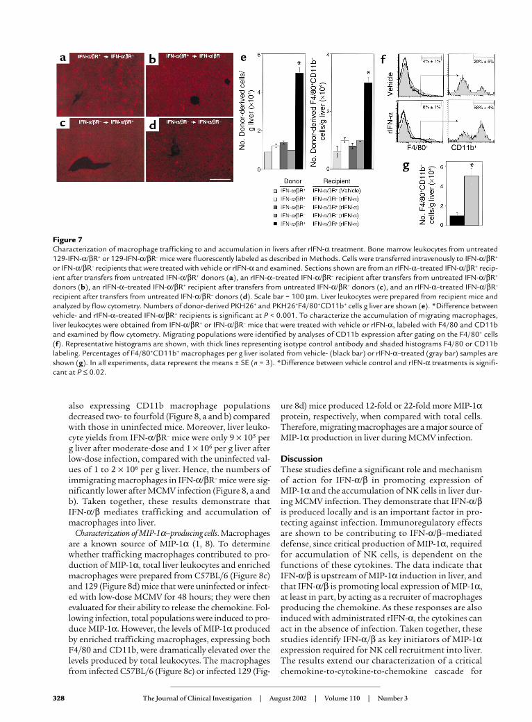
IFN-α/β effects on macrophage trafficking and accumula-tion. As macrophages can localize in liver (1), experi-ments were performed to evaluate the roles of IFN-α/βfunctions for trafficking and accumulation ofmacrophages. Macrophages are F4/80–positive, andimmigrating macrophage populations are defined byexpression of both F4/80 and CD11b (21). Donor bonemarrow–derived cells were prepared from untreatedIFN-α/βR+ or IFN-α/βR– mice, fluorescently labeledwith PKH26, and transferred intravenously into IFN-α/βR+ or IFN-α/βR– recipient mice treated with eithervehicle or rIFN-α. Livers were harvested at 24 hoursafter cell transfer. Samples were sectioned and analyzedby fluorescent microscopy. Alternatively, liver leuko-cytes were prepared and analyzed for expression ofF4/80 and CD11b to determine the numbers of donor-derived macrophages using flow cytometric and cellyield analyses. Donor-derived cells were evident in liversinusoids from all samples (Figure 7, a–d). However, thetransfer of cells from IFN-α/βR+ into rIFN-α–treatedIFN-α/βR+ mice (Figure 7a) demonstrated a fivefoldinduction in number of total and a three- to fivefoldinduction in the number of macrophage donor-derivedpopulations (Figure 7e) as compared with transfers intorIFN-α–treated IFN-α/βR– or vehicle-treated IFN-α/βR+
326 The Journal of Clinical Investigation | August 2002 | Volume 110 | Number 3
Figure 5MIP-1α induction and NK cell accumulation after treat-ments with rIFN-α. Liver homogenates or liver leukocyteswere prepared from C57BL/6 (a and b), 129 (c and d),or C57BL/6 MIP-1α+ and C57BL/6 MIP-1α– (e) micetreated with vehicle (black bars) or with rIFN-α (graybars) as described in Methods. MIP-1α protein wasmeasured in liver homogenates by ELISA (a and c). Thelevels of detection were 0.014 ng/g liver. Data representthe means ± SE (n = 4–8 mice tested individually). Liverleukocytes were analyzed by flow cytometry as describedin Methods. Numbers (b, d, and e) of NK1.1+TCR-β– andDX5+TCR-β– NK cells per g liver are shown. Data repre-sent the means ± SE (n = 4–8). Differences between vehi-cle control and rIFN-α treatments are significant at *P < 0.03, **P ≤ 0.01, and ***P ≤ 0.001.
mice (Figure 7, b and e). In contrast, donor cells isolat-ed from IFN-α/βR– and transferred into rIFN-α–treat-ed IFN-α/βR+ (Figure 7c) or IFN-α/βR– mice (Figure 7d)were limited in their ability to migrate into liver. Thenumber of total and macrophage donor-derived cellsdid not change significantly when compared with thoseisolated from IFN-α/βR+ and transferred into vehicle-treated IFN-α/βR+ (Figure 7e) mice. Thus, the resultsdemonstrate that IFN-α/β–mediated effects on bothdonor and recipient populations promote the migra-tion of macrophages into livers.
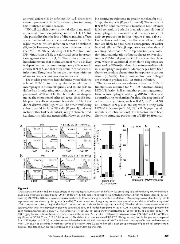
To determine the effects of IFN-α/β on overallmacrophage accumulation, the proportions and num-bers of F4/80+CD11b+ cells were determined in liverleukocyte populations after either rIFN-α treatment orMCMV infection. Following administration of rIFN-α,the frequency of F4/80+ cells, and that of F4/80+ cellsalso expressing CD11b, were elevated twofold after
rIFN-α as compared with those in mice receiving vehi-cle treatments (Figure 7f). The immigrating macro-phage cell numbers increased from the vehicle-treatedvalues of 1 × 10⁴ to 5 × 10⁴ per g liver (Figure 7g). Underthe conditions of either moderate-dose (Figure 8a) orlow-dose (Figure 8b) MCMV infections, the frequenciesof F4/80+ cells increased approximately twofold andthe frequencies of those also expressing CD11bincreased to 83–87% from the uninfected values of53–65% in IFN-α/βR+ mice. As the infection-inducedtotal liver leukocyte numbers increased to 2 × 10⁶ per gliver after moderate dose and 3 × 10⁶ per g liver afterlow dose from the uninfected values of 1 × 10⁶ per gliver, IFN-α/βR+ mice had more-than-fivefold eleva-tions in immigrating macrophage cell numbers at 48hours after infection (Figure 8, a and b). In contrast,although the frequencies of F4/80+ cells did increase ininfected IFN-α/βR– mice, the frequencies of F4/80+ cells
The Journal of Clinical Investigation | August 2002 | Volume 110 | Number 3 327
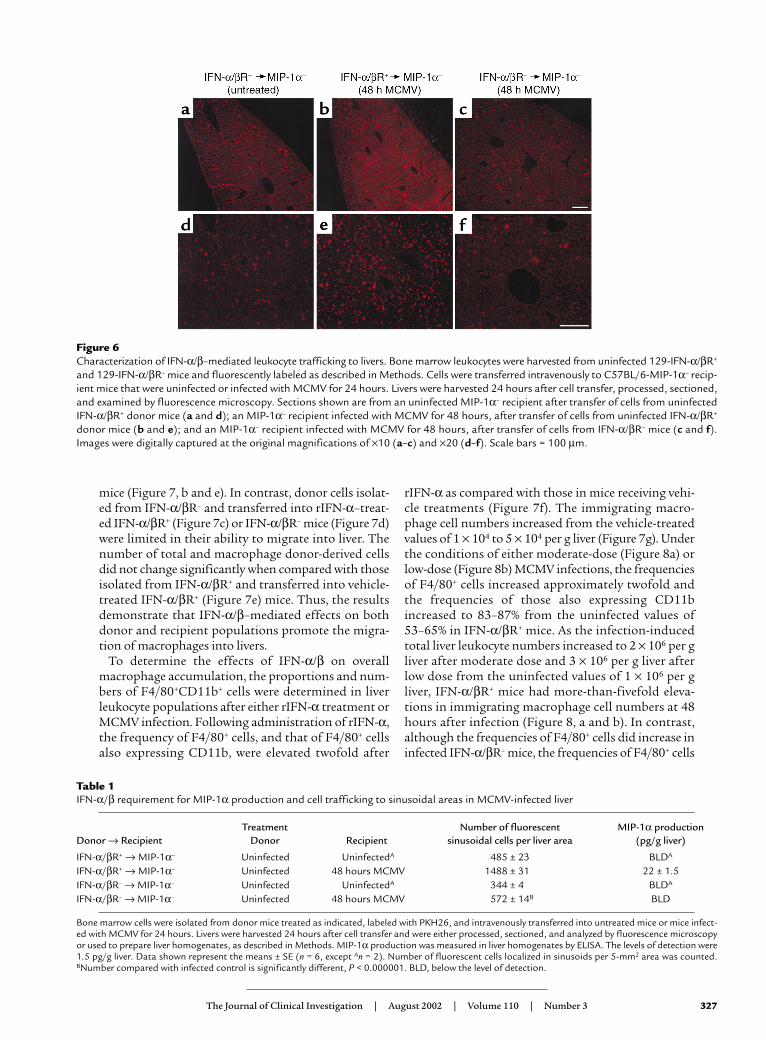
Figure 6Characterization of IFN-α/β–mediated leukocyte trafficking to livers. Bone marrow leukocytes were harvested from uninfected 129-IFN-α/βR+
and 129-IFN-α/βR– mice and fluorescently labeled as described in Methods. Cells were transferred intravenously to C57BL/6-MIP-1α– recip-ient mice that were uninfected or infected with MCMV for 24 hours. Livers were harvested 24 hours after cell transfer, processed, sectioned,and examined by fluorescence microscopy. Sections shown are from an uninfected MIP-1α– recipient after transfer of cells from uninfectedIFN-α/βR+ donor mice (a and d); an MIP-1α– recipient infected with MCMV for 48 hours, after transfer of cells from uninfected IFN-α/βR+
donor mice (b and e); and an MIP-1α– recipient infected with MCMV for 48 hours, after transfer of cells from IFN-α/βR– mice (c and f).Images were digitally captured at the original magnifications of ×10 (a–c) and ×20 (d–f). Scale bars = 100 µm.
Table 1IFN-α/β requirement for MIP-1α production and cell trafficking to sinusoidal areas in MCMV-infected liver
Treatment Number of fluorescent MIP-1α productionDonor → Recipient Donor Recipient sinusoidal cells per liver area (pg/g liver)
IFN-α/βR+ → MIP-1α– Uninfected UninfectedA 485 ± 23 BLDA
IFN-α/βR+ → MIP-1α– Uninfected 48 hours MCMV 1488 ± 31 22 ± 1.5IFN-α/βR– → MIP-1α– Uninfected UninfectedA 344 ± 4 BLDA
IFN-α/βR– → MIP-1α– Uninfected 48 hours MCMV 572 ± 14B BLD
Bone marrow cells were isolated from donor mice treated as indicated, labeled with PKH26, and intravenously transferred into untreated mice or mice infect-ed with MCMV for 24 hours. Livers were harvested 24 hours after cell transfer and were either processed, sectioned, and analyzed by fluorescence microscopyor used to prepare liver homogenates, as described in Methods. MIP-1α production was measured in liver homogenates by ELISA. The levels of detection were1.5 pg/g liver. Data shown represent the means ± SE (n = 6, except An = 2). Number of fluorescent cells localized in sinusoids per 5-mm2 area was counted.BNumber compared with infected control is significantly different, P < 0.000001. BLD, below the level of detection.
also expressing CD11b macrophage populationsdecreased two- to fourfold (Figure 8, a and b) comparedwith those in uninfected mice. Moreover, liver leuko-cyte yields from IFN-α/βR– mice were only 9 × 10⁵ perg liver after moderate-dose and 1 × 10⁶ per g liver afterlow-dose infection, compared with the uninfected val-ues of 1 to 2 × 10⁶ per g liver. Hence, the numbers ofimmigrating macrophages in IFN-α/βR– mice were sig-nificantly lower after MCMV infection (Figure 8, a andb). Taken together, these results demonstrate that IFN-α/β mediates trafficking and accumulation ofmacrophages into liver.
Characterization of MIP-1α–producing cells. Macrophagesare a known source of MIP-1α (1, 8). To determinewhether trafficking macrophages contributed to pro-duction of MIP-1α, total liver leukocytes and enrichedmacrophages were prepared from C57BL/6 (Figure 8c)and 129 (Figure 8d) mice that were uninfected or infect-ed with low-dose MCMV for 48 hours; they were thenevaluated for their ability to release the chemokine. Fol-lowing infection, total populations were induced to pro-duce MIP-1α. However, the levels of MIP-1α producedby enriched trafficking macrophages, expressing bothF4/80 and CD11b, were dramatically elevated over thelevels produced by total leukocytes. The macrophagesfrom infected C57BL/6 (Figure 8c) or infected 129 (Fig-
ure 8d) mice produced 12-fold or 22-fold more MIP-1αprotein, respectively, when compared with total cells.Therefore, migrating macrophages are a major source ofMIP-1α production in liver during MCMV infection.
DiscussionThese studies define a significant role and mechanismof action for IFN-α/β in promoting expression of MIP-1α and the accumulation of NK cells in liver dur-ing MCMV infection. They demonstrate that IFN-α/βis produced locally and is an important factor in pro-tecting against infection. Immunoregulatory effectsare shown to be contributing to IFN-α/β–mediateddefense, since critical production of MIP-1α, requiredfor accumulation of NK cells, is dependent on thefunctions of these cytokines. The data indicate thatIFN-α/β is upstream of MIP-1α induction in liver, andthat IFN-α/β is promoting local expression of MIP-1α,at least in part, by acting as a recruiter of macrophagesproducing the chemokine. As these responses are alsoinduced with administrated rIFN-α, the cytokines canact in the absence of infection. Taken together, thesestudies identify IFN-α/β as key initiators of MIP-1αexpression required for NK cell recruitment into liver.The results extend our characterization of a criticalchemokine-to-cytokine-to-chemokine cascade for
328 The Journal of Clinical Investigation | August 2002 | Volume 110 | Number 3
Figure 7Characterization of macrophage trafficking to and accumulation in livers after rIFN-α treatment. Bone marrow leukocytes from untreated129-IFN-α/βR+ or 129-IFN-α/βR– mice were fluorescently labeled as described in Methods. Cells were transferred intravenously to IFN-α/βR+
or IFN-α/βR– recipients that were treated with vehicle or rIFN-α and examined. Sections shown are from an rIFN-α–treated IFN-α/βR+ recip-ient after transfers from untreated IFN-α/βR+ donors (a), an rIFN-α–treated IFN-α/βR– recipient after transfers from untreated IFN-α/βR+
donors (b), an rIFN-α–treated IFN-α/βR+ recipient after transfers from untreated IFN-α/βR– donors (c), and an rIFN-α–treated IFN-α/βR–
recipient after transfers from untreated IFN-α/βR– donors (d). Scale bar = 100 µm. Liver leukocytes were prepared from recipient mice andanalyzed by flow cytometry. Numbers of donor-derived PKH26+ and PKH26+F4/80+CD11b+ cells g liver are shown (e). *Difference betweenvehicle- and rIFN-α–treated IFN-α/βR+ recipients is significant at P < 0.001. To characterize the accumulation of migrating macrophages,liver leukocytes were obtained from IFN-α/βR+ or IFN-α/βR– mice that were treated with vehicle or rIFN-α, labeled with F4/80 and CD11band examined by flow cytometry. Migrating populations were identified by analyses of CD11b expression after gating on the F4/80+ cells(f). Representative histograms are shown, with thick lines representing isotype control antibody and shaded histograms F4/80 or CD11blabeling. Percentages of F4/80+CD11b+ macrophages per g liver isolated from vehicle- (black bar) or rIFN-α–treated (gray bar) samples areshown (g). In all experiments, data represent the means ± SE (n = 3). *Difference between vehicle control and rIFN-α treatments is signifi-cant at P ≤ 0.02.
antiviral defense (5) by defining IFN-α/β–dependentevents upstream of MIP-1α necessary for initiatingthis multistep immune process.
IFN-α/β can elicit potent antiviral effects and medi-ate several immunoregulatory activities (11, 12–16).The possibility that the loss of direct antiviral effectsalso contributed to the increased sensitivity of IFN-α/βR– mice to MCMV infection cannot be excluded(Figure 3). However, we have previously demonstratedthat MIP-1α, NK cell delivery of IFN-γ to liver, andIFN-γ induction of Mig are all critical steps in protec-tion against this virus (1, 5). The studies presentedhere demonstrate that the induction of MIP-1α in liveris dependent on the immunoregulatory effects medi-ated by IFN-α/β, and that these occur in the absence ofinfections. Thus, these factors are upstream initiatorsof an essential chemokine-cytokine cascade.
The studies presented here definitively establish therole of IFN-α/β in driving the accumulation ofmacrophages in the liver (Figures 7 and 8). The cells aredefined as immigrating macrophages by their coex-pression of F4/80 and CD11b. The conditions also pro-moted the migration of other cell types, since the dou-ble positive cells represented fewer than 10% of thedonor-derived cells (Figure 7e). The other traffickingsubsets would include NK cells (Figure 5) and mayinclude other leukocyte subsets not yet characterized,i.e., dendritic cells and neutrophils. However, the dou-
ble positive populations are greatly enriched for MIP-1α–producing cells (Figure 8, c and d). The transfer ofIFN-α/βR– bone marrow cells to infected MIP-1α– micefailed to result in both the dramatic accumulation ofmacrophages in sinusoids and the appearance of MIP-1α production in liver (Figure 6 and Table 1).Under these conditions, the effects on cell accumula-tion are likely to have been a consequence of earlierblocked cellular IFN-α/β responsiveness rather than ofresulting reductions in MIP-1α production, since infec-tion-induced migration of macrophages to liver sinu-soids is MIP-1α independent (1). It is not yet clear, how-ever, whether additional chemokine responses areregulated by IFN-α/β and/or play an intermediary rolein macrophage migration. Macrophages have beenshown to produce chemokines in response to variousstimuli (8, 24–27). Here, immigrant liver macrophagesare shown to produce MIP-1α during infection.
The observations clearly demonstrate that IFN-α/βfunctions are required for MIP-1α induction duringMCMV infection in liver, and that promoting accumu-lation of macrophages producing MIP-1α to this site isa contributing mechanism for this. The responses ofother innate cytokines, such as IL-12, IL-15, and NKcell–derived IFN-γ, also are expressed during earlyMCMV infection (refs. 10, 28; K.B. Nguyen et al.,unpublished observations). These factors have beenshown to stimulate production of MIP-1α from cul-
The Journal of Clinical Investigation | August 2002 | Volume 110 | Number 3 329
Figure 8Characterization of IFN-α/β–mediated effects on macrophage accumulation and of MIP-1α–producing cells in liver during MCMV infection.Liver leukocytes were prepared from 129-IFN-α/βR+ or 129-IFN-α/βR– mice that were uninfected or infected with moderate-dose (a) or low-dose (b) MCMV for 48 hours. Leukocytes were analyzed by flow cytometry as described in Methods. Macrophages were identified by F4/80expression and are shown by histograms (a and b). The accumulation of migrating populations was subsequently identified by analyses ofCD11b expression after gating on the F4/80+ population and is shown by histograms (a and b). The data shown are representative his-tograms, with thick lines representing isotype control antibody and shaded histograms F4/80 or CD11b labeling. Percentages presented ineach histogram are means ± SE (n = 3–4). Numbers of F4/80+CD11b+ cells per g liver isolated from 129-IFN-α/βR+ (black bars) or 129-IFN-α/βR– (gray bars) are shown (a and b). Data represent the means ± SE (n = 3–4). Differences between control IFN-α/βR+ and IFN-α/βR– aresignificant at *P ≤ 0.03 and **P ≤ 0.01. (c and d) Total (black bars) or enriched F4/80+CD11b+ (gray bars) liver leukocytes were preparedfrom C57BL/6 (c) or 129 (d) mice that were uninfected or infected with low-dose MCMV for 48 hours. Leukocyte-CM was used to measureMIP-1α in ELISA as described in Methods. Levels of detection were 0.2 pg/million cells. Each group consisted of pooled cell samples fromsix mice. The data shown are representative of two independent experiments.
tures of purified human NK cells (29). However, theydo not appear to be required for liver MIP-1α inductionduring MCMV infection (T.P. Salazar-Mather and C.A.Biron, unpublished observations). The promoterregion for murine MIP-1α does have a potential IFN-α/β–responsive element (30, 31), suggesting a rolefor this cytokine in enhancing transcription of thechemokine. Our studies do not eliminate the possibil-ity that IFN-α/β can directly enhance induction of MIP-1α, but they do undoubtedly demonstrate thepresence of another pathway for cell delivery of MIP-1αto MCMV-infected livers.
In summary, this work has extended our characteri-zation of molecular mechanisms of cellular recruit-ment required for innate defense against viral infectionin liver. Our results identify IFN-α/β as key initiatorsof MIP-1α production that is necessary for NK cellinflammation. In addition, by showing that IFN-α/βcan mediate the recruitment of MIP-1α–producingmacrophages, our results define a cellular deliverymechanism by which these cytokines promote localimmune responses.
AcknowledgmentsThe authors wish to thank K.B. Nguyen for expertassistance with flow cytometry, and G. Yap, M. Dalod,and G. Gutierrez for experimental help. This work wassupported by NIH grants CA-79076 and CA-41268.
1. Salazar-Mather, T.P., Orange, J.S., and Biron, C.A. 1998. Early murinecytomegalovirus (MCMV) infection induces liver natural killer (NK) cellinflammation and protection through macrophage inflammatory pro-tein 1α (MIP-1α)-dependent pathways. J. Exp. Med. 187:1–14.
2. Orange, J.S., Wang, B., Terhorst, C., and Biron, C.A. 1995. Requirementfor natural killer cell-produced interferon-γ in defense against murinecytomegalovirus infection and enhancement of this defense pathway byinterleukin-12 administration. J. Exp. Med. 182:1045–1056.
3. Biron, C.A., Nguyen, K.B., Pien, G.C., Cousens, L.P., and Salazar-Mather,T.P. 1999. Natural killer cells in antiviral defense: function and regula-tion by innate cytokines. Annu. Rev. Immunol. 17:189–220.
4. Ruzek, M.C., Miller, A.H., Opal, S.M., Pearce, B.D., and Biron, C.A. 1997.Characterization of early cytokine responses and an interleukin (IL)-6-dependent pathway of endogenous glucocorticoid induction duringmurine cytomegalovirus infection. J. Exp. Med. 185:1185–1192.
5. Salazar-Mather, T.P., Hamilton, T.A., and Biron, C.A. 2000. Achemokine-to-cytokine-chemokine cascade critical in antiviral defense.J. Clin. Invest. 105:985–993.
6. Farber, J.M. 1997. Mig and IP10: CXC chemokines that target lympho-cytes. J. Leukoc. Biol. 61:246–257.
7. Miura, M., et al. 2001. Monokine induced by IFN-gamma is a dominantfactor directing T cells into murine cardiac allografts during acute rejec-tion. J. Immunol. 167:3494–3504.
8. Rollins, B.J. 1997. Chemokines. Blood. 90:909–928.9. Rossi, D., and Zlotnick, A. 2000. The biology of chemokines and their
receptors. Annu. Rev. Immunol. 18:217–242.10. Orange, J.S., and Biron, C.A. 1996. Characterization of early IL-12,
IFN-α/β, and TNF effects on antiviral state and NK cell responses dur-ing murine cytomegalovirus infection. J. Immunol. 156:4746–4756.
11. Biron, C.A. 2001. Interferons α and β as immune regulators: a new look.Immunity. 14:661–664.
12. Biron, C.A., Dalod, M., and Salazar-Mather, T.P. 2002. Innate immunityand viral infections. In Immunology of infectious diseases. S.H.E. Kaufmann,A. Sher, and R. Ahmed, editors. ASM Press. Washington, DC, USA.139–160.
13. Korngold, R., Blank, K.J., and Murasko, D.M. 1983. Effect of interferonon thoracic duct lymphocyte output: induction with either polyI:C orvaccinia virus. J. Immunol. 130:2236–2243.
14. Wiltrout, R.H., et al. 1989. Augmentation of mouse liver-associated nat-ural killer cell activity by biological response modifiers occurs largely viarapid recruitment of large granular lymphocytes from the bone marrow.J. Immunol. 143:372–378.
15. Ishikawa, R., and Biron, C.A. 1993. IFN induction and associatedchanges in splenic leukocyte distribution. J. Immunol. 150:3713–3727.
16. Salazar-Mather, T.P., and Biron, C.A. 1996. NK cell trafficking andcytokine expression in splenic compartments after IFN induction andviral infection. J. Immunol. 157:3054–3064.
17. Cook, D.N., et al. 1995. Requirement of MIP-1α for an inflammatoryresponse to viral infection. Science. 269:1583–1585.
18. Muller, U., et al. 1994. Functional role of type 1 and type II interferonsin antiviral defense. Science. 264:1918–1921.
19. Pien, G.C., and Biron, C.A. 2000. Compartmental differences in NK cellresponsiveness to IL-12 during lymphocytic choriomeningitis virusinfection. J. Immunol. 164:994–1001.
20. Arase, H., Saito, T., Phillips, J.H., and Lanier, L.L. 2001. Cutting edge: themouse NK cell-associated antigen recognized by DX5 monoclonal anti-body is CD49b (α2 integrin, very late antigen-2). J. Immunol.167:1141–1144.
21. Lepay, D.A., Steinman, R.M., Nathan, C.F., Murray, H.W., and Cohn, Z.A.1985. Liver macrophages in murine listeriosis. J. Exp. Med.161:1503–1512.
22. Dalod, M., et al. 2002. IFN-α/β and IL-12 responses to viral infections:pathways regulating dendritic cell cytokine expression in vivo. J. Exp.Med. 195:517–528.
23. Andrews, D.M., et al. 2001. NK1.1+ cells and murine cytomegalovirusinfection: what happens in situ? J. Immunol. 166:1796–1802.
24. Orange, J.S., and Biron, C.A. 1996. An absolute and restricted require-ment for interleukin-12 in natural killer (NK) cell interferon-γ produc-tion and antiviral defense: studies of NK and T cell responses in con-trasting viral infections. J. Immunol. 156:1138–1142.
25. Bluman, E.M., Bartynski, K.J., Avalos, B.R., and Caligiuri, M.A. 1996.Human natural killer cells produce abundant macrophage inflamma-tory protein-1α in response to monocyte-derived cytokines. J. Clin. Invest.97:2722–2727.
26. Fujita, T., Shibuya, H., Hotta, H., Yamanishi, K., and Taniguchi, T. 1987.Interferon-beta gene regulation: tandemly repeated sequences of a syn-thetic 6 bp oligomer function as a virus-inducible enhancer. Cell.49:357–367.
27. Widmer, U., Manogue, K.R., Cerami, A., and Sherry, B. 1993. Genomiccloning and promoter analyses of macrophage inflammatory protein(MIP)-2, MIP-1α, and MIP-1β, members of the chemokine superfamilyof proinflammatory cytokines. J. Immunol. 150:4996–5012.
28. Farber, J.M. 1990. A macrophage mRNA selectively induced by γ-inter-feron encodes a member of the platelet 4 family of cytokines. Proc. Natl.Acad. Sci. USA. 87:5238–5242.
29. Oppenheim, J.J., Zachariae, C.O.C., Mukaida, N., and Matsushima, K.1991. Properties of the novel proinflammatory supergene “intercrine”cytokine family. Annu. Rev. Immunol. 9:617–648.
30. Kopydlowski, K.M., et al. 1999. Regulation of macrophage chemokineexpression by lipopolysaccharide in vitro and in vivo. J. Immunol.163:1537–1544.
31. Lehner, T., et al. 2000. Heat shock protein generates β-chemokines whichfunction as innate adjuvant enhancing adaptive immunity. Eur. J.Immunol. 30:594–603.
330 The Journal of Clinical Investigation | August 2002 | Volume 110 | Number 3