Two new Preussia species defined based on morphological ... · Fungal Diversity Two new Preussia...
15
Fungal Diversity Two new Preussia species defined based on morphological and molecular evidence Francisco Arenal 1 , Gonzalo Platas 2 and Fernando Peláez 2* 1 Dpto. de Protección Vegetal, Centro Ciencias Medioambientales (CCMA-CSIC). Serrano, 115 Dpdo., E-28006 Madrid, Spain 2 Centro de Investigación Básica. Merck Sharp & Dohme Research Laboratories. Josefa Valcárcel 38, E-28027 Madrid, Spain Arenal, F., Platas, G. and Peláez, F. (2005). Preussia africana and Preussia isabellae, two new Preussia species based on morphological and molecular evidence. Fungal Diversity 20: 1-15. Two new Preussia species from plant debris and coprophilous substrata are described and illustrated based on molecular and morphological data. Preussia africana was isolated from Canary Islands, South Africa and Tanzania and Preussia isabellae from Puerto Rico. Morphologically Preussia africana resembled P. australis and P. minimoides. Preussia isabellae was related to P. minima. Parsimony analysis of the ITS1-5.8S-ITS2 region, the 5´end of the 28S rRNA gene and a fragment of the translation elongation factor 1α gene, supported the recognition of these fungi as new species. Key words: Ascomycota, elongation factor, ITS, LSU, phylogeny, rDNA, taxonomy. Introduction The genus Preussia was erected by Fuckel (1866). This genus comprised species of bitunicate ascomycetes with non-ostiolate ascomata, containing dark brown multi-celled ascospores (4-16 cells) with germ slits, and are covered by a gelatinous sheath. The genus Sporormiella Ellis & Everhart differs from Preussia mainly by having ostiolate ascomata and by its coprophilous habitat. In contrast, Preussia includes species isolated from soil, wood and plant debris (Cain, 1961; Ahmed and Cain, 1972; Arx and Van der Aa, 1987). Recognition of both genera has caused confusion because of their shared morphological features (Auerswald, 1866; Munk, 1957). In recent years the two genera have been proposed or accepted as synonyms (Arx, 1973; Barr, 1987; Valldosera and Guarro, 1990; Guarro et al., 1997; Arenal et al., 2004), based on the inconsistency of the character of the presence of the ostiole, which is known to be influenced by the culture conditions, and may be present or absent even in * Corresponding author: F. Peláez; e-mail: [email protected], [email protected], [email protected] 1
Transcript of Two new Preussia species defined based on morphological ... · Fungal Diversity Two new Preussia...
Fungal Diversity
Two new Preussia species defined based on morphological and molecular evidence Francisco Arenal1, Gonzalo Platas2 and Fernando Peláez2* 1Dpto. de Protección Vegetal, Centro Ciencias Medioambientales (CCMA-CSIC). Serrano, 115 Dpdo., E-28006 Madrid, Spain 2Centro de Investigación Básica. Merck Sharp & Dohme Research Laboratories. Josefa Valcárcel 38, E-28027 Madrid, Spain Arenal, F., Platas, G. and Peláez, F. (2005). Preussia africana and Preussia isabellae, two new Preussia species based on morphological and molecular evidence. Fungal Diversity 20: 1-15. Two new Preussia species from plant debris and coprophilous substrata are described and illustrated based on molecular and morphological data. Preussia africana was isolated from Canary Islands, South Africa and Tanzania and Preussia isabellae from Puerto Rico. Morphologically Preussia africana resembled P. australis and P. minimoides. Preussia isabellae was related to P. minima. Parsimony analysis of the ITS1-5.8S-ITS2 region, the 5´end of the 28S rRNA gene and a fragment of the translation elongation factor 1α gene, supported the recognition of these fungi as new species. Key words: Ascomycota, elongation factor, ITS, LSU, phylogeny, rDNA, taxonomy. Introduction
The genus Preussia was erected by Fuckel (1866). This genus comprised species of bitunicate ascomycetes with non-ostiolate ascomata, containing dark brown multi-celled ascospores (4-16 cells) with germ slits, and are covered by a gelatinous sheath. The genus Sporormiella Ellis & Everhart differs from Preussia mainly by having ostiolate ascomata and by its coprophilous habitat. In contrast, Preussia includes species isolated from soil, wood and plant debris (Cain, 1961; Ahmed and Cain, 1972; Arx and Van der Aa, 1987). Recognition of both genera has caused confusion because of their shared morphological features (Auerswald, 1866; Munk, 1957). In recent years the two genera have been proposed or accepted as synonyms (Arx, 1973; Barr, 1987; Valldosera and Guarro, 1990; Guarro et al., 1997; Arenal et al., 2004), based on the inconsistency of the character of the presence of the ostiole, which is known to be influenced by the culture conditions, and may be present or absent even in * Corresponding author: F. Peláez; e-mail: [email protected], [email protected], [email protected]
1
ascomata from the same culture (Guarro et al., 1997). Furthermore, typical species of Sporormiella, as defined by Ahmed and Cain (1972) can be isolated from substrata other than dung, making the habitat an artificial feature as well (Guarro et al., 1997; Peláez et al., 1998). Other authors have reconsidered the concept of these two genera, and argued whether or not to treat them as synonyms (Arx and Van der Aa, 1987; Barr, 1990; Barrasa and Checa, 1991).
Although new Preussia and Sporormiella species continue to be described (Barr, 1990; Lorenzo, 1994; Guarro et al., 1997; Koryolova, 2000), an accumulating body of data suggests that, unless other more relevant characters are found that can be used to differentiate between the two genera, it may be more appropriate to synonymize them, therefore newly described species would be assigned to Preussia.
During a morphological and molecular analysis of a series of wild isolates of genus Preussia from different substrata and geographic regions, we found some interesting strains isolated from herbivore dung and plant materials from Canary Islands, Puerto Rico South Africa and Tanzania (Arenal, 2001). Their morphological characters were inconsistent with any described species of Preussia or Sporormiella, even though they were apparently related to a series of species morphologically akin including P. australis (Speg.) Arx, P. intermedia (Auersw.) S.I. Ahmed, P. minimoides (S.I. Ahmed & Cain) Valldos. & Guarro, P. minima (Auersw.) Arx and P. similis (Khan & Cain) Arenal. Molecular data derived from nuclear ribosomal DNA sequences, including the ITS1-5.8S-ITS2 region and the D1-D2 domains of the 28S rRNA gene, and a portion of the translation elongation factor 1α gene, combined with morphological data, prompted us to describe two new species for these isolates. Materials and methods Strains examined
All the strains were isolated by the authors following standard indirect isolation techniques. The strains from dung or leaf litter were isolated using a particle filtration method (Bills and Polishook, 1994). The strains from living plant materials were isolated using a surface sterilization method (Collado et al., 1996). The isolation media used have been also previously described (Collado et al., 1996). The strains were grown at 22ºC and 80% relative humidity on PDA and OMA (oat meal agar) and exposed to alternate cycles of 12 hour near-UV light/daylight for at least 14 days to induce sporulation. The cultures were preserved in the CIBE-Merck, Sharp & Dohme Culture Collection in 20% glycerol vials as 0.6 cm diam. frozen agar plugs at -80ºC.
2
Fungal Diversity
The geographical origin, isolation substrata and GenBank accession numbers of the isolates are listed in Table 1. Morphological data Microscopic features were examined after sporulation of four sequential subcultures on PDA and OMA for each strain. Slides were made using a Leica Wild M8 dissection scope, in water or lactophenol cotton blue, and observed under a Leitz Diaplan microscope. Photographs were made with an Olympus DP-12 microscope digital camera system, incorporated to the Leitz microscope. Twenty one measurements were made from each sporulating culture in order to define the range of spore length, following the methodology described in Arenal et al. (2004). Measurements in descriptions are given as (minimum value-)(mean-2SD)-mean-(mean+2SD)(-maximum value), as well as the Q value and number of measurements, according to the recommendations of Heinemann and Rammeloo (1985). Ascospore measurements were made at their widest point and did not include the gelatinous sheath. The microscopic terminology of Ahmed and Cain (1972) was adopted. The colour codes from Kornerup and Wanscher (1978) were used in the description of the gross morphology of the colonies. DNA sequencing and phylogenetic analysis
DNA extraction and PCR amplification procedures were performed as
described by Bills et al. (1999). The ITS1-5.8S-ITS2 region was amplified using primers ITS1F and ITS4 (White et al., 1990), whereas the D1-D2 domains of the 28S rRNA gene was amplified using primers LR0R and LR16 (Bunyard et al., 1994). For the fragment of EF-1α, that includes one intron, we used the primers EF1-728F and EF1-986R described by Carbone and Kohn (1999). All the resulting PCR products were purified using GFXTM PCR Gel Band Purification Kit (Amersham Pharmacia Biotech Inc, USA), before sequencing. The amplified products were sequenced with an ABI PRISM Dye Terminator Cycle Sequencing Kit (Perkin Elmer). The samples were sequenced in both directions as described for the ITS region (Sánchez-Ballesteros et al., 2000), for the D1-D2 domains of the 28S rRNA gene (Acero et al., 2004), and for the EF-1α (Carbone and Kohn, 1999). DNA sequences were visually aligned with the multiple sequence alignment editor GeneDoc 2.5 and deposited in TreeBASE (M1773, M1774). The program GapCoder (Hennequin et al., 2003; Young and Healy, 2003) was used to improve the quality of the alignment of the EF-1α fragment. All sequences were deposited
3
in GenBank (Table 1). The phylogenetic relationships among the two new Preussia species and other closely related species, were examined using maximum parsimony method inferred by heuristic search of the aligned sequences using PAUP 4.0 b10 (Swofford, 2001). Heuristic search was performed with simple addition of sequences and TBR branch swapping, with MaxTrees set to 100. All characters were unordered and equally weighted, with gaps treated as missing data. Two phylogenetic analysis were made, one with the ITS sequences combined with the D1-D2 region of the 28S rRNA gene, and another with the EF-1α fragment alone. The confidence of the branches was measured by bootstrap analysis with 1000 bootstrap replicates using heuristic search (Felsenstein, 1985), and by decay indexes (Bremer, 1994), calculated with SEPAL v1.4 software. The trees were visualized with the application Treeview 1.5. Results Sequencing and phylogenetic analysis
In order to clarify the relationships of the two new species with other
morphologically similar Preussia species, we sequenced the ITS, the 5’ region of the 28S rRNA gene and a fragment of the EF-1α of all isolates of P. africana and P. isabellae. The sequences were aligned with those from a group of isolates unambiguously identified by morphology as other Preussia species collected from diverse geographic origins (Table 1). Single DNA fragments of 465-485 bp for the ITS region, 584-587 bp for the D1-D2 domains of the 28S rRNA gene and 370-450 bp for the EF-1α gene, were obtained in the amplification reactions for all the Preussia isolates analyzed (Table 1). For the ITS/D1-D2 fragment a total of 1016 characters were aligned, of which 815 were constant, 123 were variable but parsimony-uninformative, and 78 were parsimony informative. Fifty two equally most-parsimonious trees were obtained, with identical topologies regarding all the aspects discussed below. One representative tree is shown in Fig. 1. The tree length was 282 steps, with consistency index CI = 0.830, homoplasy index HI = 0.170, retention index RI = 0.817, rescaled consistency index RC = 0.678. For the EF-1α fragment a total of 564 characters were aligned, of which 185 were constant, 153 were variable and parsimony-uninformative, and 226 were parsimony informative. Six equally parsimonious trees were obtained, with identical topologies regarding all the aspects discussed below. One representative tree is shown in Fig. 2. The tree length was 600 steps, with CI = 0.827, HI = 0.173, RI = 0.853 and RC = 0.705.
4

Fungal Diversity
Table 1. Preussia isolates examined, their substrata and geographical origin. Strain Species Substrate Origin GenBank accession numbers ITS 28S EF-1α S12 P. africana Goat dung Iringa, Tanzania AY510420 AY510384 AY510405
S14 P. africana Zebra dung Kwazulu-Natal,
South Africa AY510417 AY510382 AY510403
S15 P. africana Zebra dung Kwazulu-Natal, South Africa
AY510421 AY510385 AY510404
S17 P. africana Viburnum tinus leaves
Tenerife, Canary Islands
AY510418 AY510383 AY510402
S5 P. australis Gazelle dung Cape Point, South Africa
AY510411 AY510376 AY510399
S6 P. australis Gazelle dung Luderitz, Namibia AY510412 AY510377 AY510401S7 P. australis Zebra dung Hester Malan
Reserve, South Africa
AY510413 AY510378 AY510400
S1 P. intermedia Elk dung Arizona, USA AY510415 AY510380 AY510398S3 P. intermedia Goat dung Cefalonia, Greece AY510414 AY510379 AY510396S4 P. intermedia Goat dung Cefalonia, Greece AY510416 AY510381
AY510397
S13 P. minima Gazelle dung Hobatere,
Namibia AY510426 AY510391 AY510410
S21 P. minima Rhinoceros dung
Kwazulu-Natal, South Africa
AY510425 AY510390 AY510408
S26 P. minima Leaf litter South Dakota, USA
AY510427 AY510392 AY510409
S10 P. minimoides Pig dung Chaco, Argentina AY510423 AY510388 AY510406S18 P. minimoides Prunus
lusitanica leaves
Tenerife, Canary Islands
AY510422 AY510387
AY510394
S25 P. isabellae Leaf litter Puerto Rico
AY510424 AY510389 AY510407
S19 P. similis Dung Arizona, USA AY510419 AY510386 AY510395
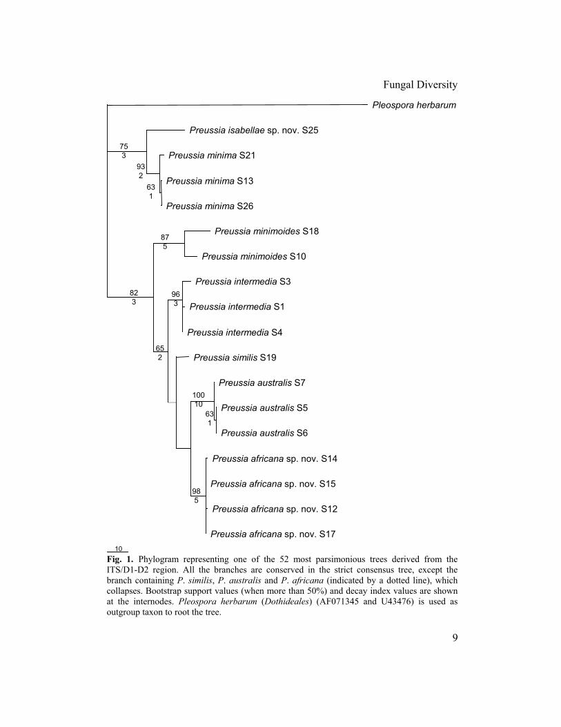
The Preussia strains were grouped as monophyletic clades corresponding to each species, supported by high bootstrap values (94-100%) in both phylogenetic trees, except for P. minimoides. The two strains sequenced from this species appeared grouped together in a monophyletic clade with a moderate bootstrap value (87%) in the ITS/D1-D2 phylogram, but not in the EF-1α tree. All the strains of P. africana fell within a clade with very high bootstrap support for both regions sequenced (100% for the EF-1α and 98% for ITS/D1-D2). Preussia isabellae, represented by a single isolate, was consistently in a basal position to P. minima, which formed a well-supported monophyletic clade in both phylogenetic trees.
5
Taxonomy Preussia africana Arenal, Platas & Peláez, sp. nov. (Figs. 3-11) Etymology: referring to its apparent geographic distribution. Pseudotheciis (180-)210-290 µm in diametro, sparsis vel aggregatis, immersis vel semiimersis, subglobosis vel piriformibus, atro-brunneis vel nigris, glabris et ostiolatis. Collo breve papilliformi vel cylindraceo, 50-60 × 20-40 µm. Peridio membranaceo pseudoparenchymatico, glabro, 10-15 µm, bistratoso. Ascis 94-110 × 15-17 µm, octosporis, cylindraceo-clavatis, superne rotundatis, inferne attenuatis, breve stipitatis, usque 13 × 4.5 µm. Pseudoparaphysibus 1-2 µm crassis, filiformibus, numerosis, ramosis et septatis. Ascosporis 32.5-44 × 4-7 µm, oblique uniseriatis vel biseriatis, cylindraceis, quattuorcellularibus, olivaceo-brunneis, transverse septatis et leviter constrictis, articulis similibus, facile sedecentibus; stria germinationis oblique usque parallela et rectis vel leviter curvatis. Vagina mucosa hyalina et angusta.
Typus: In foliis Viburni tini subsp. rigidi. Tenerife, Canary Islands, Spain, 2 Apr. 1995, leg. A. Santos. Cultura sicca (holotypus) in Herbarium AH (AH32767) Colonies on PDA attaining 70-75 mm diam. in 14 days at 23ºC. Texture cottony to floccose showing frequently white to light cream sectors (10YR 7/4), adpressed and occasionally submerged, light brown (10YR 7/3) to black. Ascomata scattered or aggregated, superficial or partially immersed in culture media when young. Pseudothecia (180-)210-290 µm diam., globose, subglobose to pyriform, smooth, ostiolate, brown to dark brown (10YR 5/3, 4/3); neck small, 50-60 × 20-40 µm, papilliform to cylindrical; glabrous, but with short ornamental hyphae measuring 10-20 × 2.5-4.5 µm, sometimes present at the base of the ascomata. Peridium dark brown (10YR 5/3, 4/3), pseudoparenchymatous in surface view, membranaceous, coriaceous and 10-15 µm thick. Asci 94-110 × 15-17 µm eight-spored, cylindrical-clavate, stipitate, nonamyloid, broadly rounded above, gradually to abruptly tapering into a short and robust stipe up to 13 × 4.5 µm. Pseudoparaphyses 1-2 µm diam., filiform, septate, interspersed with asci. Ascospores (32.5-)32.9-37.3-41.6(-44) × 4-5.3-7 µm, Q = 4.6-7.3-9.9(-10.4) (n = 168), biseriate, cylindrical, hyaline to olivaceous when young, becoming olivaceous brown to dark brown (10YR 6/3) when mature; four-celled, transversely septate, cells separable at the central septa, constrictions at septa broad and shallow, middle cells of equal length and broader than terminal cells, with rounded apices; germ slit oblique to parallel and straight to slightly sinuous; gelatinous sheath hyaline and narrow.
Anamorph: unknown. Habitat: On living plant material and herbivore dung. Known distribution: Canary Islands (Spain), Tanzania and South Africa. Material examined: SPAIN, Canary Islands (Tenerife), Llano de los Viejos, on living leaves of Viburnum tinus subsp. rigidum, 2 April 1995, col. A. Santos, S17 (AH32767; holotype here designated); South Africa, Kwazulu-Natal, zebra dung, 22 January 1995, col. M.J. Wingfield, S14 (AH32768); South Africa, Kwazulu-Natal, zebra dung, 23 January 1995,
6
Fungal Diversity
col. M.J. Wingfield, S15 (AH32769); Tanzania, Iringa, goat dung, 26 June 1992, col. D. Moyer, S12 (AH32770). Preussia isabellae Arenal, Platas & Peláez, sp. nov. (Figs. 12-20) Etymology: dedicated to Isabel Soto.
Pseudotheciis (90-)100-130(140) µm in diametro, sparsis vel aggregatis, immersis usque semiimersis, subglobosis vel piriformibus, atro-brunneis vel nigris, glabris et ostiolatis. Peridio pseudoparenchymatico et membranaceo, 12-15 µm crasso, glabro, bistratoso. Ascis 94-110 × 17-20.5 µm, octosporis, cylindraceo-clavatis, superne late rotundatis, inferne in stipitem attenuatis. Pseudoparaphysibus 2 µm crassis, filiformibus, numerosis, ramosis et septatis. Ascosporis 29-42 × 4-6.5 µm, oblique uniseriatis vel biseriatis, cylindraceis, quattuorcellularibus, olivaceo-brunneis et brunneis, transverse septatis et leviter constrictis, articulis prope similibus, cellulis maturis facile sedecentibus; stria germinationis parallela, abrupte curvata ad centrum. Vagina mucosa hyalina et angusta.
Typus: In ligno plantae. Puerto Rico, 12 Jan 1996, leg. J. Guarro. Cultura sicca (holotypus) in Herbarium AH (AH32771).
Colonies on PDA attaining 75-80 mm diam. in 14 days at 23ºC. Texture cottony, adpressed, brown to black (10YR 5/3) with white patches. Ascomata scattered or in small groups, superficial or partially immersed when young in culture media surface. Pseudothecia subglobose to pyriform, (90-)100-130(140) µm in diam., dark brown to black (10YR 4/3), smooth and membranaceous; glabrous, but with short ornamental hyphae, 7-10 × 2.5-4 µm, sometimes present at the base of the ascomata. Peridium pseudoparenchymatous in surface view, membranaceous, glabrous, peridial cells 12-15 µm in diam. Asci 94-110 × 17-20.5 µm, eight-spored, cylindrical-clavate, stipitate, nonamyloid, broadly rounded above, gradually to abruptly tapering into a short stipe. Pseudoparaphyses 2 µm in diam., filiform, abundant, branched and septate. Ascospores (29-)30.7-35.8-40.9(-42) × 4-5.3-6.5 µm, Q = 5.2-6.8-8.9(-9.7) (n = 168), uniseriate to biseriate, cylindrical, olivaceous brown to dark brown (10YR 6/3) when mature; four-celled, transversely septate, cells easily separable, constrictions at septa broad and shallow, cells nearly equal in size, terminal cells with rounded apices; germ slit parallel with a strong to sinuous curvature at the middle; gelatinous sheath hyaline and narrow.
Anamorph: unknown. Habitat: On unidentified plant debris.
Known distribution: Puerto Rico. Material examined: PUERTO RICO, on woody plant debris, 12 January 1996, col. J.
Guarro, S25 (AH32771; holotype here designated). Discussion
Employing the morphological characters in the keys of Cain (1961) and Ahmed and Cain (1972), the two new species are related to a series of Preussia
7
species characterized by the absence of hairs in the pseudothecia and neck, 8-spored cylindrical asci abruptly contracting below into a short stipe, and four-celled ascospores with a length ranging from 37 to 60 µm. The species within this group can be distinguished based on spore size, morphology and disposition of the germ slit.
Preussia africana has a spore length range (32.5-44 µm) overlapping between P. minimoides (28-36 µm) and P. australis (38-46 µm). However, the germ slit in P. africana is oblique to parallel and straight to slightly sinuous, while it is strongly oblique to diagonal in P. australis, and sigmoid or sinuous in P. minimoides (Ahmed and Cain, 1972). Other characters, such as cells not easily separable, or the terminal spore cells longer and narrower toward the end, do not fit exactly with the morphological characteristics of P. australis and P. minimoides (Ahmed and Cain, 1972). However, these three species share other features, such as the morphology of the asci, broadly rounded, frequently broader near the middle and abruptly constricted in a short stipe, as well as the presence of a narrow and hyaline gelatinous ascospore sheath. The overlapping size range makes it possible to misidentify strains of P. africana, ascribing them to either P. australis or P. minimoides, unless a significant number of ascospores are measured and the morphology and disposition of the germ slit are carefully examined (Arenal et al., 2004). Preussia africana seems to have both coprophilous and non-coprophilous habitat; three of the four isolates found were recovered from dung of herbivore species, but one strain was isolated as an endophyte from surface-sterilized living leaves of Viburnum tinus (Peláez et al., 1998). Likewise, other Preussia and Sporormiella species have been often reported as endophytes (e.g. Guarro et al., 1997; Peláez et al., 1998). Another new species, P. isabellae, is morphologically most comparable to P. minima. The two species differ only slightly in spore length range (28-34 µm in P. minima vs. 29-42 µm in P. isabellae). However, the shape of the germ slit in P. isabellae, although presenting a curvature in the middle, is not kink-like as described in P. minima, characterized by a strong wider curvature (Ahmed and Cain, 1972). Other morphological ascospore characters, such as easily separable cells at septa and the hyaline narrow gelatinous sheath, are shared by the two species. Again, both species could be confused unless enough ascospores are measured to determine a reliable size range, as described by Arenal et al. (2004). Actually, it would be difficult to justify P. isabellae as a new species solely based on morphology, given the overlapping with P. minima. However, molecular phylogenetic analyses clearly support P. isabellae as a new species, as discussed below. The single strain of P. isabellae
8
Fungal Diversity
9
Fig. 1. Phylogram representing one of the 52 most parsimonious trees derived from the ITS/D1-D2 region. All the branches are conserved in the strict consensus tree, except the branch containing P. similis, P. australis and P. africana (indicated by a dotted line), which collapses. Bootstrap support values (when more than 50%) and decay index values are shown at the internodes. Pleospora herbarum (Dothideales) (AF071345 and U43476) is used as outgroup taxon to root the tree.
10
Pleospora herbarum
Preussia isabellae sp. nov. S25
Preussia minima S21
Preussia minima S13
Preussia minima S26
Preussia minimoides S18
Preussia minimoides S10
Preussia intermedia S3
Preussia intermedia S1
Preussia intermedia S4
Preussia similis S19
Preussia australis S7
Preussia australis S5
Preussia australis S6
Preussia africana sp. nov. S14
Preussia africana sp. nov. S15
Preussia africana sp. nov. S12
Preussia africana sp. nov. S17
631
932
753
875
963
652
631
10010
985
823
10
Pleospora herbarum
Preussia minimoides S18
Preussia similis S19
Preussia intermedia S1
Preussia intermedia S3
Preussia intermedia S4
Preussia minimoides S10
Preussia isabellae sp. nov. S25
Preussia minima S13
Preussia minima S21
Preussia minima S26
Preussia australis S6
Preussia australis S5
Preussia australis S7
Preussia africana sp. nov. S17
Preussia africana sp. nov. S14
Preussia africana sp. nov. S15
Preussia africana sp. nov. S12
100
100
70
82
66
98
100
100
100
68
92
100
58
6
12
2
17
1
2
20
2
21
16
10
1
2
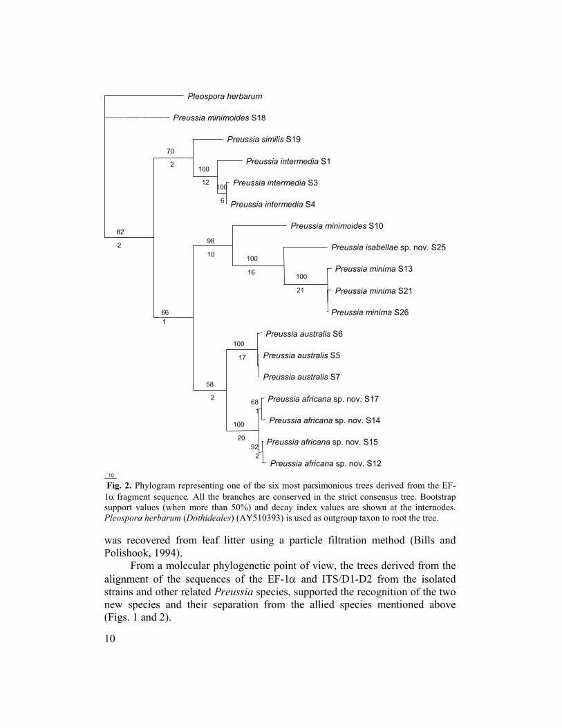
Fig. 2. Phylogram representing one of the six most parsimonious trees derived from the EF-1α fragment sequence. All the branches are conserved in the strict consensus tree. Bootstrap support values (when more than 50%) and decay index values are shown at the internodes. Pleospora herbarum (Dothideales) (AY510393) is used as outgroup taxon to root the tree. was recovered from leaf litter using a particle filtration method (Bills and Polishook, 1994).
From a molecular phylogenetic point of view, the trees derived from the alignment of the sequences of the EF-1α and ITS/D1-D2 from the isolated strains and other related Preussia species, supported the recognition of the two new species and their separation from the allied species mentioned above (Figs. 1 and 2).
10
Fungal Diversity
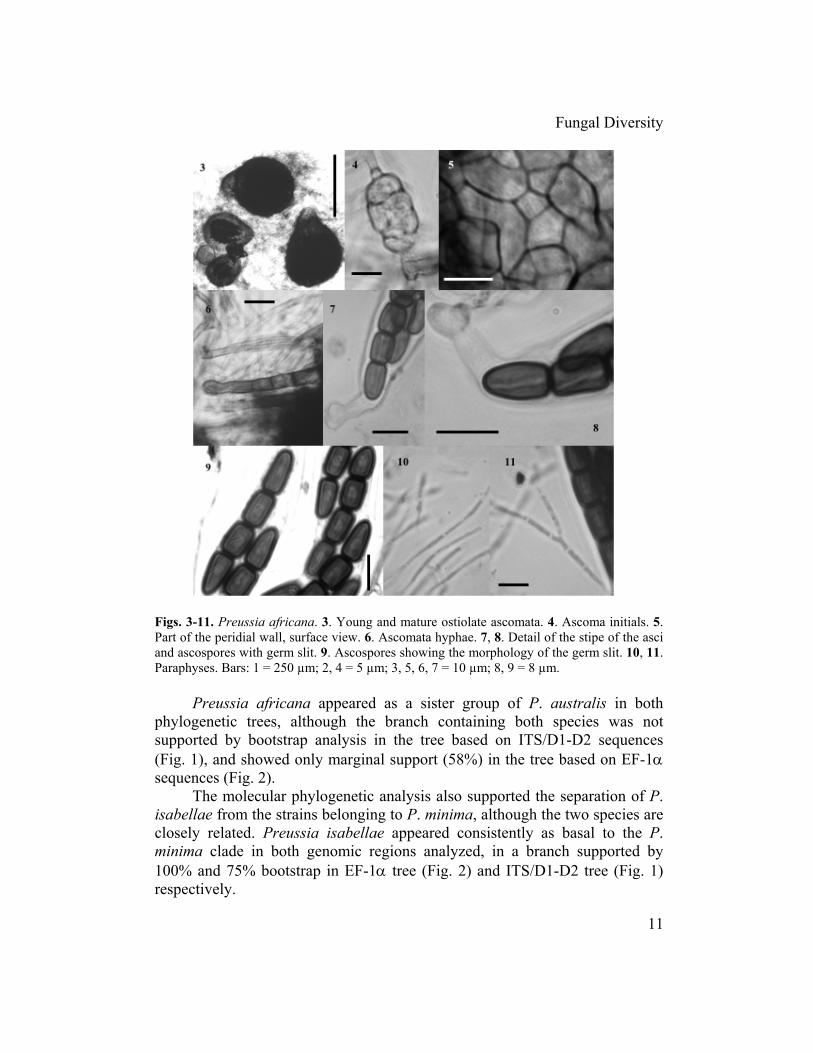
Figs. 3-11. Preussia africana. 3. Young and mature ostiolate ascomata. 4. Ascoma initials. 5. Part of the peridial wall, surface view. 6. Ascomata hyphae. 7, 8. Detail of the stipe of the asci and ascospores with germ slit. 9. Ascospores showing the morphology of the germ slit. 10, 11. Paraphyses. Bars: 1 = 250 µm; 2, 4 = 5 µm; 3, 5, 6, 7 = 10 µm; 8, 9 = 8 µm.
Preussia africana appeared as a sister group of P. australis in both phylogenetic trees, although the branch containing both species was not supported by bootstrap analysis in the tree based on ITS/D1-D2 sequences (Fig. 1), and showed only marginal support (58%) in the tree based on EF-1α sequences (Fig. 2).
The molecular phylogenetic analysis also supported the separation of P. isabellae from the strains belonging to P. minima, although the two species are closely related. Preussia isabellae appeared consistently as basal to the P. minima clade in both genomic regions analyzed, in a branch supported by 100% and 75% bootstrap in EF-1α tree (Fig. 2) and ITS/D1-D2 tree (Fig. 1) respectively.
11
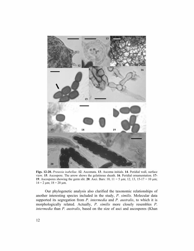
Figs. 12-20. Preussia isabellae. 12. Ascomata. 13. Ascoma initials. 14. Peridial wall, surface view. 15. Ascospore. The arrow shows the gelatinous sheath. 16. Peridial ornamentation. 17-19. Ascospores showing the germ slit. 20. Asci. Bars: 10, 11 = 5 µm; 12, 13, 15-17 = 10 µm; 14 = 2 µm; 18 = 20 µm.
Our phylogenetic analysis also clarified the taxonomic relationships of
another interesting species included in the study, P. similis. Molecular data supported its segregation from P. intermedia and P. australis, to which it is morphologically related. Actually, P. similis more closely resembles P. intermedia than P. australis, based on the size of asci and ascospores (Khan
12
Fungal Diversity
and Cain, 1979). These relationships were, at least, partially confirmed by our molecular data. In the phylogenetic tree derived from the EF-1α sequences (Fig. 2), the strain of P. similis appeared in a basal position with respect to the clade containing the P. intermedia strains, both species grouped in the same branch (70% bootstrap support). On the other hand, in the phylogeny inferred from the ITS/D1-D2 region (Fig. 1), P. similis appeared grouped with P. australis, P. intermedia and the new species P. africana within a clade, although with low bootstrap support (65%).
In conclusion, we have described two new Preussia species based on morphology and molecular evidence. Morphological characters of P. africana suggest it to be an intermediate taxon between P. australis and P. minimoides, whereas P. isabellae resembles P. minima. The taxa included in this work constitute a series of Preussia species that may often overlap in their morphological diagnostic features, being necessary to apply some caution before erecting new Preussia species based solely on morphology. However, the molecular phylogenetic analysis based on the genomic regions selected, justifies the existence of well-defined biological species and reveals their inferred relationships within genus Preussia. Acknowledgements
We want to thank Dr. Jose María Barrasa (Universidad de Alcalá de Henares, Madrid) for expert criticism of the manuscript, Mrs. Asunción Fillola and Dr. Javier Collado (CIBE-MSD, Madrid) for their technical assistance in this study and Dr. Gerald F. Bills (CIBE-MSD, Madrid) for language and grammar review. References Acero, F.J., González, V., Sánchez-Ballesteros, J., Rubio, V., Checa, J., Bills, G.F., Salazar,
O., Platas, G. and Peláez, F. (2004). Molecular phylogenetic studies on the Diatrypaceae based on rDNA-ITS sequences. Mycologia 96: 249-259.
Ahmed, S.I. and Cain, R.F. (1972). Revision of the genera Sporormia and Sporormiella. Canadian Journal of Botany 50: 419-477.
Arenal, F. (2001). Comparación de técnicas genotípicas y fenotípicas para el análisis de la diversidad en hongos, aplicadas a Epicoccum nigrum Link y varias especies del género Sporormiella Ell. & Ev. Ph.D. thesis. Universidad Autónoma de Madrid, Spain.
Arenal, F., Platas, G. and Peláez, F. (2004). Variability of spore length in some species of the genus Preussia (Sporormiella). Mycotaxon 89: 137-151.
Arx, J.A. von (1973). Ostiolate and nonostiolate pyrenomycetes. Proceedings Koninklijke Nederlandse Akademie van Wetenschappen, Series C 76: 289-296.
Arx, J.A. von and Van der Aa, H.A. (1987). Spororminula tenerifae gen. et sp. nov. Transactions of the British Mycological Society 89: 117-120.
Auerswald, B. (1866). In Rabenhorst. Fungi europaei exsiccatti No. 921. Hedwigia 5: 189.
13

Barr, M.E. (1987). Prodomus to class Loculoascomycetes. Published by the Author, Amherst, MA, USA.
Barr, M.E. (1990). Melanommatales (Loculoascomycetes). North American Flora II, part 13: 1-129.
Barrasa, J.M. and Checa, J. (1991). Dothideales del parque natural de Monfragüe (Cáceres). I. Boletín de la Sociedad Micológica de Madrid 15: 91-102.
Bills, G.F., Platas, G., Peláez, F. and Masurekar, P. (1999). Reclassification of a pneumocandin-producing anamorph, Glarea lozoyensis gen. et sp. nov., previously identified as Zalerion arboricola. Mycological Research 103: 179-192.
Bills, G.F. and Polishook, J.D. (1994). Abundance and diversity of microfungi in leaf litter of a lowland rain forest in Costa Rica. Mycologia 86: 187-198.
Bremer, K. (1994). Branch support and tree stability. Cladistics 10: 295-304. Bunyard, B.A., Nicholson, M.S. and Royse, D.J. (1994). A systematic assessment of
Morchella using RFLP analysis of the 28S ribosomal RNA gene. Mycologia 86: 762-772.
Cain, R.F. (1961). Studies of coprophilous ascomycetes. VII. Preussia. Canadian Journal of Botany 39: 1633-1666.
Carbone, I. and Kohn, L.M. (1999). A method for designing primer sets for speciation studies in filamentous Ascomycetes. Mycologia 91: 553-556.
Collado, J., Platas, G. and Peláez, F. (1996). Fungal endophytes in leaves, twigs and bark of Quercus ilex from Central Spain. Nova Hedwigia 63: 347-360.
Felsenstein, J. (1985). Confidence intervals on phylogenies: an approach using the bootstrap. Evolution 39: 783-791.
Fuckel, L. (1866). Fungi rhenani, Suppl. Fasc. 3, 1750. Guarro, J., Abdullah, S.K., Gené, J. and Al-Saadoon, A.H. (1997). A new species of Preussia
from submerged plant debris. Mycological Research 101: 305-308. Heinemann, P. and Rammeloo, J. (1985). De la mesure des spores et de son expression.
Agarica 6: 366-380. Hennequin, S., Ebihara, A., Ito, M., Iwatsuki, K. and Dubuisson, J.Y. (2003). Molecular
systematics of the fern genus Hymenophyllum s.l. (Hymenophyllaceae) based on chloroplastic coding and noncoding regions. Molecular Phylogenetics and Evolution 27: 283-301.
Khan, R.S. and Cain, R.F. (1979). The genera Sporormiella and Sporormia in East Africa. Canadian Journal of Botany 57: 1174-1186.
Kornerup, A. and Wanscher, J.H. (1978). Methuen Handbook of Colour. Methuen, UK. Korolyova, O.V. (2000). New ascomycete species Sporormiella tomilinii Korolyova.
Mycology and Phytopatology 34: 11-13. Lorenzo, L.E. (1994). A new hairy species of Sporormiella. Mycological Research 98: 10-12. Munk, A. (1957). Danish Pyrenomycetes. Dansk Botanisk Arkiv 17: 1-491. Peláez, F., Collado, J., Arenal, F., Basilio, A., Cabello, M.A., Díez, M.T., García, J.B.,
González, del Val A., González, V., Gorrochategui, J., Hernández, P., Martín, I., Platas, G. and Vicente, F. (1998). Endophytic fungi from plants living on gypsum soils as a source of secondary metabolites with antimicrobial activity. Mycological Research 102: 755-761.
Sánchez-Ballesteros, J., González, V., Salazar, O., Acero, J., Portal, M.A., Julián, M., Rubio, V., Bills, G.F., Polishook, J.D., Platas, G., Mochales, S. and Peláez, F. (2000). Phylogenetic study of Hypoxylon and related genera based on ribosomal ITS sequences. Mycologia 92: 964-977.
14
Fungal Diversity
Swofford, D.L. (2001). PAUP*, Phylogenetic Analysis using Parsimony (* and Other Methods). Ver. 4. Sinauer Associates, Sunderland, USA.
Valldosera, M. and Guarro, J. (1990). Estudios sobre hongos coprófilos aislados en España. XV. El género Preussia (Sporormiella). Boletín de la Sociedad Micologica de Madrid 14: 81-94.
White, T.J., Bruns, T., Lee, S. and Taylor, J. (1990). Amplification and direct sequencing of fungal ribosomal RNA genes for phylogenetics. In PCR Protocols: A Guide to Methods and Amplifications (eds. M.A. Innis, D.H. Gelfand, J.J. Sninsky and T.J. White). Academic Press, USA: 315-322.
Young, N.D. and Healy, J. (2003). GapCode automates the use of indel characters in phylogenetic analysis. BMC Bioinformatics 4: 6.
(Received 5 January 2005; accepted 13 September 2005)
15


![MORPHOLOGICAL CHARACTERIZATION AND OF ......Citrus gummosis is caused by several species [1].Phytophthora Morphological differences between some of the species are few and variable,](https://static.fdocuments.in/doc/165x107/60fe04d3377fa214c64c9a60/morphological-characterization-and-of-citrus-gummosis-is-caused-by-several.jpg)