Two cases of accidental hypothermia in Parkinson's disease with
8
J. Neurol. Neurosurg. Psychiat., 1966, 29, 459 Two cases of accidental hypothermia in Parkinson's disease with unusual E.E.G. findings S. S. GUBBAY AND D. D. BARWICK From the Regional Neurological Centre, Newcastle General Hospital, Newcastle upon Tyne Increasing awareness of the clinical entity of accidental hypothermia is apparent from case reports and observations in the medical literature in the last seven years. This paper described two patients, both of whom were sufferers from Parkinson's disease, who developed an episode of accidental hypothermia during which unusual and strikingly similar reversible electro-encephalographic changes were recorded. CASE REPORTS CASE 1 A 73-year-old widow, treated for the past four years with benzhexol and benztropine for Parkinson's 1'sease, was admitted to the Newcastle General Hospital 10 February 1964. For two years previously she had ,own signs of progressive dementia; her personal habits :ind recent memory had deteriorated, and she had diffi- culty in dressing herself and was becoming aggressive in her manner. A week before admission she had felt generally unwell, retired to bed, and thereafter gradually lost her appetite and complained of feeling very cold. On the morning of admission to hospital, she was found in a stuporose condition by relatives with whom she lived. When examined on admission she responded only to painful stimuli and was cyanosed. Her extremities were cold and the rectal temperature was 91°F. (32-8°C.). There was marked generalized muscular rigidity with neck stiffness and retraction. All four limbs were held in flexion and she exhibited a flapping tremor of the hands which could be intensified by sudden passive extension of the fingers as well as a slow tremulousness of all four limbs. Deep tendon reflexes were brisk and both plantar responses were flexor. The pulse rate was 100 per minute and of low volume and blood pressure was 120/80 mm.Hg. Investigations The electrocardiogram was partially obscured by a muscle tremor artefact. There were no J waves and T waves were flat in standard leads I and II. The haemoglobin was 11-7 g./100 ml., W.B.C. 5,200/ c.mm., and E.S.R. 53 mm./hour (Westergren). The serum Na was 130 mEq/l. and serum K 4-4 mEq/l. The serum glutamic oxaloacetic transaminase level was 52 inter- national units/ml., serum glutamic pyruvic transaminase 28 international units/ml., serum amylase 266 Somogyi units, and serum cholesterol 249 mg./100 ml. A chest radiograph showed extensive patchy consolidation in the lower lobe of the left lung as well as several recent frac- tures of the right lower ribs and a fracture of the left ninth rib. The cardiac outline was within normal limits. The cerebrospinal fluid was clear, colourless and under normal pressure, containing 5 mg. of protein/100 ml. and no cells. The Wassermann reaction and Reiter's protein complement-fixation test were negative in the cerebrospinal fluid. A skull radiograph was normal. Progress The patient was treated by very gradual warming in bed with an electric blanket in a well-heated room and received 100 mg. of hydrocortisone intra- muscularly daily as well as a course of intramuscular penicillin. Her rectal temperature rose to 97°F. (36 1 'C.) in 12 hours, by which time the spontaneous movements had practically ceased. By the third day her conscious state had improved markedly and there was considerable lessening of muscular rigidity. By the time of her dis- charge from hospital some five weeks after admission, she had regained her former clinical state and exhibited mask-like facies and mild extrapyramidal rigidity of her limbs with occasional mild Parkinsonian tremor of the upper limbs. Electroencephalographicfindings All E.E.G.s reported here were recorded on 16-channel Offner electroence- phalographs with scalp electrodes placed according to the international 10-20 system. Recordings were nmade with bipolar montages, the time constants being all at 1-0 sec. and the paper speed being 3 cm./second. The first E.E.G. was taken 18 hours after admission (Fig. 1), when the patient was semi-comatose, showing only slight withdrawal reponses to painful stimuli. There was, however, no evidence that the E.E.G. ap- pearances described below were modified by external stimuli. There were frequent diphasic or triphasic dis- charges of rather sharp contour and of up to 300 uV. in amplitude. These wave forms were widespread through- out both hemispheres, occurring synchronously in all channels and regularly for long periods, often at a frequency of about one per second. At times there were intervals of three to four seconds between successive discharges. The distribution of the potentials in some respects resembled those of so-called mid-vertical sharp waves. Between the sharp wave outbursts the record was often flat and featureless, but sometimes there was a little rhythmical activity at 7 c.p.s. of up to 30 /AV. either generalized or posterior in situation. The changes were interpreted as indicating a generalized disturbance with a severe disorder of function in central structures. A marked similarity between the appearances in this record and those seen in patients with subacute spongioform 459 Protected by copyright. on 5 January 2019 by guest. http://jnnp.bmj.com/ J Neurol Neurosurg Psychiatry: first published as 10.1136/jnnp.29.5.459 on 1 October 1966. Downloaded from
Transcript of Two cases of accidental hypothermia in Parkinson's disease with
J. Neurol. Neurosurg. Psychiat., 1966, 29, 459
Two cases of accidental hypothermia in Parkinson'sdisease with unusual E.E.G. findings
S. S. GUBBAY AND D. D. BARWICK
From the Regional Neurological Centre, Newcastle General Hospital, Newcastle upon Tyne
Increasing awareness of the clinical entity ofaccidental hypothermia is apparent from casereports and observations in the medical literature inthe last seven years. This paper described two patients,both of whom were sufferers from Parkinson'sdisease, who developed an episode of accidentalhypothermia during which unusual and strikinglysimilar reversible electro-encephalographic changeswere recorded.
CASE REPORTS
CASE 1 A 73-year-old widow, treated for the past fouryears with benzhexol and benztropine for Parkinson's1'sease, was admitted to the Newcastle General Hospital
10 February 1964. For two years previously she had,own signs of progressive dementia; her personal habits
:ind recent memory had deteriorated, and she had diffi-culty in dressing herself and was becoming aggressive inher manner. A week before admission she had feltgenerally unwell, retired to bed, and thereafter graduallylost her appetite and complained of feeling very cold.On the morning of admission to hospital, she was foundin a stuporose condition by relatives with whom she lived.When examined on admission she responded only to
painful stimuli and was cyanosed. Her extremities werecold and the rectal temperature was 91°F. (32-8°C.).There was marked generalized muscular rigidity withneck stiffness and retraction. All four limbs were held inflexion and she exhibited a flapping tremor of the handswhich could be intensified by sudden passive extension ofthe fingers as well as a slow tremulousness of all fourlimbs. Deep tendon reflexes were brisk and both plantarresponses were flexor. The pulse rate was 100 per minuteand of low volume and blood pressure was 120/80mm.Hg.
Investigations The electrocardiogram was partiallyobscured by a muscle tremor artefact. There were no Jwaves and T waves were flat in standard leads I and II.The haemoglobin was 11-7 g./100 ml., W.B.C. 5,200/c.mm., and E.S.R. 53 mm./hour (Westergren). The serumNa was 130 mEq/l. and serum K 4-4 mEq/l. The serumglutamic oxaloacetic transaminase level was 52 inter-national units/ml., serum glutamic pyruvic transaminase28 international units/ml., serum amylase 266 Somogyiunits, and serum cholesterol 249 mg./100 ml. A chestradiograph showed extensive patchy consolidation in thelower lobe of the left lung as well as several recent frac-
tures of the right lower ribs and a fracture of the leftninth rib. The cardiac outline was within normal limits.The cerebrospinal fluid was clear, colourless and undernormal pressure, containing 5 mg. of protein/100 ml.and no cells. The Wassermann reaction and Reiter'sprotein complement-fixation test were negative in thecerebrospinal fluid. A skull radiograph was normal.
Progress The patient was treated by very gradualwarming in bed with an electric blanket in a well-heatedroom and received 100 mg. of hydrocortisone intra-muscularly daily as well as a course of intramuscularpenicillin. Her rectal temperature rose to 97°F. (36 1 'C.) in12 hours, by which time the spontaneous movements hadpractically ceased. By the third day her conscious statehad improved markedly and there was considerablelessening of muscular rigidity. By the time of her dis-charge from hospital some five weeks after admission,she had regained her former clinical state and exhibitedmask-like facies and mild extrapyramidal rigidity of herlimbs with occasional mild Parkinsonian tremor of theupper limbs.
Electroencephalographicfindings All E.E.G.s reportedhere were recorded on 16-channel Offner electroence-phalographs with scalp electrodes placed according tothe international 10-20 system. Recordings were nmadewith bipolar montages, the time constants being all at1-0 sec. and the paper speed being 3 cm./second.The first E.E.G. was taken 18 hours after admission
(Fig. 1), when the patient was semi-comatose, showingonly slight withdrawal reponses to painful stimuli.There was, however, no evidence that the E.E.G. ap-pearances described below were modified by externalstimuli. There were frequent diphasic or triphasic dis-charges of rather sharp contour and of up to 300 uV.in amplitude. These wave forms were widespread through-out both hemispheres, occurring synchronously in allchannels and regularly for long periods, often at afrequency of about one per second. At times there wereintervals of three to four seconds between successivedischarges. The distribution of the potentials in somerespects resembled those of so-called mid-vertical sharpwaves. Between the sharp wave outbursts the recordwas often flat and featureless, but sometimes there was alittle rhythmical activity at 7 c.p.s. of up to 30 /AV. eithergeneralized or posterior in situation. The changes wereinterpreted as indicating a generalized disturbance with asevere disorder of function in central structures. A markedsimilarity between the appearances in this record andthose seen in patients with subacute spongioform
459
Protected by copyright.
on 5 January 2019 by guest.http://jnnp.bm
j.com/
J Neurol N
eurosurg Psychiatry: first published as 10.1136/jnnp.29.5.459 on 1 O
ctober 1966. Dow
nloaded from

S. S. Gubbay and D. D. Barwick
13 i
IOOgaV I Isec.
w 9 \ <_~~~I
=0,V
-- - - P r - * r-
FIG. 1. Initial E.E.G. in first patient shows generalized repetitive and triphasic sharp complexes synchronous in anlchannels. These occur at intervals ofabout oneper second.
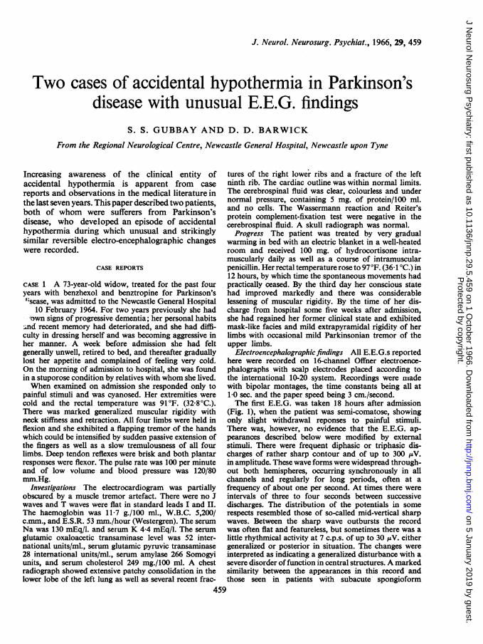
encephalopathy of the Nevin-Jones type (Jones andNevin, 1954) was noted.A second E.E.G. (Fig. 2) obtained after a further 48
hours, when the patient was responsive but confused,was greatly improved. The dominant activity was in thealpha frequency range. There were none of the sharpdischarges previously seen, but there were repeatedgeneralized and fairly symmetrical delta waves usuallytriphasic in outline and synchronous in all channels.These occurred at intervals varying between 3 and 10seconds, and were unassociated with any external arous-ing stimuli or readily detectable changes in level ofawareness, though they did not appear with the eyesopen.
CASE 2 The patient was a 61-year-old spinster who wasoriginally admitted to the Newcastle General Hospitalin March 1963. Shortly before admission she haddeveloped stiffness of gait, fixity of facial expression,and mild tremor of both hands. A diagnosis of Parkin-son's disease was made and she was given benzhexol.Six days after starting treatment she became drowsyand withdrawn and later confused and hallucinateddespite alterations in her drug therapy; because of this
deterioration she was admitted to hospital. Her facewas expressionless and her voice monotonous. There wasa slight slurring dysarthria with increased tone in all fourlimbs, and cogwheel rigidity and tremor of the upperlimbs. The glabellar tap sign was positive and the deeptendon reflexes were brisk with flexor plantar responses.The blood pressure was 170/110 mm.Hg. After withdrawalof all drug therapy, her mental state rapidly improved tonormal.Apart from the persistence of the Parkinsonian
symptoms, the patient remained well until mid-December1963, when with the onset of the very cold weather,she became slow and more unsteady on her feet. Becauseshe appeared to be depressed, she received nortryptilene25 mg. twice daily. Gradually over the next few weeksthe patient became slower, more withdrawn and drowsy,and took to her bed. For two days before her re-admnissionto Newcastle General Hospital on 22 January 1964 shehad been lying mute and staring into space unrespon-sively. When examined at home she was lying in anextremely cold room and her skin felt very cold.On admission to hospital the patient was conscious, but
mute. She was warmer to the touch in the warmly heatedroom, and her temperature per axilla was 96-6°F.
460
Protected by copyright.
on 5 January 2019 by guest.http://jnnp.bm
j.com/
J Neurol N
eurosurg Psychiatry: first published as 10.1136/jnnp.29.5.459 on 1 O
ctober 1966. Dow
nloaded from
Two cases of accidental hypothermia in Parkinson's disease with unusual E.E.G. findings
100IV IEL
-| e v 0 v r- f , v .
P ,
FIG. 2. A second recordfrom the first patient taken a little over 48 hours after the first. Alpha activity now dominatesthe record but occasional triphasic, generalized slow wave outbursts are seen.
(35 9°C.). The blood pressure was 120/80 mm.Hg andpulse rate 70 per minute and regular. There was a pecu-liar tremulousness of the limbs and face, and a flappingtremor of the hands. These abnormal movements couldbe accentuated by tapping the muscles or stretching thelimbs. The upper limbs were held in flexion and pronation,and the lower limbs in extension, and there was markedrigidity of all four limbs and neck muscles. The deeptendon reflexes were brisk and equal, and both plantarresponses were flexor.The spontaneous movements and hyperirritability of
the muscles gradually lessened over the next 48 hours,but otherwise her general clinical state remained muchthe same for seven days when she regained her speechand showed remarkable improvement, but was slightlyconfused mentally. Her axillary temperature neverfell below the level recorded on admission. Withinfour weeks she became fully ambulant, and had revertedto a normal state, but still showed the clinical features ofParkinsonism.
Investigations The haemoglobin level was 13-2g./100 ml., W.B.C. 5,800/c.mm., and E.S.R. 23 mm./hour.A chest radiograph showed clear lung fields and slightcardiomegaly, and a skull radiograph was normal.The serum calcium was 10-4 mg./100 ml., phosphate3-0 mg./100 ml., Na 143 mEq./l., K 5 9 mEq./l., and7
Cl 95 mEq./l. The cerebrospinal fluid was clear andcolourless and under normal pressure, with a proteincontent of 73 mg./100 ml., and no pleocytosis. TheWassermann reaction and Reiter's complement-fixationtest were negative in blood and cerebrospinal fluid. Theonly electrocardiographic abnormality was that of leftventricular hypertrophy.
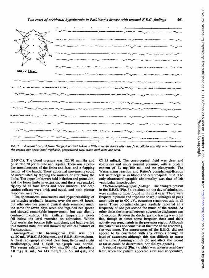
Electroencephalographic findings The changes presentin the E.E.G. (Fig. 3), obtained on the day of admission,were similar to those found in the first case. There werefrequent diphasic and triphasic sharp discharges of peakamplitude up to 400 pV., occurring synchronously in allareas. These potential changes regularly repeated at afrequency of one per second for much of the record. Atother times the interval between successive discharges was1-5 seconds. Between the discharges the tracing was oftenflat, though at times some irregular theta and deltaactivity was seen, mainly in the posterior region. Althoughthe patient was not comatose at the time of the recording,she was mute. The appearances of the E.E.G. did notappear to be correlated with any obvious change inlevel of awareness although this was difficult to assessat the time. Arousing stimuli did not affect the recordas far as could be determined, nor did eye-opening.A second record (Fig. 4), which was taken several days
later, when the patient appeared alert and cooperative,
461
Protected by copyright.
on 5 January 2019 by guest.http://jnnp.bm
j.com/
J Neurol N
eurosurg Psychiatry: first published as 10.1136/jnnp.29.5.459 on 1 O
ctober 1966. Dow
nloaded from

S. S. Gubbay and D. D. Barwick
FIG. 3. First record from case 2 which show repetitive di- and triphasic sharp waves synchronous in all channels andrepeating regularly at about one per second.
no longer showed repetitive sharp waves. With the eyesclosed there was a generalized excess of slow activityconsisting ofmainly irregular, but occasionally rhythmical,theta and delta discharges. The rhythmical slow activitywas most marked in the central regions, but at times wasgeneralized, synchronous and symmetrical, and occurredin a rather paroxysmal fashion. With the eyes open therecord was dominated by very low-voltage, irregularslow activity.The E.E.G. showed a considerable improvement
over the initial record, but was still thought to indicatea diffuse disorder of cerebral metabolism.
Further investigations Adequate temperature record-ings on this patient were not obtained for some timeafter admission to the warm hospital environment, as herclinical state did not at once suggest the diagnosis ofaccidental hypothermia. Because it seemed importantfrom the point of view of future management that a firmdiagnosis be reached in this patient, the following investi-gations were performed. The patient lay in a room cooledto 50°F. (10°C.) with one covering blanket and theE.E.G. was recorded continuously as was the rectaltemperature which was initially 100°F. (37 8°C.). Aftertwo hours of exposure under these conditions, the rectaltemperature fell to 96-0°F. (35 6°C.) and it was thoughtunjustifiable to lower the temperature below this levelin case her former serious clinical state was provoked.During this two-hour period the patient felt cold anddrowsy and eventually slept but was easily rousable.She complained of a similar unpleasant subjectivesensation to that experienced on admission to hospital.There was no overt shivering, but she briefly exhibited
some slow involuntary movements of her feet resemblingthose seen on admission.
Initially (rectal temperature 99°F. (37 2°C.) the E.E.G.(Fig. 5) was largely normal, though there were occasionalbrief runs of 4 c.p.s. activity, sometimes associated withsharp transients in both temporal regions. As hertemperature fell, the record became more abnormal withintermittent generalized outbursts of delta activity andoccasional sharp transients appeared (Figs. 6 and 7).The E.E.G. appearances, however, did not resembleentirely those seen in the initial record taken from thispatient, nor did the patient develop the striking clinicalmanifestations evident at the time of her admission.
DISCUSSION
There is a very strong circumstantial evidence thatthe second patient was suffering from the effectsof accidental hypothermia on her second admissionto hospital. First, some of her subjective and objectivechanges could be reproduced by inducing even mildhypothermia, although only minor abnormalitiescould be reproduced in her E.E.G. Secondly,despite experimental lowering of her body tempera-ture, shivering was not provoked which suggeststhat she had some derangement of the mechanismof regulation of body temperature. Her originalclinical state in many ways was similar to that of thefirst patient and certain features, such as generalizedrigidity (Duguid, Simpson, and Stowers, 1961;
462
-J..
IOONV
"A,r-V
AV...
Protected by copyright.
on 5 January 2019 by guest.http://jnnp.bm
j.com/
J Neurol N
eurosurg Psychiatry: first published as 10.1136/jnnp.29.5.459 on 1 O
ctober 1966. Dow
nloaded from

10 ' -* v W
16 IS
IOOpV sec.
FIG. 4. Recordfrom case 2, taken three days after the initial tracing, shows a generalized excess of slow activity but noparoxysms.
4._
IOopV IIsec.
TEMP 99 ( R) I st
FIG. 5. Case 2, before cooling. Rectal temperature 99°F. (372°C.). Much of the record is normal. Occasional sloivwaves and sharp transients are seen in the temporal regions.
Protected by copyright.
on 5 January 2019 by guest.http://jnnp.bm
j.com/
J Neurol N
eurosurg Psychiatry: first published as 10.1136/jnnp.29.5.459 on 1 O
ctober 1966. Dow
nloaded from

464 S. S. Gubbay and D. D. Barwick
loop v Isec.
TEMP. 97-3 8th
FIG. 6. Case 2. Rectal temperature 97-30F. (36&30C.). Runs ofslow waves and some sharp transients are seen.
Is 2> Vs --<-*a,
16" \ 5 th
IOOPV I Isec. T
FIG. 7. Case 2. Rectal temperature 965F. (3580C.). A fairly generalizedparoxysm of mixed slow activity and sharptransients is seen. At this stage some involuntary movement ofthe legs occurred.
Protected by copyright.
on 5 January 2019 by guest.http://jnnp.bm
j.com/
J Neurol N
eurosurg Psychiatry: first published as 10.1136/jnnp.29.5.459 on 1 O
ctober 1966. Dow
nloaded from

Two cases of accidental hypothermia in Parkinson's disease with unusual E.E.G. findings
Rees, 1958), flapping tremor, and neck stiffness(Rosin and Exton-Smith, 1964) have been observedpreviously in hypothermic patients. Moreover, theE.E.G. findings in both our cases were remarkablysimilar. The E.E.G. in the first patient was recordedduring the hypothermic phase and although J wavesdescribed by Osborn (1953) were not present, QTcwas prolonged, as in Emslie-Smith's (1958) experi-ence, and muscle tremor artefact was present as hasbeen observed by Duguid et al. (1961).The striking E.E.G. abnormalities in the initial
records obtained from both patients are of interest.They do not resemble those seen in induced hypo-thermia as carried out in preparation for cardiacand brain surgery (Scott, 1955; Pampiglione andWaterston, 1958). The usual changes described insuch cases take the form of gradual lowering offrequency and decrease in amplitude. However,these appearances may be in part due to anaestheticor other drugs concurrently administered. In someprevious reports of accidental hypothermia theE.E.G. has been markedly abnormal, and variousauthors (Bauer, 1954; Hockaday, Granston, Cooper,and Mottram, 1962) have demonstrated the presenceof hypothalamic lesions at necropsy in patients withaccidental hypothermia. Duff, Farrant, Leveaux,and Wray (1961) described E.E.G. studies in one oftheir cases which showed gross generalized abnor-malities thought to be unassociated with epilepsy.The changes described appear to be quite differentfrom those found in the two patients reported here.Hockaday et al. (1962) performed E.E.G.s on theirthree patients, and found grossly abnormal recordswith bilaterally synchronous high-voltage slow waves.Again these descriptions differ from our findings,and must be considered in the light of the more com-plicated cerebral pathology that was present in theircases.We have already commented upon the similarity
of the appearances present in the initial recordsobtained from our two cases to those seen in theNevin-Jones type of encephalopathy. In fact thisdiagnosis was also considered in the second patientbecause the abnormal movements were thought torepresent the myoclonic jerks described in that con-dition. It has been said that E.E.G. appearances ofthis type which occur in middle age are usually ofgloomy prognostic significance, and are associatedwith a rapidly progressive dementia and a fataloutcome (Abbott, 1959; Lesse, Hoefer, and Austin,1958; Pallis and Spillane, 1957). Similar changes havebeen reported to occur transiently in a case of cere-bral fat embolism (Muller and Klingler, 1965).The abnormal E.E.G.s in our two cases cannot
simply be due to the presence of Parkinsonism. Inthis condition the E.E.G. changes (England,
Schwab, and Peterson, 1959) are usually relativelyslight, and consist of posterior rhythms below 8 c.p.s.and a slight theta excess.
It seems possible that the combination of hypo-thermia and basal ganglion disease, in the two casesreported here, resulted in a similar though temporaryneurophysiological derangement to that associatedwith the Nevin-Jones syndrome.
Histopathological changes may be found in thehypothalamic nuclei in patients with Parkinson'sdisease (Greenfield, 1963) so that it is to be antici-pated that dysthermic episodes might occur in suchpatients. Autonomic disturbances and excessivetolerance to cold are well described clinical featuresin this disorder (Brain, 1962) and the latter feature,particularly, could be a predisposing factor toaccidental hypothermia. It is true that other pre-disposing factors also existed in our two patients.In the first case, the patient was elderly and senileand had bronchopulmonary infection (Duguidet al., 1961); the second was receiving a drugpharmacologically related to imipramine and chlor-promazine (Prescott, Peard, and Wallace, 1962;McGrath, and Paley, 1960) and was living in aninadequately heated room.
SUMMARY AND CONCLUSIONS
Two patients with Parkinson's disease who developedaccidental hypothermia are described. The E.E.G.findings in these two cases were remarkably similarand consisted of appearances not unlike those de-scribed in subacute spongioform encephalopathy.
These E.E.G. changes were reversible and werecommensurate with general clinical improvementrather than with restoration ofnormal body tempera-ture. It is suggested that Parkinson's disease may be afurther predisposing cause of accidental hypother-mia and this may in part be due to involvement ofthe hypothalamus in the disease process.
We should like to thank Dr. J. N. Walton for hisencouragement and kind permission to publish thereports of these two patients, who were under his careat the Regional Neurological Centre, and Miss B. P.Longley for technical assistance.
REFERENCES
Abbott, J. (1959). The E.E.G. in Jakob-Creutzfeldt's disease. Electro-enceph. clin. Neurophysiol., 11, 184-185.
Bauer, H. G. (1954). Endocrine and other clinical manifestations ofhypothalamic disease. J. clin. Endocr., 14, 13-31.
Brain, W. R. (1962). Diseases of the Nervous System, 6th ed., p. 476.Oxford University Press, London.
Duff, R. S., Farrant, P. C., Leveaux, V. M., and Wray, S. M. (1961).Spontaneous periodic hypothermia. Quart. J. Med., 30, 329-338.
Duguid, H., Simpson, R. G., and Stowers, J. M. (1961). Accidentalhypothermia. Lancet, 2, 1213-1219.
465
Protected by copyright.
on 5 January 2019 by guest.http://jnnp.bm
j.com/
J Neurol N
eurosurg Psychiatry: first published as 10.1136/jnnp.29.5.459 on 1 O
ctober 1966. Dow
nloaded from

S. S. Gubbay and D. D. Barwick
Emslie-Smith, D. (1958). Accidental hypothermia-a common con-dition with a pathognomonic electrocardiogram. Ibid., 2,492-495.
England, A. C., Schwab, R. S., and Peterson, E. (1959). The electro-encephalogram in Parkinson's syndrome. Electroenceph. cin.Neurophysiol., 11, 723-731.
Greenfield, J. G. (1963). Neuropathology, 2nd ed., p. 583. Arnold,London.
Hockaday, T. D. R., Cranston, W. I., Cooper, K. E., and Mottram,R. F. (1962). Temperatuw regulation in chronic hypothermia.Lancet, 2, 428-432.
Jones, D. P., and Nevin, S. (1954). Rapidly progressive cerebral de-generation (subacute vascular encephalopathy) with mentaldisorder, focal disturbances and myoclonic epilepsy. J. Neurol.Psychiat., 17, 148-159.
Lesse, S., Hoefer, P. F. A., and Austin, J. H. (1958). The eletcro-encephalogram in diffuse encephalopathies. Arch. Neurol.Psychiat. (Chic.), 79, 359-375.
McGrath, M. D., and Paley, R. G. (1960). Hypothermia induced in amyxoedematous patient by imipramine hydrochloride. Brit.med. J., 2, 1364.
Muller, H. R., and Klingler, M. (1965). The electroencephalogramin cerebral fat embolism. Electroenceph. clin. Neurophysiol.,18, 178-86.
Osborn, J. J. (1953). Experimental hypothermia; respiratory andblood pH changes in relation to cardiac function. Amer. J.Physiol., 175, 389-398.
Pallis, C. A., and Spillane, J. D. (1957). A subacute progressiveencephalopathy with mutism, hypokinesia, rigidity, and myo-
clonus. Quart. J. Med., 26, 349-373.Pampiglione, G., and Waterston, D. J. (1958). Preliminary E.E.G.
observations during partial and complete occlusion of cerebralblood flow. Electroenceph. cdin. Neurophysiol., 10, 354.
Prescott, L. F., Peard, M. C., and Wallace, I. R. (1962). Accidentalhypothermia: a common condition. Brit. med. J., 2, 1367-1370.
Rees, J. R. (1958). Accidental hypothermia. Lancet, 1, 556-559.Rosin, A. J., and Exton-Smith, A. N. (1964). Clinical features of
accidental hypothermia, with some observations on thyroidfunction. Brit. med. J., 1, 16-19.
Scott, J. W. (1955). The E.E.G. during hypothermia. Electroenceph.clin. Neurophysiol., 7, 466.
466
Protected by copyright.
on 5 January 2019 by guest.http://jnnp.bm
j.com/
J Neurol N
eurosurg Psychiatry: first published as 10.1136/jnnp.29.5.459 on 1 O
ctober 1966. Dow
nloaded from