Turning Low-Molecular-Weight Drugs into Prolonged Acting Prodrugs by Reversible Pegylation: A Study...
6
Turning Low-Molecular-Weight Drugs into Prolonged Acting Prodrugs by Reversible Pegylation: A Study with Gentamicin Yonit Marcus, † Keren Sasson, † Mati Fridkin,* ,‡ and Yoram Shechter* ,† Departments of Biological Chemistry and Organic Chemistry, The Weizmann Institute of Science, RehoVot 76100, Israel ReceiVed March 9, 2008 Pegylation is a powerful technology to prolong the action of proteins in vivo, but it is impractical for low- molecular-weight (LMW) drugs, which are usually inactivated upon such modification. Here, we have applied a recently developed strategy of reversible pegylation to gentamicin, a LMW antibiotic. Variable length polyethyleneglycol (PEG-SH) chains were covalently linked to gentamicin using two heterobifunctional agents, each containing a spontaneously hydrolyzable bond. The inactive derivatives regained full antibacterial potency upon incubation under physiological conditions in vitro, and following systemic administration to rats, they released native active gentamicin with half-lives 7- to 15-fold greater than those of systemically administered nonderivatized gentamicin. In conclusion, reversibly pegylated prodrug derivatives of gentamicin were found to be capable of releasing gentamicin for prolonged periods in vivo. Most importantly, the major drawback of conventional pegylation, namely, the loss of pharmacological potency following irreversible derivatization, has been overcome. Introduction Most low-molecular-weight (LMW) drugs are short-lived species in the circulatory system, being rapidly eliminated by glomerular filtration in the kidney. 1 This therapeutic drawback is even more severe when such drugs cannot be absorbed orally and therefore must be introduced directly into the circulatory system. It is also a major obstacle for drugs having high indices of toxicity, which therefore should be present in the circulation at low but therapeutic levels for long durations. Pegylated therapeutic proteins often exhibit clinical properties superior to those of their respective unmodified proteins. Pegylated proteins resist proteolysis, show increased solubility, and are shielded from the immune system. 2–4 Most notably, pegylated proteins turn into long-lived species in vivo, a feature attributed predominantly to decreased clearance rate of the conjugates from the circulatory system by kidney filtration. 3–6 However, substantial prolongation in vivo requires the conjuga- tion of several LMW PEG a chains or a single high-molecular- weight (HMW) PEG. This covalent introduction of PEG chains to protein drugs often results in a marked reduction in biological/ pharmacological potency. For example, a currently used 40 kDa PEG-interferon-R2A exhibits only 7% of the activity of the native cytokine, thus necessitating the administration of high doses. 7 We have recently developed an approach, termed reversible- pegylation, and demonstrated its ability to prolong the actions of peptide and protein drugs, all of which undergo substantial inactivation by conventional pegylation (refs 8–11, reviewed in ref 12). Here, we investigated whether this approach could be extended to protract the action of LMW drugs in vivo. We, a priori, assumed that with regard to this drug category, conventional pegylation would, more often than not, result in an inactive product, thereby limiting the usefulness of this approach. As a prototype for this drug category, we selected gentamicin, a LMW nonorally absorbed agent used for the treatment of many serious Gram-negative bacterial infections. 13 Since a major fraction of systemically injected gentamicin reaches the urine within a short period after administration (i.e., this study), we have developed noninvasive procedures that enabled us to investigate several pharmacokinetic parameters, including the time in which the reversibly pegylated gentamicin derivatives release active gentamicin. Experimental Section Materials. Gentamicin sulfate, cystamine-di-HCl, dithiothreitol, and 5,5′-dithiobis (2-nitrobenzoic acid) were purchased from Sigma. PEG 40 -OSu (Y-shape PEG-NHS ester, MW 40 kDa, Y-NHS-40K) was the product of Jenkem Technology Co. (Allen, TX). PEG 20 - SH (CH 3 O-PEG 20 -SH) was purchased from RAPP Polymere GmbH (Tubingen, Germany), and PEG 5 -MAL (mPEG-maleimide, MW 5000) was from Shearwater Group Inc. (Ra’anana, Israel). All other materials used in this study were of analytical grade. Chemical Procedures. The syntheses of 9-hydroxymethyl- 2(amino-3-maleimidopropionate)fluorene-N-hydroxysuccinimide es- ter (MAL-Fmoc-OSu) and of 9-hydroxymethyl-7-(amino-3-male- imidopropionate)-2-sulfo-N-hydroxysuccinimide (MAL-FMS-OSu) are described in detail in ref 8. Both syntheses were initiated from 9-hydroxymethyl-2-aminofluorene, following three and four steps, respectively, with overall yields of 84% and 65%, respectively. PEG 40 -SH. PEG 40 -SH was prepared by dissolving PEG 40 -OSu at a concentration of 40 mg/mL in 0.1 M NaHCO 3 containing 1 M * To whom correspondence should be addressed. For M.F.: phone, 972- 8-9342505; fax, 972-8-9344142; e-mail, [email protected]. For Y.S.: phone, 972-8-9344530; fax, 972-8-9344118; e-mail, y.shechter@ weizmann.ac.il. † Department of Biological Chemistry. ‡ Department of Organic Chemistry. a Abbreviations: PEG, polyethylene glycol; IC 50 , inhibitory concentration (half-maximal) needed to arrest E. coli replication; OD, optical density; PBS, phosphate buffered saline; MAL-Fmoc-OSu, 9-hydroxymethyl-2- (amino-3-maleimidopropionate)fluorene-N-hydroxysuccinimide; MAL-FMS- OSu, 2-sulfo-9-hydroxymethyl-7-(amino-3-maleimidopropionate)-fluorene- N-hydroxysuccinimide; PEG 40-SH, a 40 kDa branched polyethylene glycol linked to cystamine; PEG 5 -SH, a 5 kDa polyethylene glycol chain containing a sulfhydryl moiety; PEG 20-SH, a 20 kDa polyethylene glycol chain containing a sulfhydryl moiety; DTNB, 5,5′-dithiobis(2-ntirobenzoic acid); HSA, human serum albumin; PEG 5 -, PEG 20 -, PEG 40 -Fmoc-gentamicin, 1:1 conjugates of PEG 5 , PEG 20 , and PEG 40 -SH linked to gentamicin through MAL-Fmoc-OSu, respectively; PEG 5 , PEG 20 , PEG 40 -FMS-gentamicin, 1:1 conjugates of PEG 5 , PEG 20 , and PEG 40 -SH linked to gentamicin through MAL-FMS-OSu, respectively. J. Med. Chem. 2008, 51, 4300–4305 4300 10.1021/jm8002558 CCC: $40.75 2008 American Chemical Society Published on Web 06/25/2008
Transcript of Turning Low-Molecular-Weight Drugs into Prolonged Acting Prodrugs by Reversible Pegylation: A Study...

Turning Low-Molecular-Weight Drugs into Prolonged Acting Prodrugs by ReversiblePegylation: A Study with Gentamicin
Yonit Marcus,† Keren Sasson,† Mati Fridkin,*,‡ and Yoram Shechter*,†
Departments of Biological Chemistry and Organic Chemistry, The Weizmann Institute of Science, RehoVot 76100, Israel
ReceiVed March 9, 2008
Pegylation is a powerful technology to prolong the action of proteins in vivo, but it is impractical for low-molecular-weight (LMW) drugs, which are usually inactivated upon such modification. Here, we have applieda recently developed strategy of reversible pegylation to gentamicin, a LMW antibiotic. Variable lengthpolyethyleneglycol (PEG-SH) chains were covalently linked to gentamicin using two heterobifunctionalagents, each containing a spontaneously hydrolyzable bond. The inactive derivatives regained full antibacterialpotency upon incubation under physiological conditions in vitro, and following systemic administration torats, they released native active gentamicin with half-lives 7- to 15-fold greater than those of systemicallyadministered nonderivatized gentamicin. In conclusion, reversibly pegylated prodrug derivatives of gentamicinwere found to be capable of releasing gentamicin for prolonged periods in vivo. Most importantly, themajor drawback of conventional pegylation, namely, the loss of pharmacological potency following irreversiblederivatization, has been overcome.
Introduction
Most low-molecular-weight (LMW) drugs are short-livedspecies in the circulatory system, being rapidly eliminated byglomerular filtration in the kidney.1 This therapeutic drawbackis even more severe when such drugs cannot be absorbed orallyand therefore must be introduced directly into the circulatorysystem. It is also a major obstacle for drugs having high indicesof toxicity, which therefore should be present in the circulationat low but therapeutic levels for long durations.
Pegylated therapeutic proteins often exhibit clinical propertiessuperior to those of their respective unmodified proteins.Pegylated proteins resist proteolysis, show increased solubility,and are shielded from the immune system.2–4 Most notably,pegylated proteins turn into long-lived species in vivo, a featureattributed predominantly to decreased clearance rate of theconjugates from the circulatory system by kidney filtration.3–6
However, substantial prolongation in vivo requires the conjuga-tion of several LMW PEGa chains or a single high-molecular-weight (HMW) PEG. This covalent introduction of PEG chainsto protein drugs often results in a marked reduction in biological/pharmacological potency. For example, a currently used 40 kDa
PEG-interferon-R2A exhibits only 7% of the activity of thenative cytokine, thus necessitating the administration of highdoses.7
We have recently developed an approach, termed reversible-pegylation, and demonstrated its ability to prolong the actionsof peptide and protein drugs, all of which undergo substantialinactivation by conventional pegylation (refs 8–11, reviewedin ref 12). Here, we investigated whether this approach couldbe extended to protract the action of LMW drugs in vivo. We,a priori, assumed that with regard to this drug category,conventional pegylation would, more often than not, result inan inactive product, thereby limiting the usefulness of thisapproach.
As a prototype for this drug category, we selected gentamicin,a LMW nonorally absorbed agent used for the treatment of manyserious Gram-negative bacterial infections.13 Since a majorfraction of systemically injected gentamicin reaches the urinewithin a short period after administration (i.e., this study), wehave developed noninvasive procedures that enabled us toinvestigate several pharmacokinetic parameters, including thetime in which the reversibly pegylated gentamicin derivativesrelease active gentamicin.
Experimental Section
Materials. Gentamicin sulfate, cystamine-di-HCl, dithiothreitol,and 5,5′-dithiobis (2-nitrobenzoic acid) were purchased from Sigma.PEG40-OSu (Y-shape PEG-NHS ester, MW 40 kDa, Y-NHS-40K)was the product of Jenkem Technology Co. (Allen, TX). PEG20-SH (CH3O-PEG20-SH) was purchased from RAPP Polymere GmbH(Tubingen, Germany), and PEG5-MAL (mPEG-maleimide, MW5000) was from Shearwater Group Inc. (Ra’anana, Israel). All othermaterials used in this study were of analytical grade.
Chemical Procedures. The syntheses of 9-hydroxymethyl-2(amino-3-maleimidopropionate)fluorene-N-hydroxysuccinimide es-ter (MAL-Fmoc-OSu) and of 9-hydroxymethyl-7-(amino-3-male-imidopropionate)-2-sulfo-N-hydroxysuccinimide (MAL-FMS-OSu)are described in detail in ref 8. Both syntheses were initiated from9-hydroxymethyl-2-aminofluorene, following three and four steps,respectively, with overall yields of 84% and 65%, respectively.
PEG40-SH. PEG40-SH was prepared by dissolving PEG40-OSuat a concentration of 40 mg/mL in 0.1 M NaHCO3 containing 1 M
* To whom correspondence should be addressed. For M.F.: phone, 972-8-9342505; fax, 972-8-9344142; e-mail, [email protected]. ForY.S.: phone, 972-8-9344530; fax, 972-8-9344118; e-mail, [email protected].
† Department of Biological Chemistry.‡ Department of Organic Chemistry.a Abbreviations: PEG, polyethylene glycol; IC50, inhibitory concentration
(half-maximal) needed to arrest E. coli replication; OD, optical density;PBS, phosphate buffered saline; MAL-Fmoc-OSu, 9-hydroxymethyl-2-(amino-3-maleimidopropionate)fluorene-N-hydroxysuccinimide; MAL-FMS-OSu, 2-sulfo-9-hydroxymethyl-7-(amino-3-maleimidopropionate)-fluorene-N-hydroxysuccinimide; PEG40-SH, a 40 kDa branched polyethylene glycollinked to cystamine; PEG5-SH, a 5 kDa polyethylene glycol chain containinga sulfhydryl moiety; PEG20-SH, a 20 kDa polyethylene glycol chaincontaining a sulfhydryl moiety; DTNB, 5,5′-dithiobis(2-ntirobenzoic acid);HSA, human serum albumin; PEG5-, PEG20-, PEG40-Fmoc-gentamicin, 1:1conjugates of PEG5, PEG20, and PEG40-SH linked to gentamicin throughMAL-Fmoc-OSu, respectively; PEG5, PEG20, PEG40-FMS-gentamicin, 1:1conjugates of PEG5, PEG20, and PEG40-SH linked to gentamicin throughMAL-FMS-OSu, respectively.
J. Med. Chem. 2008, 51, 4300–43054300
10.1021/jm8002558 CCC: $40.75 2008 American Chemical SocietyPublished on Web 06/25/2008

cystamine-di-HCl. The reaction was carried out for 2 h at 25 °C.The product obtained was dialyzed overnight against 0.1 MNaHCO3, treated with 30 mM dithiothreitol (25 °C, 1 h), andredialyzed against 10 mM HCl-10 mM ascorbic acid. The PEG40-SH obtained contained 1 mol of sulfhydryl moiety per mol of PEG40
as determined by reaction with DTNB. The product was lyophilizedand kept at 7 °C until used.
PEG5-SH. PEG5-SH was prepared by dissolving PEG5-MAL inwater at 20 mg/mL (4 mM). Solid dithiothreitol (15.4 mg) wasthen added (final concentration 100 mM, 25 molar excess overPEG5-MAL). The reaction was carried out for 20 min at 25 °Cfollowed by extensive dialysis against 10 mM HCl and lyophiliza-tion. PEG5-SH at a concentration of 5.2 mg/mL (∼1 µmol/mL)contained 0.9 ( 0.03 µmol/mL of sulfhydryl moiety as determinedby reaction with DTNB. The lyophilized material was stable forweeks at 7 °C.
Preparation of PEG5,20,40/Fmoc/FMS Derivatives of Gentami-cin. PEG5-SH, PEG20-SH, or PEG40-SH (1 µmol) was dissolved in1.0 mL of 0.1 M NaHCO3 containing 50 mg of gentamicin (0.1mmol/mL, 100 molar excess over PEG-SH). Either MAL-Fmoc-OSu (1.1 µmol, 55 µL from a fresh solution of 10 mg/mL in DMF)or MAL-FMS-OSu (1.1 µmol, 64 µL from a fresh solution of 10mg/mL in DMF) was then added to the stirred solutions. Reactionswere carried out over a period of 3 h at 25 °C. Extensive dialysisagainst water was followed with several changes over a period of24 h. All reversibly pegylated gentamicin derivatives prepared bythis procedure (namely, PEG5,20,40-Fmoc/FMS-gentamicin) were 1:1PEG-gentamicin conjugates. The concentration of each conjugatewas determined both by its absorbance at 280 nm and by aminoacid analyses following acid hydrolyses. The conjugates absorb at280 nm with a molar extinction coefficient of 21 200 L mol-1 cm-1.Absorbance is exclusively due to the Fmoc and/or the FMS spacer,as neither gentamicin nor the PEG chains absorb at this wavelength.Covalently linked gentamicin in conjugates was quantitated byhydrolyzing an aliquot in 6 M HCl (110 °C, 22 h) followed byprecolumn reaction with 6-aminoquinolyl-N-hydroxysuccinimidylcarbamate (AQC). Acid-hydrolyzed gentamicin produced two peaksin a 1:1 ratio at the positions of proline (retention time, tR ) 24.94min) and leucine (tR ) 32.965 min). An amount of 100 nmol ofgentamicin yielded 179 nmol of either leucine- or proline-like peaks.By use of peak areas of leucine and proline, the obtained valueswere divided by 1.79.
Biological Procedures. Using E. coli Replication Assay forQuantitating Gentamicin in Urine Fractions. A suspension of E.coli in Luria-Bertani (LB) broth (about 104 cells/mL) was dividedinto plastic tubes (0.5 mL/tube) and incubated at 37 °C for ∼7 huntil the suspension lacking gentamicin reached a value of OD600
) 0.8-0.9. A standard inhibitory curve for gentamicin wasconstructed using 10 gentamicin concentrations ranging from 0.05µg/mL (0.1 µM) to 1.5 µg/mL (3 µM). Aliquots of increasing size
(from 1 to 40 µL) from each urine fraction were added to the assay,and the concentration was determined with values intercepting thestandard inhibitory curve. Native gentamicin inhibits E. colireplication with IC50 ) 0.3 µg/mL (0.6 ( 0.03 µM). A urine fractionin which 5 µL was added to the assay (1:100 dilution), facilitating50% inhibition of E. coli replication, contains 30 µg/mL gentamicin.Rat urine alone (up to 40 µL per tube) neither arrested nor supportedE. coli replication.
Constructing a Pharmacokinetic (PK) Profile for IntravenouslyAdministered Gentamicin in Rats Based on Glomerular Ultrafil-tration. Male Wistar rats weighting 170 ( 10 g, having received0.2 mL of saline containing 200 µg of gentamicin (iv), were placedin metabolic cages. Urine fractions were collected immediatelyfollowing excretion and their volume measured, and gentamicinconcentrations were quantitated by their ability to inhibit E. colireplication (previous paragraph). For constructing the PK profilefor intravenously administered gentamicin in rats (Figure 1B),results were expressed as micrograms of gentamicin excreted overa period of 1 h prior to collecting the fraction. Thus, the amountsof gentamicin found in five consecutive urine fractions collected15, 30, 60, 80, and 110 min following administration weremultiplied by 4, 4, 2, 3, and 2, respectively. In construction of thecurve, the sequence between the two urine fractions containing the
Figure 1. Constructing pharmacokinetic profiles (PK) for intravenously administered gentamicin. Two groups of rats (A and B, n ) 4 per group)received gentamicin intravenously (0.4 µmol/rat). In group A, blood aliquots were drawn at the indicated time points and assayed for immunoreactivegentamicin. Each point is the arithmetic mean ( SEM from four rats. In group B, rats were placed in metabolic cages and urine fractions werecollected immediately after excretion and each quantitated for gentamicin content. The PK profile was constructed according to the protocol describedin the Experimental Section.
Figure 2. Dose-dependent inhibition of E. coli replication by gen-tamicin and conventionally pegylated gentamicin. Suspensions of E.coli (∼104 cells in LB medium) were incubated for 7 h at 37 °C, inthe absence or presence of increasing concentrations of gentamicin(0.2-1.2 µM) or the nonreversible PEG20-gentamicin (20-120 µM).E. coli density was then monitored by the absorption at 600 nm. Eachpoint is the arithmetic mean ( SEM of four determinations.
Prodrugs by ReVersible Pegylation Journal of Medicinal Chemistry, 2008, Vol. 51, No. 14 4301
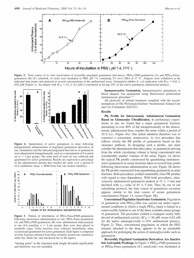
“turning point” at the maximal peak height deviated significantlyand therefore was not included.
Immunoreactive Gentamicin. Immunoreactive gentamicin inblood aliquots was quantitated using fluorescence polarizationimmunoassay procedure.14
All protocols of animal treatment complied with the recom-mendations of The Weizmann InstitutesInstitutional Animal Careand Use Committee (IACUC).
ResultsPK Profile for Intravenously Administered Gentamicin
Based on Glomerular Ultrafiltration. In preliminary experi-ments in rats we found that a major gentamicin fraction,amounting to over 80% of the intraperitoneally or the intrave-nously administered dose, reaches the urine within a period of10 h (i.e., Figure 4A). Our initial intention therefore was toconstruct a convenient, noninvasive, in vivo procedure thatreflects closely the PK profile of gentamicin based on thisclearance pathway. In designing such a profile, one mustconsider the phenomenon that takes place, as gentamicin arrivingfrom the whole circulating volume is being concentrated intoconsiderably smaller volumes of the urine. Figure 1A showsthe typical PK profile constructed by quantitating immunore-active gentamicin in serum fractions taken at several time pointsfollowing intravenous administration in rats; Figure 1B showsthe PK profile constructed from quantitating gentamicin in urinefractions. Both procedures yielded remarkably close PK profileswith regard to time dependency. With both procedures, intra-venously administered gentamicin peaked at 23 ( 3 min anddeclined with t1/2 value of 45 ( 5 min. Thus, by use of ourcalculating protocol, the time course of gentamicin excretionappears similar to the time course of serum gentamicinconcentration (Figure 1A versus Figure 1B).
Conventional Pegylation Inactivates Gentamicin. Pegylationof gentamicin with PEG20-OSu was carried out under experi-mental conditions in which a single PEG20 chain is linked in anonreversible fashion to one of the three available amino groupsof gentamicin. The procedure yielded a conjugate nearly fullydevoid of antibacterial activity (IC50 ) 56 µM versus 0.62 µMfor the native aminoglycoside (1.19% antibacterial potency,Figure 2). Thus, conventional pegylation, in which the PEGremains attached to the drug, appears to be an unsuitableapproach for prolonging the action of aminoglycosides such asgentamicin.
Reversibly Pegylated Gentamicin Derivatives Are Inactivebut Activatable Prodrugs. In Figure 3, PEG20-FMS-gentamicinor PEG40-Fmoc-gentamicin (0.1 µmoL/mL) was incubated at
Figure 3. Time course of in vitro reactivation of reversibly pegylated gentamicin derivatives. PEG20-FMS-gentamicin (A) and PEG40-Fmoc-gentamicin (B) (0.1 µmol/mL of each) were incubated in PBS, pH 7.4, containing 2% (w/v) HSA at 37 °C. Aliquots were withdrawn at theindicated time points and analyzed at several concentrations in the antibacterial assay. Gentamicin inhibits E. coli replication with IC50 ) 0.62 (0.02 µM (Figure 2). An aliquot with IC50 ) 6.2 ( 0.2 µM is considered as having 10% of the native gentamicin antibacterial potency.
Figure 4. Quantitation of active gentamicin in urine followingintraperitoneal administration of pegylated gentamicin derivatives inrats. Gentamicin and the indicated pegylated derivatives of gentamicinwere administered intraperitoneally, each at a concentration of 0.4 µmol/rat (n ) 4 per group). The urine pool of each rat was collected andquantitated for active gentamicin. Results are expressed as percentageof the administered amount that reached the urine over a period of10 h (arithmetic mean ( SEM from four rats treated similarly).
Figure 5. Pattern of distribution of PEG5-Fmoc/FMS-gentamicinfollowing intravenous administration to rats. PEG5-Fmoc-gentamicin(A) and PEG5-FMS-gentamicin (B) were administered intravenouslyto rats (0.4 µmol/rat, n ) 3 per group). Rats were then placed inmetabolic cages. Urine fractions were collected immediately whenexcreted and quantitated for active gentamicin. Each figure is composedof urine fractions obtained from three similarly treated rats (marked aswhite bar, black bar, and checkered bar in the figure).
4302 Journal of Medicinal Chemistry, 2008, Vol. 51, No. 14 Marcus et al.
37 °C in PBS buffer (pH 7.4) containing 20 mg/mL HSA.Aliquots were withdrawn at different time points and assessedfor their antibacterial potencies. Both conjugates were nearlyinactive prior to incubation (∼2% activity, time 0 in the figure).Upon incubation of PEG20-FMS-gentamicin, antibacterial po-tency was generated with a t1/2 value of 24 ( 2 h (Figure 3A)and with a t1/2 value of 66 ( 3 h upon incubation of PEG40-Fmoc-gentamicin (Figure 3B). Both PEG20-FMS-gentamicin andPEG40-Fmoc-gentamicin regained their full (100%) antibacterialpotencies following 40 h (Figure 3A) or 130 h (Figure 3B) ofincubation, respectively.
Intraperitoneal Administered Reversibly Pegylated Gen-tamicin Derivatives Release Gentamicin. Quantitation of An-tibacterial Potencies in Urine Fractions. Initially, we adminis-tered gentamicin and PEG20-gentamicin (0.4 µmol/rat) intraperito-neally and confirmed that a major fraction of the injectedgentamicin (84 ( 4%) and essentially none from PEG20-gentamicin (<1%) were excreted in the urine over a period of10 h (Figure 4A). Thus, an enzymatic system capable ofhydrolyzing the bond linking gentamicin to the PEG chainfollowing conventional pegylation is lacking. In Figure 4B thesame protocol was applied to a family of reversibly pegylatedgentamicin derivatives. Following intraperitoneal administrationof PEG5-Fmoc-, PEG20-Fmoc-, and PEG40-Fmoc-gentamicin,4 ( 0.3%, 6.0 ( 0.5%, and 6.6 ( 0.3%, respectively, of theinjected dose was excreted in the urine. With PEG5-FMS-,PEG20-FMS-, and PEG40-FMS-gentamicin, the level of gen-
tamicin found in the urine was 11 ( 1%, 14 ( 2%, and 17 (3%, respectively, of the injected dose (Figure 4B). Thus,spontaneous hydrolysis with the concomitant release of activegentamicin from the inactive, reversibly pegylated, conjugatetakes place in the circulatory system. As expected from thehydrolysis rates in vitro (Figure 3), gentamicin released fromthe PEG-FMS-gentamicin conjugates in vivo was greater than2.5 ( 0.2 times that released from the PEG-Fmoc-gentamicinconjugates (Figure 4B).
Distribution Pattern of Reversibly Pegylated GentamicinDerivatives after Intravenous Administration in Rats. InFigures 5, 6, and 7, PEG5-, PEG20-, and PEG40-gentamicinderivatives, linked through either the Fmoc or the FMSheterobifunctional spacer, were administered to rats intra-venously, each at a concentration of 0.4 µmol/rat. Rats werethen placed in metabolic cages, and urine was collected andquantitated for active gentamicin. Each derivative wasinjected into three rats in order to obtain at least two urinefractions each hour. Administered PEG5-Fmoc-gentamicinshowed a lag period of 1 h, during which no gentamicin wasdetected. It then reached a value of 3.8 ( 0.4 µg per eachexcreted urine fraction over a period of 2.5-4 h and declinedwith a t1/2 value of about 5 h. No gentamicin was found inurine fractions taken 7 and 8 h after administration (Figure5A). With PEG5-FMS-gentamicin, no lag period was ob-served; gentamicin was present in the very first urine fraction,taken about 0.5 h following administration. Elevated levels
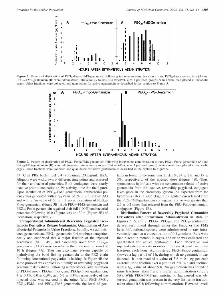
Figure 6. Pattern of distribution of PEG20-Fmoc/FMS-gentamicin following intravenous administration to rats. PEG20-Fmoc-gentamicin (A) andPEG20-FMS-gentamicin (B) were administered intravenously to rats (0.4 µmol/rat, n ) 3 per each group), which were then placed in metaboliccages. Urine fractions were collected and quantitated for active gentamicin as described in the caption to Figure 5.
Figure 7. Pattern of distribution of PEG40-Fmoc/FMS-gentamicin following intravenous administration to rats. PEG40-Fmoc-gentamicin (A) andPEG40-FMS-gentamicin (B) were administered intravenously to rats (0.4 µmol/rat, n ) 3 per each group), which were then placed in metaboliccages. Urine fractions were collected and quantitated for active gentamicin as described in the caption to Figure 5.
Prodrugs by ReVersible Pegylation Journal of Medicinal Chemistry, 2008, Vol. 51, No. 14 4303

of gentamicin were maintained in subsequent urine fractions,amounting to 5.8 ( 1 µg gentamicin/fraction over a periodof 3 h. The levels then declined with a t1/2 value of about5.5 h (Figure 5B).
The same characteristic features of distribution were obtainedfor administered PEG20-Fmoc/FMS-gentamicin, with the notabledifference of more protracted maintenance (t1/2 value of ∼9 h,Figure 6). As with the shorter conjugates, PEG20-Fmoc-gentamicin showed a lag period, amounting in this case to about2.5 h, followed by the presence of active gentamicin in urine atconcentrations in the range of 1.7-2.2 µg/fraction over a periodof 6 h before declining. With administered PEG20-FMS-gentamicin, considerably higher levels (3-7 µg gentamicin/fraction) were observed in the first 2 h of administrationfollowed by a concentration of 4.5-5.5 µg/fraction over 2-8h (Figure 6B). Essentially the same characteristic features ofdistribution were obtained with PEG40-Fmoc/FMS-gentamicinexcept that the half-life appeared to extend to a t1/2 value of∼11 h (Figure 7). With administered PEG40-Fmoc-gentamicin,a lag period of ∼1 h was obtained followed by the appearanceof gentamicin in urine fractions in the range of 0.8-1.8 µg/fraction over a period of 10 h. With PEG40-FMS-gentamicin,relatively high levels (4-10 µg) of gentamicin were found inurine fractions in the first 2 h following administration.Gentamicin then leveled off to 0.8-1.9 µg/fraction over a periodof 10 h following administration. No gentamicin was found inurine fractions taken 14 and 16 h following injection (Figure7).
Discussion
Conventional pegylation of gentamicin ends up with aninactive product (Figure 2). We therefore engineered andsynthesized a family of reversibly pegylated derivatives ofgentamicin. PEG-Fmoc- and PEG-FMS-gentamicin derivativesundergo reactivation upon incubation at physiological conditionswith t1/2 values of 66 ( 3 h (k ) 0.0105 h-1) and 24 ( 2 h (k) 0.0289 h-1), respectively (Figure 3). The release of gentami-cin was then studied in rats. Active gentamicin was generatedfrom the inactive conjugates in situ (Figure 4) and in agreementwith the hydrolysis rates found under physiological conditionsin vitro (Figure 3). PEG-FMS-gentamicin conjugates generated2.5 ( 0.2 times more gentamicin than PEG-Fmoc-gentamicinconjugates (Figure 4B).
We next studied the contribution of incresing the molecularmass of the conjugates to prolong the duration of excretedgentamicin in vivo. This has been evaluated by two differentapproaches, both of which revealed a residence time ofapproximately 5, 9, and 11 h following intraperitoneal and/or intravenous administrations of reversibly pegylated PEG5-,PEG20-, and PEG40- gentamicin, respectively. No significantdifference in this parameter was found between the Fmoc-linked and the FMS-linked conjugate for the same lengthPEG (Figures 4–7). Although PEG40-linked gentamicinconjugates are the longer-lived species in vivo, the residencetime for PEG20-linked conjugates is not that different. Theusage of PEG chain having a molecular mass of 20 kDa forfuture application of this strategy appears therefore economi-cal and sufficient.
The administration of our conjugates at a dose of 0.4 µmol/rat (2-2.4 µmol/kg body weight) was required to prolongmaintenance levels of gentamicin in microgram concentrations;fortunately with most relevant LMW drugs, the presence ofnanogram quantities in the circulatory system is generally oftherapeutic benefit.1 Thus, under these circumstances, admin-
istered doses can be reduced by 3 orders of magnitude, namely,to levels of about 2 nmol/kg body weight.
Most importantly, our reversibly pegylated derivatives areinactive but reactivatable compounds (i.e., prodrugs). Thisfeature might be especially relevant for those LMW drugshaving a narrow pharmacological window where 2-3 timesthe therapeutic level is toxic. From our study, Fmoc-linkedPEG-drug conjugates appear more suitable in this case, sincequite a stable level of gentamicin appeared to be released asa function of time, following a lag period of 1-3 h (Figures5A, 6A, 7A). For maintaining the desired therapeutic level,the size of the injected dose can be calculated from the rateof drug discharge from the conjugate at physiologicalconditions.
Finally, we recommend the application of our procedure toconstruct PK profiles for substances whose major clearancepathway takes place by glomerular filtration, as in the case ofgentamicin, although more urine fractions need to be collectedand quantitated specifically in the peak period to obtain moreaccurate PK profiles.
In summary, the conceptual approach termed “reversible-PEGylation” was applied to nonpeptidic LMW substancesthat otherwise would be inactivated by this technique. Apharmacologically “silent” conjugate that is “trapped” in thecirculatory system releases the covalently linked moleculein its active form, by spontaneous chemical hydrolysis, overmany hours with desirable pharmacokinetic patterns. Thischaracteristic feature would be a significant asset in themanagement of those drug candidates having high indicesof toxicity.
Acknowledgment. We thank Yigal Avivi for editing thismanuscript and the Horowitz, Kimmelman, and Benoziofoundations for supporting this study. Y.S. is the incumbent ofC. H. Hollenberg Chair in Metabolic and Diabetes Researchestablished by the friends and associates of Dr. C. H. Hollenbergof Toronto, Canada. M.F. is the Lester Pearson Professor ofProtein Chemistry.
Supporting Information Available: MS and HPLC data forreversible-pegylated derivatives of gentamicin. This material isavailable free of charge via the Internet at http://pubs.acs.org.
References(1) Benet, L. Z.; Mitchell, T. R.; Scheiner, L. B. In The Pharmacological
Basis of Therapeutics; Goodman, L. A. E., Gilman, T. W., Rall, A. S.,Nies, A. S., Taylor, G. M., Eds.; Pergamon Press: New York, 1990;pp 3-32.
(2) Delgado, C.; Francis, G. E.; Derek, F. F. The uses and properties ofPEG-linked proteins. Crit. ReV. Ther. Drug Carrier Syst. 1992, 9, 249–304.
(3) Fuerteges, F.; Abuchowski, A. The clinical efficacy of poly(ethyleneglycol)-modified proteins. J. Controlled Release 1990, 11, 139–148.
(4) Fung, W.-J.; Porter, J. E.; Bailon, P. Strategies for the preparationand characterization of polyethylene glycol (PEG) conjugatedpharmaceutical proteins. Polym. Prepr. 1997, 38, 565–566.
(5) Bailon, P.; Berthold, W. Polyethylene glycol-conjugated pharmaceuti-cal proteins. Pharm. Sci. Technol. Today 1998, 1, 352–356.
(6) Clark, R.; Olson, K.; Fuh, G.; Marian, M.; Mortensen, D.; Teshima,G. Long-acting growth hormones produced by conjugation withpolyethylene glycol. J. Biol. Chem. 1996, 271, 21969–21977.
(7) Bailon, P.; Palleroni, A.; Schaffer, C. A.; Spence, C. L.; Fung, W.-J.;Porter, J. E.; et al. Rational design of a potent, long-lasting form ofinterferon: a 40 kDa branched polyethylene glycol-conjugated inter-feron R-2a for the treatment of hepatitis C. Biochonjugate Chem. 2001,12, 195–202.
(8) Tsubery, H.; Mironchik, M.; Fridkin, M.; Shechter, Y. Prolonging theaction of protein and peptide drugs by a novel approach of reversible
4304 Journal of Medicinal Chemistry, 2008, Vol. 51, No. 14 Marcus et al.

polyethylene glycol modification. J. Biol. Chem. 2004, 279, 38118-38124.
(9) Peleg-Shulman, T.; Tsubery, H.; Mironchik, M.; Fridkin, M.; Schreiber,G.; Shechter, Y. Reversible PEGylation: a novel technology to releasenative interferon alpha2 over a prolonged time period. J. Med. Chem.2004, 47, 4897–4904.
(10) Shechter, Y.; Tsubery, H.; Mironchik, M.; Rubinstein, M.; Fridkin,M. Reversible PEGylation of peptide YY3-36 prolongs itsinhibition of food intake in mice. FEBS Lett. 2005, 579, 2439–2444.
(11) Nesher, M.; Vachutinsky, Y.; Fridkin, G.; Schwarz, Y.; Sasson, K.;Fridkin, M.; Shechter, Y.; Lichtstein, D. Reversible pegylation prolongsthe hypotensive effect of atrial natriuretic peptide. Bioconjugate Chem.2008, 19, 342–348.
(12) Shechter, Y.; Mironchik, M.; Saul, A.; Gershonov, E.; Precido-Patt,L.; Sasson, K.; Tsubery, H.; Mester, B.; Kapitkovsky, A.; Rubinraut,S.; Vachutinsky, Y.; Fridkin, G.; Fridkin, M. New technologies toprolong life-time of peptide and protein drugs in-vivo. Int. J. Pept.Res. Ther. 2007, 13, 105–117.
(13) Edson, R. S.; Terrell, C. L. The aminoglycosides. Mayo Clin. Proc.1999, 74, 519–528.
(14) Jolley, M. E.; Stroupe, S. D.; Wang, C.-H. J.; Panes, H. N.; Keegan,C. L.; Schmidt, R. L.; Schwenzer, K. S. Fluorescence polarizationimmunoassay I. Monitoring aminoglycoside anti-biotics in serum andplasma. Clin. Chem. 1981, 27, 1190–1197.
JM8002558
Prodrugs by ReVersible Pegylation Journal of Medicinal Chemistry, 2008, Vol. 51, No. 14 4305