Tumor vs. Necrosis - Rochester, NY...Tumor vs necr 5-11-05.ppt Author: Margaret Kowaluk Created...
43
Tumor vs. Necrosis Leena M. Ketonen, MD, Ph.D. MD Anderson Cancer Center May 2005 Presentation material is for education purposes only. All rights reserved. ©2005 URMC Radiology Page 1 of 43
Transcript of Tumor vs. Necrosis - Rochester, NY...Tumor vs necr 5-11-05.ppt Author: Margaret Kowaluk Created...

Tumor vs. Necrosis Leena M. Ketonen, MD, Ph.D.
MD Anderson Cancer Center
May 2005
Presentation material is for education purposes only. All rights reserved. ©2005 URMC Radiology Page 1 of 43

Presentation material is for education purposes only. All rights reserved. ©2005 URMC Radiology Page 2 of 43
Presentation material is for education purposes only. All rights reserved. ©2005 URMC Radiology Page 3 of 43

Various MDA CC Buildings
Presentation material is for education purposes only. All rights reserved. ©2005 URMC Radiology Page 4 of 43

MD Anderson Cancer Center
Division of Diagnostic Imaging, Section of Neuroradiology
!! Neuro: Chief and 5.5 neuroradiologists -(7 faculty on call for neuro), 1-3 residents, 1-2 fellows -35 phycisist, one full time for neuro section -CNI Lab (computational neuroimaging lab) for post processing: 1 phycisist, 1 tech
!! ~200 Neuro CT and MRI scans daily + procedures –! 9 Clinical GE 1.5T MR scanners –! 3 Clinical GE 3T MRI –! 17 multislice CT scanners (4-64 slice scanners)
Presentation material is for education purposes only. All rights reserved. ©2005 URMC Radiology Page 5 of 43

28 y/o with Anaplastic Oligodendroglioma in 2003, s/p glial wafers and chemoradiation
!! Examination: Brain MRI, 6-25-04
!! Clinical History: AO
!! IMPRESSION:
!! FULL RESULT: Routine brain imaging was obtained. This is the first study at M.D. Anderson and outside imaging is not available.
!! There is an irregularly shaped, 3.0 or so cm, operative site in the right high posterior parietal lobe. There is internal heterogeneity, and on the FLAIR images as well as the other sequences, internal areas of linear-shape that correspond to the wafers that were placed surgically elsewhere. There is a rim of enhancement that is not clearly tumor. I do not see any clearly internal enhancing tumor, and the study is somewhat difficult to interpret in the absence of any prior imaging. This will serve as a baseline M.D. Anderson scan. I do not see surrounding infiltrative tumor or nonenhancing disease elsewhere.
Presentation material is for education purposes only. All rights reserved. ©2005 URMC Radiology Page 6 of 43

Postradiation Necrosis May Mimic Tumor
!! Late delayed radiation injury can be progressive and fatal
!! May need surgery
–! …….Diagnostic dilemma
Presentation material is for education purposes only. All rights reserved. ©2005 URMC Radiology Page 7 of 43

Dg of Radiation Injury Is Challenging
!! The pattern of abnormal enhancement closely mimics that of recurrent brain tumor
Presentation material is for education purposes only. All rights reserved. ©2005 URMC Radiology Page 8 of 43

Proposed Mechanism of Radiation Induced Neurotoxicity
!! Vascular Injury: thrombosis, infarction necrosis
!! Glial and white matter damage: oligodendrocytes sensitive to radiation "" destruction leads to demyelination
!! Effect on the fibrinolytic enzyme system !! Immune mechanismm (?autoimmune vaculitis)
Presentation material is for education purposes only. All rights reserved. ©2005 URMC Radiology Page 9 of 43

Diagnostic Dilemmas….
!! Mild form of radiation injury: white matter enhancement: nodular, linear, curvilinear
!! Suspect progression to radiation necrosis if: increase in size, edema, mass effect
!! Cortical gyral enhancement may simulate infarction
Presentation material is for education purposes only. All rights reserved. ©2005 URMC Radiology Page 10 of 43

Suspect Chemoradiation Injury on MRI
!! Soap bubble or Swiss cheese interior –lace-like appearance
!! Cave: can be solid lesion
!! Proximity to original lesion: –! Edge of the treated tumor –! Several cm from tumor
!! Ipsilateral, but also contralateral –! Within the tumor site
Presentation material is for education purposes only. All rights reserved. ©2005 URMC Radiology Page 11 of 43

Brain Has Limited Number of Ways to Respond to Various Insults
Both tumor recurrence and radiation injury can cause: –! Vasogenic edema –! Disruption of BBB –! Cavitations
Presentation material is for education purposes only. All rights reserved. ©2005 URMC Radiology Page 12 of 43

Follow up of Treated Tumors
!! CT, MRI with contrast !! MRS, DWI/ADC !! CT / MR perfusion studies !! PET, SPECT
Presentation material is for education purposes only. All rights reserved. ©2005 URMC Radiology Page 13 of 43
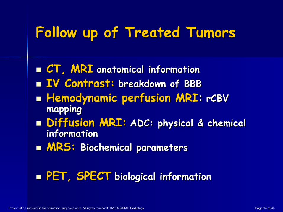
Follow up of Treated Tumors
!! CT, MRI anatomical information !! IV Contrast: breakdown of BBB !! Hemodynamic perfusion MRI: rCBV
mapping !! Diffusion MRI: ADC: physical & chemical
information !! MRS: Biochemical parameters !! PET, SPECT biological information
Presentation material is for education purposes only. All rights reserved. ©2005 URMC Radiology Page 14 of 43

Radiation Injury Area
!! Irradiated tumor cells !! Coagulation necrosis !! Reactive gliosis !! Active fibrosis
Presentation material is for education purposes only. All rights reserved. ©2005 URMC Radiology Page 15 of 43

Necrosis & Tumor Recurrence AJNR Aug 2001, Schlemmer et al.
Presentation material is for education purposes only. All rights reserved. ©2005 URMC Radiology Page 16 of 43
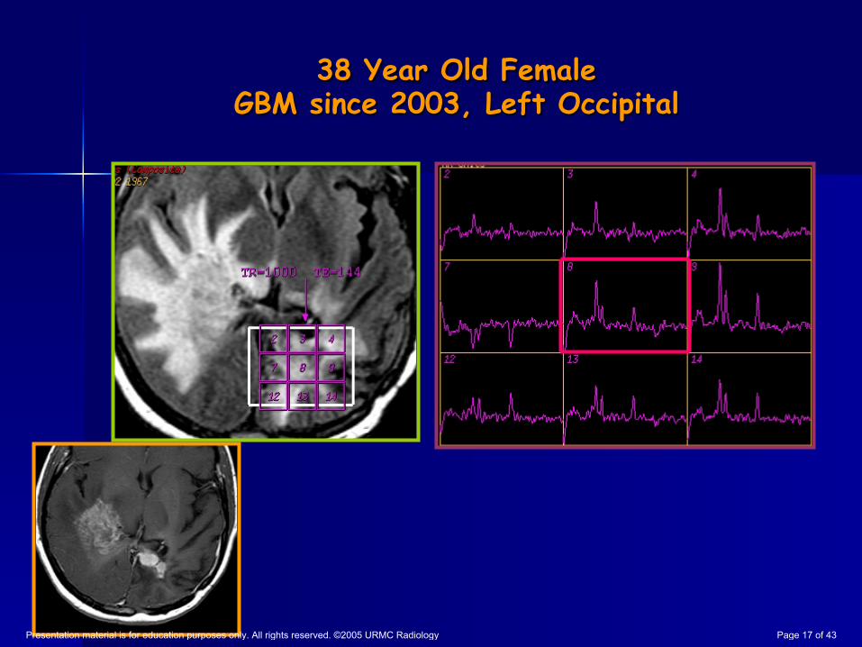
38 Year Old Female GBM since 2003, Left Occipital
Presentation material is for education purposes only. All rights reserved. ©2005 URMC Radiology Page 17 of 43

GBM left occipital: First Crani 4/03, Rx 7/ 03. Second Crani 11/ 04 37 year old female
Presentation material is for education purposes only. All rights reserved. ©2005 URMC Radiology Page 18 of 43

Anaplastic Oligoastrocytoma, status post chemoradiation, follow-up in 3/2005 with new periventricular enhancement
Presentation material is for education purposes only. All rights reserved. ©2005 URMC Radiology Page 19 of 43
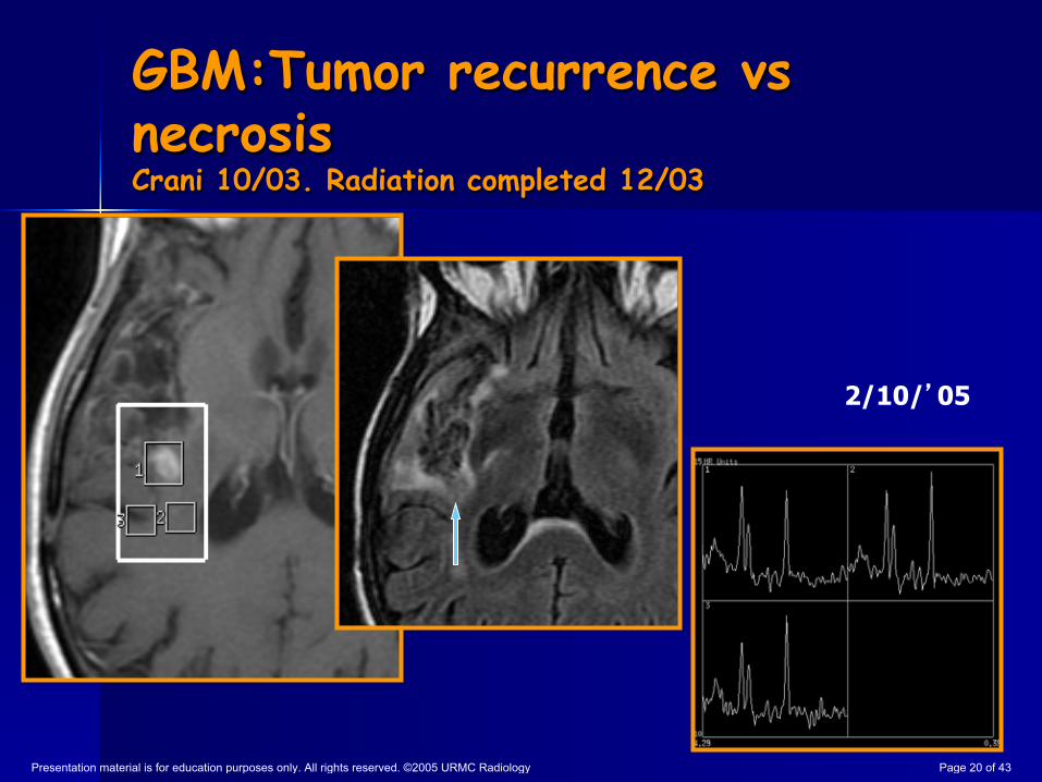
GBM:Tumor recurrence vs necrosis Crani 10/03. Radiation completed 12/03
2/10/ 05
Presentation material is for education purposes only. All rights reserved. ©2005 URMC Radiology Page 20 of 43
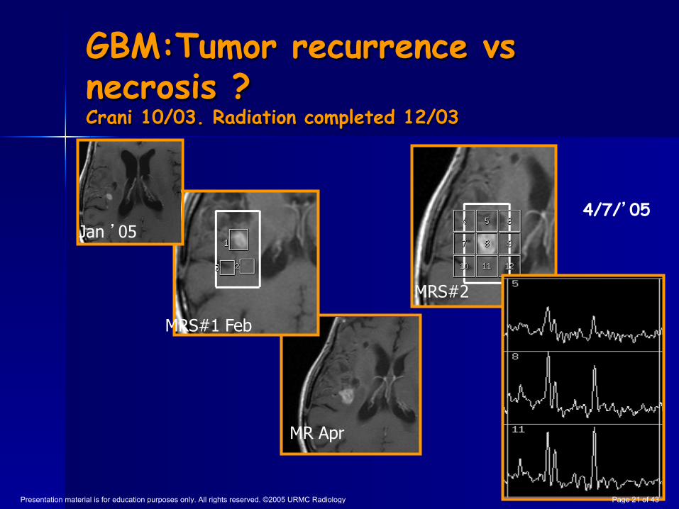
GBM:Tumor recurrence vs necrosis ? Crani 10/03. Radiation completed 12/03
MRS#1 Feb
MR Apr
Jan 05 4/7/ 05
MRS#2
Presentation material is for education purposes only. All rights reserved. ©2005 URMC Radiology Page 21 of 43

Radiation I njury: Predilection of Periventricular White Matter Involvement
!! Poor blood supply from long medullary arteries
!! Lack of collateral supply
!! Subependymal necrosis may mimic tumor spread
Presentation material is for education purposes only. All rights reserved. ©2005 URMC Radiology Page 22 of 43
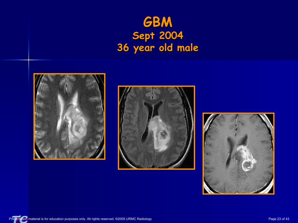
GBM Sept 2004
36 year old male
Presentation material is for education purposes only. All rights reserved. ©2005 URMC Radiology Page 23 of 43

Necrosis GBM in a 36 year old male, 5 month follow-up post
radiation and 3 cycles of temozolomide
Presentation material is for education purposes only. All rights reserved. ©2005 URMC Radiology Page 24 of 43

Recurrent GBM, Triple Dose of Contrast, 3T Magnet
20 min later: Gd leaks into surgical cavity MR xxx008
Presentation material is for education purposes only. All rights reserved. ©2005 URMC Radiology Page 25 of 43

Recurrent GBM, 3T
Presentation material is for education purposes only. All rights reserved. ©2005 URMC Radiology Page 26 of 43

Recurrent GBM, 3T
rCBV: Positive Enhancement Maximum Slope of Increase Integral
Postsurgical
Presentation material is for education purposes only. All rights reserved. ©2005 URMC Radiology Page 27 of 43

Change In Tumor Grade 12/ 04. Radiation Injury 4/ 05 Tumor & Rad. injury
Presentation material is for education purposes only. All rights reserved. ©2005 URMC Radiology Page 28 of 43

Summary Suspect Radiation Injury If:
1.! If the tumor was nonenhancing before surgery and enhancing foci subsequently develop : more likely radiation injury than progression to a higher grade
2.! If an enhancing focus develops at a distance from the primary lesion
3.! Enhancing periventricular lesion develops 4.! New lesions exhibits soap bubble, Swiss
cheese or lace like pattern
Presentation material is for education purposes only. All rights reserved. ©2005 URMC Radiology Page 29 of 43

MRS Protocols
Tumor vs Necrosis !! TE =~144msec
!! For: Lipid, Cho/Cr
Gliomatosis, Low grade astrocytoma
!! TE =35msec !! TE =144msec
!! For: m-Ins & Gly
Presentation material is for education purposes only. All rights reserved. ©2005 URMC Radiology Page 30 of 43

MRS
MRS before administration of contrast despite of problems in voxel location, but verify the location in postcontrast image: –! Decrease of Cho signal intensity after Gd
Ref: Sijens PE et al: 1H chemical shift imaging reveals loss of
brain tumor choline signal after admin. of Gd-contrast. Magn Reson Med (1997) 37;222-225
Presentation material is for education purposes only. All rights reserved. ©2005 URMC Radiology Page 31 of 43
MD Anderson CC, Alkek Building
Presentation material is for education purposes only. All rights reserved. ©2005 URMC Radiology Page 32 of 43

Anaplastic Oligo (gr III) (3/22/05) MRS 3/21/05
Presentation material is for education purposes only. All rights reserved. ©2005 URMC Radiology Page 33 of 43

16 year old female, NF-1 asymptomatic
Presentation material is for education purposes only. All rights reserved. ©2005 URMC Radiology Page 34 of 43

Gliomatosis Cerebri TE=20 & 135 msec
Saraf AJNR 24/03
Short Echo Long Echo
Presentation material is for education purposes only. All rights reserved. ©2005 URMC Radiology Page 35 of 43

Diffuse Astrocytoma High m-Ins/Gly
Presentation material is for education purposes only. All rights reserved. ©2005 URMC Radiology Page 36 of 43

Summary
!! Hyperintense lesion with minimal or No enhancement on post Gd T1WI and
!! lack of Cho/Cr elevation does not exclude primary glial neoplasm
Presentation material is for education purposes only. All rights reserved. ©2005 URMC Radiology Page 37 of 43
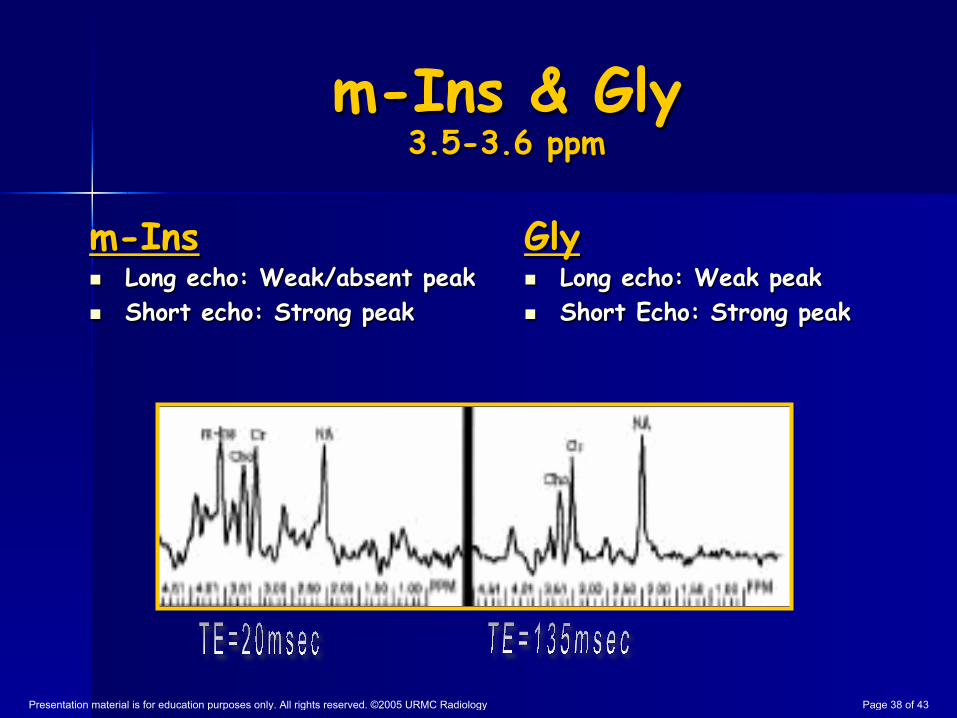
m-Ins & Gly 3.5-3.6 ppm
m-Ins !! Long echo: Weak/absent peak !! Short echo: Strong peak
Gly !! Long echo: Weak peak !! Short Echo: Strong peak
TE=20
Presentation material is for education purposes only. All rights reserved. ©2005 URMC Radiology Page 38 of 43

Summary MRS
If m-Ins or m-Ins/Cr elevated, include low grade astrocytoma or gliomatosis in the differential diagnosis
Presentation material is for education purposes only. All rights reserved. ©2005 URMC Radiology Page 39 of 43

Low Grade Astrocytoma
m-Ins: Glial marker !! Change in phospholipid composition or abundance of cell membranes
Presentation material is for education purposes only. All rights reserved. ©2005 URMC Radiology Page 40 of 43

Presentation material is for education purposes only. All rights reserved. ©2005 URMC Radiology Page 41 of 43

Anaplastic oligo (grIII), Crani 12/ 03 28 year old female
Presentation material is for education purposes only. All rights reserved. ©2005 URMC Radiology Page 42 of 43
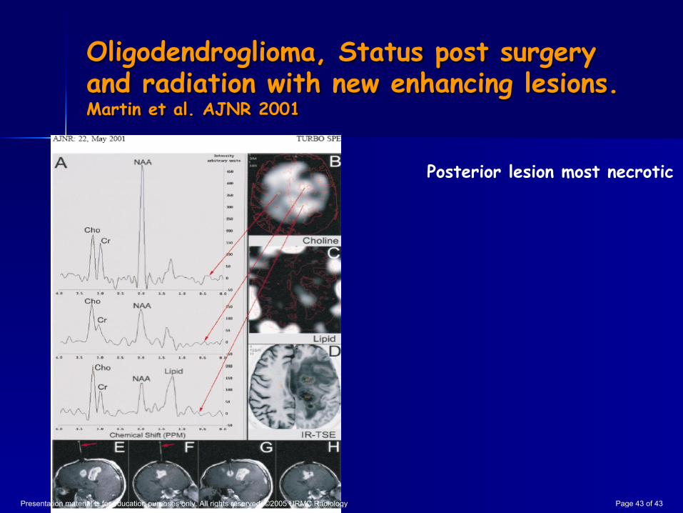
Oligodendroglioma, Status post surgery and radiation with new enhancing lesions. Martin et al. AJNR 2001
Posterior lesion most necrotic
Presentation material is for education purposes only. All rights reserved. ©2005 URMC Radiology Page 43 of 43