Tuesday, 14 May, 2013 - radiation-medicine.de · Contents ConRad 2017 - Global Conference on...
100
Contents ConRad 2017 - Global Conference on Radiation Topics - Preparedness, Response, Protection and Research 1 Contents Organizing committee ……………….…………………………………..……………………………………….2 Welcoming addresses ……………….……………………………………………………………..…………….3 Program at a glance ……………….…………………………………………………………………..……….6 Scientific program ……………….……………………………………………………………………..…….7 Tuesday, May 09 .………………….……………………………………………………………………..…8 Wednesday, May 10 ……………….………………………………………………….……………………….12 Thursday, May 11 ……………….……………………………………………….………………………….16 Poster numbers ……………….……………………………………………….………………………….19 Abstracts conference lectures ……………….…………………………….…………………………………………….26 Abstracts poster presentations .……………….………………………………………………………………………….55 General information .……………….………………………………………………………………………….86 Industrial exhibition .……………….………………………………………………………………………….91 Deutsch-Schweizerischer Fachverband für Strahlenschutz e.V. ……….……………………………….…….93 EPR Biodose 2018 .…………………………………………………………………………………………..95 Notes .……………….………………………………………………………………………….96 The ConRad Logo and the cover page have been designed with friendly support of Ms. Isabell Schütz (Dez VI 5; Zentrum für Geoinformationswesender Bundeswehr, Euskirchen, Leiter: Hr. Venker-Metarp)
Transcript of Tuesday, 14 May, 2013 - radiation-medicine.de · Contents ConRad 2017 - Global Conference on...

Contents ConRad 2017 - Global Conference on Radiation Topics - Preparedness, Response, Protection and Research
1
Contents
Organizing committee ……………….…………………………………..……………………………………….2
Welcoming addresses ……………….……………………………………………………………..…………….3
Program at a glance ……………….…………………………………………………………………..……….6
Scientific program ……………….……………………………………………………………………..…….7
Tuesday, May 09 .………………….……………………………………………………………………..…8
Wednesday, May 10 ……………….………………………………………………….……………………….12
Thursday, May 11 ……………….……………………………………………….………………………….16
Poster numbers ……………….……………………………………………….………………………….19
Abstracts conference lectures ……………….…………………………….…………………………………………….26
Abstracts poster presentations .……………….………………………………………………………………………….55
General information .……………….………………………………………………………………………….86
Industrial exhibition .……………….………………………………………………………………………….91
Deutsch-Schweizerischer Fachverband für Strahlenschutz e.V. ……….……………………………….…….93
EPR Biodose 2018 .…………………………………………………………………………………………..95
Notes .……………….………………………………………………………………………….96
The ConRad Logo and the cover page have been designed with friendly support of Ms. Isabell Schütz (Dez VI 5; Zentrum für Geoinformationswesender Bundeswehr, Euskirchen, Leiter: Hr. Venker-Metarp)

Organizing committee ConRad 2017 - Global Conference on Radiation Topics - Preparedness, Response, Protection and Research
2
Organizing committee
Conference Chair Conference Secretary
Colonel PD Dr. Matthias Port Dr. Christina Beinke Director of the Bundeswehr Institute of Radiobiology Neuherbergstraße 11, D-80937 Munich Email: [email protected] [email protected] Phone: +49-(0)89-992692-2250 +49-(0)89-992692-2273 affiliated to the University of Ulm
Scientific Committee
Prof. Dr. D. Bazyka, Ukraine Dr. M. Drouet, France Dr. F. Groß-Alltag, Germany Prof. Dr. A. Kesminiene, France Prof. Dr. C. Piekarski, Germany Dr. R. Rambousky, Germany Prof. Dr. K. Rothkamm, UK Dr. R. Schneider, Germany Prof. Dr. M. Stiemer, Germany Prof. Dr. A. Wojcik, Sweden Prof. Dr. L. Zablotska, USA

Welcoming addresses ConRad 2017 - Global Conference on Radiation Topics - Preparedness, Response, Protection and Research
3
Written welcome address by the Director of the Bundeswehr Institute of Radiobiology affiliated to the University of Ulm on the occasion of the
22nd Nuclear Medical Defence Conference - ConRad 2017
Dear Colleagues, It is a pleasure for us to welcome you to ConRad 2017, the 22nd Nuclear Medical Defense Conference, from May 8th to 11th 2017 in Munich. This unique conference is hosted by the Bundeswehr Institute of Radiobiology affiliated to the University of Ulm in Munich.
As in 2015 there will be two key sessions introduced with a brief overview followed by several key lectures: The first one will give an update on the concept of “Biomarker of exposure and bioindicator of effect”. The second key session will summarize and update the current knowledge of the “Effects of electromagnetic fields”. Furthermore, we will focus in a session on Radio-nuclear terrorism - a massive threat to civilians, as well as for military personnel.
In the proven concept of this conference, further subjects comprise the presentation of new insights in radiation emergency medical preparedness and response as well as new research findings in radiation epidemiology, medicine, biology and physics as well as low dose effects and radiation protection. This event offers the particular opportunity to bring together a broad spectrum of German and international experts, civilian and military, as a forum for professional and multidisciplinary exchange of experience and expertise in this particular domain. Special attendance is attached to the fact that these aspects will be presented in a comprehensive form for a broader audience thus facilitating the idea of continuous education.
The conference will be accompanied by an industrial exhibition arranged by the German Society for Military Medicine and Pharmacy (GSMMP/DGWMP). There, companies will present their technologies, products and latest developments as well as their service in the field of radiation protection and laboratory equipment for radiation research.
We invite you to attend this scientific, but also practice-oriented event, which offers plenty of opportunities for extensive discussions, to establish new contacts and to strengthen existing relationships after the oral presentations, during the poster session and the poster exhibition, while visiting the exhibition or at the social events.
We are certain that you will enjoy being in Munich, the capital city of the Free State of Bavaria, for this exciting conference. We look forward to personally welcome you as a participant to this further key event dealing with radiation topics affecting all of us.
Sincerely, Colonel PD Dr. Matthias Port Chair, ConRad 2017 Director Bundeswehr Institute of Radiobiology affiliated to the University of Ulm

Welcoming addresses ConRad 2017 - Global Conference on Radiation Topics - Preparedness, Response, Protection and Research
4
Message from the President of the German Society for Military Medicine and Pharmacy (GSMMP/DGWMP) on the occasion of the
22nd Nuclear Medical Defence Conference - ConRad 2017
Dear Participants, As the President of the German Society for Military Medicine and Pharmacy (GSMMP/DGWMP), I am delighted by your interest in the subject matter and welcome you to the 22nd Nuclear Medical Defence Conference. For more than 50 years, the Bundeswehr Institute of Radiobiology has called the Ernst von Bergmann Barracks in Munich its home. This research institute of the Federal Ministry of Defence, which is unique in Germany, represents tradition and innovation, research and analysis, networking and advisory skills in the Bundeswehr, in all of Germany and in the European research community. Globalization has found its way into scientific fields such as medical and biological radiation research. For years, researchers from all over the world have thus come together at this conference. It is a sign of the excellent reputation acquired by the Institute of Radiobiology through many years of hard work that participants from more than 40 countries regularly gather in Munich to attend the Nuclear Medical Defence Conference and its forerunners. I congratulate the Institute on this achievement. In 2014, the German Society for Military Medicine and Pharmacy celebrated its 150th anniversary. As one of the oldest medical societies in Germany, we are grateful and proud to be able to once more support the organization and funding of the NMDC. Matters concerning radiation sources – such as the transport of spent nuclear fuel elements to their storage sites – often trigger a disproportionate, even hysteric public response, while interest in truly threatening risks and scenarios and the corresponding public investment and precautions remain low. Once "the genie is out of the bottle", be it intended or not, people are confronted with an invisible power. Even before anything has happened, this naturally causes fear, which is further stoked by ideologists and false prophets. Facts and evidence often fall by the wayside. Your task as a small group of experts is to give advice to policy makers, the military, civil defense personnel and the population on possible scenarios involving low- or high-energy radiation exposure, and to provide precautionary instructions – with no warning whatsoever, as it has happened before. I invite you to seize the opportunity for scientific exchange provided by this conference, get in touch and gather as much information and experience as possible to integrate into your everyday life back at home. I would also like to invite you to visit the presentations by industry representatives and talk to them about their experiences and about opportunities for technological development. I hope that you will have a successful 22nd Nuclear Medical Defence Conference in the Bavarian capital. Yours sincerely, Jürgen Blätzinger, MD Lieutenant General, MC (ret.) President of the DGWMP

Welcoming addresses ConRad 2017 - Global Conference on Radiation Topics - Preparedness, Response, Protection and Research
5
Written welcome address by the XXX on the occasion of the
22nd Nuclear Medical Defence Conference - ConRad 2017

Program at a glance ConRad 2017 - Global Conference on Radiation Topics - Preparedness, Response, Protection and Research
6
Please note: Posters in the basement have to be removed Wednesday evening between 5:15 and 6.00 pm due to transportation of poster walls.

Program Monday, 08 May, 2017 ConRad 2017
7
Monday, 08 May 2017
Arrival Conference office opened from 10:00 am until 11:00 pm 07:00 pm Icebreaker (Officers` mess, Goercke Haus, build. no. 09)
Note: In the following time schedule only presenting authors are mentioned. Co-authorships are outlined in the abstracts section.

Scientific program Tuesday, 09 May, 2017 ConRad 2017
8
Tuesday, 09 May 2017 8:00 – 8:30 am Welcoming addresses 8:30 – 9:45 am Radio-nuclear terrorism, threats and specific preparedness 10:15 – 11:45 am Radiation emergency medical preparedness and response 1:00 – 5:10 pm Key session I: Biomarker of exposure and bioindicator of
effect
8:00 – 8:30 am Welcoming addresses General (MC) MD Dr. N. Weller Director of Military Medical Science and Capability Development of the Medical Service and Deputy Commandant Bundeswehr Medical Office, Munich, Germany
Colonel (MC) Prof. Dr. Horst Becker Vice President of the DGWMP (GSMMP), Germany
Colonel (MC) MD PD Dr. Matthias Port Bundeswehr Institute of Radiobiology affiliated to the University of Ulm, Munich, Germany Conference Chairperson 8:30 – 9:45 am Radio-nuclear terrorism, threats and specific preparedness
(O1 – O5) Chairpersons Prof. Dr. Nicholas Dainiak and Dr. Wolfram Rother 8:30 – 8:45 am (O-1) Chemical Terror: sudden changes in awareness - facts and medical consequences H. Thiermann, Bundeswehr Institute of Pharmacology and Toxicology, Germany 8:45 – 9:00 am (O-2) CBRN event: pre-hostpital victims management at the Paris Fire Brigade D. Riccobono, French Armed Biomedical Research Institute (IRBA), France 9:00 – 9:15 am (O-3) Predicting the Public Health Consequences of Nuclear Terrorist Attacks T. Jorgensen, Georgetown University, USA

Scientific program Tuesday, 09 May, 2017 ConRad 2017
9
9:15 – 9:30 am (O-4) Triage and medical management of high-volume mass casualties after a nuclear detonation: Hiroshima revisited D. F. Flynn, Adjunct Faculty Armed Forces Radiobiology Research Institute USA; Visiting Faculty, REAC/TS USA 9:30 – 9:45 am (O-5) The Decision Support Model LASAIR as a Useful Tool for Radiation Accident Management within the Scope of Terroristic Attacks H. Walter, Federal Office for Radiation Protection, Germany
9:45 – 10:15 am Coffee break and visit of the industrial exhibition
10:15 – 11:45 am Radiation emergency medical preparedness and response
(O6 – O11)
Chairpersons Prof. Dr. Ann-Barry Flood and Prof Dr. Dymytrii Bazyka 10:15 – 10:30 am (O-6) RENEB – contribution to individualised retrospective dosimetry U. Kulka, Federal Office for Radiation Protection, Germany 10:30 – 10:45 am (O-7) Clinical triage of radiation victims - the hematological module of the Bundeswehr InstRadBioBw applied during the recent NATO exercise on clinical signs & symptoms M. Port, Bundeswehr Institute of Radiobiology affiliated to the University of Ulm, Germany 10:45 – 11:00 am (O-8) Filling Gaps in Public Health Radiological Preparedness C. Case, Radiation Injury Treatment Network, USA 11:00 – 11:15 am (O-9) European Blood and Transplant Group (EBMT) Preparednes for an RDD R. Powles, Cancer Centre London, UK 11:15 – 11:30 am (O-10) Study of the effect of repeated potassium iodide prophylaxis in an adult rat model D. Lebsir, Institute for Radiological Protection and Nuclear Safty (IRSN), France 11:30 – 11:45 am (O-11) Deployment of the « Dosikit » system in operational conditions: experience from a French defence national nuclear exercise F. Entine, French defense radiation protection service (SPRA), France

Scientific program Tuesday, 09 May, 2017 ConRad 2017
10
11:45 – 12:00 noon Group photo (inside Audimax)
12:00 – 1:00 pm Lunch and visit of the industrial exhibition
1:00 – 2:55 pm Key session I: Biomarker of exposure and bioindicator of
effect (O12 – O17) Chairpersons Dr. William F. Blakely and Dr. Christina Beinke 1:00 – 1:05 pm Introduction C. Beinke, Bundeswehr Institute of Radiobiology affiliated to the University of Ulm, Germany 1:05 – 1:20 pm (O-12) Enhancing National Preparedness through Biodosimetry L. Wathen, Biomedical Advanced Research and Development Authority (BARDA), USA 1:20 – 1:35 pm (O-13) High-Throughput Microfluidics Automated Cytogenetic processing for effectively lowering biological process time and aid triage during Radiation Accidents. A. Ramakumar, Armed Forces Radiobiology Research Institute (AFRRI), USA 1:35 – 1:50 pm (O-14) French Armed Forces Biological Dosimetry Laboratory – An overview of 8 years of research M. Valente, French Armed Biomedical Research Institute (IRBA), France 1:50 – 2:15 pm (O-15) Three-color FISH analysis using human peripheral blood lymphocytes for cytogenetic dose assessment Y. Suto, National Institute of Radiological Sciences (NIRS), Japan 2:15 – 2:40 pm (O-16) Transcriptional expression monitoring for rapid determination of dose exposure C. Badie, Public Health England (PHE), UK 2:40 – 2:55 pm (O-17) EPR Biodosimetry Based on Fingernails and Toenails in vivo H. Swartz, Geisel School of Medicine at Dartmouth, USA

Scientific program Tuesday, 09 May, 2017 ConRad 2017
11
2:55 – 3:25 pm Coffee break and visit of the industrial exhibition
3:25 – 5:10 pm cont.: Biomarker of exposure and bioindicator of effect
(O18 – O24)
Chairpersons Prof. Dr. Andrzej Wojcik and Dr. Ulrike Kulka 3:25 – 3:40 pm (O-18) Genomic signature of radiation-induced DNA-sequence alterations in human gingiva fibroblasts A. Kuss, University Medicine Greifswald, Germany 3:40 – 3:55 pm (O-19) Expression analysis of white blood cells from cancer patients treated with 131I-mIBG; identifying biomarkers of internalized radiation in patients with cancer M. Coleman, Lawrence Livermore National Laboratory, USA 3:55 – 4:10 pm (O-20) A Method for Personalized Measurement of Radiation-Induced Mutations P. Okunieff, University of Florida, USA 4:10 – 4:25 pm (O-21) MicroRNA signatures Associated with Neutron-Induced Neoplastic Transformation A. Miller, Uniformed Services University of the Health Sciences (USUHS), USA 4:25 – 4:40 pm (O-22) Establishing gene expression for prediction of the hematological acute radiation syndrome M. Abend, Bundeswehr Institute of Radiobiology affiliated to the University of Ulm, Germany 4:40 – 4:55 pm (O-23) Use of Proteomic and Hematology Biomarkers for Prediction of Hematopoietic Acute Radiation Syndrome in Baboon Radiation Models W.F. Blakely, Armed Forces Radiobiology Research Institute (AFRRI), USA 4:55 – 5:10 pm (O-24) Biomarkers for improving radiation therapy P. Prasanna, National Cancer Institute (NCI), USA
6:00 pm Departure to conference dinner at “Event-Stadl – Zur Alten Säge”

Scientific program Wednesday, 10 May, 2017 ConRad 2017
12
Wednesday, 10 May 2017 8:00 – 10:15 am Key session II: Biological effects of electromagnetic fields/
other non-ionizing radiation 10:45 – 12:00 noon Poster presentation (poster exhibition/basement) 1:00 – 2:50 pm Radiation epidemiology 3:20 – 5:15 pm Radiation health effects and countermeasures (O40 - O46)
8:00 – 10:15 am Key session II: Biological effects of electromagnetic fields / other non-ionizing radiation (O25 – O31)
Chairpersons Prof. Dr. Carmel Mothersill and
Major Dr. Andreas Lamkowski 8:00– 8:05 am Introduction A. Lamkowski, Bundeswehr Institute of Radiobiology affiliated to the University of Ulm, Institute and Outpatient Clinic for Occupational, Social and Environmental Medicine, AllergieZENTRUM, Klinikum der Universität München, Germany 8:05 – 8:25 am (O-25) Biological Effects of Non-Ionizing Radiation: Measurement Environments and Dosimetry M. Stiemer, Helmut-Schmidt-University/University of the Bundeswehr Hamburg, Germany 8:25 – 8:45 am (O-26) Gene expression in human blood cells after Radiofrequency EMF exposure A. Lamkowski, Bundeswehr Institute of Radiobiology affiliated to the University of Ulm, Institute and Outpatient Clinic for Occupational, Social and Environmental Medicine, AllergieZENTRUM, Klinikum der Universität München, Germany 8:45 – 9:05 am (O-27) Genotocic effects in human fibroblasts exposed to microwave radiation V. Franchini, Scientific Department - Policlinico Militare of Rome, Italy 9:05 – 9:25 am (O-28) Radiofrequency electromagnetic field induced effects on brain activity in sleep and waking state H. Danker-Hopfe, Charité - University Medicine Berlin, Germany

Scientific program Wednesday, 10 May, 2017 ConRad 2017
13
9:25 – 9:45 am (O-29) Synoptic analysis of the epidemiologic evidence on mobile phone EMF and cancer N. Leitgeb, formerly Institute of Health Care Engineeriing, Graz University of Technology, Austria 9:45 – 10:00 am (O-30) Growth factors and telomerase activity induced by low level laser therapy in chronic leg ulcer S. Korraa, National Center for Radiation Research and Technology – Egyptian Atomic Energy Authority, Egypt 10:00 – 10:15 am (O-31) Exosomes or UV as candidate bystander signals: a unifying hypothesis C. Mothersill, McMaster University, Canada
10:15 – 10:45 am Coffee break and visit of the industrial exhibition
10:45 – 12:00 noon Poster presentation in the poster exhibition (basement)
12:00 noon – 1:00 pm Lunch and visit of the industrial exhibition
1:00 – 2:50 pm Radiation epidemiology (O32 – O37) Chairpersons Colonel Prof. Dr. Michael Abend and Prof. Dr. Lydia Zablotska 1:00 –1:05 pm Introduction M. Abend, Bundeswehr Institute of Radiobiology affiliated to the University of Ulm, Germany 1:05 –1:25 pm (O-32) Chernobyl and Fukushima lessons learned E. Cardis, IS Global Barcelona Institute for Global Health, Spain 1:25 – 1:40 pm (O-33) Epidemiology of Late Health Effects in Chornobyl Cleanup Workers D. Bazyka, National Research Centre for Radiation Medicine, Ukraine 1:40 – 2:00 pm (O-34) "New insights into thyroid cancer epidemiology: Chernobyl, Fukushima and beyond." A. Kesminiene, International Agency for Research on Cancer (IARC), France

Scientific program Wednesday, 10 May, 2017 ConRad 2017
14
2:00 – 2:20 pm (O-35) "New Evidence on Radiogenic Risks of CLL - Where do we go from here?" L. Zablotska, University of California San Francisco, USA 2:20 – 2:35 pm (O-36) Radiation-epidemiological study of the incidence of complexes of disease of the circulatory system and comorbidities disease among Chernobyl recovery operation workers S. Karpenko, A. Tsyb Medical Radiological Research Center - branch of the National Medical Research Radiological Center of the Ministry of Health (A. Tsyb MRRC), Russia 2:35 – 2:50 pm (O-37) Incidence of cardiac disease after breast cancer radiotherapy: A retrospective cohort study on 12264 women D. Wollschläger, Institute of Medical Biostatistics, Epidemiology and Informatics, Germany
2:50 – 3:20 pm Coffee break and visit of the industrial exhibition
3:20 – 5:15 pm Radiation health effects and countermeasures Chairpersons Colonel Dr. Michel Drouet and Prof. Dr. Juliann Kiang 3:20 – 3:45 pm (O-38) REAC/TS approach to rapid dose estimation and decontamination of plutonium following a puncture wound N. Dainiak, Radiation Emergency Assistance Center Training Site (REAC/TS), USA 3:45 – 4:00 pm (O-39) New paradigms of the treatment of acute radiation syndrome M. Drouet, French Armed Biomedical Research Institute (IRBA), France 4:00 – 4:15 pm (O-40) Animal Model Development of Gastrointestinal-Acute Radiation Syndrome (GI-ARS) in Minipigs: Approaches to Model Development, and Harmonization. J. Esker, BARDA, US Dept of Health & Human Services, USA 4:15 – 4:30 pm (O-41) Autologous adipose tissue derived stromal cells: a new strategy for muscles fibrosis management in cutaneous radiation syndrome D. Riccobono, French Armed Biomedical Research Institute (IRBA), France

Scientific program Wednesday, 10 May, 2017 ConRad 2017
15
4:30 – 4:45 pm (O-42) Delayed Treatment with Thrombopoietin or Eltrombopag after Acute Radiation Exposure Y. Chen, University of Rochester, USA 4:45 – 5:00 pm (O-43) Actinide-contaminated skin: comparing the decontamination efficacy of water, gels, DTPA and soap N. Griffiths, CEA Université de Paris-Saclay, France 5:00 – 5:15 pm (O-44) Plasma-based Biomaterials for the Treatment of Cutaneous Radiation Injury N. Jacob, Ohio State University Medical Center, USA

Scientific program Thursday, 11 May, 2017 ConRad 2017
16
Thursday, 11 May 2017 8:00 – 10:15 am Radiation health effects and countermeasures /
Effects of low dose ionizing radiation / Radiation protection 10:45 – 12:15 am Biomarker of exposure and bioindicator of effect /
Radiation biology - Radiation physics 12:15 – 12:30 pm Closing remarks
8:00 – 10:15 am Radiation health effects and countermeasures (O45)
Effects of low dose ionizing radiation (O46 - O50) Radiation protection (O51 - O53) Chairpersons Prof. Dr. Harry Scherthan and Lieutenant Colonel Stefan Eder Radiation health effects and countermeasures (O45) 8:00 – 8:15 am (O-45) Development of the Toll-Like Receptor-5 Agonist, Entolimod, as a Medical Radiation Countermeasure L. Miller, Cleveland BIoLabs, Inc, USA Effects of low dose ionizing radiation (O46 – O50) 8:15 – 8:30 am (O-46) Automated dicentric chromosome detection - a new approach to investigate age dependent radiosensitivity after Computer Tomography U. Oestreicher, Federal Office of Radiation Protection, Germany 8:30 – 8:45 am (O-47) Anaplastic lymphoma kinase (ALK) – rearrangements in radiation-induced papillary thyroid carcinomas: a study on post Chernobyl tissue samples A. Arndt, Institute of Pathology and Molecular Pathology, Bundeswehrkrankenhaus Ulm, Germany 8:45 – 9:00 am (O-48) Risk of Cataract Incidence in a Cohort of Mayak PA Workers following Chronic Occupational Radiation Exposure E. Bragin, Southern Urals Biophysics Institute (SUBI), Russia

Scientific program Thursday, 11 May, 2017 ConRad 2017
17
9:00 – 9:15 am (O-49) Radiation effect on the murine brain S. Tapio, Institute of Radiation Biology, German Research Center of Environmental Health, Germany 9:15 – 9:30 am (O-50) DNA damage response to internal systemic low dose irradiation by beta and alpha emitters H. Scherthan, Bundeswehr Institute of Radiobiology affiliated to the University of Ulm, Germany Radiation protection (O51 - O53) 9:30 – 9:45 am (O-51) Development of gamma-tocotrienol as a radiation countermeasure V. Singh, F. Edward Hébert School of Medicine/Armed Forces Radiobiology Research Institute, USUHS, USA 9:45 – 10:00 am (O-52) BBT-059, a potential prophylactic radiation countermeasure as well as a mitigator – accelerates recovery from peripheral blood cytopenia S. Ghosh, Armed Forces Radiobiology Research Institute (AFRRI), USA 10:00 – 10:15 am (O-53) „Ärztliche Stelle der Bundeswehr“ national and international M. Hoffmann, Bundeswehr Medical Service Headquarters, Germany
10:15 – 10:45 am Coffee break and visit of the industrial exhibition
10:45 – 12:15 pm Biomarker of exposure and bioindicator of effect
(O54) Radiation biology - Radiation physics (O55 – O59)
Chairpersons Prof. Dr. Reinhard Ullmann and Major Matthäus Majewski Biomarker of exposure and bioindicator of effect (O54) 10:45 – 11:00 (O-54) Immunologic biomarkers associated with chronic radiation exposure V.L. Rybkina, Southern Urals Biophysics Institute (SUBI), Russia

Scientific program Thursday, 11 May, 2017 ConRad 2017
18
Radiation Biology - Radiation physics (O55 – O59) 11:00 – 11:15 am (O-55) Radiobiological, genomic and epigenomic characteristics of a cell line with high tolerance towards genotoxic exposure R. Ullmann, Bundeswehr Institute of Radiobiology affiliated to the University of Ulm, Germany 11.15 – 11:30 am (O-56) Role of spatial organization of chromosome domains in ionizing radiation induced chromosomal aberrations A. Balajee, Radiation Emergency Assistance Center and Training Site, Oak Ridge Institute for Science and Education, Oak Ridge Associated Universities, USA 11:30 – 11:45 am (O-57) Impact of ionizing radiation on electrophysiological behavior of human iPS cardiomyocytes on a multi electrode array B. Becker, Bundeswehr Institute of Radiobiology affiliated to the University of Ulm, Germany 11:45 – 12:00 noon (O-58) A dose-dependent perturbation in cardiac energy metabolism is linked to radiation-induced ischemic heart disease in Mayak nuclear workers O. Azimzadeh, Institute of Radiation Biology, German Research Center of Environmental Health, Germany 12:00 – 12:15 pm (O-59) “Liquid Biopsy” from whole blood in testis tumor patients – “proof of principal“ M. Majewski, Bundeswehr Institute of Radiobiology affiliated to the University of Ulm, Germany
12:15 – 12:30 pm Closing remarks Colonel PD Dr. Matthias Port, Conference chairperson
Bundeswehr Institute of Radiobiology affiliated to the University of Ulm, Munich, Germany
12:30 – 1:15 pm Lunch

Poster numbers Poster exhibition (Wednesday, 10 May) ConRad 2017
19
Biomarkers of exposure and bioindicators of effect P-1-1 Accurate dicentric chromosome biodosimetry through automation of image selection. Liu J1, Li Y1, Wilkins R2, Flegal F3, Knoll JH4,5, Rogan PK1,5 1Department of Biochemistry, University of Western Ontario, 2Radiobiology and Protection Division, Health Canada, 3Canadian Nuclear Laboratories, 4Department of Pathology and Laboratory Medicine, University of Western Ontario, 5Cytognomix Inc. P-1-2 Markers of Neural Degeneration and Regeneration in Blood of Cardiac Catheterization Personals Mohamed AK1, Korraa S2, Noussier N2, Elhammady W3, Elgazzar E2, Diab H4 1Institute of Environmental Studies and Research Ain Shams University, 2National Center for Radiation Research and Technology (NCRRT), Atomic Energy Authority, 3Department of Cardiology, Faculty of medicine, Ain Shams University, 4Department of Environmental Risk Assessment, Center for Radiation Safety, Atomic Energy Authority. P-1-3 Use of Multivariate Discriminant Analysis for Radiation Dose Assessment using the Premature Chromosome Condensation Assay Involving Use of Length Ratios, Fragment Number, and Rings Blakely WF1, Subramanian U1, Miura T2, Hsiao KH1, and Bolduc DL1 1Scientific Research Department, Armed Forces Radiobiology Research Institute, Uniformed Services University, Bethesda, MD, USA; 2Institute of Radiation Medicine, Hirosaki University, Hirosaki, Japan. P-1-4 Redox parameters in medical workers exposed to LLD of ionizing radiation Durovic B, Vukmirovic D, MMA, Occupational Medicine Institute; University of Defense, Medical Faculty of MMA, Belgrade, Serbia P-1-5 Utilizing Irradiation Schemes Mimicking Total Body or Fractionated Dose Regimens to Evaluate Biomarkers of Acute Irradiation in Rhesus Monkeys Doyle-Eisele M Lovelace Respiratory Research Institute, Albuquerque, USA P-1-6 Clinical Observation, Genomic Bioindication, and Medicolegal Interests in Assessment of Chronic Exposure to Low-Dose Ionizing Radiation in the US FUSRAP Madison Site of the St. Louis District Durakovic A, Bell DE Uranium Medical Research Center, Washington, USA P-1-7 Radiological or nuclear emergency set up of a novel tool for early detection of radiation exposure: preliminary results Giovanetti A1, Marconi R2, Bartoleschi C1, Pardini MC1, Turchi 1, Awad N3, Pinnaro P4, Canfora M5, Strigari L2 1Laboratory of Biosafety and Risk Assessment, Division of Health Technologies, Department of Sustainable Territorial and Production Systems (IT); 2Laboratory of medical physics Regina Elena National Cancer institute (IT); 3HIPH University of Alexandria (ET); 4Radiotherapy Regina Elena National Cancer institute (IT); 5Scientific Direction, Regina Elena National Cancer Institute (IT). P-1-8 Decreased intracellular concentrations of heat shock proteins in irradiated human blood lymphocytes Hotz ME, Port M Bundeswehr Institute of Radiobiology affiliated to the University of Ulm, Munich, Germany P-1-9 Modifying influence of ionizing radiation on the genetic regulation of apoptosis, aging and proliferation of immune cells in the early and remote periods after exposure Ilienko I1, Bazyka D1, Sergeeva A2 1National Research Centre for Radiation Medicine, Kyiv, Ukraine; 2Bogomolets National Medical University, Kyiv, Ukraine e-mail: [email protected] P-1-10 Overview of radiation protection and safety of training courses for occupationally exposed workers Inkoom S1, Darko EO, Amoako J K, Adjei DN, Kpeglo DO, Adukpo OK, Otoo F, Owusu-Banahene J, Charles DF, Glover ET 1Radiation Protection Institute, Ghana Atomic Energy Commission, Accra, Ghana. e-mail: [email protected] / [email protected] P-1-11 CLOR’s fission-neutron calibration curve for the lymphocyte dicentric assay 1Kowalska M, 2Golnik N, 2Gryziński MA, 2Tulik P, 1Szkarłat Z 1Central Laboratory for Radiological Protection, Warsaw, Poland, 2National Centre for Nuclear Research, Otwock-Świerk, Poland

Poster numbers Poster exhibition (Wednesday, 10 May) ConRad 2017
20
P-1-12 Preliminary study of expression of γ- H2AX and 53BPI in medical radiation workers Kurnia I, Lusiyanti Y, Alatas Z Center for Technology of Radiation Safety and Metrology, National Nuclear Energy Agency, Jakarta, Indonesia, e-mail: [email protected] P-1-13 cancelled P-1-14 Chromosome aberrations as biomarkers of partial body exposure in cancer patients undergone radiotherapy with different irradiation volume Maznyk N, Sypko T, Pshenichna N, Sukhina O, Starenkiy V Grigoriev Institute for Medical Radiology of NAMSU, Kharkov, Ukraine (IMR) P-1-15 Multiparametric Biodosimetry and Acute Radiation Sickness Prognosis/Outcome after Mixed-Field (Neutron and Gamma) and 60Co -ray Radiation in a Mouse Total-body Irradiation Model Ossetrova NI, Stanton P, Krasnopolsky K, Ismail M, Doreswamy A, Hieber KP Armed Forces Radiobiology Research Institute (AFRRI), USA P-1-16 Instrument and Methodological Advancements for X-band Electron Paramagnetic Resonance In Vivo Fingernail Dosimetry Swarts SG*, Sidabras JW, Grinberg O, Tipikin DS, Feldman M, Mariani M, Kmiec M, Petryakov S, Spencer S, Varanasi S, Schreiber W, Marsh S, Boyle H, Satinsky V, Williams BB, Flood AB, Swartz HM *Department of Radiation Oncology, University of Florida, Gainesville, USA P-1-17 Human blood lymphocytes mitochondrial DNA changes as a marker for biological dosimetry? Tichy A Faculty of Military Health Sciences, University of Defence, Tschechien
Biological effects of electromagnetic fields P-2-1 Engineering and bio-engineering approach of the research carried out in Romania on the effects of electromagnetic fields exposure Miclaus S1, Morega M2, Rosu G3, Baltag O4 1Dept. of Technical Sciences, Land Forces Academy, Sibiu, Romania, 2Fac. of Medical Engineering, University “Politehnica”, Bucharest, Romania, 3Fac. of Military Electronic and Information Systems, Military Technical Academy, Bucharest, Romania, 4Fac. of Medical Bioengineering, University of Medicine and Pharmacy, Iasi, Romania P-2-2 Optical ocular hazard assessment of intense incandescent light sources for dental treatment Schirmer A1, Warnecke U2, Graf MJ2 1Bundesamt für Infrastruktur, Umweltschutz und Dienstleistungen der Bundeswehr (BAIUDBw) GS I 3 Strahlenmessstelle der Bundeswehr, 2Bundesamt für Infrastruktur, Umweltschutz und Dienstleistungen der Bundeswehr (BAIUDBw)
Radiation epidemiology P-3-1 The National Health Programme for the years 2016-2020: The concept and aims of the research task “Assessment of the health risk associated with exposure of the military personnel to ionizing radiation.” Cheda A, Nowosielska EM, Janiak MK Department of Radiobiology and Radiation Protection, Military Institute of Hygiene and Epidemiology, Warsaw, Poland P-3-2 Spectrometric Isotope Analysis of Urine and Whole Body of a Child from Tokyo after Exposure to the Radioactive Outfall from Fukushima – A Case Study 1Klimaschewski F, 2Durakovic, A 1UMRI, London, UK; 2UMRC, Washington, USA P-3-3 Malignant Neoplasms Incidence among the Chernobyl NPP accident emergency workers from Armenia Hovhannisyan NM, Mirijanyan MI, Dallakyan AM Scientific Center of Radiation Medicine and Burns, Armenia

Poster numbers Poster exhibition (Wednesday, 10 May) ConRad 2017
21
Radiation accident management P-4-1 Recommendations for developing of information-analytical systems for simulation of the external exposure of staff in the aftermath of radiation accident Chizhov K1, Mark NK2, Szőke I2, Sneve MK3, Ivakina A1, Kryuchkov V1
1State Research Center Burnasyan Federal Medical Biophysical Center of Federal Medical Biological Agency, RF Ministry of Health and Social Development, Moscow, Russian Federation. 2Institute for Energy Technology, Halden, Norway. 3Norwegian Radiation Protection Authority, Osterеs, Norway. P-4-2 DosiKit, a new solution for external radiation biodosimetry on field *Etzol-Bensimon J, *Bouvet S, *Bettencourt C, Altmeyer S, Corre Y, Faye B, Ugolin N, Chevillard S *Acubens, sas and CEA/DSV/IRCM/SREIT/Laboratoire de Cancérologie Expérimentale, France; e-mail: [email protected] P-4-3 The European Union`s Missing Strategy for Civilian Nuclear/Radiological Emergency Preparedness for Mass Casualties Klimaschewski F UMRI, London, UK P-4-4 The impact of the treatment initiation time on decorporation efficacy after the incorporation of radionuclides Rump A Bundeswehr Institute of Radiation Biology affiliated to the University of Ulm, Munich, Germany
Radiation emergency medical preparedness and response P-5-1 “United States Department of Health and Human Services Radiological/Nuclear Medical Countermeasures Programs “ Homer M (presented by: J. Esker) United States Department of Health and Human Services; Office of the Assistant Secretary of Preparedness and Response, Biomedical Advanced Research and Development Authority, Washington, USA P-5-2 Further development of virtual biodosimetry laboratory concept and its role in radiation accident countermeasures Maznyk N1, Fehringer F2, Johannes C3, Sypko T1, Pshenichna N1, Müller WU3 1Grigoriev Institute for Medical Radiology of NAMSU, Kharkov, Ukraine (IMR), 2Institute for Radiation Protection of the BG ETEM and BG RCI (IfS), Cologne, Germany, 3University Duisburg-Essen, Essen, Germany P-5-3 Development and Performance of a Gene Expression based High Throughput Biodosimetry Test System 1Terbrueggen R, 1Cleveland J, 1Duymich C, 1Flores F, 1Goldman T, 1Guiang K, 1Guyon T, 1Jacobs AR, 1Jacobsen GE, 1Headley V, 1,2Dressman H, 3Iversen E, 3Lipton G, 4Gary Phillips G, 4Ross J, 5Zenhausern F, 6Chao N 1DxTerity Diagnostics, Inc. Los Angeles, CA, 2Department of Molecular Genetics and Microbiology, Duke University, Durham, NC, 3Department of Statistical Science, Duke University, Durham, NC, 4Duke University Medical Center, Durham, NC, 5University of Arizona Medical Center, Phoenix, AZ, 6Chief – Division of Hematologic Malignancies & Cellular Therapy/BMT, Duke University Medical Center, Durham NC
Radiation health effects and medical countermeasures P-6-1 Eltrombopag, an alternative to thrombopoietin for the treatment of acute radiation bone marrow injury Chen Y, Nowak I, Tsai Y, Keng PC, Wu JHD, Lee SO Radiation Oncology, University of Rochester, Rochester,USA P-6-2 Ghrelin Therapy: Attenuation of Acute Radiation Syndromes and Increases in Survival after Ionizing Radiation Followed by Wound Trauma Kiang JG, Smith JT, Anderson MN Armed Forces Radiobiology Research Institute (AFRRI), Bethesda, Maryland, USA

Poster numbers Poster exhibition (Wednesday, 10 May) ConRad 2017
22
P-6-3 The Toll-Like Receptor-5 Agonist, Entolimod, Increases Survival after Lethal Irradiation in Nonhuman Primates Krivokrysenko V, Bittman RM, Hards A, Purmal A, Gudkov AV, Miller LL, Kogan Y Cleveland BioLabs, USA P-6-4 Pharmacokinetics (PK), Pharmacodynamics (PD), and Safety of the Toll-Like Receptor-5 Agonist, Entolimod, in Healthy Male and Female Human Subjects Miller LL, Hards A, Krivokrysenko V, Purmal A, Bittman RM, Gudkov AV, Kogan Y Cleveland BioLabe, Inc., USA P-6-5 Low Level Laser Therapy (670nm) for the treatment of cutaneous radiation syndrome: radioprotective effects in vitro on irradiated fibroblast Riccobono D, François S, Agay D, Baert P, Foray N, Drouet M, Forcheron F French Armed Forces Biomedical Research Institute (IRBA), France P-6-6 Potential targets of radiomitigative action of BP-C2, a novel metal-organic compound based on polyphenolic ligand Pigarev SE1, Gubareva EA2, Bykov VN2, Drachev IS2, Fedoros EI2, Panchenko AV2 1Meabco A/S, Copenhagen, Denmark and 2N.N. Petrov Research Institute of Oncology, St. Petersburg, Russian Federation; e-mail: [email protected] P-6-7 The Role of TGF Beta and PPAR Alpha Signalling Pathways in Radiation Response of Locally Exposed Heart: Integrated Global Transcriptomics and Proteomics Analysis Subramanian V Institute of radiation biology, Helmholtz Zentrum München, Neuherberg
Effects of low dose ionizing radiation P-7-1 Low-dose X-ray exposures during the early-stage diabetes: Effects on the development and progression of endothelial dysfunction Cheda A1, Nowosielska EM1, Wrembel-Wargocka J1, Zdanowski R2, Lewicki S2, Chłopicki S3, Janiak MK1 1Department of Radiobiology and Radiation Protection, Military Institute of Hygiene and Epidemiology, Warsaw, Poland; 2Department of Regenerative Medicine, Military Institute of Hygiene and Epidemiology, Warsaw, Poland; 3Jagiellonian Centre for Experimental Therapeutics, Krakow, Poland This study was supported by the EU FP7 DoReMi (grant agreement 249689) on “Low Dose Research Towards Multidisciplinary Integration.” P-7-2 Proteome Analysis of Murine Hippocampus 18 Months after Total Body Low-Dose Irradiation Hladik D1,8, Azimzadeh O1, von Törne C2, Garrett L3, Hölter SM3, Dalke C3, Neff F4, Rößler U7, Greiter M5, Unger K6, Atkinson MJ1,8, Graw JS3, Tapio S1,8 1Institute of Radiation Biology, Helmholtz Zentrum München, German Research Centre for Environmental Health GmbH (HMGU), Germany, 2Research Unit Protein Science, HMGU, 3Institute of Developmental Genetics, HMGU, 4Institute of Pathology, HMGU, 5Research Unit Medical Radiation Physics and Diagnostics, HMGU, 6Research Unit of Radiation Cytogenetics, HMGU, 7Federal Office for Radiation Protection, Department SG Radiation Protection and Health, Germany, 8Technical University Munich (TUM), Germany P-7-3 Developing a Methodology to Assess Human Lung Failure due to Uranium Dust Contamination - A Progress Report Klimaschewski F1, Durakovic A2, Dullin C3, Pacilѐ S4, Tromba G4, Dreossi D4 1Uranium Medical Research Institute, London, UK, 2UMRC, Washington, DC, US,3Dept. Diagnostic and Interventional Radiology, University Medical Center Goettingen, Goettingen, Germany, 4Elettra Synchrotron Light Laboratory, Trieste, Italy P-7-4 Internal contamination with HTO: Effects on some immune parameters in mice Nowosielska EM1, Cheda A1, Wrembel-Wargocka J1, Zdanowski R2, Lewicki S2, Janiak MK1 1Department of Radiobiology and Radiation Protection, Military Institute of Hygiene and Epidemiology, Warsaw, Poland; 2Department of Regenerative Medicine, Military Institute of Hygiene and Epidemiology, Warsaw, Poland P-7-5 Low-dose X-ray exposures during the fully developed diabetes: Effects on the progression of endothelial dysfunction Nowosielska EM1, Cheda A1, Wrembel-Wargocka J1, Zdanowski R2, Lewicki S2, Chłopicki S3, Janiak MK1
1Department of Radiobiology and Radiation Protection, Military Institute of Hygiene and Epidemiology, Warsaw, Poland; 2Department of Regenerative Medicine, Military Institute of Hygiene and Epidemiology, Warsaw, Poland; 3Jagiellonian Centre for Experimental Therapeutics, Krakow, Poland

Poster numbers Poster exhibition (Wednesday, 10 May) ConRad 2017
23
P-7-6 Assessment of Genome Damage Using Micronucleus Assay in High Background Radiation Area in Indonesia Ramadhani D Nuclear Medicine Technique and Radiation Biology Division, Center for Technology of Radiation Safety and Metrology, National Nuclear Energy Agency of Indonesia, Jakarta, Indonesia P-7-7 Chromosomal Aberrations Analysis of Mamuju Inhabitants Using Single Color FISH and Conventional Giemsa Stain Syaifudin M Nuclear Medicine Technique and Radiation Biology Division, Center for Technology of Radiation Safety and Metrology, National Nuclear Energy Agency of Indonesia, Jakarta, Indonesia
Decontamination measures and monitoring P-9-1 Decontamination strategies for the response to nuclear security events in Germany Kroeger EA, Eisheh JT Working Group Defence Against Nuclear Hazards, Federal Office for Radiation Protection (Bundesamt fuer Strahlenschutz), Berlin, Germany
Radiation risk perception of the public P-10-1 Protecting the population depending on the differentiation of populations Freitinger-Skalicka Z Institute of Radiology, Toxicology and Civil Protection, Faculty of Health and Social Sciences University of South Bohemia, Czech Republic
Radiation protection P-11-1 Monte Carlo Simulations and Experiments to Detect Radioactive Sources in Steel Scrap de Groot J1, Merk R2 1Brenk Systemplanung GmbH, Deutschland, 2Federal Office for Radiation Protection (BfS), Salzgitter, Germany P-11-2 Effective doses of uranium processing workers at MAPE Mydlovary (Czech Republic) Havránková R, Havránek J, Freitinger Skalická Z, Zölzer F Institute of Radiology, Toxicology and Civil Protection, University od South Bohemia in České Budějovice, Faculty of Health and Social Sciencies, Ceske Budejovice, Czech Republic P-11-3 Characterization of a CLYC Detector Köble T, Bornhöft C, Schumann O, Risse M, Friedrich H, Berky W, Chmel S, Lieder E, Rosenstock W Fraunhofer Institut Naturwissenschaftlich-Technische Trendanalysen INT, D-53864 Euskirchen, Germany P-11-4 Molecular and Biochemical Studies on the Therapeutic Effects of Shark Cartilage in Rats Exposed to Ionizing Radiation Korraa S1, Monir El Ganzour S2, Mahmoud R2, Darwish S3 1Department of Health Radiation Research, National Center for Radiation Research and Technology – Egyptian Atomic Energy Authority, 2Department of Zoology, Faculty of Science - Ain Shams University.3 Department of National Research Institute, Cairo, Egypt. P-11-5 The impact оf astaxanthin on radiation-induced genomic damages in human peripheral blood lymphocytes in vitro Кurinnyi D1, Rushkovsky S2, Smetansk I3, Pilinska M1 1State institution "National Research Center for Radiation Medicine of the National Academy of Medical Sciences of Ukraine", 2 Institute of Biology and Medicine, Taras Shevchenko National University of Kyiv, 3 University of Applied Sciences Weihenstephan-Triesdorf, Triesdorf P-11-6 Estimate of individualized radiation risk coefficients under internal exposure for the cohort of Russian emergency workers Lovachev S National Radiation and Epedemiological Registry, A. Tsyb Medical Radiological Research Center - branch of the National Medical Research Radiological Center of the Ministry of Health of the Russian Federation (A. Tsyb MRRC)

Poster numbers Poster exhibition (Wednesday, 10 May) ConRad 2017
24
P-11-7 Rescue effect in irradiated human lymphocytes incubated with yeast cells Saccharomyces cerevisiae. Vasylenko OP, Pronina OB, Rushkovsky SR Institute of Biology and Medicine, National Taras Shevchenko University of Kyiv, Volodymyrska, 64, 01033 Kyiv, Ukraine [email protected] P-11-8 Radiation risk from CT scans from perspective of physicians Vukmirovic D1, Durovic B1,2. 1Military Medical Academy, Occupational Medicine Institute, Belgrade, Serbia, 2University of Defense, Medical Faculty of Military Medical Academy Belgrade, Serbia
Radiation biology / Radiation physics P-12-1 Irradiation-induced dsDNA breaks at meiotic chromosome-ends are repaired by NHEJ Ahmed EA1, Scherthan H2
1Lab of Immunology and Molecular Physiology, Zoology Department. Assiut University, Assiut, Egypt. 2Bundeswehr Institute of Radiobiology, affil. to the Univ. of Ulm, Munich, Germany P-12-2 Gene expression changes in PHA-M stimulated lymphocytes - Unraveling PHA activity as a prerequisite of the dicentric chromosome assay Beinke C, Port M, Ullmann R, Gilbertz K, Majewski M, Abend M Bundeswehr Institute of Radiobiology affiliated to the University Ulm, Munich, Germany P-12-3 Radiation-induced re-arrangements of molecular complexes studied in 3D-conserved cells and cell nuclei by super-resolution localization microscopy Hausmann M1, Krufczik M1, Wagner E1, Chojowski R1, Theda F1, Eryilmaz M1, Lee JH1, Peper M3, Pilarczyk G1, Hildenbrand G1,2, Scherthan H3 1Kirchhoff-Institute for Physics, University of Heidelberg; 2Dept. Radiation Oncology, Universitätsmedizin Mannheim, University of Heidelberg,; 3Bundeswehr Institute of Radiobiology, Germany; e-mail: [email protected] P-12-4 Protective signaling pathways in the cellular radiation response: Nuclear Factor κB (NF-κB) and Nrf2 Henschenmacher B1, Crellin A1, Enyong E1, Chishti AA1,2, Diegeler S1, Spitta LF1, Koch K1, Konda B1, Feles S1, Schmitz C1, Baumstark-Khan C1, Hellweg CE1 1 German Aerospace Centre (DLR), Institute of Aerospace Medicine, Radiation Biology Department, Köln, Germany. 2 Present adress: The Karachi Institute of Biotechnology and Genetic Engineering (KIBGE), University of Karachi, Karachi, Pakistan. P-12-5 Clinical application of OBI® CBCT systems and image quality using Taguchi technique: a phantom study Hsieh YC 1 Department of Radiation Oncology, CHIA-YI Christian Hospital Department, Taiwan, 2 Department of Medical Imaging and Radiological Sciences, Central Taiwan University of Science and Technology, Taichung, Taiwan, 3 Department of Medical Imaging and Radiological, CHIA-YI Christian Hospital Department, Taiwan P-12-6 Setup for tumor growth delay studies in small animals for low energy x-rays and small irradiation fields Hunger A1,2, Burger K2,3,4, Stein M1,2, Dierolf M3,4, Günther B3,4, Porth AK2, Bartzsch S2, Urban T2, Achterhold K3,4, Gleich B4, Beyreuther E7, Pfeiffer F3,4,5,6, Combs S1,2, Wilkens JJ1,2,3, Schmid TE1,2 1Institute of Innovative Radiotherapy, Helmholtz Zentrum München, Germany, 2Department of Radiation Oncology, Technical University of Munich, Klinikum rechts der Isar, Germany 3Chair of Biomedical Physics, Department of Physics and School of BioEngineering, Technical University of Munich, Germany, 4Munich School of BioEngineering, Technical University of Munich, Germany, 5Department of Diagnostic and Interventional Radiology, Klinikum rechts der Isar, Technical University of Munich, Germany, 6Institute for Advanced Study, Technical University of Munich, Germany, 7OncoRay – National Center for Radiation Research in Oncology, Faculty of Medicine and University Hospital Carl Gustav Carus, Technische Universität Dresden, Helmholtz-Zentrum Dresden - Rossendorf, Germany P-12-7 From Roentgen Discovery to Stereotactic Radiosurgery Maciejewski B Cancer Center – M. Skłodowska-Curie, Memorial Institute, Gliwice, POLAND P-12-8 Generation of radio-resistant pancreatic cancer cell lines Nguyen L1,2, Dobiasch S1, Schilling D1,2, Schmid TE1,2 and Combs SE1,2 1Technische Universität München (TUM), Department of Radiation Oncology, Ismaninger Straße 22, Munich, Germany, 2Institute for Innovative Radiotherapy (iRT), Helmholtz Zentrum München, Neuherberg, Germany

Poster numbers Poster exhibition (Wednesday, 10 May) ConRad 2017
25
P-12-9 Ionizing Radiation Leads to Senescence and Inflammation in Human Coronary Artery Endothelial Cell Line Phillipp J1, Azimzadeh O1, Lowe D2, Raj K2, Merl-Pham J3, Atkinson MJ1,4, Tapio S1 1Institute of Radiation Biology, Helmholtz Zentrum München, German Research Center for Environmental Health GmbH, Germany, 2Radiation Effects Department, Centre for Radiation, Chemical and Environmental Hazards, Public Health England, Chilton, Didcot, Oxfordshire, United Kingdom, 3Research Unit Protein Science, Helmholtz Zentrum München, German Research Center for Environmental Health GmbH, Germany, 4Chair of Radiation Biology, Technical University Munich, Germany P-12-10 The impact of radiation on the invasion of glioblastoma cells Wank M1,2, Lämmer F1, Reindl J4, Dollinger G4, Schilling D1,2, Schlegel J3, Wilkens JJ2, Schmid TE1, 2, Combs SE1,2
1) Department of Radiation Oncology, Technical University of Munich (TUM), Munich, Germany, 2) Institute of Innovative Radiotherapy (iRT), Helmholtz Zentrum München, Neuherberg, Germany, 3) Institute of Pathology, Technical University of Munich (TUM), Munich, Germany, 4) Institute for Applied Physics and Metrology, Bundeswehr University Munich, Neubiberg, Germany P-12-11 Synergistic interaction of low and high LET radiation at the level of DNA damage and repair - significance for radiation protection Sollazzo A1, Cheng L 1, Lundholm L1, Brzozowska B2, Haghdoost S1, Scherthan H3, Wojcik A1 1Centre for Radiation Protection Research, MBW Department, Stockholm University, Sweden; 2Department of Medical Physics, University of Warsaw, Poland, 3 Bundeswehr Institute of Radiobiology affil. to the University of Ulm, Germany

Abstracts Conference lectures (Tuesday, 09 May) ConRad 2017
26
Conference lectures (after conference) available online: www.wehrmed.de and www.sanitaetsdienst-bundeswehr.de)
Radio-nuclear terrorism, threats and specific preparedness O-1 Chemical Terror: sudden changes in awareness - facts and medical consequences Thiermann H, Worek F Bundeswehr Institute of Pharmacology and Toxicology The recent events in Syria and Iraq shifted the old threat of chemical warfare again in public interest. Indeed, the use of the nerve agent sarin and the blistering agent sulfur mustard has been verified by the organization for the prohibition of chemical weapons. Although the first attack with sulfur mustard took place 100 years ago and the first nerve agents have been synthesized in the 30ies of the last century, poisoning with these highly toxic chemicals is still a major challenge for medical services. These problems are enhanced by the fact that nowadays asymmetric conflicts or terrorist attacks are considered more likely scenarios. Upon deliberate release of chemical warfare agents arising of a high number of victims within very short time may be the first sign. Immediate care will be necessary but providing of adequate countermeasures (e.g. infrastructure for showering, distribution of antidotes) will be very difficult without preparation. Moreover, protection of unexposed people including medical stuff as well as facilities, including hospitals and transport capacitation are decisive. In consequence, adequate measures have to be prepared long in advance and unloved procedures trained. As elaborated techniques for detection may be not available on the spot, clinical diagnosis and simple fielded laboratory methods become of utmost importance. Thus, the clinical pictures and options for treatment have to be part of the curricula in medical training in each level of care. Unfortunately, however, therapy of chemical warfare agents is complex, resource consuming and complicated by a variety of limitations. Hence, improvement of diagnostic and therapeutic options is necessary. This goal can only be achieved with innovative and intense research as well transfer of newest scientific findings in clinical practice. O-2 CBRN event: pre-hospital victims management at the Paris Fire Brigade Riccobono D1, Drouet M1, Calamai F2, Abriat A2
1 French Armed Forces Biomedical Research Institute; Brétigny sur Orge ; France; 2 Paris Fire Brigade; Paris; France Due to the current international context, emergency medical services have to be prepared for CBRN events. In Paris, emergency response requires coordination between many components: the fire brigade composed of military firemen, nurses and physicians, civilian emergency services with nurses and physicians, policemen, hospital etc. To optimize efficiency, victim management is governed by a specific text called Yellow Plan. This plan is inspired by military CBRN victim management on operational theatre. The Yellow plan is based on extraction, decontamination, triage and treatment, CBRN agent identification and training. It is also supported by specific CBRN dotation. Pre-hospital victim management will be described in this communication as well as French CBRN supplies. However is important to remember that the complexity of the NRBC context subjects this response plan to a constant evolution. O-3 Predicting the Public Health Consequences of Nuclear Terrorist Attacks Jorgensen T Radiation Medicine, Georgetown University, Washington, USA In addition to prevention, we need to prepare for the aftermath of an improvised nuclear device (IND) attack by nuclear terrorists. Since such an attack has yet to occur, we have no direct experience from which to draw insight. But we do know that a ground level detonation of an IND will simultaneously produce both blast and fallout radiation casualties -- a combined public health threat that we have not encountered before. With regard to blast casualties, the Hiroshima bombing provides a good public health preparedness model, since an IND would likely be

Abstracts Conference lectures (Tuesday, 09 May) ConRad 2017
27
of similar size. But the Hiroshima bombing produced little fallout (because of its detonation at altitude), so it provides no insight into fallout casualties. In contrast, nuclear power plant accidents produce fallout similar to the detonation of a uranium fission bomb, but there are no blast radiation casualties involved. So we need to draw inferences from both types of events, in order to best predict and prepare for the public health consequences of an IND attack. Models of blast and fallout dose distributions coupled with census data allow for fairly precise estimates of types of radiation effects that will be seen and the numbers of people experiencing those effects. Current medical countermeasures for radiation exposures will likely have little impact on the casualty statistics because very few victims will experience doses within the windows where countermeasures are medically effective, and poor individual dosimetry data will make it difficult to identify the victims where medical treatment would be beneficial. When to evacuate attack victims will be a problem. But also, exactly when people can safely return to radioactively contaminated areas will also be a major recovery issue that needs to be addressed. Public education about radiation risks should ideally begin prior to potential attacks, not after, because elevated fear and misunderstanding about radiation further plays to terrorist interests. O-4 Triage and medical management of high-volume mass casualties after a nuclear detonation: Hiroshima revisited Flynn DF Adjunct Faculty Armed Forces Radiobiology Research Institute USA; Visiting Faculty, REAC/TS USA Today, nuclear terrorism is a greater threat than nuclear war. The U.S. Department of Homeland Security, issued a number of different major threat scenarios. National Planning Scenario #1 (NPS1) is a 10-kiloton ground-level nuclear detonation in a densely populated area. This would result in a surge of overwhelming casualties. The death toll, particularly during the first few days, would be very high; however there still would be the potential to save many seriously injured people by implementing appropriate triage, treatment, and evacuation strategies. Casualties within a five kilometer blast zone radius will sustain various combinations of physical trauma, thermal burns, and radiation injuries. In addition, because of the ground-level detonation in NPS1, there will be a downwind radioactive fallout plume that extends well beyond the blast zone. Here there will not be physical trauma or burns but there will be delayed radiation-alone casualties including some fatalities. The Hiroshima nuclear 15-20 kiloton detonation was at low altitude so that fallout was not considered an issue. Of the estimated 136,000 casualties, at four months following the detonation, there were 64,000 fatalities including 45,000 on day one and 19,000 between day two and four months. More than half of the fatalities were attributed to the thermal injuries alone, physical trauma alone, or combined. Other fatalities were attributed to radiation-alone or combined injuries with thermal and/or trauma. Of those who survived the first 20 days, some developed alopecia, purpura, and painful oropharyngeal lesions. These symptoms were attributed to serious, sometimes fatal, exposures in the hematopoietic syndrome range. Recognizing these three delayed radiation manifestations would be important in NPS1 ground level detonation fallout zone. The U.S.military triage system for mass casualties originally adopted for trauma (physical injuries and burns) consists of four categories: IMMEDIATE(highest priority) - requires immediate treatment to save a life; DELAYED - requires treatment that can be delayed as less urgent than IMMEDIATE category; MINIMAL - requires minor treatment which can be administered as an outpatient if necessary (“walking wounded”); and EXPECTANT - requires extensive treatment and resources but has a poor prognosis even with treatment. It is expected that within 12-48 hours post-detonation, that clinical (time-to-emesis) and laboratory (such as lymphocyte depression) data will be available to modify conventional triage. However laboratory results may be delayed with high-volume mass casualties and damaged area infrastructure including communication. In that event, for unshielded casualties, rough radiation dose assessment may be made by the degree of thermal burn or the visible blast injuries. This assessment should provide the likely distance from ground zero and subsequent expected radiation dose. This has the potential, in addition to time- to-emesis, to speed up combined injury triage.

Abstracts Conference lectures (Tuesday, 09 May) ConRad 2017
28
O-5 The Decision Support Model LASAIR as a Useful Tool for Radiation Accident Management within the Scope of Terroristic Attacks Walter H Federal Office for Radiation Protection, Germany In present times, even now more than in the past, there is a fear that terrorists might thread population, military forces or a State to enforce illegal demands. An often-discussed possibility is to disperse explosive material combined with radioactive substances somewhere in public or military areas (dirty bomb- or RDD- (Radiological Dispersive Device) scenario). These areas consist generally of urban structures, with a range of smaller buildings like housing areas to complex buildings like areas in the center of big cities or military camps. A decision support model (LASAIR) has been developed to simulate atmospheric dispersion of radionuclides after an accidental release and assist in such a case of malevolent threads to provide quick and relevant information on the radiation exposure. The microscale model with a model domain of 20 km x 20 km and a fine grid size is based on a well-accepted mathematical procedure (Lagrange-particle procedure), with a state of the art turbulence parameterisation (developed in 2016). The model allows to assess the radiation exposure after explosion or short term releases with special consideration of the radiation dose from inhalation, cloud- and ground-shine as well as activity concentration and deposition as a function of time. The model is especially dedicated for operational use but can be applied as well for analysis of building structures in order to provide a maximum of shelter against attacks. The presentation gives an overview of the model and especially on the influence of simple urban structures (e.g. urban areas, military camp) to the dispersion of radioactive substances and related radiation exposure.
Radiation emergency medical preparedness and response O-6 RENEB – contribution to individualised retrospective dosimetry Kulka U1, Abend M2, Ainsbury E3, Badie C3, Barquinero F4, Barrios L4, Beinke C2, Bortolin E5, Cucu A6, De Amicis A7, Della Monaca S5, Dolo JM8, , Domínguez I9, Fattibene P5, Frovig AM10, Gregoire E11, Guogyte K12, Hadjidekova V13, Jaworska A10, Kriehuber R14, Lloyd D3a, Lumniczky K15, Lyng F16, Meschini R17, Monteiro Gil O18, Montoro A19, Moquet J3, Moreno M20, Oestreicher U1, Pantelias G21, Patrono C22, Port M2, Prieto M20, Quintens R23, Ricoul M8, Roy L11, Sáfrány G15, Sabatier L8, Sebastià N19, Sommer S24, Terzoudi G21, Testa A22, Thierens H25, Turai I15a, Trompier F11, Valente M26, Vaz P18, Voisin P11a, Vral A25, Woda C27, Zafiropoulos D28 and Wojcik A29 1 Bundesamt fuer Strahlenschutz, Department Radiation Protection and Health, Oberschleissheim, Germany 2 Bundeswehr Institute of Radiobiology affiliated to the University of Ulm, Munich, Germany 3 Public Health England, CRCE, Chilton, Didcot, Oxon, United Kingdom 3a affiliated to Public Health England, CRCE, Chilton, Didcot, Oxon, United Kingdom 4 Universitat Autònoma de Barcelona, Barcelona, Spain 5 Istituto Superiore di Sanità, Rome, Italy 6 Institutul National de Sanatate Publica, Romania 7 Army Medical and Veterinary Research Center, Rome, Italy 8 PROCyTOX, Commissariat à l’Energie Atomique et aux Energies Alternatives, Fontenay-aux-Roses, and Université Paris-Saclay, France 9 University of Sevilla, Spain 10 Norwegian Radiation Protection Authority, Norway 11 Institut de Radioprotection et de Sûreté Nucléaire, Fontenay-aux-Roses, France 11a affiliated to Institut de Radioprotection et de Sûreté Nucléaire, Fontenay-aux-Roses, France 12 Radiation Protection Center, Vilnius, Lithuania 13 National Center for Radiobiology and Radiation Protection, Bulgaria 14 Forschungszentrum Jülich, Germany 15 National Public Health Centre - National Research Directorate for Radiobiology and Radiohygiene, Budapest, Hungary 15a affiliated to National Public Health Centre - National Research Directorate for Radiobiology and Radiohygiene, Budapest, Hungary 16 Dublin Institute of Technology, Ireland 17 University of Tuscia, Italy; 18 Centro de Ciências e Tecnologias Nucleares, Instituto Superior Técnico, Universidade de Lisboa, Bobadela-LRS, Portugal 19Hospital Universitario y Politécnico La Fe, Valencia, Spain 20 Servicio Madrileño de Salud - Hospital General Universitario Gregorio Marañón, Spain 21 National Centre for Scientific Research Demokritos, Greece 22 Agenzia Nazionale per le Nuove Tecnologie, L´Energia e lo Sviluppo Economico Sostenibile, Rome, Italy 23 Belgian Nuclear Research Centre, Belgium 24Institut Chemii i Techniki Jadrowej, Poland 25 Universiteit Gent, Faculty of Medicine and Health Sciences, Gent, Belgium 26 French Army Biomedical Research Institute, France 27 HelmholtzZentrum München, Oberschleissheim, Germany 28 Laboratori Nazionali di Legnaro, Italy 29 Stockholm University, Centre for Radiation Protection Research, Stockholm, Sweden The European Network of biological dosimetry and retrospective physical dosimetry – RENEB (www.reneb.eu) – was established with the support of the European Commission and is currently based on a Memorandum of Understanding between the members, which include research organisations, universities, hospitals, regulators and radiation protection authorities [1]. Together they provide competences in various fields of emergency preparedness and/or radiation science [2]. This configuration assures access to laboratories with expert knowledge in different biological assays and physical techniques for individualised dose assessment. It also facilitates contacts to special

Abstracts Conference lectures (Tuesday, 09 May) ConRad 2017
29
national units and international organisations, which are involved in emergency preparedness and management of radiological or nuclear incidents. The future of the network is challenging, but also encouraging. With its ready-to-use operational basis, quality assurance and education & training plans, RENEB is of benefit for emergency preparedness and response as well as for radiation research [3] [4]. Even though RENEB is a European network, successful collaborations have been initiated with colleagues from all over the world and links to global emergency preparedness and response systems have been established. First and foremost, RENEB focuses on emergency preparedness and response in large-scale radiological incidents by enabling individualised retrospective dose assessment for possibly exposed people, first responders, but also for distressed “worried well” individuals. The concerted action of the network can help to rebuild trust and prevent a confidence crisis in the affected population groups. Beyond that, the knowledge of the actual received dose is of overriding importance for the optimal medical care of the actually exposed people. Many of the partners are also involved in radiation research and are members of European radiation protection platforms. For further consolidation, RENEB is transferred to an Association, which is complementary to existing platforms EURADOS, MELODI, NERIS, ALLIANCE and EURAMED. References: [1] Kulka et al. 2016. RENEB – Running the European Network of biological dosimetry and physical retrospective dosimetry. Int J Rad Biol. http://dx.doi.org/10.1080/09553002.2016.1230239; [2] Gil et al. 2016. Capabilities of the RENEB network for research and large scale radiological and nuclear emergency situations. Int J Rad Biol. http://dx.doi.org/10.1080/09553002.2016.1227107; [3] Wojcik et al. 2016. The RENEB operational basis: complement of established biodosimetric assays. Int J Rad Biol. http://dx.doi.org/10.1080/09553002.2016.1235296 [4] Gregoire et al. 2016. The harmonization process to set up and maintain an operational biological and physical retrospective dosimetry network : QA QM applied to the RENEB network. Int J Rad Biol. http://dx.doi.org/10.1080/09553002.2016.1206232 O-7 Clinical triage of radiation victims - the hematological module of the Bundeswehr InstRadBioBw applied during the recent NATO exercise on clinical signs & symptoms Port M Bundeswehr Institute of Radiobiology affiliated to the University of Ulm, Munich, Germany Rapid clinical triage of radiation injury patients within the first days after a radio nuclear attack or accident is mandatory to guide therapeutic interventions. We developed an early prediction model for the acute hematological syndrome (H-ARS) using complete blood counts (CBC), based on real radiation-accident data. This tool (H-module) enables the first responder to differentiate between worried well and patients that develop different severity degrees of H-ARS. It also provides information on the medical management such as hospitalization required and treatment decisions. The "H-module", was tested during an international NATO exercise, again using real patient data. In this exercise the “H-module” together with clinical signs and symptoms proved to predict the later developing ARS in more than 90% of the cases correctly within the first 3 days after exposure. Also, clinical decisions regarding hospitalization were made correctly in more than 90% of the cases. Encouraged by the promising results of the NATO exercise we developed a short training course for students of the Munich mastercourse of radiobiology and again were able to reproduce prior results. Surprisingly, these students (with background e.g. in biology or pharmacology) performed as well as the best performer of the NATO exercise. This experience underlines the requirement of medical training courses which are planned to be conducted in near future under the umbrella of the NATO. O-8 Filling Gaps in Public Health Radiological Preparedness Case C Radiation Injury Treatment Network, USA In 2016 RITN coordinated a survey of public health professionals in the US through the National Association of City and County Health Officials (NACCHO) and the Association of State and Territorial Heath Officials (ASTHO). The objective of this project were to address areas for improvement in radiation emergency preparedness at state and local health departments across the United States: 1) Increasing the understanding of the preparedness coordinators’ awareness of radiation emergency concerns nationally; 2) Enhancing the understanding of the radiation emergency response capabilities of preparedness coordinators and

Abstracts Conference lectures (Tuesday, 09 May) ConRad 2017
30
available planning activities; 3) Expanding the level of awareness and interaction of preparedness coordinators with the RITN and NDMS. Over 300 officials responded to the survey. The results highlighted self evaluated states and large municipality preparedness from radiological disasters, ranging from well prepared to completely unprepared. We will share some high level results of this survey and how the information will be used to fill the identified gaps in medical and public health preparedness. O-9 European Blood and Transplant Group (EBMT) Preparednes for an RDD Powles R Haemato-oncology, Cancer Centre London, London, UK At the fourth Nuclear Security Summit it was stated that a likely scenario for a terrorist attack is an RDD. The EBMT meeting of experts was convened in Paris 3/6/16 it was agreed if the right measures are taken immediately, the risk of clinically significant effects on the bone marrow will be eliminated. We will present information on the contrasts between RDD and hidden radiation sources for preparedness/clinical care. O-10 Study of the effect of repeated potassium iodide prophylaxis in an adult rat model Lebsir D Institute for Radiological Protection and Nuclear Safety (IRSN), France Background: Following severe reactor emergencies with release of radioactive iodine, increased thyroid cancer incidence is considered as the major health consequence. Administration of a single dose of potassium iodide (KI) is imperative to reduce this risk. However in case of repeated or continued radioiodine release as was noticed during Chernobyl and Fukushima accidents, more than one dose of KI may be basic to ensure adequate protection. Whereas the effect of a single dose of KI has largely been studied ensuring its safety, scientifically sound studies of adverse effects of repeated KI administration are scarce and consequently the evidence base to apply this prophylaxis is weak. Objective: This work aims to evaluate the effects of a repeated administration of an optimal dose of KI particularly on the thyroid, in an adult model. Methods: Adult male Wistar rats were subjected for 24 h, 4 days or 8 days to either KI or saline water. Clinical biochemistry, hormone levels (TSH, FT4 and FT3), antithyroid antibodies levels (anti-TPO and anti-Tg), thyroid genes expression and morphology were analyzed after the prophylaxis at different time (24 h, 4 days, 8 days, 10 days and 30 days). Results: Repeated administration of KI did not modify biochemical and hormonal status. Contrariwise, we observed a sequential genic Wolff-Chaikoff effect, resulting first in a prompt decrease of NIS and MCT8 mRNA expression (-58% and -26% respectively), then in a delayed decrease of TPO mRNA expression (-33%) in conjunction with a stimulation of PDS mRNA expression (+62%). Other’s genes (AIT, Tg and DUOX) expression was unaffected. These modifications were transient. Thirty days later, autoimmune and morphological status of treated rats were similar to controls. Conclusion: Considering the lack of toxic effects, these data provide evidence of the safety of repeated KI administration in adult model, and may contribute to ongoing developments of KI guidelines and marketing authorization.

Abstracts Conference lectures (Tuesday, 09 May) ConRad 2017
31
O-11 Deployment of the « Dosikit » system in operational conditions Experience from a French defence national nuclear exercise Entine F1, Bensimon J2, Bettencourt C2, Dondey M1, Michel X1, Gagna G1, Gellie G1, Corre Y3, Chevillard S3, Amabile JC1 1 French defense radiation protection service, 2 Acubens, 3 French Atomic Energy and Alternative Energies Commission In the context of a nuclear or radiological accident involving high doses of ionizing radiation, priority must go to dose assessment for irradiated victims. The aim is to sort the patients according to the severity of the exposure, and then lead them to the most appropriate health structure. But at present there is yet only very few field techniques that are capable of rapidly characterizing an external radiation exposure in case of an accident involving a large amount of victims. Nevertheless scientific, industrial and military applications as well as terrorist menace generate a significant probability of such an event. Dosimetry diagnosis is based on a tripod: clinical, biological and physical dosimetry. In each of the three sectors, military scientific teams have recently tested in real conditions the tools that are already developed, or that are still in development. It was decided to make this type of experiment for the Dosikit, collaborating with the SPRA, during a French defence national nuclear exercise that occurred the November 17th and 18th 2016. Dosikit is a new operational, mobile radiation biodosimetry device allowing the measurement of external irradiation directly on the site of a radiological accident using a portable laboratory and individual kits. Biodosimetry is performed on blood and hair samples, allowing identification of irradiated body area in case of partial-body irradiation. Results are provided in about 45 minutes, by non-specialists, after a relatively simple training. The scientific validation is performed in collaboration with the French Army Biomedical Research Institute (IRBA). The Toulon exercise scenario was based on a major accident concerning the reactor of a nuclear attack submarine. Following a chain of events imagined by the organization crew, several players had to intervene in the direct vicinity of the nuclear reactor compartment. Some of them had to fight a fire in this area and others had to put the installation back in a safe configuration. For several players, the scenario was designed so as to make it impossible for them to leave the area before one or two hours. That led to submit these sailors to a high dose rate, due to the default of integrity in the confinement barriers (more than 1 Gy/h). In the exercise conventions, all these victims were asymptomatic, in order to let the physical (emergency development of dosimeters) and biological (Dosikit experiment) tools estimate the exposures. The whole Dosikit process was evaluated, from the technicians training before the deployment, to the material preparation, its carriage, the field setting as well as the step-by-step analysis process. Conclusions of the operational test of the Dosikit system are really compelling for its use in an authentic radiological emergency, not only about the deployment, but also concerning the time needed to obtain the analyses results.
Biomarkers of exposure and bioindicators of effect O-12 Enhancing National Preparedness through Biodosimetry Wathen L, Shen B, Boston D, Eder P, Wallace R Biomedical Advanced Research and Development Authority (BARDA), Office of Assistant Secretary of Preparedness and Response (ASPR), US Dept. of Health and Human Services, Washington DC, USA In all potential radiation disasters, the population is likely to encounter a number of complex radiation exposure scenarios, including different dose ranges and dose rates. Therefore, performing triage and definitive radiation biodosimetry will require multiple tests to measure absorbed dose. Qualitative point-of-care tests are being designed to be administered quickly to determine whether an individual has absorbed a minimum threshold radiation dose and needs further medical care. Quantitative high-throughput laboratory-based tests that estimate the actual absorbed dose a person has received to enable more accurate clinical management. Five promising biodosimetry tests are currently funded by BARDA to identify the most relevant proteomic, genomic and cytologic radiation biomarkers and validate their utility using animal models and humans. Algorithms integrate multiple individual biomarker results into a single test result. The two point of care tests in development use immune-capture technology. One uses multiple test lines on a nitrocellulose lateral flow device with upconverting phosphor signal output, and the other uses a spotted array in a cartridge with an electrochemiluminescent reporter. Both technologies use capillary

Abstracts Conference lectures (Tuesday, 09 May) ConRad 2017
32
(finger stick) blood samples to detect host protein biomarker levels that increase (or decrease) following gamma or x-ray exposure. Of the three high-throughput tests under development, two use changes in gene expression patterns to determine the extent of radiation damage, and the third measures chromosomal damage and micronucleus generation to predict absorbed dose. ASPR’s BARDA working with federal and industry partners will enable the development, regulatory review by the United States Food and Drug Administration, and acquisition of radiation biodosimeters. The Biodosimetry Program’s continued success will help the United States prepare for and respond more effectively to a nuclear incident. O-13 High-Throughput Microfluidics Automated Cytogenetic processing for effectively lowering biological process time and aid triage during Radiation Accidents. Ramakumar A Armed Forces Radiobiology Research Institute (AFRRI), Germany Nuclear or Radiation mass casualties require individual, rapid, and accurate dose-based triage of exposed subjects for cytokine therapy and supportive care, to save life. Radiation mass casualties will demand high-throughput individual diagnostic dose assessment for medical management of exposed subjects. Cytogenetic techniques are widely used for triage and definitive radiation biodosimetry. Dicentric chromosome assay (DCA) is the current “gold standard” cytogenetic biodosimetry assay accepted by IAEA and ISO. DCA is highly radiation-specific, shows low background levels and very sensitive. Similarly Cytokinesis-Block Micronucleus Cytome assay is also being adapted widely by International bodies for routine biodosimetry. However many of these established cytogenetic assays are generally manually performed, either in part or in whole, and are very inadequate for high-throughput requirements, particularly during radiation mass casualty. Cytogenetic methods are extremely time consuming and laborious. Today, there is a clear demand for a simple and efficient cytogenetic methods and microfluidics that can be applied in public health situations; be it a simple screening of potential victims of an act of nuclear terrorism or triage the exposed individuals. Effective medical management; response and treatment to the exposed cohort (within short time window) can only be maximized and achieved with the aid of robotic tools, microfluidics and automated systems. Automated cytogenetic and microfluidics systems can also reduce significant level of human error caused by fatigue due to the magnitude of samples to be processed, particularly during mass casualty events. Advances in computerized image analysis allow automation of DCA, Cytome assays for higher throughput. Using Artificial Intelligence, contextual image mapping and neural network systems, we have developed Cytogenetic Laboratory Automated Scoring Platform (CLASP) for DCA and CBMN. Another critical aspect of the triage is logistics. We have developed prototype platform to demonstrate high-throughput microfluidic micro incubation to support the logistics of sample in miniaturized incubators from the site of accident to analytical labs. We have focused our efforts both at the level of developing concepts and advanced system for higher throughput in processing the samples and also implementing better and efficient methods of logistics leading to performance of lab-on-chip analyses. Automated high-throughput platform with automated feature extraction, storage, cross platform data linkage, cross platform validation and inclusion of multi-parametric biomarker approaches will provide the first generation high-throughput platform systems for effective medical management, particularly during radiation mass casualty events. O-14 French Armed Forces Biological Dosimetry Laboratory – An overview of 8 years of research Valente M French Armed Forces Biomedical Research Institute (IRBA), Brétigny sur Orge, France The French Military Medical Service established the Radiation Biological Dosimetry Laboratory (LDBI in French) within the French Armed Forces Biomedical Research Institute to have its own resources in the event of a nuclear or radiological incident. Being located inside a research institute enables this lab a continuous evolution of its expertise with the participation in different scientific collaborations. Here are presented the most important scientific results of the LDBI since its creation in 2008 and a few results from the ongoing projects. For dose estimations the LDBI uses the chromosomal techniques recommended by the International Atomic Energy Agency and it is involved in inter-comparisons of both national and international dosimetry networks. In an effort to

Abstracts Conference lectures (Tuesday, 09 May) ConRad 2017
33
improve our current retrospective dosimetry, we are establishing the background of chromosome translocations in the military population to determine if certain military activities should be taken in to consideration when trying to determine if an operative has been exposed to ionizing radiation. The most important study done in the LDBI so far involved the search of exposure biomarkers that could give relevant information for clinical prognostics after an ionizing radiation exposure. This project involving a NHP irradiation model allowed for the identification of several plasmatic biochemical biomarkers useful for the distinction of partial from total exposures. Here we will suggest a few post-exposure diagnostic guidelines based on these results. Finally we will present our current efforts to validate these biomarkers in a clinical research project and improve diagnostics in the field. O-15 Three-color FISH analysis using human peripheral blood lymphocytes for cytogenetic dose assessment Suto Y National Institute of Radiological Sciences (NIRS), Japan Fluorescence in situ hybridization (FISH) using differentially colored chromosome painting probes is a useful tool to detect exchange-type chromosome aberrations. To elucidate the relationship between biological effects and absorbed radiation at low doses in cultured human peripheral blood lymphocytes, a number of in vitro experiments using chromosome aberrations as biomarkers have been documented so far. In our recent study, a reference dose response curve was constructed using blood samples from a female donor by FISH with three differentially colored chromosome painting probes (3-color FISH; chromosomes 1, 2 and 4). The DNA amount of the three painted chromosome pairs comprises about 23% of the total DNA of the human genome. The genome-equivalent chromosomal exchanges deduced from the results of the 3-color FISH are about 39% of the total exchanges in the genome. Aberration yields were studied for a total of about 155 thousand metaphases obtained from seven dose-points of gamma irradiations (0, 50, 100, 150, 200, 250 and 300 mGy). With the aid of an automated image-capturing method, exchange-type aberrations involving painted chromosomes were detected with considerable accuracy and speed. The results on the exchange-type aberrations (dicentrics plus translocations) showed a good fit to the linear-quadratic model (y = 0.0023 + 0.0015x + 0.0819x^2, P = 0.83). The combined yields of exchange-type aberrations could reach the statistically significant level at 150 mGy irradiations when compared to the baseline frequency. The reference dose response curve may serve as a means to assess the individual differences in cytogenetical radio-sensitivities. By using the 3-color FISH method, detection of inter-individual differences and application for retrospective dosimetry of survivors of the past radiation accidents are in progress. The utility and limitations of cytogenetic dosimetry based on FISH analyses will also be discussed. O-16 Transcriptional expression monitoring for rapid determination of dose exposure Badie C Punlic Health England (PHE), Chilton, United Kingdom In the hypothesis of a large-scale acute radiological incident, molecular assays and high-throughput platforms would be required to provide rapid individual dose estimates for initial triage, clinical monitoring and potential medical treatment. Studies of gene expression have proved important in identifying biomarkers of ionising radiation exposure. Measuring accurately the modification of expression of specific genes with high sample throughput can potentially allow dose-assessment in whole blood from individuals and is therefore an attractive alternative to the cytogenetic methods used for radiation biological dosimetry purposes. By analysing gene expression in response to radiation using several techniques such as microarrays and quantitative real-time polymerase chain reaction in human peripheral blood leukocytes exposed ex vivo, several research laboratories across the world have established panels of highly radiation responsive genes suitable for biological dosimetry (e.g. Kabacik 2011). Dose-responses of these transcriptionally responsive genes were investigated as well as the potential impact of inter-individual variation in response (Manning 2013). Recent NATO and RENEB exercises allowed laboratories' inter-comparison and blinded test sample dose-assessments (Badie 2013, Abend, Manning 2016). Importantly, these exercises demonstrated, (1) Comparable dose estimates of blinded whole blood samples can be obtained irrespectively of culture conditions, analytical approaches

Abstracts Conference lectures (Tuesday, 09 May) ConRad 2017
34
or platforms, (2) They can be reported quickly (<8 h), (3) accuracy and sensitivity are comparable to established cytogenetic assay, (4) in vitro-constructed calibration curves can be successfully used for in vivo exposed radiotherapy patient blood samples dose estimation. Overall, these studies confirm the robustness of gene expression for dose estimation and its potential as a biological dosimetry method for triage purposes in large-scale radiological incidents. O-17 EPR Biodosimetry Based on Fingernails and Toenails in vivo Swartz H Geisel School of Medicine at Dartmouth, Lebanon, USA The use of physical biodosimetry for triage after a large scale radiation event has a number of potential advantages. We report here on the progress in a new approach using Electron Paramagnetic Resonance (EPR) spectrometry measurement in vivo of the radiation-induced signal (RIS) in finger/toe nails to rapidly and accurately determine individual radiation dose for triage in a radiological/nuclear event. The method potentially can be used in the field by operators with no previous training. It has the advantages of being minimally perturbed by previous disease, concurrent trauma or stress, and physiological variations. It also has the very important capability of providing a robust indication of the homogenity of the exposure. Key components under development are resonators with unique geometries that allow for large sampling volumes but limiting the measurements to the nail plate. One resonator under development is a Surface Array Resonator (SRA) consisting of parallel elements which restricts the electric field component of the microwave from penetrating the nail plate and limits the depth sensitivity of the RIS measurements to within the nail plate. Several SRA geometries have been tested in tissue-equivalent nail models and in vivo nail measurements of simulated RIS in fingernails of healthy volunteers, by applying thin plastic films (containing an EPR active singlet signal) to the surface of nails. The 9-element SRA was found to provide the best detection sensitivity of the nail background and simulated RIS in in vivo measurements of the nail plate while minimizing losses due to the lossiness of the soft tissues underlying the nail plate. It has been integrated into an ergonomic platform for secure positioning of the nail and finger. Current results show that X-band EPR in vivo measurements of the RIS in nails in the clinically relevant range of 1-10 Gy required for use in triage of individuals suspected of radiation exposure are achievable. O-18 Genomic signature of radiation-induced DNA-sequence alterations in human gingiva fibroblasts Kuss AW1, Esche J1, Müller J2, Jensen LR1, Peper M2, Haug J1, van Diepen L1, Port M2, Scherthan H2, 1Department of Functional Genomics, Interfaculty Institute of Genetics and Functional Genomics, University Medicine Greifswald, Greifswald, Germany, 2Bundeswehr Institute of Radiobiology affiliated to the University of Ulm, Munich, Germany Ionizing radiation (IR) can induce genomic lesions such as DNA double strand breaks whose incomplete or faulty repair can result in mutations, which in turn can influence cellular functions and alter the fate of affected cells and organ systems. IR-induced sequence alterations/mutations occur in a stochastic manner, which contribute to an increased cancer risk in low dose irradiated individuals. Higher doses, exceeding 1.5 Gy (as often observed in radiation accidents), may also lead to effects, which may in part relate to specific characteristics of the DNA damage response and the repair mechanisms involved. As the genomic consequences of a radiation insult are only fragmentarily understood at a high resolution, we wanted to identify radiation-specific signatures in the genomes of primary human gingiva fibroblasts (HGF) as our model system. HGFs were exposed to acute doses of ionizing radiation (0.2-10 Gy) and samples obtained after different repair intervals. In all, we investigated 109 DNA and/or RNA samples from accordingly exposed cells by exome-, whole genome- and/or RNA-sequence analysis. The transcriptomic response of HGFs after 0.5 and 5 Gy X ray exposures and 0.5h and 16h repair intervals disclosed several IR-induced fusion transcripts and dose-dependent expression changes of 3383 genes. Overrepresentation analyses furthermore revealed significant IR modulation of microRNA targets. Comparing DNA from irradiated and non-irradiated cells resulted in a characteristic variation of the frequency of IR-induced single nucleotide variants (SNPs) per megabase among chromosomes and their subregions, indicating that certain chromosomal regions may be prone to preferentially accumulating SNPs than other regions. These results indicate a characteristic meta-signature of the irradiated genome. Since eu- and heterochromatin can

Abstracts Conference lectures (Tuesday, 09 May) ConRad 2017
35
influence repair pathway choice, our results suggest structural and/or functional chromatin domain differences at different sub-chromosomal regions. O-19 Expression analysis of white blood cells from cancer patients treated with 131I-mIBG; Identifying biomarkers of internalized radiation in patients with cancer. Coleman M Biology and Biotechnology Division, Lawrence Livermore National Laboratory, Livermore, USA 131I-Metaiodobenzylguanidine (MIBG) is used for targeted radiation treatment in patients with neuroblastoma, a sympathetic nervous system cancer of children. In these children, we have shown previously that peripheral blood gene expression analysis can be used to predict internal ionizing radiation (IR) dose after 131I-mIBG exposure. In the current study, we used genome-wide microarray expression analysis to conduct a pilot study on serial blood samples from 3 patients treated with 131I-mIBG for comparison to an in-house biodosimetry panel of transcripts. Samples were taken prior to treatment (baseline) and 72 hours after (treated). Total RNA was isolated and screened using Human Gene 2.0 Affymetrix arrays. Principle Component Analysis of the gene expression data showed that the 131I-mIBG treatment resulted in similar patterns of expression in the three patients, whereas the baseline samples were not closely clustered to one another. Differentially expressed transcripts contained 149 significantly under- and 84 over-expressed genes in the treated samples. Selected transcripts were validated using PCR and showed an R2 value >0.66 with microarray data. Unsupervised Hierarchical Clustering of the gene expression profiles identified two major clusters of differentially regulated transcripts. The top three key pathways associated with the 72 hour response after treatment included Hematopoietic Cell Lineage pathway, which had 16 transcripts with overrepresentation (p value = 3E-9), the Natural Killer Cell Mediated pathway, which had 11 transcripts from this specific pathway showing overrepresentation (p value = 0.002), and the Graft vs. Host Disease pathway, which had 6 transcripts showing overrepresentation (p value = 0.003). These results have led us to expand our TP53 biomarker panel for internalized 131I exposures. Ongoing work at 15 days after exposure will help identify additional genes and pathways of late biomarkers of internalized exposure in humans. O-20 A Method for Personalized Measurement of Radiation-Induced Mutations Okunieff P, Swarts S, Gupta R, Zhang SB, Zhang Z, Vidyasagar S University of Florida, USA Most cancers are caused by mutations that accumulate randomly in the genome over time. As there are many DNA repair pathways, each with different fidelities of repair, and as every individual has a different mix of these repair pathways, it is not yet possible to determine individual sensitivity to radiation-induced mutations. We have now developed a method for quantitating the number of mutations that are pre-existing and that occur following a radiation or other genotoxic exposure. The newly developed QClamp technology (DiaCarta, Inc., Hayward, CA) takes advantage of highly repeated genetic sequences in mice and humans. It utilizes a sequence-specific, wild-type template xenonucleic acid (XNA) “clamp” that suppresses PCR amplification of the wild-type DNA but permits selective PCR amplification of genes with even simple point mutations. This occurs because an amino glycol rather than a phosphate between bases in the XNA causes it to bind tightly to the DNA template, but only when the match is perfect. We have used this technique to detect mutations in tumor specimens in the presence of a large excess of normal DNA. In standard PCR mix, it quantitatively detects ≤10 mutant DNA fragments in a pool of 100,000 wild-type template DNA, without false positives. Using digital drop PCR, we expect to reach detection levels of 1:1,000,000. Preliminary data in mice exposed to different qualities of radiation will be presented. QClamp technology can count the total point mutations and short deletions and insertions (indels) that account for over 95% of all non-synonymous and early cancer- related mutations. The method is quantitative and should detect even the few mutations that arise from a single genotoxic event.

Abstracts Conference lectures (Tuesday, 09 May) ConRad 2017
36
O-21 MicroRNA signatures Associated with Neutron-Induced Neoplastic Transformation Miller A Armed Forces Radiobiology Research Institute, Uniformed Services University of the Health Sciences Epigenetic markers include microRNAs (miRNAs) which are short single-stranded RNA molecules that regulate the stability or translational efficiency of target messenger RNAs. Specific miRNAs also have roles in many human tumor malignancies and their expression is specifically regulated on each stage of oncogenic process. Therefore, miRNA expression profiling can be used as a new class of biomarker that indicates the development of cancer. Unexpected events like the Fukushima Daiichi incident demonstrated the possibility that military personnel could potentially be exposed to low dose radiation (LDR). LDR cancer risks are uncertain and research has yet to establish cancer risks at LDRs because epidemiological studies are difficult to conduct. Identifying biomarkers of LDR induced cancers is critical since it is difficult to predict the long-term consequences of Fukushima, or of any future accidental or terrorist-based large-scale radiological event. The aim of this study was to identify miRNAs associated with three stages (early, mid, and late) neoplastic transformation in human osteoblast cells (HOS) exposed to low dose neutrons. HOS cells exposed to neutron radiation (240 cGy; 2.5 cGy/min) were then analyzed for miRNA expression using a microarray platform concomitant with neoplastic cell transformation at 14, 28, and 42 days post-radiation. Microarray data from neutron exposed cells showed that nine miRNAs (miR-20b*, miR-28, miR-99a, miR-125b, miR-151, miR-151:9.1, miR-216a, miR-223* and miR-1296) were differentially expressed fully transformed clones at day 42. Early transformed clones demonstrated differential expression of (miR-20b*, miR-28-5p, miR-99a, and miR-125b). In contrast, mid-progression transformed clones showed differential expression of (miR-20b*, miR-28-5p, miR-99a, miR-125b, miR-216a, and miR-223*). This study suggests that expression changes of specific miRNAs could be a potential biomarker for neutron-induced neoplastic cell growth. O-22 Establishing gene expression for prediction of the hematological acute radiation syndrome Majewski M1, Herodin F2, Valente M2, Drouet M2, Lamkowski A1, Port M1, Abend M1
1Bundeswehr Institute of Radiobiology, Germany, 2Institut de Recherche Biomedicale des Armees, France We aimed to predict the later occurring hematological acute radiation syndrome (HARS) and its severity based on early detected changes in gene expression. Using peripheral blood from baboons irradiated with 2.5 or 5 Gy (whole body equivalent dose) we examined changes in gene expression occurring 1 and 2 days after exposure in relation to unexposed blood samples (pre-exposure samples). Utilizing whole genome microarrays and validating candidate genes with qRT-PCR finally allowed us to identify a set of baboon genes (n=29) forwarded for cross-species validation using human samples. Within this presentation we will provide first results on this cross-species validation and share preliminary results on our envisioned 1,000 sample exercise to examine the feature of high-throughput diagnostic of the HARS using gene expression. O-23 Use of Proteomic and Hematology Biomarkers for Prediction of Hematopoietic Acute Radiation Syndrome in Baboon Radiation Models Blakely WF1, Herodin F2, Valente M2, Drouet M2, Manjula N3, Debad J3, Sigal G3, and Bolduc DL1 (1) Scientific Research Department, Armed Forces Radiobiology Research Institute, Uniformed Services University, Bethesda, USA; (2) Department of Radiobiology, Institut de Recherche Biomedicale des Armees, Bretigny sur Orge, France; (3) Meso Scale Diagnostics, Rockville, USA Use of plasma proteomic biomarkers combined with hematology parameters represent a promising approach to provide useful diagnostic information for assessment of the severity of hematopoietic−acute radiation syndrome (H-ARS). Seventeen baboons were evaluated in a radiation model that underwent total-body and partial-body irradiations at doses of 60Co gamma rays from 2.5 to 10 Gy at dose rates (6.25 cGy/min, 32 cGy/min) (Valente et al. Plos One 2015 July 15: 10(7): e0132194). H-ARS severity levels determined by an analysis of blood count changes measured up to 60 d after irradiation were used to gauge overall H-ARS severity classifications (Port et al. Radiat Res 186(1): 39-54, 2016). A panel of protein biomarkers was measured on plasma samples collected at 0 to 200 h after exposure and measured using the Meso Scale Diagnostics, LLC (MSD) MULTI-ARRAY® electrochemi-luminescence-detection technology.

Abstracts Conference lectures (Tuesday, 09 May) ConRad 2017
37
The database was split into two distinct models (i.e., effect of shielding; TBI vs PBI) and a step-wise regression multivariate model fitting approach was used to identify subpanels of H-ARS responsive biomarkers. The identified biomarker subpanels were combined with hematology parameters to produce multivariate linear-regression models. The shielding and TBI vs PBI datasets were used to produce least squares multiple regression fits of multivariate discriminant regression models to assess H-ARS severity levels and showed fit confidences of R2 = 0.66 and R2 = 0.42 respectively using time and 21 (shielding) and 14 (TBI vs PBI) proteomic and hematology biomarkers. In the shielding model the predicted H-ARS levels for controls (i.e., 0 H-ARS) in general were over-estimated between 1 and 1.5, however, the predictive values without 0 H-ARS data when compared with known H-ARS levels showed a reasonable accuracy as judged by a slope of 0.91 (±0.048) and intercept of 0.13 (± 0.10) (R2 = 0.73). Future studies will attempt to improve on the TBI vs PBI model fit. [The views expressed are those of the authors and do not necessarily reflect the official policy or position of DoD, AFRRI, USUHS, nor the U.S. Government. Funding support provided by AFRRI RBB43523 and the Department of Health and Human Services, Office of the Assistant Secretary for Preparedness and Response, Biomedical Advanced Research and Development Authority (HHS/ASPR/BARDA) under Contract No. HHSO100201000009C (MSD) and Inter-Agency Agreement No. AFR.10.064 (AFRRI).]
O-24 Biomarkers for improving radiation therapy Prasanna P National Cancer Institute, Bethesda, USA Current treatment decisions in radiation therapy do not take into account individual patients’ or cohorts of patients’ sensitivities. Patients treated with radiation therapy experience a large variation in normal tissue toxicity that results in dose-limiting acute and irreversible progressive side effects. These adverse effects include mucositis, pneumonitis, and cognitive damage, respectively representing acute, intermediate, and late effects. Stratification of patients based on radiation sensitivities will allow delivery of suitable alternative treatments to high-risk patients and dose escalation to tumors in less sensitive patients. Current focus on radiation biomarkers appears to be primarily to assess radiation doses after catastrophic accidental radiation exposure, which includes measurement of DNA damage foci, various types of chromosome aberrations, gene, protein and microRNA expression changes, etc. Recent advances have brought together several cross-disciplinary areas such as biological assays, analytical platforms, and algorithms to rapidly assess dose to individuals. These technologies are at different maturation levels. This immense progress is also an opportunity to use these technologies to predict heterogeneity of radiation sensitivities among cancer patients to improve radiation therapy outcome and their quality of life. This talk will emphasize the need for discovery, development and validation of predictive biomarkers, provide examples and discuss challenges involved in leveraging advances in radiation-specific biomarker research to radiotherapy, which for the foreseeable future is likely to remain a cornerstone of the treatment of majority of cancer patients. Acknowledgement and Disclaimer: This work is supported by the National Cancer Institute’s (NCI) Radiation Research Program. Author declares no conflict of interest. Views expressed here reflect the personal views of the author and does not represent the views of NCI.

Abstracts Conference lectures (Wednesday, 10 May) ConRad 2017
38
Biological effects of electromagnetic fields O-25 Biological Effects of Non-Ionizing Radiation: Measurement Environments and Dosimetry Stiemer, M Helmut-Schmidt-University/University of the Bundeswehr Hamburg, Germany The influences of high frequent electromagnetic (EM), non-ionizing radiation (NIR) on biological cells and more complex organisms are not as good characterized as those due to ionizing radiation. As a consequence, only the thermal impact of NIR on biological tissue is usually considered for risk assessment. Further, NIR dosimetry is mainly based on the specific absorption rate (SAR) and exposure time (absorbed dose). Additional consideration of the type of EM radiation (effective dose) and its impact on a whole organism (equivalent dose) requires further research. With an extended use of electromagnetically emitting devices for, e.g., communication or cyber physical systems (e.g., smart factories), the daily exposition of humans increases rapidly. Moreover, biological effects of high power EM fields, e.g., for medical treatment need identification. A comprehensive theory of the impact of EM fields on biological systems requires systematic field exposure experiments. Here, two settings, TEM-cells and reverberation chambers, are introduced and the characteristics of the generated EM fields are discussed. While the first produces homogeneous EM fields with defined parameters, the latter provides randomly distributed fields with exactly reproducible statistic distribution. This is more adequate to risk assessment in an everyday context. For both types of environment, the determination of an absorbed dose via accurate SAR computation is discussed. To relate doses to biological effects, suitable models for the interaction of EM fields and biological tissue are helpful. Hence, it is proposed here to support experiments by numerical simulation: As an example the frequency depending polarization of exposed cells and the corresponding change of the transmembrane potential are computed. In the long term, exposure studies may lead to an understanding of the relation between EM dose and effect including the determination of effective and equivalent doses. O-26 Gene expression in human blood cells after Radiofrequency EMF exposure Lamkowski A1,4, Kreitlow M2, Sabath F2, Schuhn W2, Stiemer M3, Majewski M1, Port M1 and Abend M1 1 Bundeswehr Institute of Radiobiology affiliated to the University of Ulm, Munich, Germany; 2 NBC‐Protection, Bundeswehr Research Institute for Protective Technology, Munster, Germany; 3 Theory of Electrical Engineering, Helmut-Schmidt-University/University of the Bundeswehr Hamburg, Germany, 4 Institute and Outpatient Clinic for Occupational, Social and Environmental Medicine, AllergieZENTRUM, Klinikum der Universität München Purpose: In the last decades, the global use of mobile communication devices has shown exponential growth, leading to increased levels of exposure to Radiofrequency electromagnetic fields (RF-EMF). The biological response to RF-EMF is proven only for thermal energy deposition in cells which implies no serious health risk as long as immission values keep statutory thresholds. However, it remains unclear whether RF-EMFs are able to additionally induce non-thermal biological effects in cells. In our study we used gene expression analysis to reveal possible directed cellular responses following RF-EMF exposure at the most elementary biological control level. Material and Methods: We analyzed gene expression alterations in donated peripheral blood cells after exposure to 900 MHz EMF according to a two stage study design. RF-EMF responsive candidate transcripts meeting the selection criteria (p≤0.05 and > 2-fold differences above or ≤0.5-fold differences below the sham exposed samples) were designated through whole genome screening by using microarrays (8x60k v2) in the first stage. The validation of promising candidate transcripts is performed with quantitative real-time polymerase chain reaction (qRT-PCR) to confirm the microarray measurements in the second stage. We controlled cell culture- and temperature-induced effects on gene expression by analyzing sham- and hyperthermia-exposed cells at the corresponding time points. SAR values were computed based on mathematical modelling. Results: Our microarray analyses suggest 257 deregulated transcripts related to sham in RF-EMF exposed cells which show no expression in any hyperthermia control. The deregulated microarray data points comply with different types of RNA (e.g. mRNAs, snRNAs, linc RNAs). We selected 20 hot candidates which have to be confirmed through a validation by qRT-PCR.

Abstracts Conference lectures (Wednesday, 10 May) ConRad 2017
39
O-27 Genotoxic effects in human fibroblasts exposed to microwave radiation Franchini V1,5 , De Sanctis S1, Marinaccio J5, De Amicis A1, Coluzzi E5, Di Cristofaro S1, Regalbuto E1,5, Doria A2, Giovenale E2, Gallerano GP2, Bei R3, Benvenuto M3, Masuelli L4, Sgura A5 and Lista F1 1Scientific department of Policlinico Militare of Rome – Italy, 2ENEA - Radiation Sources Laboratory, Frascati Research Center – Italy, 3University of Rome "Tor Vergata" - Department of Clinical Sciences and Translational Medicine, Italy, 4 University of Rome "Sapienza" - Department of Experimental Medicine, Italy, 5University “Roma Tre” Department of Science, Italy The everyday use of devices and systems emitting Microwaves (MW), in particular telecommunications equipment, is continuously increasing. This public exposure is raising concern about the potential adverse effects on human health, especially long-term effects. In this perspective biological effects of MW radiation have been the focus of many studies but the reported scientific data are unclear and contradictory. The aim of this study is to evaluate the potential genotoxic effects associated with the in vitro exposure of human foetal and adult fibroblasts to MW radiation at the frequency of 25 GHz. Several end-points were used to assess genetic damage: comet assay, phosphorylation of H2AX histone, MN-CREST analysis, chromosome malsegregation and telomere length modulation. Results from all these biological endpoints are reported. O-28 Radiofrequency electromagnetic field induced effects on brain activity in sleep and waking state. Danker-Hopfe H Competence Center for Sleep Medicine, Charité - University Medicine Berlin, Berlin, Germany High frequency electromagnetic fields are ubiquitous in our everyday life. Sources are wireless communication systems like mobile phones and their base stations, DECT telephones, WLAN but also radio- and TV – stations. Especially the rapid development of mobile telephony has led to concerns about possible health effects in parts of the population. An EU study on electromagnetic fields (EU, 2010), which assessed the perception of possible health risks in a sample of 26.600 participants from 27 EU member states, revealed that 33% of the subjects believe that their health is affected by mobile phone base stations to a large extent. The corresponding figure for health impairment due to mobile phones is 27%. Sleep disturbances and headache are the most non-specific complaints. Early experimental studies on short term effects of radiofrequency exposure on sleep in healthy human volunteers observed an increase in the NREM EEG power, especially in the spindle frequency range. More recent studies underline that effects 1) are not restricted to power spectra values in the spindle frequency range, 2) are not only observed in NREM sleep, and 3) do not only occur at the beginning of the night (SCENIHR 2015). Overall studies vary among others with 1) exposure condition and setups, 2) number of investigated variables, 3) statistical analysis and consideration of multiple testing, and 4) measures taken to control the interference between the exposure system and the recording device. Results of an own balanced, randomized, placebo-controlled (placebo = sham exposure), double-blind, cross-over study in 30 healthy young men (18-30 years) conform effects on power spectra values of the sleep EEG. The study used TETRA signals (TETRA 1.5 W/kg and TETRA 6.0 W/kg) as in comparison to a sham exposure. All exposures were applied for the whole night. The results of this experimental study will be presented. O-29 Synoptic analysis of the epidemiologic evidence on mobile phone EMF and cancer Leitgeb N Formerly Graz University of Technology, Austria The increase of prevalence and use intensity of mobile phones in the population worldwide makes clarification of potential health risks of utmost importance. Mostly based on epidemiologic evidence radio frequency electromagnetic fields such as from mobile phones have been classified probably carcinogenic to humans. However, this is challenged by contradictory results and conclusions in epidemiological studies as well as by striking systematic discrepancies among published results. In spite of an already existent large body of data, the controversy among believers and deniers of a cancer risk has even increased. With a new approach, the synoptic data analysis (SDA) the entire pool of available epidemiological data in terms of published odds ratios (OR) was analysed. This provided new insight with regard to a suspected link between mobile phone use and all cancers as well as regarding specific cancer types such as glioma and meningioma. By analyzing over thousand ORs two pools of systematical different data could be identified with results of one research group opposing those of all other national and international studies. However, it could be shown that in

Abstracts Conference lectures (Wednesday, 10 May) ConRad 2017
40
spite of puzzling differences, overall, there are important similarities, anyway, in particular with regard to a common trend of decreasing cancer risk with increasing number of exposed cases on which ORs were based on. Interestingly, this trend finally converged towards zero or even reduced risk. To account for potential masking a long-term risk by reassuring short-term data SDA was performed not just on the entire pool but also on data subsets such as of dose surrogates like cumulated use time, cumulated call time or cumulated number of calls as well as of user groups such as long time or heavy users and short time or occasional users. Overall, SDA supports reassuring rather than alarming conclusions on cancer risk in general as well as with regard to specific cancer types. O-30 Growth factors and telomerase activity induced by low level laser therapy in chronic ulcer El El Batanouny M1,2, Korraa S2 and Kamali A3 1 Department Of General and Vascular Surgery – Faculty of Medicine- Cairo University, 2 Institute of Laser Enhanced Sciences – Cairo University, 3National Center For Radiation Research and Technology For more than three decades, the technology of low level laser therapy (LLLT) has been increasingly used in photomodulation of ulcer and wound healing; however the exact mechanism of action of LLLT is not yet elucidated. To better understand the utility of LLLT in cutaneous wound healing, good clinical studies that correlate cellular effects and biologic processes are necessary. The aim of this present study was to investigate clinical and biochemical effects of LLLT on chronic wounds. This was achieved by clinical follow up of 48 patients, whose chronic leg ulcers were of different aetiologies: venous insufficienciey, diabetic, traumatic and ischemic ulces. These cases were amendable to surgical treatment and also not responding to comprehensive conservative treatment . Ulcer biopsies and blood were obtained just before treatment and after the eighth session of exposure to 10 mW He-Ne laser (wavelength 632.8 nm) at energy fluence of 0.5 - 4 J/cm2 thrice a week and their treatment continued for an average period of 20 weeks. Biochemical tests included tissue and serum estimation of growth factors' level: stromal derived growth Factor, (SDF-1), basic fibroblast growth factor (bFGF) and epidermal growth factor (EGF). Tissue telomerase activity, protein carbonyls and DND fragmentation and content were also estimated. Results indicated a significant increase in the level of tissue EGF, bFGF and telomerase activity post LLLT compared to their level before LLLT. There was no significant increase in DNA content but DNA fragmentation and tissue protein carbonyls significantly decreased post LLLT. Since telomerase is exclusively expressed in the cells that are normally capable of long-term proliferation but not in normal differentiated somatic cells, except for lymphocytes, LLLT can be regarded as a "photoceutical" for enhancing production of stem cell growth and chemoattractant factors, stimulation of angiogenesis, and directly augmenting proliferation of stem cells. O-31 Exosomes or UV as candidate bystander signals: a unifying hypothesis Mothersill C, Seymour C, Le M, Fernandez-Palomo CF, McNeill F Department of Medical Physics and Applied Radiation Sciences, Hamilton, Canada Recent efforts to discover the nature of the initial signal triggering the ionising radiation-induced bystander effect have led in two separate directions which appear to be mutually exclusive. Many groups have evidence that exosomes in transferred medium carry information in the form of miRNA which trigger responses in unirradiated cells. However our group has published several papers showing that no medium transfer is necessary and that a UVA signal generated as a result of secondary excitation decay following irradiation is able to trigger the bystander effect in unirradiated cells which are in separate sealed flasks. There are two possible explanations; either these are two alternative mechanisms or the exosomes are generated by the UV signal in both bystander and directly irradiated flasks. In order to test the latter hypothesis, we cultured HCT+/+ cells in medium containing exosome free serum and used our established protocols to generate bystander effects in unirradiated cells in separate flasks. The exosome fractions were harvested from both sets of flasks and from appropriate controls. These fractions were then added to new flasks of never exposed cells to see whether the bystander exosomes could in fact induce a bystander effect. The results suggest that bystander exosomes could indeed induce a bystander effect. Our conclusion is that secondary UV from excitation decay in irradiated cells may have an important role in triggering exosome production in bystander cells without the need for actual medium transfer.

Abstracts Conference lectures (Wednesday, 10 May) ConRad 2017
41
Radiation epidemiology O-32 Chernobyl and Fukushima lessons learned Cardis E IS Global Barcelona Institute for Global Health, Spain O-33 Epidemiology of Late Health Effects in Chornobyl Cleanup Workers Bazyka D Dept. of Clinical Immunology, National Research Centre for Radiation Medicine, Kyiv, Ukraine Studies of health effects in exposed with acute radiation syndrome included clinical follow-up, haematopoietic system and immune function. Number of persons under follow-up in RCRM varied from 179 in 1986-1991 to 105 in 2011-2015. The main causes of late deaths (52) included cancers and leukemia (18), cardiovascular diseases (20). On stochastic effects the main findings after the 2008 UNSCEAR report demonstrated increased radiation risks of leukemia comparable with hibakusha data (ERR/Gy for 20 years – 2.38; 95% CI 0.49; 5.87), the excess of chronic lymphocytic leukemia for 26 years after the exposure (SIR 1.44 (95% CI: 1.21; 1.68), new evidence on the association of radiation dose and younger age at exposure with shorter survival and gene expression. A significant excess was registered in incidence of multiple myeloma (SIR 1.61, 95% CI 1.01; 2.21), thyroid cancer (TC) (SIR 3.50, 95% CI 3.04; 4.03) and all solid cancers (SIR 1.08; 95% CI 1.05; 1.11). High prevalence was demonstrated for cardio- and cerebrovascular diseases, mental health changes. However the mechanisms have to be further investigated. Last studies have demonstrated an involvement of genes regulating the basic cell response to exposure and disease. Conclusions and future direction. Expected effects for the next period include increased rates of thyroid, breast and lung cancers; multiple myeloma, reduction of radiation risks of leukemia to population levels, increased morbidity and mortality of cleanup workers from cardio- and cerebrovascular pathology. Analytical cohort and case-control studies are in need on circulation pathology, late high dose effects, specific types of radiogenic cancers using molecular epidemiology approach. O-34 New insights into thyroid cancer epidemiology: Chernobyl, Fukushima and beyond Kesminiene A International Agency for Research on Cancer, Lyon, France Worldwide, thyroid cancer occupies place outside the ten most frequent malignancies. However, the increase in thyroid cancer incidence observed in many countries over the last three decades initiated concerns about the causes of such rise. Ionising radiation, as a risk factor for thyroid cancer, was established following studies of atomic bomb survivors of Hiroshima and Nagasaki, exposed instantaneously to external radiation, as well as of patients treated with radiotherapy. Initially, the increase in thyroid cancer in young residents of the areas of Belarus, Ukraine and Russia, contaminated after the Chernobyl accident, was met with scepticism based on the “evidence of non-carcinogenicity” of iodine-131 from the previous studies of medically exposed populations with underlying thyroid diseases and limited data on childhood exposure. Chernobyl produced a new evidence of the radiosensitivity of paediatric thyroid gland following exposure to internally incorporated radioactive iodine. A role of various modifying factors, such as age at exposure (including exposure in utero and in adulthood), attained age, sex and iodine deficiency present in the areas affected by the accident has been hypothesised but remains to be established, as well as the pattern of the risk in the longer term. Although contribution of screening in the increase of thyroid cancer observed after Chernobyl was questioned, it is obvious that an important fraction of these thyroid cancers is attributable to radioiodine intake in 1986. Long-term increases are difficult to quantify in the population which is aging and in which spontaneous thyroid cancer risk is also increasing.

Abstracts Conference lectures (Wednesday, 10 May) ConRad 2017
42
Finally, after several decades of data accumulation, the dominating hypothesis for explaining the worldwide increase in thyroid cancer shifted to improved surveillance and diagnostics. In this context, the findings of the large-scale thyroid screening survey in Fukushima prefecture should be interpreted with caution. O-35 New Evidence on Radiogenic Risks of CLL - Where do we go from here? Zablotska LB Department of Epidemiology and Biostatistics, School of Medicine, University of California, San Francisco, USA One of the biggest conundrums of radiation epidemiology is whether chronic lymphocytic leukemia (CLL) is radiogenic. The radiogenic risks of CLL have an important public health implication, as it is the most common adult leukemia in the Western hemisphere (~30% of all leukemias in adults over 50 years old.) Although for many years it was generally accepted that radiation does not induce CLL, the recent report based on 55 years of follow-up of survivors of atomic bombings in Japan showed a significant linear radiation dose-response. Recent cancer incidence studies from our group and other incidence studies of occupationally exposed radiation workers also reported significantly increased radiation risks of CLL in Chernobyl cleanup workers. The emergence of CLL as a radiation-induced disease has raised questions as to whether these cases demonstrate any unusual clinical or genetic characteristics that might differ from idiopathic CLL. We recently examined associations between bone marrow radiation doses from the Chornobyl accident and clinical manifestations of the CLL in the cleanup workers. We observed that higher radiation doses and younger age at first exposure to radiation during Chornobyl cleanup work were associated with significantly shorter survival. A detailed characterization of the genomic landscape in CLL cases among Chernobyl cleanup workers (UR-CLL) and comparison with age- and sex-matched unexposed Ukrainian general population CLL (UN-CLL) and Western general population CLL (W-CLL) showed mutations in telomere-maintenance pathway genes more frequently in UR-CLL compared to UN-CLL and W-CLL. Additionally, tumor telomere length was significantly longer in UR-CLL than in UN-CLL. These novel associations between radiation exposure, telomere maintenance and CLL prognosis merits further investigation of larger patient sets. O-36 Radiation-epidemiological study of the incidence of complexes of disease of the circulatory system and comorbidities disease among Chernobyl recovery operation workers Karpenko S A. Tsyb Medical Radiological Research Center - branch of the National Medical Research Radiological Center of the Ministry of Health of the Russian Federation (A. Tsyb MRRC), Russia Author presents retrospective cohort study of the incidence of complexes of disease of the circulatory system and comorbidities among liquidators of the Chernobyl accident, for the follow-up period 1986-2012. Cohort selected for analysis consists of more than 100 thousands Russian liquidators registered in National Radiation and Epidemiology Registry (NRER) who worked in the Chernobyl zone. External radiation whole-body dose varied from 0.0001 Gy to 1.41 Gy with average dose of 0.113 Gy. Evaluation of radiation risks obtained in terms of the excess relative risk (ERR) and relative risk (RR). Maximizing the likelihood function for ERR, RR and calculation of 95% confidence intervals were performed using Epicure software. In our previous studies, we provided estimates of ERR for cerebrovascular disease (CeVD), taking into account comorbidities. (ERR / Gy = 1.29 – CeVD with diabetes mellitus (E10-E14) and ERR / Gy = 0.35 - CeVD without diabetes mellitus). In this study we estimated ERR for some complexes of the following diseases: hypertension (I10-I15), cerebrovascular disease (I60-I69), ischemic heart disease (IHD) (I20-I25), diabetes mellitus (E10-E14), overweight and obesity (E66). The time at risk calculated as the difference between the date of entry into the Chernobyl zone and the last (maximum) date of three cases diagnosed first time. The result is statistically significant estimates for the following complexes of diseases: • Hypertension (I10-I15), CeVD (I60-I69), diabetes mellitus (E10-E14): ERR/Gy = 1.88; p < 0.001 • Hypertension (I10-I15), IHD (I20-I25), diabetes mellitus (E10-E14): ERR/Gy = 1.32; p = 0.002 • Hypertension (I10-I15), IHD (I20-I25), CeVD (I60-I69): ERR/Gy = 0.69; p < 0.001 These estimates obtained for the liquidators, who entered in the Chernobyl zone in the period of 26.04.1986 to 26.04.1987 and worked there less than 6 weeks.

Abstracts Conference lectures (Wednesday, 10 May) ConRad 2017
43
O-37 Incidence of cardiac disease after breast cancer radiotherapy: A retrospective cohort study on 12264 women Wollschläger D1, Merzenich H1, Blettner M1, Schmidberger H2 1 Institute of Medical Biostatistics, Epidemiology and Informatics, University Medical Center Mainz, Germany. 2 Department of Radiation Oncology, University Medical Center Mainz, Germany. The German BMBF-funded PASSOS project includes a retrospective cohort study on late cardiac effects in 12264 women treated in 1998-2008 for breast cancer. 9057 patients (73.9%) received radiotherapy. We ascertained patient's vital status at the end of 2013. In 2014, a questionnaire was sent to 9191 women. 5386 (59%) women with information on radiotherapy responded to questions on cardiac events (CE) after breast cancer therapy like myocardial infarction, angina pectoris, congestive heart disease, valvular heart disease, arrythmia, and stroke as well as on surgical interventions including bypass surgery, balloon dilatation, coronary stenting, and pacemaker implantation. Median follow-up time among responders was 8.3 years (maximum: 17 years). Compared to non-responders, responders were younger, more often had T0/T1 tumor stage and N0 status, and had higher proportions of breast conserving therapy, radiotherapy, and chemotherapy. Out of 5386 responders, 574 (10.7%) reported having had one of the aforementioned CEs after treatment. In 4474 patients with radiotherapy, 505 (11.3%) reported a CE whereas in 912 patients without radiotherapy, it was 69 (7.6%). In 4096 patients with radiotherapy and no preexisting cardiac comorbidity at time of diagnosis, multivariate Cox regression showed that higher age, self-reported diabetes, and chemotherapy were significant risk factors for CE after therapy. Even though a validation sample of 774 patients with detailed heart dosimetry showed that left-sided radiotherapy was associated with higher mean heart dose (average 4.6 Gy) than right-sided radiotherapy (average 1.7 Gy), there was no significantly elevated CE morbidity risk for left-sided compared to right-sided radiotherapy. Possible reasons failing to confirm previous reports on increased risk for patients with left-sided radiotherapy include shorter follow-up, application of newer radiotherapy techniques, and improved health monitoring in modern breast cancer care.
Radiation health effects and medical countermeasures O-38 Early, rapid dose magnitude estimation and monitoring for internal decontamination following accidental exposure to an actinide Dainiak N, Sugarman S, Iddins C, Jenkins M, Findley WM Radiation Emergency Assistance Center and Training Site (REAC/TS), Oak Ridge Institute for Science and Education, Oak Ridge Associated Universities, Oak Ridge, TN; and MJW Corporation, Aiken, SC, USA Prompt assessment of the magnitude of ionizing radiation (IR) dose involved in a radiological/nuclear incident is essential to inform medical management, including the use of medical countermeasures. Reliable activity measurements are needed to compare potential intakes to the Clinical Decision Guide (CDG) for a particular radionuclide (ICRP Report No. 161). In the case of exceeding regulatory limits for cutaneous wounds, the derived reference level (DRL) is used for early dose assessment. These tools provide comparisons of dose magnitude that is in a general range of absorbed doses rather than a precise estimate of absorbed dose. Radiation dose magnitudes should be communicated to first responders and receivers, clinicians and other healthcare providers in terms of the risk posed to the exposed individual(s). In contrast to absorbed radiation doses that are measured at the time of external exposure, radiation doses are calculated for internal exposures. Regulations in the US require that internal doses be reported as a committed dose based on a protracted exposure over 50 years. Internalization of radionuclides occurs not only by inhalation, ingestion, parenteral injection (i.e, the administration of a radioactive material for a medical purpose) and direct transdermal absorption, but also by absorption through open wounds. A case will be discussed of a puncture wound that resulted in a plutonium (Pu-238) intake requiring prolonged decorporation therapy with diethylenetriaminepentaacetic acid (DTPA). Although the initial count rate measured at the wound site was minimal and would not have predicted a serious medical threat (particularly as alpha-emitting particles penetrate poorly through barriers such as intact skin), subsequent detection of associated x-rays with a high

Abstracts Conference lectures (Wednesday, 10 May) ConRad 2017
44
purity germanium (HPGe) detector and Pu-238 in the urine, augured a more significant medical condition. Monitoring of the effectiveness of DTPA therapy in this individual will be discussed in the context of the averted committed effective dose (CED). O-39 New paradigms ofthe treatment of acute radiation syndrome Drouet M French Armed Forces Biomedical Research Institute, Brétigny sur Orge, France Acute Radiation Syndrome (ARS) represents the pathophysiological consequences of total body or large partial body exposure to high doses of ionizing radiation (accident or terrorist attack). In such cases the hematopoietic syndrome (HS, is frequently the first therapeutic challenge in clinic. Importantly the bone marrow damages appear heterogeneous in most accidental cases. Therefore intermediate damages (H3 Metrepol scoring) may benefit of cytokine treatment. Thus the current gold standard consists in administering Growth Stimulating Factor (G-CSF) to counteract neutropenia, albeit mitigating the crucial thrombopenia parameter remains a target for future optimization. Following the FDA statement in 2015, accidental radiation-induced bone marrow aplasia represents now a full indication for G-CSF treatment. In addition, preclinical models (especially monkey) have clearly established the benefit of the early injection of G-CSF towards delayed administration. This is the reason why NATO is now recommending a rapid injection schedule within the first 24 hours after the diagnosis, even if the classical cytogenetic dosimetry is not available. This pointed out the importance of clinical dosimetry to be ideally completed with blood cell counts to discard the worried well and prevents the sparing of available G-CSF. It is now obvious that thus a recommendation may imply huge logistic constraints and necessitate adequate stockpiling based on ad-hoc realistic attack/accident scenarii to be selected. O-40 Animal Model Development of Gastrointestinal-Acute Radiation Syndrome (GI-ARS) in Minipigs: Approaches to Model Development, and Harmonization. Esker J, Raulli R, and Homer M Biomedical Advanced Research and Development Authority, Washington, DC, USA, Models for acute radiation syndrome (ARS) typically include murine, canine or non-human primate (NHP) species. There is now a trend to avoid canines and NHPs in pharmaceutical development. Minipigs are an attractive alternative large animal species for models, due to cost, ease of handling, genetic uniformity and physiological similarity to humans. The Biomedical Advanced Research and Development Authority (BARDA) has invested in developing animal models for chemical, biological, radiological, and nuclear (CBRN) threats. Previously, BARDA had developed a robust minipig hematopoietic-ARS (h-ARS) model and is now developing a GI-centric, partial body irradiation (PBI) GI-ARS model in minipigs. Similarly to the h-ARS minipig model, BARDA conducted studies across multiple institutions to help define, control, and test conditions for establishing a harmonized GI-ARS model. Studies in minipigs have assessed radiation exposure levels, shielding needs, and histological procedures necessary for use of the models in medical countermeasure (MCM) development. For this GI-ARS model, BARDA focused on both GI structure and function endpoints to define the natural history. Four key elements are presented; 1) A dose dependent GI-Injury profile with hemibody PBI radiation between 8 and 16 Gy; 2) Natural history over a time course of injury and recovery; 3) Identification of clinically useful physiologic, histological, and other biomarkers; and 4) Comprehensive technology transfer package of all parameters necessary to replicate experiments. MCM efficacy is difficult to assess using short duration acute GI injury models, as they are insufficient to assess longer term recovery. BARDA has focused on GI-ARS models with later stage structure/function endpoints for model development and harmonization across multiple institutions. Work supported by BARDA Contracts No. HHSO100201100004I, HHSO100201100009I, and HHSO100201300023C, U.S. Department of Health and Human Services.

Abstracts Conference lectures (Wednesday, 10 May) ConRad 2017
45
O-41 Autologous adipose tissue derived stromal cells: a new strategy for muscles fibrosis management in cutaneous radiation syndrome Riccobono D1, Nikovics K, Favier AL, Francois S, Scherthan H, Schrock G, Forcheron F, Drouet M 1French Armed Forces Biomedical Research Institute (IRBA), France The cutaneous radiation syndrome caused by a local high dose irradiation, is characterized by poor revascularization, extensive inflammation of muscles and skin and fibrosis. Due to their paracrine secretion properties, adipose tissue derived stromal cells (ASC) sub-cutaneous injections have shown favorable effects on skin wound healing in a minipig model of gamma acute local irradiation (50Gy). However a persistent muscle fibrosis remained, being a consequence of radio-induced muscles inflammation. Thus, for the management of muscle fibrosis after irradiation, the main issue is the control of acute inflammation and the polarization of the inflammatory response. Based on the pro- myogenic and immunomodulation potential of ASC, a new protocol combining subcutaneous and intra-muscular injections of ASC has being evaluated. Six minipigs were locally irradiated (gamma ray; 50 Gy) and randomly divided into 2 groups. Three controls
animals received the vehicle (phosphate-buffer-saline) and three animals received 3 injections of 75x106 ASC (intra-muscular and subcutaneous injections). The muscle regeneration pathway, the tissue remodeling marker and the polarization of the inflammatory response of irradiated muscle were analyzed by western-blot, immunohistochemistry and RTqPCR 76 days after irradiation. A prominent fibrosis was observed in PBS group. A muscle regeneration pathway activation, a recruitment of myofibroblasts and a macrophage (M2) polarization of the inflammatory response after ASC intramuscular injections were also highlighted. This effect on inflammatory response is currently under validation by cytokines assessment (RTqPCR). Finally, after local irradiation, intramuscular injections of ASC favour biological processes implied in muscle regeneration especially inflammatory response. Work is ongoing to further investigate this key process and to prevent subcutaneous muscles fibrosis in cutaneous radiation syndrome. Keywords: cutaneous radiation syndrome, muscle repair, adipose tissue derived stromal cells, minipigs, inflammatory response polarization O-42 Delayed Treatment with Thrombopoietin or Eltrombopag after Acute Radiation Exposure Chen Y, Tsai Y, Nowak I, Lee SO, Keng PC, Liesveld JL, David Wu JH Radiation Oncology, University of Rochester, Rochester,USA Background: Medical countermeasure development for ARS must demonstrate efficacy with delayed intervention because of delayed access to radiation victims. Cumulative evidence supports thrombopoietin (TPO) and TPO mimetic “eltrombopag” (e-pag) in promoting multi-lineage hematopoiesis, ideal for ARS intervention. We examined effects of delayed treatment of TPO w/wo parathyroid hormone (PTH), a cytokine supporting hematopoietic stem cell niche in C57Bl/6 mice after total body irradiation (TBI). We also examined effects of e-pag, an oral agent in a humanized mouse model, and in 3D human bone marrow (BM) cultures, due to strict species specifications to human and chimpanzee. Methods: (1) C57Bl/6 mice were exposed to sublethal TBI (6.75 Gy) and treated with TPO at early (2 hours-post) or delayed (24 hours-post) time point at 0.3 µg/mouse/day for 7 days w/wo PTH. Blood count, BM cell count, stem cell (Lin-), CD41+CD42d+, CD45+cell, and Ter119+CD71+ were analyzed on days 10, 17, and 24 after TBI. (2) C57Bl/6 mice were irradiated with lethal TBI (8 Gy, LD80/30) an survival was followed. (3) Human TPO receptor knock-in mice were exposed to 7.75 Gy TBI. 24 hours after irradiation, mice were gavaged with either water (vehicle) or e-pag at doses of 12.5 mg/kg, 25 m g/kg, or 50 mg/kg daily for 15 days. The survival was monitored. (4) Human BM cells were established in 3D BM cultures and then irradiated (2Gy). 24 hours later, cultures were treated with e-pag (8 µg/mL) and maintained for 14 days and analyzed for megakaryocyte counts and CD41+CD34- cells. Results: Both early and delayed TPO treatment showed survival improvement and hematopoietic recovery after TBI. PTH did not improve outcome. Delayed e-pag treatment also improved survival in irradiated mice. E-pag promoted megakaryocytes and BM CD41+CD34- recovery after radiation in 3D human BM culture. Conclusions: Delayed treatment with TPO or e-pag improved survival and hematopoietic recovery after acute irradiation.
Abstracts Conference lectures (Wednesday, 10 May) ConRad 2017
46
O-43 Actinide-contaminated skin: comparing the decontamination efficacy of water, gels, DTPA and soap Griffiths N Laboratoire de RadioToxicologie, CEA Université de Paris-Saclay, France Background: Actinide skin contamination is one of the most common risks for workers during nuclear fuel production and reactor decommissioning. Also the list of items for potential use in radiological dispersal devices includes Plutonium (Pu) and Americium, (Am). The actinide chemical form is important and solvents such as Tributylphosphate, (TBP) used to extract Pu from spent nuclear fuel could influence Pu behavior. Am is mainly associated with aging nuclear fuels and reactor decommissioning. In these contexts effective decontamination protocols are necessary to prevent actinide fixation to the skin and penetration resulting in internal contamination and alpha irradiation. Methods: Decontamination of actinide-contaminated skin was evaluated using two systems a- bovine hide preparation (in vitro) or b- porcine skin explants mounted in the Franz cell diffusion system (ex vivo). Samples were incubated with actinides Pu, Am or Pu-TBP for 2 h and the bound fraction was washed with different decontaminant solutions including water, soap, DTPA solution or a DTPA-gel preparation. Results: In vitro and ex vivo binding studies demonstrated that more Pu nitrate is bound to skin compared to Pu-TBP. Fixation of Am to skin is also significant. Both experimental approaches showed DTPA or soap washes to be particularly effective for all three actinide preparations with removal of around 90% of bound actinide. DTPA hydrogel form presented a good efficacy compared to osmogel and an equivalent efficacy to the DTPA solution. This formulation offers many advantages and may be useful where water is scarce or liquid radioactive waste disposal is problematic. Conclusions: These two approaches allow a rapid evaluation of potential decontaminants. However treatments did not significantly decontaminate deeper skin layers. This suggests a need for further improvement of decontamination procedures particularly for industrial Pu complexes that modify skin structures. Funded by DGA and CEA O-44 Plasma-based Biomaterials for the Treatment of Cutaneous Radiation Injury Miller ED1, Song F1, Smith, JD2, Ayan A1, Mo X3, Weldon M1, Weiss LE2, Campbell PG2, Chakravarti A1, Jacob NK1
1Department of Radiation Oncology, 3Center for Biostatistics, and the Comprehensive Cancer Center, The Ohio State University, Columbus, OH, 2Carnegie Mellon University, Pittsburgh, PA Cutaneous wounds caused by an exposure to high doses of ionizing radiation remain a therapeutic challenge. There are currently no off-the-shelf therapies for the treatment of cutaneous radiation injury that have been proven to promote repair of the damaged tissues. Plasma-based biomaterials (PBMs) are biomaterials made from blood plasma and platelets, which contain growth factors that naturally stimulate wound repair. PBMs can be made into various forms, are inexpensive, and are available as off-the-shelf, non-refrigerated products. Our hypothesis for this study was that PBM-based topical therapies delivered within 24 hours after acute localized radiation exposure may result in accelerated resolution of cutaneous injury while possibly mitigating late toxicity including fibrosis. Male Mus musculus strain BALB/c between 9 and 11 weeks old were used for all experiments. Briefly, the dorsal surface of the mice was shaved and irradiated using a single dose of 35 Gy with 165 kV X-rays with the remaining body shielded. Topical treatment was initiated within 24 hours following radiation exposure. Group 1 (n=22) was treated with aquaphor alone while group 2 (n=22) was treated with 10% PBM by weight mixed in aquaphor with treatments administered for 5 days per week for 5 weeks post-irradiation. The wound area was the largest at 3-4 weeks post-irradiation. Weekly imaging of the wound area demonstrated more complete wound resolution in the PBM group compared to the vehicle group which became statistically significant (p<0.05) at weeks 12, 13, and 14 post-maximum wound area. Despite more complete wound healing, at 17 weeks post-irradiation there was no difference in collagen deposition and skin thickness between the PBM and vehicle groups based on Masson trichrome staining nor was there a difference in fibrosis-related gene expression between the groups. While PBMs were effective at promoting wound closure, there was no improvement in late toxicity including fibrosis.

Abstracts Conference lectures (Thursday, 11 May) ConRad 2017
47
Radiation health effects and medical countermeasures O-45 Development of the Toll-Like Receptor-5 Agonist, Entolimod, as a Medical Radiation Countermeasure Miller LL, Krivokrysenko V, Hards A, Purmal A, Shyshynova I, Bittman RM, Gudkov AV, Kogan Y Cleveland BioLabs, Inc., USA Radiation injury caused by a hostile nuclear attack or an accident could cause thousands of deaths. Medical radiation countermeasures (MRC) are needed to improve survival among victims of a radiation disaster. Entolimod is a recombinant Toll-like receptor-5 (TLR5) agonist in advanced development as an MRC to reduce the risk of death following a radiation disaster. Entolimod binding to TLR5 induces production of granulocyte colony-stimulating factor (G-CSF) and interleukin-6 (IL-6), and mobilizes neutrophils. These effects promote multiorgan tissue protection and regeneration. Because humans cannot be lethally irradiated to test drug activity, entolimod is being developed based on efficacy data in animals and safety data in humans. Efficacy studies in nonhuman primates (NHP) show that a single intramuscular (IM) injection of entolimod within 48 hr after lethal irradiation decreases radiation-related myelosuppression and gastrointestinal injury resulting, in highly significant survival benefits, even in animals that receive minimal supportive care. Studies of single IM injections in healthy male and female humans have confirmed dose-dependent increases in G-CSF, IL-6, and neutrophils consistent with effects in NHP. The human safety profile has been well characterized, comprising transient flu-like symptoms, hemodynamic changes, and asymptomatic laboratory findings that resolve spontaneously. A formal dose-conversion paradigm has established a human dose based on cross-species comparisons of changes in the 3 circulating biomarkers of drug effect (G-CSF, IL-6, and neutrophils). Entolimod meets military and civilian requirements for an MRC that can be administered after radiation exposure as a single-injection solution for deployment during field operations or in a mass-causality situation to enhance the survival of military and civilian victims of a radiation catastrophe.
Effects of low dose ionizing radiation O-46 Automated dicentric chromosome detection - a new approach to investigate age dependent radiosensitivity after Computer Tomography Oestreicher U1, Samaga D1, Lindholm C2, Kulka U1 , Baatout S3, Kesminiene A4, Rößler U1, Gomolka M1 1Bundesamt für Strahlenschutz, BfS, Oberschleissheim, Germany; 2Radiation and Nuclear Safety Authority, STUK, Helsinki, Finland; 3Belgian Nuclear Research Centre, SCK-CEN, Mol, Belgium; 4International Agency for Research on Cancer, IARC, Lyon, France 1e-mail: [email protected] The dicentric chromosome assay is currently the most specific method to detect radiation induced DNA damage and is regarded as the “gold standard” for biodosimetry (Pernot et al. 2012).In the last years, new approaches have been developed to increase the throughput of the method. Automated scoring of dicentric chromosomes in peripheral human blood lymphocytes has clearly enhanced the capacity and speed of the method compared to manual scoring. In the frame of the European study of risks from paediatric CT (EPI-CT), a feasibility study was conducted to investigate age-dependent radiosensitivity in the blood samples taken from healthy individuals representing three different age groups (new-born, young children (2 – 5 years) and adults (>18 years)). Individual blood samples from each of the three groups were exposed in vitro to 41 mGy (120 kVp, 50mA, 15sec ) and 978 mGy (120 kVp, 400 mA, 3 x 15 sec) X–rays in a CT scanner. Chromosomal aberrations were first manually analysed with 2000 – 2400 cells / dose point / age group. In a second step the automated scoring mode, based on a considerable higher number of scored cells (13 000 – 23 000 / dose point / age group), was performed. For manual scoring of the cells, irradiated with 978 mGy, a statistically significant higher increase in the cytogenetic damage was observed in umbilical cord blood and young children compared to the adult group (p<0.0001), whereas no difference was shown for the low dose (41 mGy) between the age groups. In contrast, the automated scoring mode allows a considerable increase in the number of scored cells and thus resolves differences between the age

Abstracts Conference lectures (Thursday, 11 May) ConRad 2017
48
groups at the low dose level, too. Blood cells from young donors have a statistically significant (p = 0.004) higher risk at 41 mGy to obtain dicentrics than adults. The results demonstrate very clearly the usefulness of the automated dicentric scoring method for the detection of age-dependent radiosensitivity at low dose level after CT exposure. Funded by the EC (FP7 under Grant Agreement n◦ 269912) O-47 Anaplastic lymphoma kinase (ALK) – rearrangements in radiation-induced papillary thyroid carcinomas: a study on post Chernobyl tissue samples Arndt A1, Steinestel K1, Port M2, and Eder S2 1Institute of Pathology and Molecular Pathology, Bundeswehrkrankenhaus Ulm, Germany; 2Bundeswehr Institute of Radiobiology, Munich, Germany Background: Previous analysis of data derived from the Chernobyl accident has shown a strong correlation between absorbed doses of IR and the induction of papillary thyroid cancer (PTC). Specific genetic alterations, such as rearrangements of the RET oncogene, are linked to previous exposure to radioiodine. Recently, rearrangements of the anaplastic lymphoma kinase (ALK) gene have been found to be selectively expressed in papillary thyroid cancer (PTC) amongst atomic bomb survivors (ABS), but not in PTC patients lacking radiation exposure. In PTC, ALK rearrangements have been shown to be associated with tumor aggressiveness and, importantly, represent a possible therapeutic target for several protein kinase inhibitors. Interestingly, radiation-induced PTCs that show ALK rearrangements lack additional genetic alterations that are frequently found in sporadic thyroid cancer, such as RET, NTRK1, BRAF, or RAS; these findings underline the oncogenic potential of ALK rearrangements in radiation-induced PTC. Preliminary results: In a first approach we determined the mutational status in hot spot regions of the oncogenes BRAF, KRAS and NRAS in 99 tissue samples from PTC patients provided by the Chernobyl tissue bank using pyrosequencing. Of these samples, 24.24% harbored a c.1799T>A mutation in BRAF leading to a p.V600E amino acid exchange, whereas 1.01% showed a c.182A>G mutation in NRAS resulting in a p.Q61R substitution. Interestingly, no KRAS mutations were detected. Using fluorescence in situ hybridization (FISH) analysis, we now plan to look for ALK rearrangements in this patient cohort and therefore, assess the correlation of radiation exposure to this specific genetic alteration. O-48 Risk of Cataract Incidence in a Cohort of Mayak PA Workers following Chronic Occupational Radiation Exposure Bragin E Southern Urals Biophysics Institute, Ozyorsk, Russia This is the first study of cataract incidence in a cohort of Mayak Production Association workers first employed at one of the main facilities in 1948–1982 and followed up till the end of 2008 (22,377 workers). Principal advantages of the study are the large size of the cohort, long-term follow-up and sufficient statistical power, available results of annual eye examinations over the entire follow-up period and detailed information on non-radiation confounders. Individual measured doses from external gamma-rays and neutrons used in the analyses were provided by the Mayak Worker Dosimetry System 2008 (MWDS-2008). Mean cumulative dose from external gamma-rays was 0.54 (SD 0.76) Gy in males and 0.44 (SD 0.65) Gy in females. Alpha activity due to plutonium was measured in urine samples from 31% of the whole cohort members. Mean cumulative alpha-particle dose from incorporated plutonium absorbed in liver was 0.23 (SD 0.77) Gy in males and 0.44 (SD 2.11) Gy in females. Relative risk (RR) and excess relative risk (ERR) per unit dose (Gy) were calculated based on maximum likelihood using the AMFIT module of the EPICURE software. The RR of cataract incidence was found to be the highest in workers exposed at doses above 2.0 Gy. A significant linear association of cataract incidence with cumulative dose from external gamma-rays was found with ERR/Gy equal to 0.28 (95% confidence intervals: 0.20, 0.37). The results obtained varied slightly with inclusion of additional adjustments for non-radiation factors (smoking index, hypertension, glaucoma and body mass index). Adjusting for the dose from neutrons gave a considerable increase in ERR/Gy for cataract incidence.

Abstracts Conference lectures (Thursday, 11 May) ConRad 2017
49
O-49 Radiation effect on the murine brain Tapio S Institute of Radiation Biology, Helmholtz Zentrum München, German Research Center for Environmental Health, Germany Epidemiological studies on the atomic-bomb survivors, cancer survivors and occupational cohorts provide strong evidence for multifaceted damage to brain after ionizing radiation. Decreased neurogenesis and differentiation, alteration in neural structure and synaptic plasticity as well as increased oxidative stress and inflammation are suggested to contribute to adverse effects in the brain. In addition to neural stems cells, several brain-specific mature cell types including endothelial and glial cells are negatively affected by ionizing radiation. The radiation-induced changes in hippocampus using different mouse models irradiated with low to moderate doses of either total body or cranial exposure will be discussed. Not only the dose but also the age at exposure and the dose rate seem to play a significant role in the outcome. A better understanding of how irradiation impairs hippocampal neurogenesis at low and moderate doses is crucial to minimize adverse effects of therapeutic and environmental irradiation, contributing also to radiation safety regulations. O-50 DNA damage response to internal systemic low dose irradiation by beta and alpha emitters Scherthan H1, Schumann S2, Muhtadi R1, Eberlein U2, Müller J1, Port M1, Lassmann M2 1Bundeswehr Institute of Radiobiology affiliated to the Univ. of Ulm, Munich, Germany, 2Department of Nuclear Medicine, University of Würzburg, Germany Application of ionizing radiation (IR) in clinical therapeutic procedures is the major source of man-made radiation exposure. While high dose and dose rate exposures with penetrating sparsely ionizing radiation can induce acute radiation sickness, incorporation of radionuclides often leads to protracted internal low dose irradiation at low dose rates. Of all biodosimetry assays at hand the immunofluorescent detection of foci of DNA double strand break (DSB)-associated proteins/modifications provides the most rapid and sensitive way of verification of an IR exposure. Here, we used the enumeration of microscopically visible and colocalizing foci of phospho-histone (γ) H2AX and the DNA damage sensor protein 53BP1 in the chromatin surrounding a DSB to investigate the dose-dependent DSB induction in blood lymphocytes in solution and in nuclear medicine patients. By exposing a defined volume of blood with diluted radionuclides we generated an in vitro DSB focus calibration-curve for the β--emitters I-131 and Lu-177. For these two beta emitters a linear regression fitted to the data showed a good correlation between the number of IR-induced foci (RIF) per cell and the absorbed doses to the blood. Testing the high energy β+ emitter Ga-68 with this setup, revealed similar results but at a ~23% reduced slope of the Ga-68 calibration-curve relative to the values obtained for the β- emitters. Since the mean positron energy emitted by Ga-68 (829 keV) is significantly higher compared to the mean electron energy of I-131 (182 keV) and Lu-177 (134 keV), it seems possible that the different energy deposition patterns may lead to an altered DSB foci yield. However, further experiments with different nuclides are needed to clarify an energy dependency of RIF induction. In contrast to low LET IR, the transit of high LET alpha particles through cells and their nuclei cause a track of closely spaced ionization events leading to complex DNA damage including clustered DSBs. Complex DNA damage is difficult to repair and may lead to chromothryptic type of translocation series. We will report on the DNA damage response of lymphocytes after low and high LET radionuclide exposures in vitro and in vivo.
Radiation protection O-51 Development of gamma-tocotrienol as a radiation countermeasure Singh VK Department of Pharmacology and Experimental Therapeutics, F. Edward Hébert School of Medicine, Armed Forces Radiobiology Research Institute, USUHS, Bethesda, MD, USA; [email protected] Gamma-tocotrienol (GT3) is one of the eight isomers (tocols) of vitamin E and appears to be one of the most promising radioprotective tocols. GT3 has been demonstrated to increase survival in rodents, through ameliorating the radiation-induced injuries of the hematopoietic and gastrointestinal systems. When administered 24 h prior to irradiation, GT3 significantly protected irradiated mice and induced high levels of G-CSF. Injection of a G-CSF neutralizing antibody to the GT3-treated mice resulted in complete neutralization of G-CSF and abrogation of its

Abstracts Conference lectures (Thursday, 11 May) ConRad 2017
50
radioprotective efficacy in murine model. Similar observations were made with several radiation countermeasures. GT3 mobilized progenitors from bone marrow to peripheral circulation and mobilized progenitors mitigated radiation injury in lethally irradiated mice. Recently, GT3 was evaluated in nonhuman primates (NHPs) for its efficacy against lethal doses of radiation. GT3 was administered subcutaneously 24 h before irradiation and its efficacy was tested without supportive care. Results demonstrated that the GT3 treatment significantly decreased the duration and severity of neutropenia and thrombocytopenia in irradiated NHPs. GT3 administered in one dose was comparable to multiple G-CSF and two PEGylated G-CSF administrations in combination with supportive care, in terms of improving radiation-induced neutropenia and thrombocytopenia. GT3 administration demonstrated modulation of several microRNAs and metabolomics biomarkers in serum, and mitigated radiation-dependent transcriptomic changes within the fronto-limbic circuit in NHPs. Our studies indicate that GT3 is a promising radiation countermeasure. Currently, GT3 is under advanced development with the support from Congressionally Directed Medical Research Program for humans against the potentially lethal effects of radiation exposure. The views expressed do not necessarily represent the Armed Forces Radiobiology Research Institute, the Uniformed Services University of the Health Sciences, or the Department of Defense. O-52 BBT-059, a potential prophylactic radiation countermeasure as well as a mitigator – accelerates recovery from peripheral blood cytopenia Ghosh SP1, Biswas S1, Stone S1, Fam C2, Cox G2, Kumar VP1 1Armed Forces Radiobiology Research Institute, Bethesda, MD 20889, 2Bolder Biotechnology, Boulder, CO 80301 BBT-059, developed by Bolder Biotechnology (BBT), is a long acting PEGylated IL-11 analog created using site-specific PEGylation technology modifying with a single branched 40 kDa-PEG at a cysteine residue added to the C-terminus of the protein. BBT-059 is being developed as a potential treatment for thrombocytopenia, myelodysplastic syndromes, bleeding disorders and acute kidney injury. Since peripheral blood cells decline following radiation and BBT-059 was found to stimulate production of platelets and red blood cells by binding and activating the IL-11 receptors on cells, we tested radioprotective efficacy of the drug in a mouse model. An effective countermeasure when it is given either 24 h before or after radiation exposure would be beneficial for first responders and military personnel deployed for rescue and cleanup operation following an incident, as well as for civilians exposed to radiation. We report here that BBT-059 is effective in CD2F1 mice when administered either 24 h before, 4 h or 24 h after total body radiation (TBI). A single dose of 1.2 mg/kg the drug was found to be non-toxic in a 14 day toxicity study in 12 weeks old male CD2F1 mice. No abnormalities in behavior or changes in body weight in the drug treated groups were observed. The drug was then tested for its survival efficacy in male CD2F1 mouse model (12-14 weeks old) in AFRRI Co-60 gamma facility. BBT-059 was formulated in 10 mM sodium phosphate, 4% mannitol, 1% sucrose, pH 6.2, which was used as vehicle control. A single dose of 300 µg/kg BBT-059 was administered to two groups of mice (N=24) subcutaneously 1) 24 h before, 2) 4 h, and 3) 24 h post-TBI (9.25 Gy, 0.6 Gy/min). Two groups (N=24) of vehicle treated mice (-24 and +4 h) were also irradiated along with the drug groups. Significant survival benefit was found in the BBT-059 treated groups at all time-points compared to vehicle control. To determine the optimum drug dose for maximum efficacy in CD2F1 mice, we performed the dose response study for the drug administered either 24 h pre- or post-radiation. Moreover, BBT-059 showed accelerated recovery from peripheral blood cytopenia and bone marrow progenitor cell survival post-radiation. Significant survival benefit with BBT-059 suggests that the drug could be developed as a novel radiation countermeasure for soldiers as well as civilians, which can be used either before or after radiation in the aftermath of a radiation event. Disclaimer: The views expressed here are those of the authors and do not reflect the official policy of AFRRI, USUHS, DoD, or the US government. Authors have no financial interest in the product or the company. (Supported by NIAID IAA and AFRRI Intramural Research)

Abstracts Conference lectures (Thursday, 11 May) ConRad 2017
51
O-53 „Ärztliche Stelle der Bundeswehr“ national and international Hoffmann M Ärztliche Stelle der Bundeswehr – Medical Quality Control Board – Radiation Protection – National and International Operations of the Bundeswehr The National and International Medical Quality Control Board of the Bundeswehr (Bw) named „Ärztliche Stelle der Bundeswehr“ is responsible for the quality assurance program according to the German Radiation Protection Ordinance and the X-Ray Ordinance. This includes nuclear and radiological medecine institutions. The challenge is to equally maintain the criteria for the association`s body medical self-administration and autonomy considering the National and International Operations of the Bw at the same time as to provide the legal framework from the Radiation Protection Regulations. The German central conference of federal states for clinical audit in Radiation Protection implemented a standardized evaluation system. These regulations build the basis for the work of the „Ärztliche Stelle der Bundeswehr“, which already achieved a quality improvement in nuclear and x-ray medical diagnostics and therapy.
Biomarkers of exposure and bioindicators of effect O-54 Immunologic biomarkers associated with chronic radiation exposure Rybkina VL1, Azizova TV1, Bannikova MV1, Adamova GV1, Scherthan H 2, Doerr H 2 1Southern Urals Biophysics Institute (SUBI), Ozyorsk, Chelyabinsk Region, Russian Federation. 2Bundeswehr Institute of Radiobiology affiliated to the Univ. of Ulm, Munich, Germany. The study was aimed to identify immunological biomarkers associated with occupational chronic radiation exposure. The study group consisted of: a) 14 workers chronically exposed to external gamma-rays at cumulative doses 0.5Gy – 3.0Gy; b) 77 workers exposed to combined radiation (external gamma-rays at cumulative doses 0.7Gy – 5.1Gy and internal alpha-radiation from incorporated plutonium with a body burden of 0.3kBq – 16.4kBq), and c) 43 age- and sex-matched control individuals who had neither been occupationally exposed to radiation, nor participated in clean-up operations following nuclear accidents, nor resided in radiation-contaminated districts. ELISA assay and flow cytofluorometry were used to perform this study. T-lymphocyte absolute concentration, T-helpers relative and absolute concentrations, interferon gamma level in blood were inversely and linearly correlated with the cumulative external dose in individuals occupationally exposed to radiation. T-helpers absolute and T-NK-lymphocyte relative levels were found to be linearly related to cumulative dose from internal alpha-particles absorbed to the red bone marrow in a negative and positive manner, respectively. Thus, the absolute concentration of T-lymphocytes, relative and absolute concentrations of T-helpers, interferon gamma blood level may be regarded as immunological biomarkers associated with chronic external radiation exposure, while relative concentration of T-NK-lymphocytes may be considered as a potential biomarker of internal alpha-particle radiation exposure after plutonium incorporation.
Radiation biology / Radiation physics O-55 Radiobiological, genomic and epigenomic characteristics of a cell line with high tolerance towards genotoxic exposure Ullmann U1, Schmidt A2, Rothmiller S2, Thiermann H2, Port M1, Becker B1, Scherthan H1 1Bundeswehr Institute of Radiobiology, affil. to the Univ. of Ulm, Munich, Germany, 2Bundeswehr Institute of Pharmacology and Toxicology, Munich, Germany The skin represents the first line organismal defense against environmental stressors, toxins and radionuclides. HaCaT cells are skin-derived immortal, but non-tumorigenic and phenotypically normal keratinocytes that are widely used as model system in radiobiological and toxicological investigations. The karyotype of the cell line is dominated by hypotetraploid cells since an early shift from the originally hypodiploid clone. Apart from this, the genome of HaCaT has been rather stable over time. Recently, a stressor-resistant HaCat subline was established after long-term exposure to increasing doses of the skin toxin sulfur mustard (SM), which resulted in a 4.7-fold increased tolerance towards SM. So far, the causes for this behavior have remained elusive. Since we were

Abstracts Conference lectures (Thursday, 11 May) ConRad 2017
52
interested to see whether SM resistance translates also to stressors like ionizing radiation (IR), we studied the radiation response of HaCaT and its SM-resistant derivative HaCat/SM and searched for genomic and epigenomic characteristics that distinguish the two sub-clones. First, we determined the chromosome numbers in HaCaT and SM resistant HaCaT cells. Structural chromosome abnormalities were identified at basepair resolution using whole genome, paired-end deep sequencing. Results were verified by array CGH employing a genomewide 400k oligonucleotide microarray that was also used for a genomewide screen for differentially methylated DNA segments by means of Methylated DNA Immunoprecipitation. DNA double strand break (DSB) response and repair capacity was investigated using γ-H2AX+53BP1 immunostaining after IR exposure. While the parental cell line displayed sub-clones with 76 and 56 chromosomes on average, chronic exposure to the SM toxin wiped out the hypotetraploid clone leading to a karyotype with 55 chromosomes on average in HaCaT/SM. DSB induction in the two lines was similar 1h after 1Gy, while DNA repair progressed faster in the resistant cell line, indicating an altered response to genotoxic exposure. Genomic analyses revealed shared patterns of point mutations and structural chromosome abnormalities, which underpin the common origin of both HaCaT clones, but also identified a number of changes specific to the SM-resistant HaCaT/SM. These include differentially methylated regions encompassing several 100kb as well as DNA copy number changes affecting genes involved in DNA repair and tumor suppression. In all, we suggest that the mechanisms causing the observed higher tolerance towards toxic stressors in the resistant HaCaT line also includes the cell’s capacity to cope with IR-induced DNA damage. We currently search for genomic and epigenomic modifications specific to the resistant HaCaT line, which may contribute to its particular features. The genes highlighted by our analysis will provide an excellent starting point for functional follow-up studies aiming at the elucidation of mechanisms mediating increased cellular robustness towards physical and chemical stressors like SM and IR. O-56 Role of spatial organization of chromosome domains in ionizing radiation induced chromosomal aberrations Balajee AS, Dainiak N Radiation Emergency Assistance Center and Training Site, Oak Ridge Institute for Science and Education, Oak Ridge Associated Universities, Oak Ridge, TN, USA In this study, a mechanistic approach has been undertaken to analyze ionizing radiation induced organizational changes in chromosome domains and how such changes can lead to simple and complex chromosome exchange events. Inter- and intrachromosomal aberrations induced by low (X-rays) and high LET (Neutrons) radiations were analyzed by multicolor fluorescence in situ hybridization (mFISH) and chromosome specific multicolor band (mBAND) techniques in human lymphocytes with special emphasis on proximal positioning of chromosomes in nuclear space. Our results indicate that some of the complex chromosomal exchange events observed after radiation exposure involved proximally positioned chromosomes in the interphase nuclei. Further, chromosomes 1 through 5 were predominantly involved in more than 50% of the total number of stable (translocations) and unstable (dicentrics) exchange events. Experiments with human fibroblasts during early (30 min) and late (8 hr and 24 hr) times after radiation exposure revealed a significant diffusion of chromosome fragments in the nuclear space. The mFISH data are currently being verified using chromosome conformation capture technique (Hi-C) to determine whether or not proximally positioned chromosome domains participate in ionizing radiation induced chromosome translocation events. Results will be discussed in the light of our recent knowledge on spatial reorganization of 3-dimensional chromosome territories/domains after radiation exposure and their impact on chromosome aberration formation. The financial support received from the Oak Ridge Associated Universities Directed Research and Development (ODRD) Program is gratefully acknowledged.

Abstracts Conference lectures (Thursday, 11 May) ConRad 2017
53
O-57 Impact of ionizing radiation on electrophysiological behavior of human iPS cardiomyocytes on a multi electrode array Becker B Bundeswehr Institute of Radiation Biology affiliated to the University of Ulm, Munich, Germany Cardiac arrhythmia presumably induced through cardiac fibrosis is a recurrent long term consequence of exposure to ionizing radiation. However, there is also controversial evidence that cardiac arrhythmia can occur in patients shortly after irradiation. Yet, the underlying mechanism of this phenomenon remains elusive. In this pilot study we employed a multi electrode array (MEA) to investigate the short-term effects of X-ray radiation on the electrophysiological behavior of cardiomyocytes derived from human induced pluripotent stem cell (hiPS). Human iPS cardiomyocytes with pace maker activity (Cor4U©, Axiogenesis, Cologne, Germany) were cultured as a monolayer on a single well MEA with 60 electrodes (MCS, Reutlingen, Germany). After exposure with 0, 0.5 or 1Gy, respectively, electric activity was measured at different time points ranging from 10min to 96h using a MEA-Reader (USB-ME128, MCS). Data acquisition and analysis was performed with dedicated software tools (Cardio2D©, Cardio2D+©, MCS). While we have failed to identify any radiation induced effect with respect to RR-interval, QT-time, propagation velocity and propagation direction with 0Gy and 0.5Gy, we have observed extensive prolongation of the RR-interval and concordant decrease of beating rate in cells exposed to 1Gy. The maximum effect was recorded 48h after irradiation and attenuated again at later time points. Cell viability did not decrease up to an exposure of 5 Gy. Cor4U cells used in this study are commonly used for drug testing in preclinical studies because of their similarities to mature cardiomyocytes. Our study revealed a dose dependent and temporarily confined effect on the pacemaker function of hIPS cardiomyocytes and provides first hints on a possible connection between irradiation and short-term changes in electrophysiological function. Future studies will focus on the reproducibility of this effect in other test systems and the elucidation of the molecular basis of our observation. O-58 A dose-dependent perturbation in cardiac energy metabolism is linked to radiation-induced ischemic heart disease in Mayak nuclear workers Azimzadeh O Institute of Radiation Biology, Helmholtz Zentrum München, German Research Center for Environmental Health, Germany Epidemiological studies show a significant increase in ischemic heart disease (IHD) incidence associated with total external gamma-ray dose among Mayak plutonium enrichment plant workers. Our previous studies using mouse models suggest that persistent alteration of heart metabolism due to the inhibition of peroxisome proliferator-activated receptor (PPAR) alpha accompanies cardiac damage after high doses of ionising radiation. The aim of the present study was to elucidate the mechanism of radiation-induced IHD in humans. The cardiac proteome response to irradiation was analysed in Mayak workers who were exposed only to external doses of gamma rays. All participants were diagnosed during their lifetime with IHD that also was the cause of death. Label-free quantitative proteomics analysis was performed on tissue samples from the cardiac left ventricles of individuals stratified into four radiation dose groups (0 Gy, <100 mGy, 100 - 500 mGy, and >500 mGy). The groups could be separated using principal component analysis based on all proteomics features. Proteome profiling showed a dose-dependent increase in the number of downregulated mitochondrial and structural proteins. Both proteomics and immunoblotting showed decreased expression of several oxidative stress responsive proteins in the irradiated hearts. The phosphorylation of transcription factor PPAR alpha was increased in a dose-dependent manner, which is indicative of a reduction in transcriptional activity with increased radiation dose. These data suggest that chronic external radiation enhances the risk for IHD by inhibiting PPAR alpha and altering the expression of mitochondrial, structural, and antioxidant components of the heart.

Abstracts Conference lectures (Thursday, 11 May) ConRad 2017
54
O-59 “Liquid Biopsy“ from whole blood in testis tumor patients – “proof of principal“ Majewski M1, Ruf C2, Kägler S1, Schmelz HU2, Matthies C3, Wagner W3, Port M1, Abend M1 1Bundeswehr Institute of Radiobiology affiliated to the University of Ulm, Munich; 2Bundeswehr Central Hospital, Koblenz, Department for Urology; 3 Bundeswehr Hospital Hamburg, Department for Urology; e-mail: [email protected] Introduction: Healthy and tumor cells both emit microvesicles, which represent a communication system. By analyzing tumor associated microvesicles from blood it is possible to take a “liquid biopsy” of the tumor. Methods: Samples from non-metastasized seminoma (Stage I, n=5) and from the contralateral testicle (control) were taken during surgery. Whole blood and serum samples were taken before and 5-7 days after surgery. RNA was isolated, examined and a mRNA whole genome screening was performed. As a “proof of principal“ of an gene-expression-based early diagnostic of occult metastasis in whole blood we selected genes that were detectable in the tumor and the blood sample prior to the surgery and showed a 2-fold downregulation in the blood sample after surgery. Results • The spectrophotometric measurement of the RNA in serum proved to be an artefact but we were able to perform a valid measurement using fluorescence. • Of the 20 serum samples only 5 had measurable amounts of RNA. The mean yield was low (0.6 ng/µl). • From whole blood we isolated high amounts of high quality RNA (8.4 µg/100 µl; RNA integrity number 8.2) • The principal component analysis (PCA) and hierarchical clustering showed huge inter-individual differences of the blood samples before and after surgery. On the contrary the tumors seemed to form two clusters (Tumor 3, 5 and 1, 2, 4). • For each patient and for both clusters several candidate genes could be found in the blood sample after surgery. Summary and prospects: PAXGene proved to be a robust link between hospital and laboratory. We found a large number of candidate genes in whole blood, even without considering the control samples from the contralateral testicle. We aim to validate the candidate genes and will investigate whether the surgery per se is a confounder by taking an additional blood sample several weeks after the operation into account.

Abstracts Poster presentations (Wednesday, 10 May) ConRad 2017
55
Poster Presentations (after conference) available online: www.wehrmed.de and www.sanitaetsdienst-bundeswehr.de
Biomarkers of exposure and bioindicators of effect P-1-1 Accurate dicentric chromosome biodosimetry through automation of image selection. Liu J1, Li Y1, Wilkins R2, Flegal F3, Knoll JH4,5, Rogan PK1,5 1Department of Biochemistry, University of Western Ontario, 2Radiobiology and Protection Division, Health Canada, 3Canadian Nuclear
Laboratories, 4Department of Pathology and Laboratory Medicine, University of Western Ontario, 5Cytognomix Inc.
Software to automate digital pathology relies on image quality and the rates of of false positive and negative objects in these images. Cytogenetic biodosimetry is an important digital pathology application that detects dicentric chromosomes (DCs) that arise from ionizing radiation exposures. We present image segmentation methods to rank images by novel quality measures and to reduce errors in recognizing dicentric chromosomes. These improvements reduce false DC detection at low radiation exposure levels, which would otherwise overestimate radiation dose. A set of chromosome morphology segmentation methods selectively filtered out false dicentrics, notably targeting sister chromatid separation or chromosome fragmentation. This reduced false dicentrics by 55% and was highly specific to the abnormal structures (≥97.7%). This reduced average dose estimation error >2-fold, from 0.4Gy to <0.2Gy, though some images were still excluded manually. Additional procedures were then developed to fully automate image review, resulting in 6 image-level filters that, when combined, selectively remove images with consistently unparsable or incorrectly segmented chromosome morphologies. These filters can eliminate half of the false positive DCs detected by manual image review. With the combination of digital filters to ensure image quality and reduce false DCs, our image filtering method promises to deliver a reliable and scalable solution for cytogenetic biodosimetry. P-1-2 Markers of Neural Degeneration and Regeneration in Blood of Cardiac Catheterization Personals Mohamed AK1, Korraa S2, Noussier N2, Elhammady W3, Elgazzar E2, Diab H4 1Institute of Environmental Studies and Research Ain Shams University, 2National Center for Radiation Research and Technology (NCRRT), Atomic Energy Authority, 3Department of Cardiology, Faculty of medicine, Ain Shams University, 4Department of Environmental Risk Assessment, Center for Radiation Safety, Atomic Energy Authority. The catheterization laboratory is considered an area where exposure to ionizing radiation (IR) is particularly high during fluoroscopic procedures. Neuro-vascular and cerebro-vascular damage are considered to be induced by IR. Such damage is postulated to be repaired by circulating endothelial and neural circulating progenitor cells originating from the Bone Marrow. The aim of the present study was to evaluate neural damage and rejuvenation capacity among cardiac catheterization (CC) staff. Subjects and Methods: Venous blood samples were obtained from 70 cardiac catheterization staff exposed to x-ray during fluoroscopy procedures at three busy hospitals in Cairo – Egypt vs. 40 controls. Blood was assayed for the amyloid beta peptide, the frequency of micronuclei (FMN), plasma nerve growth factor (NGF) and cell phenotype of circulating neural progenitor cells (NPCs), whose surface markers were identified as the nestin, CD45 and CD34. Amyloid beta peptide was non significantly increased among CC staff compared to controls. The individual three month collective dose information, as measured by thermoluminscent personal dosimeters (TLD), ranged between 2.16 and 14.9 mSv/y. Results: NFG and FMN were significantly higher among CC staff compared to controls. Nestin, CD45 and CD34 were also significantly higher among CC staff compared to the controls. Smoking seemed to have a positive effect on the FMN and SDF-1, while negative on circulating prpgenitor cells. Conclusion: It is found that among CC staff, the numbers of EPCs had increased indicating an increased capacity for tissue repair. This regenerative process is hindered by smoking, evidenced by increased levels of NFG and decreased numbers of PCs. Further studies are required to prove whether changes in of EPCs’ levels can offer a reliable detection marker for radiation exposure.

Abstracts Poster presentations (Wednesday, 10 May) ConRad 2017
56
P-1-3 Use of Multivariate Discriminant Analysis for Radiation Dose Assessment using the Premature Chromosome Condensation Assay Involving Use of Length Ratios, Fragment Number, and Rings Blakely WF1, Subramanian U1, Miura T2, Hsiao KH1, and Bolduc DL1 1Scientific Research Department, Armed Forces Radiobiology Research Institute, Uniformed Services University, Bethesda, MD, USA; 2Institute of Radiation Medicine, Hirosaki University, Hirosaki, Japan. The lymphocyte premature chromosome condensation (PCC) assay is one of several cytogenetic assays utilized for radiation dose assessment and is useful for high doses and partial-body exposure predictions. Typically one of several endpoints (i.e., number of fragments, rings, length ratio, etc.) are used to establish radiation dose-response calibration curves in order to assess the dose, however, there is no general consensus on the ideal endpoint to use. Here we evaluated the use of multivariate discriminant analysis to compare these endpoints used singly and in combination for dose assessment. Blood from healthy human donors were exposed to 137Cs gamma rays at dose rates of 0.59 to 0.58 Gy/min spanning 0 and 26.3 Gy dose range. Culturing procedures were the same as previously described (Miura et al. Cytometry Part A; 2011 Nov 3. doi: 10.1002/cyto.a.21154) except whole blood was used instead of isolated lymphocytes. Calyculin A was added at a final concentration of 100 nM for 30 min prior to harvest. Cells were harvested using conventional methods. Yields of fragments, rings, and length ratios (LR) were measured in each of the spreads, which were stained with Giesma. Length ratio were measured using CellProfiler software and pipeline tool as described by Gonzalez et al. J Radiat. Res. 55(5): 862-865, 2014. Three repeated independent experiments were performed and 25 spreads were scored for each dose for each of the 3 experiments. A least squares multiple regression fit of multivariate discriminant regression models were developed to assess radiation dose using MedCalc software. Use of the combined biomarkers (R2= 0.7703) resulted in an enhanced fit to the combined model compared with using the single biomarkers (Rings: R2 = 0.1632; Fragments: R2 = 0.6687; LR: R2 = 0.5976). Future plans are to include a blind test experiment to assess the accuracy of dose assessment using this combined endpoint approach compared with the endpoints used singly. [The views expressed in this abstract are those of the authors and do not necessarily reflect the official policy or position of DoD, AFRRI, USUHS, nor the U.S. Government. Funding support provided by AFRRI RBB42675 and RBB44313.] P-1-4 Redox parameters in medical workers exposed to LLD of ionizing radiation Durovic B, Vukmirovic D, MMA, Occupational Medicine Institute; University of Defense, Medical Faculty of MMA, Belgrade, Serbia Background: Occupational exposure (OE) to ionizing radiation (IR) can induce harmful effects by LLD as well as severe effects in the accidental situation. It is extremely important to identify biomarkers and establish tests and bioindicators which could be rapid, reliable, sensitive and easy to use for diagnosis. Chromosomal aberration analysis (CA) and micronucleus test (MN) are reliable on higher doses, but time consuming. Oxidative stress, production of radicals and activity of antioxidants are important steps in radiation-induced damage. Reliability of bioindicators could be altered by potential adaptation in exposed workers. Aim of this paper is to examine correlation between rate of CA and MN and values of redox parameters in occupationally exposed medical workers (OEMW) and to examine their correlation on higher doses, in comparison with the same data of unexposed medical workers (UEMW). Methods: Cytogenetic tests were performed by standard procedures and production of superoxide anion, MDA as well as activity of SOD, catalase, and glutathione were measured spectrophotometrically. Blood samples were irradiated with 2Gy gamma radiation to simulate accidental situation and all analysis were repeated. The data were analyzed by standard statistics. Results: We found increased: rate of CA and MN, production of superoxide anion and MDA and activity of enzymes in OEMW than in UEMW, and decreased level of glutathione. After high doses CA and MN score, production of superoxide anion and MDA increased in both groups, but much higher in OEMW. On the contrary, activity of enzymes decreased in OEMW and increased in UEMW. Conclusions: OE didn’t induce adaptation, but significantly altered redox status of OEMW in correlation with CA and MN.

Abstracts Poster presentations (Wednesday, 10 May) ConRad 2017
57
P-1-5 Utilizing Irradiation Schemes Mimicking Total Body or Fractionated Dose Regimens to Evaluate Biomarkers of Acute Irradiation in Rhesus Monkeys Doyle-Eisele M Lovelace Respiratory Research Institute, Albuquerque, USA To fulfill their mission to protect the public in the event of a radiation exposure, BARDA is working with multidisciplinary teams of researchers to identify and validate biomarkers of absorbed dose. The use of biomarkers to triage treatments for a large number of people with varying levels of exposure can only be conducted if well controlled studies are completed to differentiate between biomarkers that are more refined than simply detecting exposed vs. non-exposed. Since 2012, LBERI has conducted several non-human primate studies focused on generating samples that will be used by BARDA subcontractors to develop cutting-edge biodosimetry assays in multiple irradiation model scenarios associated with sub-lethal or lethal hematopoietic acute radiation syndrome (hARS). These studies focused on the conservation of animals by conducting studies at a central location and distributing samples to laboratories for genomic, proteomic, and DNA damage biomarker analysis. Animals were exposed to external photon radiation at nominal doses ranging from 0 to 13 Gy, in single or fractionated doses, using a 6 MV Varian 600c LINAC. All animals were administered supportive care. The irradiation schemes employed during these studies could be utilized to mimic both real-world disaster scenarios or for clinical treatment with cancer patients including sensitive populations (aged). LBERI has shown consistent hematological changes associated with the varying irradiation doses throughout the studies as a biological control measure for each of the biomarkers under development. Dynamic sampling strategies allowed for increased blood sampling in immune compromised animals to successfully complete the mission for reduction of overall animal use. To date, these methods have advanced several assays to human confirmation for submission to the FDA. (Work supported under Contract HHS0100200100004I: HHSO10033005T, HHSO10033006T, HHSO10033009T with BARDA within the Office of the ASPR in the US HHS) P-1-6 Clinical Observation, Genomic Bioindication, and Medicolegal Interests in Assessment of Chronic Exposure to Low-Dose Ionizing Radiation in the US FUSRAP Madison Site of the St. Louis District Durakovic A, Bell DE Uranium Medical Research Center, Washington, USA This paper examines long-term health consequences from low-dose ionizing radiation and internal contamination with uranium isotopes through clinical observation, genomic aberration as a bioindicator of effect, and concerns for liability from contamination-based illnesses. In particular, this work examines chronic multi-symptomatic illnesses, as opposed to stochastic events, as a result of increased significance of biomarkers and bioindicators. Emphasizing variable perspectives for causation, this study highlights health concerns of both residents and industrial workers in Madison, IL. The Madison Site in the US Formerly Utilized Sites Remedial Action Program (FUSRAP) centers an active industrial area extruding aluminum and magnesium metal, now operated as Custom Steel Processing. In the 1950s and 1960s, the site was utilized by Dow Metal Products, Mallinckrodt Chemical, and the Atomic Energy Commission for purposes of uranium extrusion and rod- straightening. A 1989 survey found low-level radioactive dust on multiple overhead surfaces in active production areas and, in 2000, over 40 cubic yards of radioactive dust were finally vacuumed and sent to a licensed out-of-state facility for disposal storage. While the Madison Site has been declared a non-active FUSRAP site since 2002, numerous health concerns and ambiguous illness complaints remain. Currently, over 60 workers have been granted some form of compensation for contamination-based illnesses. Over 200 cases remain pending, including many from nearby local residents who may have been exposed to contaminated grounds linked to uranium transportation. Understanding the challenge of validating causation associated with this contamination, particularly through bioindication from genomic aberration, is the focus of this paper. Specifically, uranium isotope analysis from urine is discussed as a biomarker to be used in conjunction with bioindication from preliminary comet assay and comprehensive SKY analysis.

Abstracts Poster presentations (Wednesday, 10 May) ConRad 2017
58
P-1-7 Radiological or nuclear emergency set up of a novel tool for early detection of radiation exposure: preliminary results Giovanetti A1, Marconi R2, Bartoleschi C1, Pardini MC1, Turchi 1, Awad N3, Pinnaro P4, Canfora M5, Strigari L2 1Laboratory of Biosafety and Risk Assessment, Division of Health Technologies, Department of Sustainable Territorial and Production Systems (IT); 2Laboratory of medical physics Regina Elena National Cancer institute (IT); 3HIPH University of Alexandria (ET); 4Radiotherapy Regina Elena National Cancer institute (IT); 5Scientific Direction, Regina Elena National Cancer Institute (IT). Radiological or nuclear (R/N) emergencies may cause mass casualties so as to overwhelm the medical facilities in charge. In this scenario, it is highly needed to sort the unaffected or “worried- well” subjects from those patients likely to develop health consequences, requiring medical evaluation and intervention. Dicentrics or micronuclei count in peripheral lymphocytes are currently adopted as biomarkers to measure absorbed dose. Results of these tests become available after at least 50 h and 74 h, respectively, impairing an appropriate management of the first phase of the R/N emergency. Other biomarkers able to determinate radiation exposure in a short time are available but used alone are not considered reliable due to their lack of specificity or sensitivity. Our Italian-Egyptian collaborative project, funded by NATO SPS, is focusing on this issue in order to develop a novel tool of biomarkers for detecting the absorbed dose within the first hours after exposure. Specific biomarkers will be validated in patients undergoing radiotherapy (RT) at different dose/fraction. The selected biomarkers measure genetic damage, oxidative stress or radio-inducible proteins, provide results in few hours, show dose-effect relationship and not require experienced staff and expensive equipment. Patients are recruited in Rome (Italy) and in Alexandria (Egypt), samples are processed at ENEA (Italy) and HIPH (Egypt). The project includes the training of Egyptian young scientists and the creation of a web-based software. Blood samples of 70 patients for each country are collected before and 3h after RT (also after 24h in Egypt). Fresh samples are immediately processed for blood count, alpha-amylase test, micronuclei count, comet assay, while serum and plasma are stored at -80°C for measuring oxidative stress and proteins; lymphocytes are isolated and stored at-20°C for pH2AX analysis. The study is in progress and results collected so far are being statistical analysed and presented. P-1-8 Decreased intracellular concentrations of heat shock proteins in irradiated human blood lymphocytes Hotz ME, Port M Bundeswehr Institute of Radiobiology affiliated to the University of Ulm, Munich, Germany The stress-inducible heat shock protein 70 (Hsp70) predominantly resides in the cytosol, where it maintains homeostasis by supporting the folding, refolding, and assembly of polypeptides also after exposure to cellular stressors. Apart from heat, the synthesis of Hsp70 is increased by a large variety of different stressors including irradiation, which cause the production of reactive oxygen species (ROS). Comparable properties are achieved by the mitochondria-encoded heat shock protein 60 (Hsp60) and the small molecular weight heat shock protein 27 (Hsp27). During oxidative stress, Hsp27 functions as an antioxidant regulator, lowering the levels of ROS by raising the levels of intracellular glutathione, like Hsp70, and lowering the levels of intracellular iron. Furthermore, Hsp27 is particularly involved in protection from programmed cell death by inhibition of caspase-dependent apoptosis. Under physiological conditions, all three heat shock proteins are expressed constitutively in the cytosol. In recent in vitro studies the effect of IR on the production and intracellular amounts of Hsp70, Hsp60 and Hsp27 was measured in lysates of human peripheral blood lymphocytes exposed to 0.2 – 10 Gy 240kV X-rays (DR: 1 Gy/min) before and 8 – 48 hours post irradiation by sandwich-Enzyme-Linked Immunosorbent Assay (ELISA). This study revealed decreased concentrations of the heat shock proteins with growing dose. The present results indicate that these heat shock proteins are unequivocally involved in the regulation of the cellular radiation response (repair, detoxification mechanisms, etc.). Furthermore, the results suggest that IR modulates Hsp expression whose decrease in expression usually is linked to an increased radiation sensitivity.

Abstracts Poster presentations (Wednesday, 10 May) ConRad 2017
59
P-1-9 Modifying influence of ionizing radiation on the genetic regulation of apoptosis, aging and proliferation of immune cells in the early and remote periods after exposure Ilienko I1, Bazyka D1, Sergeeva A2 1National Research Centre for Radiation Medicine, Kyiv, Ukraine; 2Bogomolets National Medical University, Kyiv, Ukraine e-mail: [email protected] Aim. To identify a role gene regulation of apoptosis, cell cycle and telomere-telomerase complex in the formation of radiation-induced disorders of immune system in the early and remote periods after exposure to a wide range of doses. Materials and Methods. Study groups included 310 Chernobyl cleanup workers in 28-years after exposure (dose: 360.82 ± 32.3 mSv), 113 staff of “Shelter” (dose: 25.5 ± 1.74 mSv) and 77 control persons. Expression of CD95, phosphatidylserine receptors, bcl-2, p53 proteins and cellular immunity status was studied by flow cytometry. Relative quantification of gene expression was performed with RT-PCR and TagMan technology on 22 genes-regulators of signal transduction, proliferation, apoptosis and cell aging. Relative telomere length was quantified by flow-FISH assay. Results. An incompleteness of apoptosis was shown at doses over 500 mSv with imbalance in expression of pro- and anti-apoptotic genes (TP53, TP53I3, FASLG, BAX, BIRC5); cellular aging and TERT-associated risk of immune cells transformation due to TP53-mediated regulation; violations in cyclin-kinase cell cycle regulation and signal transduction (CCND1, CDKN1A, CDKN2A, CDKN1B, TGFBR1, MAPK14); presence of radiogenic TCR mutations and МАРК14, MKNK2, CDKN1A, CDKN1B, CDKN2A, TGFBR1, CSF2 genes overexpression
in the effector CD16+CD57+, HLADR+ subsets. In the early period after occupational exposure under 20 mSv limit an activation of compensatory processes with an increased apoptosis, telomere length regulation changes (TERF1, TERF2, TERT, DDB2) and overexpression of antigens associated with antiviral and anti-tumor immune surveillance. Conclusion. Our study shows the modifying influence of ionizing radiation on gene regulation of immune cells apoptosis, proliferation and aging in a group of Chеrnobyl cleanup workers exposed to a wide range of doses and personnel exposed to occupational limits. P-1-10 Overview of radiation protection and safety of training courses for occupationally exposed workers Inkoom S1, Darko EO, Amoako J K, Adjei DN, Kpeglo DO, Adukpo OK, Otoo F, Owusu-Banahene J, Charles DF, Glover ET 1Radiation Protection Institute, Ghana Atomic Energy Commission, Accra, Ghana. e-mail: [email protected] / [email protected] The new International Basic Safety Standards of 2014 published by the International Atomic Energy Agency (IAEA) requires employers, registrants and licensees to provide adequate instruction and training and periodic retraining in radiation protection and safety so as to ensure safety standards for protecting people and the environment. It is also mandatory for all operators to be trained in radiation protection and safety in Ghana, as required by the Nuclear Regulatory Authority (NRA). The Radiation Protection Institute of the Ghana Atomic Energy Commission recently established a Radiation Protection Training and Consultancy Centre. The Centre was tasked to provide training in radiation protection and safety at the following levels: Local, National Training, African Sub-Regional, On the Job Training and IAEA/ International Fellowship Training activities for various end users of ionizing radiation. The training course also certifies Radiation Protection Officers as required by the NRA. Within a period of 12 months, more than 150 occupationally exposed workers have been trained. The training modules consist of theoretical presentation and practical demonstrations. The background of the trainees covers Radiographers, X-ray Technicians, Biomedical Engineers, Radiation Protection Practitioners, Operators of Density Nuclear Gauges, Well Logging Sources and Transportation of Nuclear and Radioactive Materials among others. The training courses have equipped them with the requisite knowledge and skills and recent trends in radiation protection and safety. It is anticipated that the trainees would demonstrate a good safety culture to help prevent accidents and incidents in the utilization of ionizing radiation applications.
Abstracts Poster presentations (Wednesday, 10 May) ConRad 2017
60
P-1-11 CLOR’s fission-neutron calibration curve for the lymphocyte dicentric assay 1Kowalska M, 2Golnik N, 2Gryziński MA, 2Tulik P, 1Szkarłat Z 1Central Laboratory for Radiological Protection, Warsaw, Poland, 2National Centre for Nuclear Research, Otwock-Świerk, Poland The main human exposures to neutrons released from the nuclear fission reactions are related to occupation, medical irradiation and cosmic rays. Occupational accidents or overexposures of people to fission neutrons are not frequent, but more complex in investigation than those where a single type of radiation occurs. Fission-neutron beams contain not only neutrons but also γ rays, which are generated in the course of their interaction with matter. Moreover, these beams are never monoenergetic, and their effectiveness at inducing specified health effects in exposed people is strongly dependent on the energy spectrum. Because radiation doses absorbed during an accident are from combination of fission spectrum neutrons and γ rays, it is desirable to estimate the total dose as well as its both components. Among different biological markers of radiation exposure and dose, the analysis of dicentric chromosomes in peripheral blood lymphocytes of the exposed person is the most effective and reliable tool for individual dose estimation. For biological dose assessment the measured dicentric frequency is converted to the absorbed dose by reference to an appropriate in vitro calibration curve. Such curve is produced in the same laboratory using comparable quality of radiation and under reliable and accurate physical dosimetry support. In order to establish a fission-neutron calibration curve for the lymphocyte dicentric assay at the CLOR, a simple facility for irradiation of blood samples was prepared at the H8 horizontal channel at the research reactor Maria in the NCNR. The radiation field, composed mainly by γ rays and thermal neutrons, was characterised in terms of kerma using twin detectors and recombination chambers. Whole blood samples obtained from six volunteers were irradiated at ambient temperature with seven total doses between 0 and 2 Gy. A neutron contribution to the total absorbed dose in blood samples was 8%. P-1-12 Preliminary study of expression of γ- H2AX and 53BPI in medical radiation workers Kurnia I, Lusiyanti Y, Alatas Z Center for Technology of Radiation Safety and Metrology, National Nuclear Energy Agency, Jakarta, Indonesia, e-mail: [email protected] High expression of histone γ-H2AX is one of a sensitive marker of DNA double strand break damage. It is believed to be an indication of defective DNA repair pathway or genomic instability that begins with the presence of mutations that may cause cancer. DNA damage can be caused by ionizing radiation exposure. The radiation worker in medical area have risk exposed by ionizing radiation that also potential caused DNA damaged. In this study we collect the blood from 40 volunteers from radiation worker and administrative staff as control from Hasan Sadikin, Medistra, Betsaida Hospital that grouped into sexes man or woman and duration working time viz; <5 years, 5 – 10 years and > 10 years. The expression of foci γ- H2AX and 53BPI were detected by using antibody of γ- H2AX Ser-139 and 53BPI under fluorescence microscope observation. Results showed that average the expression foci in worker were 0.22 and in control 0.23 (p=0.90>0.05), it was still in normal range, there was positive correlation between foci γ- H2AX Ser-139 and 53BPI (p<0.0001) and no statistical different of foci of γ-H2AX in different sexes worker (p=0.60>0.05) and working time (0.14>0.05). In this preliminary study we conclude potency of DNA double strand break radiation worker is not different between radiation worker and administrative staff, sexes and duration working time. Keywords : γ-H2AX, 53BPI, DNA damage, radiation worker. P-1-13 cancelled

Abstracts Poster presentations (Wednesday, 10 May) ConRad 2017
61
P-1-14 Chromosome aberrations as biomarkers of partial body exposure in cancer patients undergone radiotherapy with different irradiation volume Maznyk N, Sypko T, Pshenichna N, Sukhina O, Starenkiy V Grigoriev Institute for Medical Radiology of NAMSU, Kharkov, Ukraine (IMR) For biological dosimetry as a part of radiation accident countermeasures it is important not only to get the dose estimation but also to provide information about cases of partial body exposure. The considerable part of the data in radiation cytogenetic biodosimetry concerning this task was obtained in in vitro experiments. We investigated cytogenetic effects in lymphocytes of cancer patients and one of the tasks was to determine the informativeness of chromosome aberrations as the radiation biomarkers for partial body acute and partial body fractionated exposure depending on the irradiation volume. Chromosome type aberrations were analyzed in lymphocytes of 16 radiotherapy patients divided on three groups depending on tumor localization: with uterine body cancer, lung cancer and with head and neck cancer. Blood sampling was performed during of gamma rays radiotherapy or megavolt radiotherapy course on linear accelerator: before radiotherapy, after first fraction for all patients and also after second fraction of radiotherapy for 12 patients. Dose per fraction was 1.8 – 2 Gy. It was shown the possibility to detect radiation exposure even after first irradiation and subsequently after second fraction in all groups of patients with different tumor localization, but in some cases the data obtained could be interpreted not as partial body but as whole body exposure. The peculiarity of chromosome aberrations assay and data treatment for biological dosimetry of partial body acute and fractionated exposure in cancer patients will be discussed. P-1-15 Multiparametric Biodosimetry and Acute Radiation Sickness Prognosis/Outcome after Mixed-Field (Neutron and Gamma) and 60Co γ-ray Radiation in a Mouse Total-body Irradiation Model Ossetrova NI, Stanton P, Krasnopolsky K, Ismail M, Doreswamy A, Hieber KP Armed Forces Radiobiology Research Institute (AFRRI), USA The increasing terrorist threat from a radiological weapon of mass destruction has exposed the need for early biodosimetric evaluation as part of initial medical triage. The medical- management situation requires the biodosimetry for early initiation of cytokine therapy in individuals exposed to life-threatening radiation doses and acute radiation sickness (ARS) as well as effective triage tools for first-responders in mass-casualty radiological incidents. Our previously established animal total-body irradiation (TBI) models have succeeded in evaluating a panel of radiation-responsive proteins that, when applied with other biomarkers in a multiparametric biodosimetry algorithm provide a threshold for γ-exposure detection of ~1 Gy from 1 to 7 d after exposure and demonstrated the ARS severity score systems. Herein, we present further demonstration of multiparametric biodosimetry and the METREPOL-like ARS severity scoring system created in mouse (B6D2F1) TBI model following the mixed-field (neutrons and γ-rays) exposure using the AFRRI Training, Research, Isotope, General Atomic Mark-F nuclear research reactor compared with results from earlier studies performed using the C 60Co γ-ray source. Multiparametric biodosimetry using the radiation- responsive protein expression profile combined with peripheral blood cell counts was evaluated for the different percentage of neutrons/gammas, dose-rates and animal gender. Protein biomarker measurements were performed using the Meso Scale Diagnostics’ MULTI-ARRAY platform. Results were compared with earlier funded γ-radiation studies. This effort expands on an ongoing project to deliver an FDA approved biodosimetry capability by including a broader spectrum of radiation exposure. The research was supported by S&T Priorities Program, BARDA and DTRA grants awarded to Dr. Ossetrova. The views expressed do not necessarily represent the opinions or policies of the AFRRI, USUHS, the Department of Defense, or the US Government.

Abstracts Poster presentations (Wednesday, 10 May) ConRad 2017
62
P-1-16 Instrument and Methodological Advancements for X-band Electron Paramagnetic Resonance In Vivo Fingernail Dosimetry Swarts SG*, Sidabras JW, Grinberg O, Tipikin DS, Feldman M, Mariani M, Kmiec M, Petryakov S, Spencer S, Varanasi S, Schreiber W, Marsh S, Boyle H, Satinsky V, Williams BB, Flood AB, Swartz HM *Department of Radiation Oncology, University of Florida, Gainesville, USA Instrumentation and application methodologies are being developed for an in vivo nail X-band electron paramagnetic resonance (EPR) dosimetry method to directly measure the radiation-induced signal (RIS) in finger/toe nails as the basis for rapidly and accurately estimating individual radiation dose in the field for triage in a radiological/nuclear event. The primary components under development are key instrument features, such as resonators with unique geometries that allow for large sampling volumes but limit RIS measurements to the nail plate, and methodological approaches for addressing interfering signals in the nail and calibration of dose from RIS measurements. One resonator development effort that will be highlighted is a Surface Array Resonator (SRA) which reduces signal detection losses due to the soft tissues underlying the nail plate. Several SRA geometries have been tested, along with ergonomic features to stabilize fingernail placement, in tissue-equivalent nail models and in vivo nail measurements of simulated RIS in fingernails of healthy volunteers. These studies have demonstrated RIS detection sensitivities and quantitation limits within the clinically relevant range of <10 Gy. Supporting ex vivo nail studies have provided informed approaches to accommodating for additional signals in the nail as well as defining the impact of demographic variables on the RIS dose-response. These studies have reduced the variability in calculating RIS amplitudes and increased the accuracy of the radiation dose calibrations, resulting in the dosimetry from the assay approaching the 2 Gy threshold for field application. (This work was supported by NIH/NIAID grant U19AI09117) P-1-17 Human blood lymphocytes mitochondrial DNA changes as a marker for biological dosimetry? Tichy A Faculty of Military Health Sciences, University of Defence, Tschechien The increasing risk of acute large-scale radiation exposure of population underlines the necessity of development of new biodosimetric tools. Mitochondrial DNA (mtDNA) was reported to be sensitive to radiation-induced oxidative stress, which may result in insufficient DNA repair. The presented work aimed to verify this hypothesis on peripheral blood lymphocytes of cancer patients with indicated radiotherapy and undergoing partial body irradiation (PBI) of a large body volume; i.e. 10 patients with tumour of endometrium and 7 patients with tumour of head and neck. We assessed the changes in mtDNA content, since it was described previously as a potential indicator of radiation damage in vitro, using a novel quantitative digital PCR technique. Heparin-treated peripheral blood samples were collected soon before the first treatment and then after 24, 48 hours, and 5 weeks; i.e. after 1, 2, and 25 RT fractions. The samples were processed adequately for assessment of mtDNA content changes, cytogenetic analysis (incidence of dicentrics and micronuclei), and apoptosis. Our cytogenetic data confirmed the expected dose-dependent increase in chromosomal aberrations and induction of micronuclei in peripheral lymphocytes. We measured the percentage of apoptotic cells by flow-cytometry but observed no statistically significant change in both groups of patients throughout all time intervals probably due to macrophage system. Importantly, our results indicate that increasing the number of RT fractions leads to a decrease in the mtDNA content in lymphocytes. Therefore, it is an interesting parameter for radiation dosimetry mainly due to its minimally invasive character and methodological premise of rapid and high-throughput tool. Obviously, a larger study with higher number of patients is needed, but this work provides a platform for our future work, which will involve not only PBI but also TBI patients.

Abstracts Poster presentations (Wednesday, 10 May) ConRad 2017
63
Biological effects of electromagnetic fields P-2-1 Engineering and bio-engineering approach of the research carried out in Romania on the effects of electromagnetic fields exposure Miclaus S1, Morega M2, Rosu G3, Baltag O4 1Dept. of Technical Sciences, Land Forces Academy, Sibiu, Romania, 2Fac. of Medical Engineering, University “Politehnica”, Bucharest, Romania, 3Fac. of Military Electronic and Information Systems, Military Technical Academy, Bucharest, Romania, 4Fac. of Medical Bioengineering, University of Medicine and Pharmacy, Iasi, Romania This paper presents an overview of the research conducted by engineers and bioengineers in Romania, on the biological effects of exposure to electromagnetic fields, analysis of exposure in professional military environments and proposed solutions for limiting exposure. The analysis of biological effects is composed of several studies. Some were performed on Wistar rats samples and were focused on behavior changes, weight variation, sensitivity of the haemato-immunological system, and histological examination resulting from exposure to low frequency electromagnetic fields and disruption of circadian cycle. Other studies focused on indirect exposure, investigating the effects on biological batches of seeds watered with water exposed to microwaves, compared to seeds watered with non-irradiated water. Another study investigated the genetic effect of low power microwave radiation on vegetal embryos of Zea mays developed from exposed seeds at several frequencies. The assessment of electromagnetic fields professional exposure presents several field measurements made in specific military environments within the Romanian Army and Navy. One study investigated the relations between induced foot-current and the characteristics of the incident electromagnetic field, as well as human body features in the case of indoor exposure in the near field of a VHF radiator. Another component of the exposure analysis was aimed at building a numerical model for determining the SAR distribution in a mobile phone user's head. For the Zea mays seeds, there was performed the numerical analysis of electromagnetic field distribution in the TEM cell, in order to determine the microwave exposure conditions of the biological sample and to estimate the sample heating during exposure. There are also described the simulation results and experimental research on shielding materials made in Romania and there are proposed solutions for military personnel protection against occupational exposure. P-2-2 Optical ocular hazard assessment of intense incandescent light sources for dental treatment Schirmer A1, Warnecke U2, Graf MJ2 1Bundesamt für Infrastruktur, Umweltschutz und Dienstleistungen der Bundeswehr (BAIUDBw) GS I 3 Strahlenmessstelle der Bundeswehr, 2Bundesamt für Infrastruktur, Umweltschutz und Dienstleistungen der Bundeswehr (BAIUDBw) Although LED-based light sources become available also for illumination in medical treatment, intense incandescent halogen light bulbs are the most common light source in dental health-care equipment. The broad optical spectrum of incandescent light sources bears a potential hazard to different parts of the eye. Corresponding to different interaction- and damage mechanisms of light with tissue as ‘blue light’ chemical injury and visible/near infrared thermal injury of the retina as well as thermal injury to the crystalline lens, the hazard evaluation has to consider both, the spectral irradiance and the spectral radiance emitted by the lamps. For comparison with exposure limits given by ICNIRP, which current permissible limits are based on, different spectral weighting functions have to be taken into account. The assessment covers both, the personnel of the dental care team and the patient. For the latter, hampered aversion reactions to optical exposure are considered too. In order to account for the large variation of the spectral weighting functions, the evaluation relies on a combination of measurements with high and low wavelength resolution and low and high dynamic range, respectively.

Abstracts Poster presentations (Wednesday, 10 May) ConRad 2017
64
Radiation epidemiology P-3-1 The National Health Programme for the years 2016-2020: The concept and aims of the research task “Assessment of the health risk associated with exposure of the military personnel to ionizing radiation.” Cheda A, Nowosielska EM, Janiak MK Department of Radiobiology and Radiation Protection, Military Institute of Hygiene and Epidemiology, Warsaw, Poland The strategic objective of the National Health Programme for the years 2016-2020 (NPZ2016-2020) is to extend people’s lifespan, improve the quality of life related to health, and reduce social inequalities in health. These goals can be achieved through implementation of operational targets aimed at reduction of the exposure of the public to major health hazards. The aim of the studies within NPZ2016-2020 is to assess the effects of exposure to chemical, biological, and physical factors specific for the type of service of the Polish armed forces’ personnel with the exception of the Air Force and the Navy. Special emphasis will be put on exposure to ionizing radiation. Radioactive sources are used in the units subordinate to the Minister of National Defence for the control, warning, calibration, training and research purposes, as well as in medical diagnostics and therapy. Additionally, depleted uranium is a component of the stored munitions. However, under specific circumstances, such as military operations, countering terrorism, elimination of consequences of natural disasters and industrial accidents, troops may be forced to act in the areas radioactively contaminated and/or with elevated background radiation. In such situations, long-term effects of the absorption of excessive doses of ionizing radiation are likely to occur of which the most important is the increase in cancer incidence in the exposed population. Therefore, it is important to pin down possible health risks associated with exposures to ionizing radiation resulting from the service-specific situations. In this context, the aim of our study is to assess the risk of potential long-term health effects of exposure of the military personnel to ionizing radiation and to determine the need for implementation of individual dosimetry measures. This study is funded by the National Health Programme for the years 2016-2020 (NPZ 2016-2020) - Military NPZ 4-3.2.a (grant agreement No. 517/2016/DA) P-3-2 Spectrometric Isotope Analysis of Urine and Whole Body of a Child from Tokyo after Exposure to the Radioactive Outfall from Fukushima – A Case Study 1Klimaschewski F, 2Durakovic, A 1UMRI, London, UK; 2UMRC, Washington, USA Introduction: In March 2011, three nuclear reactor blocks exploded in Fukushima, Japan following a meltdown of reactor cores due to the impact of a tsunami wave that had hit the nuclear power station. Since then radioactive isotopes have contaminated parts of the main land and ocean. The centre of Tokyo is located only ca. 141 miles (227 km) south of the Fukushima powerplant (air line) and suffered part of these first outfalls of airborne isotopes. Materials and Methods: The single subject of the research study is a boy who, at the time of exposure, was 8½ year old and was living in Tokyo when the nuclear accident occurred. Approximately 130 days afterwards he moved to Europe. Radioactive contamination measurements of the boy’s urine samples as well as thyroid, chest and abdomen were conducted by a spectrometry laboratory in Udine, northern Italy in July 2011, 4½ months after the accident. Results: Spectrometric examinations of urine samples measured the following activities: (1) Iodine-131< Minimun Detectable Activity, MDA, (0.374 Bq/kg), (2) Cesium-137 could be measured with 0.573 Bq/kg (MDA = 0.34 Bq/kg) proving that traces of this reactor made isotope were present in the body and excreted by urine. Spectrometric examination of the total body showed non-detectable activities in (1) thyroid: Cesium-137 < MDA (39 Bq), Iodine-131 < MDA (19 Bq), (2) chest: Cesium-137 < MDA (44 Bq), Iodine-131 < MDA (55 Bq), and (3) abdomen: Cesium-137 < MDA (33 Bq) and Iodine-131< MDA (35 Bq). Conclusion: The amount of Cs-137 detected in the urine sample of the child after a limited period of 4 ½ months of exposure in Tokyo is negligible according to radiation protection standards. The effective whole body dose received is ca. 27 micro Sv and minimal considering that the average annual effective dose for an individual due

Abstracts Poster presentations (Wednesday, 10 May) ConRad 2017
65
to natural and artificial sources is ca. 2.5 mSv/year and therefore 90 times higher. The measured activity of Cs-137 in the urine, however, proves that a build up of internal contamination with radionucleides from the Fukushima reactors had begun. The further assessment of long-term internal contamination levels with isotopes by independent research and sampling of the population in urban and rural areas is warranted. P-3-3 Malignant Neoplasms Incidence among the Chernobyl NPP accident emergency workers from Armenia Hovhannisyan NM, Mirijanyan MI, Dallakyan AM Scientific Center of Radiation Medicine and Burns, Armenia Results on malignant neoplasms cumulative incidence is presented as identified among the Armenian cohort of Chernobyl Nuclear Power Plant accident emergency workers (Liquidators) during the last 4 years, that is 25 years after exposure, in comparison to the comparable common Armenian male population. The number of Armenians among Liquidators makes 98,9%, and in the general population – 98,6%. Correction of our data with the national data was done according to age. A direct dependence of the incidence of malignant tumors on the year of stay in the Chernobyl zone is recorded among Liquidators (n=98). Tumors (totally 8 cases) were mainly detected in Liquidators exposed in 1986 as the most unfavourable post-accident period. The cumulative incidence of malignant neoplasms among Armenian Liquidators (8163/100000 population) is almost 4 times higher than in the general Armenian population (2097/100000 population). The comparative analysis of morbidity structure in Liquidators and in the general population revealed some differences in certain types of malignant tumors. In the general population patients with malignant tumors of lung, stomach and prostate significantly prevail, whereas among the Liquidators patients with cancer of the larynx achieve the highest percent.
Radiation accident management P-4-1 Recommendations for developing of information-analytical systems for simulation of the external exposure of staff in the aftermath of radiation accident Chizhov K1, Mark NK2, Szőke I2, Sneve MK3, Ivakina A1, Kryuchkov V1
1State Research Center Burnasyan Federal Medical Biophysical Center of Federal Medical Biological Agency, RF Ministry of Health and Social Development, Moscow, Russian Federation. 2Institute for Energy Technology, Halden, Norway. 3Norwegian Radiation Protection Authority, Osterеs, Norway. In this paper guidelines for optimizing work procedures in terms of radiation protection for planned field work are presented. The general provisions, procedures and methods for applying the principles of optimization are provided in accordance with the Radiation Safety Standards and Basic sanitary rules of radiation safety. Implementation of the principle of optimization should include considerations for human and organizational aspects for ensuring high level safety. The planning and optimization process includes education and training of personnel, estimation of radiation doses for the upcoming work, preparations for unplanned situations, and implementation of practical safety measures within the targeted radiation-hazardous works. The principle of optimization is most important in the planning phase where uncertainties in planned exposure must be considered. Variability of radiation risks related to different scenarios can be managed by modern simulation technology, and use of software for simulating planned activities and conditions in digital models including the environment with dynamic visualization of the radiation exposure conditions. Application of advanced information technology can reduce uncertainties related to the radiation environment by turning invisible radiation into directly perceivable risk information. In addition, virtual reality enables the user to create different scenarios (alternatives) for planned work, and compare these with a numerical assessment of the radiological consequences for the staff. Software providing such functionalities needs radiological input data (measurements or activity and radionuclide composition, as well as the geometry of the radiation sources) for simulating radiation conditions. This paper provides basic requirements for application of such advanced support systems for solving the challenges of radiation safety related to complex work in nuclear environments.

Abstracts Poster presentations (Wednesday, 10 May) ConRad 2017
66
P-4-2 DosiKit, a new solution for external radiation biodosimetry on field Etzol-Bensimon J1, Bouvet S1, Bettencourt C1, Altmeyer S2, Corre Y2, Faye B2, Ugolin N2, Chevillard S2 1 Acubens, sas and 2CEA/DSV/IRCM/SREIT/Laboratoire de Cancérologie Expérimentale, France; e-mail: [email protected] Nuclear/radiological accidents or terrorist attacks can expose individuals to radiations and can lead to total body radiation exposure or high dose partial body exposure. The need for fast triage of potentially exposed people, identified by the European Commission1, has led to the development of the DosiKit, a non-invasive field-oriented operational biodosimetry device for fast on-site triage after individual external radiation exposure. Numerous approaches have been developed to quantify the radiation dose absorbed by an individual. Among these, cytogenetic analysis is the “gold standard” for radiation biodosimetry. However, cytogenetic analysis is very time-consuming and requires well-trained specialists. Other radio-biodosimetry technologies are in development; however none of these methods can be used directly on site after an accident since they all require both a dedicated laboratory and specialized staff. Furthermore, they do not provide any mapping of the irradiation of the body and results can only be available D+1 or D+2 after receipt of biological samples. To develop appropriate emergency management tools, we invented a new operational, portable radiation biodosimetry device, allowing the measurement of external irradiation directly on the field of a radiological accident. This device consists in a portable field laboratory and individual kits, and allows the classification of irradiated individuals into three groups (<0.5 Gy, 0.5-3 Gy, >3 Gy). Biodosimetry is performed on blood and hair samples, allowing identification of irradiated body area in case of partial-body irradiation. Results are provided in less than 45 minutes, by non-specialists, after a simple training. Scientific and operational validations are performed in collaboration with the French Army Biomedical Research Institute (IRBA) and the French Army Radiological Protection Service (SPRA). P-4-3 The European Union`s Missing Strategy for Civilian Nuclear/Radiological Emergency Preparedness for Mass Casualties Klimaschewski F UMRI, London, UK In 2016, Russia held the largest post World War II nuclear/radiation drill involving approximately 40 million Russian citizens. In view of recent terror attacks, international tensions and the number of nuclear reactors, it is remarkable that the European Union has no similar program in place involving active participation of the larger population. Instead, the emphasis of emergency management concepts is on the efficient coordination of existing services, i.e. police, firebrigade and EMS, the build up of highly trained hazmat teams, CBRN centres of excellence and other expert networks. Organisations, such as schools, hospitals, private enterprises or even public administration, for example, have hardly any training programs that include ongoing learning cycles with practical training drills and performance assessments that prepare for a nuclear/radiological threat from an accident, terror attack or military conflict. Currently, the EU fails to provide a practical concept for civilian protection, particularly in cities which have the highest vulnerability and dependency on technical infrastructure, despite having existing expert networks, which could easily play a major role in a preparedness approach based on active participation of the population.

Abstracts Poster presentations (Wednesday, 10 May) ConRad 2017
67
P-4-4 The impact of the treatment initiation time on decorporation efficacy after the incorporation of radionuclides Rump A Bundeswehr Institute of Radiation Biology affiliated to the University of Ulm, Munich, Germany Background: In the case of a nuclear or radiological incident, there is a risk of external and internal contamination with radionuclides in addition to external irradiation. There is no consensus whether decorporation treatment should be initiated right away on spec or pending the results of internal dosimetry to determine the indication. Method: Based on biokinetic models for plutonium-239, americium-241 and cesium-137, the efficacy of a decorporation treatment using DTPA or Prussian blue was simulated depending on the initiation time and the duration of treatment for different invasion pathways and physicochemical properties of the inhaled compounds. Results: For the same level of radioactivity incorporated, the committed effective dose increases with the speed of the invasion process. The impact of the initiation time of a decorporation treatment is particularly important when the absorption of the radionuclide is fast. Even if started early after incorporation, the therapeutic efficacy is less for americium-241 or cesium-137 compared to plutonium-239. Therapeutic efficacy increases with treatment duration up to about 90 days for plutonium-239 and cesium-137, whereas a prolongation of the treatment over this limit may further enhance efficacy in the case of americium-241. Conclusion: In the case of a nuclear incident, several fractions with different but a priori unknown physicochemical properties may be inhaled. Thus, decorporation therapy should be started as soon as possible after the incorporation of the radionuclide(s), as a loss of efficacy caused by a delay of treatment initiation possibly cannot be compensated later on. Treatment should be pursued for several months.
Radiation emergency medical preparedness and response P-5-1 “United States Department of Health and Human Services Radiological/Nuclear Medical Countermeasures Programs “ Homer M (presented by J. Esker) United States Department of Health and Human Services; Office of the Assistant Secretary of Preparedness and Response, Biomedical Advanced Research and Development Authority, Washington, USA The United States Department of Health and Human Services (HHS) is fully committed to the development of medical countermeasures (MCM) to address national security threats from chemical, biological, radiological, and nuclear (CBRN) agents. Through the Public Health Emergency Medical Countermeasures Enterprise, HHS has launched and managed a multi-agency, comprehensive effort to develop and operationalize medical countermeasures. Within HHS, development of MCMs includes the National Institutes of Health (NIH) (the MCM effort is led by the National Institute of Allergy and Infectious Diseases (NIAID)), the Office of the Assistant Secretary of Preparedness and Response (ASPR)/Biomedical Advanced Research and Development Authority (BARDA) and the Division of Medical Countermeasure Strategy and Requirements, the Centers for Disease Control and Prevention (CDC), and the Food and Drug Administration (FDA) as primary partners in this endeavor. The presentation describes the BARDA portfolio and strategy as well as the coordinating efforts of BARDA and NIH for the development of countermeasures for radiological and nuclear threats. Establishment of product development tools, strategies for biomarker and target identification, and strategies for reducing product development costs will be briefly discussed in the context of BARDA portfolio management. P-5-2 Further development of virtual biodosimetry laboratory concept and its role in radiation accident countermeasures Maznyk N1, Fehringer F2, Johannes C3, Sypko T1, Pshenichna N1, Müller WU3 1Grigoriev Institute for Medical Radiology of NAMSU, Kharkov, Ukraine (IMR), 2Institute for Radiation Protection of the BG ETEM and BG RCI (IfS), Cologne, Germany, 3University Duisburg-Essen, Essen, Germany Cytogenetic assays play one of the key roles in radiation accident medical countermeasures as they provide the necessary information about biological dose and this is required for successful medical treatment and monitoring of casualties.

Abstracts Poster presentations (Wednesday, 10 May) ConRad 2017
68
The dicentric assay as the golden standard for cytogenetic biodosimetry has its limitations for triage and among them is the lack of biodosimetry laboratories around the world. As one of the ways to solve this problem we have suggested and applied the concept of virtual cytogenetic biodosimetry laboratory. In order to determine the narrow points in all stages of biodosimetry procedures starting from cell cultivation to dose estimation we have conducted several in vitro and in vivo studies. Inter-comparisons study design with increasing complexity required for harmonization the most importing steps including scoring criteria has been developed, successfully tried and will be presented. As the further step we have conducted our dicentric dose-response curve with 7 dose points which can be used both for virtual and participants’ laboratories. The main points and requirements for virtual cytogenetic biodosimetry laboratory development, lessons learned and the advantages of further implementation of the concept suggested to the system of radiation emergencies medical countermeasures will be discussed. P-5-3 Development and Performance of a Gene Expression based High Throughput Biodosimetry Test System 1Terbrueggen R, 1Cleveland J, 1Duymich C, 1Flores F, 1Goldman T, 1Guiang K, 1Guyon T, 1Jacobs AR, 1Jacobsen GE, 1Headley V, 1,2Dressman H, 3Iversen E, 3Lipton G, 4Gary Phillips G, 4Ross J, 5Zenhausern F, 6Chao N 1DxTerity Diagnostics, Inc. Los Angeles, CA, 2Department of Molecular Genetics and Microbiology, Duke University, Durham, NC, 3Department
of Statistical Science, Duke University, Durham, NC, 4Duke University Medical Center, Durham, NC, 5University of Arizona Medical Center,
Phoenix, AZ, 6Chief – Division of Hematologic Malignancies & Cellular Therapy/BMT, Duke University Medical Center, Durham NC
A direct from stabilized blood, gene expression based high throughput biodosimetry system has been developed. The system consists of a 1.0 ml draw blood collection tube, an 18-plex gene expression assay, the Thermo Fisher 3500xL Dx Genetic Analyzer, and a custom software module enabling automated processing of assay results. The collected blood samples are stable at ambient temperature and the system is able to deliver results in less than 6 hours with a throughput of 600 samples per 24- hour day. The system was developed using samples from total body irradiated (TBI) non-human primate (NHP) and TBI human cancer patients. An overview of the system performance and key comparisons between NHP and human model systems will be presented.
Funding statement "This project has been funded in whole or in part with Federal funds from the Biomedical Advanced Research and Development Authority, Office of the Assistant Secretary for Preparedness and Response, Office of the Secretary, Department of Health and Human Services, under Contract No: HHSO100201000001C and Contract No:HHSO100201600034C"
Radiation health effects and medical countermeasures P-6-1 Eltrombopag, an alternative to thrombopoietin for the treatment of acute radiation bone marrow injury Chen Y, Nowak I, Tsai Y, Keng PC, Wu JHD, Lee SO Radiation Oncology, University of Rochester, Rochester,USA Background: Thrombopoietin (TPO) is the endogenous thrombopoietic cytokine that promotes thrombopoiesis. TPO was previously found to promote the recovery of multi-lineage hematopoiesis after sublethal total body irradiation in mice and in non-human primates, presumably through its effects on bone marrow (BM) hematopoietic stem cells via TPO-R. Clinically, both recombinant human thrombopoietin (rh-TPO) and the shorter, pegylated recombinant megakaryocyte growth and development factor (PEG-rHuMGDF) were developed, but unfortunately were found to have autoantibody formation, which led to ultimate thrombocytopenia. Eltrombopag (e-pag) is a TPO mimetic that binds to TPO-R and activates JAK/STAT and MAPK pathways similar to TPO. E-pag is approved by FDA for the treatment of idiopathic thrombocytopenic purpura (ITP), thrombocytopenia associated with hepatitis-C, and severe aplastic anemia. Its utility in enhancing post-radiation BM recovery has not been explored, primarily due to species specificity to human and chimpanzee. Our team first reported that e-pag promoted multi-lineage hematopoiesis,

Abstracts Poster presentations (Wednesday, 10 May) ConRad 2017
69
supporting its function at the multi-potent stem cell level. We hypothesized that e-pag could promote post-radiation thrombopoiesis through eliciting cellular responses similar to the endogenous cytokine TPO. Methods: (1) Human BM mononuclear cells were exposed to irradiation (2 Gy), and were treated with either TPO (70 ng/ml) or e-pag (30 uM) for 1 hour. Western blot analyses was conducted for the activation of the two known signaling pathways Stat5 and MAPK. (2) Human BM cells were inoculated in 3D BM cultures and stimulated with TPO and IL11 (5 ng/mL each) for 6-7 days and then irradiated (2Gy). TPO was replaced with e-pag (8 µg/mL) and cultures were maintained for another 14 days and analyzed for megakaryocyte counts and CD41+CD34- cells (by flow cytometry) every 7 days. Results: After radiation exposure, e-pag activated Stat5 (p-Stat5) and MAPK (p-MAPK) of BM mononuclear cells to the same extent as TPO. Likewise, e-pag promoted the growth of megakaryocytes and BM progenitors and precursors (CD41+CD34-) of thrombopoiesis to the same extent as TPO after radiation. Conclusions: E-pag and TPO are comparable in post-irradiation activation of intracellular signaling of Stat 5 and MAPK. Both e-pap and TPO promoted the recovery of megakaryocytes and progenitors/precursors of human BM cells in the 3D BM culture. P-6-2 Ghrelin Therapy: Attenuation of Acute Radiation Syndromes and Increases in Survival after Ionizing Radiation Followed by Wound Trauma Kiang JG, Smith JT, Anderson MN Armed Forces Radiobiology Research Institute (AFRRI), Bethesda, Maryland, USA Radiation exposure (RI) combined with burns, wounding, hemorrhage, or bacterial infectionvresults in greater
mortality than radiation exposure alone. We fund that B6D2F1/J female mice receiving 60Co-γ photon radiation combined with 15% total body surface area wounding (CI) reduced the LD50/30 to 8.95 Gy (CL: 8.74, 9.11) from the 9.65 Gy (CL: 9.51, 9.82) determined in RI mice. This non-lethal wounding enhanced radiation responses including γ-H2AX increases and survivin decreases in bone marrow cells, circulatory blood cell losses, increases in serum cytokine concentrations, and activation of NF-kB/NF–IL6/iNOS pathway in ileum and skin. CI decreased AKT activation but increased JNK activation. CI decreased β-cadherin, increased MMPs and TLRs in ileum and skin, and produced an earlier onset of sepsis in liver, spleen, and heart blood. In ileum, cell death occurred via apoptosis and autophagy. Ghrelin is a 28-amino acid peptide secreted from stomach into circulation during hunger. Its treatment (113 μg/kg, i.v., +24h, +48h, +72h) significantly increased survival after irradiation at 9.5 Gy alone or radiation combined with wound (CI) in mice. This therapy accelerated wound healing in CI mice, decreased body weight loss, ameliorated neutropenia, thrombocytopenia and bone marrow damage, decreased IL-1β, IL-6, IL-17A, IL-18, KC, and TNF-α in serum. G-CSF was remarkably increased and sustained by ghrelin. Ghrelin therapy greatly improved bone marrow and ileum cellularity and blocked brain hemorrhage. In ileum, ghrelin therapy maintained AKT activation, suppressed JNK activation, and reduced NF-kB and caspase-3 activation. The results suggest that ghrelin therapy to reduce tissue injury is mediated by NF-kB, AKT and JNK. In conclusion, ghrelin is a potential therapy for reducing tissue damages and saving life after CI. (Supported by NIAID AAI-12044-001-03000, AFRRI RAB32164, and RAB33529; the views expressed do not represent NIH, AFRRI, USUHS, Department of Navy, or US DoD). P-6-3 The Toll-Like Receptor-5 Agonist, Entolimod, Increases Survival after Lethal Irradiation in Nonhuman Primates Krivokrysenko V, Bittman RM, Hards A, Purmal A, Gudkov AV, Miller LL, Kogan Y Cleveland BioLabs, USA Medical radiation countermeasures (MRC) are needed to ameliorate life-threatening morbidities and improve survival after a radiation disaster. This randomized, blinded, placebo-controlled study evaluated the effects of the Toll-like receptor-5 agonist, entolimod, in rhesus macaques (N=179) exposed to total body irradiation (7.2 Gy) predicted to cause death in 70% of control animals within 60 days (LD70/60). Animals were randomized to receive a single intramuscular injection of 0.0 (placebo) (n=40), 0.3 (n=20), 1.0 (n=19), 3.0 (n=20), 6.6 (n=20), 10 (n=20), 40 (n=20), or 120 (n=20) µg/kg of entolimod 25 hr after irradiation. The animals did not receive transfusions, systemic antibiotics, or intravenous fluids.

Abstracts Poster presentations (Wednesday, 10 May) ConRad 2017
70
Entolimod generated a dose-dependent increase in survival at doses ≥10 µg/kg. Survival was 27.5% with placebo and 75.0% with 10 µg/kg of entolimod (p=0.002), representing an absolute 47.5% survival increase. At doses >10 µg/kg, entolimod improved hematological nadirs (p=0.015 to p<0.001) and the proportions of days alive without severe neutropenia, thrombocytopenia, or anemia (p<0.001). Radiation injury patterns in the gastrointestinal tract and other tissues were consistent with better outcomes for doses ≥10 μg/kg. Entolimod increased neutrophil counts and plasma levels of granulocyte colony-stimulating factor and interleukin-6 (p<0.001 for all tests); these biomarkers of drug activity strongly predicted survival benefit (r=0.84 for each biomarker). In conclusion, entolimod improves survival and reduces radiation injury in lethally irradiated nonhuman primates. Biomarker data support translation of the 10-µg/kg optimal animal dose to a relevant human dose. Entolimod is an effective MRC in animals when administered under conditions of minimal supportive care, supporting its use in military or civilian victims of a large- scale radiation disaster. P-6-4 Pharmacokinetics (PK), Pharmacodynamics (PD), and Safety of the Toll-Like Receptor-5 Agonist, Entolimod, in Healthy Male and Female Human Subjects Miller LL, Hards A, Krivokrysenko V, Purmal A, Bittman RM, Gudkov AV, Kogan Y Cleveland BioLabe, Inc., USA Entolimod, a recombinant Toll-like receptor 5 agonist, is in advanced development as a medical radiation countermeasure (MRC). In irradiated nonhuman primates (NHP) entolimod induces neutrophilia, increases plasma granulocyte colony-stimulating factor (G-CSF) and interleukin (IL)-6, providing highly significant survival benefit. To assess entolimod in humans, 2 studies enrolled 150 healthy subjects (123 men/27 women; ages 18 to 55 years; weights 46.0 to 109.5 kg). One study assessed single IM doses of 2 (n=6), 6 (n=6), 12 (n=6), 24 (n=6), 30 (n=12), 35 (n=6), 40 (n=5), or 50 µg (n=3). The second studyevaluated IM injections: 25 μg x 1 (n=25), 30 μg x 2 (72 hr apart) (n=24), 35 μg x 1 (n=25), or 35 μg x 1 preceded by ibuprofen, 400 mg (n=26). PK data indicated a mean Tmax of 3-6 hr, greater-than-dose-proportional increases in Cmax and AUC, and a mean t1/2 of ~3 hr. PD data showed dose-dependent neutrophilia peaking at 7-24 hr and resolving by 5 days postdose. Dose-related increases in plasma G-CSF and IL-6 peaked by 4 hr and decreased to baseline levels by 7-12 hr postdose. Entolimod did not increase plasma cytokines associated with “cytokine storm” (eg, IL-1α/β, IL-2, IL-12, interferons). The safety profile reflected IL-6 induction, with a flu-like syndrome; hyperglycemia; hypophosphatemia; serum transaminase increases; and decreased blood pressure with increased pulse. These effects were transient and resolved spontaneously. There were no renal function abnormalities. Cardiac conduction and rhythm were unaffected. Ibuprofen did not change PK and increased plasma G-CSF and IL-6 levels while reducing flu-like symptoms. In humans as in NHP, entolimod increases neutrophils, G-CSF, and IL-6 biomarkers and shows acceptable safety. The data support conversion of an effective MRC dose in irradiated animals to an appropriate dose for human victims of a radiation disaster. P-6-5 Low Level Laser Therapy (670nm) for the treatment of cutaneous radiation syndrome: radioprotective effects in vitro on irradiated fibroblast Diane Riccobono, Sabine François, Diane Agay, Patrice Baert, Nicolas Foray, Michel Drouet, Fabien Forcheron, French Armed Forces Biomedical Research Institute (IRBA), France Cutaneous radiation syndrome occurs after a localized skin exposure to high dose irradiation. The main chemical and biological effects of ionizing radiations in the cells are the creation of reactive oxygen species (ROS) which interact with cellular components leading to several damages, especially DNA breaks. Low Level Laser Therapy particularly light emitting diode (670 nm) is a painless non-ionizing radiation which can penetrate the tissue to the dermis and activate ROS defenses. Thus it has recently been demonstrated to promote skin healing. In this contest LED exposure effects after irradiation has been evaluated. Skin minipigs fibroblasts in culture were irradiated with gamma-ray at 5 Gy in a clinical irradiator and then immediately exposed to the LED radiation at 670 nm (4J, 50mW.cm-2). Mitotic death was investigated through

Abstracts Poster presentations (Wednesday, 10 May) ConRad 2017
71
the number of micronuclei. DNA double strand breaks and signaling were examined through the assessment of gamma-H2AX, p-ATM, 53BP1 and DNA-PK foci. Early radiation induced chemical events were also studied using ROS specific assay (n=4 in triplicates). LED exposure significantly decreased ROS production and the number of micronuclei after irradiation (p<0.05) respectively 1h and 24h after irradiation. Furthermore, the yield of residual gamma-H2AX, 53BP1 and p-DNA foci induced after irradiation were also reduced by the exposure to LED radiation 24h post-irradiation (p<0.05). In conclusion, the exposure to LED radiation may improve skin fibroblasts response after gamma-irradiation and work is ongoing to investigate further this new therapeutic tool for the treatment of cutaneous radiation syndrome. Keywords: Cutaneous radiation syndrome, LED treatment, minipig fibroblasts, DNA repair P-6-6 Potential targets of radiomitigative action of BP-C2, a novel metal-organic compound based on polyphenolic ligand Pigarev SE1, Gubareva EA2, Bykov VN2, Drachev IS2, Fedoros EI2, Panchenko AV2 1Meabco A/S, Copenhagen, Denmark and 2N.N. Petrov Research Institute of Oncology, St. Petersburg, Russian Federation; e-mail: [email protected] BP-C2 is a novel metal-organic compound comprising polyphenolic ligand, derived from natural lignin, and molybdenum (BP-C2 LD50=7000 mg/kg in mice). Radiomitigative effect of BP-C2 was demonstrated in Total Body Irradiation (TBI, 4.0 - 8.0 Gy) models in both СBA (radiosensitive) and C57Bl/6 (radioresistant) mice. BP-C2 at 81 mg/kg efficiently mitigated Acute Radiation Syndromes (ARS), increasing survival of the experimental animals, progenitor cell recovery (CFUs) and GI mucosa recovery (SIC), apparently due to stimulation of a range of cytokines. Both combined prophylactic-therapeutic or therapeutic administration of BP-C2 mitigated severity of ARS. Potential molecular targets of the organic ligand were further elucidated in in vitro pharmacological assay (CEREP Diversity Profile panel). In silico screening of potential targets, using online SwissTargetPrediction tool, has been performed for some small molecules comprised by the polyphenolic ligand. In in vitro test system interaction of the ligand with some 40 different membrane, nuclear receptors, ion channels and enzymes has been identified. One of the exciting findings was interaction of the compound with a range of G-protein coupled receptors, A1 and A2A adenosine receptors, in particular, as adenosinergic pathways exert effect of proliferation of hematopoietic precursor cells. Another identified target that can play an important role in radiomitigative effect of the compound is GR nuclear receptor (glucocorticoids), primary mechanism of action of which is regulation of gene transcription and immunomodulation. What also appears to be interesting is that no interaction of the compound with nuclear oestrogen and progesterone receptors was observed in vitro, even though such interactions are well described for this class of compounds and it was also expected based on the results of in silico screening system. P-6-7 The Role of TGF Beta and PPAR Alpha Signalling Pathways in Radiation Response of Locally Exposed Heart: Integrated Global Transcriptomics and Proteomics Analysis Subramanian V Institute of radiation biology, Helmholtz Zentrum München, Neuherberg Epidemiological data from patients undergoing radiotherapy for thoracic tumours clearly show the damaging effect of ionising radiation on cardiovascular system. The long-term impairment of heart function and structure after local high-dose irradiation is associated with systemic inflammatory response, contraction impairment, microvascular damage and cardiac fibrosis. The goal of the present study was to investigate molecular mechanisms involved in this process. C57BL/6J mice received a single X-ray dose of 16 Gy given locally to the heart at the age of 8 weeks. Radiation-induced changes in the heart transcriptome and proteome were investigated 40 weeks after the exposure. The omics data were analysed by bioinformatics tools and validated by immunoblotting. Integrated network analysis of transcriptomics and proteomics data elucidated the signalling pathways that were similarly affected at gene and protein level. Analysis showed induction of transforming growth factor (TGF) beta signalling but inactivation of peroxisome proliferator-activated receptor (PPAR) alpha signalling in irradiated heart. The putative mediator role of mitogen-activated protein kinase (MAPK) cascade linking PPAR alpha and TGF beta signalling was supported by data from immunoblotting and ELISA. This study indicates that both signalling pathways are involved in radiation-

Abstracts Poster presentations (Wednesday, 10 May) ConRad 2017
72
induced heart fibrosis, metabolic disordering and impaired contractility, a pathophysiological condition that is often observed in patients that received high radiation doses in thorax.
Effects of low dose ionizing radiation P-7-1 Low-dose X-ray exposures during the early-stage diabetes: Effects on the development and progression of endothelial dysfunction Cheda A1, Nowosielska EM1, Wrembel-Wargocka J1, Zdanowski R2, Lewicki S2, Chłopicki S3, Janiak MK1 1Department of Radiobiology and Radiation Protection, Military Institute of Hygiene and Epidemiology, Warsaw, Poland; 2Department of Regenerative Medicine, Military Institute of Hygiene and Epidemiology, Warsaw, Poland; 3Jagiellonian Centre for Experimental Therapeutics, Krakow, Poland Endothelial dysfunction is associated with impaired function of vasoprotective mechanisms and with the excessive production of pro-oxidant, pro-thrombotic, and pro- inflammatory factors leading to various pathologies. Vascular derangements are also a hallmark of diabetes which confers a two-fold excess risk for vascular diseases independently from other risk factors. Recently, it has been demonstrated that exposures to low-level low-LET ionizing radiation applied at a low dose rate during the early stage of atherothrombosis hamper progression of the disease. Hence, the aim of the study was to evaluate the effects of low doses of X-rays on the structure and function of vascular endothelium in mice before the development of hyperglycaemia – a cause of the vascular complications in diabetes. The experiments were carried out on the diabetic db/db, non-diabetic db/+, and background C57Bl/6 mice that from the age of 6 weeks were whole-body irradiated (WBI) at 0.002, 0.01, or 0.02 Gy X-rays per day for five consecutive days. Eight and 14 weeks after completion of the WBI the selected parameters associated with endothelial dysfunction were assessed. The obtained results show that db/db mice develop hyperglycaemia associated with alterations in the blood lipids, pro-inflammatory cytokines, and markers of endothelial dysfunction and that the low-level WBI with X-rays during the early-stage diabetes do not seem to induce significant and consistent changes in the measured parameters in these mice. This study was supported by the EU FP7 DoReMi (grant agreement 249689) on “Low Dose Research Towards Multidisciplinary Integration.” P-7-2 Proteome Analysis of Murine Hippocampus 18 Months after Total Body Low-Dose Irradiation Hladik D1,8, Azimzadeh O1, von Törne C2, Garrett L3, Hölter SM3, Dalke C3, Neff F4, Rößler U7, Greiter M5, Unger K6, Atkinson MJ1,8, Graw JS3, Tapio S1,8 1Institute of Radiation Biology, Helmholtz Zentrum München, German Research Centre for Environmental Health GmbH (HMGU), Germany, 2Research Unit Protein Science, HMGU, 3Institute of Developmental Genetics, HMGU, 4Institute of Pathology, HMGU, 5Research Unit Medical Radiation Physics and Diagnostics, HMGU, 6Research Unit of Radiation Cytogenetics, HMGU, 7Federal Office for Radiation Protection, Department SG Radiation Protection and Health, Germany, 8Technical University Munich (TUM), Germany The impact of low-dose radiation on the human brain has recently attracted attention due to the increasing medical use of ionising radiation in diagnosis and therapy. High doses are known to induce cognitive impairment, however little is known about the effects of low radiation doses. This study investigated long term proteome alterations after total body low- dose radiation in hippocampus of mice heterozygous for Ercc2 mutation and that of wild-type mice of the same strain background. The Ercc2 gene codes for the XPD protein that has an important function in transcription-related DNA repair. Mutations in this protein have been associated with radiation sensitivity and neurodegeneration. The effects of radiation dose, genetic status, and gender were investigated with a special focus on the mice showing aberrations in the cognitive testing. At the age of 10 weeks mice were irradiated with a single dose of 0, 0.063, 0.125 or 0.5 Gy. 18 month post irradiation quantitative proteomic analysis was performed by LC-MS/MS using label free methodology. Data was evaluated applying INGENUITY software and validated by western blotting and functional assays. In line with the results from behavioural tests, the molecular analysis indicated long-term effects of low-dose exposure on the hippocampus. The proteome analysis showed significant differences between the radiation doses depending on gender and mutational status. One of the main deregulated pathways was the PI3K/Akt signalling that is involved in normal neuronal functioning by blocking apoptosis induction pathways and inducing protein synthesis mechanisms. The proteomics results were validated by targeted transcriptomics and functional assays. This work will clarify the correlation between behavioural and proteomic changes in the brain and

Abstracts Poster presentations (Wednesday, 10 May) ConRad 2017
73
elucidate the signalling pathways responsible for these alterations. Such knowledge will be important in order to balance the risk against benefit in the use of medical radiology. Acknowledgement: This work is supported by the German Federal Ministry of Research and Technology (FKZ 02NUK045C). P-7-3 Developing a Methodology to Assess Human Lung Failure due to Uranium Dust Contamination – A Progress Report Klimaschewski F1, Durakovic A2, Dullin C3, Pacilѐ S4, Tromba G4, Dreossi D4 1Uranium Medical Research Institute, London, UK, 2UMRC, Washington, DC, US,3Dept. Diagnostic and Interventional Radiology, University Medical Center Goettingen, Goettingen, Germany, 4Elettra Synchrotron Light Laboratory, Trieste, Italy Introduction: The purpose of our work is the development of an adequate methodology to assess the contamination and failure of a human lung through the inhalational of depleted uranium dust. Perfecting this method may allow future studies to create an image not only of embedded uranium particles but also visualize the effects on the lung cells, in particular the paratracheal lymph nodes. Methods and Materials: The first step of our methodology was a X-ray micro tomography imaging study of a lung sample of a former Canadian worker of a uranium processing facility, who had undergone a successful lung transplant following a lung failure. A basic lung histology and histo-autoradiography images were not available. The first scan was performed to image a cross section, a slice of 16 mm thickness. To refine the analysis a second micro-CT scan of a cylindrical section of the lung was taken at the Elettra Synchrotron Laboratory (Trieste, Italy) in free propagation phase contrast modality. 3D rendering was used later to localize particles with 3D imaging software. MIP z-projections were a tool for fast reviewing the data. Findings: The lung appeared collapsed and its anatomy was difficult to analyze. The formalin could not be completely removed from the sample. Consequently, the inner part of the sample showed airways still filled with formalin providing little contrast to identify finer anatomical details except for the surface of the lung samples’ air filled alveoli. Nothing on the scans resembled the histology of lymph nodes. Numerous very bright spots, however, were scattered in the lung sample and may have been inclusions of metal particles such as uranium. The smallest inclusions found were much more frequent than the medium size ones and appeared in all scans in all locations as well as deep within the sample. Determining the exact material proved challenging. A more accurate quantitative study on distribution of these bright spots will be performed analyzing 3D volumes obtained from the micro-CT scans. Conclusion: The 3D X-ray scans do not provide enough data to identify paratracheal lymph nodes and particles as uranium nano particles or to make any logical conclusion about the extent of the uranium contamination. Different types of strongly absorbing particles within the lung, however, could be detected, while the chemical analysis of the particles based on the CT data seemed challenging. Since the sample was not embedded it is unlikely that the histology can be done and neither can the same region in CT be found.The work so far, however, provides the starting point of a search for suitable methodology, therefore further work is warranted. P-7-4 Internal contamination with HTO: Effects on some immune parameters in mice Nowosielska EM1, Cheda A1, Wrembel-Wargocka J1, Zdanowski R2, Lewicki S2, Janiak MK1 1Department of Radiobiology and Radiation Protection, Military Institute of Hygiene and Epidemiology, Warsaw, Poland; 2Department of Regenerative Medicine, Military Institute of Hygiene and Epidemiology, Warsaw, Poland Tritium (H-3), a low-LET radioactive isotope of hydrogen, is today one of the significant sources of internal radiation exposure of workers and members of the public. It is a common by-product of nuclear reactors and is used by a number of industries as well as for research and diagnostic purposes. H-3 binds with hydroxyl radicals to form the easily internalized tritiated water (HTO). Internal accumulation of HTO is likely to be carcinogenic. Thus, the aim of the present study was to estimate whether internal contamination of mice with HTO modifies production of anti- or pro-inflammatory factors by anti-neoplastic immune cells. The study was conducted using radio-sensitive BALB/c and radio-resistant C57BL/6 mice that were i.p. injected with HTO so that the total absorbed doses of radiation were 0.01, 0.1, or 1.0 Gy per mouse. From day 7 post-injection of HTO we estimated the blood levels of IL-2, IL-4, IL-6, IL-10, IL-17a, IFN-γ, and TNF-α as well as the cytotoxic activity of NK lymphocytes, production of nitric oxide (NO) by macrophages, and percentages of these cells in the spleen and peritoneal exudates, respectively. Internal contamination of both radio-sensitive and radio-

Abstracts Poster presentations (Wednesday, 10 May) ConRad 2017
74
resistant mice with HTO at all the total absorbed doses of radiation resulted in a slightly elevated production of NO by peritoneal macrophages and of the cytotoxic activity of NK cells. No significant changes were detected in the blood levels of the tested pro- and anti- inflammatory cytokines. This study was supported by the grant No. DEC-2011/01/D/NZ7/05389 from the Polish National Science Centre. P-7-5 Low-dose X-ray exposures during the fully developed diabetes: Effects on the progression of endothelial dysfunction Nowosielska EM1, Cheda A1, Wrembel-Wargocka J1, Zdanowski R2, Lewicki S2, Chłopicki S3, Janiak MK1
1Department of Radiobiology and Radiation Protection, Military Institute of Hygiene and Epidemiology, Warsaw, Poland; 2Department of Regenerative Medicine, Military Institute of Hygiene and Epidemiology, Warsaw, Poland; 3Jagiellonian Centre for Experimental Therapeutics, Krakow, Poland Healthy endothelium is essential for undisturbed functioning of the cardiovascular system. Diabetes mellitus confers at least a two-fold excess risk for a wide range of vascular diseases independently from other conventional risk factors and diabetic patients represent a major group of individuals with atherothrombosis. It has been demonstrated that low-level low-LET irradiation of atherosclerosis-prone mice hampers progression of the disease and in diabetic mice attenuates the development of nephropathy, vascular and cardiac inflammation, impaired proliferation of neurons, and disturbed wound healing. Thus, the aim of the present investigation was to evaluate the effects of low doses of X-rays on the structure and function of vascular endothelium in mice after the development of hyperglycaemia – a cause of the vascular complications in diabetes. The experiments were carried out on diabetic db/db, non- diabetic db/+, and C57Bl/6 mice which at the 16. week of age were whole-body irradiated (WBI) at 0.002, 0.01, or 0.02 Gy X-rays per day for five consecutive days. Eight and 14 weeks after the WBI the selected parameters associated with endothelial dysfunction were assessed. The obtained results show that the low-level X-ray exposures applied during the fully-developed diabetes do not significantly affect most of the measured parameters in the db/db mice. The only exception was the significantly improved endothelial-dependent vasodilation in response to acetylcholine detected in the 30-week-old mice pre-irradiated for five days at 0.01 Gy/day. This study was supported by the EU FP7 DoReMi (grant agreement 249689) on “low dose research towards multidisciplinary integration.” P-7-6 Assessment of Genome Damage Using Micronucleus Assay in High Background Radiation Area in Indonesia Ramadhani D Nuclear Medicine Technique and Radiation Biology Division, Center for Technology of Radiation Safety and Metrology, National Nuclear Energy Agency of Indonesia, Jakarta, Indonesia Mamuju inhabitants in Indonesia received radiation exposure higher than other areas in Indonesia. This because Mamuju area recorded has a higher average dose rate compared to other regions and considered as a high natural background radiation area. Evaluation of genome damage in Mamuju inhabitants can provide an opportunity to understand better the biological effects of low-dose exposures. The aim of this study was to assess the genome damage in peripheral blood lymphocytes of Mamuju inhabitants using micronucleus assays. A total 147 donors from Mamuju and 46 healthy adult subjects from normal background radiation area were included in this study. Results showed that there was no statistically different of micronucleus frequency in Mamuju and control samples. It is possible that the level of radiation exposure was not adequate to induce significant genome damage in Mamuju inhabitants. Another possibility is the repair process of in Mamuju inhabitants was very efficient. It can be concluded that chronic low radiation dose exposure in Mamuju has no effect on MN numbers among Mamuju inhabitants. Further study using a larger sample number and measurement of DNA repair capacities in Mamuju inhabitants should be conducted to ensure this study result.

Abstracts Poster presentations (Wednesday, 10 May) ConRad 2017
75
P-7-7 Chromosomal Aberrations Analysis of Mamuju Inhabitants Using Single Color FISH and Conventional Giemsa Stain Syaifudin M Nuclear Medicine Technique and Radiation Biology Division, Center for Technology of Radiation Safety and Metrology, National Nuclear Energy Agency of Indonesia, Jakarta, Indonesia In this study we analyzed the chromosomal aberrations in lymphocytes of Mamuju inhabitants that received a high natural background radiation exposure in their daily life. Mamuju area has considered as a high natural background radiation area cause has a higher average dose rate compared to other regions in Indonesia. Unstable chromosomal aberrations (dicentrics and rings) were analyzed using a conventional Giemsa stain in 144 individuals from Mamuju and 45 individuals in control area. Stable chromosomal aberrations (translocations) were examined using single color fluorescence in situ hybridization (FISH) in 10 individuals from Mamuju and 10 individuals in control area. Results showed that statistically there was no different of both unstable and stable chromosomal aberrations between Mamuju and control samples. A possible explanation for this phenomenon is the level of natural radiation exposure in Mamuju was not enough to induce chromosomal damages in lymphocytes of Mamuju inhabitants. Further investigation with more number of metaphase to be analyzed should be conducted in the next study.
Decontamination measures and monitoring P-9-1 Decontamination strategies for the response to nuclear security events in Germany Kroeger EA, Eisheh JT Working Group Defence Against Nuclear Hazards, Federal Office for Radiation Protection (Bundesamt fuer Strahlenschutz), Berlin, Germany The German Federal Office for Radiation Protection (Bundesamt für Strahlenschutz, BfS) offers support to police or other radiation protection authorities, on the request of those authorities, during a nuclear security event. The type of support offered by the BfS during the response to a nuclear security event ranges from expert advice on radiation protection for the deployed forces and the public, lab analysis and measurements at the scene to reachback and technical support. An important application of this support is to assist the decontamination measures at the scene. The BfS can advise on decontamination measures carried out by other authorities (e.g. by first responders) and carry out contamination measurements at the scene using mobile equipment. The BfS works together with the Federal Criminal Police Office (BKA) and the Federal Police (BPOL) for the response to nuclear security events within the German Federal Unit for the defence against nuclear hazards (ZUB). This contribution will describe part of the decontamination strategies adopted by the ZUB for the response to nuclear security events. In particular, the decontamination strategies that have been developed jointly by the BfS and the BPOL for the deployed forces working in a crime scene contaminated with open radioactive or nuclear material (for instance, in a scene in which a radioactive source has been opened for intended misuse) will be described. The aim of this contribution is to present the joint decontamination strategies of the BfS and the BPOL for the forces deployed in a crime scene contaminated with radioactive or nuclear material and to explain why these strategies have been adopted within the ZUB. This will include information in the form of “lessons learned” from previous deployments and exercises [1], which in turn will include examples of what can go wrong in a deployment situation, focusing in particular on the decontamination of injured persons. In addition, decontamination considerations for larger, more open contaminated areas will be described, drawing on experience gained by the BfS during an exercise in the Chernobyl exclusion zone in 2016 [2]. This will serve to share the experience gathered by the BfS over the last ten years and to open a discussion on best practice for the decontamination of deployed forces during a nuclear security event. [1] KROEGER, E.A., et al., “Joint Exercises and Training of Law Enforcers and Radiation Protection Advisors for the Defence against Nuclear Hazards in Germany - Experience Gathered on the Federal Level”, Vienna, IAEA International Symposium on Nuclear Security, 2009, IAEA-CN-166/16 [2] http://www.bfs.de/SharedDocs/Pressemitteilungen/BfS/DE/2016/010.html

Abstracts Poster presentations (Wednesday, 10 May) ConRad 2017
76
Radiation risk perception of the public P-10-1 Protecting the population depending on the differentiation of populations Freitinger-Skalicka Z Institute of Radiology, Toxicology and Civil Protection, Faculty of Health and Social Sciences University of South Bohemia, Czech Republic Within the project VG 20132015122 “Protecting the population depending on the differentiation of populations“ the realisation team conducted an analysis of the current status of the evacuation planning from the emergency zone planning and suggest new methods of evacuation planning with the disruption of ethical issues. The safety of the nuclear power plant´s is achieved by the design safety and the power plant’s operational culture level, which includes qualified personnel, quality documentation, use of operating experience, technical control, protection against radiation, fire safety, etc. To facilitate a preplanner strategy for protective actions during an emergency, there are two emergency planning zones (Temelin Nuclear Power Plant) or three emergency planning zones (Dukovany Nuclear Power Plant) around each nuclear power plant. Emergency evacuation from is the immediate and urgent movement of people away from the threat or actual occurrence of a hazard. In these often emotionally stressful situations, it is also necessary to ensure effective communication with the evacuated population. Evacuation must be well managed, not only on the technical side, but also on the biopsychosocial side. This plane, which is viewed from the evacuated population is important, it is often neglected.
Radiation protection P-11-1 Monte Carlo Simulations and Experiments to Detect Radioactive Sources in Steel Scrap de Groot J1, Merk R2 1Brenk Systemplanung GmbH, Deutschland, 2Federal Office for Radiation Protection (BfS), Salzgitter, Germany “Orphan” radioactive sources in steel scrap represent a topic of continuous concern. They endanger workers and may contaminate steel products. Initial PENELOPE-2008 Monte Carlo simulations by the German Federal Office for Radiation Protection (BfS) were aimed at identifying ways to better detect radioactive sources in scrap and to estimate possible detection thresholds. Based on this work, the BfS has commissioned Brenk Systemplanung GmbH (Brenk) to compile a report on radiation incidents involving orphan sources in the scrap metal economic cycle. This investigation is part of the German effort to implement the Council Directive 2013/59/EURATOM and carried out by BfS and Brenk on behalf of the German Federal Ministry for the Environment, Nature Conservation, Building and Nuclear Safety (Bundesministerium für Umwelt, Naturschutz, Bau und Reaktorsicherheit, BMUB). The emphasis of this study is to determine the current detection limits for Co-60, Cs-137 and Am-241 sources in scrap metal as well as identify possibilities to improve the detection. In order to assess the current situation in Germany, a survey was carried out to determine typical measurement technology of the involved companies. This survey also identified the typical geometry and loading of a scrap container for the German market. Based on the results of the survey, a realistic model of a scrap container filled with metal brackets was developed. Monte Carlo simulations based on the MCNP6 software package were performed to determine the detection limits for typical and widely used detection devices under standard conditions. Thus, the photon fluxes of Co-60, Cs 137 and Am-241 sources at different positions inside the scrap container were simulated. The results of these theoretical investigations were compared to those of the homogeneous density approximation and confirmed by experiments under realistic conditions.

Abstracts Poster presentations (Wednesday, 10 May) ConRad 2017
77
P-11-2 Effective doses of uranium processing workers at MAPE Mydlovary (Czech Republic) Havránková R, Havránek J, Freitinger Skalická Z, Zölzer F Institute of Radiology, Toxicology and Civil Protection, University od South Bohemia in České Budějovice, Faculty of Health and Social Sciencies, Ceske Budejovice, Czech Republic The mining and processing of uranium has a long tradition in the Czech Republic, (resp. Czechoslovakia). One of the places where uranium ore was processed was MAPE Mydlovary near České Budějovice. Results are presented of a survey of almost 1 000 dosimetric records of employees at the former uranium processing plant MAPE. Our analysis contains all three kinds of exposures (short-lived radon progeny, long-lived radionuclides and external gamma exposure). We also looked at the dependence of doses received on the kind of work that was carried out, and compared our results with the limits stipulated at the time and currently. The employees whose records were available worked at MAPE Mydlovary 10.4 ± 7.3 years on average, minimum 0.5 years, maximum 35 years. The records refer to incorporation of short-lived radon progeny, long-lived radionuclides and external gamma exposure. The average annual doses were 2.7 ± 1.4, 5.0 ± 3.1 and 1.7 ± 0.9 mSv from all sources, resp. No legal limits applicable at the time were exceeded, as the inclusion of long-lived radionuclides in the dose calculations was not yet obligatory. P-11-3 Characterization of a CLYC Detector Köble T, Bornhöft C, Schumann O, Risse M, Friedrich H, Berky W, Chmel S, Lieder E, Rosenstock W Fraunhofer Institut Naturwissenschaftlich-Technische Trendanalysen INT, D-53864 Euskirchen, Germany Many detection systems detect either gamma or neutron radiation or combine the detection of both nuclear radiation types by integrating two detectors in one system. For hand-held systems a very small 3He-tube is often combined with a scintillation crystal of e.g. NaI or LaBr3. In general it would be of considerable advantage if one material could be used to detect gammas and neutrons simultaneously with good resolution and efficiency for fast and reliable isotope identification and efficient neutron counting. Recently the new detector material CLYC came to the market. This material has very promising characteristics. In the paper we report on tests with a CLYC detector. The scintillation material of CLYC-detectors (Cs2LiYCl6:Ce) contains enriched 6Li. Via the nuclear reaction 6Li(n,α)t alpha particles and high energetic tritons are generated by neutron radiation. The ions generate a light pulse while travelling through the crystal. Gamma radiation excites electrons in the scintillator. Neutron and gamma radiation have a unique pulse shape, enabling the distinct discrimination of induced pulses. For neutrons, due to the limited range of the ions all energy of the nuclear reaction is deposited within the material. Neutron pulses with low amplitudes are basically absent which greatly improves the discrimination. Pulse shape discrimination (PSD) was performed with a FPGA by a processing algorithm, comparing the produced data with a library in real-time. The energy resolution determined in preliminary tests using a conventional setup was 4.0% for 137Cs with a shaping time of 6 μs. New detector materials like CLYC, which are able to detect gammas and neutrons simultaneously, may lead to a new type of small and efficient hand-held devices. These detectors have the potential to improve the detection of nuclear and radioactive material and may be used successfully in various applications like interdiction of illicit trafficking or radiation protection. P-11-4 Molecular and Biochemical Studies on the Therapeutic Effects of Shark Cartilage in Rats Exposed to Ionizing Radiation Korraa S1, Monir El Ganzour S2, Mahmoud R2, Darwish S3 1Department of Health Radiation Research, National Center for Radiation Research and Technology – Egyptian Atomic Energy Authority, 2Department of Zoology, Faculty of Science - Ain Shams University.3 Department of National Research Institute, Cairo, Egypt. The ionizing radiation (IR) induced bystander effect, whereby cells that are not traversed by IR exhibit various responses when in proximity to irradiated cells, is well documented in the field of radiation biology. To date, the vast majority of these effects are described in cell-culture systems, while in vivo validation and assessment of biological consequences within an organism remain uncertain. Accordingly, this work was carried out in order to elucidate the bystander effect of IR in bone marrow of rats exposed to sub lethal dose of 1 Gу γ-radiation measured in terms of gene p53 mRNA expression and mutation. Rats’ right femurs were exposed to Gamma irradiation while the remainder of the body was completely protected by a medical-grade shield. The frequency of polymorphisms in

Abstracts Poster presentations (Wednesday, 10 May) ConRad 2017
78
exons 5 and 6 of gene p53 were studied in whole bone marrow cell population of both irradiated and non-irradiated femurs, while levels of nitrite, malondialdehyde (MDA) and protein carbonyls were assayed in plasma. Shark cartilage (SC) administration was carried out as an antioxidant. Results revealed that localized femur radiation exposure led to the induction of bystander effects in the lead-shielded remote femur bone marrow tissue but with a lesser frequency in the SC dieting group post irradiation. It induced increased levels of p53 mutation, while gene p53 mRNA expression gave negative results. Nitrite and protein carbonyl levels in plasma were significantly increased by IR. Both direct and bystander effects can cause mutation in exons 5 and 6 of gene p53. Shark cartilage (SC) administration decreased mutation in both direct and remote bone marrow cell populations and significantly decreased plasma levels of nitrite, MDA and protein carbonyls. These results provide proof-of-principle that bystander effects are factual in vivo with carcinogenic potential and implicates the need for re-evaluation of approaches currently used to estimate radiation health risks. P-11-5 The impact оf astaxanthin on radiation-induced genomic damages in human peripheral blood lymphocytes in vitro Кurinnyi D1, Rushkovsky S2, Smetansk I3, Pilinska M1 1State institution "National Research Center for Radiation Medicine of the National Academy of Medical Sciences of Ukraine", 2 Institute of Biology and Medicine, Taras Shevchenko National University of Kyiv, 3 University of Applied Sciences Weihenstephan-Triesdorf, Triesdorf The purpose of our study was investigation of modifying effect of astaxanthin (a carotenoid of xanthophyll group) on radiation-induced genomsic damages in human peripheral blood lymphocytes (PBL). To establish astaxanthin radioprotective activity we used method for scoring of structural chromosomal aberrations (ChA) in PBL and neutral single-cell gel electrophoresis (comet assay) for estimation of the level of DNA breaks in cells (expressed as tail moment (TM) for “classical comets” and rate of “atypical comets” for apoptotic cells). Blood samples was obtained by venepuncture from five conditionally healthy volunteer donors. Astaxanthin was added into the culture medium in final concentration 20.0 µg/ml before beginning of cultivation, prior to γ- irradiation of PBL in vitro in dose 1 Gy. Cultures were incubated at 37 °C for 48 h. Our data indicated that astaxanthin in chosen concentration per se has not mutagenic activity. Effect of astaxanthin on exposed cells manifested in significant decreasing of the radiation-induced ChA level: from 26.06±1.81 per 100 metaphases (phm) to 9.03±0.73 phm (p<0,05). Comparative analysis of the ChA spectra indicated the most substantial decreasing of classic unstable cytogenetic markers of radiation exposure (dicentric and centric ring chromosomes). Likewise, statistical significant decreasing of TM in irradiated lymphocytes treated by astaxanthin compared with untreated irradiated samples was observed (5.27±1.77 and 12.86±0.74 respectively, p<0,05). But contrary to TM data, the level of AC in irradiated treated with astaxanthin cultures was approximately in 2 time higher as compared with irradiated samples (7.15±1.13% and 3.57±0,81% respectively, p<0,05). Thus, our study indicated that astaxanthin in chosen concentration demonstrated evident radioprotective properties by reduction of the ChA level, decreasing of DNA damages and increasing of the apoptotic rate. Mechanisms of astaxanthin radioprotective action will be discussed. P-11-6 Estimate of individualized radiation risk coefficients under internal exposure for the cohort of Russian emergency workers Lovachev S National Radiation and Epedemiological Registry, A. Tsyb Medical Radiological Research Center - branch of the National Medical Research Radiological Center of the Ministry of Health of the Russian Federation (A. Tsyb MRRC) International basic radiation safety standards 2011 of the International International Atomic Energy Agency require to inform the irradiated persons about possible effects of radiation on their health. This information applies to all types of exposure situations (planned, emergency and existing). In accordance with the Recommendations of the ICRP in 2007 (Publication 103), the nominal risk coefficients and the effective doses cannot be used for assessment of the individual radiological exposure consequences and harm to health. This is relevant for the radiation risk estimation of internal exposure, when the exposure of different organs and tissues is uneven. The purpose of the study is to determine the dependence of lifetime radiation risk (LAR) by sex and age at exposure (inhalation intake for internal exposure) for Russian exposed cohorts.

Abstracts Poster presentations (Wednesday, 10 May) ConRad 2017
79
The ICRP radiation risk models (Publication 103), Russian age-sex cancer incidence and mortality rates and rates from all causes were used for the risk calculation. The LAR calculation for a single inhalation intake can be generalized to the case of repeated or prolonged exposure, provided that the excess absolute risk (EAR) are additive. We used the standard conditions - the aerosol U234 intermediate type of solubility (P) with AMAD=1 µm. The equivalent doses dynamics in tissues and organs can be found in the ICRP database. Was shown, that expected equivalent dose in lungs makes the main contribution (99.7%) to the expected effective dose of internal exposure from inhalation intake of U234 aerosol. The value of LAR/Sv for lungs may be approximated by a polynomial of the fifth degree. The risk coefficient for Russian irradiated cohorts exceeds the nominal risk coefficient from Radiation Safety Standards 99/2009 for adults (0.041/Sv) in next cases: -internal exposure: men younger than 54, women younger than 69. P-11-7 Rescue effect in irradiated human lymphocytes incubated with yeast cells Saccharomyces cerevisiae. Vasylenko OP, Pronina OB, Rushkovsky SR Institute of Biology and Medicine, National Taras Shevchenko University of Kyiv, Volodymyrska, 64, 01033 Kyiv, Ukraine [email protected] Previously we have demonstrated the presence of bystander effect between evolutionary distant organisms: irradiated yeast cells may induce chromosomal aberration in human peripheral blood lymphocytes. The purpose of present study was investigation how cocultivation with S. cerevisiae cells affects the level of chromosomal damages in X-ray irradiated human lymphocytes. The cultures of X-ray irradiated (1 Gy) human peripheral blood lymphocytes were experimentally contaminated with nonirradiated or X-ray irradiated yeast cells (haploid strains of S. cerevisiae, 10 Gy). Well spread human metaphases were scored for aberration metaphases (AM). It was found that irradiated yeast cells had no effect on chromosomal stability level in irradiated lymphocytes. The statistically significant decreasing of the AM level was observed in irradiated lymphocytes incubated with non-irradiated yeast cells compared with irradiated cultures without yeast cells (18.50±2.75% and 30.00±2.65% respectively, p>0,05). Thus, our findings suggest that the yeast bystander cells able to induce rescue effect in irradiated human lymphocytes. P-11-8 Radiation risk from CT scans from perspective of physicians Vukmirovic D1, Durovic B1,2. 1Military Medical Academy, Occupational Medicine Institute, Belgrade, Serbia, 2University of Defense, Medical Faculty of Military Medical Academy Belgrade, Serbia Background / Aim: It is ongoing debate about radiation risk from CT scans due to non-critical growing use of CT. The aim of this study was to examine how physicians are informed about the potentially harmful effects of CT, especially when they are repeated and without clear indication. Method: The study included 84 physicians from the same hospital: 40 specialists - not occupationally exposed to ionizing radiation (“non-radiologists”) and 44 specialists occupationally exposed to ionizing radiation. Descriptive research method included two questionnaires. The data were analyzed using χ2 and Kruskal-Wallis test. Results: 72.5% of “non-radiologists” warned their patients to potential risk of CT high doses, but only 25% physicians from the second group thought they warned patients enough. 70% of physicians from both groups were aware that doses from CT are 10-100 times higher than classic radiography, but still most of “non-radiologists” did not considered them as increased risk of malignancy. On the contrary, more than 50% physicians from the second group considered these doses as increased risk of malignancy. 71.4% of physicians believed that lower rate of patients undergoing CT diagnostic in Serbia comparing to EU countries, was caused just by economic reasons. Conclusion: It would be desirable to involve radiological community in programs and education of physicians and the entire society to raise awareness about the risks associated with CT scans.

Abstracts Poster presentations (Wednesday, 10 May) ConRad 2017
80
Radiation biology / Radiation physics P-12-1 Irradiation-induced dsDNA breaks at meiotic chromosome-ends are repaired by NHEJ Ahmed EA1, Scherthan H2
1Lab of Immunology and Molecular Physiology, Zoology Department. Assiut University, Assiut, Egypt. 2Bundeswehr Institute of Radiobiology, affil. to the Univ. of Ulm, Munich, Germany Accidental exposure to ionizing radiation (IR), chemo- or radiotherapy can induce DNA double strand breaks (DSBs) in cell nuclei. To repair DNA damage correctly, cells must distinguish between DNA ends of chromosomes (telomeres) and free DNA ends produced by DSBs. DSBs in in telomeres may lead to telomere shortening, mutagenesis or impair meiotic chromosome mobility. Eukaryotic cells repair DSBs primarily by two mechanisms: homologous recombination (HR) and non-homologous end-joining (NHEJ). HR repair is accurate but kinetically slow, since it involves the search for a correct DNA template. NHEJ repair in the other hand is fast, repairing DSB ends without homology, which comes at the cost of error proneness and mutagenicity. Meiosis is the process by which gametes are formed and genotoxic exposures like IR put meiosis at risk to transmit mutations. Meiotic chromosomes display proteinaceous chromosome cores (synaptonemal-complex, SC) that protect from DSB-induced losses. The ends of SCs contain telomere DNA that is important for execution of the meiotic process. Previously, IR-induced large (L) γ-H2AX foci (marking DSBs) were detected at the ends of SCs. However, it is still unclear whether and through which repair pathway these IR-induced telomeric L-foci are repaired. Using HR-deficient (Rad54/Rad54B) and NHEJ-deficient (SCID) mice, we investigated the kinetics of gamma-H2AX L-foci repair at meiotic chromosomes ends instantly (5 min) and several hours post exposure to 0.5 Gy of gamma irradiation. Using DSB markers and telomere PNA FISH we found that some L-foci located at chromosomes ends overlap with the telomere TTAGGG FISH marker, indicating telomeric DNA damage. Telo-L γ-H2AX foci were repaired faster (~1hr) as compared to foci located along chromosomes. In agreement, most of foci that persisted up to 12 hr post-IR were seen along the SC and not at telomeres. In SCID mice we noted a noticeable increase in the number of foci located at telomeres 5 min post IR, relative to wildtype and Rad54/Rad54B-deficient mice. Our data thus indicate that DSBs in telomeric DNA repeats at the chromosome end are repaired by the fast NHEJ repair pathway. P-12-2 Gene expression changes in PHA-M stimulated lymphocytes - Unraveling PHA activity as a prerequisite of the dicentric chromosome assay Beinke C, Port M, Ullmann R, Gilbertz K, Majewski M, Abend M Bundeswehr Institute of Radiobiology affiliated to the University Ulm, Munich, Germany Background: A limiting factor of dicentric chromosome analysis (DCA), the gold standard for individual radiobiodosimetry, is the time consuming lymphocyte proliferation using PHA-M. Here, we investigated whole genome gene expression changes after PHA-M stimulation to gain a better understanding of this process and to search for suitable gene targets for accelerating the DCA culture time. Material and Method: Human peripheral whole blood from three healthy donors was separately cultured and stimulated with PHA-M. Thereafter diluted whole blood samples were transferred into PaxGene tubes at 0, 12, 24 and 36h. RNA was isolated (miRVana Kit) and aliquots were used for whole genome gene expression screening. Microarray results were validated using qRT-PCR and differentially expressed genes (significantly [FDR corrected] 2-fold different from the 0h value-reference) were analyzed using several bioinformatic tools (e.g. PANTHER classification). The cell cycle positions and DNA synthetic activities of lymphocytes have been determined analyzing the correlated total DNA content and incorporated BrdU levels. Flow cytometric analysis was performed either after continued or pulsed BrdU incorporation. Results: From 42,545 transcripts of the whole genome microarray on average 47.6% appeared expressed. Number of differentially expressed genes increased linearly from 855 to 2,858 and 4,607 at 12, 24 and 36 h after PHA-M incubation, respectively. About two-times more up-regulated than down-regulated genes were observed at each time point with several hundred genes differentially expressed at all three time points. Earliest enrichment of genes was

Abstracts Poster presentations (Wednesday, 10 May) ConRad 2017
81
observed in relation to the nucleus (12h) followed by genes targeting intracellular structures such as organelles (24h) and finally genes related to the membrane and the extracellular matrix were overrepresented (36h). Early gene expression changes at 12h after PHA-M incubation were in particular associated with molecular functions such as chemokines/cytokines and chaperones. Genes coding for biological processes involved in cell cycle control appeared overrepresented at 24h and later. Flow cytometry data correlated with the findings based on gene expression analysis. With slight inter-individual differences cell cycle transition into S-phase was observed at 24-28h, progression into G2/M at 30-34h and appearance of the next generation G1 cells was detected after 34-38h. Conclusion: Gene set enrichment analysis over time identifies two molecular categories of gene targets (cytokines and cell cycle control genes) and it remains to be shown whether the culture time of lymphocytes for DCA can be accelerated by influencing both or only one of these molecular categories. P-12-3 Radiation-induced re-arrangements of molecular complexes studied in 3D-conserved cells and cell nuclei by super-resolution localization microscopy Hausmann M1, Krufczik M1, Wagner E1, Chojowski R1, Theda F1, Eryilmaz M1, Lee JH1, Peper M3, Pilarczyk G1, Hildenbrand G1,2, Scherthan H3 1Kirchhoff-Institute for Physics, University of Heidelberg; 2Dept. Radiation Oncology, Universitätsmedizin Mannheim, University of Heidelberg,; 3Bundeswehr Institute of Radiobiology, Germany; e-mail: [email protected] Novel light microscopic super-resolution techniques enable optical resolution down to about 10 nm even in 3D conserved cells or cell nuclei. Localization microscopy, as one of these techniques, is based on the concept of using fluorescent labels that can be switched between two different spectral states (e.g. off/on) to achieve temporal isolation and thus spatial separation of molecular signals leading to pointillist images and quantitative spatial and structural parameter. Multi-colour localization microscopy has been applied in multiple studies of molecular re-arrangements after exposure to ionizing radiation and during repair processes. These experiments include ErbB2-receptor arrangements in membranes, conformational changes of chromatin, and recruitment of repair proteins and repair foci formation under different radiation and repair conditions. After specific labelling by antibodies against heterochromatin or oligo-nucleotide nano-probing against ALU-repeats, network-like structures were detected and characteristic changes were elucidated after X-irradiation and during an up to 48 hrs time course of repair. The data indicate dose and repair process-dependent de-compaction and re-compaction of the different types of chromatin addressed. The recruitment to and loss of repair proteins and foci formation (e.g. γ-H2AX, MRE11) at DNA damage sites was analysed at the nanoscale together with local compaction changes of their chromatin surroundings. The data show a dose-dependent early increase of the γ-H2AX DSB marker. During the early repair, the formation of of dense γ-H2AX signals at dsDNA damage chromatin regions was increasing followed by continuous foci relaxation in the later repair phase. In addition the spatial interaction between foci formation and local chromatin re-arrangements was shown. A dose response was also observed by the spatial arrangement of ErbB2 receptors and their internalisation into the cytoplasm after X-irradiation. Since these receptors are involved in repair pathways, our measurements revealed radiation induced spatio-temporal modifications of these pathway endpoints. In conclusion, the investigations shown here demonstrate the broad potential of localization microscopy in biological radiation research in order to better understand spatial re-arrangements of molecular complexes and mechanisms behind radiation and repair response of individual cells.al cells. P-12-4 Protective signaling pathways in the cellular radiation response: Nuclear Factor κB (NF-κB) and Nrf2 Henschenmacher B1, Crellin A1, Enyong E1, Chishti AA1,2, Diegeler S1, Spitta LF1, Koch K1, Konda B1, Feles S1, Schmitz C1, Baumstark-Khan C1, Hellweg CE1 1 German Aerospace Centre (DLR), Institute of Aerospace Medicine, Radiation Biology Department, Köln, Germany. 2 Present adress: The Karachi Institute of Biotechnology and Genetic Engineering (KIBGE), University of Karachi, Karachi, Pakistan. As a prerequisite for developing appropriate countermeasures to mitigate acute effects and late risks of ionizing radiation exposure, the role of protective pathways in the cellular radiation response needs to be better understood. The Nuclear Factor κB (NF-κB) pathway is generally regarded as protective by upregulating anti-apoptotic genes and is known to be activated by DNA double strand breaks. The transcription factor Nuclear Factor Erythroid 2 Like 2 (Nrf2) is activated in response to oxidative stress and increases the expression of anti-oxidative enzymes. In this

Abstracts Poster presentations (Wednesday, 10 May) ConRad 2017
82
work, the production of reactive oxygen species (ROS), the activation of NF-κB and Nrf2 and the expression of selected target genes after ionizing radiation exposure (X-rays, heavy ions) were analyzed in human cell lines. ROS were determined with CellROX® Green. NF-κB activation was quantified by a NF-κB reporter cell line (HEK-pNF-κB-d2EGFP/Neo L2). Nrf2 activation was measured using the Dual Luciferase Assay. Nrf2 and NF-κB target gene expression was analyzed by real time reverse transcriptase quantitative PCR (RT-qPCR). X-irradiation increased ROS dose-dependently and they persisted several days after irradiation. NF-κB activation and NF-κB dependent gene expression occurred as an early step in the cellular radiation response. The expression of several chemokines and cytokines (CXCL1, CXCL2, CXCL10, IL-8 and TNF) was up-regulated. Nrf2 was not activated up to 48 hours after exposure, and only NAD(P)H quinone dehydrogenase 1 (NQO1) was transiently upregulated. Nrf2 seems to play a minor role in the radiation induced signaling compared to NF-κB. The upregulated chemokines and cytokines might be important for cell-cell communication. Their role in the cellular and tissue response to ionizing radiation needs to be further examined as they might induce proinflammatory effects. Acknowledgement: AAC, KK, BH, SD, BK were supported by the Helmholtz Space Life Sciences Research School (SpaceLife). P-12-5 Clinical application of OBI® CBCT systems and image quality using Taguchi technique: a phantom study Hsieh YC 1 Department of Radiation Oncology, CHIA-YI Christian Hospital Department, Taiwan, 2 Department of Medical Imaging and Radiological Sciences, Central Taiwan University of Science and Technology, Taichung, Taiwan, 3 Department of Medical Imaging and Radiological, CHIA-YI Christian Hospital Department, Taiwan The purpose of this study is to establish a comprehensive set of dose measurements data by the Varian Medical Systems. Image quality and relative dose of CBCT had been important. CBCT is the mainstream of the technique to verify the imaging quality in radiation oncology.The study was performed on Varian iX OBI® CBCT systems . This study used Taguchi method to optimize the operative parameters in CBCT to reach better image from practical viewpoint.CBCT images become more and more essential technique in Radiation oncology. Image quality and relative dose of CBCT had been more and more concerned. This study used Taguchi method to optimize the operative parameters in CBCT to reach better image from practical viewpoint. To this end, The customized dynamic water moving phantom ( 30 x 30 cm) was used for all measurements .The water phantom was support with adjustable speed device from 20-100 per minute turn, a total of 10 speed, up and down amplitude 20-40 mm, simulated human organs for small lesions displacement. Taguchi methods analysis parameters produce different saturation, and find the major factor is (1) Reconstruction Filter,(2)Reconstruction Volume,(3)Slice Distance,(4) Diameter. The optimized parameters setting were Reconstruction filter smooth, Reconstruction volume for 256*256, Slice Distance for1.5, diameter for 45*45, Reconstruction filter for smooth and artifact suppression from the ANOVA test in this work. The optimized recommendation of the factors was also confirmed by the ANOVA and was shown above 99% confidence level in statistical significance. The recommended combination of the factors could successfully reduce the motion shift uncertainties of the Image quality. It offers the best available combination of imaging completeness and accuracy for the radiation exposure required of any imaging technology. P-12-6 Setup for tumor growth delay studies in small animals for low energy x-rays and small irradiation fields
Hunger A1,2, Burger K2,3,4, Stein M1,2, Dierolf M3,4, Günther B3,4
, Porth AK2, Bartzsch S2, Urban T2, Achterhold K3,4, Gleich B4, Beyreuther E7, Pfeiffer F3,4,5,6, Combs S1,2, Wilkens JJ1,2,3, Schmid TE1,2 1Institute of Innovative Radiotherapy, Helmholtz Zentrum München, Germany, 2Department of Radiation Oncology, Technical University of Munich, Klinikum rechts der Isar, Germany 3Chair of Biomedical Physics, Department of Physics and School of BioEngineering, Technical University of Munich, Germany, 4Munich School of BioEngineering, Technical University of Munich, Germany, 5Department of Diagnostic and Interventional Radiology, Klinikum rechts der Isar, Technical University of Munich, Germany, 6Institute for Advanced Study, Technical University of Munich, Germany, 7OncoRay – National Center for Radiation Research in Oncology, Faculty of Medicine and University Hospital Carl Gustav Carus, Technische Universität Dresden, Helmholtz-Zentrum Dresden - Rossendorf, Germany Introduction: The tumor growth delay is a widely accepted method in experimental animal tumor models for assessment of treatment modalities. Most commonly, the tumor growth delay assay measures the growth of

Abstracts Poster presentations (Wednesday, 10 May) ConRad 2017
83
subcutaneous xenograft tumors in the hind leg of small animals. However, some radiation qualities with low energy and/or very small irradiation fields cannot use this method. This study was performed to test a new irradiation setup at the Small Animal Radiation Research Platform (SARRP, Xtrahl Ltd.) which can be especially used to irradiate very small tumors with low energy X-rays. Methods: This study was performed with the human head and neck cancer cell line (FaDu). 100 000 FaDu cells were subcutaneously co-injected with Matrigel® at the right ear of immunocompromised NMRI nu/nu mice. Tumors with a size of 2 mm in diameter were irradiated with 3 Gy and 6 Gy operating the SARRP at 70 kVp X-rays. Growth of homogeneously and non-irradiated tumors was determined over a follow-up of 20 days with a caliper. 20 days after irradiation a single cell suspension was prepared from the xenograft tumors. Results: In this pilot study six tumor-bearing mice were irradiated with 70 kVp X-rays at a dose of 3 or 6 Gy. Three tumor-bearing mice served as a control. One mouse out of three showed a clear tumor growth delay at 3 Gy. However, all tumors were controlled which were irradiated with 6 Gy. The volume doubling time of unirradiated tumors was 2.75 ± 0.4 days. Tumor cells which were transferred into cell culture medium showed normal growth characteristics. Conclusion and Outlook: We successfully implemented a mouse ear tumor model and irradiations of xenograft tumors at the SARRP. This model represents an accurate and simple method to determine the tumor volume. In future, the mouse ear tumor model will be suitable for irradiations which are limited due to small irradiation fields and/or low X-ray energies. Moreover, it is possible to isolate tumor cells out of the mouse ear for future analysis. This new method could be used at the first brilliant and compact synchrotron X-ray source (Munich Compact Light Source) where the dose can be deposited by spatially fractionated X-ray beamlets like microbeam radiation therapy (MRT). Acknowledgements: This work was supported by the DFG-Cluster of Excellence ‘‘Munich-Centre for Advanced Photonics’’. P-12-7 From Roentgen Discovery to Stereotactic Radiosurgery Maciejewski B Cancer Center – M. Skłodowska-Curie, Memorial Institute, Gliwice, POLAND Enormous, progress in technology and radiotherapy equipment - from simple orthovoltage machines and single dose irradiation through linear accelerators, IMRT, IGRT, particle beams to stereotactic robotic radiosurgery is described and discussed. Also step-by-step progression from 1 H (Holtzknecht) to biologically equivalent doses (NBED) expressed in izobioGy2.0 in relation to dose fractionation modified from conventional to altered hyper-, accelerated and hypofractionation is exemplified by clinical results. Key role of overall treatment time and initial tumor volume seems to be - major factors determining treatment outcome in radiotherapy. Sequential combined therapy is confronted against teragnostic therapy showing therapeutic gain when combined therapeutic methods are individually designed. Importance of molecular margins and genetic and molecular profiling is discussed for selected solid malignant tumors. Significant gain in locoregional control due to combination of microvascular reconstructive surgery combined with postoperative IMRT is illustrated by clinical cases of H&N cancer. Impact of Diffusion Tensor Tractography MRI on efficacy of neurosurgery of glioblastomas is shown as example of stereotactic NeuroRadiosurgery System developed and working in the Gliwice Institute. Examples of clinical importance of molecular profiling of medullary thyroid cancer and breast, stomach and rectal cancers are presented. Significant technological and empirical improvement since Roentgen discovery about 120 years ago result in an increased efficacy of radiotherapy being on important part of individually designed combined treatment strategy. P-12-8 Generation of radio-resistant pancreatic cancer cell lines Nguyen L1,2, Dobiasch S1, Schilling D1,2, Schmid TE1,2 and Combs SE1,2 1Technische Universität München (TUM), Department of Radiation Oncology, Ismaninger Straße 22, Munich, Germany, 2Institute for Innovative Radiotherapy (iRT), Helmholtz Zentrum München, Neuherberg, Germany Introduction: Pancreatic cancer is one of the most lethal human cancers. The mean survival after diagnosis is only around 6-8 months and the 5-year survival rate is less than 5 %. Radiotherapy alone or in combination with chemotherapy has been used as a major therapeutic method in pancreatic cancer patients who are not eligible for surgery. However, only 12-40 % show response to radiation treatment. Recently, studies have shown that specific

Abstracts Poster presentations (Wednesday, 10 May) ConRad 2017
84
microRNAs (miRNAs) are involved in the development and progression of pancreatic cancer and additionally that their expression is associated with the overall response to radiation treatment. The aim of our study is to compare established pancreatic cancer cell lines regarding their radiosensitivity and to generate new radio-resistant (RR) cell lines in order to analyse their different miRNA profiles. Methods: Different pancreatic cancer cell lines were investigated concerning their radiosensitivity by colony formation assay. For the generation of radio-resistant (RR) cell lines, cells were exposed to repeated cycles of radiation treatment. Results: The results of the colony formation assay showed that the parental MiaPaCa-2 and T3M4 show higher radiosensitivity than the cell line Panc-1. Conclusion and Outlook: Our results demonstrate that established pancreatic cancer cell lines differ in their radiosensitivity. To generate new cell lines with even higher radiosensitivity these cell lines are exposed to repeated cycles of radiation. After the successful generation of RR cell lines, their miRNA profile will be investigated by miRNA sequencing. P-12-9 Ionizing Radiation Leads to Senescence and Inflammation in Human Coronary Artery Endothelial Cell Line Phillipp J1, Azimzadeh O1, Lowe D2, Raj K2, Merl-Pham J3, Atkinson MJ1,4, Tapio S1 1Institute of Radiation Biology, Helmholtz Zentrum München, German Research Center for Environmental Health GmbH, Germany, 2Radiation Effects Department, Centre for Radiation, Chemical and Environmental Hazards, Public Health England, Chilton, Didcot, Oxfordshire, United Kingdom, 3Research Unit Protein Science, Helmholtz Zentrum München, German Research Center for Environmental Health GmbH, Germany, 4Chair of Radiation Biology, Technical University Munich, Germany It has been shown that women undergoing radiotherapy for left-sided breast cancer have a higher risk for developing cardiovascular disease later in life than those with right-sided breast cancer, probably due to the radiation damage of cardiac endothelium. The objective of this study was to elucidate proteins that are involved in the radiation response of human coronary artery endothelial cell line. Therefore a label free proteomics approach was performed on the cells and the surrounding medium 14 days after radiation exposure using an X-ray dose of 10 Gy. For the secretome analysis, the medium of the irradiated cells was changed on day 13 to serum-free medium and the secretomal proteins were isolated 24 h after the medium change. Non-irradiated recipient cells were grown in this medium for 24 h before harvesting. The number of protein quantifications in the irradiated cells was 3028 (significantly deregulated 271), in the secretome 1621 (383) and in non-irradiated recipient cells 2926 (9). Proteins having a q- value < 0.05, fold change > 2.0 or < -2.0, and identification with at least 2 unique peptides were considered as significantly deregulated. CD44, an adhesion molecule on the cell surface that has previously been associated with radiation-induced cellular senescence, was quantified in all three compartments. In particular, the inflammatory response was characterized by interferon gamma-related proteins which were deregulated in all three compartments. The inflammatory reaction was modulated by STAT-proteins involved in the transcription of several pro-inflammatory proteins. Irradiation of endothelial cells resulted in increased inflammation and premature senescence. Adhesion molecules ICAM1 and CD44 played an important role in both intra- and extracellular radiation response transferred by the secretome. The recipient cells responded quickly (24 h) to signals carried by the “radiation” secretome by inducing similar pathways as found in the irradiated cells. Acknowledgements: This work is supported by the German Federal Ministry of Research and Technology (FKZ: 02NUK038B) P-12-10 The impact of radiation on the invasion of glioblastoma cells Wank M1,2, Lämmer F1, Reindl J4, Dollinger G4, Schilling D1,2, Schlegel J3, Wilkens JJ2, Schmid TE1, 2, Combs SE1,2
1) Department of Radiation Oncology, Technical University of Munich (TUM), Munich, Germany, 2) Institute of Innovative Radiotherapy (iRT), Helmholtz Zentrum München, Neuherberg, Germany, 3) Institute of Pathology, Technical University of Munich (TUM), Munich, Germany, 4) Institute for Applied Physics and Metrology, Bundeswehr University Munich, Neubiberg, Germany Glioblastoma multiforme (GBM) is the most aggressive primary brain tumour. The standard therapy is a multimodal treatment concept. The prognosis remains poor with a 5-year survival < 10%. The major obstacle is diffuse invasion.

Abstracts Poster presentations (Wednesday, 10 May) ConRad 2017
85
Our study compares the effects of low and high-LET radiation on the migration and invasion of GBM cells. GBM cell lines LN18, LN229, U87 and primary GBM cell lines were used. Low LET radiation was performed using an X-ray irradiation device (RS225, Gulmay Medical, UK; with ~ 2 keV/µm), high LET radiation was performed using an alpha particle irradiation device (americium-241 source, with 146 keV/µm, with 2.5 MeV). Matrigel coated membrane inserts were used to compare the extent of invasion of un-irradiated vs. irradiated cell lines after x-ray or alpha particle irradiation. Gene expression changes were measured with qRT-PCR. The migration assay showed no change for any established or primary cell line after 2Gy, 4Gy or 6Gy photon irradiation nor with 0.36Gy alpha irradiation. However, data of the matrigel invasion analysis upon 4Gy and 6Gy of photon irradiation showed radiation induced increase of invasion. LN229 was significantly increased by 1.8x after 4Gy irradiation, LN18 and U87 were increased by 1.3x and 1.4x after 6Gy of photon irradiation. First results of gene expression studies revealed that basal migration gene expression between the cell lines is heterogeneous, additionally after clinically relevant doses of photon irradiation there is no significant change in expression pattern of migratory relevant genes. First results demonstrate that photon irradiation with doses of 4Gy, 6Gy doesn’t affect migration but increases GBM invasion. Studies with high LET radiation are in progress. Potential genes for the changed invasion of the GBM cells are analyzed and the underlying pathways examined. P-12-11 Synergistic interaction of low and high LET radiation at the level of DNA damage and repair - significance for radiation protection Sollazzo A1, Cheng L 1, Lundholm L1, Brzozowska B2, Haghdoost S1, Scherthan H3, Wojcik A1 1Centre for Radiation Protection Research, MBW Department, Stockholm University, Sweden; 2Department of Medical Physics, University of Warsaw, Poland, 3 Bundeswehr Institute of Radiobiology affil. to the University of Ulm, Germany The survivors of atomic bomb explosions in Hiroshima and Nagasaki represent the most important source of knowledge about cancer effects of ionising radiation and the results serve as basis for radiation protection recommendations of the ICRP. Several uncertainties exist in connection with the risk factors derived from these results and one of them is related to the problem of radiation quality. The atomic bomb explosions generated a mixed beam of gamma radiation and neutrons. Neutrons have a higher biological effectiveness than gamma radiation and this factor is taken into account by multiplying the physical dose by a radiation quality weighing factor. The thus weighted dose is used to predict the cancer risk in people exposed to pure gamma radiation, for example in consequence of 137Cs contamination. What is not taken into consideration is the problem of a possible interaction of high and low LET radiation in inducing biological effects. The existence of such interaction would suggest that the risk factors derived from Hiroshima and Nagasaki survivors must be corrected before being applied to pure gamma exposure scenarios. Without appropriate correction, the lifetime attributable risks calculated for residents of areas contaminated by 137Cs would be overestimated. The occurrence and mechanisms of an interaction between radiations of different qualities can be studied in cells exposed to radiation of single and combined mixed beam components. To this end we have built a mixed beam exposure facility where cells can be separately and simultaneously exposed to alpha particles and X-rays. We have carried out a series of experiments focusing on induction and repair DNA damage. The results suggest that exposure of cells to mixed beams of alpha particles and X-rays leads to cellular effects not predictable based on assuming simple additivity of the individual mixed beam components.

General information ConRad 2017 - Global Conference on Radiation Topics - Preparedness, Response, Protection and Research
86
General information Conference venue and date ConRad 2017 will take place from 08 to 11 May 2017 at the Bundeswehr Medical Academy in Munich:
Bundeswehr Medical Academy Ernst von Bergmann Barracks, Lecture Hall Neuherbergstrasse 11 80937 Munich, Germany
Security advice The conference will be held on the military premises of the Bundeswehr Medical Academy. Access to the premises will only be allowed to registered participants with a valid passport or military identity card. Please make sure that the number entered in the registration form exactly corresponds to the number shown on your passport/ID. The name list of registered participants will be checked prior to the conference by the military security authorities. The security staff reserves the right to deny access to the military premises for any security concerns to anyone without giving reason or prior notice. The conference organizers as well reserve the right to deny registration to anyone without giving reason.
Conference office (registration desk) The conference office is located in bldg. 9, entry B, room 13.
Opening hours:
Monday, 8 May 2017 10:00 am - 11:00 pm Tuesday, 9 May 2017 7:00 am - 7:00 pm Wednesday, 10 May 2017 7:00 am - 7:00 pm Thursday, 11 May 2017 7:00 am - 4:00 pm Friday, 12 May 2017 7.30 am - 9.00 pm
Conference fee / meals / military accommodation / conference dinner
The mandatory conference fee is 40,00 €. Meals and military accommodation: payment upon registration. Participation in the conference dinner is optional and has to be booked separately (charged at 50,00 €). Only cash payment.
Note: Following registration at the conference office, you are requested to wear your name tag inside the barracks at all times.
For military accommodation key return please pay attention to information on the pin board. Friday 12th May: keys have to be returned before 8.30 am.

General information ConRad 2017 - Global Conference on Radiation Topics - Preparedness, Response, Protection and Research
87
Meals and beverages Dining hall (bldg. 8) / Opening hours:
Participants may use the canteen in the Dining hall (according to online-booking and on-site payment upon registration or for “Bundeswehr-Angehörige” with prepaid card issued by “Verpflegungsmanagement der Bundeswehr”).
Monday
Breakfast 6:00 am - 7:30 am Lunch 11:00 am - 12:30 pm Dinner 4:00 pm - 5:30 pm
Tuesday/Wednesday:
Breakfast 6:00 am - 8:00 am Lunch 11:00 am - 13:00 pm Dinner 4:00 pm - 5:45 pm
Thursday:
Breakfast 6:00 am - 8:00 am Lunch 11:00 am - 13:15 pm Dinner 4:00 pm - 5:30 pm
Friday:
Breakfast 6:00 am - 7:30 am Lunch 11:00 am - 12:30 pm Dinner: not available
Coffee breaks (snacks, coffee, tea, soft drinks) are covered by the conference fee and served in front of the lecture hall.
Officers mess (bldg. 9, payment in cash only: á la carte menus available) / Opening hours:
Monday:
Breakfast/Lunch not available Dinner 05:00 pm – 11:00 pm
Tuesday - Thursday:
Breakfast not available Lunch 10:00 am - 01:30 pm Dinner 04:00 pm - 11:30 pm
Friday: closed
CME points The conference is recognized by the Bavarian Medical Association. To earn CME credits, participants are asked to register daily in the lists of the Bavarian Medical Association displayed at the registration desk. Please indicate your central CME reference number.

General information ConRad 2017 - Global Conference on Radiation Topics - Preparedness, Response, Protection and Research
88
Dress code Dress code for the conference is uniform (military) or casual for all occasions. For the social events dress code is casual only.
Phone numbers Conference office (during opening hours): +49 (0)89 992692 4513 Officer on duty (room key pickup after closing of the conference office): +49 (0)89 992692 4600 Bundeswehr Institute of Radiobiology: +49 (0)89 992692 2251
In case of medical emergency call: Medical center of the Academy: +49 (0)89 992692 3333 or Civilian Ambulance: 112
Abstracts Abstract numbers are composed of a first letter (O/P), indicating an oral or poster presentation. The oral presentation no. includes a serial number. The poster no. includes a second letter indicating the session or topic to which the poster has been assigned and a serial number. All abstracts are included in this booklet and are soon available online: www.wehrmed.de and www.sanitaetsdienst-bundeswehr.de
Conference language The official conference language is English.
Advice for speakers 1. Please prepare your presentation in Powerpoint (Maximum Version 2010) or pdf file format.
2. Please don't use special characters in the file names.
3. The presentation file(s) should be made available on USB stick or CD-ROM.
4. Make sure to bring your presentation to the media room (next to conference office) at least 2 h before your session starts.
5. Files for early morning presentations should ideally be handed in the day before.
6. Check the Corrigenda to determine if there are any changes to the program that might relate to your presentation.
6. If possible, arrive at the lecture hall prior to the start of the session and introduce yourself to the chair. Keep strictly to the scheduled presentation time, which includes the time for discussion.
6. Keep strictly to the scheduled presentation time, which includes the time for discussion. The chairpersons are advised to start the sessions in time and to terminate the lectures on schedule to avoid any overrun.
Advice for poster presentations Posters should be prepared in English and can be presented up to a size of A0 (or 84.1 x 118.9 cm) at the conference venue. We will provide some material for mounting posters at the conference office. If you have certain preferences feel free to bring your own material for fixation. Your poster should be presented on the assigned poster board during the whole conference. There will be a special poster presentation session during which one of the authors should be personally present at the poster.
Poster exhibition The poster exhibition is organized in the basement. Please note: Posters have to be removed Wednesday evening between 5:15 and 6.00 pm, due to transportation of poster walls.

General information ConRad 2017 - Global Conference on Radiation Topics - Preparedness, Response, Protection and Research
89
Travel
The venue (Ernst von Bergmann Barracks) can be reached as follows (see also map):
By airplane
The nearest airport is Franz-Josef-Strauss Airport in Munich (MUC). To reach the center of Munich you can use the Lufthansa Airport Shuttle Bus (operating from 6:20 to 21:40 hrs every 15 min; approx. 10 EUR for a single ticket) or the suburban train (S-Bahn - S8, approx. 11 EUR for a single ticket) to the main station. You will also find car rental agencies or taxis at the airport (one-way taxi fare approx. 45 EUR).
To directly reach the conference venue from airport, use suburban train S1 to “Feldmoching” and change to the bus line 170 to “Kieferngarten/Heidemannstraße”. Get off at “Rose-Pichler-Weg”, which is right next to the main gate.
By train
From the main station, take underground train U2 to “Feldmoching” and get off at the stop “Harthof”. There, take the bus no. 141 (to “Münchner Freiheit”) or 170 (to “Kieferngarten/Heidemannstraße”). Get off at “Rose-Pichler-Weg” and you will find yourself right next to the main gate.
By car
Highway A8 Stuttgart-Munich:
At Eschenried intersection change to highway A99 to Salzburg. Take the exit Neuherberg, then drive in the direction of Munich. After passing the city boundaries, turn right into Neuherbergstrasse at the first traffic lights; after 400m you will find the main gate on the left hand side.
Highway A9 Nuremberg-Munich:
At Neufahrn intersection change to highway A92 to Stuttgart, then at Feldmoching junction to highway A99 to Salzburg. Take the exit Neuherberg, then drive in the direction of Munich. After passing the city boundaries, turn right into Neuherbergstrasse at the first traffic lights; after 400m you will find the main gate on the left hand side.
Highway A8 Salzburg-Munich:
At the intersection München Brunnthal change to highway A99 to Nuremberg/Stuttgart, then stay on the A99. Take the exit Neuherberg, then drive in the direction of Munich. After passing the city boundaries, turn right into Neuherbergstrasse at the first traffic lights; after 400m you will find the main gate on the left hand side.
When arriving by car, please note there are parking restrictions on the barracks' premises. Please do not park on reserved parking spaces.

General information ConRad 2017 - Global Conference on Radiation Topics - Preparedness, Response, Protection and Research
90
Transportation Please pay attention to the information at the pinboard in front of the conference office.
Hotel shuttle bus from recommended conference hotels
Guests staying at the recommended conference hotels (Hotel Leopold, Fleming`s Schwabing, Ibis München Garching) may use the complimentary hotel shuttle bus: Monday, May 8th (after icebreaker) Tuesday, May 9th – Thursday, May 11th: to the conference venue in the morning and back in the evening (Please note: Thursday afternoon: Shuttle to U2 Dülferstraße, not to the hotels). Tuesday, May 9th: busses will bring you after the conference dinner to the recommended hotels Firday, May 12th: shuttle to underground U2 “Dülferstraße” in the morning (see pinboard)
You will be informed about departure times of the busses when checking in at the conference office (see pinboard).
Guests staying at other hotels should use public transport.
Shuttle to underground on Thursday, May 11th and Friday, May 12th
You will be informed about departure times of the shuttles from the Medical Academy directly (please take your lagguage from a hotel to the academy in the morning) to the underground station U2 Dülferstraße in front of the conference office. The shuttles leave in front of bldg. 2 (lecture hall, see pinboard).
Military accommodation
Please note:
If you leave on Friday, May 12th, the keys have to be returned by Friday morning 8.30 am at the conference office. Please take off the bedlinen by yourself and leave it within the room.

Industrial Exihibition ConRad 2017 - Global Conference on Radiation Topics - Preparedness, Response, Protection and Research
91
Industrial Exhibition
During the conference an industrial exhibition will be held by the German Society for Military Medicine and Pharmacy in the foyer 2 of the lecture hall.
The German Society for Military Medicine and Pharmacy (GSMMP/DGWMP) Deutsche Gesellschaft für Wehrmedizin und Wehrpharmazie e.V. - Vereinigung deutscher Sanitätsoffiziere (VdSO) - - Bundesgeschäftsstelle - Karl-Wilhelm-Wedel-Haus Neckarstraße 2a, 53175 Bonn, Germany Phone: +49 228 632420, Fax: +49 228 698533 E-Mail: [email protected] Internet: www.dgwmp.de
Kindly supported by: Gold Partner
HEYL Chemisch-pharmazeutische Fabrik GmbH & Co. KG http://www.heyl-berlin.de/

Industrial Exihibition ConRad 2017 - Global Conference on Radiation Topics - Preparedness, Response, Protection and Research
92
Exihibitors/Sponsors Crystal Photonics GmbH http://www.crystal-photonics.com/ Pluristem Therapeutics Inc http://www.pluristem.com/ Cytognomix Inc http://www.cytognomix.com/ ADS Biotech www.adsbiotec.com
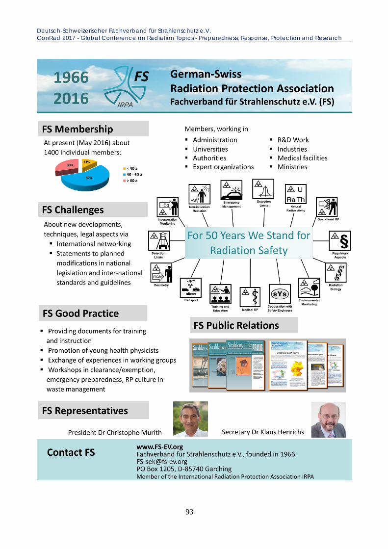
Deutsch-Schweizerischer Fachverband für Strahlenschutz e.V. ConRad 2017 - Global Conference on Radiation Topics - Preparedness, Response, Protection and Research
93

Deutsch-Schweizerischer Fachverband für Strahlenschutz e.V. ConRad 2017 - Global Conference on Radiation Topics - Preparedness, Response, Protection and Research
94

EPR-BioDose 2018, 14th – 18th May, Munich, Germany ConRad 2017 - Global Conference on Radiation Topics - Preparedness, Response, Protection and Research
95
ANNOUNCEMENT
EPR-BIODOSE CONFERENCE 2018
in Munich, Germany
14. – 18. May 2018
organized by
Bundeswehr Institute of Radiobiology affiliated to the University of Ulm, Munich
German Research Center for Environmental Health
(HelmholtzZentrum München), Neuherberg
Federal Office for Radiation Protection (BFS), Neuherberg

Notes ConRad 2017 - Global Conference on Radiation Topics - Preparedness, Response, Protection and Research
96

Notes ConRad 2017 - Global Conference on Radiation Topics - Preparedness, Response, Protection and Research
97

Notes ConRad 2017 - Global Conference on Radiation Topics - Preparedness, Response, Protection and Research
98

Notes ConRad 2017 - Global Conference on Radiation Topics - Preparedness, Response, Protection and Research
99

Notes ConRad 2017 - Global Conference on Radiation Topics - Preparedness, Response, Protection and Research
100