Tuberculous dilated cardiomyopathy: an under-recognized entity?
5
BioMed Central Page 1 of 5 (page number not for citation purposes) BMC Infectious Diseases Open Access Case report Tuberculous dilated cardiomyopathy: an under-recognized entity? Ritesh Agarwal* 1 , Puneet Malhotra 1 , Anshu Awasthi 2 , Nandita Kakkar 2 and Dheeraj Gupta 1 Address: 1 Department of Pulmonary Medicine, Post-Graduate Institute of Medical Education and Research, Sector-12, Chandigarh-160012, India and 2 Department of Histopathology, Post-Graduate Institute of Medical Education and Research, Sector-12, Chandigarh-160012, India Email: Ritesh Agarwal* - [email protected]; Puneet Malhotra - [email protected]; Anshu Awasthi - [email protected]; Nandita Kakkar - [email protected]; Dheeraj Gupta - [email protected] * Corresponding author Abstract Background: Tuberculosis (TB) is a common public health problem in many parts of the world. TB is generally believed to spare these four organs-heart, skeletal muscle, thyroid and pancreas. We describe a rare case of myocardial TB diagnosed on a post-mortem cardiac biopsy. Case presentation: Patient presented with history suggestive of congestive heart failure. We describe the clinical presentation, investigations and outcome of this case, and review the literature on the involvement of myocardium by TB. Conclusion: Involvement of myocardium by TB is rare. However it should be suspected as a cause of congestive heart failure in any patient with features suggestive of TB. Increasing recognition of the entity and the use of endomyocardial biopsy may help us detect more cases of this "curable" form of cardiomyopathy. Background Tuberculosis (TB) is generally believed to spare these four organs-heart, thyroid, pancreas and skeletal muscle. Involvement of myocardium by TB is rare, and generally occurs in conjunction with pericardial involvement. Iso- lated myocardial TB is a rare finding, and definitive diag- nosis during life requires a myocardial biopsy. Herein we describe a patient who presented with features suggestive of congestive heart failure, and was finally diagnosed to have myocardial TB on a post-mortem cardiac biopsy. Case presentation A 25-year old female presented to the emergency depart- ment with a one week history of low grade fever, increas- ing cough and dyspnea on exertion. At presentation she had dyspnea at rest, orthopnea and paroxysmal nocturnal dyspnea. Around two and a half months before the present illness, she had low grade fever, anorexia and weight loss, cough with expectoration and hemoptysis. She was investigated at another center and a sputum smear for acid-fast bacilli was positive. She was started on antituberculous therapy with which she improved symp- tomatically. She experienced weight gain, and her appetite became normal. A sputum smear performed after two months for acid-fast bacilli was negative. On examina- tion, the patient was conscious and afebrile with a pulse rate of 128 beats/minute, blood pressure of 100/60 mm Hg and a respiratory rate of 38/minute. She had bilateral pitting pedal edema. Examination of the cardiovascular system revealed tachycardia, elevated jugular venous pres- sure, diffuse apical impulse and left ventricular third heart sound. Auscultation of the chest showed bibasal Published: 27 April 2005 BMC Infectious Diseases 2005, 5:29 doi:10.1186/1471-2334-5-29 Received: 03 March 2005 Accepted: 27 April 2005 This article is available from: http://www.biomedcentral.com/1471-2334/5/29 © 2005 Agarwal et al; licensee BioMed Central Ltd. This is an Open Access article distributed under the terms of the Creative Commons Attribution License (http://creativecommons.org/licenses/by/2.0 ), which permits unrestricted use, distribution, and reproduction in any medium, provided the original work is properly cited.
-
Upload
ritesh-agarwal -
Category
Documents
-
view
213 -
download
0
Transcript of Tuberculous dilated cardiomyopathy: an under-recognized entity?

BioMed CentralBMC Infectious Diseases
ss
Open AcceCase reportTuberculous dilated cardiomyopathy: an under-recognized entity?Ritesh Agarwal*1, Puneet Malhotra1, Anshu Awasthi2, Nandita Kakkar2 and Dheeraj Gupta1Address: 1Department of Pulmonary Medicine, Post-Graduate Institute of Medical Education and Research, Sector-12, Chandigarh-160012, India and 2Department of Histopathology, Post-Graduate Institute of Medical Education and Research, Sector-12, Chandigarh-160012, India
Email: Ritesh Agarwal* - [email protected]; Puneet Malhotra - [email protected]; Anshu Awasthi - [email protected]; Nandita Kakkar - [email protected]; Dheeraj Gupta - [email protected]
* Corresponding author
AbstractBackground: Tuberculosis (TB) is a common public health problem in many parts of the world.TB is generally believed to spare these four organs-heart, skeletal muscle, thyroid and pancreas.We describe a rare case of myocardial TB diagnosed on a post-mortem cardiac biopsy.
Case presentation: Patient presented with history suggestive of congestive heart failure. Wedescribe the clinical presentation, investigations and outcome of this case, and review the literatureon the involvement of myocardium by TB.
Conclusion: Involvement of myocardium by TB is rare. However it should be suspected as a causeof congestive heart failure in any patient with features suggestive of TB. Increasing recognition ofthe entity and the use of endomyocardial biopsy may help us detect more cases of this "curable"form of cardiomyopathy.
BackgroundTuberculosis (TB) is generally believed to spare these fourorgans-heart, thyroid, pancreas and skeletal muscle.Involvement of myocardium by TB is rare, and generallyoccurs in conjunction with pericardial involvement. Iso-lated myocardial TB is a rare finding, and definitive diag-nosis during life requires a myocardial biopsy. Herein wedescribe a patient who presented with features suggestiveof congestive heart failure, and was finally diagnosed tohave myocardial TB on a post-mortem cardiac biopsy.
Case presentationA 25-year old female presented to the emergency depart-ment with a one week history of low grade fever, increas-ing cough and dyspnea on exertion. At presentation shehad dyspnea at rest, orthopnea and paroxysmal nocturnal
dyspnea. Around two and a half months before thepresent illness, she had low grade fever, anorexia andweight loss, cough with expectoration and hemoptysis.She was investigated at another center and a sputumsmear for acid-fast bacilli was positive. She was started onantituberculous therapy with which she improved symp-tomatically. She experienced weight gain, and her appetitebecame normal. A sputum smear performed after twomonths for acid-fast bacilli was negative. On examina-tion, the patient was conscious and afebrile with a pulserate of 128 beats/minute, blood pressure of 100/60 mmHg and a respiratory rate of 38/minute. She had bilateralpitting pedal edema. Examination of the cardiovascularsystem revealed tachycardia, elevated jugular venous pres-sure, diffuse apical impulse and left ventricular third heartsound. Auscultation of the chest showed bibasal
Published: 27 April 2005
BMC Infectious Diseases 2005, 5:29 doi:10.1186/1471-2334-5-29
Received: 03 March 2005Accepted: 27 April 2005
This article is available from: http://www.biomedcentral.com/1471-2334/5/29
© 2005 Agarwal et al; licensee BioMed Central Ltd. This is an Open Access article distributed under the terms of the Creative Commons Attribution License (http://creativecommons.org/licenses/by/2.0), which permits unrestricted use, distribution, and reproduction in any medium, provided the original work is properly cited.
Page 1 of 5(page number not for citation purposes)

BMC Infectious Diseases 2005, 5:29 http://www.biomedcentral.com/1471-2334/5/29
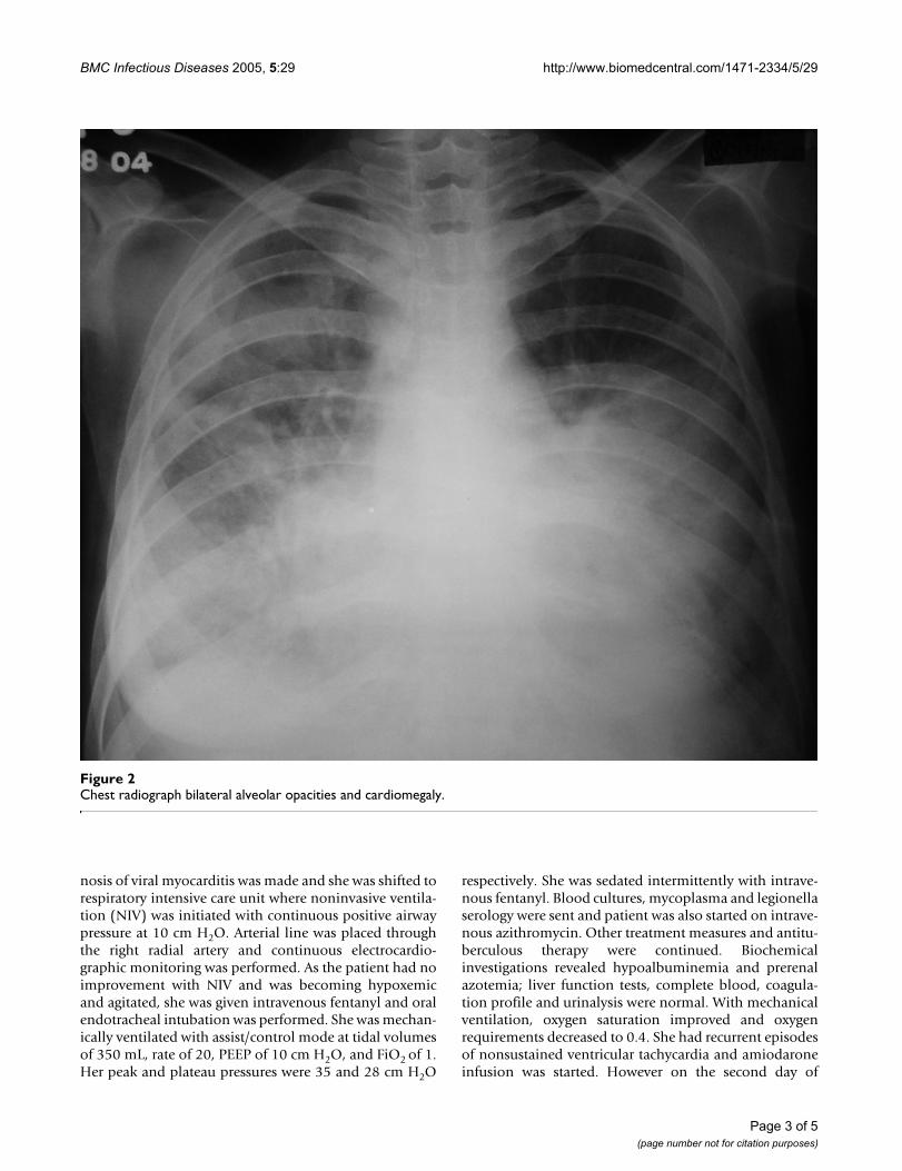
inspiratory crackles. She also had tender hepatomegalyand free fluid in the abdomen. Her oxygen saturation was85% on pulse oximetry. Arterial blood gases showed type1 respiratory failure [(FiO2 0.5) – pH 7.48, PaO2 8.2 kPa,PaCO2 3.6 kPa, HCO3 21 mEq/L]. She was started onsupplemental oxygen, intravenous morphine, nitroglyc-erin, furosemide and dobutamine, oral captopril, stressulcer and deep venous thrombosis prophylaxis. Chest X-ray (Figure 1) done two months ago showed right upperlobe consolidation with normal cardiac silhouette. Achest radiograph (Figure 2) performed at admission
revealed cardiomegaly and bilateral alveolar opacities.Electrocardiogram revealed sinus tachycardia and non-specific ST-T changes in the lateral leads. Echocardiogra-phy was performed which revealed global hypokinesia,enlarged left atrium and left ventricle, mild mitral regurgi-tation, severe left ventricular systolic dysfunction with anejection fraction of 20%; left ventricular end systolic andend diastolic dimensions were 45 and 51 mm respec-tively. Right atrium and right ventricle were dilated withassociated mild tricuspid regurgitation and pulmonaryartery systolic pressure of 47 mm Hg. A provisional diag-
Chest radiograph showing right upper lobe consolidation (cardiac silhouette is normal)Figure 1Chest radiograph showing right upper lobe consolidation (cardiac silhouette is normal).
Page 2 of 5(page number not for citation purposes)
BMC Infectious Diseases 2005, 5:29 http://www.biomedcentral.com/1471-2334/5/29
nosis of viral myocarditis was made and she was shifted torespiratory intensive care unit where noninvasive ventila-tion (NIV) was initiated with continuous positive airwaypressure at 10 cm H2O. Arterial line was placed throughthe right radial artery and continuous electrocardio-graphic monitoring was performed. As the patient had noimprovement with NIV and was becoming hypoxemicand agitated, she was given intravenous fentanyl and oralendotracheal intubation was performed. She was mechan-ically ventilated with assist/control mode at tidal volumesof 350 mL, rate of 20, PEEP of 10 cm H2O, and FiO2 of 1.Her peak and plateau pressures were 35 and 28 cm H2O
respectively. She was sedated intermittently with intrave-nous fentanyl. Blood cultures, mycoplasma and legionellaserology were sent and patient was also started on intrave-nous azithromycin. Other treatment measures and antitu-berculous therapy were continued. Biochemicalinvestigations revealed hypoalbuminemia and prerenalazotemia; liver function tests, complete blood, coagula-tion profile and urinalysis were normal. With mechanicalventilation, oxygen saturation improved and oxygenrequirements decreased to 0.4. She had recurrent episodesof nonsustained ventricular tachycardia and amiodaroneinfusion was started. However on the second day of
Chest radiograph bilateral alveolar opacities and cardiomegalyFigure 2Chest radiograph bilateral alveolar opacities and cardiomegaly.
Page 3 of 5(page number not for citation purposes)

BMC Infectious Diseases 2005, 5:29 http://www.biomedcentral.com/1471-2334/5/29
admission she had a sudden episode of ventricular fibril-lation and despite all resuscitative measures the patientcould not be revived. Her husband did not give consentfor an autopsy; however he agreed for a post-mortem car-diac biopsy, which revealed multi-focal areas of caseousmyocardial necrosis, Ziehl-Neelson stain for acid-fastbacilli was positive (Figure 3). HIV serology received post-mortem was nonreactive.
ConclusionInvolvement of heart in tuberculosis (TB) occurs in one totwo percent of patients with tuberculosis [1,2]. The mostcommon site involved is pericardium, and tuberculousinvolvement of the myocardium is exceedingly rare. The
introduction of effective antituberculous therapy has fur-ther decreased the incidence [3]. The earliest report ofmyocardial TB was in 1664 by Maurocordat and secondreport in 1761 by Morgagni [4]. The myocardium can beaffected either by direct extension or by retrograde lym-phatic drainage from mediastinal nodes; direct spreadfrom tuberculous pericarditis can also occur [5]. Moreo-ver, during the hematogenous phase of dissemination ofprimary TB, any and every tissue and organ in the body isliable to seeding by mycobacteria and consequent patho-logical changes.
Myocardial TB is often not diagnosed during life, but ifsuspected the diagnosis can be established by an endomy-
Photomicrograph demonstrates large areas of necrosis with interspersed myocytes showing degeneration and regeneration [Hematoxylin & Eosin, original magnification × 33]Figure 3Photomicrograph demonstrates large areas of necrosis with interspersed myocytes showing degeneration and regeneration [Hematoxylin & Eosin, original magnification × 33]. Inset shows photomicrograph demonstrating acid-fast bacilli [Ziehl-Neelson stain, original magnification × 33].
Page 4 of 5(page number not for citation purposes)
BMC Infectious Diseases 2005, 5:29 http://www.biomedcentral.com/1471-2334/5/29
ocardial biopsy [6]. Three types of myocardial involve-ment have been described viz. tuberculomas of themyocardium with central caseation (seen in our patient),miliary tubercles of the myocardium complicating gener-alized miliary disease and the uncommon diffuse infiltra-tive type associated with tuberculous pericarditis [7]. Theright heart, particularly the right atrium, is most oftenaffected, probably because of the frequent involvement ofthe right mediastinal lymph nodes with consequentinvolvement of the myocardium [8], although right ven-tricle [3] and left ventricle [4] have been found to beinvolved most frequently in different series. MyocardialTB can manifest in various forms. Rhythm disturbancesinclude supraventricular arrhythmias [3,6], ventriculararrhythmias [9] or varying degrees of conduction blocks[10], and sudden cardiac death is also described [5,11].Right ventricular outflow tract obstruction [3,12,13], ven-tricular aneurysm [4,13], ventricular pseudoaneurysm[14], aortic insufficiency [15], coronary arteritis [10,13],or congestive heart failure [6,16,17] have also beendescribed in literature. Recently, magnetic resonanceimaging has also been used in the diagnosis of myocardialTB [18].
Antitubercular drugs are the cornerstone of therapy [6],and surgery [12,14-16] is indicated only in complicatedcases. Paradoxically, our patient presented with features ofcongestive heart failure, and echocardiography showedsevere left ventricular systolic dysfunction inspite of beingon anti-tuberculous therapy which had resulted in bacte-riological improvement of the pulmonary lesions. Thereason for this is not clear, but one likely reason is proba-bly enhanced immunogenicity of the host to tuberclebacilli. This has been called as a 'paradoxical response'[19]. In this regard, at least theoretically, glucocorticoidsmay have a role along with antituberculous therapy.
In conclusion, although myocardial involvement bytuberculosis is rare, it should be suspected as a cause ofcongestive heart failure in any patient with features sug-gestive of TB, as cases of myocardial TB almost alwaysshow evidence of TB at other sites [6]. Increasing recogni-tion of the entity, and the use of endomyocardial biopsymay help us detect more cases of this "curable" form ofcardiomyopathy especially in areas of high prevalence ofTB. It may not be out of place to state.... Look and ye shallfind....
Competing interestsThe author(s) declare that they have no competinginterests.
Authors' ContributionsRA was involved in patient care and drafting themanuscript
PM was involved in patient care and editing themanuscript
AA was involved in histopathological examination andediting the manuscript
NK was involved in histopathological examination
DG conceived the study and was also involved in patientcare
References1. Anders JM: Tuberculosis of the myocardium. JAMA 1902,
39:1081-1086.2. Fowler NO: Tuberculous pericarditis. JAMA 1991, 266:199-203.3. Kapoor OP, Marcarenhas E, Rananaware MM, Gadgil RK: Tubercu-
loma of the heart. Report of 9 cases. Am Heart J 1973,86:334-340.
4. Rose AG: Cardiac tuberculosis: a study of 19 patients. ArchPathol Lab Med 1987, 111:422-426.
5. Wallis PJW, Branfoot AC, Emerson PA: Sudden death due tomyocardial tuberculosis. Thorax 1984, 39:155.
6. Bali HK, Wahi S, Sharma BK, Anand IS, Datta BN, Wahi PL: Myocar-dial tuberculosis presenting as restrictive cardiomyopathy.Am Heart J 1990, 120:703-706.
7. Horn H, Saphir O: The involvement of the myocardium intuberculosis: a review of the literature and report of threecases. Am Rev Tuberc 1935, 32:492-506.
8. Maeder M, Ammann M, Rickli H, Schoch OD: Fever and nightsweats in a 22-year-old man with a mediastinal mass involv-ing the heart. Chest 2003, 124:2006-2009.
9. Behr G, Palin HC, Temperley JM: Myocardial tuberculosis. Br MedJ 1977, 1:951.
10. Kinare SG: Interesting facets of cardiovascular tuberculosis.Indian J Surg 1975, 37:144-151.
11. Kinare SG, Deshmukh MM: Complete atrioventricular blockdue to myocardial tuberculosis. Report of a case. Arch Pathol1969, 88:684-687.
12. Rawls WJ, Shuford WH, Logan WD, Hurst JW, Schlant RC: Rightventricular outflow tract obstruction produced by a myocar-dial abscess in a patient with tuberculosis. Am J Cardiol 1968,21:738-745.
13. Kinare SG, Bhatia BI: Tuberculous coronary arteritis with aneu-rysm of the ventricular septum. Chest 1971, 60:613-616.
14. Halim MA, Mercer EN, Guinn GA: Myocardial tuberculoma withrupture and pseudoaneurysm formation: successfultreatment. Br Heart J 1985, 54:603-604.
15. Soyer R, Brunet A, Chavalier B, Leroy J, Morere M, Redonnet M:Tuberculous aortic insufficiency. Report of a case with suc-cessful surgical treatment. J Thorac Cardiovasc Surg 1981,82:254-256.
16. Wilbur EL: Myocardial tuberculosis: a case of congestive car-diac failure. Am Rev Tuberc 1938, 38:769-776.
17. Danbauchi SS, Odigie VI, Rafindadi AH, Kalayi GD, Mohammed I:Tuberculous myocarditis: A case report. Niger Postgrad Med J2001, 8:199-202.
18. Jagia P, Gulati GS, Sharma S, Goyal NK, Gaikwad S, Saxena A: MRIfeatures of tuberculoma of the right atrial myocardium. Pedi-atr Radiol 2004, 34:904-907.
19. Chambers ST, Hendrickse WA, Record C, Rudge P, Smith H: Para-doxical expansion of intracranial tuberculomas duringchemotherapy. Lancet 1984, 2:181-184.
Pre-publication historyThe pre-publication history for this paper can be accessedhere:
http://www.biomedcentral.com/1471-2334/5/29/prepub
Page 5 of 5(page number not for citation purposes)
http://www.ncbi.nlm.nih.gov/entrez/query.fcgi?cmd=Retrieve&db=PubMed&dopt=Abstract&list_uids=4199393
http://www.ncbi.nlm.nih.gov/entrez/query.fcgi?cmd=Retrieve&db=PubMed&dopt=Abstract&list_uids=4199393
http://www.ncbi.nlm.nih.gov/entrez/query.fcgi?cmd=Retrieve&db=PubMed&dopt=Abstract&list_uids=3566473
http://www.ncbi.nlm.nih.gov/entrez/query.fcgi?cmd=Retrieve&db=PubMed&dopt=Abstract&list_uids=6701827
http://www.ncbi.nlm.nih.gov/entrez/query.fcgi?cmd=Retrieve&db=PubMed&dopt=Abstract&list_uids=6701827
http://www.ncbi.nlm.nih.gov/entrez/query.fcgi?cmd=Retrieve&db=PubMed&dopt=Abstract&list_uids=2389712
http://www.ncbi.nlm.nih.gov/entrez/query.fcgi?cmd=Retrieve&db=PubMed&dopt=Abstract&list_uids=2389712
http://www.ncbi.nlm.nih.gov/entrez/query.fcgi?cmd=Retrieve&db=PubMed&dopt=Abstract&list_uids=5357722
http://www.ncbi.nlm.nih.gov/entrez/query.fcgi?cmd=Retrieve&db=PubMed&dopt=Abstract&list_uids=5357722
http://www.ncbi.nlm.nih.gov/entrez/query.fcgi?cmd=Retrieve&db=PubMed&dopt=Abstract&list_uids=4230731
http://www.ncbi.nlm.nih.gov/entrez/query.fcgi?cmd=Retrieve&db=PubMed&dopt=Abstract&list_uids=4230731
http://www.ncbi.nlm.nih.gov/entrez/query.fcgi?cmd=Retrieve&db=PubMed&dopt=Abstract&list_uids=4230731
http://www.ncbi.nlm.nih.gov/entrez/query.fcgi?cmd=Retrieve&db=PubMed&dopt=Abstract&list_uids=5126198
http://www.ncbi.nlm.nih.gov/entrez/query.fcgi?cmd=Retrieve&db=PubMed&dopt=Abstract&list_uids=5126198
http://www.ncbi.nlm.nih.gov/entrez/query.fcgi?cmd=Retrieve&db=PubMed&dopt=Abstract&list_uids=4074594
http://www.ncbi.nlm.nih.gov/entrez/query.fcgi?cmd=Retrieve&db=PubMed&dopt=Abstract&list_uids=4074594
http://www.ncbi.nlm.nih.gov/entrez/query.fcgi?cmd=Retrieve&db=PubMed&dopt=Abstract&list_uids=4074594
http://www.ncbi.nlm.nih.gov/entrez/query.fcgi?cmd=Retrieve&db=PubMed&dopt=Abstract&list_uids=7253687
http://www.ncbi.nlm.nih.gov/entrez/query.fcgi?cmd=Retrieve&db=PubMed&dopt=Abstract&list_uids=7253687
http://www.ncbi.nlm.nih.gov/entrez/query.fcgi?cmd=Retrieve&db=PubMed&dopt=Abstract&list_uids=7253687
http://www.ncbi.nlm.nih.gov/entrez/query.fcgi?cmd=Retrieve&db=PubMed&dopt=Abstract&list_uids=6146749
http://www.ncbi.nlm.nih.gov/entrez/query.fcgi?cmd=Retrieve&db=PubMed&dopt=Abstract&list_uids=6146749